Embed Size (px)
Citation preview

RESEARCH ARTICLE
Genetic structure of four plasmids found in
Acinetobacter baumannii isolate D36 belonging
to lineage 2 of global clone 1
Mohammad HamidianID1*, Ruth M. Hall2
1 The ithree institute, University of Technology Sydney, Ultimo, New South Wales, Australia, 2 School of Life
and Environmental Sciences, The University of Sydney, New South Wales, Australia
Abstract
Four plasmids ranging in size from 4.7 to 44.7 kb found in the extensively antibiotic resistant
Acinetobacter baumannii isolate D36 that belongs to lineage 2 of global clone 1 were exam-
ined. D36 includes two cryptic plasmids and two carrying antibiotic resistance genes. The
smallest plasmid pD36-1 (4.7 kb) carries no resistance genes but includes mobA and mobC
mobilisation genes related to those found in pRAY* (pD36-2, 6,078 bp) that also carries the
aadB gentamicin, kanamycin and tobramycin resistance gene cassette. These two plasmids
do not encode a Rep protein. Plasmid pRAY* was found to be mobilised at high frequency
by the large conjugative plasmid pA297-3 but a pRAY* derivative lacking the mobA and
mobC genes was not. The two larger plasmids, pD36-3 and pD36-4, encode Rep_3 family
proteins (Pfam1051). The cryptic plasmid pD36-3 (6.2 kb) has RepAci1 and pD36-4 (44.7
kb) encodes two novel Rep_3 family proteins suggesting a co-integrate. Plasmid pD36-4
includes the sul2 sulfonamide resistance gene, the aphA1a kanamycin/neomycin resistance
gene in Tn4352::ISAba1 and a mer module in a hybrid Tn501/Tn1696 transposon conferring
resistance to mercuric ions. New examples of dif modules flanked by pdif sites (XerC-XerD
binding sites) that are part of many A. baumannii plasmids were also identified in pD36-3
and pD36-4 which carry three and two dif modules, respectively. Homologs of three dif mod-
ules, the sup sulphate permease module in pD36-3, and of the abkAB toxin-antitoxin module
and the orf module in pD36-4, were found in different contexts in diverse Acinetobacter plas-
mids, consistent with module mobility. A novel insertion sequence named ISAba32 found
next to the pdif site in the abkAB dif module is related to members of the ISAjo2 group which
also are associated with the pdif sites of dif modules. Plasmids found in D36 were also
found in some other members of GC1 lineage 2.
Introduction
Acinetobacter baumannii is a Gram negative opportunistic pathogen and a member of the
ESKAPE group of bacteria that are the leading cause of difficult to treat nosocomial infections
throughout the world [1]. A. baumannii has emerged as a global challenge mainly because of
PLOS ONE | https://doi.org/10.1371/journal.pone.0204357 September 27, 2018 1 / 15
a1111111111
a1111111111
a1111111111
a1111111111
a1111111111
OPENACCESS
Citation: Hamidian M, Hall RM (2018) Genetic
structure of four plasmids found in Acinetobacter
baumannii isolate D36 belonging to lineage 2 of
global clone 1. PLoS ONE 13(9): e0204357. https://
doi.org/10.1371/journal.pone.0204357
Editor: Finbarr Hayes, University of Manchester,
UNITED KINGDOM
Received: June 8, 2018
Accepted: September 6, 2018
Published: September 27, 2018
Copyright: © 2018 Hamidian, Hall. This is an open
access article distributed under the terms of the
Creative Commons Attribution License, which
permits unrestricted use, distribution, and
reproduction in any medium, provided the original
author and source are credited.
Data Availability Statement: All relevant data are
within the paper.
Funding: MH is supported by the Chancellor’s
Postdoctoral Research Fellowship (CPDRF)
received from the University of Technology Sydney
(https://www.uts.edu.au/). Funding for this study
was received from National Health and Medical
Research Council (NHMRC) grant 1079616
(https://www.nhmrc.gov.au/). The funders had no
role in study design, data collection and analysis,
decision to publish, or preparation of the
manuscript.

treatment failure due to its high levels of antibiotic resistance [2]. The majority of globally-dis-
tributed A. baumannii strains that are resistant to multiple antibiotics are members of the two
main successful, globally-distributed clones, namely global clone 1 and global clone 2 or simply
GC1 and GC2 [3–7].
We previously showed that the most recent common ancestor of GC1 arose around 1960
and subsequently around 1967 members of GC1 diverged into two phylogenetically distinct
lineages [8]. In A. baumannii, unlike other Gram negative bacteria, resistance genes are often
located in genomic resistance islands in the chromosome [5, 8, 9]. Strains belonging to the
main GC1 lineage, lineage 1, all included either an AbaR-type resistance island or a remnant
of it located in the chromosomal comM gene [8]. However, strains falling into lineage 2 con-
tained either an intact comM gene or Tn6022 or AbaR4 in comM [8]. AbaR4 is a 16.8 kb trans-
poson consisting of Tn6022, a transposon related to the backbone transposon of the AbaR-
type islands, interrupted by Tn2006, a transposon that carries the oxa23 carbapenem resistance
gene [10]. Recently, several studies have highlighted the significance of A. baumannii plasmids
in introducing antibiotic resistance genes [11–14].
In an earlier study, we showed that D36 is a carbapenem resistant A. baumannii isolate that
belongs to lineage 2 and ST81 (Institut Pasteur MLST scheme), which is a single locus variant
(SLV) of the predominant GC1 sequence type, ST1 [8]. D36 was known to carry the AbaR4
resistance island in the comM gene [10]. However, D36 is resistant to other antibiotics includ-
ing third generation cephalosporins, sulfamethoxazole, ciprofloxacin, gentamicin, kanamycin,
neomycin and tobramycin [10, 15, 16]. The ISAba1-ampC structure accounts for its resistance
to third generation cephalosporins [16] and mutations in gyrA and parC account for the fluo-
roquinolone resistance [8]. D36 also carries the aadB gene in a small plasmid called pRAY�
explaining its resistance to kanamycin, tobramycin and gentamicin [15]. However, the context
of the aphA1a and sul2 genes that confer resistance to kanamycin and neomycin and to sulfa-
methoxazole, respectively, [17] had not been found.
In addition, the genome of D36 has been completely sequenced revealing three additional
plasmids in this strain [17] but their properties were not described. Hence, here we sought to
analyse the genetic structure of the plasmids carried by D36, compare them to other Acineto-bacter plasmids and explore their distribution in other members of GC1, lineage 2 that are
closely related to D36.
Materials and methods
Sequence analysis and bioinformatics
The strain D36 is an extensively antibiotic resistant strain recovered in 2008 at a Sydney hospi-
tal from a wound infection of a 27-year-old male who was a member of the armed forces.
The complete genome sequence of strain D36 was determined previously using a combina-
tion of Illumina HiSeq and PacBio technologies [17].
A range of bioinformatics tools was used to annotate the sequences of the three novel plas-
mids, pD36-1, pD36-3 and pD36-4 found in D36 (GenBank accession numbers CP012953,
CP012955 and CP012956, respectively). The outputs of the automatic annotation program
Prokka [18] were manually modified where further information such as assigned gene names
was available. Antibiotic resistance genes were identified using ResFinder (https://cge.cbs.dtu.
dk/services/ResFinder/) [19] and ISFinder (https://www-is.biotoul.fr/) was used to identify
insertion sequences. Sequence repeats including iterons were found using Unipro UGENE
v1.29.0 (http://ugene.net/). Pfam searches (http://pfam.xfam.org/) were also used to identify
possible protein functions. The copy number of each plasmid was estimated by dividing the
coverage of contigs containing plasmid sequences by the coverage of chromosomal contigs.
Plasmids of Acinetobacter baumannii D36
PLOS ONE | https://doi.org/10.1371/journal.pone.0204357 September 27, 2018 2 / 15
Competing interests: The authors have declared
that no competing interests exist.

Gene Construction Kit (GCK 4.0.3) was used to visualize and manipulate the sequences
studied. Figures were drawn to scale using GCK 4.0.3, SnapGene Viewer 4.1.7 and Adobe Illus-
trator CS6.
Construction of pRAY�-Δ1
Plasmid DNA was isolated from D36, using the Wizard1 Plus SV Minipreps DNA Purifica-
tion kit (Promega), and pRAY� was gel purified and digested with HindIII according to the
manufacturer’s instructions. Approximately 50 ng of the HindIII digestion mix was then re-
ligated using 1 μl of T4 DNA ligase (New England BioLabs, Ipswich, USA) and 2x Ligation
buffer. The ligation mix was then used to transform the antibiotic susceptible A. baumanniistrain AB307-0294 [7] by electroporation using 0.2 cm cuvettes and with the following param-
eters 2.5 kV, 25 μF and 200 O. Potential transformants were selected on L-agar supplemented
with 20 mg/L kanamycin to select for pRAY� derivatives. To screen the deletion derivatives,
the PCR primers RH1370 5'-CGTTATCGGATTTACTGCTTTAC-3' and RH1377 5'-CGTCAGCCCAATTACAGGTT-3', were designed to generate a product spanning all HindIII
restriction sites present in pRAY� and used to amplify products with various length depending
on the number of HindIII fragments present. The smallest PCR product obtained, represent-
ing the shortest pRAY� deletion derivative with only one HindIII site, was sequenced to con-
firm the deletion and the resulting corresponding plasmid, which lacked the mobA and mobCgenes (Fig 1B) was named pRAY�-Δ1. Plasmid pRAY�-Δ1 was transformed into AB307-0294
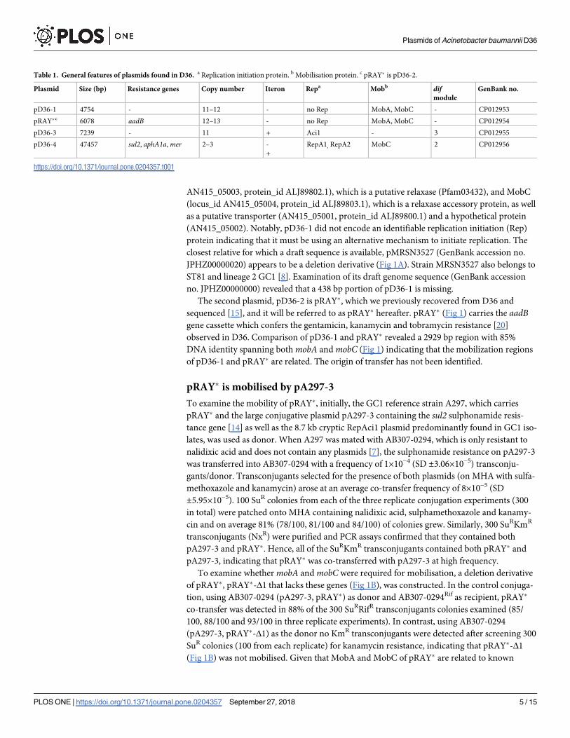
Fig 1. Linearized map of pD36-1 compared with pRAY� (pD36-2) and pMRSN 3527 (A) and comparison of pRAY� with pRAY�-Δ1 (B). Central
horizontal lines indicate the plasmid backbones. Arrows represent the extent and orientation of genes and the gene cassette is boxed. The grey shadings
indicate regions with significant identity with the % identities indicated in red. Scale bar is shown. Drawn to scale from GenBank accession numbers
CP012954 (pRAY�), CP012953 (pD36-1), and JPHZ00000000 (pMRSN3527).
https://doi.org/10.1371/journal.pone.0204357.g001
Plasmids of Acinetobacter baumannii D36
PLOS ONE | https://doi.org/10.1371/journal.pone.0204357 September 27, 2018 3 / 15

(pA297-3) cells for mobilization assays. Transformants were selected on L-agar supplemented
with 20 mg/L kanamycin and 100 mg/L sulphamethoxazole to select for both pA297-3 and
pRAY�-Δ1. The resulting transformants were purified and the presence of both pA297-3 and
pRAY�-Δ1 was confirmed by PCRs.
Conjugation and mobilisation experiments
Conjugation was performed by mixing 100 μl of donor and recipient on an L-agar plate with
no antibiotic selection. Following overnight incubation, cells were resuspended and diluted in
0.9% saline. Potential transconjugants were selected on Muller Hinton Agar (MHA) supple-
mented with nalidixic acid (25 mg/L) and sulfamethoxazole (100 mg/L), to select for pA297-3
transfer, and MHA agar containing nalidixic acid (25 mg/L), sulfamethoxazole (100 mg/L)
and kanamycin (20 mg/L) to select for co-transfer of pA297-3 and pRAY� or pRAY�-Δ1. To
examine whether the colonies selected with only sulfamethoxazole also carried pRAY� they
were patched onto MHA containing kanamycin (20 mg/L). To ensure the colonies were trans-
conjugants they were patched onto L-agar supplemented with 10 mg/L tetracycline, to which
the recipient was sensitive and the donor resistant. Transconjugants were also screened by
PCR to confirm plasmid transfer, pRAY� co-transfer and that the RepAci1 cryptic plasmid
had not been co-transferred.
A rifampicin resistant mutant of the strain AB307-0294 [7], was generated by growing the
cells overnight on L-agar supplemented with 100 mg/L rifampicin. Potential mutants were
purified, confirmed phenotypically and one was named 307-0294rif. 307-0294rif was then used
as recipient and mated with AB307-0294 (pA297-3, pRAY�) as donor generated in the previ-
ous conjugation round. Transconjugants were selected on MHA containing 100 mg/L rifampi-
cin and 100 mg/L sulfamethoxazole, to select for pA297-3 and on MHA with rifampicin,
sulfamethoxazole and 20 mg/L kanamycin to select for pA297-3 and pRAY� co-transfer. Strain
AB307-0294 (pA297-3, pRAY�-Δ1) was used as a donor and mated with AB307-0294rif using
the conditions and antibiotics specified above. The transfer and co-transfer frequencies were
calculated using an average of values obtained from three replicates.
Resistance to mercury
To examine whether D36 is resistant to mercury, 10 fresh colonies were patched onto L-agar
supplemented with 25 μg/ml HgCl2 followed by overnight incubation at 37˚C and visual
inspection for the presence and absence of growth.
Results
Properties of the four plasmids carried by D36
The D36 genome consists of a chromosome (GenBank accession no. CP012952, 4063596 bp)
and four plasmids named pD36-1 to pD36-4 and ranging in size from 4.7 to 47.2 kb [17]. The
general features of these plasmids are summarised in Table 1. Two plasmids, pRAY� (pD36-2,
6 kb) described previously [15] and pD36-4 (47.4 kb) carry antibiotic resistance genes while
the other two, pD36-1 (4.7 kb) and pD36-3 (7.2 kb) are cryptic [8]. The copy number of pD36-
1, pRAY� and pD36-3 was estimated to be between 11–13 copies/cell while that of the largest
plasmid, pD36-4, was 2–3 copies/cell.
pD36-1 and pRAY�
The smallest plasmid found in D36, the 4757 bp pD36-1, encodes only four proteins (Fig 1 and
Table 1). The proteins include two potential mobilization proteins MobA (locus_id
Plasmids of Acinetobacter baumannii D36
PLOS ONE | https://doi.org/10.1371/journal.pone.0204357 September 27, 2018 4 / 15
AN415_05003, protein_id ALJ89802.1), which is a putative relaxase (Pfam03432), and MobC
(locus_id AN415_05004, protein_id ALJ89803.1), which is a relaxase accessory protein, as well
as a putative transporter (AN415_05001, protein_id ALJ89800.1) and a hypothetical protein
(AN415_05002). Notably, pD36-1 did not encode an identifiable replication initiation (Rep)
protein indicating that it must be using an alternative mechanism to initiate replication. The
closest relative for which a draft sequence is available, pMRSN3527 (GenBank accession no.
JPHZ00000020) appears to be a deletion derivative (Fig 1A). Strain MRSN3527 also belongs to
ST81 and lineage 2 GC1 [8]. Examination of its draft genome sequence (GenBank accession
no. JPHZ00000000) revealed that a 438 bp portion of pD36-1 is missing.
The second plasmid, pD36-2 is pRAY�, which we previously recovered from D36 and
sequenced [15], and it will be referred to as pRAY� hereafter. pRAY� (Fig 1) carries the aadBgene cassette which confers the gentamicin, kanamycin and tobramycin resistance [20]
observed in D36. Comparison of pD36-1 and pRAY� revealed a 2929 bp region with 85%
DNA identity spanning both mobA and mobC (Fig 1) indicating that the mobilization regions
of pD36-1 and pRAY� are related. The origin of transfer has not been identified.
pRAY� is mobilised by pA297-3
To examine the mobility of pRAY�, initially, the GC1 reference strain A297, which carries
pRAY� and the large conjugative plasmid pA297-3 containing the sul2 sulphonamide resis-
tance gene [14] as well as the 8.7 kb cryptic RepAci1 plasmid predominantly found in GC1 iso-
lates, was used as donor. When A297 was mated with AB307-0294, which is only resistant to
nalidixic acid and does not contain any plasmids [7], the sulphonamide resistance on pA297-3
was transferred into AB307-0294 with a frequency of 1×10⁻⁴ (SD ±3.06×10⁻⁵) transconju-
gants/donor. Transconjugants selected for the presence of both plasmids (on MHA with sulfa-
methoxazole and kanamycin) arose at an average co-transfer frequency of 8×10⁻⁵ (SD
±5.95×10⁻⁵). 100 SuR colonies from each of the three replicate conjugation experiments (300
in total) were patched onto MHA containing nalidixic acid, sulphamethoxazole and kanamy-
cin and on average 81% (78/100, 81/100 and 84/100) of colonies grew. Similarly, 300 SuRKmR
transconjugants (NxR) were purified and PCR assays confirmed that they contained both
pA297-3 and pRAY�. Hence, all of the SuRKmR transconjugants contained both pRAY� and
pA297-3, indicating that pRAY� was co-transferred with pA297-3 at high frequency.
To examine whether mobA and mobC were required for mobilisation, a deletion derivative
of pRAY�, pRAY�-Δ1 that lacks these genes (Fig 1B), was constructed. In the control conjuga-
tion, using AB307-0294 (pA297-3, pRAY�) as donor and AB307-0294Rif as recipient, pRAY�
co-transfer was detected in 88% of the 300 SuRRifR transconjugants colonies examined (85/
100, 88/100 and 93/100 in three replicate experiments). In contrast, using AB307-0294
(pA297-3, pRAY�-Δ1) as the donor no KmR transconjugants were detected after screening 300
SuR colonies (100 from each replicate) for kanamycin resistance, indicating that pRAY�-Δ1
(Fig 1B) was not mobilised. Given that MobA and MobC of pRAY� are related to known
Table 1. General features of plasmids found in D36. a Replication initiation protein. b Mobilisation protein. c pRAY� is pD36-2.
Plasmid Size (bp) Resistance genes Copy number Iteron Repa Mobb difmodule
GenBank no.
pD36-1 4754 - 11–12 - no Rep MobA, MobC - CP012953
pRAY�c 6078 aadB 12–13 - no Rep MobA, MobC - CP012954
pD36-3 7239 - 11 + Aci1 - 3 CP012955
pD36-4 47457 sul2, aphA1a, mer 2–3 -
+
RepA1, RepA2 MobC 2 CP012956
https://doi.org/10.1371/journal.pone.0204357.t001
Plasmids of Acinetobacter baumannii D36
PLOS ONE | https://doi.org/10.1371/journal.pone.0204357 September 27, 2018 5 / 15

relaxase/accessory combinations [15] this indicates a likely role for MobA and MobC in mobi-
lisation of pRAY�. However, further work will be needed to establish whether both the relaxase
(MobA) and the relaxase accessory protein (MobC) are required, as is usual in homologous
systems.
pD36-3—A RepAci1 plasmid
The third plasmid, pD36-3 (9,276 bp), in strain D36 includes 15 orfs ranging in size from 144
bp to 1461 bp but carries no antibiotic resistance genes (Fig 2). It also encodes a Rep protein
that is 100% identical to the previously identified RepAci1 replication initiation protein [21].
Examination of the region surrounding the repAci1 gene indicated that it was preceded by a
putative 88 bp iteron region consisting of four copies of the 22 bp repeat 5'-ATATGTCCACGTTTACCTTGCA-3' that is identical to the corresponding region in other RepAci1 plasmids
such as pA1-1 (GenBank accession no. CP010782), which is an 8731 bp cryptic plasmid pre-
dominantly found in many members of global clone 1, lineage 1 including A297 (see above).
In total a 1997 bp region on the left in Fig 2 (bases 1–1997 of pD36-3 including the repAci1
Fig 2. Linearized map of pD36-3 compared with pA1-1 (A) and pACNIH1 and pMRSN5540 (B). Central lines indicate the plasmid backbones with
horizontal arrows indicating the extent and direction of genes/orfs. Genes/orfs are also colour coded according to the function they encode (shown
below) and the key is shown on the right. The filled boxes coloured orange at the beginning of each line represent the iteron regions and small vertical
bars indicated by C/D or D/C represent the two pdif orientations (XerC/D or XerD/C) binding sites. The scale bar is also shown. Drawn to scale from
GenBank accession numbers CP012955 (pD36-3), CP010782 (pA1-1), CP026427 (pACNIH1) and LNCW01000064 (pMRSN5540).
https://doi.org/10.1371/journal.pone.0204357.g002
Plasmids of Acinetobacter baumannii D36
PLOS ONE | https://doi.org/10.1371/journal.pone.0204357 September 27, 2018 6 / 15
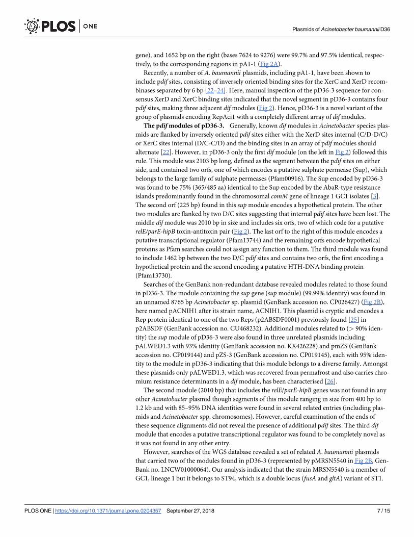
gene), and 1652 bp on the right (bases 7624 to 9276) were 99.7% and 97.5% identical, respec-
tively, to the corresponding regions in pA1-1 (Fig 2A).
Recently, a number of A. baumannii plasmids, including pA1-1, have been shown to
include pdif sites, consisting of inversely oriented binding sites for the XerC and XerD recom-
binases separated by 6 bp [22–24]. Here, manual inspection of the pD36-3 sequence for con-
sensus XerD and XerC binding sites indicated that the novel segment in pD36-3 contains four
pdif sites, making three adjacent dif modules (Fig 2). Hence, pD36-3 is a novel variant of the
group of plasmids encoding RepAci1 with a completely different array of dif modules.
The pdif modules of pD36-3. Generally, known dif modules in Acinetobacter species plas-
mids are flanked by inversely oriented pdif sites either with the XerD sites internal (C/D-D/C)
or XerC sites internal (D/C-C/D) and the binding sites in an array of pdif modules should
alternate [22]. However, in pD36-3 only the first dif module (on the left in Fig 2) followed this
rule. This module was 2103 bp long, defined as the segment between the pdif sites on either
side, and contained two orfs, one of which encodes a putative sulphate permease (Sup), which
belongs to the large family of sulphate permeases (Pfam00916). The Sup encoded by pD36-3
was found to be 75% (365/485 aa) identical to the Sup encoded by the AbaR-type resistance
islands predominantly found in the chromosomal comM gene of lineage 1 GC1 isolates [3].
The second orf (225 bp) found in this sup module encodes a hypothetical protein. The other
two modules are flanked by two D/C sites suggesting that internal pdif sites have been lost. The
middle dif module was 2010 bp in size and includes six orfs, two of which code for a putative
relE/parE-hipB toxin-antitoxin pair (Fig 2). The last orf to the right of this module encodes a
putative transcriptional regulator (Pfam13744) and the remaining orfs encode hypothetical
proteins as Pfam searches could not assign any function to them. The third module was found
to include 1462 bp between the two D/C pdif sites and contains two orfs, the first encoding a
hypothetical protein and the second encoding a putative HTH-DNA binding protein
(Pfam13730).
Searches of the GenBank non-redundant database revealed modules related to those found
in pD36-3. The module containing the sup gene (sup module) (99.99% identity) was found in
an unnamed 8765 bp Acinetobacter sp. plasmid (GenBank accession no. CP026427) (Fig 2B),
here named pACNIH1 after its strain name, ACNIH1. This plasmid is cryptic and encodes a
Rep protein identical to one of the two Reps (p2ABSDF0001) previously found [25] in
p2ABSDF (GenBank accession no. CU468232). Additional modules related to (> 90% iden-
tity) the sup module of pD36-3 were also found in three unrelated plasmids including
pALWED1.3 with 93% identity (GenBank accession no. KX426228) and pmZS (GenBank
accession no. CP019144) and pZS-3 (GenBank accession no. CP019145), each with 95% iden-
tity to the module in pD36-3 indicating that this module belongs to a diverse family. Amongst
these plasmids only pALWED1.3, which was recovered from permafrost and also carries chro-
mium resistance determinants in a dif module, has been characterised [26].
The second module (2010 bp) that includes the relE/parE-hipB genes was not found in any
other Acinetobacter plasmid though segments of this module ranging in size from 400 bp to
1.2 kb and with 85–95% DNA identities were found in several related entries (including plas-
mids and Acinetobacter spp. chromosomes). However, careful examination of the ends of
these sequence alignments did not reveal the presence of additional pdif sites. The third difmodule that encodes a putative transcriptional regulator was found to be completely novel as
it was not found in any other entry.
However, searches of the WGS database revealed a set of related A. baumannii plasmids
that carried two of the modules found in pD36-3 (represented by pMRSN5540 in Fig 2B, Gen-
Bank no. LNCW01000064). Our analysis indicated that the strain MRSN5540 is a member of
GC1, lineage 1 but it belongs to ST94, which is a double locus (fusA and gltA) variant of ST1.
Plasmids of Acinetobacter baumannii D36
PLOS ONE | https://doi.org/10.1371/journal.pone.0204357 September 27, 2018 7 / 15

In pMRSN5540, two short regions with ~95% sequence identity, compared to their corre-
sponding regions on the left-end of the sup module and spanning the internal orf of the supmodule on the right, were found next to each other (Fig 2B). Consequently, pMRSN5540
lacked the sup gene due to an internal deletion starting from 4 bp inside the dif module on the
left and extending 1755 bp to the 3’-end of the internal orf on the right (Fig 2B). The cause of
this deletion was unclear as careful inspection of this module did not reveal additional pdifsites. The sequence of this region could not be confirmed as MRSN5540 was not available to
be examined further.
pD36-4 carrying resistance genes and dif modules
The largest plasmid found in D36 was pD36-4 (GenBank accession no. CP012956), which is
47457 bp in length [17] and includes 57 orfs ranging in size from 138–1686 bp. Two novel Rep
proteins (Fig 3), which belong to the Rep_3 family of replication initiation proteins
(Pfam01051), were encoded by pD36-4 and were named RepA1 (locus_taq AN415_8001, pro-
tein_id ALJ89824.1) and RepA2 (locus_taq AN415_8021, protein_id ALJ89842.1), respectively
(Fig 3). Compared to known RepA proteins reported in the published typing scheme [25], the
closest match to RepA1 was a RepA protein encoded by p3ABAYE (290/392 aa) (protein_id
CAM84695.1; locus_id p3ABAYE0002 in GenBank accession no. CU459140) that is 74% iden-
tical, and RepA2 was 57% identical (176/307 aa) to the RepA encoded by pAB1, which is a 13
kb (GenBank accession no. CP000522) plasmid found in ATCC 17978. RepA2 is also 91%
identical to one of the RepA proteins found subsequently in the unrelated plasmid pAb242_25
(GenBank accession no. KY984047), which was sequenced recently [27, 28]. Examination of
the regions surrounding the repA genes showed that repA2 is preceded by three copies of a
putative 19 bp iteron sequence 5'-AGGTGGGAAAACGTAGACT-3' (orange vertical line in
Fig 3) but iterons were not found in the region upstream of repA1.
Fig 3. Linearized map of pD36-4. The thick horizontal line represents the plasmid backbone and arrows indicate the extent and orientation of genes/
orfs. Filled boxes coloured different shades of green indicate insertion sequences with the white arrows inside indicating the direction of the transposase
genes. Small vertical red bars indicated by C/D or D/C represent pdif binding sites. The vertical bar coloured orange upstream of repA2 indicates the
iteron region. The scale bar is also shown. Drawn to scale from GenBank accession no. CP012956.
https://doi.org/10.1371/journal.pone.0204357.g003
Plasmids of Acinetobacter baumannii D36
PLOS ONE | https://doi.org/10.1371/journal.pone.0204357 September 27, 2018 8 / 15
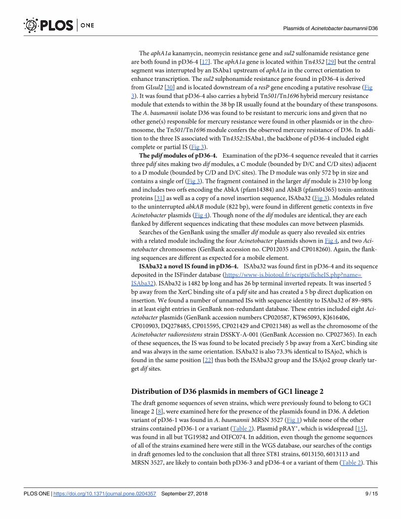
The aphA1a kanamycin, neomycin resistance gene and sul2 sulfonamide resistance gene
are both found in pD36-4 [17]. The aphA1a gene is located within Tn4352 [29] but the central
segment was interrupted by an ISAba1 upstream of aphA1a in the correct orientation to
enhance transcription. The sul2 sulphonamide resistance gene found in pD36-4 is derived
from GIsul2 [30] and is located downstream of a resP gene encoding a putative resolvase (Fig
3). It was found that pD36-4 also carries a hybrid Tn501/Tn1696 hybrid mercury resistance
module that extends to within the 38 bp IR usually found at the boundary of these transposons.
The A. baumannii isolate D36 was found to be resistant to mercuric ions and given that no
other gene(s) responsible for mercury resistance were found in other plasmids or in the chro-
mosome, the Tn501/Tn1696 module confers the observed mercury resistance of D36. In addi-
tion to the three IS associated with Tn4352::ISAba1, the backbone of pD36-4 included eight
complete or partial IS (Fig 3).
The pdif modules of pD36-4. Examination of the pD36-4 sequence revealed that it carries
three pdif sites making two dif modules, a C module (bounded by D/C and C/D sites) adjacent
to a D module (bounded by C/D and D/C sites). The D module was only 572 bp in size and
contains a single orf (Fig 3). The fragment contained in the larger dif module is 2310 bp long
and includes two orfs encoding the AbkA (pfam14384) and AbkB (pfam04365) toxin-antitoxin
proteins [31] as well as a copy of a novel insertion sequence, ISAba32 (Fig 3). Modules related
to the uninterrupted abkAB module (822 bp), were found in different genetic contexts in five
Acinetobacter plasmids (Fig 4). Though none of the dif modules are identical, they are each
flanked by different sequences indicating that these modules can move between plasmids.
Searches of the GenBank using the smaller dif module as query also revealed six entries
with a related module including the four Acinetobacter plasmids shown in Fig 4, and two Aci-netobacter chromosomes (GenBank accession no. CP012035 and CP018260). Again, the flank-
ing sequences are different as expected for a mobile element.
ISAba32 a novel IS found in pD36-4. ISAba32 was found first in pD36-4 and its sequence
deposited in the ISFinder database (https://www-is.biotoul.fr/scripts/ficheIS.php?name=
ISAba32). ISAba32 is 1482 bp long and has 26 bp terminal inverted repeats. It was inserted 5
bp away from the XerC binding site of a pdif site and has created a 5 bp direct duplication on
insertion. We found a number of unnamed ISs with sequence identity to ISAba32 of 89–98%
in at least eight entries in GenBank non-redundant database. These entries included eight Aci-netobacter plasmids (GenBank accession numbers CP020587, KT965093, KJ616406,
CP010903, DQ278485, CP015595, CP021429 and CP021348) as well as the chromosome of the
Acinetobacter radioresistens strain DSSKY-A-001 (GenBank Accession no. CP027365). In each
of these sequences, the IS was found to be located precisely 5 bp away from a XerC binding site
and was always in the same orientation. ISAba32 is also 73.3% identical to ISAjo2, which is
found in the same position [22] thus both the ISAba32 group and the ISAjo2 group clearly tar-
get dif sites.
Distribution of D36 plasmids in members of GC1 lineage 2
The draft genome sequences of seven strains, which were previously found to belong to GC1
lineage 2 [8], were examined here for the presence of the plasmids found in D36. A deletion
variant of pD36-1 was found in A. baumannii MRSN 3527 (Fig 1) while none of the other
strains contained pD36-1 or a variant (Table 2). Plasmid pRAY�, which is widespread [15],
was found in all but TG19582 and OIFC074. In addition, even though the genome sequences
of all of the strains examined here were still in the WGS database, our searches of the contigs
in draft genomes led to the conclusion that all three ST81 strains, 6013150, 6013113 and
MRSN 3527, are likely to contain both pD36-3 and pD36-4 or a variant of them (Table 2). This
Plasmids of Acinetobacter baumannii D36
PLOS ONE | https://doi.org/10.1371/journal.pone.0204357 September 27, 2018 9 / 15

is consistent with the ST81 isolates being part of a clade within lineage 2 of GC2 as shown pre-
viously [8].
Discussion
Our previous study indicated that the two phylogenetically distinct lineages of A. baumanniiGC1 acquired resistance to antibiotics via different routes [8]. However, how members of line-
age 2 became resistant to several antibiotics is not well understood. Here, the plasmids found
in D36, an extensively antibiotic resistant member of lineage 2 [17], were studied. The largest
Fig 4. Comparison of dif modules found in pD36-4 to related modules from diverse Acinetobacter plasmids. Only
1–2 kb of the surrounding region of each dif module is shown. Central black lines represent the backbone of plasmids
and vertical lines indicate pdif sites (marked C/D or D/C to indicate their orientation). Horizontal arrows indicate the
extent and orientation of orfs. Open reading frames encoding hypothetical proteins are white. Segments with
significant identities (>90%) are connected using a shade of grey with the numbers indicating identities. In
pNDM-GJ02 the usp gene encodes a universal stress protein and mtr, in pMS32-2, encodes a DNA methyltransferase.
The figure is drawn to scale using the sequence retrieved from the GenBank accession numbers indicated for each
plasmid. Scale bar is shown.
https://doi.org/10.1371/journal.pone.0204357.g004
Plasmids of Acinetobacter baumannii D36
PLOS ONE | https://doi.org/10.1371/journal.pone.0204357 September 27, 2018 10 / 15

plasmid, pD36-4, contains two antibiotic resistance genes, aphA1a in Tn4352 interrupted by
ISAba1 and sul2 in a fragment from GIsul2. It also included a Tn501/Tn1696 hybrid merregion responsible for resistance to mercuric ions (Fig 3). Hence, two plasmids, pRAY� and
pD36-4 carry resistance genes, complementing the chromosomal resistance determinants.
Plasmids pRAY� (pD36-2), pD36-3 and pD36-4 were found to be present in all ST81 isolates
regardless of the country of origin (Table 2). Hence, it appears that the ST81 group represent a
distinct multiply resistant clade of lineage 2 isolates and this is consistent with the recombina-
tion-free phlyogeny generated from the chromosomes of three members of this group [8].
Plasmid pRAY� was mobilised by pA297-3, a large conjugative plasmid, and this is likely to
be an important route for the spread of the aadB gentamicin, tobramycin and kanamycin resis-
tance gene. We previously reported that the N-terminal end of the putative MobA encoded by
pRAY� is related to MbeA of ColE1, and is therefore classified as a member of the MOBP5 (for-
merly MOBHEN [32]) group of the MobA superfamily [15]. Here, we demonstrated that
pRAY� was mobilised by pA297-3 only if mobA and mobC genes were present. Plasmid pA297
[14] is of a type seen to date only in Acineobacter baumannii. The small cryptic plasmid pD36-
1 includes a region spanning the mobA and mobC genes related to the corresponding region in
pRAY�. The predicted MobA and MobC proteins of pD36-1 and pRAY� were 77% and 94%
identical, respectively, and it is possible that pD36-1 can also be mobilised by pA297-3 and
related conjugative plasmids. A small plasmid pALWED1.8, which was found in an A. lwoffiirecovered from permafrost and carries the aadA27 streptomycin/spectinomycin resistance
gene also carries a mobilisation region related to that of pRAY� [33]. In fact, mobilisation of
pALWED1.8 was demonstrated [33] but the sequence of the conjugative plasmid used is not
available precluding comparison to pA297-3. Further work is needed to examine these
questions.
Neither pD36-1 nor pRAY� included an identifiable rep gene suggesting an alternative rep-
lication mechanism. Indeed, it is possible that like ColE1 [34] pRAY� uses an antisense RNA
mechanism to initiate its replication. Notably, pRAY�-Δ1, which was constructed here for
mobilisation assays, is only 2808 bp in size (2217 bp + aadB cassette) but is still able to replicate
indicating that it contains all of the sequences required for initiation of replication. However,
further deletions will be required to determine the exact sequences essential for replication
initiation.
Table 2. Distribution of plasmids found in D36 in other members of GC1, lineage2.
Isolate Date Country Source pD36-1 pD36-2
(pRAY�)
pD36-3 pD36-4 aphAa mer sul2 STIP GenBank no.
TG19582 nkb nk nk - - - - - - + 1 AMIV00000000
Naval-21 2006 US Wound - + - - 1b - + 19 AMSY00000000
OIFC074 2003 US nk - - - - 1b - + 19 AMDE01000000
D36 2008 Australia Wound + + + + 1a + + 81 CP012952
6013150 2007 UK Skin - + +c + 1a + + 81 ACYQ00000000
6013113 2007 UK Skin - + +c + 1a + + 81 ACYR00000000
MRSN 3527 2011 US Wound +d + +e +f 6 + + 81 JPHZ00000000
a aphA1a, aphA1b or aphA6.b not known.c Includes indels ~ 0.3 kb and 1.3 kb.d 438 bp missing.e Containing only 2 fragments (0.7 and 2.2 kb) of pD36-3.f Missing the Tn4352::ISAba1 structure likely due to an IS26-mediated deletion event.
https://doi.org/10.1371/journal.pone.0204357.t002
Plasmids of Acinetobacter baumannii D36
PLOS ONE | https://doi.org/10.1371/journal.pone.0204357 September 27, 2018 11 / 15

D36, the GC1 lineage 2 A. baumannii isolate studied here, carried two plasmids, pD36-3
and pD36-4, that contained dif modules. One of them, pD36-3 is a RepAci1 plasmid.
RepAci1, is seen in the 8.7 kb pA1-1 plasmid (Fig 2A) frequently found in members of GC1
[11, 35, 36]. However, pD36-3 has a novel structure due to a different array of dif modules.
Though we did not find identical modules in GenBank, close relatives of some of the pD36-
3 and pD36-4 dif modules were found in different contexts in other A. baumannii plasmids
providing evidence that these modules are mobile and can spread amongst Acinetobacterplasmids. The divergence in the sequences of these sets of related dif modules speaks of a
long history for this type of mobile element and this is supported by the finding of one of
them in a bacterium recovered from permafrost [26]. Clearly, dif modules are playing a sig-
nificant part in the evolution of Acinetobacter plasmids and, though an initial insight into
how they move has been provided recently [28], this warrants further investigation. A fur-
ther insight into our understanding of the properties of dif modules comes from our analy-
sis of the abkAB dif module in pD36-4 encoding the AbkAB toxin-antitoxin system. This
module is also related (90% identical) to one end of the large abkAB-tonB-septicolysin mod-
ule in pA1-1 (see Fig 2A). The similarity extends from the left hand pdif sites to the inner
edge of the pdif site at the other end which is missing from the module in pA1-1. Hence, as
we suggested previously [22], the pdif site at the right-hand end of the abkAB module has
been lost, explaining the fact that the larger module in pA1-1 is flanked by directly-oriented
pdif sites.
ISAba32, a novel IS that was found in pD36-4, along with a number of related but unnamed
IS found in GenBank, was located 5 bp away from the XerC binding site of a pdif site. Recently,
another IS, ISAjo2 and related several IS have also been reported to be inserted 5 bp away from
the XerC binding site within a pdif site of several Acinetobacter plasmids [22]. Both ISAba32
and ISAjo2 are 1482 bp in size and are yet to be classified into a named IS family. The ISAba32
and ISAjo2 DNA sequences are 73.3% identical, and the ISAba32 encoded transposase is 72%
identical to that of ISAjo2. Hence, it appears that the variants of ISAjo2 and ISAba32 can spe-
cifically target dif modules and insert 5 bp away from a XerC binding site. The targeting mech-
anism is unknown and warrants further investigation.
Conclusions
The three novel plasmids found in an A. baumannii strain D36 increase our knowledge of the
features and structures found in the unique pantheon of plasmids found in this species and
more broadly in the Acinetobacter genus. Two plasmids contributed to antibiotic resistance
and pRAY�, the fourth plasmid in D36, is mobilizable at high frequency by a conjugative plas-
mid of a type found to date only in A. baumannii. Some of the dif modules found in two of the
plasmids were found elsewhere adding to the increasing evidence that dif modules are mobile
elements, and an IS in one of them clearly represents a so far unrecognised family of IS that
target dif modules.
Acknowledgments
We thank Dr Steven Nigro for first noticing pRAY� mobilisation and Dr Grace Blackwell for
insights into the abkAB and ISAba32 stories.
Author Contributions
Conceptualization: Mohammad Hamidian, Ruth M. Hall.
Data curation: Mohammad Hamidian, Ruth M. Hall.
Plasmids of Acinetobacter baumannii D36
PLOS ONE | https://doi.org/10.1371/journal.pone.0204357 September 27, 2018 12 / 15

Formal analysis: Mohammad Hamidian, Ruth M. Hall.
Funding acquisition: Mohammad Hamidian, Ruth M. Hall.
Methodology: Mohammad Hamidian, Ruth M. Hall.
Project administration: Mohammad Hamidian, Ruth M. Hall.
Visualization: Mohammad Hamidian.
Writing – original draft: Mohammad Hamidian.
Writing – review & editing: Mohammad Hamidian, Ruth M. Hall.
References
1. Rice LB. Federal funding for the study of antimicrobial resistance in nosocomial pathogens: no
ESKAPE. J Infect Dis. 2008; 197(8):1079–81. https://doi.org/10.1086/533452 PMID: 18419525.
2. Zarrilli R, Pournaras S, Giannouli M, Tsakris A. Global evolution of multidrug-resistant Acinetobacter
baumannii clonal lineages. Int J Antimicrob Agents. 2013; 41(1):11–9. https://doi.org/10.1016/j.
ijantimicag.2012.09.008 PMID: 23127486.
3. Post V, White PA, Hall RM. Evolution of AbaR-type genomic resistance islands in multiply antibiotic-
resistant Acinetobacter baumannii. J Antimicrob Chemother. 2010; 65(6):1162–70. https://doi.org/10.
1093/jac/dkq095 PMID: 20375036.
4. Post V, Hall RM. AbaR5, a large multiple-antibiotic resistance region found in Acinetobacter baumannii.
Antimicrob Agents Chemother. 2009; 53(6):2667–71. https://doi.org/10.1128/AAC.01407-08 PMID:
19364869.
5. Nigro SJ, Hall RM. Loss and gain of aminoglycoside resistance in global clone 2 Acinetobacter bauman-
nii in Australia via modification of genomic resistance islands and acquisition of plasmids. J Antimicrob
Chemother. 2016; 71(9):2432–40. https://doi.org/10.1093/jac/dkw176 PMID: 27246238.
6. Adams MD, Chan ER, Molyneaux ND, Bonomo RA. Genomewide analysis of divergence of antibiotic
resistance determinants in closely related isolates of Acinetobacter baumannii. Antimicrob Agents Che-
mother. 2010; 54(9):3569–77. https://doi.org/10.1128/AAC.00057-10 PMID: 20530228.
7. Adams MD, Goglin K, Molyneaux N, Hujer KM, Lavender H, Jamison JJ, et al. Comparative genome
sequence analysis of multidrug-resistant Acinetobacter baumannii. J Bacteriol. 2008; 190(24):8053–64.
https://doi.org/10.1128/JB.00834-08 PMID: 18931120.
8. Holt K, Kenyon JJ, Hamidian M, Schultz MB, Pickard DJ, Dougan G, et al. Five decades of genome evo-
lution in the globally distributed, extensively antibiotic-resistant Acinetobacter baumannii global clone 1.
Microb Genom. 2016; 2(2):e000052. https://doi.org/10.1099/mgen.0.000052 PMID: 28348844.
9. Blackwell GA, Hamidian M, Hall RM. IncM plasmid R1215 is the source of chromosomally located
regions containing multiple antibiotic resistance genes in the globally disseminated Acinetobacter bau-
mannii GC1 and GC2 clones. mSphere. 2016; 1(3). https://doi.org/10.1128/mSphere.00117-16 PMID:
27303751.
10. Hamidian M, Hall RM. AbaR4 replaces AbaR3 in a carbapenem-resistant Acinetobacter baumannii iso-
late belonging to global clone 1 from an Australian hospital. J Antimicrob Chemother. 2011; 66
(11):2484–91. https://doi.org/10.1093/jac/dkr356 PMID: 21873287.
11. Hamidian M, Kenyon JJ, Holt KE, Pickard D, Hall RM. A conjugative plasmid carrying the carbapenem
resistance gene blaOXA-23 in AbaR4 in an extensively resistant GC1 Acinetobacter baumannii isolate. J
Antimicrob Chemother. 2014; 69(10):2625–8. https://doi.org/10.1093/jac/dku188 PMID: 24907141.
12. Nigro SJ, Holt KE, Pickard D, Hall RM. Carbapenem and amikacin resistance on a large conjugative
Acinetobacter baumannii plasmid. J Antimicrob Chemother. 2015; 70(4):1259–61. https://doi.org/10.
1093/jac/dku486 PMID: 25433005.
13. Jones LS, Toleman MA, Weeks JL, Howe RA, Walsh TR, Kumarasamy KK. Plasmid carriage of
blaNDM-1 in clinical Acinetobacter baumannii isolates from India. Antimicrob Agents Chemother. 2014;
58(7):4211–3. https://doi.org/10.1128/AAC.02500-14 PMID: 24752257.
14. Hamidian M, Ambrose SJ, Hall RM. A large conjugative Acinetobacter baumannii plasmid carrying the
sul2 sulphonamide and strAB streptomycin resistance genes. Plasmid. 2016; 87–88:43–50. https://doi.
org/10.1016/j.plasmid.2016.09.001 PMID: 27601280.
15. Hamidian M, Nigro SJ, Hall RM. Variants of the gentamicin and tobramycin resistance plasmid pRAY
are widely distributed in Acinetobacter. J Antimicrob Chemother. 2012; 67(12):2833–6. https://doi.org/
10.1093/jac/dks318 PMID: 22888272.
Plasmids of Acinetobacter baumannii D36
PLOS ONE | https://doi.org/10.1371/journal.pone.0204357 September 27, 2018 13 / 15

16. Hamidian M, Hall RM. Tn6168, a transposon carrying an ISAba1-activated ampC gene and conferring
cephalosporin resistance in Acinetobacter baumannii. J Antimicrob Chemother. 2014; 69(1):77–80.
https://doi.org/10.1093/jac/dkt312 PMID: 23920428.
17. Hamidian M, Hawkey J, Holt KE, Hall RM. Genome sequence of Acinetobacter baumannii strain D36,
an antibiotic-resistant isolate from lineage 2 of global clone 1. Genome announcements. 2015; 3(6).
https://doi.org/10.1128/genomeA.01478-15 PMID: 26679588.
18. Seemann T. Prokka: rapid prokaryotic genome annotation. Bioinformatics. 2014; 30(14):2068–9.
https://doi.org/10.1093/bioinformatics/btu153 PMID: 24642063.
19. Zankari E, Hasman H, Cosentino S, Vestergaard M, Rasmussen S, Lund O, et al. Identification of
acquired antimicrobial resistance genes. J Antimicrob Chemother. 2012; 67(11):2640–4. https://doi.org/
10.1093/jac/dks261 PMID: 22782487.
20. Cameron FH, Groot Obbink DJ, Ackerman VP, Hall RM. Nucleotide sequence of the AAD(2’’) aminogly-
coside adenylyltransferase determinant aadB. Evolutionary relationship of this region with those sur-
rounding aadA in R538-1 and dhfrII in R388. Nucleic acids research. 1986; 14(21):8625–35 PMID:
3024112.
21. Holt KE, Hamidian M, Kenyon JJ, Wynn MT, Hawkey J, Pickard D, et al. Genome sequence of Acineto-
bacter baumannii strain A1, an early example of antibiotic-resistant global clone 1. Genome announce-
ments. 2015; 3(2). https://doi.org/10.1128/genomeA.00032-15 PMID: 25767221.
22. Blackwell GA, Hall RM. The tet39 determinant and the msrE-mphE genes in Acinetobacter plasmids
are each part of discrete modules flanked by inversely oriented pdif (XerC-XerD) sites. Antimicrob
Agents Chemother. 2017; 61(8). https://doi.org/10.1128/AAC.00780-17 PMID: 28533235.
23. D’Andrea MM, Giani T, D’Arezzo S, Capone A, Petrosillo N, Visca P, et al. Characterization of
pABVA01, a plasmid encoding the OXA-24 carbapenemase from Italian isolates of Acinetobacter bau-
mannii. Antimicrob Agents Chemother. 2009; 53(8):3528–33. https://doi.org/10.1128/AAC.00178-09
PMID: 19487447.
24. Merino M, Acosta J, Poza M, Sanz F, Beceiro A, Chaves F, et al. OXA-24 carbapenemase gene flanked
by XerC/XerD-like recombination sites in different plasmids from different Acinetobacter species iso-
lated during a nosocomial outbreak. Antimicrob Agents Chemother. 2010; 54(6):2724–7. https://doi.
org/10.1128/AAC.01674-09 PMID: 20385865.
25. Bertini A, Poirel L, Mugnier PD, Villa L, Nordmann P, Carattoli A. Characterization and PCR-based repli-
con typing of resistance plasmids in Acinetobacter baumannii. Antimicrob Agents Chemother. 2010; 54
(10):4168–77. https://doi.org/10.1128/AAC.00542-10 PMID: 20660691.
26. Mindlin S, Petrenko A, Petrova M. Chromium resistance genetic element flanked by XerC/XerD recom-
bination sites and its distribution in environmental and clinical Acinetobacter strains. FEMS Microbiol
Lett. 2018; 365(8). https://doi.org/10.1093/femsle/fny047 PMID: 29514194.
27. Cameranesi M, Limansky A, Moran-Barrio J, Repizo G, Viale A. Three novel Acinetobacter baumannii
plasmid replicase-homology groups inferred from the analysis of a multidrug-resistant clinical strain iso-
lated in Argentina. J Infect Dis Epidemiol. 2017; 3:046.
28. Cameranesi MM, Moran-Barrio J, Limansky AS, Repizo GD, Viale AM. Site-specific recombination at
XerC/D Sites mediates the formation and resolution of plasmid co-integrates carrying a blaOXA-58 and
TnaphA6-resistance module in Acinetobacter baumannii. Front Microbiol. 2018; 9:66. https://doi.org/10.
3389/fmicb.2018.00066 PMID: 29434581.
29. Wrighton CJ, Strike P. A pathway for the evolution of the plasmid NTP16 involving the novel kanamycin
resistance transposon Tn4352. Plasmid. 1987; 17(1):37–45 PMID: 3033719.
30. Nigro SJ, Hall RM. GIsul2, a genomic island carrying the sul2 sulphonamide resistance gene and the
small mobile element CR2 found in the Enterobacter cloacae subspecies cloacae type strain ATCC
13047 from 1890, Shigella flexneri ATCC 700930 from 1954 and Acinetobacter baumannii ATCC
17978 from 1951. J Antimicrob Chemother. 2011; 66(9):2175–6. https://doi.org/10.1093/jac/dkr230
PMID: 21653606.
31. Jurenaite M, Markuckas A, Suziedeliene E. Identification and characterization of type II toxin-antitoxin
systems in the opportunistic pathogen Acinetobacter baumannii. J Bacteriol. 2013; 195(14):3165–72.
https://doi.org/10.1128/JB.00237-13 PMID: 23667234.
32. Garcillan-Barcia MP, Francia MV, de la Cruz F. The diversity of conjugative relaxases and its applica-
tion in plasmid classification. FEMS Microbiol Rev. 2009; 33(3):657–87. https://doi.org/10.1111/j.1574-
6976.2009.00168.x PMID: 19396961.
33. Kurakov A, Mindlin S, Beletsky A, Shcherbatova N, Rakitin A, Ermakova A, et al. The ancient small
mobilizable plasmid pALWED1.8 harboring a new variant of the non-cassette streptomycin/spectinomy-
cin resistance gene aadA27. Plasmid. 2016; 84–85:36–43. https://doi.org/10.1016/j.plasmid.2016.02.
005 PMID: 26896789.
Plasmids of Acinetobacter baumannii D36
PLOS ONE | https://doi.org/10.1371/journal.pone.0204357 September 27, 2018 14 / 15

34. Brantl S. Plasmid Replication Control by Antisense RNAs. Microbiology spectrum. 2014; 2(4):Plas-
0001-2013. https://doi.org/10.1128/microbiolspec.PLAS-0001-2013 PMID: 26104196.
35. Hamidian M, Holt KE, Pickard D, Dougan G, Hall RM. A GC1 Acinetobacter baumannii isolate carrying
AbaR3 and the aminoglycoside resistance transposon TnaphA6 in a conjugative plasmid. J Antimicrob
Chemother. 2014; 69(4):955–8. https://doi.org/10.1093/jac/dkt454 PMID: 24235096.
36. Hamidian M, Venepally P, Hall RM, Adams MD. Corrected genome sequence of Acinetobacter bau-
mannii strain AB0057, an antibiotic-resistant isolate from lineage 1 of global clone 1. Genome
announcements. 2017; 5(35). https://doi.org/10.1128/genomeA.00836-17 PMID: 28860239.
Plasmids of Acinetobacter baumannii D36
PLOS ONE | https://doi.org/10.1371/journal.pone.0204357 September 27, 2018 15 / 15