Embed Size (px)
Citation preview
RESEARCH Open Access
Genome-wide placental DNA methylationanalysis of severely growth-discordantmonochorionic twins reveals novelepigenetic targets for intrauterinegrowth restrictionMaian Roifman1,2,3,4, Sanaa Choufani3, Andrei L. Turinsky3,5, Sascha Drewlo6, Sarah Keating7,8, Michael Brudno3,5,9,John Kingdom8,10 and Rosanna Weksberg1,2,3,11*
Abstract
Background: Intrauterine growth restriction (IUGR), which refers to reduced fetal growth in the context of placentalinsufficiency, is etiologically heterogeneous. IUGR is associated not only with perinatal morbidity and mortalitybut also with adult-onset disorders, such as cardiovascular disease and diabetes, posing a major health burden.Placental epigenetic dysregulation has been proposed as one mechanism that causes IUGR; however, the spectrum ofepigenetic pathophysiological mechanisms leading to IUGR remains to be elucidated. Monozygotic monochorionictwins are particularly affected by IUGR, in the setting of severe discordant growth. Because monozygotic twins havethe same genotype at conception and a shared maternal environment, they provide an ideal model system forstudying epigenetic dysregulation of the placenta.
Results: We compared genome-wide placental DNA methylation patterns of severely growth-discordant twins to identifynovel candidate genes for IUGR. Snap-frozen placental samples for eight severely growth-discordant monozygoticmonochorionic twin pairs were obtained at delivery from each twin. A high-resolution DNA methylation arrayplatform was used to identify methylation differences between IUGR and normal twins. Our analysis revealeddifferentially methylated regions in the promoters of eight genes: DECR1, ZNF300, DNAJA4, CCL28, LEPR, HSPA1A/L,GSTO1, and GNE. The largest methylation differences between the two groups were in the promoters of DECR1and ZNF300. The significance of these group differences was independently validated by bisulfite pyrosequencing,implicating aberrations in fatty acid beta oxidation and transcriptional regulation, respectively. Further analysis of thearray data identified methylation changes most prominently affecting the Wnt and cadherin pathways in theIUGR cohort.
Conclusions: Our results suggest that IUGR in monozygotic twins is associated with impairments in lipid metabolismand transcriptional regulation as well as cadherin and Wnt signaling. We show that monozygotic monochorionictwins discordant for growth provide a useful model to study one type of the epigenetic placental dysregulationthat drives IUGR.
Keywords: Intrauterine growth restriction, Monozygotic twins, Monochorionic twins, DNA methylation, Wntpathway, Cadherin pathway, DECR1, ZNF300, LEPR
* Correspondence: [email protected] of Clinical and Metabolic Genetics, The Hospital for Sick Children,Toronto, Ontario, Canada2Department of Paediatrics, University of Toronto, Toronto, Ontario, CanadaFull list of author information is available at the end of the article
© 2016 The Author(s). Open Access This article is distributed under the terms of the Creative Commons Attribution 4.0International License (http://creativecommons.org/licenses/by/4.0/), which permits unrestricted use, distribution, andreproduction in any medium, provided you give appropriate credit to the original author(s) and the source, provide a link tothe Creative Commons license, and indicate if changes were made. The Creative Commons Public Domain Dedication waiver(http://creativecommons.org/publicdomain/zero/1.0/) applies to the data made available in this article, unless otherwise stated.
Roifman et al. Clinical Epigenetics (2016) 8:70 DOI 10.1186/s13148-016-0238-x
BackgroundFetal growth restriction is considered a disorder with sig-nificant etiologic heterogeneity, as there are many knownfetal and maternal causes of growth restriction [1]. Whengrowth restriction occurs in the context of placental insuffi-ciency, it is termed intrauterine growth restriction (IUGR).IUGR, which affects 10–15 % of pregnancies [2], is associ-ated not only with significant perinatal morbidity andmortality [2] but also with adult-onset diseases, such as car-diovascular disease and diabetes [3–5]. Thus, IUGR poses amajor human health burden. The association betweenIUGR and adult-onset disease is thought to resultfrom fetal programming, wherein fetal adaptive mech-anisms “set” metabolic regulatory pathways in re-sponse to fetal environments, such as limitednutrition. Consequently, in a society where postnatalnutrient supply exceeds the prenatally “sensed” de-mands, these metabolic set points predispose the or-ganism to disease later in life [3–5]. The molecularbasis of fetal programming is likely to be mediated byepigenetic mechanisms such as DNA methylation.Early embryonic development depends on normally func-
tioning epigenetic modifications such as DNA methylation,to guide spatial and temporal gene activation and repres-sion that mediate normal growth, cell differentiation andmorphogenesis [6]. Likewise, normal placental growth re-quires appropriate epigenetic regulation of gene expression.The spectrum of epigenetic pathophysiological mecha-
nisms leading to IUGR remains to be elucidated. Theseepigenetic alterations, which can involve the fetus and/orthe placenta, may affect expression of imprinted or non-imprinted genes important for normal fetal growth anddevelopment. Imprinted genes are normally expressed froma single allele in a parent of origin-specific manner [6].This expression pattern is coordinated via CpG-richdifferentially methylated regions (DMRs) in imprinteddomains. Loss of imprinting, characterized by aberrantmethylation of an imprinted DMR, may restrict the growthof both the placenta and the fetus, causing IUGR, as inthe undergrowth disorder Russell-Silver syndrome (OMIM180860) [6].IUGR due to placental insufficiency typically exhibits
specific diagnostic vascular and pathological placental fea-tures. It has been proposed that the intrauterine environ-ment can affect placental development and alter placentalgene expression, thereby reducing the placenta’s ability tosupport optimal fetal growth. Studies investigating placen-tal epigenetic dysregulation have identified altered DNAmethylation of genes involved in placental development,including trophoblast differentiation, angiogenesis, andendocrine signaling [7]. Placental epigenetic dysregula-tion, in particular altered DNA methylation, has beenproposed as a mechanism underlying the aberrant geneexpression in the pathophysiology of IUGR [7].
Monochorionic (MC) twins are monozygotic (MZ; i.e.,identical) twins that share the same placenta. They pro-vide an ideal model for studying epigenetic regulation ofthe placenta because of their identical genotypes andshared maternal environment. Monochorionic twins arealso particularly affected by IUGR in the setting of se-vere discordant growth, which occurs in 20 % of MCpregnancies [8, 9]. Severe discordant growth is a majorcontributor to perinatal morbidity and mortality in MCtwin pregnancies. Growth discordance is 2.5-fold higherin MC versus dichorionic twin pregnancies [10]. IUGRin the setting of severe growth discordance of MC twinsis typically characterized by unequal placental share, wherethe smaller twin has a smaller share of the placenta [11]. Abetter understanding of the placental dysfunction under-lying MC growth discordance not only holds promise forimproving postnatal outcomes in this population but willalso offer clues to understanding IUGR on a wider scale insingletons and the molecular basis of fetal programmingrelevant to increased risks for adult-onset disease.In this study, we compared genome-wide placental
DNA methylation (DNAm) patterns of severely growth-discordant MC twins. Our microarray analysis identifiednovel candidate differentially methylated regions forIUGR, including those overlapping the promoters of threegenes DECR1, LEPR, and ZNF300. These differences werestatistically significant when validated by an independentmethodology, bisulfite pyrosequencing. A pathway ana-lysis supported a role for epigenetic dysregulation in IUGRpathogenesis of the Wnt and cadherin pathways, whichare known to be important in embryonic development.While a single prior study reported targeted gene expres-sion in growth-discordant MC twins [12], the currentstudy is the first genome-wide investigation of DNAm inMC growth-discordant twins.
ResultsWe sought to identify novel candidate genes for IUGRby comparing genome-wide placental DNAm using Illu-mina HumanMethylation450 arrays of eight severelygrowth-discordant MC twin pairs. The clinical charac-teristics of the MZ twins in this study are detailed inAdditional file 1: Table S1. Growth-discordant twin pairshad birth weight differences between 21 and 59 %. Allgrowth-restricted twins measured below the tenth cen-tile in weight for gestational age, while their normalweight counterparts measured above the tenth centile.A description of placental pathology for these twins is
found in Additional file 1: Table S2. Pathological featurestypically associated with IUGR, such as velamentouscord insertion, advanced villous maturity, thrombosis,and infarction, were consistently present in the IUGR-affected twin’s placental share.
Roifman et al. Clinical Epigenetics (2016) 8:70 Page 2 of 13
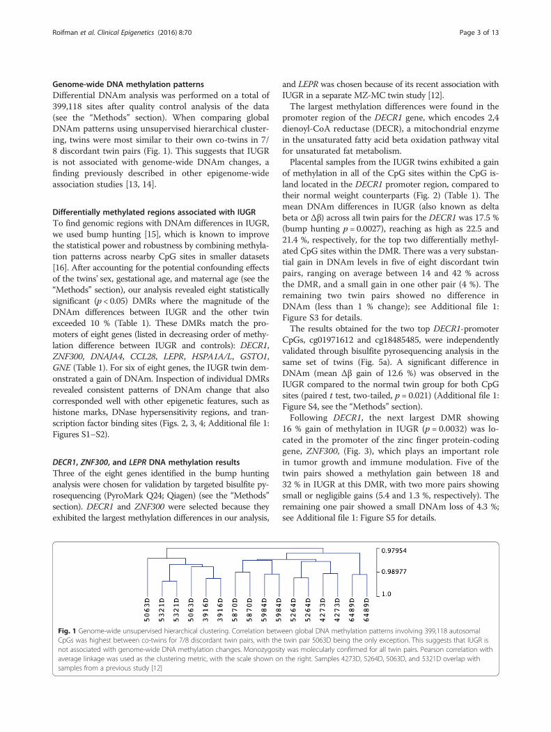
Genome-wide DNA methylation patternsDifferential DNAm analysis was performed on a total of399,118 sites after quality control analysis of the data(see the “Methods” section). When comparing globalDNAm patterns using unsupervised hierarchical cluster-ing, twins were most similar to their own co-twins in 7/8 discordant twin pairs (Fig. 1). This suggests that IUGRis not associated with genome-wide DNAm changes, afinding previously described in other epigenome-wideassociation studies [13, 14].
Differentially methylated regions associated with IUGRTo find genomic regions with DNAm differences in IUGR,we used bump hunting [15], which is known to improvethe statistical power and robustness by combining methyla-tion patterns across nearby CpG sites in smaller datasets[16]. After accounting for the potential confounding effectsof the twins’ sex, gestational age, and maternal age (see the“Methods” section), our analysis revealed eight statisticallysignificant (p < 0.05) DMRs where the magnitude of theDNAm differences between IUGR and the other twinexceeded 10 % (Table 1). These DMRs match the pro-moters of eight genes (listed in decreasing order of methy-lation difference between IUGR and controls): DECR1,ZNF300, DNAJA4, CCL28, LEPR, HSPA1A/L, GSTO1,GNE (Table 1). For six of eight genes, the IUGR twin dem-onstrated a gain of DNAm. Inspection of individual DMRsrevealed consistent patterns of DNAm change that alsocorresponded well with other epigenetic features, such ashistone marks, DNase hypersensitivity regions, and tran-scription factor binding sites (Figs. 2, 3, 4; Additional file 1:Figures S1–S2).
DECR1, ZNF300, and LEPR DNA methylation resultsThree of the eight genes identified in the bump huntinganalysis were chosen for validation by targeted bisulfite py-rosequencing (PyroMark Q24; Qiagen) (see the “Methods”section). DECR1 and ZNF300 were selected because theyexhibited the largest methylation differences in our analysis,
and LEPR was chosen because of its recent association withIUGR in a separate MZ-MC twin study [12].The largest methylation differences were found in the
promoter region of the DECR1 gene, which encodes 2,4dienoyl-CoA reductase (DECR), a mitochondrial enzymein the unsaturated fatty acid beta oxidation pathway vitalfor unsaturated fat metabolism.Placental samples from the IUGR twins exhibited a gain
of methylation in all of the CpG sites within the CpG is-land located in the DECR1 promoter region, compared totheir normal weight counterparts (Fig. 2) (Table 1). Themean DNAm differences in IUGR (also known as deltabeta or Δβ) across all twin pairs for the DECR1 was 17.5 %(bump hunting p = 0.0027), reaching as high as 22.5 and21.4 %, respectively, for the top two differentially methyl-ated CpG sites within the DMR. There was a very substan-tial gain in DNAm levels in five of eight discordant twinpairs, ranging on average between 14 and 42 % acrossthe DMR, and a small gain in one other pair (4 %). Theremaining two twin pairs showed no difference inDNAm (less than 1 % change); see Additional file 1:Figure S3 for details.The results obtained for the two top DECR1-promoter
CpGs, cg01971612 and cg18485485, were independentlyvalidated through bisulfite pyrosequencing analysis in thesame set of twins (Fig. 5a). A significant difference inDNAm (mean Δβ gain of 12.6 %) was observed in theIUGR compared to the normal twin group for both CpGsites (paired t test, two-tailed, p = 0.021) (Additional file 1:Figure S4, see the “Methods” section).Following DECR1, the next largest DMR showing
16 % gain of methylation in IUGR (p = 0.0032) was lo-cated in the promoter of the zinc finger protein-codinggene, ZNF300, (Fig. 3), which plays an important rolein tumor growth and immune modulation. Five of thetwin pairs showed a methylation gain between 18 and32 % in IUGR at this DMR, with two more pairs showingsmall or negligible gains (5.4 and 1.3 %, respectively). Theremaining one pair showed a small DNAm loss of 4.3 %;see Additional file 1: Figure S5 for details.
Fig. 1 Genome-wide unsupervised hierarchical clustering. Correlation between global DNA methylation patterns involving 399,118 autosomalCpGs was highest between co-twins for 7/8 discordant twin pairs, with the twin pair 5063D being the only exception. This suggests that IUGR isnot associated with genome-wide DNA methylation changes. Monozygosity was molecularly confirmed for all twin pairs. Pearson correlation withaverage linkage was used as the clustering metric, with the scale shown on the right. Samples 4273D, 5264D, 5063D, and 5321D overlap withsamples from a previous study [12]
Roifman et al. Clinical Epigenetics (2016) 8:70 Page 3 of 13
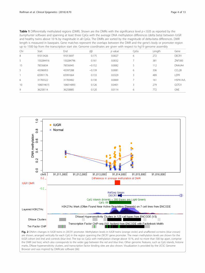
Table 1 Differentially methylated regions (DMR). Shown are the DMRs with the significance level p < 0.05 as reported by thebumphunter software and spanning at least three CpGs with the average DNA methylation differences (delta beta) between IUGRand healthy twins above 10 % by magnitude in all CpGs. The DMRs are sorted by the magnitude of delta-beta differences. DMRlength is measured in basepairs. Gene matches represent the overlaps between the DMR and the gene’s body or promoter regionup to 1500 bp from the transcription start site. Genome coordinates are given with respect to hg19 genome assembly
Chr Start End Δβ p value CpGs Length Gene
8 91013426 91013697 0.175 0.0027 6 272 DECR1
5 150284416 150284796 0.161 0.0032 7 381 ZNF300
15 78556834 78556945 −0.152 0.0082 5 112 DNAJA4
5 43396953 43397288 −0.139 0.0081 6 336 CCL28
1 65991176 65991664 0.133 0.0329 3 489 LEPR
6 31783322 31783482 0.130 0.0069 7 161 HSPA1A/L
10 106014615 106014893 0.126 0.0401 3 279 GSTO1
9 36258114 36258885 0.120 0.0114 6 772 GNE
Fig. 2 DNAm changes in IUGR twins in DECR1 promoter. Methylation levels in IUGR twins (orange circles) and unaffected co-twins (blue crosses)are shown, arranged vertically for each CpG in the region spanning the DECR1 gene promoter. The mean methylation levels are shown for theIUGR cohort (red line) and controls (blue line). The top six CpGs with methylation change above 10 %, and no more than 500 bp apart, comprisethe DMR (red box), which also corresponds to the wider gap between the red and blue lines. Other genomic features, such as CpG islands, histonemarks, DNase hypersensitivity clusters, and transcription factor binding sites are also shown. Visualization is provided by the UCSC GenomeBrowser and was inspired by DMRcate software [66]
Roifman et al. Clinical Epigenetics (2016) 8:70 Page 4 of 13
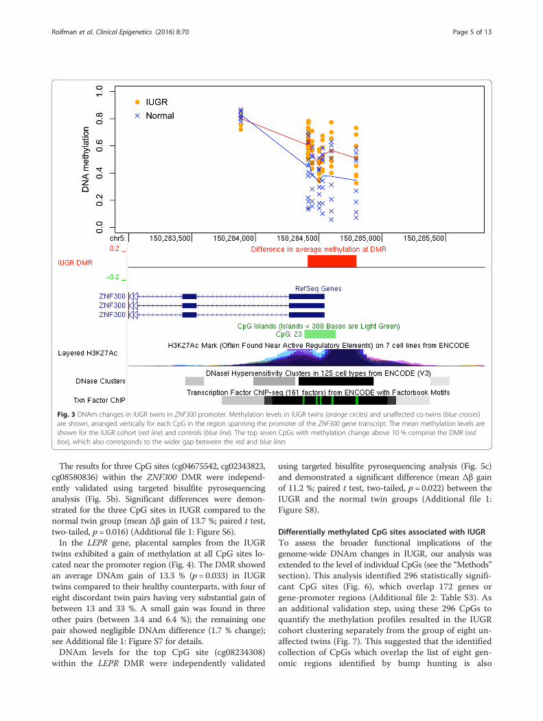
The results for three CpG sites (cg04675542, cg02343823,cg08580836) within the ZNF300 DMR were independ-ently validated using targeted bisulfite pyrosequencinganalysis (Fig. 5b). Significant differences were demon-strated for the three CpG sites in IUGR compared to thenormal twin group (mean Δβ gain of 13.7 %; paired t test,two-tailed, p = 0.016) (Additional file 1: Figure S6).In the LEPR gene, placental samples from the IUGR
twins exhibited a gain of methylation at all CpG sites lo-cated near the promoter region (Fig. 4). The DMR showedan average DNAm gain of 13.3 % (p = 0.033) in IUGRtwins compared to their healthy counterparts, with four ofeight discordant twin pairs having very substantial gain ofbetween 13 and 33 %. A small gain was found in threeother pairs (between 3.4 and 6.4 %); the remaining onepair showed negligible DNAm difference (1.7 % change);see Additional file 1: Figure S7 for details.DNAm levels for the top CpG site (cg08234308)
within the LEPR DMR were independently validated
using targeted bisulfite pyrosequencing analysis (Fig. 5c)and demonstrated a significant difference (mean Δβ gainof 11.2 %; paired t test, two-tailed, p = 0.022) between theIUGR and the normal twin groups (Additional file 1:Figure S8).
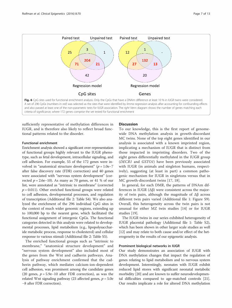
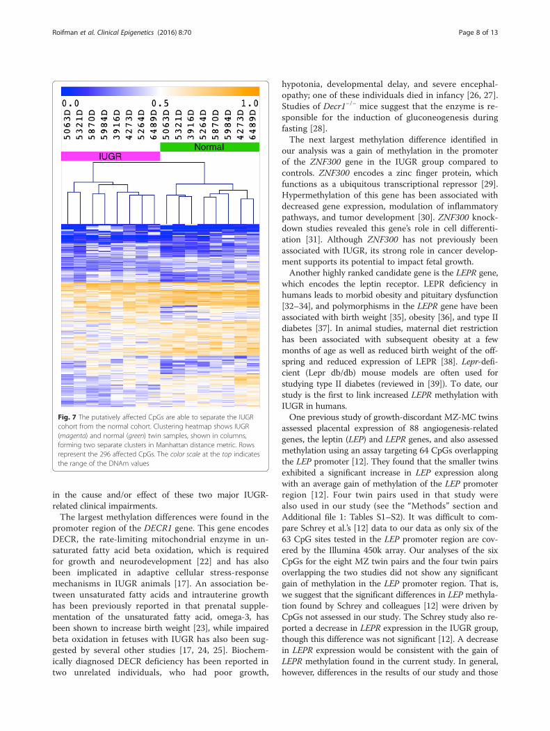
Differentially methylated CpG sites associated with IUGRTo assess the broader functional implications of thegenome-wide DNAm changes in IUGR, our analysis wasextended to the level of individual CpGs (see the “Methods”section). This analysis identified 296 statistically signifi-cant CpG sites (Fig. 6), which overlap 172 genes orgene-promoter regions (Additional file 2: Table S3). Asan additional validation step, using these 296 CpGs toquantify the methylation profiles resulted in the IUGRcohort clustering separately from the group of eight un-affected twins (Fig. 7). This suggested that the identifiedcollection of CpGs which overlap the list of eight gen-omic regions identified by bump hunting is also
Fig. 3 DNAm changes in IUGR twins in ZNF300 promoter. Methylation levels in IUGR twins (orange circles) and unaffected co-twins (blue crosses)are shown, arranged vertically for each CpG in the region spanning the promoter of the ZNF300 gene transcript. The mean methylation levels areshown for the IUGR cohort (red line) and controls (blue line). The top seven CpGs with methylation change above 10 % comprise the DMR (redbox), which also corresponds to the wider gap between the red and blue lines
Roifman et al. Clinical Epigenetics (2016) 8:70 Page 5 of 13
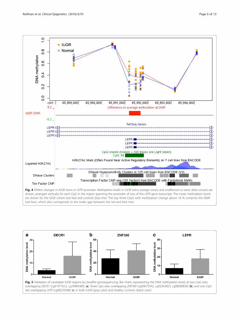
Fig. 4 DNAm changes in IUGR twins in LEPR promoter. Methylation levels in IUGR twins (orange circles) and unaffected co-twins (blue crosses) areshown, arranged vertically for each CpG in the region spanning the promoter of one of the LEPR gene transcripts. The mean methylation levelsare shown for the IUGR cohort (red line) and controls (blue line). The top three CpGs with methylation change above 10 % comprise the DMR(red box), which also corresponds to the wider gap between the red and blue lines
Fig. 5 Validation of candidate IUGR regions by bisulfite pyrosequencing. Bar charts representing the DNA methylation levels at two CpG sitesoverlapping DECR1 (cg01971612, cg18485485) (a), three CpG sites overlapping ZNF300 (cg04675542, cg02343823, cg08580836) (b), and one CpGsite overlapping LEPR (cg08234308) (c) in both IUGR (gray color) and healthy co-twins (black color)
Roifman et al. Clinical Epigenetics (2016) 8:70 Page 6 of 13
sufficiently representative of methylation differences inIUGR, and is therefore also likely to reflect broad func-tional patterns related to the disorder.
Functional enrichmentEnrichment analysis showed a significant over-representationof functional groups highly relevant to the IUGR pheno-type, such as fetal development, intracellular signaling, andcell adhesion. For example, 55 of the 172 genes were in-volved in “anatomical structure development” (p = 1.0e−7after false discovery rate (FDR) correction) and 40 geneswere associated with “nervous system development” (cor-rected p = 2.6e−10). As many as 70 genes, or 41 % of ourlist, were annotated as “intrinsic to membrane” (correctedp = 0.011). Other enriched functional groups were relatedto cell adhesion, developmental processes, and regulationof transcription (Additional file 2: Table S4). We also ana-lyzed the enrichment of the 296 individual CpG sites inthe context of much wider genomic regions, extending upto 100,000 bp to the nearest gene, which facilitated thefunctional assignment of intergenic CpGs. The functionalcategories detected in this analysis were related to develop-mental processes, lipid metabolism (e.g., lipopolysacchar-ide metabolic process, response to cholesterol) and cellularresponse to various stimuli (Additional file 2: Table S5).The enriched functional groups such as “intrinsic to
membrane,” “anatomical structure development” and“nervous system development” also included most ofthe genes from the Wnt and cadherin pathways. Ana-lysis of pathway enrichment confirmed that the cad-herin pathway, which mediates calcium-ion-dependentcell adhesion, was prominent among the candidate genes(20 genes, p = 5.9e−10 after FDR correction), as was therelated Wnt signaling pathway (23 affected genes, p = 5.0e−8 after FDR correction).
DiscussionTo our knowledge, this is the first report of genome-wide DNA methylation analysis in growth-discordantMC twins. None of the top eight genes identified in ouranalysis is associated with a known imprinted region,implicating a mechanism of IUGR that is distinct fromthose impacted in imprinting disorders. Two of theeight genes differentially methylated in the IUGR group(DECR1 and GSTO1) have been previously associatedwith IUGR (in animals and singleton humans, respect-ively), suggesting (at least in part) a common patho-genic mechanism for IUGR in singletons versus that inMC growth-discordant twins [17, 18].In general, for each DMR, the patterns of DNAm dif-
ferences in IUGR (Δβ) were consistent across the major-ity of twin pairs, although the magnitude of Δβ acrossdifferent twin pairs varied (Additional file 1: Figure S9).Overall, this heterogeneity across the twin pairs is notunusual for either MZ twin studies [14] or for IUGRstudies [19].The IUGR twins in our series exhibited heterogeneity of
IUGR placental pathology (Additional file 1: Table S2),which has been shown in other larger scale studies as well[12] and may relate to both cause and/or effect of the het-erogeneity in the results of our epigenetic analysis.
Prominent biological networks in IUGROur study demonstrates an association of IUGR withDNA methylation changes that impact the regulation ofgenes relating to lipid metabolism and to nervous systemdevelopment. Interestingly, neonates with IUGR exhibitreduced lipid stores with significant neonatal metabolicmorbidity [20] and are known to suffer neurodevelopmen-tal difficulties compared to age-matched controls [21].Our results implicate a role for altered DNA methylation
Fig. 6 CpG sites used for functional enrichment analysis. Only the CpGs that have a DNAm difference at least 10 % in IUGR twins were considered.A set of 296 CpGs (numbers in red) was selected as the sites that were identified by limma regression analysis after accounting for confounding effectsand also passed at least one of the non-parametric tests for IUGR association. The right Venn diagram shows the number of genes matching eachcriteria of significance, where 172 genes comprise the set tested for functional enrichment
Roifman et al. Clinical Epigenetics (2016) 8:70 Page 7 of 13
in the cause and/or effect of these two major IUGR-related clinical impairments.The largest methylation differences were found in the
promoter region of the DECR1 gene. This gene encodesDECR, the rate-limiting mitochondrial enzyme in un-saturated fatty acid beta oxidation, which is requiredfor growth and neurodevelopment [22] and has alsobeen implicated in adaptive cellular stress-responsemechanisms in IUGR animals [17]. An association be-tween unsaturated fatty acids and intrauterine growthhas been previously reported in that prenatal supple-mentation of the unsaturated fatty acid, omega-3, hasbeen shown to increase birth weight [23], while impairedbeta oxidation in fetuses with IUGR has also been sug-gested by several other studies [17, 24, 25]. Biochem-ically diagnosed DECR deficiency has been reported intwo unrelated individuals, who had poor growth,
hypotonia, developmental delay, and severe encephal-opathy; one of these individuals died in infancy [26, 27].Studies of Decr1−/− mice suggest that the enzyme is re-sponsible for the induction of gluconeogenesis duringfasting [28].The next largest methylation difference identified in
our analysis was a gain of methylation in the promoterof the ZNF300 gene in the IUGR group compared tocontrols. ZNF300 encodes a zinc finger protein, whichfunctions as a ubiquitous transcriptional repressor [29].Hypermethylation of this gene has been associated withdecreased gene expression, modulation of inflammatorypathways, and tumor development [30]. ZNF300 knock-down studies revealed this gene’s role in cell differenti-ation [31]. Although ZNF300 has not previously beenassociated with IUGR, its strong role in cancer develop-ment supports its potential to impact fetal growth.Another highly ranked candidate gene is the LEPR gene,
which encodes the leptin receptor. LEPR deficiency inhumans leads to morbid obesity and pituitary dysfunction[32–34], and polymorphisms in the LEPR gene have beenassociated with birth weight [35], obesity [36], and type IIdiabetes [37]. In animal studies, maternal diet restrictionhas been associated with subsequent obesity at a fewmonths of age as well as reduced birth weight of the off-spring and reduced expression of LEPR [38]. Lepr-defi-cient (Lepr db/db) mouse models are often used forstudying type II diabetes (reviewed in [39]). To date, ourstudy is the first to link increased LEPR methylation withIUGR in humans.One previous study of growth-discordant MZ-MC twins
assessed placental expression of 88 angiogenesis-relatedgenes, the leptin (LEP) and LEPR genes, and also assessedmethylation using an assay targeting 64 CpGs overlappingthe LEP promoter [12]. They found that the smaller twinsexhibited a significant increase in LEP expression alongwith an average gain of methylation of the LEP promoterregion [12]. Four twin pairs used in that study werealso used in our study (see the “Methods” section andAdditional file 1: Tables S1–S2). It was difficult to com-pare Schrey et al.’s [12] data to our data as only six of the63 CpG sites tested in the LEP promoter region are cov-ered by the Illumina 450k array. Our analyses of the sixCpGs for the eight MZ twin pairs and the four twin pairsoverlapping the two studies did not show any significantgain of methylation in the LEP promoter region. That is,we suggest that the significant differences in LEP methyla-tion found by Schrey and colleagues [12] were driven byCpGs not assessed in our study. The Schrey study also re-ported a decrease in LEPR expression in the IUGR group,though this difference was not significant [12]. A decreasein LEPR expression would be consistent with the gain ofLEPR methylation found in the current study. In general,however, differences in the results of our study and those
Fig. 7 The putatively affected CpGs are able to separate the IUGRcohort from the normal cohort. Clustering heatmap shows IUGR(magenta) and normal (green) twin samples, shown in columns,forming two separate clusters in Manhattan distance metric. Rowsrepresent the 296 affected CpGs. The color scale at the top indicatesthe range of the DNAm values
Roifman et al. Clinical Epigenetics (2016) 8:70 Page 8 of 13
found by Schrey and colleagues [12] may largely be attrib-uted to the use of different methodologies: genome-wideDNA methylation with limited coverage at individual pro-moter regions in this study, and gene expression analysisby Schrey and colleagues.The remainder of the genes with methylation differences
identified in the IUGR group (DNAJA4, CCL28, HSPA1A/L, GSTO1, and GNE) function in growth, immunity, andcell metabolism. The GSTO1 gene, which encodes anarsenic detoxification enzyme, glutathione S-transferaseomega 1, is the only gene in this list to have a prior associ-ation with fetal growth restriction [18]. This enzyme alsoplays a role in the inflammatory response, apoptosis, andtumor development (reviewed in [40]). The mouse Gsto1knockout model shows impaired detoxification of arsenic,a known human carcinogen [41]. DNAJA4 and HSPA1A/Lencode heat-shock proteins. A variety of tumors exhibithypermethylation of DNAJA4, which has been associ-ated with gene silencing [42, 43]. Increased expressionof DNAJA4 has been shown to suppress tumor invasion[42–44]. CCL28, which encodes the growth factor che-mokine ligand 28 [45], increases placental cell apoptosis[46] and is downregulated in a variety of human malig-nancies [47, 48]. GNE encodes a key enzyme in sialicacid biosynthesis, a process that is often upregulated intumor development [49]. Reduced sialylation has beenshown in heterozygous Gne-deficient mice, while completeknockouts are embryonically lethal [50]. Gne-deficientmouse embryonic stem cells exhibit impaired proliferationand cell differentiation, particularly affecting muscle [51].In humans, GNE deficiency causes Nonaka myopathy [52].Collectively, the aberrant methylation of the genes foundin our IUGR cohort appears to drive increased apoptosis,impaired cellular metabolism as well as poor growth, andinvasion of the placenta.
Prominent cellular pathways in IUGRThe Wnt and cadherin pathways were significantly over-represented in the list of candidate genes generated from296 individual CpGs representative of the IUGR epige-nome. Wnt signaling is well known for its role in carcino-genesis and embryonic development, specifically body axispatterning, cell proliferation, cell migration and cell differ-entiation [53, 54]. Wnt signaling plays a role in placentaldevelopment (trophoblast proliferation and invasion) andis altered in gestational diseases (reviewed in [55]). Ourgroup has previously shown that the promoter region ofthe WNT2 gene is differentially methylated in the placen-tas of IUGR singletons versus controls [19]. WNT2 geneexpression has also been proposed to mediate poor mater-nal nutrition and fetal growth restriction [56]. Our resultssuggest that upregulation of DNAJA4 may be an importantdownstream effect of impaired Wnt signaling in IUGR.Cadherins are a group of “calcium-dependent adhesion”
transmembrane proteins that facilitate normal embryonictissue organization and trophoblast invasion via cell-to-celladhesion and cell migration (reviewed in [57]).The precise mechanisms by which Wnt and cadherin
signaling play a role in IUGR is unknown. To the best ofour knowledge, this is the first report that prominentlyhighlights these pathways in the pathogenesis of IUGRin growth-discordant MC twins.
ConclusionsThis is the first report of a genome-wide DNA methyla-tion analysis using the Infinium HumanMethylation450BeadChip platform to investigate IUGR in MC growth-discordant twins. We identified eight candidate genes thatare epigenetically altered in IUGR. Our results suggest thatdysregulation of lipid metabolism and transcription, as wellas impaired cadherin and Wnt signaling, are associatedwith IUGR. Collectively, the genes exhibiting differentialregulation in growth-restricted fetuses may underlie fetalprogramming and may generate candidate biomarkers forIUGR and/or its related adult-onset diseases. These resultsshould be validated by further investigation of DNAmethylation and/or gene expression of the identified candi-date genes in a large cohort of MZ-MC growth-discordanttwins. Finally, we propose the growth-discordant MC twinmodel as a useful tool in the genome-wide study of IUGR.
MethodsSample selectionThis study was approved by the Mount Sinai Hospital(MSH) and the Hospital for Sick Children (SickKids)Research Ethics Boards (REB # 10-0128-E). Participantswere recruited from the MSH high-risk obstetrical clinic,following diagnosis of MC discordant growth and pla-cental insufficiency. Informed consent was obtainedfrom each participant. Growth discordance was definedas a birth weight difference >20 % between the co-twins[58]. Birth weight was plotted as percentile for gesta-tional age using a Canadian population-based referencechart [59]. Placental insufficiency was defined as intermit-tent absent or reversed end-diastolic flow. A full list of theinclusion and exclusion criteria is found in Additional file 1:Table S6.Following these criteria, we recruited eight MC growth-
discordant twin pregnancies.Placental characteristics are detailed in Additional file 1:
Table S2. Four of these pairs overlap with a previousMZ twin study of IUGR that evaluated expression ofangiogenesis-related genes and targeted DNA methyla-tion of one gene [12].Placental sampling was performed immediately after de-
livery by members of the Research Center for Women’sand Infants’ Health BioBank, Toronto. At delivery, cordclamps were labeled as twin “A” or “B” to distinguish each
Roifman et al. Clinical Epigenetics (2016) 8:70 Page 9 of 13
twin’s placental share. Snap-frozen placental samples wereobtained from each twin according to a previously stan-dardized protocol (http://biobank.lunenfeld.ca/). The re-mainder of each placenta was sent for pathologicalexamination. Monozygosity was molecularly confirmedfor all twin pairs.
Genome-wide DNA methylation analysisGenomic DNA from the placental samples was bisulfitetreated (Qiagen). DNA methylation was then analyzedusing the Infinium HumanMethylation450 BeadChiparray (Illumina, San Diego, CA, USA) following thestandard Infinium HD Assay Methylation ProtocolGuide (Part #15019519, Illumina). The array assessesDNA methylation at single CpG sites genome-wide; how-ever, it does not differentiate between 5-methylcytosineand 5-hydroxymethylcytosine, which is a recognized limi-tation of this platform [60].All samples were run on the array together within the
same batch and passed quality control. Data pre-processingsuch as background subtraction and normalization to in-ternal array controls was performed in Illumina GenomeS-tudio v2011.1 Methylation Module 1.9 software (http://www.illumina.com).
CpG probe filteringThe 485,577 methylation probes from the Human-Methylation450 array were filtered by removing probeswith missing DNA methylation values (beta values eithermissing or exactly zero) and with detection p valuesabove the significance threshold 0.05. We also removed allcross-reactive probes annotated as such in [61]. Tominimize possible confounding effect by ethnicity or sex,especially in the context of unpaired statistical tests be-tween IUGR and control groups (Fig. 6), all probes forwhich there was a known SNP of at least 1 % minor allelefrequency within 5 bp of the targeted cytosine (based onthe annotation in [61]) as well as probes located on sexchromosomes were excluded, leaving 399,118 autosomalCpG sites available for analysis.
Detection of differentially methylated regionsTo detect the differentially methylated regions, we usedthe bump hunting method of Jaffe and colleagues [15] im-plemented in the bumphunter Bioconductor package, set-ting the cutoff threshold of 10 % for the Δβ methylationdifference in IUGR. To account for confounding effects,the model design matrix consisted of an intercept term,IUGR indicator variable, and twin sex, maternal age andtwin’s gestational age as additional confounders. Statisticalsignificance of the detected bumps was assessed in com-parison to B = 1000 randomized bootstrap iterations.From the resulting collection of bumps, we retained onlythe DMRs that contained at least three CpGs with gaps
no more than 500 bp, and exhibited methylation differ-ences of 10 % or higher in IUGR while satisfying the sig-nificance level p < 0.05.
Selection of CpG sites for functional enrichment analysisFor the analysis of methylome-wide functional enrich-ment in IUGR, we included not only the eight DMRsfrom the bump hunting analysis, but also the CpG sitesthat were significantly different in IUGR compared tohealthy co-twins. These individual CpG sites were iden-tified using a combination of statistical criteria to ac-count for the small sample size.Three sets of statistical tests were performed on eight
growth-discordant twin pairs at each CpG site. First, weused limma regression modeling, which is particularlywell suited for small sample sizes thanks to its method-ology of information borrowing across CpG sites. Themodel was applied to DNAm data log-transformed intoM-values to improve statistical accuracy [62]. The twin’ssex, gestational age and maternal age were included inthe model as covariates. CpGs were considered signifi-cant if the limma regression p values corresponding tothe IUGR status satisfied the significance level p < 0.05.Second, we used a non-parametric approach to test
the between-group differences, comparing the placentalmethylation levels in the group of eight healthy individ-uals (controls) and the group of eight affected IUGR in-dividuals. The unpaired Mann–Whitney U test was usedfor each CpG site.Finally, the changes in DNA methylation level in healthy
twins versus their IUGR-affected counterparts were com-pared using the paired Wilcoxon signed-rank test, whichattempted to identify a consistent pattern of DNAm shiftacross all eight pairs, although not necessarily an overalldifference between the IUGR and the healthy cohorts.Thus, the two non-parametric tests are complementary infinding pattern of differences across the twin pairs.Given the large number of probes tested and a rela-
tively small sample size, the standard methods of cor-rection for multiple testing (such as Bonferroni orBenjamini-Hochberg FDR procedures) were too re-strictive to confirm the validity of any of the individualtests. Therefore, we decided on a pragmatic strategy ofusing the probe-level statistical tests as guidance andcomplemented them with an effect-size threshold. Weconsidered the threshold of 10 % as sufficiently strin-gent because relatively few CpG sites had a DNAmethylation change exceeding 10 % by magnitude.For validation purposes, we used unsupervised clustering
to confirm that the resulting set of CpG sites contained asufficiently discernible biological signal unobscured by thepotential false positives or other stochastic noise in the se-lected loci. All DNAm profiles were first reduced to the setof probes with significant differences in IUGR according to
Roifman et al. Clinical Epigenetics (2016) 8:70 Page 10 of 13
our selection criteria. Thereafter, hierarchical clusteringwas applied to discern the different groups in our cohort.
Functional enrichment analysisTo identify prominent clinical mechanisms affectingand/or affected by growth restriction in our cohort, the172 genes with evidence of differential methylation wereinvestigated with respect to their functions and majorbiological roles. The analysis was performed using DA-VID [63]. All p values were FDR corrected.The 296 putatively affected CpG set was also analyzed
in the context of wider genomic regions using GREAT[64]. We set the distal extension to the nearest genes as100 kbp. The background set of probes to which the com-parison was made was defined as the 399,118 autosomalCpGs used as the initial input to our analysis pipeline.
Validation analysis of DNA methylation using bisulfitepyrosequencingResults of top candidate CpG sites (cg01971612 andcg18485485 for DECR1; cg04675542, cg02343823, andcg08580836 for ZNF300; and cg08234308 for LEPR)were validated using pyrosequencing of bisulfite convertedDNA (PyroMark Q24; Qiagen). Polymerase chain reaction(PCR) and sequencing primers were designed using Pyro-Mark Assay Design 2.0. All procedures were performedaccording to the manufacturer’s protocols [65]. The pres-ence of 5-methylcytosine versus 5-hydroxymethylcytosinecould not be distinguished using this method.
Additional files
Additional file 1: Supplementary Figures and Tables. (PDF 1827 kb)
Additional file 2: Supplementary Tables S3–S5. (XLSX 86 kb)
AbbreviationsDMR, differentially methylated region; DNAm, DNA methylation; FDR, falsediscovery rate; IUGR, intrauterine growth restriction; MC, monochorionic;MZ, monozygotic; PCR, polymerase chain reaction
AcknowledgementsWe would like to thank Chunhua Zhao, Youliang Lou, and Yi-an Chen fortheir contributions to this work. This study was supported by the MclaughlinCentre and the Canadian Institute for Health Research. Bioinformatic ana-lyses were supported by the Canadian Centre for Computational Genomics(C3G), part of the Genome Innovation Network (GIN), funded by GenomeCanada through Genome Quebec and Ontario Genomics.
Authors’ contributionsMR participated in the design of the study, carried out the sample selection,methylation array interpretation, and validation analysis, and drafted themanuscript. SC participated in the design of the study, carried out themethylation array interpretation and validation analysis, and drafted themanuscript. ALT participated in the design of the study, performed thebioinformatic analyses of the data, and drafted the manuscript.MB participated in the design of the study and oversaw bioinformaticanalyses. SD, and JK participated in the design of the study. SK performedthe pathological analyses on the samples. RW participated in the design,data analyses, and writing of the manuscript as corresponding senior author.All authors read and approved the final manuscript.
Competing interestsThe authors declare that they have no competing interests.
Author details1Division of Clinical and Metabolic Genetics, The Hospital for Sick Children,Toronto, Ontario, Canada. 2Department of Paediatrics, University of Toronto,Toronto, Ontario, Canada. 3Genetics and Genome Biology Program, TheHospital for Sick Children, Toronto, Ontario, Canada. 4The Prenatal andMedical Genetics Program, Department of Obstetrics and Gynaecology,Mount Sinai Hospital, Toronto, Ontario, Canada. 5Centre for ComputationalMedicine, The Hospital for Sick Children, Toronto, Ontario, Canada. 6C.S. MottCenter for Human Growth and Development, Wayne State School ofMedicine, Wayne State University, Detroit, MI, USA. 7Maternal-Fetal MedicineDivision, Department of Obstetrics and Gynecology, Mount Sinai Hospital,Toronto, Ontario, Canada. 8Department of Laboratory Medicine andPathology, University of Toronto, Toronto, Ontario, Canada. 9Department ofComputer Science, University of Toronto, Toronto, Ontario, Canada.10Department of Obstetrics and Gynecology, University of Toronto, Toronto,Ontario, Canada. 11Institute of Medical Science, University of Toronto,Toronto, Ontario, Canada.
Received: 25 April 2016 Accepted: 12 June 2016
References1. Hendrix N, Berghella V. Non-placental causes of intrauterine growth
restriction. Semin Perinatol. 2008;32(3):161–5.2. Bernstein IM, Horbar JD, Badger GJ, Ohisson A, Golan A. Morbidity and
mortality among very-low-birth-weight neonates with intrauterine growthrestriction. The Vermont Oxford Network. Am J Obstet Gynecol. 2000;182(1 Pt 1):198–206.
3. Barker DJ. The fetal and infant origins of adult disease. BMJ. 1990;301(6761):1111.
4. Gluckman PD, Hanson MA, Beedle AS. Early life events and theirconsequences for later disease: a life history and evolutionary perspective.Am J Hum Biol. 2007;19(1):1–19.
5. Morley R, Dwyer T, Carlin JB. Studies of twins: can they shed light on thefetal origins of adult disease hypothesis? Twin Res. 2003;6(6):520–5.
6. Inbar-Feigenberg M, Choufani S, Butcher DT, Roifman M, Weksberg R. Basicconcepts of epigenetics. Fertil Steril. 2013;99(3):607–15.
7. Nelissen EC, van MontFoort AP, Dumoulin JC, Evers JL. Epigenetics and theplacenta. Hum Reprod Update. 2011;17(3):397–41.
8. Lewi L, Gucciardo L, Huber A, Jani J, Van Mieghem T, Done E, et al. Clinicaloutcome and placental characteristics of monochorionic diamniotic twinpairs with early- and late-onset discordant growth. Am J Obstet Gynecol.2008;199(5):511 e1–7.
9. Sebire NJ, Snijders RJ, Hughes K, Sepulveda W, Nicolaides KH. The hiddenmortality of monochorionic twin pregnancies. Br J Obstet Gynaecol.1997;104(10):1203–7.
10. Bejar R, Vigliocco G, Gramajo H, Solana C, Benirschke K, Berry C, et al.Antenatal origin of neurologic damage in newborn infants. II. Multiplegestations. Am J Obstet Gynecol. 1990;162(5):1230–6.
11. Lopriore E, Pasman SA, Klumper FJ, Middeldorp JM, Walther FJ, Oepkes D.Placental characteristics in growth-discordant monochorionic twins: amatched case-control study. Placenta. 2012;33(3):171–4.
12. Schrey S, Kingdom J, Baczyk D, Fitzgerald B, Keating S, Ryan G, et al. Leptinis differentially expressed and epigenetically regulated acrossmonochorionic twin placenta with discordant fetal growth. Mol HumReprod. 2013;19(11):764–72.
13. Gordon L, Joo JE, Powell JE, Ollikainen M, Novakovic B, Li X, et al. NeonatalDNA methylation profile in human twins is specified by a complex interplaybetween intrauterine environmental and genetic factors, subject to tissue-specific influence. Genome Res. 2012;22(8):1395–406.
14. Wong CC, Meaburn EL, Ronald A, Price TS, Jeffries AR, Schalkwyk LC, et al.Methylomic analysis of monozygotic twins discordant for autism spectrumdisorder and related behavioural traits. Mol Psychiatry. 2014;19(4):495–503.
15. Jaffe AE, Murakami P, Lee H, Leek JT, Fallin MD, Feinberg AP, et al. Bumphunting to identify differentially methylated regions in epigeneticepidemiology studies. Int J Epidemiol. 2012;41(1):200–9.
16. Li D, Xie Z, Pape ML, Dye T. An evaluation of statistical methods for DNAmethylation microarray data analysis. BMC Bioinformatics. 2015;16:217.
Roifman et al. Clinical Epigenetics (2016) 8:70 Page 11 of 13
17. Arroyo JA, Garcia-Jones P, Graham A, Teng CC, Battaglia FC, Galan HL.Placental TonEBP/NFAT5 osmolyte regulation in an ovine model ofintrauterine growth restriction. Biol Reprod. 2012;86(3):94.
18. Thomas S, Arbuckle TE, Fisher M, Fraser WD, Ettinger A, King W. Metalsexposure and risk for small-for-gestational age birth in a Canadian birthcohort: the MIREC study. Environ Res. 2015;140:430–9.
19. Ferreira JC, Choufani S, Grafodatskaya D, Butcher DT, Zhao C, Chitayat D, etal. WNT2 promoter methylation in human placenta is associated with lowbirthweight percentile in the neonate. Epigenetics. 2011;6(4):440–9.
20. Longo S, Bollani L, Decembrino L, Di Comite A, Angelini M, Stronati M.Short-term and long-term sequelae in intrauterine growth retardation(IUGR). J Matern Fetal Neonatal Med. 2013;26(3):222–5.
21. Geva R, Eshel R, Leitner Y, Valevski AF, Harel S. Neuropsychological outcomeof children with intrauterine growth restriction: a 9-year prospective study.Pediatrics. 2006;118(1):91–100.
22. Herrera E. Lipid metabolism in pregnancy and its consequences in the fetusand newborn. Endocrine. 2002;19(1):43–55.
23. Szajewska H, Horvath A, Koletzko B. Effect of n-3 long-chain polyunsaturatedfatty acid supplementation of women with low-risk pregnancies onpregnancy outcomes and growth measures at birth: a meta-analysis ofrandomized controlled trials. Am J Clin Nutr. 2006;83(6):1337–44.
24. Cetin I, Giovannini N, Alvino G, Agostoni C, Riva E, Giovannini M, et al.Intrauterine growth restriction is associated with changes inpolyunsaturated fatty acid fetal-maternal relationships. Pediatr Res. 2002;52(5):750–5.
25. Alvino G, Cozzi V, Radaelli T, Ortega H, Herrera E, Cetin I. Maternal and fetalfatty acid profile in normal and intrauterine growth restriction pregnancieswith and without preeclampsia. Pediatr Res. 2008;4(6):615–20.
26. Roe CR, Millington DS, Norwood DL, Kodo N, Sprecher H, Mohammed BS,Nada M, Schulz H, McVie R. 2,4-Dienoyl-coenzyme A reductase deficiency: apossible new disorder of fatty acid oxidation. J Clin Invest. 1990;85(5):1703–7.
27. Houten SM, Denis S, te Brinke H, Jongejan A, van Kampen AHC, Bradley EJ,Baas F, Hennekam RCM, Millington DS, Young SP, Frazier DM, Gucsavas-Calikoglu M, Wanders RJA. Mitochondrial NADP(H) deficiency due tomutation in NADK2 causes dienoyl-CoA reductase deficiency withhyperlysinemia. Hum Mol Genet. 2014;23(18):5009–16.
28. Miinalainen IJ, Schmitz W, Huotari A, et al. Mitochondrial 2,4-dienoyl-CoAreductase deficiency in mice results in severe hypoglycemia with stressintolerance and unimpaired ketogenesis. PLoS Genet. 2009;5(7):e1000543.
29. Gou D, Wang J, Gao L, Sun Y, Peng X, Huang J, Li W. Identification andfunctional analysis of a novel human KRAB/C2H2 zinc finger gene ZNF300.Biochim Biophys Acta. 2004;1676(2):203–9.
30. Zhao Y, Xue F, Sun J, Guo S, Zhang H, Qiu B, et al. Genome-widemethylation profiling of the different stages of hepatitis B virus-relatedhepatocellular carcinoma development in plasma cell-free DNA revealspotential biomarkers for early detection and high-risk monitoring ofhepatocellular carcinoma. Clin Epigenetics. 2014;6(1):30.
31. Cai J, Gong R, Yan F, Yu C, Liu L, Wang W, et al. ZNF300 knockdown inhibitsforced megakaryocytic differentiation by phorbol and erythrocytic differentiationby arabinofuranosyl cytidine in K562 cells. PLoS One. 2014;9(12):e114768.
32. Clement K, Viasse C, Lahlou N, Cabrol S, Pelloux V, Cassuto D, et al. Amutation in the human leptin receptor gene causes obesity and pituitarydysfunction. Nature. 1998;392(6674):398–401.
33. Farooqi IS, Wangensteen T, Collins S, Kimber W, Matarese G, Keogh JM, etal. Clinical and molecular genetic spectrum of congenital deficiency of theleptin receptor. N Engl J Med. 2007;356(3):237–47.
34. Jiang B, Liu Y, Liu Y, Fang F, Wang X, Li B. Association of four insulinresistance genes with type 2 diabetes mellitus and hypertension in theChinese Han population. Mol Biol Rep. 2014;41(2):925–33.
35. Marginean CO, Marginean C, Voidazan S, Melit L, Crauciuc A, Duicu C,Banescu C. Correlations between leptin gene polymorphisms 223A/G, 1019G/A, 492 G/C, 976 C/A, and anthropometrical and biochemical parametersin children with obesity: a prospective case-control study in Romanianpopulation—the Nutrichild study. Medicine. 2016;95(12):e3115.
36. Yang MM, Wang J, Fan JJ, Ng TK, Sun DJ, Guo X, Teng Y, Li YB. Variations inthe obesity gene “LEPR” contribute to risk of type 2 diabetes mellitus:evidence from a meta-analysis. J Diab Res. 2016;2016:5412084.
37. Souren NY, Paulussen AD, Steyls A, Loos RJ, Stassen AP, Gielen M, et al.Common SNPs in LEP and LEPR associated with birth weight and type 2diabetes-related metabolic risk factors in twins. Int J Obes. 2008;32:1233–9.
38. Ovilo C, Gonzalez-Bulnes A, Benitez R, Ayuso M, Barbero A, Perez-Solana ML,et al. Prenatal programming in an obese swine model: sex-related effects ofmaternal energy restriction on morphology, metabolism and hypothalamicgene expression. Br J Nutr. 2014;111(4):735–46.
39. King AJF. The use of animal models in diabetes research. Br J Pharmacol.2012;166(3):877–94.
40. Zmorzynski S, Swiderska-Kolacz G, Koczkodaj D, Filip AA. Significance ofpolymorphisms and expression of enzyme-encoding genes related to glutathionein hematopoietic cancers and solid tumors. Biomed Res Int. 2015;2015:853573.
41. Chowdhury UK, Zakharyan RA, Hernandez A, Avram MD, Kopplin MJ,Aposhian HV. Glutathione-S-transferase-omega [MMA(V) reductase]knockout mice: enzyme and arsenic species concentrations in tissues afterarsenate administration. Toxicol Appl Pharmacol. 2006;216(3):446–57.
42. Alholle A, Brini AT, Gharanei S, Vaiyapuri S, Arrigoni E, Dallol A, et al.Functional epigenetic approach identifies frequently methylated genes inEwing sarcoma. Epigenetics. 2013;8(11):1198–204.
43. Mahoney SE, Yao Z, Keyes CC, Tapscott SJ, Diede SJ. Genome-wide DNAmethylation studies suggest distinct DNA methylation patterns in pediatricembryonal and alveolar rhabdomyosarcomas. Epigenetics. 2012;7(4):400–8.
44. Pencheva N, Tran H, Buss C, Huh D, Drobnjak M, Busam K, Tavazoie SF.Convergent multi-miRNA targeting of ApoE drives LRP1/LRP8-dependentmelanoma metastasis and angiogenesis. Cell. 2012;151(5):1068–82.
45. Karlsson C, Baudet A, Miharada N, Soneji S, Gupta R, Magnusson M, et al.Identification of the chemokine CCL28 as a growth and survival factor forhuman hematopoietic stem and progenitor cells. Blood. 2013;121(19):3838–42.
46. Sun C, Zhang YY, Tang CL, Wang SC, Piao HL, Toa Y, et al. ChemokineCCL28 induces apoptosis of decidual stromal cells via binding CCR3/CCR10in human spontaneous abortion. Mol Hum Reprod. 2013;19(10):676–86.
47. Dimberg J, Hugander A, Wågsäter D. Protein expression of the chemokine,CCL28, in human colorectal cancer. Int J Oncol. 2006;28(2):315–9.
48. Liu GX, Lan J, Sun Y, Hu YJ, Jiang GS. Expression of the chemokine CCL28 inpleomorphic adenoma and adenolymphoma of the human salivary glands.Exp Ther Med. 2012;4(1):65–9.
49. Kemmner W, Kessel P, Sanchez-Ruderisch H, Moller H, Hinderlich S, Schlag PM,Detjen K. Loss of UDP-N-acetylglucosamine 2-epimerase/N-acetylmannosaminekinase (GNE) induces apoptotic processes in pancreatic carcinoma cells. FASEB J.2012;26(2):938–46.
50. Schwarzkopf M, Knobeloch KP, Rohde E, Hinderlich S, Wiechens N, Lucka L,Horak I, Reutter W, Horstkorte R. Sialylation is essential for earlydevelopment in mice. Proc Natl Acad Sci U S A. 2002;99:5267–70.
51. Krentsis IM, Sela I, Eiges R, Blanchard V, Berger M, Cohen MB, Mitrani-RosenbaumS. GNE is involved in the early development of skeletal and cardiac muscle. PLoSOne. 2011;6(6):e21389.
52. Eisenberg I, Avidan N, Potikha T, Hochner H, Chen M, Olender T, Barash M,Shemesh M, Sadeh M, Grabov-Nardini G, Shmilevich I, Friedmann A, KarpatiG, Bradley WG, Baumbach L, Lancet D, Ben Asher E, Beckmann JS, Argov Z,Mitrani-Rosenbaum S. The UPD-N-acetylglucosamine 2-epimerase/N-acetylmannosamine kinase gene is mutated in recessive hereditary inclusionbody myopathy. Nature Genet. 2001;29:83–7.
53. Clevers H. Wnt/beta-catenin signaling in development and disease. Cell.2006;127(3):469–80.
54. Logan CY, Nusse R. The Wnt signaling pathway in development anddisease. Annu Rev Cell Dev Biol. 2004;20:781–810.
55. Knofler M, Pollheimer J. Human placental trophoblast invasion anddifferentiation: a particular focus on Wnt signaling. Front Genet. 2013;4:190.
56. Reamon-Buettner SM, Buschmann J, Lewin G. Identifying placentalepigenetic alterations in an intrauterine growth restriction (IUGR) rat modelinduced by gestational protein deficiency. Reprod Toxicol. 2014;45:117–24.
57. Kokkinos MI, Murthi P, Wafai R, Thompson EW, Newgreen DF. Cadherins inthe human placenta—epithelial-mesenchymal transition (EMT) andplacental development. Placenta. 2010;31(9):747–55.
58. Breathnach FM, McAuliffe FM, Geary M, Daly S, Higgins JR, Dornan J, et al.Prediction of safe and successful vaginal twin birth. Am J Obstet Gynecol.2011;205(3):237 e1–7.
59. Kramer MS, Platt RW, Wen SW, Joseph KS, Allen A, Abrahamowicz M, et al.A new and improved population-based Canadian reference for birth weightfor gestational age. Pediatrics. 2001;108(2):E35.
60. Sandoval J, Heyn H, Moran S, Serra-Musach J, Pujana MA, Bibikova M, et al.Validation of a DNA methylation microarray for 450,000 CpG sites in thehuman genome. Epigenetics. 2011;6(6):692–702.
61. Chen YA, Lemire M, Choufani S, Butcher DT, Grafodatskaya D, Zanke BW,et al. Discovery of cross-reactive probes and polymorphic CpGs in the IlluminaInfinium HumanMethylation450 microarray. Epigenetics. 2013;8(2):203–9.
Roifman et al. Clinical Epigenetics (2016) 8:70 Page 12 of 13
62. Du P, Zhang X, Huang CC, Jafari N, Kibbe WA, Hou L, et al. Comparison ofBeta-value and M-value methods for quantifying methylation levels bymicroarray analysis. BMC Bioinformatics. 2010;11:587.
63. da Huang W, Sherman BT, Lempicki RA. Systematic and integrative analysisof large gene lists using DAVID bioinformatics resources. Nat Protoc. 2009;4(1):44–57.
64. McLean CY, Bristor D, Hiller M, Clarke SL, Schaar BT, Lowe CB, et al. GREATimproves functional interpretation of cis-regulatory regions. Nat Biotechnol.2010;28(5):495–01.
65. Qiagen. PyroMark: PCR handbook. Sample & Assay Technologies. 2009;http://cmgm.stanford.edu/pan/doc/Pyrosequencing/PyroMark_Q24_PCR_Handbook.pdf
66. Peters TJ, Buckley MJ, Statham AL, Pidsley R, Samaras K, Lord VR, et al. Denovo identification of differentially methylated regions in the humangenome. Epigenetics Chromatin. 2015;8:6.
• We accept pre-submission inquiries
• Our selector tool helps you to find the most relevant journal
• We provide round the clock customer support
• Convenient online submission
• Thorough peer review
• Inclusion in PubMed and all major indexing services
• Maximum visibility for your research
Submit your manuscript atwww.biomedcentral.com/submit
Submit your next manuscript to BioMed Central and we will help you at every step:
Roifman et al. Clinical Epigenetics (2016) 8:70 Page 13 of 13