Embed Size (px)
Citation preview
SM Ophthalmology Journal
Gr upSM
How to cite this article Taleb EA. Giant Retinal Tear Retinal Detachment Etiologies, Surgical Outcome and Incidence of Recurrent Retinal Detachment after Silicon Oil Removal.
SM Opthalmol J. 2016; 2(1): 1008.OPEN ACCESS
IntroductionA Giant Retinal Tear (GRT) is defined as a full-thickness retinal break extending circumferentially
for 3 clock hours (90°) in the presence of a posteriorly detached vitreous [1]. GRTs are rare; their incidence has not been well established in the literature. The true incidence of GRTs is difficult to assess given their rarity, but one recent study estimates 0.094 per 100,000 of the general population per year [2].
Giant retinal tears may arise spontaneously, but approximately 25% of cases occur in association with ocular trauma [3]. The fellow eye of patients who have experienced a spontaneous giant retinal tear is at an increased risk of developing giant retinal tears, retinal detachment, or both [4]. Previous studies have reported various risk factors for giant retinal tears; these include trauma, high myopia, aphakia and pseudophakia, and young age [3]. The surgical approach for GRT has always been a challenge for vitreoretinal surgeons, as these patients have a high risk of Proliferative Vitreoretinopathy (PVR) formation (40-50%) [5]. Many approaches [5,6] to repositoning and fixating the inverted retinal flap, reattaching the retina, and reducing the risk of redetachment have been reported with varying success rates. The use of Perfluorocarbon Liquids (PFCLs) demonstrated by Chang et al [2] to unfold and flatten the inverted retina provides several advantages.
Despite improvement in the surgical maneuvers and tamponade agents, recurrence of the detachment still occurs due to several factors, such as reopening of the tear, formation of a new tear, or extension of the existing tear due to concurrent PVR [7]. PVR is one of the late complications of giant retinal breaks and the leading cause of surgical failure.6 Increased access to the exposed Retinal Pigment Epithelium (RPE) allows greater spillage of cells and pigment into the vitreous cavity and on the retinal surface, thereby increasing the risk of PVR [8].
In this study, we studied the etiology and the demographic and clinical characteristics of GRT and the safety and efficacy of PPV or combined PPV with encircling scleral buckle, 360° laser retinopexy and postoperative silicon oil tamponade in management. In addition, we sought to study complications of the surgery and the risk factor for re-detachment and determine the final anatomic and visual outcomes of the surgery. We also studied the incidence of retinal detachment in fellow eyes of patients with GRT.
Research Article
Giant Retinal Tear Retinal Detachment Etiologies, Surgical Outcome and Incidence of Recurrent Retinal Detachment after Silicon Oil RemovalEman Abo TalebDepartment of Ophthalmology, Sana’a University, Sana’a, Yemen
Article Information
Received date: Oct 03, 2016 Accepted date: Dec 09, 2016 Published date: Dec 19, 2016
*Corresponding author
Eman Abo Taleb, Department of Ophthalmology, Sana’a University, Sana’a, Yemen, Email: [email protected]
Distributed under Creative Commons CC-BY 4.0
Abstract
Purpose: To evaluate etiologies, management, and outcomes of patients with Giant Retinal Tears (GRT) undergoing primary surgery at tertiary referral center.
Methods: A Retrospective, consecutive case series of 94 patients with at least 3 months follow up after silicon oil removal. 57 eyes (60.6%) underwent vitrectomy, 36 eyes (38.3%) underwent combined vitrectomy with buckling and 1 eye (1.1%) underwent scleral buckling. Perfluorocarbon (PFCL) heavy liquid to flatten GRT flap intraoperative has been used then PFCL air exchange then air silicon exchange in all eyes undergoing vitrectomy. Fellow eye was observed for retinal detachment.
Results: Idiopathic cause constitute 47 eyes (50%), 25 eyes (26.6%) are myopic and 22 eyes (23.4%) have history of trauma. 85 eyes (90.4%) achieved anatomic success. Visual acuity at the last follow-up was at least 20/400 in 71 eyes (75.5%) of patients. Recurrent retinal detachment after Silicon Oil Removal (SOR) was found in 21 eyes (22.3%) of which, 50% had Proliferative Vitreoretinopathy Grade C (PVR-C) or more (p value 0.03) and 20% had GRT size more than 180° (p value 0.04). Pars Plana Vitrectomy (PPV) alone (p value 0.89) or combined PPV with buckling (p value 0.98) have no significant correlation with recurrent Retinal Detachment (RD). 21% of the fellow eye had retinal detachment.
Conclusion: Idiopathic cause constitutes the majority 50%. Patients with GRT who underwent surgery achieved high anatomic success rate. PVR-C or more remain the most significant risk factor for recurrent RD after SOR whereas PPV alone or combined PPV with buckling have no significant correlation with recurrent RD.
Citation: Taleb EA. Giant Retinal Tear Retinal Detachment Etiologies, Surgical Outcome and Incidence of Recurrent Retinal Detachment after Silicon Oil Removal. SM Opthalmol J. 2016; 2(1): 1008. Page 2/5
Gr upSM Copyright Taleb EA
MethodsThis retrospective study was performed on consecutive 94 patients
(94 eyes) who underwent retinal detachment surgery over a 10-year period from May 2004 to November 2013 at Retina Foundation and Asopalav Eye hospital Ahmadabad, India. Informed written consent was obtained from each patient to do Pars Plana Vitrectomy (PPV) and scleral buckle or only PPV for treatment of giant retinal tear. Medical records of these patients were reviewed and subjects with giant retinal tears were identified. Data sheets were designed and patient information including age, sex, lens status (phakic, pseudophakic, or aphakic), laterality (right or left eye) were noted.
The patients were stratified into two age groups: older than 30 years and 30 years of age or younger. History of trauma and myopic refractive errors of the patients were taken into account.
Each patient underwent complete preoperative ophthalmic examinations including Best-Corrected Visual Acuity (BCVA) using the Snellen chart, slit-lamp biomicroscopy, Intraocular Pressure (IOP) measurement using non-contact tonometer, fundus examination by indirect ophthalmoscopy, and B-scan ultrasonography if required.
For statistical analysis, Snellen visual acuity was converted to the logarithm of the Minimum Angle of Resolution (logMAR). Mean was used for description of quantitative data, and percentages were used for qualitative data. Univariate analyses, such as the chi-square test and Fisher exact test, were used to compare qualitative data, whereas the two-sample t-test was used to compare quantitative data. Statistical analyses were done using SPSS statistical software (version 19.0; SPSS, Inc., Chicago, IL). For all statistical tests, P ≤ 0.05 was considered significant.
Surgical technique
All eyes were operated under general or local anaesthesia. At the beginning of surgery, 360° peritomy was done followed by slinging
of the four recti muscle and placement of an encircling equatorial band No. 240 (2.5 mm), but not tied till the retina was flattened. Convent PPV procedure using 23-gauge or 25-gauge vitrectomy system coupled with contact wide field viewing system. Vitrectomy was performed and then PFCL was injected into the vitreous cavity to unroll the retina and displace the subretinal fluid. This was followed by diathermy of the edges of the tear, excision of the anterior flap, and smoothening of the edges of the posterior flap. Meticulous removal of the peripheral vitreous base under wide field viewing with indentation with all efforts made to remove as much vitreous as possible. Under PFCL tamponade, 360° laser (several rows extended up to the retinal periphery) was applied to seal the retina. Finally, PFCL air exchange followed by silicon oil air exchange was done. The height of the buckle aimed to be relatively low and broad to minimize radial folds formation. All patients were instructed for postoperative face down for 10 hours daily for at least seven days. All surgeries were done by one surgeon (Manish Nagpal). In phakic eyes, the lens was spared in all cases. This treatment was a part of standard patient care and not specific for the study. Follow-up examinations were done at postoperative Day 1, Months 1, 3, and 6.
Silicon oil removal, with or without cataract surgery, was planned following signs of oil emulsification. All patients were followed up regularly for at least three months after silicone oil removal with complete ophthalmological examination each visit with special attention to Best-Corrected Visual Acuity (BCVA), lens status, IOP and peripheral retinal status. Complete anatomical success was defined as complete retinal attachment after silicone oil removal at the third postoperative month, while incomplete success was considered in eyes where the retina remained detached under silicon oil or redetected after silicon oil removal.
ResultsNinety four eyes (94 patients) with GRT were included in the
current study. Baseline characteristics, available for all patients are summarized in Table 1.
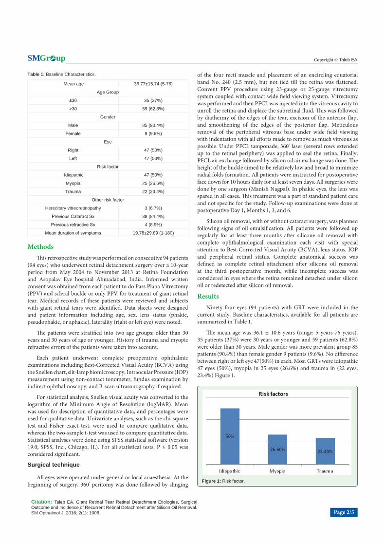
The mean age was 36.1 ± 10.6 years (range: 5 years-76 years). 35 patients (37%) were 30 years or younger and 59 patients (62.8%) were older than 30 years. Male gender was more prevalent group 85 patients (90.4%) than female gender 9 patients (9.6%). No difference between right or left eye 47(50%) in each. Most GRTs were idiopathic 47 eyes (50%), myopia in 25 eyes (26.6%) and trauma in (22 eyes, 23.4%) Figure 1.
Figure 1: Risk factor.
Table 1: Baseline Characteristics.
Mean age 36.77±15.74 (5-76)
Age Group
≤30 35 (37%)
>30 59 (62.8%)
Gender
Male 85 (90.4%)
Female 9 (9.6%)
Eye
Right 47 (50%)
Left 47 (50%)
Risk factor
Idiopathic 47 (50%)
Myopia 25 (26.6%)
Trauma 22 (23.4%)
Other risk factor
Hereditary vitreoretinopathy 3 (6.7%)
Previous Cataract Sx 38 (84.4%)
Previous refractive Sx 4 (8.9%)
Mean duration of symptoms 19.78±29.89 (1-180)
Citation: Taleb EA. Giant Retinal Tear Retinal Detachment Etiologies, Surgical Outcome and Incidence of Recurrent Retinal Detachment after Silicon Oil Removal. SM Opthalmol J. 2016; 2(1): 1008. Page 3/5
Gr upSM Copyright Taleb EA
Hereditary vitreoretinopathy were found in 3 eyes (6.7%), previous cataract surgery in 38 eyes (84.4%) and previous refractive surgery in 4 eyes (8.9%). Clinical Characteristics at Presentation summarized in Table 2.
The mean presenting vision was logMAR 2.20±0.94. A significant proportion of eyes presented with vision of less than 20/200 in 82 eyes (87.2%) and only 4 eyes (4.3%) presented with vision 20/40 or better. Majority had detached fovea at time of presentation. 78 eyes (83%) with 16 eyes (17%) had attached fovea. PVR in 82 eyes (87.2%) and 12 eyes (12.8%) no PVR. PVR grade C or greater in 27 eyes (28.7%).
The circumference of the GRT was between 90°-180° in 58 eyes (61.7%), between 180°-270° in 31 eyes (33%) and more than 270° in 5 eyes (5.3%) Table 3.
Fellow Eye
The median presenting BCVA for the fellow eye was better than 20/40 in 52 (71.3%) and less than 20/200 in 14 (19.2%). Of the 72 nontraumatic, noniatrogenic GRT fellow eyes, vitreoretinal disease was noted in 30 (31.9%) white without pressure was seen in 22.3%
and lattice degeneration was seen in 9.6%. 15 eyes (21%) of the fellow had previous/current RD 8 eyes (11.1%) are non-GRT RD and 7 eyes (9.7%) are GRT RD. Only 1 eye (1.4%) has GRT without RD Table 3.
Surgical Treatment
The intraoperative management techniques used in the surgical repair of these cases are presented in Table 4. Majority of GRTs 57 eyes (60.6%) were treated by PPV and 36 eyes, (38.3%) were treated by combined scleral buckling and PPV. One eye (1.1%) macula on GRT was treated with cryotherapy and scleral buckle surgery. The retina in this eye remained attached at the last available follow-up visit (9 months) without any additional procedure.
Outcomes
Outcomes data were available for 94 eyes are summarized in Table 5. Primary retinal reattachment (after the first operation) was 75 eyes (80%) and the final retinal reattachment (after one or multiple operation) was 85 eyes (90.4%), with final visual acuity more than 20/400 in 71 eyes (75.5%).
Most of the postoperative complication were related to silicon oil with cataract formation was the most common 24 eyes (25.2%). Recurrent RD (retinal detachment) was found in 21 eyes (22.3%). The mean time for SOR was 10.69±10.17 months range between (6 months-72 months).
Table 2: Clinical Characteristics at Presentation.
Presenting vision 20/40 or better 4(4.3%)
Presenting vision worse than 20/200 82(87.2%)
Lens status
Phakic 52(55.3%)
Pseudophakic 34(36.2%)
Aphakic 6(6.4%)
Dislocated lens 2(2.1%)
Retinal detachment
Fovea off 78(83%)
Fovea on 16(17%)
Size of GRT
Between 90˚-180˚ 58(61.7%)
Between 180˚-270˚ 31(33%)
≥270˚ 5(5.3%)
PVR
Yes 82(87.2%)
No 12(2.8%)
PVR C or greater
Yes 27(28.7%)
No 67(71.3%)
Table 3: Clinical Characteristics of the fellow eye at Presentation.
Presenting vision 20/40 or better 52 (71.2%)
Presenting vision worse than 20/200 14(19.2%)
Retinal finding
normal 56 (77.8%)
Previous/current non-GRT RD 8 (11.1%)
Previous/current GRT RD 7 (9.7%)
GRT without RD 1 (1.4%)
Table 4: Management Data.
Surgery
Pars plana vitrectomy 57 (60.6%)
cryobuckle 1(1.1%)
Combined buckle and vitrectomy 36 (38.3%)
Retinoctomy 6 (6.4%)
Relaxing retinoctomy 3 (3.2%)
Tamponada
silicon 92 (97.9%)
PFCL 1 (1.1%)
No tamponade 1(1.1%)
Table 5: Outcome.
Visual acuity
More than 20/400 71 (75.5%)
Less than 20/400 23 (24.5%)
Final anatomy
attached 85 (90.4%)
Not attached 9 (9.6%)
Post operative complication
Cataract 24 (25.2%)
Recurrent RD 21 (22.3%)
High IOP 19 (20.2%)
Band keratopathy 7 (7.4%)
Epiretinal membrane 5 (5.3%)
Endophthalmitis 0 (0%)
Citation: Taleb EA. Giant Retinal Tear Retinal Detachment Etiologies, Surgical Outcome and Incidence of Recurrent Retinal Detachment after Silicon Oil Removal. SM Opthalmol J. 2016; 2(1): 1008. Page 4/5
Gr upSM Copyright Taleb EA
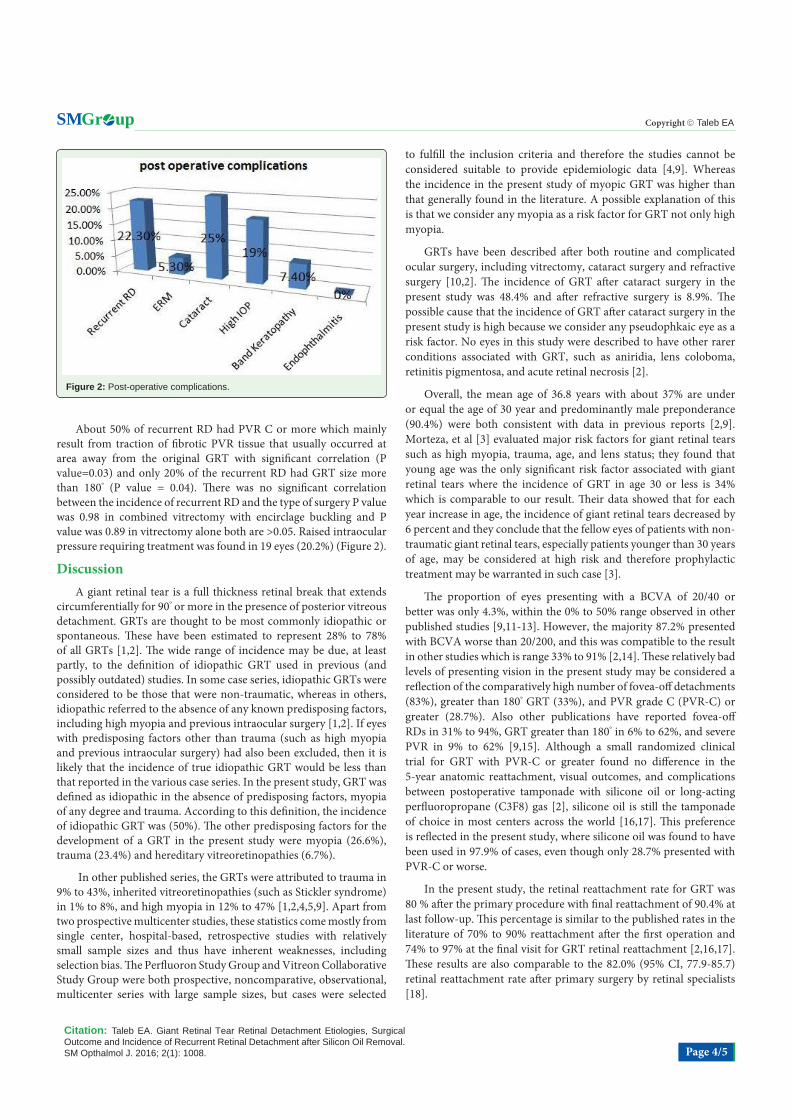
About 50% of recurrent RD had PVR C or more which mainly result from traction of fibrotic PVR tissue that usually occurred at area away from the original GRT with significant correlation (P value=0.03) and only 20% of the recurrent RD had GRT size more than 180° (P value = 0.04). There was no significant correlation between the incidence of recurrent RD and the type of surgery P value was 0.98 in combined vitrectomy with encirclage buckling and P value was 0.89 in vitrectomy alone both are >0.05. Raised intraocular pressure requiring treatment was found in 19 eyes (20.2%) (Figure 2).
DiscussionA giant retinal tear is a full thickness retinal break that extends
circumferentially for 90° or more in the presence of posterior vitreous detachment. GRTs are thought to be most commonly idiopathic or spontaneous. These have been estimated to represent 28% to 78% of all GRTs [1,2]. The wide range of incidence may be due, at least partly, to the definition of idiopathic GRT used in previous (and possibly outdated) studies. In some case series, idiopathic GRTs were considered to be those that were non-traumatic, whereas in others, idiopathic referred to the absence of any known predisposing factors, including high myopia and previous intraocular surgery [1,2]. If eyes with predisposing factors other than trauma (such as high myopia and previous intraocular surgery) had also been excluded, then it is likely that the incidence of true idiopathic GRT would be less than that reported in the various case series. In the present study, GRT was defined as idiopathic in the absence of predisposing factors, myopia of any degree and trauma. According to this definition, the incidence of idiopathic GRT was (50%). The other predisposing factors for the development of a GRT in the present study were myopia (26.6%), trauma (23.4%) and hereditary vitreoretinopathies (6.7%).
In other published series, the GRTs were attributed to trauma in 9% to 43%, inherited vitreoretinopathies (such as Stickler syndrome) in 1% to 8%, and high myopia in 12% to 47% [1,2,4,5,9]. Apart from two prospective multicenter studies, these statistics come mostly from single center, hospital-based, retrospective studies with relatively small sample sizes and thus have inherent weaknesses, including selection bias. The Perfluoron Study Group and Vitreon Collaborative Study Group were both prospective, noncomparative, observational, multicenter series with large sample sizes, but cases were selected
to fulfill the inclusion criteria and therefore the studies cannot be considered suitable to provide epidemiologic data [4,9]. Whereas the incidence in the present study of myopic GRT was higher than that generally found in the literature. A possible explanation of this is that we consider any myopia as a risk factor for GRT not only high myopia.
GRTs have been described after both routine and complicated ocular surgery, including vitrectomy, cataract surgery and refractive surgery [10,2]. The incidence of GRT after cataract surgery in the present study was 48.4% and after refractive surgery is 8.9%. The possible cause that the incidence of GRT after cataract surgery in the present study is high because we consider any pseudophkaic eye as a risk factor. No eyes in this study were described to have other rarer conditions associated with GRT, such as aniridia, lens coloboma, retinitis pigmentosa, and acute retinal necrosis [2].
Overall, the mean age of 36.8 years with about 37% are under or equal the age of 30 year and predominantly male preponderance (90.4%) were both consistent with data in previous reports [2,9]. Morteza, et al [3] evaluated major risk factors for giant retinal tears such as high myopia, trauma, age, and lens status; they found that young age was the only significant risk factor associated with giant retinal tears where the incidence of GRT in age 30 or less is 34% which is comparable to our result. Their data showed that for each year increase in age, the incidence of giant retinal tears decreased by 6 percent and they conclude that the fellow eyes of patients with non-traumatic giant retinal tears, especially patients younger than 30 years of age, may be considered at high risk and therefore prophylactic treatment may be warranted in such case [3].
The proportion of eyes presenting with a BCVA of 20/40 or better was only 4.3%, within the 0% to 50% range observed in other published studies [9,11-13]. However, the majority 87.2% presented with BCVA worse than 20/200, and this was compatible to the result in other studies which is range 33% to 91% [2,14]. These relatively bad levels of presenting vision in the present study may be considered a reflection of the comparatively high number of fovea-off detachments (83%), greater than 180° GRT (33%), and PVR grade C (PVR-C) or greater (28.7%). Also other publications have reported fovea-off RDs in 31% to 94%, GRT greater than 180° in 6% to 62%, and severe PVR in 9% to 62% [9,15]. Although a small randomized clinical trial for GRT with PVR-C or greater found no difference in the 5-year anatomic reattachment, visual outcomes, and complications between postoperative tamponade with silicone oil or long-acting perfluoropropane (C3F8) gas [2], silicone oil is still the tamponade of choice in most centers across the world [16,17]. This preference is reflected in the present study, where silicone oil was found to have been used in 97.9% of cases, even though only 28.7% presented with PVR-C or worse.
In the present study, the retinal reattachment rate for GRT was 80 % after the primary procedure with final reattachment of 90.4% at last follow-up. This percentage is similar to the published rates in the literature of 70% to 90% reattachment after the first operation and 74% to 97% at the final visit for GRT retinal reattachment [2,16,17]. These results are also comparable to the 82.0% (95% CI, 77.9-85.7) retinal reattachment rate after primary surgery by retinal specialists [18].
Figure 2: Post-operative complications.
Citation: Taleb EA. Giant Retinal Tear Retinal Detachment Etiologies, Surgical Outcome and Incidence of Recurrent Retinal Detachment after Silicon Oil Removal. SM Opthalmol J. 2016; 2(1): 1008. Page 5/5
Gr upSM Copyright Taleb EA
With regard to final BCVA of 20/400 or better in 75.5% and worse than 20/400 in 24.5%. This result was comparable to the BCVA outcomes of previously published series where to final BCVA of 20/400 or better in 84.9% [19].
The fellow eye of patients with GRTs is at an increased risk of GRT and RD. In a large series of 228 fellow eyes of nontraumatic GRTs in a study by Freeman [2], the 124 eyes that did not receive prophylactic treatment had an 11.3% incidence of GRT over a mean follow-up of 3.7 years.2, 13 Furthermore, RDs not associated with GRT may occur in up to 36% of fellow eyes [1,3,20]. It should be noted that in the present study, among the nontraumatic and noniatrogenic cases at presentation, 9.7% were fellow eyes of patients who had a history of GRT, compared with 6.6% in the Freeman4 series. In addition, present or previous RD, retinal breaks (other than GRT) were observed in 11.1% of fellow eyes of nontraumatic and noniatrogenic GRT. Although somewhat lower than the 31% to 81% reported in the literature [1,2,20]. The rate still represents a high proportion of fellow eyes at risk of visual loss due to RD. Currently, there is no strong evidence in the form of a RCT or a case-control study to support or refute the use of 360-degree prophylactic treatment for fellow eye of patient with unilateral GRT [20].
Fellow eye with pre-existing retinal tears and PVDs can go in to retinal detachment in spite of laser prophylactic. When PVD is not detectable or partial PVD is present, the progression of posterior vitreous separation can account for retinal tear and arising in formerly healthy area [21]. The most important postoperative complication in giant retinal surgery is recurrent detachment, which is principally due to the development of PVR. This complication developed in 49.4% and 31.5% of patients in the two large multicenter series [21,22] whereas it is lesser in the present study 22.3% because the highly efficient new instruments and machines.
In the present study 22% had recurrent retinal detachment of which 50% had PVR-C or more p value 0.3 and only 20% had GRT size more than 180° p value 0.04. Visually significant postoperative epimacular membranes developed in 7.4% and 15% of patients in the previous series [23,24] which was comparable to our study 5.3%.
ConclusionNowadays in the era of PFCL and new instruments, patients with
GRT who underwent surgery achieved high anatomic success rate. PVR C or more remain the most significant risk factor for recurrent RD. PPV alone or combined PPV with encirclage buckling have no significant correlation with recurrent RD.
References
1. Schepens CL, Dobble JG, McMeel JW. Retinal detachments with giant breaks: preliminary report. Trans Am Acad Ophthalmol Otolaryngol. 1962; 66:471-479.
2. Ang GS, Townend J, Lois N. Epidemiology of giant retinal tears in the United Kingdom: the British Giant Retinal Tear Epidemiology Eye Study (BGEES). Invest Ophthalmol Vis Sci. 2010; 51: 4781-4787.
3. Mehdizadeh M, Afarid M, Haqiqi MS. Risk Factors for Giant Retinal Tears. J Ophthalmic Vis Res. 2010; 5: 246-249.
4. Scott IU, Murray TG, Flynn HW, Feuer WJ, Schiffman JC. Outcomes and complications associated with giant retinal tear management using perfluoro-n-octane. Ophthalmology. 2002; 109: 1828-1833.
5. Ghosh YK, Banerjee S, Savant V, kotamarthi V, Benson MT, Scott RA, et al. Surgical treatment and outcome of patients with giant retinal tears. Eye. 2004; 18: 996-1000.
6. Schiff W, Chang S, Reppucci V, et al. Surgical management of giant retinal tears. In: Guyer DR, eds. Retina-vitreous-macula. 2nd edn. Pennsylvania: WB Saunders, 1999: 1338-1349.
7. Chang S, Lincoff H, Zimmerman NJ, Fuchs W. Giant retinal tears. Surgical techniques and results using perfluorocarbon liquids. Arch Ophthalmol. 1989; 107: 761-766.
8. Campochiaro PA, Kaden IH, Vidaurri-Leal J, Glaser BM. Cryotherapy enhances intravitreal dispersion of viable retinal pigment epithelial cells. Arch Ophthalmol. 1985; 103: 434-436
9. Kertes PJ, Wafapoor H, Peyman GA, Calixto N, Thompson H. The management of giant retinal tears using perfluoroperhydrophenanthrene: a multicenter case series. Vitreon Collaborative Study Group. Ophthalmology. 1997; 104: 1159-1165.
10. Batman C, Cekic O. Giant retinal tears after pars plana vitrectomy. Eye. 1998; 12: 163-164.
11. Kreiger AE, Lewis H. Management of giant retinal tears without scleral buckling: use of radical dissection of the vitreous base and perfluoro-octane and intraocular tamponade. Ophthalmology. 1992; 99: 491-497.
12. Ie D, Glaser BM, Sjaarda RN, Thompson JT, Steinberg LE, Gordon LW. The use of perfluoro-octane in the management of giant retinal tears without proliferative vitreoretinopathy. Retina. 1994; 14: 323-328.
13. Hoffman ME, Sorr EM. Management of giant retinal tears without scleral buckling. Retina. 1986; 6: 197-204.
14. Drivers Medical Group, DVLA, Swansea. At a glance Guide to the current Medical Standards of Fitness to Drive.
15. Chen CH, Tsai MH, Su CC, Kou HK, Kao ML, Tsai SH, et al. Results of 12-year clinical study of giant retinal tear. Chang Gung Med J. 2001; 24: 633-639.
16. Glaser BM. Treatment of giant retinal tears combined with proliferative vitreoretinopathy. Ophthalmology. 1986; 93: 1193-1197.
17. Kapetanios AD, Donati G, Pournaras CJ. Idiopathic giant retinal tears: treatment with vitrectomy and temporary silicone oil tamponade (in French). J Fr Ophtalmol. 2000; 23: 1001-1005.
18. Thompson JA, Snead MP, Billington BM, Barrie T, Thompson JR, Sparrow JM. National audit of the outcome of primary surgery for rhegmatogenous retinal detachment. II. Clinical outcomes. Eye. 2002; 16: 771-777.
19. Gonzalez MA, Flynn HW, Smiddy WE, Albini TA, Tenzel P. Surgery for retinal detachment in patients with giant retinal tear: etiologies, management strategies, and outcomes. Ophthalmic surg laser imaging retina. 2013; 44: 232-237.
20. Freeman HM. Fellow eyes of giant retinal breaks. Trans Am Ophthalmol Soc. 1978; 76: 343-382.
21. Ang Gs, Townend J, Lais N. Intervantions for prevention of giant retinal tear in the fellow eye. Cochrane Database study Syst Rev. 2012. 2: CD006909.
22. Mastropasqua L, Carpineto P, Ciancaglini M, Falconio G, Gallenga P. Treatment of retinal tear and lattice degenerations in fellow eyes in high risk patients suffering retinal detachment: a prospective study. Br J Ophthalmol. 1999; 83: 1046-1049.
23. Kertes PJ, Wafapoor H, Peyman GA, Calixto N, Thompson H. The management of giant retinal tears using perfluoroperhydrophenanthrene: a multicenter case series. Ophthalmology. 1997; 104: 1159-1165.
24. Freeman HM. Giant retinal tears: 207 cases from the Perfluoron study. Am Acad Ophthalmol Vitreoretinal Update. 1997; 168-171.