Embed Size (px)
Citation preview
2013/2014
Mafalda Maria Laracho de Seabra
Retinal detachment: a prognostic
factor analysis
março, 2014
Mestrado Integrado em Medicina
Área: Oftalmologia
Trabalho efetuado sob a Orientação de:
Dr. Manuel Alberto de Almeida e Sousa Falcão
Trabalho organizado de acordo com as normas da revista:
European Journal of Ophtalmology
Mafalda Maria Laracho de Seabra
Retinal detachment: a prognostic
factor analysis
março, 2014
1
RETINAL DETACHMENT: A PROGNOSTIC FACTOR ANALYSIS
Prognostic factors in retinal detachment
Mafalda Seabra, Manuel Falcão MD
Department of Sense Organs, Faculty of Medicine of University of Porto
Department of Ophthalmology of Hospital São João, Porto
Corresponding author:
Mafalda Seabra
Alameda Hernani Monteiro
4200-319 Porto
Portugal
Tel.: +351918432178.
Email: [email protected]
Conflict of Interest - None of the authors has conflict of interest with the
submission
Financial support - No financial support was received for this submission

2
Abstract
Purpose: To evaluate prognostic factors for retinal detachment.
Methods: The patient’s medical records were reviewed and preoperative and
intraoperative data analysed to ascertain an association with the outcomes: VA
≤0,52 logMAR, VA ≤0,3 logMAR, VA ≤0,52 logMAR in eyes with macula-off and
redetachment.
Results: The difference in final visual acuity between the population of macula-
on and macula off was statistically significant (t-test: p <0,001). Mean
postoperative VA was 0,41 ± 0,51 logMAR (n= 39; Snellen: 20/51) and 0,62 ±
0,59 logMAR (n=109; Snellen: 20/83), respectively. Macula-off was a factor of
poor prognosis (final VA worse than 0,3 logMAR). Mean time to surgery was 4
days. The time to surgery did not affect final VA < 0,52 logMAR (p=0,694).
Conclusions: The state of the macula only influenced the prognosis in a
negative way when the final VA considered was 0,3 logMAR. These situations
can be managed as urgent procedures without the need of emergency
interventions. In our series, time to surgery and pre-operative visual acuity were
not prognostic factors.
Keywords: retinal detachment, prognostic factors, macula
3
Introduction
Retinal detachment occurs when the sensory retina and the retinal pigment
epithelium separate (1,2,3).
Three types of retinal detachment have been described: Rhegmatogenous,
tractional and exsudative. Only the first type will be considered in this paper.
The most common type (2,3,4), rhegmatogenous retinal detachment (RRD), is
due to a retinal tear or break that may be instigated by trauma or posterior
vitreous detachment (1). This break allows the accumulation of liquefied
vitreous between the sensory retina and the retinal pigment epithelium.
Posterior vitreous detachment is characteristic of the elderly, but there are other
risk factors that can lead to this condition such as myopia, aphakia, focal retinal
atrophy, trauma (1,4), family history and retinal detachment in the fellow eye (3).
Affected individuals may be asymptomatic. However, most people have
symptoms: photopsia, floaters, visual field loss, diminished visual acuity (VA) or
blurred vision (1,4). The diagnosis can be confirmed by ophthalmoscopy (3).
If the tear is not repaired, the progressive accumulation of fluid between layers
will have degenerative effects and eventually lead to blindness (3).
A study undertaken in Portugal in 2010 estimated an incidence of 19 cases per
100000 inhabitants (2).
There are many treatment options. If there is just a tear in the retina, laser or
cryotherapy may prevent the progression of fluid in the subretinal space
preventing a complete retinal detachment. Nonetheless, if the retinal
detachment is established, the first line treatment is surgical. Up to now the
4
preferred surgical technique lacks consensus (5) but several options are
available, being retinopexy, scleral buckling and vitrectomy with internal
tamponade the most frequently used surgical approaches (3).
Up to now, the definition of prognostic factors still raises substantial discussion.
Several definitions have been put forward, among which the most consensual
ones are macula on/off (2,6,7,8,11), number of days until surgery
(2,5,8,9,12,13), pre-operatory visual acuity (6,9) and age (9,10).
Notwithstanding the results are yet to be widely accepted, particularly with
regards to the number of days between the retinal detachment and surgery.
This paper aims to analyse the prognostic factors in our population in the total
number of retinal detachments and in patients with macula-off retinal
detachments.
Methods
During a 29-month period, from January 2008 until May 2010, all patients with a
retinal detachment who were admitted to Hospital de São João from the
emergency department or referred by ophthalmologists, were enrolled in this
study.
Data was collected retrospectively from the medical records. Initially, all patients
were included (n=265). Subsequently, those with retinal redetachment or retinal
detachment other than rhegmatogenous were excluded.
The following variables were analysed: age, sex, type of retinal detachment,
affected eye, date of diagnosis, number of days from the appearance of the first
symptoms until diagnosis, number of days until surgery, pre-operative visual
5
acuity (logMAR), myopia, phakia, simultaneous phacoemulsification with intra-
ocular lens implantation, number of quadrants involved, number of tears,
location of the tears, presence of a giant tear, macula on/off, pre-operative
proliferative vitreoretinopathy (PrePVR), vitreous haemorrhage, surgical
technique (vitrectomy, scleral buckling or combined vitrectomy and scleral
buckling), vitreous substitute, primary surgeon, pneumopexy used (cryotherapy
or laser), final visual acuity at least 5 months after surgery (logMAR), and
redetachment rate.
A literature review of the studies focusing on prognostic factors of retinal
detachment has been conducted in the MEDLINE database up to March 2014.
Studies have been identified by using combinations of key words and through
MeSH-based electronic searches. The reference lists of the relevant studies
were thoroughly searched for additional studies.
Statistical analysis was performed using SPSS version 22 (SPSS inc, Chicago,
Illinois, USA). Descriptive statistics were obtained for all variables. Univariate
analysis was performed to assess the association between the explanatory
variables and the final outcome, using Mann-Whitney U test and Chi-square
test. Logistic regression models were developed to identify factors that might
influence the prognosis. Four outcomes were analysed: post-operative VA of
0,52 logMAR or better, postoperative VA of 0,3 logMAR or better, redetachment
during follow-up, and postoperative VA of 0,52 logMAR or better in the macula-
off population. The level of statistical significance was set at p<0,10.
Results
6
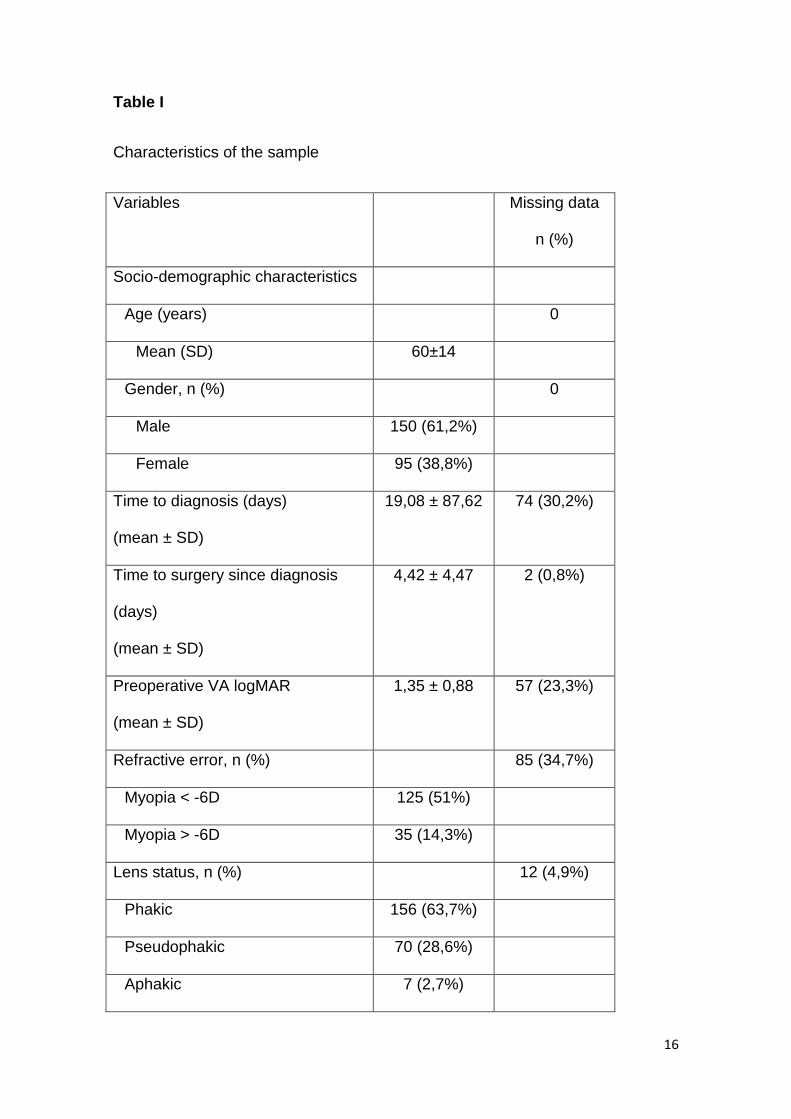
The study included 245 eyes (Table I). The mean age of all patients was 60±14
years (mean ± standard deviation) and there were 150 men (61,2%). A total of
156 eyes were phakic (63,7%), 70 were pseudophakic (28,6%) and 7 were
aphakic (2,9%). The macula was detached in 164 patients (66,9%), pre-
operative proliferative vitreoretinopathy was present in 12 patients (4,9%)
(missing data: 17 (6,9%)) and vitreous haemorrhage in 18 patients (7,3%)
(missing data: 15 (6,1%)).
The mean time since the appearance of symptoms to diagnosis and to surgery
was 19,08 ± 87,62 and 4,42 ± 4,47 days respectively. In the subpopulation with
macula-on, the mean time to surgery was 4,67 ± 7,10 days while in those with
macula-off it was 4,32 ± 2,64 days since diagnosis.
The most common procedure performed was vitrectomy in 185 patients (75,5%)
(missing data: 3 (1,2%)). As for the others, 51 (20,8%) had vitrectomy + scleral
buckle and 6 (2,4%) performed scleral buckling. A vitreous substitute was used
in 236 patients (96,3%) (missing data: 9 (3,7%)): gas tamponade (70,6%) and
silicone oil tamponade (25,7%).
Simultaneous cataract surgery and intra-ocular lens implantation was performed
in 121 patients (49,4%) (missing data: 2 (0,8%)). Retinopexy was performed by
cryotherapy in 10 patients (4,1%) and laser in 228 patients (93,1%). In 2.8% of
cases, there was no information regarding retinopexy procedures.
Pre-operative VA was documented in 188 patients (1,35 ± 0,88 logMAR). Post-
operative VA was recorded in 148 patients. The information regarding pre-
operative macular status was missing in 9 of these patients. Mean
postoperative VA was 0,58 ± 0,59 logMAR (20/76). In the macula-on population,
7
the mean postoperative VA was 0,41 ± 0,51 logMAR (n= 39; Snellen: 20/51).
Considering the macula-off population, mean postoperative visual acuity was
0,62 ± 0,59 logMAR (n=109; Snellen: 20/83). This difference in visual acuity
was statistically significant (t-test: p <0,001).
Visual acuity was then dichotomized using two different outcomes: 0,3 logMAR
(Snellen: 20/40) and 0,52 logMAR (Snellen: 20/66). From the 148 patients, 85
(57,4%) had a visual acuity worse than 0,3 logMAR, whereas 47 (31,8%) had a
visual acuity worse than 0,52 logMAR. 145 patients were considered when
comparing the patients final VA and macular status. Among those patients with
a final VA worse than 0,52 logMAR, 7 were macula-on eyes (17,9% of the total
macula on eyes) and 38 were macula-off eyes (35,8% of macula-off eyes). As
for those with a final VA worse than 0,3 logMAR, 16 (41%) were macula-on and
66 (62,3%) were macula-off eyes.
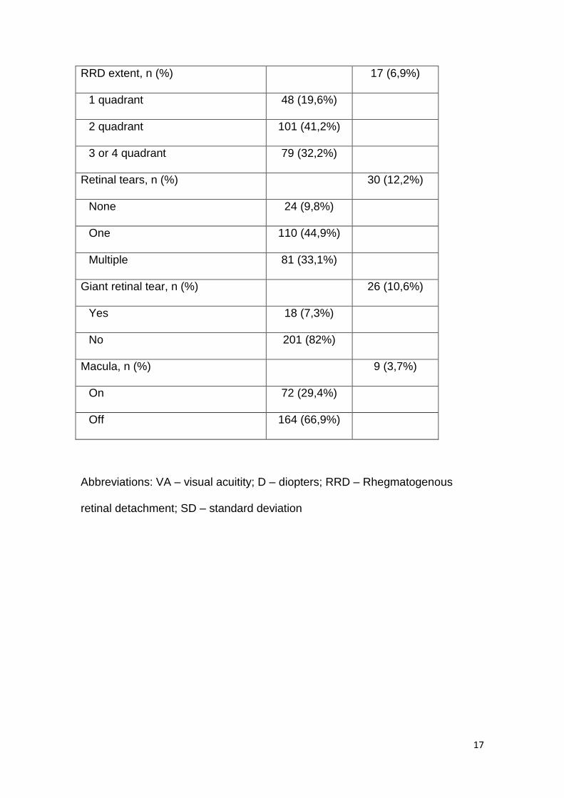
A univariate analysis to identify the factors that could be associated with a final
visual acuity better than 0,52 logMAR was initially performed. The following
factors were selected to perform a logistic regression: preoperative VA, number
of retinal tears, giant retinal tear, macula on/off, type of vitreous substitute and
cryotherapy (the variables with p>0,2 were excluded). The logistic regression
analysis revealed that multiple retinal tears (p=0,083) and gas tamponade
(p=0,025) correlated statistically with the outcome (Table II). The involvement of
the macula was not associated with the end result (p=0,807).
To evaluate the independent predictors of poor outcome for a final visual acuity
worse than 0,3 logMAR (Table III), the risk factors selected after the univariate
analysis were: gender, time to surgery, number of quadrants involved, giant

8
retinal tear, macula on/off, surgical technique and type of vitreous substitute
(the variables with p>0,2 were excluded). Two significant associations with final
visual acuity were established with the logistic regression analysis: macula off
(p=0,060) and use of silicone oil tamponade (p=0,054). In this case, time to
surgery did not show an association with the outcome (p=0,694).
A univariate analysis was performed to select the variables for the logistic
regression model in the subpopulation with macula-off, comparing those with a
final VA better or worse than 0,52 logMAR. Time to surgery, number of retinal
tears, vitreous haemorrhage and cryotherapy entered the logistic regression
model (the variables with p>0,2 were excluded). Table IV shows that the
presence of a vitreous haemorrhage (p=0,082), the use of silicone oil
tamponade (p=0,054) and cryotherapy (p=0,071) are statistically significant
predictors of a final visual acuity worse than 0,52 logMAR.
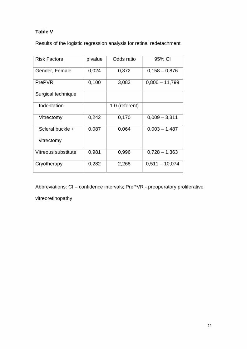
Retinal redetachment occurred in 44 patients (18%). In this case, the variables
selected for the logistic regression model by the univariate analysis were:
gender, presence of PrePVR, surgical technique and cryotherapy (the variables
with p>0,2 were excluded). With regards to this outcome (Table V), performing
indentation plus vitrectomy (p=0,064), as well as female gender (p=0,024), were
associated with the outcome (no redetachment).There was some evidence of
association between redetachment and the presence of PrePVR (p=0,1).
9
Discussion
Multiple surgical techniques can be used to approach retinal detachment. It is
therefore very difficult to try and evaluate factors associated with a better
prognosis, as several confounding variables may be present.
Nonetheless, in this series, our aim was to ascertain the prognostic factors of
rhegmatogenous retinal detachment. Two visual outcomes were defined: 0,3
logMAR, usually considered driving vision, and 0,52 logMAR – reading vision.
This series demonstrated that there are two factors associated with better
prognosis in order to have a VA 0,52 logMAR or better. These are: multiple
tears (ORs (95% (CI): 0,145 (0,016 – 1,288)) and use of gas tamponade (ORs
(95% (CI): 0,241 (0,070 – 1,834)). On the other hand, the use of silicone oil
tamponade was linked with bad prognosis if the outcome was 0,3 logMAR (ORs
(95% (CI): 2,780 (0,938 – 7,856)). Time to surgery was not associated with
either of these outcomes.
Our series revealed that the type of vitreous substitute used (gas or silicone oil)
can be an important prognostic factor. Silicone oil was a factor of poor
prognosis for two of the outcomes analysed: final VA 0,3 and final VA 0,52 in
the macula-off population. We must take into consideration that silicone oil is
generally used when surgeons feel that there is a higher risk of redetachment
and when a poorer prognosis is already expected. Therefore, it is likely that it is
not the silicone oil that is a factor of poorer prognosis but rather that it is the
tamponade that is used in patients that has a predisposition for a poorer
prognosis. It is possible that silicone oil is a confounding variable. Nonetheless,
10
with this retrospective analysis, we were not able to determine which isolated
variables led to the decision of using silicone oil as the internal tamponade.
In our series, we found an association between multiple tears and having a final
visual acuity better than 0.52 logMAR. However, when we raised the threshold
for good prognosis to the level of 0.3 logMAR, the number of retinal tears was
not associated with the final prognosis. Caution must be taken in interpreting
these results. Visual acuity is not related with the peripheral retina and
therefore, theoretically speaking, the number of tears should not interfere with
visual acuity. Further studies that focus on the number of retinal tears may help
to explain these results.
Our findings showed that, although there was no association between visual
acuity and macula using the cut-off of 0,52 logMAR (p=0,807), if we used the
cut-off 0,3 logMAR, the association could be made (p=0,060). The reason is
that many patients with macula-off retinal detachments can achieve visual
acuities better than the cut-off we defined; as such, the probability of having at
least reading vision after a retinal detachment is the same in people that
present an attached or detached macula and it is greater than 50% in both
studied populations. However, the patients that presented a macula-on retinal
detachment had a better final visual acuity (0,41 ± 0,51 logMAR) compared with
those with macula-off (0,62 ± 0,59 logMAR) (odds ratios (ORs) (95% confidence
interval (CI): 2,286 (0,965 – 5,414)). These findings are consistent with other
reports that have been published (7,11). Salicone (7) reported that 78% of
patients with macula-off had final visual acuity worse than 0,3 logMAR
compared with 28% in the macula-on group. We demonstrated that the
probability of having a final visual acuity better than 0.3 logMAR was greater in
11
the patients with macula on, showing that an attached macula is an important
prognostic factor for better final visual acuities. However, in our series, pre-
operative visual acuity was not associated with final visual acuity. We expect
patients with a macula-on retinal detachment to have a better visual acuity at
diagnosis and these two variables could have similar significance. However,
there are patients with bullous retinal detachments with hidden macula-on
detachments. This is probably why the status of the macula is a better
prognostic factor than pre-operative visual acuity.
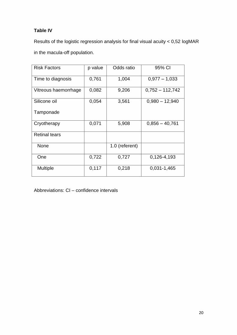
When analysing the macula off population and using the endpoint of 0,52
logMAR, we found three risk factors associated with poor prognosis: vitreous
haemorrhage on diagnosis (ORs (95 %CI): 9,206 (0,752 – 112,742)), the use of
silicone oil tamponade (ORs (95% (CI): 3,561 (0,980 – 12,940)) and
cryotherapy (ORs (95% (CI): 5,908 (0,856 – 40,761)). However, since vitreous
haemorrhage was present in 4 patients only and cryotherapy was performed in
8 patients, these variables presented very wide confidence intervals, and
therefore their results must be carefully interpreted.
Focusing on the group of patients with macula-off, many authors have
discussed the role of the time to surgery. This series showed that there is no
association between the number of days until surgery (p=0,694) and the final
outcome (using the endpoint: final visual acuity 0,3 logMAR). Other authors
have reported the same findings (7-9, 12-13). However, the number of days in
which this hypothesis can be verified is still under discussion. Thelen (13)
reported that the surgery could be postponed for 3 days without compromising
the prognosis, while Ross (8) reported a 7-day period, Hassan (9) a 10 day
period and Doyle (13) a 30 day period. Our series showed that a 4-day wait
12
from diagnosis to surgery probably does not interfere with final visual acuity.
These findings have clinical relevance as the decision to have surgery can be
postponed for a short period of time until the best conditions for surgery can be
optimized. Surgery for retinal detachment can be considered an urgent, but not
an emergent, condition.
We must emphasize that 242 of our patients had surgery within 2 weeks from
the initial symptoms. We cannot infer results for patients that have had retinal
detachments for more than two weeks.
With resgards to redetachment rates, Foster (14) reported a 12% incidence of
retinal detachment. In our series, 18% of cases redetached. Performing scleral
buckling along with a vitrectomy was associated with better prognosis (ORs (95
%CI): 0,064 (0,003 – 1,487)), as well as female gender (ORs (95 %CI): 0,372
(0,158 – 0,876)). These results differ from those of Kinori (15) who reported
equal redetachment rates for cases treated solely with vitrectomy and for cases
treated with vitrectomy and scleral buckle. Our series show that there may still
be a role for a combined procedure in selected patients. Finding a protective
effect in the female patients has not previously been reported. Although genetic
markers have recently been associated with proliferative vitreoretinopathy (16),
gender has not been classically associated. We may hypothesise that these
differences can be related to unknown hormonal factors that must be confirmed
and investigated by further studies.
Preoperatory proliferative vitreoretinopathy showed some degree of association
with retinal redetachment (p=0,1; ORs (95% (CI): 3,083 (0,806 – 11,799)).
13
Nevertheless, only 12 patients (4,9%) presented with this condition rendering
the variable a poor estimate.
Our study has limitations. The major weakness is the retrospective nature of the
study that used clinical records of regular clinical practice that lacks important
data, making statistical analysis challenging. As far as visual acuity is
concerned, this variable was not evaluated at the same post-operative stage.
The range varies from five to 24 months. It has been suggested that visual
acuity continues to improve for a period of up to 5 years (17), and this may limit
the extrapolation of the results.
Conversely, we have a large number of patients, which allows us to draw some
conclusions.
The majority of macula-on and macula-off patients present a visual acuity better
than 0.52 logMAR. Nevertheless, having a macula-on increases the probability
of having a visual acuity better than 0.3 logMAR. Furthermore, those patients
with macula off can obtain reading vision in the majority of cases and surgery
can be postponed for some days without compromising the results. Lastly. there
might still be a role for combined vitrectomy and scleral buckling for retinal
detachments for the prevention of redetachment.
14
1. Das T. Guidelines for the management of rhegmatogenous retinal
detachment. Indian J Ophtalmol 1993; 41:37-40
2. Gil Calvão-Santos. Epidemiologia do Descolamento da Retina na nossa
Área de Actuação. Oftalmologia – Vol.34; pp.315-320
3. John I. Lane. Retinal Detachment: Imaging of Surgical Treatments and
Complications. RadioGraphics 2003; 23: 983-994
4. Ray F. Gariano. Evaluation and Management of Suspected Retinal
Detachment. American Family Physician 2004; volume 69, number 7:
1691-1698
5. Peter Walker. Retinal Detachment Surgeries. The Dilemma Between
Personal Experience and Clinical Trials. Expert Ver Ophtalmol.
2012;7(5):441-447
6. Charles C. Wykoff. Fovea-Sparing Retinal Detachments: Time to Surgery
and Visual Outcomes. American Journal of Ophtalmology 2010: 205-210
7. Alberto Salicone. Visual Recovery after Scleral Buckling Procedure for
Retinal Detachment. American Academy of Ophtalmmatology. 1734-
1742
8. William H. Ross. Visual Recovery in Macula-off Rhegmatogenous Retinal
Detachments. Ophtalmology, Volume 105, Number 11:2149-2153
9. Tarek S. Hassan. The Effect of Duration of Macular Detachment on
Results after Scleral Buckle Repair of Primary, Macula-off Retinal
Detachments. American Academy of Ophtalmology 2002: 146-151
10. Otacílio de Oliveira Maia Junior. Descolamento regmatogéneo de retina:
avaliação pós-operatória da mácula. Arq Bras Oftalmol. 2007;70(6);996-
1000.
15
11. del’OMO. Short-time prone posturing is well-tolerated and reduces the
rate of unintentional retinal displacement in elderly patients operated o
for retinal detachment. BMC Surgery 2013, 13(Suppl 2):S55.
12. Ulrich Thelen. Outcome of surgery after macula-off retinal detachment –
results from MUSTARD, one of the largest databases in Europe. Acta
Ophtalmologica. 2012: 90: 481-486.
13. E. Doyle. How effective is macula-off retinal detachment surgery. Might
good outcome be predicted? Eye (2007) 21, 534-540
14. Robert E. Foster. Recurrent Retinal Detachment More than 1 Year after
Reattachment. Ophtalmology Volume 109, Number 10, October 2002.
15. Michael Kinori. Comparison of Pars Plana Vitrectomy With and Without
Scleral Buckle for the repair of Primary Rhegmatogenous Retinal
Detachment. American Journal of Ophtalmology Vol. 152, No.2, 2012
16. Salvador Pastor-Idoate, Irene Rodriguez-Hernández, Jimena Rojas, et
al. The p53 Codon 72 Polymorphism (rs1042522) Is Associated with
Proliferative Vitreoretinopathy. Ophtalmology 2013; 120:623-628
17. Sung Dong Chang. Long-term Visual Recovery After Scleral Buckling
Procedure of Rhegmatogenous Retinal Detachment Involving the
Macula. Korean J Ophtalmol. Vol 13:20-26, 2000
16
Table I
Characteristics of the sample
Variables Missing data
n (%)
Socio-demographic characteristics
Age (years) 0
Mean (SD) 60±14
Gender, n (%) 0
Male 150 (61,2%)
Female 95 (38,8%)
Time to diagnosis (days)
(mean ± SD)
19,08 ± 87,62 74 (30,2%)
Time to surgery since diagnosis
(days)
(mean ± SD)
4,42 ± 4,47 2 (0,8%)
Preoperative VA logMAR
(mean ± SD)
1,35 ± 0,88 57 (23,3%)
Refractive error, n (%) 85 (34,7%)
Myopia < -6D 125 (51%)
Myopia > -6D 35 (14,3%)
Lens status, n (%) 12 (4,9%)
Phakic 156 (63,7%)
Pseudophakic 70 (28,6%)
Aphakic 7 (2,7%)
17
RRD extent, n (%) 17 (6,9%)
1 quadrant 48 (19,6%)
2 quadrant 101 (41,2%)
3 or 4 quadrant 79 (32,2%)
Retinal tears, n (%) 30 (12,2%)
None 24 (9,8%)
One 110 (44,9%)
Multiple 81 (33,1%)
Giant retinal tear, n (%) 26 (10,6%)
Yes 18 (7,3%)
No 201 (82%)
Macula, n (%) 9 (3,7%)
On 72 (29,4%)
Off 164 (66,9%)
Abbreviations: VA – visual acuitity; D – diopters; RRD – Rhegmatogenous
retinal detachment; SD – standard deviation
18
Table II
Results of the logistic regression analysis for final visual acuity lower than 0,52
logMAR.
Risk Factors p value Odds ratio 95% CI
Preoperative VA 0,154 1,873 0,791-4,433
Retinal tears
None 1.0 (referent)
One 0,422 0,427 0,053-3,408
Multiple 0,083 0,145 0,016-1,288
Giant tear 0,231 2,878 0,510-16,228
Macula on/off 0,807 1,230 0,233-6,494
Gas Tamponade 0,025 0,241 0,070-0,834
Cryotherapy 0,095 5,077 0,755-34,160
Abbreviations: VA – visual acuity; CI – confidence intervals

19
Table III
Results of the logistic regression analysis for final visual acuity lower than 0,3
logMAR.
Risk Factors p value Odds ratio 95% CI
Gender, Female 0,398 1,409 0,637 – 3,121
Time to surgery 0,694 1,034 0,874-1,225
RDD extent
1 quadrant 1.0 (referent)
2 quadrant 0,435 1,438 0,578– 3,578
3 or 4 quadrant 0,204 1,990 0,965- 5,760
Macula on/off 0,060 2,286 0,965- 5,414
Giant tear 0,130 3,137 0,714-13,790
Scleral buckle +
vitrectomy
0,312 1,601 0,634-3,991
Silicone oil
Tamponade
0,054 2,780 0,983-7,856
Abbreviations: RDD - Rhegmatogenous retinal detachment; CI – confidence
intervals
20
Table IV
Results of the logistic regression analysis for final visual acuity < 0,52 logMAR
in the macula-off population.
Risk Factors p value Odds ratio 95% CI
Time to diagnosis 0,761 1,004 0,977 – 1,033
Vitreous haemorrhage 0,082 9,206 0,752 – 112,742
Silicone oil
Tamponade
0,054 3,561 0,980 – 12,940
Cryotherapy 0,071 5,908 0,856 – 40,761
Retinal tears
None 1.0 (referent)
One 0,722 0,727 0,126-4,193
Multiple 0,117 0,218 0,031-1,465
Abbreviations: CI – confidence intervals
21
Table V
Results of the logistic regression analysis for retinal redetachment
Risk Factors p value Odds ratio 95% CI
Gender, Female 0,024 0,372 0,158 – 0,876
PrePVR 0,100 3,083 0,806 – 11,799
Surgical technique
Indentation 1.0 (referent)
Vitrectomy 0,242 0,170 0,009 – 3,311
Scleral buckle +
vitrectomy
0,087 0,064 0,003 – 1,487
Vitreous substitute 0,981 0,996 0,728 – 1,363
Cryotherapy 0,282 2,268 0,511 – 10,074
Abbreviations: CI – confidence intervals; PrePVR - preoperatory proliferative
vitreoretinopathy

Anexos
CONTENT TYPE
Original articles. Previously unpublished manuscripts, directed to ophthalmologists and visual
science specialists describing clinical investigations, clinical observations, relevant clinical
laboratory investigations. An original article should consist of around 16-18 double-spaced,
typewritten pages, corresponding to 6-8 printed pages. The text of articles must be divided into
sections with the headings Introduction, Methods, Results and Discussion
GUIDELINES FOR THE MANUSCRIPT FILE
All manuscript pages must be numbered.
TITLE PAGE
The title page must include:
a) the complete manuscript title (max 135 characters including letters and spaces);
b) short title (max 75 characters including letters and spaces);
c) all authors listed as first name, initials and last name (i.e. Paul M. Smith) with highest
academic or medical degree and departmental affiliation (identified by arabic numerals)
d) corresponding Author's information (full mailing address, phone and fax numbers, e-mail
address);
e) Disclosures on
- Conflict of Interest (state any financial interest held by the authors or their families; if there is
no conflict of interest add a statement “None of the authors has conflict of interest with the
submission”);
- Financial support in form of Grants and funds received in support of the Study. If no financial
support was received, add a statement “No financial support was received for this submission”;
- Meeting presentation. If the paper was presented at a meeting, state its name, place and
date on which it was read;
- Informed consent. In case of manuscripts reporting the results of experimental investigation
on human subjects, human derived material or human medical records include a statement that
the study was performed with informed consent and following all the guidelines for experimental
investigations required by the Institutional Review Board or Ethics Committee of which all
authors are affiliated.
f) Structured abstract, which must not exceed 250 words and which must be structured and
divided in the sections Purpose, Methods, Results, and Conclusions. It must be acceptable for
use without revision.
g) Below the abstract, identify 3 to 6 keywords in alphabetical order under which you believe the
article should be indexed.
h) If you are submitting a manuscripts which has been previously rejected please inform the
Journal and the Reviewers of the previous review comments, how and where the manuscript
has been improved according to the reviewers’ comments.
MANUSCRIPT TEXT
Starting on a new page, type manuscript using Arial font size 12, as this creates less problems
when building your PDF, and save it as Word document (.doc). Use double spacing and do not
justify the right margin. Use only standard abbreviations and avoid abbreviations in the title. The
full term for which an abbreviation stands for should preceed its first use in the text. The
average published manuscript in European Journal of Ophthalmology, including references, is
up to 6 pages in length. This corresponds to between 16 and 20 double-spaced typewritten
pages. Type your manuscript as a single Word file, divided in the following sections:
Introduction: should be pertinent to the study but not an in-depth review of the literature.
Materials and methods: should be clearly defined so that the study may be duplicated by other
investigators.
Results: should be as concise as possible.
Discussion: offers an explanation of the results of the study and should limit itself to the subject
matter of the paper.
Cite figures consecutively in the text and number them in the order in which they are presented.
Figures must be submitted as separate files and not embedded in the word document.
CANCER CLASSIFICATION SCHEME
The European Journal of Ophthalmology encourages Authors to use the classification scheme
proposed by the American Joint Commission on Cancer. Please use these when describing
patients with ophthalmic malignancies (see AJCC Cancer Staging Manual, 7th Edition, Springer
New Yok)
ACKNOWLEDGEMENTS
Acknowledge statistical consultation and assistance or writing assistance (when provided by a
person different from the author) in an acknowledgement at the end of the article before the
references Indicate the name, degree and affiliation of the individual. For all others assisting in
the preparation of a manuscript acknowledgements cannot be done, however valuable their
service.
REFERENCES
1. If you use automated reference numbering software or bibliography software, turn it off
before submitting the manuscript.
2. References should follow the text and begin on a separate page.
3. They must be double-spaced and numbered consecutively in order of appearance in the
text, using the automated numbering tool of Word.
4. Identify references in text, tables, and legends in Arabic numerals in parentheses, i.e. (7).
5. If there are 6 or fewer authors, all authors should be listed. If there are more than 6
authors, list the first three and then "et al"
6. References used within tables should appear as footnotes in the table legend. These
references should not be repeated in the main reference list unless they are also cited
within the text.
7. List only references pertinent to the manuscript, which you have read and that the reader
can retrieve in a literature research.
8. Journals’ names should be abbreviated according to Index Medicus/Medline. If there is
any doubt about abbreviation of a journal name, it should be spelled out completely.
9. All references must be verified by the Author(s) against the original documents.
10. Personal communications, unpublished data, abstracts, oral or poster presentations
should be limited and incorporated in parentheses within the text without a reference number.
11. Any references to studies (including books or articles) that have been accepted for
publication, but not yet published, should indicate where they will be published and have the
term "in press" in the reference in place of volume and page numbers. These must be updated
prior to publication, if possible.
12. Delete digits when in the same range: 534-7 or 1007-11 (NOT 534-537; 1007-1011)
13. Do not add a discussion or comment to a reference. If applicable indicate it as Eur J
Ophthalmol. 2007;17:534-7, Comment in: Eur J Ophthalmol. 2009;19:327; author reply 327.
14. Suffixes such as Jr, Sr, and III follow authors initials
The inclusion references available onlline only should be limited: if also available in print, then it
is preferred to include the print citation. The online reference should be listed with complete
information including title and authors, adding the URL address and date of access, which
should always be confirmed with every revision submission.
Reference formatting examples:
Standard journal article: (List all Authors when six or less; when seven or more, list only first
three and add et al.) Gass JD, Harbin TS Jr, Del Piero EJ. Exudative stellate neuroretinopathy
and Coats' Syndrome in patients with progressive hemifacial atrophy. Eur J
Ophthalmol. 1991;1:2-10.
Book: Harrington DO, Drake MV. The visual field. Text and atlas of clinical perimetry, 6th
ed. St Louis: CV Mosby, 1990; 156.
FIGURE LEGENDS
Starting on a new page, type legends for figures double-spaced, with Arabic numerals
corresponding to the figures. All figures must have a legend. When symbols, arrows, numbers,
or letters are used to identify parts of the figures, identify and explain each one clearly in the
legend. Any figure that has been published elsewhere should have an acknowledgment to the
original source; a copy of the release to publish the figure, signed by the copyright holder, must
also be submitted.
TABLES
As a general rule, tables should not unnecessarily offer duplicate information given in the
text. Starting on a new page, type each table on a separate sheet, using double spacing. Tables
should be created in a Word document using the table tools.Do not format tables as
columns or tabs and do not submit tables as figures. Tables should be numbered consecutively
in Roman numerals by order of citation in the text. Each table must include title, appropriate
column heads and explanatory legends, including definitions of any abbreviation used.
References used within tables should appear as footnotes in the table legend. These references
should not be repeated in the main reference list unless they are also cited within the text.
SUMMARY STATEMENT
On a separate file please supply a summary statement (90/100 words) describing the
purpose, the methodological outline and the main outcomes of your submission. The objective
of this is to provide the reader with a brief, quick and focused summary of your work in the
perspective of other data. This is different from a version of the Abstract and is not a cover
letter.
(An example: This pilot study, the first of its type, was conducted to determine the features of
five different types of metals on computed tomographic (CT) scan. Pre-measured spherical
pieces of iron, copper, lead, aluminum and silver were inserted into animal eyes. All five metal
types measured on CT were larger than actual size. Iron was enlarged by a factor of 2.29;
silver, 1.77; copper, 1.26; and aluminum, 1.17. Features including central core, ring density and
artifacts varied for each type of metal, giving each one a characteristic appearance.)
FIGURES AND ILLUSTRATIONS
Cite figures consecutively in the text, and number them in the order in which they are
discussed. Figures must be submitted as individual files, choosing "figure" in the pull down
menu in the “Attach file” step during the submission. Below it there is the "Description" box;
where you should enter the figure number. Do not enter legends here, just the figure number.
Please name figure files as fig. 1, fig. 2 etc. Always ensure that the file extension is present to
ensure quick and easy format identification.
Clinical photographs (including those generated electronically from machines such as MRIs,
fluorescein angiography, visual fields, etc.) must be masked to prevent identification of the
patient. Clinical photographs that permit identification of an individual (those exposing anything
more than just the eyes) must be accompanied by a signed statement by the patient or guardian
granting permission for publication of the pictures for educational purposes.
Do not embed figures in the Word document .
If figures are not submitted in a high enough resolution for publishing, they will be
returned to the author.
Digital art should be created/scanned, saved and submitted as either a TIFF (tagged image file
format) or an EPS (encapsulated postscript) file. Do not submit figures as PPT files (Powerpoint
files). Electronic photographs and scanned images must have a resolution of at least 300 dpi .
Line art must have a resolution of at least 1200 dpi. Any figure containing text should be
saved only as TIFF file. Color images must be created/scanned and saved and submitted as
CMYK files. The physical dimensions of any artwork must fit within the dimensions of the pages
within the Journal. (i.e., width no more than 10 cm)
No text should appear on the face of a figure. Lettering, arrows, and other symbols should be
large enough to remain legible after reduction to a figure with a base of 10 cm. All symbols or
letters that appear on the figures should be defined in the legend. Composites are
recommended for figures in more parts (e.g., Fig 1A, 1B, 1C, 1D, 1E), labeled using typed text
in a corner of the each image. Composite are encouraged for multipanel figures. Arial font
should be used for any lettering or text on a figure. If possible use the same font type and size
in all artworks (we recommend Arial 12).
The Author should use colour figures only when necessary. If a manuscript has been
submitted, reviewed and accepted with colour figures, then it MUST be published with
colour figures. The publisher charges authors directly for colour figures included in their
manuscript. Colour figure charge is Euro 500,00 for the first page plus Euro 80,00 for each
additional page. Authors will receive a colour charge form from the publisher together with the
typeset proofs, to be returned completed with the corrected proofs.