Embed Size (px)
Citation preview
OPEN ACCESS International Journal of Pharmacology
ISSN 1811-7775DOI: 10.3923/ijp.2017.946.957
Research ArticleGinger Nanoparticles Modulate the Apoptotic Activity in MaleRats Exposed to Dioxin-Induced Cancer Initiation1Suzan Bakr Abdu, 1Faiza Abdu and 2Wagdy Khalil Bassaly Khalil
1Department of Biological Science, Faculty of Science, King Abdulaziz University, P.O. Box 42699, 21551 Jeddah, Saudi Arabia2Department of Cell Biology, National Research Centre, 33 Bohouth St. 12622 Dokki, Giza, Egypt
AbstractBackground and Objective: Ginger (Zingiber officinale Rosc.) has been known to exhibit various biological activities such as antioxidantand anti-carcinogenic actions. Nanoparticles of natural compounds believed to have high percentage of flavonoids which present abiological capability for treatment of several diseases including cancer. This study investigated the apoptotic activity as protectiveaction of ginger nanoparticles (GNPs) against the 2,3,7,8-Tetracholorodibenzo-p-dioxin (TCDD) induced initiation of colon cancer in malerats. Materials and Methods: Animals were allocated in nine groups treated with single oral injection/week with TCDD (0.2, 1, 5 and 20 µg kgG1 5 mLG1 corn oil) for one month. GNPs (50 mg kgG1 b.wt.,/everyday) were given to the rats after termination of TCDD injection(at the initiation stage of carcinogenesis) for 2 months. The antioxidant status of treated rats was determined by measuring of superoxidedismutase (SOD), catalase (CAT), glutathione peroxidase (GPx) and glutathione-S-transferase (GST) using standard kits. The expressionof several apoptotic related genes Bcl-2, Bax and p53 were analyzed by qRT-PCR. Apoptotic alterations in colon cells were determinedmorphologically by fluorescent microscope. All obtained data were analyzed using the General Linear Models (GLM) technique.Results: Levels of the antioxidant activity of SOD, CAT, GPx and GST were decreased in TCDD-rats. However, GNPs supplementationsignificantly enhanced p<0.05 the levels of these antioxidants in TCDD-rats. Expression of Bcl-2, Bax and p53 genes in TCDD-rats wassignificantly up-regulated. However, GNPs decreased the expression of Bcl-2, Bax and p53 genes in TCDD-GNPs rats. Additionally, thenecrotic/apoptotic rate was low in TCDD+GNPs groups, while, high necrotic/apoptotic rate was estimated in TCDD groups.Conclusion: It was concluded that GNPs supplementation inhibits the initiation of colon cancer due to enhancement of the antioxidantactivity.
Key words: Zingiber officinale, 2,3,7,8-Tetracholorodibenzo-p-dioxin, antioxidant enzymes, apoptosis related genes
Received: May 17, 2017 Accepted: July 27, 2017 Published: October 15, 2017
Citation: Suzan Bakr Abdu, Faiza Abdu and Wagdy Khalil Bassaly Khalil, 2017. Ginger nanoparticles modulate the apoptotic activity in male rats exposedto dioxin-induced cancer initiation. Int. J. Pharmacol., 13: 946-957.
Corresponding Author: Wagdy Khalil Bassaly Khalil, Department of Cell Biology, National Research Centre, 33 Bohouth St. 12622 Dokki, Giza, Egypt Tel: +202-33353498/+2012-27410600
Copyright: © 2017 Suzan Bakr Abdu et al. This is an open access article distributed under the terms of the creative commons attribution License, whichpermits unrestricted use, distribution and reproduction in any medium, provided the original author and source are credited.
Competing Interest: The authors have declared that no competing interest exists.
Data Availability: All relevant data are within the paper and its supporting information files.
Int. J. Pharmacol., 13 (8): 946-957, 2017
INTRODUCTION
Cancer initiation is the process in which several steps areinvolved in its activation to induce malignant cells. From thesesteps, alteration in the expression of several factors andproteins produce which involved in various biological actionssuch as regulation the cell cycle, differentiation, proliferationand metastasis1,2.
It has been reported that some of Reactive NitrogenSpecies (RNS) and Reactive Oxygen Species (ROS) such assinglet oxygen (1O2), hydrogen peroxide (H2O2), nitrosamines,peroxynitrite and nitrates are considered as not radicalspecies3,4. However, chemicals of xenobiotic may exhibit theirpathological impacts by ROS generation mechanism5. Thus,RNS and ROS stimulate the nitrative and oxidative stress inwhich they are major reasons for carcinogenesis initiation6,7.
TCDD is considered as the most dangerous toxic dioxinand defined as environmental teratogen and mutagen8. It ispromoting various toxic mechanisms in which the majorhazardous effect caused by TCDD is the carcinogenic impact9.In this point of view, long exposure period of TCDD in rodentshas been found to induce several tumor types such as thyroid,skin, liver and lung tumors9,10.
The biological action of TCDD was reported to be inseveral ways. It has been reported that TCDD inhibit theimmunity but it does not act directly as a completecarcinogen. Also, TCDD was not able to induce directly theDNA adducts. Moreover, no DNA damage was showed at theshort time exposure of TCDD or from the reaction between itsderivatives or metabolites11. On the other hand, it has beenreported that TCDD is considered as a human carcinogenicagent by the IARC (International Agency for Research onCancer) at the long exposure time intervals. Moreover, TCDDwas reported to induce multisite tumors in animals. Itsmechanism of action in cancer induction is that it affects thecells in humans and animals through a protein namely arylhydrocarbon receptor (AhR) exists in many cells and tissues.The AhR protein is considered as transcription factor regulatesthe expression pathway of different set of genes12.
On the other hand, there is a growing interest indiscovering natural products having adverse effects againstthe environmental mutagens. It has been reported that severalbiological active compounds such as flavonoids which are themost important phytochemicals group have been recognizedin plant materials. Therefore, feeding a regimen containinghigh percentage of plant foods can supply high levels ofthe phytochemicals and additionally anti-nutritive plantcompounds that acquire health-protecting impacts.Furthermore, natural plant and products such as vegetables,herbs and fruits have a great interest from both the general
public and the scientific community because their capabilityto suppress several diseases including cancers13.
Ginger (Zingiber officinale Rosc.) is a medicinal plantbelonging to sub-tropical and tropical regions. It has beenfound first in South-East Asia and then distributed to othermany regions of the world. It is extensively used as atraditional medicine and spices either in the dried or freshforms14.
It has been found that ginger components exhibitedanti-inflammatory activities during in vitro investigations15.Moreover, using of ginger components revealed otherbiological activities against several diseases and disorderssuch as migraine headaches, atherosclerosis, ulcers, highcholesterol, rheumatoid arthritis, impotence and depression16.Due to the high use of ginger in Asia, it has been found thatAsian countries have a great resistance against many of cancerdiseases related to breast, gastrointestinal, colon and prostatecompared to other countries17. It is suggested that theconstituents of the food in the Asian countries could play animportant action in the cancer protection.
In fact, several in vivo and in vitro experimentsrevealed that phenolic compounds including flavonoids existin medicinal plants, vegetables and fruits exhibited cancerchemo-preventive actions in animal models18-20. Interestingly,these compounds are supposed to inhibit the inflammatory,hyper-proliferative and transformative processes that they arecapable not only to initiate carcinogenesis but also to enhancethe later steps of tumorigenesis, specifically metastasis andangiogenesis. Therefore, ginger components are exerting highcapability against mutagenicity and cancer due to severalphenolic substances existing in its materials which possessstrong anti-inflammatory and anti-oxidative20.
The mechanism for arriving the natural products to thetarget tissues is playing an important role in the therapeuticregimen. Transforming the natural particles to nanoparticlesis believed to give good therapeutic results. Moreover,nanoparticles of natural compounds provide a safe andcompetent carrier method for delivery and improved drugbioavailability within the tissues and cells 21. Thus, an attempthas been conducted in the current study to investigate one ofthe recent methods of pharmaceutical interventions tooriginate nano-encapsulation of the ginger materials.Therefore, the main objective of the present study was toevaluate the preventive potential of GNPs against coloncancer induced by different doses of TCDD in male rats.
MATERIALS AND METHODS
Plant material and extract preparation: During June, 2015-March, 2016, ginger (Zingiber officinale) collected
947
Int. J. Pharmacol., 13 (8): 946-957, 2017
from local market in Cairo, Egypt was dried by oven at 50EC.Dry plant material was grinded and methanol extract of gingerwas collected according to Tasanarong et al.22. The extract wasdried by freeze dry as water extract of Zingiber officinale. Thesamples have been preserved in the deep freezer (-20EC).Authentication of plant materials was identified by comparingagainst the specimens deposited National Research Center,Egypt, where herbarium vouchers have been kept.
Formation of Ginger Loaded Nanoparticles (GNPs): Toprepare the poly-lactic-co-glycolic acid (PLGA) encapsulationof ginger extract solvent displacement technique ofSamadder et al.23 was deployed under optimal conditions.
Experimental animals: Ninety adult albino male rats(100-120 g, purchased from the Animal House Colony,National Research Center, Giza, Egypt) were maintained onstandard laboratory diet (protein, 16.04%; fat, 3.63%; fiber,4.1% and metabolic energy, 0.012 MJ) and water ad libitum.After an acclimation period of 1 week, animals were dividedinto nine groups (10 rats/group) and housed in filter-toppolycarbonate cages at temperature-controlled (23±1EC) andartificially illuminated (12 h dark/light cycle) room free fromany source of chemical contamination. All animals receivedhumane care in compliance with the guidelines of the AnimalCare and Use Committee of National Research Center, Egypt.
Experimental design: Animals were divided into following9 groups. Each group consists of 10 rats. Group 1 -control:Animals treated orally with corn oil (C), Groups 2-5: Rats wereinjected by single oral dose/week with TCDD (0.2, 1, 5 and20 µg kgG1 5 mLG1 corn oil) for 4 weeks, Groups 6-9: Similartreatment to groups 2-5 plus 50 mg kgG1 b.wt.,/everyday ofGNPs. The doses of TCDD used in this study were selectedaccording to Takeda et al.24, however, the ginger dose wasselected according to Manju and Nalini25.
Determination of antioxidant enzymes activitiesDetermination of SOD activity: Total SOD activity wasassayed according to Li et al.26 following the inhibition of thephotochemical reduction of nitroblue tetrazolium (NBT).
Determination of CAT activity: The CAT activity was assayedby the method of Netto et al.27 using spectrophotometery.This method is based on the disappearance of H2O2 at 240 nmin a reaction medium containing 20 mM H2O2, 0.1% TritonX-100 and 10 mM potassium phosphate buffer pH 7.0. CATactivity is represented as absorption change in time unit(1 min)/per mg protein.
Determination of GPx and GST activities: GPx and GSTactivities measurements were carried out by a procedureaccording to Sakharov et al.28. The reaction mixture consistedof 8 mM H2O2, 40 mM guaiacol, 50 mM sodium acetate buffer,pH 5.5 and a suitable amount of the enzyme preparation. Thechange in absorbance at 470 nm due to guaiacol oxidationwas followed at 30 sec intervals. One unit of glutathioneperoxidase and glutathione-S-transferase activities wasdefined as the amount of enzyme which increases the opticaldensity (O.D.) 1.0 min under standard assay conditions.
Apoptosis analysis Staining of colon cells with acridine orange/ethidiumbromide: Apoptotic changes in colon tissues weredetermined morphologically by fluorescent microscopeafter labelling with Acridine Orange/Ethidium Bromide(AO/EB) according to Czene et al.29. In addition, necrosis andnecrotic index were measured according to Pitocco et al.30.The calculation of the indexes was performed using thefollowing formula:
Necrotic cells in test-necrotic cells in control condition (%) Necrosis index = 100100 -dead cells in control condition (%)(%)
The apoptosis index was obtained by replacing thepercentage of necrotic cells by the percentage of apoptoticcells.
Gene expression analysisRNA Extraction: Total RNA was isolated from 100 :g of colontissues of female rats by the standard TRIzol extractionmethod (Invitrogen, Paisley, UK) and recovered in 100 µLmolecular biology grade water. In order to remove anypossible genomic DNA contamination, the total RNA sampleswere pre-treated using DNA-free™ DNase removal reagents kit(Ambion, Austin, TX, USA) following the manufacturer'sprotocol.
Reverse transcription: The complete Poly(A)+ RNA sampleswere reverse transcribed into cDNA in a total volume of 20 µLusing 1 µL oligo (dT) primer. The composition of the reactionmixture, termed as Master Mix (MM), consisted of 50 mMMgCl2, 10x Reverse Transcription (RT) buffer (50 mMKCl; 10 mM Tris-HCl; pH 8.3; Perkin-Elmer), 10 mM of eachdNTP (Amersham, Brunswick, Germany) and 50 µM of oligo(dT) primer. The RT reaction was carried out at 25EC for10 min, followed by 1 h at 42EC and finished withdenaturation step at 99EC for 5 min. Afterwards the reactiontubes containing RT preparations were flash-cooled in an ice
948
Int. J. Pharmacol., 13 (8): 946-957, 2017
chamber until being used for DNA amplification throughPolymerase Chain Reaction (PCR).
Quantitative real Time-PCR: The first strand cDNA fromdifferent samples was used as templates for RT-PCR with a pairof specific. The sequences of specific primer and product sizesare listed in Table 1. $-actin was used as a housekeeping genefor normalizing mRNA levels of the target genes.
PCR reactions were set up in 25 µL reaction mixturescontaining 12.5 µL 1× SYBR® Premix Ex TaqTM (TaKaRa,Biotech. Co. Ltd., Germany), 0.5 µL 0.2 µM sense primers, 0.5 µL0.2 µM antisense primer, 6.5 µL distilled water and 5 µL ofcDNA template. The reaction program was allocated to3 steps. First step was at 95.0EC for 3 min. Second stepconsisted of 40 cycles in which each cycle divided to 3 steps:(a) at 95.0EC for 15 sec; (b) at 55.0EC for 30 sec and (c) at72.0EC for 30 sec. The third step consisted of 71 cycles whichstarted at 60.0EC and then increased about 0.5EC every 10 secup to 95.0EC. At the end of each qRT-PCR a melting curveanalysis was performed at 95.0EC to check the quality of theused primers. Each experiment included a distilled watercontrol.
The quantitative values of RT-PCR (qRT-PCR) of P53, Bcl2and Bax were normalized on the bases ß-actin expression(Table 1). At the end of each qRT-PCR a melting curve analysiswas performed at 95.0EC to check the quality of the usedprimers.
Calculation of gene expression: The relative quantification ofthe target to the reference was determined by using the 2!))CT
method21 as follows:
ΔCT(test)= CT(target, test)! CT(reference, test)
ΔCT(calibrator) = CT(target, calibrator)! CT(reference, calibrator)
ΔΔCT = ΔCT(Test)! ΔCT(calibrator)
The relative expression was calculated by 2!))CT.
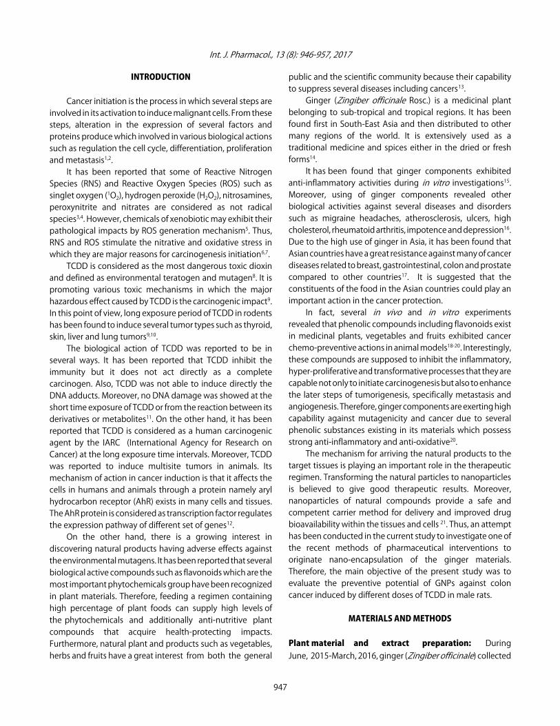
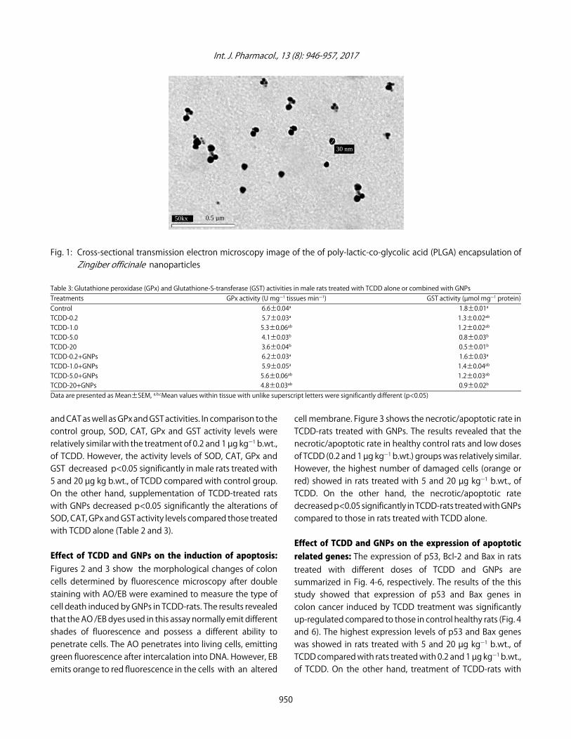
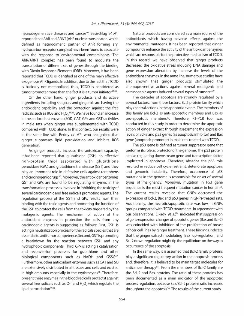
Transmission electron microscopy: The particle size andshape were characterized using high resolution transmissionelectron microscopy (HR-TEM) (Tecnai model G2-F20,Hillsboro, Oregon, OR, USA) JEM 2100 LB6 under operatingvoltage of 200 kV to investigate the micrograph of preparedPLGA encapsulation of Zingiber officinale extract underoperating voltage of 200 kV for different samples (Fig. 1).
Statistical analysis: All data were analyzed using the GeneralLinear Models (GLM) technique of Statistical Analysis Systemfollowed by Scheffé-test to assess significant differencesbetween groups33. The values are uttered as Mean±SEM. Allstatements of significance were based on probability ofp<0.05.
RESULTS
Effect of TCDD and GNPs on the antioxidant enzymeactivities: The measurements of oxidative markers includingSOD, CAT, GPx and GST activities are summarized in Table 2and 3, respectively.
Table 2 and 3 showed suppression effect of GNPs onTCDD-induced alterations in the antioxidant enzymes SOD
Table 1: Primers and reaction parameters in RT-PCRTarget cDNA Primer name Primer sequence (5'‒3') References$-Actin F GTG GGC CGC TCT AGG CAC CAA Khalil and Booles 31
R CTC TTT GAT GTC ACG CAC GAT TTp53 F GCG GTA CCC CAG GTC GGC GAG AAT CC Qin et al. 32
R GGG CTC GAG TCT AGA CTT TTG AGA AGCBcl-2 F CTC AGT CAT CCA CAG GGC GA Khalil and Booles 31
R AGA GGG GCT ACG AGT GGG ATBax F ACA AAG ATG GTC ACG GTC TGC C Khalil and Booles 31
R GGT TCA TCC AGG ATC GAG ACG G
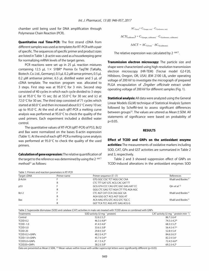
Table 2: Superoxide dismutase (SOD) and catalase (CAT) activities in male rats treated with TCDD alone or combined with GNPsTreatments SOD activity (U mgG1 protein) CAT activity (U mgG1 protein minG1)Control 52.6±5.2a 86.7±6.4a
TCDD-0.2 46.3±4.8ab 74.5±4.2ab
TCDD -1.0 41.4±4.6b 68.3±5.2b
TCDD-5.0 33.6±3.8b 56.4±4.1bc
TCDD-20 29.8±3.6b 52.8±5.4c
TCDD-0.2+GNPs 48.2±4.2ab 84.6±6.3a
TCDD-1.0+GNPs 45.7±5.6ab 82.2±5.6a
TCDD5.0+GNPs 41.1±4.2b 72.4±4.6ab
TCDD20+GNPs 38.2±3.8b 69.2±4.2b
Data are presented as Mean±SEM, a,b,cMean values within tissue with unlike superscript letters were significantly different (p<0.05)
949
Int. J. Pharmacol., 13 (8): 946-957, 2017
Fig. 1: Cross-sectional transmission electron microscopy image of the of poly-lactic-co-glycolic acid (PLGA) encapsulation ofZingiber officinale nanoparticles
Table 3: Glutathione peroxidase (GPx) and Glutathione-S-transferase (GST) activities in male rats treated with TCDD alone or combined with GNPsTreatments GPx activity (U mgG1 tissues minG1) GST activity (µmol mgG1 protein)Control 6.6±0.04a 1.8±0.01a
TCDD-0.2 5.7±0.03a 1.3±0.02ab
TCDD-1.0 5.3±0.06ab 1.2±0.02ab
TCDD-5.0 4.1±0.03b 0.8±0.03b
TCDD-20 3.6±0.04b 0.5±0.01b
TCDD-0.2+GNPs 6.2±0.03a 1.6±0.03a
TCDD-1.0+GNPs 5.9±0.05a 1.4±0.04ab
TCDD-5.0+GNPs 5.6±0.06ab 1.2±0.03ab
TCDD-20+GNPs 4.8±0.03ab 0.9±0.02b
Data are presented as Mean±SEM, a,b,cMean values within tissue with unlike superscript letters were significantly different (p<0.05)
and CAT as well as GPx and GST activities. In comparison to thecontrol group, SOD, CAT, GPx and GST activity levels wererelatively similar with the treatment of 0.2 and 1 µg kgG1 b.wt.,of TCDD. However, the activity levels of SOD, CAT, GPx andGST decreased p<0.05 significantly in male rats treated with5 and 20 µg kg b.wt., of TCDD compared with control group.On the other hand, supplementation of TCDD-treated ratswith GNPs decreased p<0.05 significantly the alterations ofSOD, CAT, GPx and GST activity levels compared those treatedwith TCDD alone (Table 2 and 3).
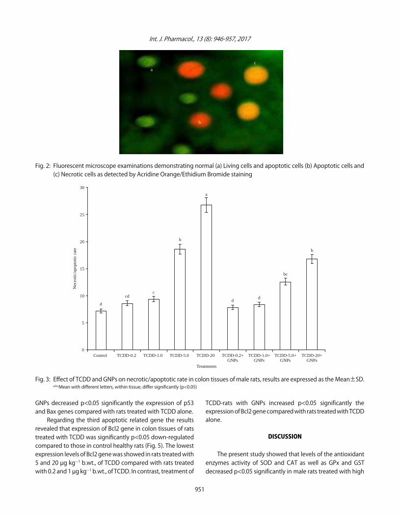
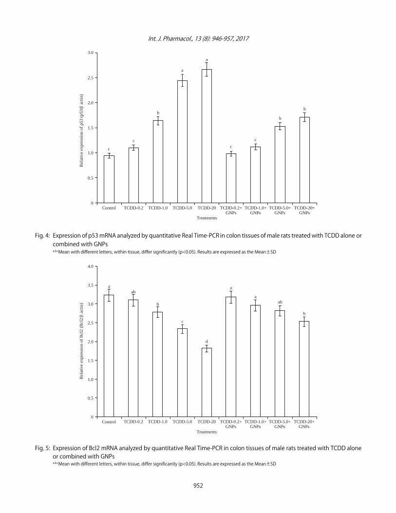
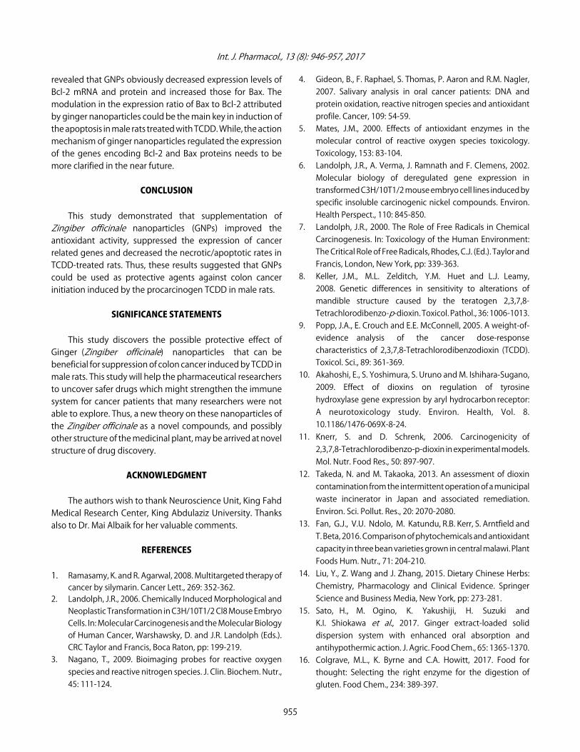
Effect of TCDD and GNPs on the induction of apoptosis:Figures 2 and 3 show the morphological changes of coloncells determined by fluorescence microscopy after doublestaining with AO/EB were examined to measure the type ofcell death induced by GNPs in TCDD-rats. The results revealedthat the AO /EB dyes used in this assay normally emit differentshades of fluorescence and possess a different ability topenetrate cells. The AO penetrates into living cells, emittinggreen fluorescence after intercalation into DNA. However, EBemits orange to red fluorescence in the cells with an altered
cell membrane. Figure 3 shows the necrotic/apoptotic rate inTCDD-rats treated with GNPs. The results revealed that thenecrotic/apoptotic rate in healthy control rats and low dosesof TCDD (0.2 and 1 µg kgG1 b.wt.) groups was relatively similar.However, the highest number of damaged cells (orange orred) showed in rats treated with 5 and 20 µg kgG1 b.wt., ofTCDD. On the other hand, the necrotic/apoptotic ratedecreased p<0.05 significantly in TCDD-rats treated with GNPscompared to those in rats treated with TCDD alone.
Effect of TCDD and GNPs on the expression of apoptoticrelated genes: The expression of p53, Bcl-2 and Bax in ratstreated with different doses of TCDD and GNPs aresummarized in Fig. 4-6, respectively. The results of the thisstudy showed that expression of p53 and Bax genes incolon cancer induced by TCDD treatment was significantlyup-regulated compared to those in control healthy rats (Fig. 4and 6). The highest expression levels of p53 and Bax geneswas showed in rats treated with 5 and 20 µg kgG1 b.wt., ofTCDD compared with rats treated with 0.2 and 1 µg kgG1 b.wt.,of TCDD. On the other hand, treatment of TCDD-rats with
950
30 nm
50kx 0.5 µm
Int. J. Pharmacol., 13 (8): 946-957, 2017
Control TCDD-0.2
b
bc
dd
a
b
ccd
d
TCDD-1.0 TCDD-5.0 TCDD-20 TCDD-0.2+GNPs
TCDD-1.0+GNPs
TCDD-5.0+GNPs
TCDD-20+GNPs
30
25
20
15
10
5
0
Nec
rotic
/apo
ptot
ic r
ate
Treatments
Fig. 2: Fluorescent microscope examinations demonstrating normal (a) Living cells and apoptotic cells (b) Apoptotic cells and (c) Necrotic cells as detected by Acridine Orange/Ethidium Bromide staining
Fig. 3: Effect of TCDD and GNPs on necrotic/apoptotic rate in colon tissues of male rats, results are expressed as the Mean±SD.a,b,cMean with different letters, within tissue, differ significantly (p<0.05)
GNPs decreased p<0.05 significantly the expression of p53and Bax genes compared with rats treated with TCDD alone.
Regarding the third apoptotic related gene the resultsrevealed that expression of Bcl2 gene in colon tissues of ratstreated with TCDD was significantly p<0.05 down-regulatedcompared to those in control healthy rats (Fig. 5). The lowestexpression levels of Bcl2 gene was showed in rats treated with5 and 20 µg kgG1 b.wt., of TCDD compared with rats treatedwith 0.2 and 1 µg kgG1 b.wt., of TCDD. In contrast, treatment of
TCDD-rats with GNPs increased p<0.05 significantly theexpression of Bcl2 gene compared with rats treated with TCDDalone.
DISCUSSION
The present study showed that levels of the antioxidantenzymes activity of SOD and CAT as well as GPx and GSTdecreased p<0.05 significantly in male rats treated with high
951
a
c
b
Int. J. Pharmacol., 13 (8): 946-957, 2017
Control TCDD-0.2
b
b
c
c
a
a
b
c
c
TCDD-1.0 TCDD-5.0 TCDD-20 TCDD-0.2+GNPs
TCDD-1.0+GNPs
TCDD-5.0+GNPs
TCDD-20+GNPs
3.0
2.5
2.0
1.5
1.0
0.5
0
Treatments
Rel
ativ
e ex
pres
sion
of
p53
(p53
/ a
ctin
)
Control TCDD-0.2
b
aba
b
d
aba
TCDD-1.0 TCDD-5.0 TCDD-20 TCDD-0.2+GNPs
TCDD-1.0+GNPs
TCDD-5.0+GNPs
TCDD-20+GNPs
4.0
3.5
3.0
2.5
2.0
1.5
1.0
0.5
0
Treatments
Rel
ativ
e ex
pres
sion
of
Bcl
2 (B
cl2/
act
in)
a
c
Fig. 4: Expression of p53 mRNA analyzed by quantitative Real Time-PCR in colon tissues of male rats treated with TCDD alone orcombined with GNPsa,b,cMean with different letters, within tissue, differ significantly (p<0.05). Results are expressed as the Mean±SD
Fig. 5: Expression of Bcl2 mRNA analyzed by quantitative Real Time-PCR in colon tissues of male rats treated with TCDD aloneor combined with GNPsa,b,cMean with different letters, within tissue, differ significantly (p<0.05). Results are expressed as the Mean±SD
952
Int. J. Pharmacol., 13 (8): 946-957, 2017
Control TCDD-0.2
c
d
TCDD-1.0 TCDD-5.0 TCDD-20 TCDD-0.2+GNPs
TCDD-1.0+GNPs
TCDD-5.0+GNPs
TCDD-20+GNPs
3.5
3.0
2.5
2.0
1.5
1.0
0.5
0
Treatments
Rel
ativ
e ex
pres
sion
of
Bax
(B
ax/
act
in)
a
b
d d
d
dcc
Fig. 6: Expression of Bax mRNA analyzed by quantitative Real Time-PCR in colon tissues of male rats treated with TCDD alone orcombined with GNPsa,b,c,dMean with different letters, within tissue, differ significantly (p<0.05). Results are expressed as the Mean±SD
doses (5 and 20 µg kgG1 b.wt.) of TCDD compared with controlgroup. In the same line of our results, Bulmusa et al.34 foundthat exposure to TCDD decreased SOD, CAT and GPx activitiesas well as glutathione levels in liver, kidney, brain and hearttissues of male rats.
The alteration in the antioxidant enzyme activities dueto exposure to TCDD was clarified by the study ofMacDonald et al.35. They indicated that TCDD may inducecytochromes P450 (CYPs) enzymes to enhance ROSgeneration and therefore trigger lipid peroxidation, modifiednitrogenous base formation and DNA strand breaks as well asdecreases the levels of hepatic glutathione levels.
Moreover, Fromme et al.36 indicated that TCDD enhancedblood pressure and oxidation in mice. In addition, TCDD hasbeen found to cause impairment of the competence of theavian heart endoplasmic reticulum to appropriate Ca2+andcontractile defects37. Thus, change in the intracellular Ca2+
levels due to the oxidation effect of TCDD in male rats is themain reason in generation of high rate of ROS.
Therefore, increasing the levels of the peroxidation in ratstreated with TCDD might be attributed to ROS mediatingmembrane damage which coincide with decrease levels of the
antioxidants resulting in oxidative stress.The present study revealed that TCDD elevated high
levels of apoptosis in male rats. Expression of apoptotic relatedgenes such as Bcl-2, Bax and p53 genes in TCDD-rats wassignificantly p<0.05 increased. Furthermore, the highestnumber of damaged cells (orange or red) showed in TCDDgroups. In agreement with our findings, Longini et al.38 andChen et al.39 suggested that the shift in the intracellularpro-oxidants stimulated by TCDD may induce celldamage/death due to direct damage in the cell membranethrough lipid peroxidation38 or due to apoptosis bytranscription factors activation or by DNA damage 39. So, theobserved structural changes including both forms of necrosisand apoptosis in male rats after TCDD treatment in thecurrent study was related to oxidative stress.
The DNA damage generated by oxidation process inhepatic cells may be resulted from extremely induction ofenzymes and proteins that regulated by transcriptional controlof the aryl hydrocarbon receptor (AhR) activated by TCDDtreatment11.
AhR is an essential transcriptional regulator occurs duringthe pathophysiological and physiological processes such as
953
Int. J. Pharmacol., 13 (8): 946-957, 2017
neurodegenerative diseases and cancer40. Besischlag et al.41
reported that AhR and ARNT (AhR nuclear translocator, whichdefined as heterodimeric partner of AhR forming arylhydrocarbon receptor complex) have been found to associatewith the response to environmental contaminants. TheAhR/ARNT complex has been found to modulate thetranscription of different set of genes through the bindingwith Dioxin Response Elements (DRE). Moreover, it has beenreported that TCDD is identified as one of the main effectiveexogenous AhR ligands. In addition, due to the fact that TCDDis basically not metabolized, thus, TCDD is considered astumor promoter more than the fact it is a tumor initiator42,43.
On the other hand, ginger products and its activeingredients including shagoals and gingerols are having theantioxidant capability and the protection against the freeradicals such as ROS and H2O2
44,45. We have found an increasein the antioxidant enzyme (SOD, CAT, GPx and GST) activitiesin male rats when ginger was supplemented with TCDDcompared with TCDD alone. In this context, our results werein the same line with Reddy et al.46, who recognized thatginger suppresses lipid peroxidation and inhibits ROSgeneration.
As ginger products increase the antoxidant capacity,it has been reported that glutathione (GSH) an effectivenon-protein thiol associated with glutathioneperoxidase (GPX) and glutathione transferase (GST) and theyplay an important role in defensive cells against teratohensand carcinogenic drugs 47. Moreover, the antioxidant enzymesGST and GPx are found to be regulating enzymes in thetransformation processes involved in inhibiting the toxicity ofseveral carcinogenic and free radicals promoting agents. Theregulation process of the GST and GPx results from theirbinding with the toxic agents and promoting the function ofthe GSH to protect the cells from the toxicity triggered by themutagenic agents. The mechanism of action of theantioxidant enzymes in protection the cells from anycarcinogenic agents is suggesting as follows: First, GSH isacting a neutralization process for the radicals species that areessential to antitumor competence. Second, GST is promotinga breakdown for the reaction between GSH and anyhydrophobic components. Third, GPx is acting a catalyzationand reconversion processes for gutathione and otherbiological components such as NADH and GSSG47.Furthermore, other antioxidant enzymes such as CAT and SOare extensively distributed in all tissues and cells and existedin high amounts especially in the erythrocytes48. Therefore,present these enzymes in the blood red cells protect it againstseveral free radicals such as O2G and H2O2 which regulate thelipid peroxidation49,50.
Natural products are considered as a main sourse of theantioxidants which having adverse effects against theenvironmental mutagens. It has been reported that gingercompounds enhance the activity of the antioxidant enzymeswhich are responsible for the protective mechanism of TCDD.In this regard, we have observed that ginger productsdecreased the oxidative stress inducing DNA damage andgene expression alteration by increase the levels of theantioxidant enzymes. In the same line, numerous studies havealso shown that ginger products stimulated thechemopreventive actions against several mutagenic andcarcinogenic agents induced several types of tumors46,51.
The cascades of apoptosis are strongly regulated by aseveral factors; from these factors, Bcl2 protein family whichplays central actions in the apoptotic events. The members ofthis family are Bcl-2 as anti-apoptotic members and Bax aspro-apoptotic members52. Therefore, RT-PCR tool wasconducted in this study in order to determine the apoptoticaction of ginger extract through assessment the expressionlevels of Bcl-2 and p53 genes (as apoptotic inhibitor) and Baxgene (apoptotic promoter) in male rats treated with TCDD.
The p53 gene is defined as tumor suppressor gene thatperforms its role as protector of the genome. The p53 proteinacts as regulating downstream gene and transcription factorimplicated in apoptosis. Therefore, absence the p53 roleresulted in reduce cell cycle restraint, deteriorate apoptosisand genomic instability. Therefore, occurrence of p53mutations in the genome is responsible for onset of severaltypes of malignancy. Moreover, mutation in P53 genesequence is the most frequent mutation cancer in human53.The current results revealed that GNPs decreased theexpression of Bcl-2, Bax and p53 genes in GNPs-treated rats.Additionally, the necrotic/apoptotic rate was low in GNPsgroups compared with TCDD treatments. In agreement withour observations, Elkady et al.54 indicated that suppressionof gene expression changes of apoptotic genes (Bax and Bcl-2)was coincided with inhibition of the proliferation of breastcancer cell lines by ginger treatment. These findings indicatethat the ginger extract modulating Bax up-regulation andBcl-2 down-regulation might tip the equilibrium on the way tooccurrence of the apoptosis.
In the same way, it is assumed that Bcl-2 family proteinsplay a significant regulatory action in the apoptosis processand, therefore, it is believed to be main target molecules foranticancer therapy55. From the members of Bcl-2 family arethe Bcl-2 and Bax proteins. The ratio of these proteins hasbeen documented as a main indicator of the apoptoticprocess regulation, because Bax/Bcl-2 proteins ratio increasesthroughout the apoptosis56. The results of the current study
954
Int. J. Pharmacol., 13 (8): 946-957, 2017
revealed that GNPs obviously decreased expression levels ofBcl-2 mRNA and protein and increased those for Bax. Themodulation in the expression ratio of Bax to Bcl-2 attributedby ginger nanoparticles could be the main key in induction ofthe apoptosis in male rats treated with TCDD. While, the actionmechanism of ginger nanoparticles regulated the expressionof the genes encoding Bcl-2 and Bax proteins needs to bemore clarified in the near future.
CONCLUSION
This study demonstrated that supplementation ofZingiber officinale nanoparticles (GNPs) improved theantioxidant activity, suppressed the expression of cancerrelated genes and decreased the necrotic/apoptotic rates inTCDD-treated rats. Thus, these results suggested that GNPscould be used as protective agents against colon cancerinitiation induced by the procarcinogen TCDD in male rats.
SIGNIFICANCE STATEMENTS
This study discovers the possible protective effect ofGinger (Zingiber officinale) nanoparticles that can bebeneficial for suppression of colon cancer induced by TCDD inmale rats. This study will help the pharmaceutical researchersto uncover safer drugs which might strengthen the immunesystem for cancer patients that many researchers were notable to explore. Thus, a new theory on these nanoparticles ofthe Zingiber officinale as a novel compounds, and possiblyother structure of the medicinal plant, may be arrived at novelstructure of drug discovery.
ACKNOWLEDGMENT
The authors wish to thank Neuroscience Unit, King FahdMedical Research Center, King Abdulaziz University. Thanksalso to Dr. Mai Albaik for her valuable comments.
REFERENCES
1. Ramasamy, K. and R. Agarwal, 2008. Multitargeted therapy ofcancer by silymarin. Cancer Lett., 269: 352-362.
2. Landolph, J.R., 2006. Chemically Induced Morphological andNeoplastic Transformation in C3H/10T1/2 Cl8 Mouse EmbryoCells. In: Molecular Carcinogenesis and the Molecular Biologyof Human Cancer, Warshawsky, D. and J.R. Landolph (Eds.).CRC Taylor and Francis, Boca Raton, pp: 199-219.
3. Nagano, T., 2009. Bioimaging probes for reactive oxygenspecies and reactive nitrogen species. J. Clin. Biochem. Nutr.,45: 111-124.
4. Gideon, B., F. Raphael, S. Thomas, P. Aaron and R.M. Nagler,2007. Salivary analysis in oral cancer patients: DNA andprotein oxidation, reactive nitrogen species and antioxidantprofile. Cancer, 109: 54-59.
5. Mates, J.M., 2000. Effects of antioxidant enzymes in themolecular control of reactive oxygen species toxicology.Toxicology, 153: 83-104.
6. Landolph, J.R., A. Verma, J. Ramnath and F. Clemens, 2002.Molecular biology of deregulated gene expression intransformed C3H/10T1/2 mouse embryo cell lines induced byspecific insoluble carcinogenic nickel compounds. Environ.Health Perspect., 110: 845-850.
7. Landolph, J.R., 2000. The Role of Free Radicals in ChemicalCarcinogenesis. In: Toxicology of the Human Environment:The Critical Role of Free Radicals, Rhodes, C.J. (Ed.). Taylor andFrancis, London, New York, pp: 339-363.
8. Keller, J.M., M.L. Zelditch, Y.M. Huet and L.J. Leamy,2008. Genetic differences in sensitivity to alterations ofmandible structure caused by the teratogen 2,3,7,8-Tetrachlorodibenzo-p-dioxin. Toxicol. Pathol., 36: 1006-1013.
9. Popp, J.A., E. Crouch and E.E. McConnell, 2005. A weight-of-evidence analysis of the cancer dose-responsecharacteristics of 2,3,7,8-Tetrachlorodibenzodioxin (TCDD).Toxicol. Sci., 89: 361-369.
10. Akahoshi, E., S. Yoshimura, S. Uruno and M. Ishihara-Sugano,2009. Effect of dioxins on regulation of tyrosinehydroxylase gene expression by aryl hydrocarbon receptor:A neurotoxicology study. Environ. Health, Vol. 8.10.1186/1476-069X-8-24.
11. Knerr, S. and D. Schrenk, 2006. Carcinogenicity of2,3,7,8-Tetrachlorodibenzo-p-dioxin in experimental models.Mol. Nutr. Food Res., 50: 897-907.
12. Takeda, N. and M. Takaoka, 2013. An assessment of dioxincontamination from the intermittent operation of a municipalwaste incinerator in Japan and associated remediation.Environ. Sci. Pollut. Res., 20: 2070-2080.
13. Fan, G.J., V.U. Ndolo, M. Katundu, R.B. Kerr, S. Arntfield andT. Beta, 2016. Comparison of phytochemicals and antioxidantcapacity in three bean varieties grown in central malawi. PlantFoods Hum. Nutr., 71: 204-210.
14. Liu, Y., Z. Wang and J. Zhang, 2015. Dietary Chinese Herbs:Chemistry, Pharmacology and Clinical Evidence. SpringerScience and Business Media, New York, pp: 273-281.
15. Sato, H., M. Ogino, K. Yakushiji, H. Suzuki andK.I. Shiokawa et al., 2017. Ginger extract-loaded soliddispersion system with enhanced oral absorption andantihypothermic action. J. Agric. Food Chem., 65: 1365-1370.
16. Colgrave, M.L., K. Byrne and C.A. Howitt, 2017. Food forthought: Selecting the right enzyme for the digestion ofgluten. Food Chem., 234: 389-397.
955
Int. J. Pharmacol., 13 (8): 946-957, 2017
17. Dorai, T. and B.B. Aggarwal, 2004. Role of chemopreventiveagents in cancer therapy. Cancer Lett., 215: 129-140.
18. Mahmoud, N.N., A.M. Carothers, D. Grunberger, R.T. Bilinskiand M.R. Churchill et al., 2000. Plant phenolics decreaseintestinal tumors in an animal model of familial adenomatouspolyposis. Carcinogenesis, 21: 921-927.
19. Kim, S.O., K.S. Chun, J.K. Kundu and Y.J. Surh, 2004. Inhibitoryeffects of [6]-gingerol on PMA-induced COX-2 expression andactivation of NF-?B and p38 MAPK in mouse skin. Biofactors,21: 27-31.
20. Kujawski, R., K. Dziekan, H. Wolski, M. Barlik andA. Seremak-Mrozikiewicz, 2015. The influence of certain plantsubstances and their chemopreventive activity in ovariancancer. Ginekol. Polska, 86: 468-472.
21. Khalil, W.K.B., G.T. El-Bassyouni and H.F. Booles, 2016.Nano-encapsulated form of citrus medica for osteoporosistreatment in animal model. Int. J. Pharm. Clin. Res., 8: 49-59.
22. Tasanarong, A., S. Kongkham and A. Itharat, 2014.Antioxidant effect of Phyllanthus emblica extract preventscontrast-induced acute kidney injury. BMC Complem. Altern.Med., Vol. 14. 10.1186/1472-6882-14-138.
23. Samadder, A., S. Das, J. Das, A. Paul and A.R. Khuda-Bukhsh,2013. Ameliorative effects of syzygium jambolanum extractand its poly (lactic-co-glycolic) acid nano-encapsulated formon arsenic-induced hyperglycemic Stress: A Multi-parametricevaluation. J. Acupuncture Meridian Stud., 5: 310-318.
24. Takeda, T., J. Taura, Y. Hattori, Y. Ishii and H. Yamada, 2014.Dioxin-induced retardation of development through areduction in the expression of pituitary hormones andpossible involvement of an aryl hydrocarbon receptor in thisdefect: A comparative study using two strains of mice withdifferent sensitivities to dioxin. Toxicol. Applied Pharmacol.,278: 220-229.
25. Manju, V. and N. Nalini, 2005. Chemopreventive efficacy ofginger, a naturally occurring anticarcinogen during theinitiation, post-initiation stages of 1,2 dimethylhydrazine-induced colon cancer. Clin. Chim. Acta, 358: 60-70.
26. Li, W.Y., B.X. Chen, Z.J. Chen, Y.T. Gao, Z. Chen and J. Liu,2017. Reactive oxygen species generated by NADPHoxidases promote radicle protrusion and root elongationduring rice seed germination. Int. J. Mol. Sci., Vol. 18.10.3390/ijms18010110.
27. Netto, L.E.S., A.J. Kowaltowski, R.F. Castilho and A.E. Vercesi,2002. Thiol enzymes protecting mitochondria againstoxidative damage. Methods Enzymol., 348: 260-270.
28. Sakharov, I.Y., J.L. Castillo, J.C. Areza and I.Y. Galaev, 2000.Purification and stability of peroxidase of African oil palmElaies guineensis. Bioseparation, 9: 125-132.
29. Czene, S., E. Testa, J. Nygren, I. Belyaev and M. Harms-Ringdahl, 2002. DNA fragmentation and morphologicalchanges in apoptotic human lymphocytes. Biochem. Biophys.Res. Commun., 294: 872-878.
30. Pitocco, D., A. Crino, E. Di Stasio, S. Manfrini andC. Guglielmi et al., 2006. The effects of calcitriol andnicotinamide on residual pancreatic $-cell function inpatients with recent-onset type 1 diabetes (IMDIAB XI).Diabetic Med., 23: 920-923.
31. Khalil, W.K. and H.F. Booles, 2011. Protective role ofselenium against over-expression of cancer-related apoptotic genes induced by O-cresol in rats. Arch. Ind. Hyg.Toxicol., 62: 121-129.
32. Qin, C., T. Nguyen, J. Stewart, I. Samudio, R. Burghardt andS. Safe, 2002. Estrogen up-regulation of p53 gene expressionin MCF-7 breast cancer cells is mediated by calmodulin kinaseIV-dependent activation of a nuclear factor 6B/CCAAT-binding transcription factor-1 complex. Mol. Endocrinol.,16: 1793-1809.
33. SAS., 2003. SAS User's Guide: Statistics. SAS Institute Inc., Cary,NC., USA.
34. Bulmus, F.G., F. Sakin, G. Turk, M. Sonmez and K. Servi, 2013.Protective effects of curcumin on antioxidant status, bodyweight gain and reproductive parameters in male ratsexposed to subchronic 2,3,7,8-tetrachlorodibenzo-p-dioxin.Toxicol. Environ. Chem., 95: 1019-1029.
35. MacDonald, C.J., R.Y.S. Cheng, D.D. Roberts, D.A. Wink andG.C. Yeh, 2009. Modulation of carcinogen metabolism bynitric oxide-aspirin 2 is associated with suppression ofDNA damage and DNA adduct formation. J. Biol. Chem.,284: 22099-22107.
36. Fromme, H., B. Hilger, M. Albrecht, W. Gries, G. Lengand W. Volkel, 2016. Occurrence of chlorinated andbrominated dioxins/furans, PCBs and brominated flameretardants in blood of German adults. Int. J. Hyg. Environ.Health, 219: 380-388.
37. Wang, Q., H. Kurita, V. Carreira, C.I. Ko and Y. Fan et al., 2016.Ah receptor activation by dioxin disrupts activin, BMP andWNT signals during the early differentiation of mouseembryonic stem cells and inhibits cardiomyocyte functions.Toxicol. Sci., 149: 346-357.
38. Longini, M., E. Belvisi, F. Proietti, F. Bazzini, G. Buonocore andS. Perrone, 2017. Oxidative stress biomarkers: Establishmentof reference values for isoprostanes, AOPP and NPBI in cordblood. Mediators Inflamm. 10.1155/2017/1758432.
39. Chen, Y., Y. Li, H. Xu, G. Li, Y. Ma and Y.J. Pang, 2017. Morinmitigates oxidative stress, apoptosis and inflammation incerebral ischemic rats. Afr. J. Tradit. Complement. Altern.Med., 14: 348-355.
40. Murray, I.A., J.L. Morales, C.A. Flaveny, B.C. DiNataleand C. Chiaro et al., 2010. Evidence for ligand-mediatedselective modulation of aryl hydrocarbon receptor activity.Mol. Pharmacol., 77: 247-254.
41. Beischlag, T.V., J.L. Morales, B.D. Hollingshead andG.H. Perdew, 2008. The aryl hydrocarbon receptor complexand the control of gene expression. Crit. Rev. Eukaryotic GeneExpr., 18: 207-250.
956
Int. J. Pharmacol., 13 (8): 946-957, 2017
42. Patel, R.D., D.J. Kim, J.M. Peters and G.H. Perdew, 2006. Thearyl hydrocarbon receptor directly regulates expression of thepotent mitogen epiregulin. Toxicol. Sci., 89: 75-82.
43. Patel, R.D., B.D. Hollingshead, C.J. Omiecinski andG.H. Perdew, 2007. Aryl-hydrocarbon receptor activationregulates constitutive androstane receptor levels in murineand human liver. Hepatology, 46: 209-218.
44. Kabuto, H., M. Nishizawa, M. Tada, C. Higashio, T. Shishiboriand M. Kohno, 2005. Zingerone [4-(4-hydroxy-3-methoxyphenyl)-2-butanone] prevents 6-hydroxydopamine-induced dopamine depression in mouse striatum andincreases superoxide scavenging activity in serum.Neurochem. Res., 30: 325-332.
45. Lanzafame, G.M., M. Sarakha, D. Fabbri and D. Vione, 2017.Degradation of methyl 2-aminobenzoate (methylanthranilate) by H2O2/UV: Effect of inorganic anions andderived radicals . Molecules, Vol . 22, No. 4.10.3390/molecules22040619
46. Reddy, Y.A., M. Chalamaiah, B. Ramesh, G. Balaji and P. Indira,2014. Ameliorating activity of ginger (Zingiber officinale)extract against lead induced renal toxicity in male rats. J. FoodSci. Technol., 51: 908-914.
47. Velazquez, I., M. Plaud, V. Wojna, R. Skolasky, J.P. Laspiur andL.M. Melendez, 2009. Antioxidant enzyme dysfunction inmonocytes and CSF of Hispanic women with HIV-associatedcognitive impairment. J. Neuroimmunol., 206: 106-111.
48. Silva, R., H. Carmo, V. Vilas-Boas, P.G. de Pinho andR.J. Dinis-Oliveira et al., 2013. Doxorubicin decreasesparaquat accumulation and toxicity in Caco-2 cells. Toxicol.Lett., 218: 34-41.
49. Rocha, S., D. Gomes, M. Lima, E. Bronze-da-Rocha andA. Santos-Silva, 2015. Peroxiredoxin 2, glutathione peroxidaseand catalase in the cytosol and membrane of erythrocytes under H2O2-induced oxidative stress. Free Radic. Res.,49: 990-1003.
50. Rieger, H. and M. Welter, 2015. Integrative models of vascularremodeling during tumor growth. Wiley Interdisciplin. Rev.:Syst. Biol. Med., 7: 113-129.
51. Murakami, A., T. Tanaka, J.Y. Lee, Y.J. Surh and H.W. Kim et al.,2004. Zerumbone, a sesquiterpene in subtropical ginger,suppresses skin tumor initiation and promotion stages in ICRmice. Int. J. Cancer, 110: 481-490.
52. Burlacu, A., 2003. Regulation of apoptosis by Bcl-2 familyproteins. J. Cell. Mol. Med., 7: 249-257.
53. Smith, N.D., J.N. Rubenstein, S.E. Eggener and J.M. Kozlowski,2003. The p53 tumor suppressor gene and nuclear protein:Basic science review and relevance in the management ofbladder cancer. J. Urol., 169: 1219-1228.
54. Elkady, A.I., O.A. Abuzinadah, N.A. Baeshen and T.R. Rahmy,2012. Differential control of growth, apoptotic activity andgene expression in human breast cancer cells by extractsderived from medicinal herbs Zingiber officinale. J. BioMed.Res. 10.1155/2012/614356.
55. Baell, J.B. and D.C.S. Huang, 2002. Prospects for targeting theBcl-2 family of proteins to develop novel cytotoxic drugs.Biochem. Pharmacol., 64: 851-863.
56. Karmakar, I., S. Haldar, M. Chakraborty, K. Chaudhury,S. Dewanjee and P.K. Haldar, 2016. Regulation of apoptosisthrough bcl-2/bax proteins expression and DNA damageby Zanthoxylum alatum. Pharmaceut. Biol., 54: 503-508.
957


![THE GINGER GUIDE · PDF file · 2017-09-27GINGER GUIDE THE [ORIGINS OF FLAVOR ] ... omes of ginger ingiber officinale) ... ORGANIC GINGER SYRUP #25400 INGREDIENTS: Organic ginger,](https://img.pdfslide.net/doc/110x75/5aadf2a57f8b9a22118b6437/the-ginger-guide-guide-the-origins-of-flavor-omes-of-ginger-ingiber-officinale.jpg)


![Synthesis And Characterization Of Gold Nanoparticles · Gold nanoparticles are synthesized by liquid chemical method and green synthesis method [7-9]. GNPs are produced by reducing](https://img.pdfslide.net/doc/110x75/5f20c230712d9d535d185368/synthesis-and-characterization-of-gold-nanoparticles-gold-nanoparticles-are-synthesized.jpg)