Embed Size (px)
Citation preview

Glucocorticoid Activation of Chromogranin A Gene ExpressionIdentification and Characterization of a Novel Glucocorticoid Response Element
David J. Rozansky, Hongjiang Wu, Kechun Tang, Robert J. Parmer, and Daniel T. O'ConnorDepartment of Medicine and Center for Molecular Genetics, University of California, San Diego and Department of Veterans AffairsMedical Center, San Diego, California 92161
Abstract
Glucocorticoids regulate catecholamine biosynthesis andstorage at several sites. Chromogranin A, an abundant pro-tein complexed with catecholamines in secretory vesicles ofchromaffin cells and sympathetic axons, is also augmentedby glucocorticoids. This study reports isolation of the ratchromogranin A promoter to elucidate transcriptional regu-lation of chromogranin A biosynthesis by glucocorticoidsin neuroendocrine cells. Endogenous chromogranin A geneexpression was activated up to 3.5-fold in chromaffin cellsby glucocorticoid, in time-dependent fashion. Inhibition ofnew protein synthesis by cycloheximide did not alter the risein chromogranin A mRNA, suggesting that glucocorticoidsdirectly activate the chromogranin A promoter; nuclearrunoff assays confirmed a 3.3-fold increased rate of initia-tion of new chromogranin A transcripts after glucocorticoid.Transfected rat chromogranin A promoter/luciferase re-porter constructs were activated 2.6-3.1-fold by glucocorti-coid, and selective agonist/antagonist studies determinedthat dexamethasone effects were mediated by glucocorticoidreceptors. Both rat and mouse chromogranin A promoter/luciferase reporter constructs were activated by glucocorti-coid. A series of promoter deletions narrowed the region ofglucocorticoid action to a 93-bp section of the promoter,from position -526 to -619 bp upstream of the cap site. A15-bp sequence ([-583 bpJ 5'-ACATGAGTGTGTCCT-3'[-597 bp]) within this region showed partial homology to aglucocorticoid response element (GRE; half-site in italics)consensus sequence, and several lines of experimental evi-dence confirmed its function as a GRE: (a) site-directedmutation of this GREprevented glucocorticoid activationof a chromogranin A promoter/reporter; (b) transfer ofthis GREto a heterologous (thymidine kinase) promoter/reporter conferred activation by glucocorticoid, in copynumber-dependent and orientation-independent fashion;and (c) electrophoretic gel mobility shifts demonstratedbinding of this GREby ligand-activated glucocorticoid re-ceptor, though at 2.75-fold lower affinity than the glucocorti-coid receptor interaction with a consensus GRE. The ratchromogranin A GREshowed functional and structuralsimilarities to GREsin other genes proportionally regulatedby glucocorticoids. Weconclude that a discrete domain of
Address correspondence to Daniel T. O'Connor, M.D., Department ofMedicine (911 1H), University of California, San Diego, 3350 La JollaVillage Drive, San Diego, CA 92161.
Received for publication 16 June 1994 and in revised form 10August 1994.
The Journal of Clinical Investigation, Inc.Volume 94, December 1994, 2357-2368
the chromogranin A promoter is both necessary and suffi-cient to confer glucocorticoid regulation onto the gene, andthat the activity of this region also explains the degree ofactivation of the endogenous gene by glucocorticoid. (J. Clin.Invest. 1994. 94:2357-2368.) Key words: chromogranin A -
adrenal medulla * catecholamine * glucocorticoid * steroid- promoter . enhancer . pheochromocytoma * PC-12 * chro-maffin.
Introduction
After release from the adrenal cortex, glucocorticoids first entersinusoids that traverse the adrenal medulla before entering thesystemic circulation. Exposure to high local glucocorticoid con-centration plays a crucial developmental role in tissue-specificactivation of genes that characterize the chromaffin cell pheno-type (1). In the adult (2), two genes of the catecholaminebiosynthetic pathway are directly activated by glucocorticoids:phenylethanolamine-N-methyltransferase (3) and tyrosine hy-droxlyase (4, 5 and references therein). The expression of chro-mogranin A, the major soluble protein in chromaffin vesicles,is also augmented by glucocorticoids, but the mechanism ofactivation has not been elucidated (6).
Chromogranin A is the index member of a family of acidic,soluble proteins found in neuroendocrine secretory granules(7). Within granules, chromogranin A binds catecholaminesand calcium (8, 9), and may inhibit prohormone processingenzymes (10). After release into the extracellular space, chro-mogranin A is processed into several biologically active pep-tides (11, 12). Even though chromogranin A is already abun-dant, representing 46%of soluble protein in chromaffin vesicles(13), it remains sensitive to glucocorticoids (14-17). In vivo,hypophysectomy decreases adrenal chromogranin A (14), withrestoration after glucocorticoid replacement ( 15). In vitro, chro-mogranin A protein is consistently up-regulated by glucocorti-coid in bovine chromaffin (16) and rat pheochromocytoma(PC- 12) cells ( 17), with proportional induction of itsmRNA(16, 17).
Since glucocorticoids and catecholamines play importantregulatory roles in metabolic and cardiovascular responses, andchromogranin A influences catecholamine storage and release(8), it is crucial to understand chromogranin A gene regulationby glucocorticoids. Indeed, a thorough understanding of thisregulation may assist in elucidating the protein's many intracel-lular and extracellular functions.
This investigation presents evidence that glucocorticoids di-rectly activate chromogranin A gene expression. Weisolated aregion of the rat chromogranin A promoter with resemblanceto a consensus glucocorticoid response element (GRE),1 and
1. Abbreviations used in this paper: GR, Glucocorticoid receptor; GRE,glucocorticoid response element; hGR, human glucocorticoid receptor;RSV, Rous sarcoma virus; TK, thymidine kinase.
Chromogranin A Glucocorticoid Response Element 2357
Table L Plasmids and Oligonucleotides Used to Investigate Glucocorticoid Regulation of Rat Chromogranin A
Plasmid orExperiment type oligonucleotide Description
Gene isolation
Sequencing/subcloning
Transcriptional nuclear runoff assay
General transient transfection
Promoter deletion and expression
Promoter GREmutation
Response elements retardationstudies
Rat CgA GREtransfer study
sCos- 1
sCos-lrCgA-1sCos- lrCgA-2pBSrCgAP/P1594
pBSrCgAB/S489
pSV2ALA5pXp2
pXp2RCgApXp2rCgAA-523(+)
pXp2rCgAA-523(-)pBSm-gDNA5.1
pRSVCATpRSVhGRpXp2
pXp2rCgA
pXp2rCgAA-523pXp2rCgAA-619pXp2rCgAA-756pXp2rCgAA- 1053pXp2rCgAA-1281pXp2rCgAA-756m
cGRE
*cGRErGRE
*rGREpTKluc
pTKlucC17pTKlucCl9pTKlucE9pcGRETKluc-IpcGRETKluc-2
Supercos-I cosmid (Stratagene); carries 32-42 kb genomic DNAinserts (18, 19)Rat genomic DNAcosmid that spans entire rat CgA geneCosmid containing rat CgA gene with - 20-kb overlap with sCos-lrCgA-1pBluescriptKS- with a 1,594-bp PstI/PstI fragment of rCgA gene subcloned into
the multiple cloning site (MCS) at PstIpBluescriptKS- with 489 bp BamHI/SstI fragment of rCgA gene subcloned into
the MCSLuciferase reporter gene vector under control of the SV40 early promoter (41)Promoterless luciferase reporter gene vector with MCSimmediately upstream of
luciferase open reading frame (35)Rat CgA promoter fragment inserted into MCSof pXp2pXp2rCgA restriction digest-derivedt insert: SstI/SstI [5' -523 to +75§ -3']
positive orientationpXp2rCgA restriction digest-derived insert: SstlI/SstI [5' +75 to -523 -3']5.1-kb EcoRI/EcoRI fragment of mouse CgA gene (includes exons 1-3)
subcloned into MCSof pBluescriptKS- (34)Chloramphenicol acetyltransferase (CAT) expression driven by RSVpromoter (39)Human glucocorticoid receptor expression driven by RSVpromoter (38)Promoterless luciferase reporter-gene plasmid with MCSimmediately upstream of
luciferase open reading frame (35)Rat CgA promoter fragment inserted into MCSof pXp2 (specifics of a fragment
are indicated by number following the deletion symbol "A")pXp2rCgA RE-derivedt insert: SstlISstI [5' -523 to +75§ -3]pXp2rCgA PCR-derivedll insert: HindIIlIXhoI [5' -619 to + 112 -3'1]pXp2rCgA PCR-derivedll insert: HindIII/XhoI [5' -756 to + 112 -3']pXp2rCgA PCR-derivedll insert: HindIII/XhoI [5' -1053 to +112 -3']pXp2rCgA RE-derivedt insert: SmaI/SmaI [5' -1281 to +75 -3']pXp2rCgAA-756 mutated from position -597 to -590 by changing [-597:5'-
AGGACACA-3':-590] to [-597:5'-gcGgtACc-3':-590] where bold lettersindicate the rGRE motif, lower case letters indicate mutated residues, and newlyintroduced KpnI site (GGTACC) is underlined. Vector constructed by ligatingPCR-derived fragments HindIII/KpnI [-756: 5' to 3':-591] and KpnI/XhoI[-595:5' to 3':+ 112] into MCSupstream of luciferase reporter in pXp2.Mutated plasmid sequence confirmed by dideoxy chain termination sequencing.
Consensus GREti 5'-AGAACAgagTGTTCT-3' (54, 55), with capital lettersindicating consensus motifs
y-[32P]-end-labeled consensus cGREttGREtt from the rat CgA promoter: [-583 bp] 5'-ACATGAGTGTGTCCT-3'
[-597 bp]y-[32P]-end-labeled GREtt from the rat CgA promoterThymidine kinase promoter/luciferase reporter plasmid with MCSupstream of TK
promoter (36)One rGRE inserted§§ (in reverse orientation) into BamHI site of MCSof pTKlucOne rGRE inserted§§ (in forward orientation) into BamHI site of MCSof pTKlucTwo rGREs inserted into BamHI site of MCSof pTKlucOne cGREinserted into BamHI site of MCSof pTKlucTwo cGREs inserted into BamHI site of MCSof pTKluc
This table serves as a reference guide for plasmids and oligonucleotides used in this investigation. It is organized by experiment type in the orderpresented in Results. Plasmids and oligonucleotides are described alphabetically in their respective experimental type subsection. Promoter compo-nents are in the "positive" or endogenous orientation, unless indicated otherwise. t Derived from a restriction enzyme digest of pBSrCgAP/P1594.§ Sequence from +1 to +75 bp is within the untranslated region in exon 1 of the rat chromogranin A gene. 11 Derived by PCRwith primersflanking the reported sequence. HinduII site placed near 5' end of forward primer; XhoI site placed near 5' end of reverse primer. ¶ Sequencefrom +1 to + 112 bp is within the untranslated region in exon 1 of the rat chromogranin A gene. 14 In gel retardation studies, cGREand rGREare the central sequences in a 22 bp double-stranded oligonucleotide organized as follows: [5'-GATC-(cGRE or rGRE)-CTA-3'] (sense strand) and[5'-TAG-(complementary sequence of cGREor rGRE)-GATC-3'] (antisense strand). In the rat chromogranin A rGRE transfer studies, cGREandrGRE are inserted into a BamHI site immediately upstream of the thymidine kinase promoter on pTKluc using double-stranded oligonucleotideswith 5' BamHI overhangs: [5'-GATC-(cGRE or rGRE)-3'] (sense strand) and [5'-GATC-(complementary sequence of cGREor rGRE)-3'] (antisensestrand). §§ Refer to Table III for more specific information regarding orientation of the insert. MCS, multiple cloning site; RE, restriction enzyme;CgA, chromogranin A.
S-
10 kbp
R R RRR I/BB B BB B
IjI IC IjIjISCOMAC%-2 II
R
B B
x
B B B
I IL ITf T3
.O 1\ sCos-IrCgA-1
R R Ssil | d
1594 bp 34mwr
Figure 1. Restriction map of two overlapping rat genomic clones (sCos-1rCgA-1 and sCos-lrCgA-2) derived from the Stratagene SuperCos-Icosmid vector (18) established the - 50-kbp local chromosomal regionof the chromogranin A (CgA) gene. This region contains the gene'spromoter, which 5' flanks exon 1. Cosmid screening and isolation wasby colony hybridization using as probes a 288-bp Aval/ApaI 5' frag-ment of the rat CgA cDNA (20) and a synthetic 34-bp oligonucleotide(34-mer) complementary to the 5' most known sequence of rat CgAcDNA [5 '-AGCGGTGGTGGTGGCAGTGGCGGTGATGGTGGTG-3'] (34). Southern hybridization using the 34-bp oligonucleo-tide yielded a 1,594 bp PstI/PstI fragment which, upon subcloning,resulted in a partial restriction map of the 5' regulatory region. Thefigure depicts on a 10-kbp scale the restriction map from two isolatedcosmids (sCos-lrCgA-2 above sCos-lrCgA-1), using three restrictionenzymes: BamHI (B), EcoRI (R), and XhoI (X). T3 and T7 indicateorientation of bacteriophage promoters flanking rat genomic DNAin-serts. Hybridization regions for the 34 bp oligonucleotide and the 288-bp Aval/Apal fragment of rat CgA cDNA (rCgA cDNA) are indicatedby arrows; *denotes the location of the transcription initiation or "cap"site. Within the dashed lines, a more detailed and enlarged restrictionmap of the 1,594 bp Pst/Pst fragment is given. This region contains thefirst 1,482 bp of the 5' regulatory region upstream of the cap site. Thelocations of the 34-mer and cap site are indicated as before: *cap site;P, PstI; B, BamHI; R, EcoRI; and Sma, SmaI.
demonstrated its functional activation of transcription in re-sponse to glucocorticoid, as well as its binding to ligand-acti-vated glucocorticoid receptor.
CTG CAGCTAGTTTTTACGAGATGGTATTTGGAGACAGCATGCCGGGAGCT7G-1430
CGTGTGATCACAGTGACTTCA3TAGGACCT&GGAACACTCTGGAATTCTCCCAGTGC -1370hGRE-MTIIA
TGAGC7GGAG_r=,TAGGAACTAATATATATGAATGGAGACGCCTCAGT¶GCAGAAT -1310hGRE.7
AAATGCTTTAAATCTGCTGGCCCTTGTCCCCGGGTAGAGCCAGCCTCTCCCATACATTCC -1250
TGTCCCTCACTAGAACTCTGCCGTCTTCTCCCCTTTATGCCTGTAGCACGGCCATGACCC -1190
AGCATAGTGTACACCTTGCTTCTTTCTCTGGAAAGGGAATTCTATAAGGGTTGGGTTTGC -1130
TGTTTGTTTACTGCCGTGTCTTTGGCATCTGGCACAGTCAAGTGG=SCTGAGGvGT-1070hGRE.3 hGRE.3
CAAGCCCACGTTGATGCTTAACACATGATTGTTGAATGAATGCATGCAAAGCAGTTTCT -1010
CATTTAGGGGCATGAGTGGGCAAGAGGSTGTGGGCAGGAAGCAGGGAAGAGCAGAAGCAGG -950
TGGGGACGGAAGGG 2G GCTC¶'TGAAGG ATGCCAGTCAGTGCCAAACT GTCATCCAGA-890Spl
TACCAGGCTCATTATGGCACTGGGTGCAGGCTTCACAGGGCTTCCCATGTGGTCCACAGG -830
GTGAGAGCAGAGCTGGGGATGGAGCGGGGCAGAAGGAAACCAACCAGGAAGCAAGCTCAC -770
ACCCAAAATATCCAGCmT AAGAGCATTAAAAAAAAAAAAAAAGACAAGGCGTiGCTCT -710
TCAAGACAGAGGITG'CCTG GAGTGCTGGACTAGGACTGACTACTTTTT TTTAGCTTAAhGRE. 7
T3GTGAGAACTGCCTCCCACTGCTACCTGCCTTACTTGCCACTTGAAATACTAGGACACAhGRE-MTIIA
CTCATGTGT GGCTGGATCTTCAATGCACACATTGAACTTGTGTGAAGCCATTGGTTGTC
-650
-590
-530
AGTGAGGAGC TCTCAGCACTGAGAAAGCAGTGACCACTACCCCTATCAAATAACTATTAA -470
ATACACACAGAACGAGGCACGGGGCTGAGTTTCAGGAGACGCCTCACTCAGGTAGGGATC -410
CAAGAGCCTTCTCTGGGACCCGCTGTAATCTTCCAGGGAGTTCTGAAAGACACAGCGTGC -350
CTCCAACCGACTGAAATCAAGAGAAAAGTACGCTAAGTATAGGAAAATTCAGCACCCTGG -290
AGAGGAACCCTAAACACGGAAGGGATGTGAGGCTCAGAGACAGGAGAgTTGCCCAAQGA -230hGRE.7 hGRE-MTIIA
GACACAGCAAATTGACAGGTGGAAGTTCAGCTGTGCCACCTTCTGAAGCCGTGTATCCTT -170
CACAGCCACCAAATAGAAGCAGGATGGAGGCAGCTCACCGAGAAGCTGGAGGTAGGGQGSpi
QGACCCCGAAGGTGGGGAAAGGGCGCAGGGGGCGGTCTATGACGTAATTGCCTGGGTGTSpl CREB
GTGCGTGTGCGTGCGTGTGTIAT&AMTAGGGCATAGCATTGCTTCGGGGCTGCTGTACCGTATA +
CCACCACCATCACCGCCACTOCCACCACCACCGCTComplementary to 34 bp screening oligonucleotide
-110
-50
+11
Figure 2. This figure records the 5' regulatory region sequence of therat chromogranin A gene (plus strand). Nucleotide numbering indicatedin the right column is based on the particular nucleotide's position 5'(upstream) of the transcription initiation or "cap" site. Sequence ofeach strand was determined by the dideoxynucleotide chain terminationmethod (23). The cap site (+) is assigned by homology to the mousechromogranin A gene sequence. Within the proximal promoter regionof the gene (from -100 to +1 bp), rat and mouse chromogranin Agenes share > 85% homology. Consensus response elements are under-lined and include: TATA-TATA box (5'-TATAAA-3') (46); Spl -
Spi (stimulation protein) promoter element (5 '-CCGCCC-3') (48);and CREB-cAMPresponse element (7/8 bp match; 5'-TGACGTAA-3') (47). In addition, several consensus glucocorticoid response elementhalf-sites (hGRE) are underlined: hGRE.3 (5'-TGTTCT-3') (50);hGRE.7 (5 '-AGTCCT-3') (51); and hGRE-MTIIA (5 '-TGTCCT-3')(49). Some of these consensus maches are on the minus strand (seeResults). The position of the 34-bp oligonucleotide used in isolatingthe gene is indicated (21).
Methods
Cosmids, plasmids, and many of the oligonucleotides used in this inves-tigation are listed and briefly described in Table I. They are categorizedby experiment type as presented in Results, and listed alphabeticallywithin each subsection, with additional details on each.
Isolation of the rat chromogranin A genomic DNA clones. From arat genomic DNAcosmid (sCos-1 vector) library (obtained from Dr.Glen Evans, Salk Institute, San Diego, CA) (18, 19), 5 x 10⁵ colonieswere screened with a random primer-labeled 288-bp 5' fragment (Aval/Apal) of rat chromogranin A cDNA (20). After initial hybridizationyielded three positive colonies, secondary and tertiary screenings weredone with a y- [32P] -end-labeled 34-bp synthetic oligonucleotide (5'-AGCGGTGGTGGTGGCAGTGGCGGTGATGGTGGTG-3'),corre-sponding to the complementary (antisense) strand of the most upstreamavailable (5' untranslated) sequence of rat chromogranin A cDNA(21 ).
Two colonies remained positive (sCos-lrCgA-l and Scos-lrCgA-2).Restriction mapping (Stratagene, La Jolla, CA) by BamHI, EcoRI, andXhoI showed that the genomic DNAinserts overlapped by - 20 kbp,and spanned - 50 kbp of the rat genome (see Fig. 1).
Southern hybridization (22) of the 34-bp probe (see above) to re-striction-digested cosmids yielded several positive fragments, notably1,594 bp PstI/PstI and 489-bp SstI/BamHI bands. Since SstI and PstIsites occurred near the 5' end of rat chromogranin A cDNA (but down-stream [3'] of the 34 bp sequence), these fragments were subclonedinto pBluescript-KS - (Stratagene), resulting in pBSrCgAP/P1594 andpBSrCgAB/S489. Insert sequencing was done by the dideoxy chaintermination method (Sequenase; United States Biochemical Co., Cleve-land, OH), initially with "universal" 17-bp primers to phage T3 (5'-ATTAACCCTCACTAAAG-3') and T7 (5 '-AATACGACTCAC-
Chromogranin A Glucocorticoid Response Element 2359
-~~~~~~~
Table IL Activity of Rat Chromogranin A 5' Regulatory Region in Adrenal Chromaffin and Fibroblast Cells
Promoter/enhancer (plasmid)
Rat CgA 0.6 kbp, Rat CgA 0.6 kbp, SV-40 earlyforward orientation reverse orientation promoter None
Cell line Cell type (pXp2rCgAA-523[+]) (pXp2rCgAA-523[-]) (pSV2ALA5) (pXp2)
PC-12 Adrenal chromaffin 8.2 1.3 1.0 0.13NIH 3T3 Fibroblast 0.28 0.025 1.0 0.007
Activity of rat chromogranin A promoter/enhancer is assessed in PC-12 (neuroendocrine) and NIH 3T3 (nonneuroendocrine) cells by a luciferasereporter transient transfection assay. A - 0.6 kbp fragment of the rat CgA gene (containing 523 bp of sequence 5' of the "cap" site) was insertedin either orientation into the multiple cloning site of the promoterless luciferase reporter vector pXp2, yielding plasmids pXp2rCgAA-523(+)(forward or endogenous orientation) and pXp2rCgAA-523(-) (reverse orientation). PC-12 and NIH3T3 cells were compared for luciferase reporteractivity after lipofection-mediated transfection with pXp2rCgAA-523(+), pXp2rCgAA-523(-), pXp2 (negative control), or pSV2ALA5 (positivecontrol, in which luciferase activity is expressed under control of the SV-40 early promoter). Cells were cotransfected with pRSVCATto correctfor differences in transfection efficiency. Values in the table represent mean luciferase activity of a given plasmid, corrected for transfectionefficiency and normalized to the activity of the SV-40 early promoter (= 1.0). n 2 3 transfections.
TATAG-3') promoters flanking subcloned fragments, and later withsequence-derived primers (23, 24).
Cell culture. Rat pheochromocytoma (PC-12) (25), mouse anteriorpituitary corticotrope (AtT-20) (26), mouse fibroblast (NIH-3T3) (27),and transformed monkey kidney (Cos) (28) cells were grown in mono-layer under 6.2% CO2 in DME-high glucose media supplemented withserum depleted of steroids by charcoal/dextran adsorption, as previouslydescribed (29). Serum supplements included 5% fetal bovine serumand 10% horse serum for PC-12 cells, 10% fetal bovine serum for AtT-20 and NIH-3T3 cells, and 5% fetal bovine serum for Cos cells. Char-coal/dextran-adsorbed serum had cortisol < 1 nM. Cells were split onceweekly, and growth medium was replaced every three to four days.
mRNAisolation and quantitation. Total RNAwas isolated from PC-12 monolayers in 10-cm tissue culture dishes (- 5 x 10⁶ cells) bythe guanidinium thiocyanate extraction method (RNAzol B; Tel-Test,Friendswood, TX) (30), quantified by UV absorption (A260), and itsquality verified by A260/A280 absorbance ratio (= 1.7-2.0) and by ap-pearance on ethidium bromide-stained agarose gel. A typical RNAyieldper 10 cmplate was 75 ,ug. Relative amounts of chromogranin A mRNAwere determined by either slot blotting or northern analysis (22) usinga random primer radiolabeled rat chromogranin AcDNAfragment (288-bp AvaI/Apal) probe (20, 31). Slot blot lanes received 5, 10, and 15jig of total RNA, while agarose gels for Northern blots were loadedwith 10,ug total RNAper lane. Chromogranin A mRNAwas normalizedin slot blot studies to the mRNAof a constitutively expressed("housekeeping") gene, cyclophilin, using a PCR-derived randomprimer-labeled rat cyclophilin cDNAprobe (32). Northern blot autora-diographic bands were quantified by densitometry (StratoScan 7000densitometer; Stratagene) after equivalent 18 S and 28 S ribosomalRNAbands were verified on ethidium bromide-stained gel lanes. Insome studies, cells were pretreated with cycloheximide (5 lsg/ml, 8 h)to block protein synthesis at the level of translation; such treatmentdecreased [35S] methionine incorporation into newly biosynthesized, tri-chloroacetic acid-precipitable protein by > 95% (33).
Nuclear runoff assay. To measure directly transcriptional events(rate of initiation of new chromogranin A transcripts) associated withdexamethasone induction of chromogranin A biosynthesis, a nuclearrunoff transcription assay was performed. In this study, heterogeneousnuclear RNA (hnRNA) was isolated (33) from AtT-20 corticotropecell nuclei, after 8-24 h of dexamethasone (100 nM) or vehicle. Inbrief, 3 x 10⁷ nuclei were isolated from two confluent 15-cm cell cultureplates by treatment with 0.5% hypotonic buffer (10 mMTris pH 7.4,3 mMCaCl2, 2 mMMgCl2, 0.5% NP-40) on ice, and stored frozen at-70°C, before biosynthetic labeling of hnRNA with a-[32P]-UTP.
Labeled hnRNA was hybridized to filters, on which S ug of thedesired DNAtarget had been previously affixed by slot-blotting. Thechromogranin A genomic DNAprobe was a - 5.1-kbp mouse chro-
mogranin A EcoRI/EcoRI genomic DNAfragment which spanned ex-ons one through three and introns A through C (34). The negativecontrol probe was the plasmid pBluescriptKS- (Stratagene). Newlylabeled transcripts were hybridized for 36 h at 65°C, at two levels ofradioactivity (1.0 x 10⁶ cpm/ml and 4.0 x 10⁵ cpm/ml) in 10 mMTES, pH 7.4, 10 mMEDTA, 0.2% SDS, and 0.3 MNaCl. Each blotwas washed twice in 2X SSCat 65°C for 1 h. Newly initiated and labeledhnRNAwas quantified by transmission densitometry of autoradiographs(StratoScan 7000 densitometer).
Promoter/reporter plasmids. Plasmids were constructed to providetemplates for the nuclear runoff assay and to test glucocorticoid re-sponses of the rat chromogranin A promoter and a consensus glucocorti-coid response element. Plasmids used are recorded in Table I. Vectorsand inserts are grouped alphabetically according to type of experiment,as categorized in Results.
Several restriction fragments of the 5' regulatory region flankingand extending into the 5' untranslated (leader, exon 1) region wereinserted into pXp2, a promoterless luciferase reporter plasmid whosepolylinker is just 5' of the luciferase open reading frame (35). A 1,389-bp SmaI/SmaI fragment or a 598-bp SstI/SstI fragment frompBSrCgAP/P1594 were inserted in the sense (correct) orientation intothe multiple cloning site of the promoterless luciferase reporter pXp2(35). The resulting expression plasmids were pXp2rCgAA-1281 andpXp2rCgAA-523 (where A = deletion). The nomenclature (Table I)for these and other chromogranin A promoter/luciferase reporter plas-mids derives from the number of base pairs of promoter sequence up-stream (5') of the transcriptional initiation or "cap" site. UsingpXp2rCgAA-1281 as a template, and HindIII (upstream) or XhoI(downstream) restriction sites engineered into polymerase chain reac-tion (PCR) primer ends, PCR created additional promoter deletionswhich were subcloned into the pXp2 reporter: pXp2rCgAA-1053,pXp2rCgAA-756, and pXp2rCgAA-619.
To create a disrupting mutation within the putative GRE,pXp2rCgAA-756 was mutated to pXp2rCgAA-756m, by substituting aKpnI (GGTACC) site, as well as two bases (GC) just upstream of theKpnI site, into the candidate GREsequence. The 15 bp wild-type ratchromogranin A GREmotif, [-597 bp] 5'-AGGACACACTCATGT-3' (-583 bp) was mutated to (-597 bp) 5'-gcGgtACcCTCATGT-3'(-583 bp); newly substituted (mutated) bases are shown in lower case,while the newly created KpnI site is underlined. The mutation was bybase substitution, rather than insertion or deletion. Two pairs of PCRprimers (Table I) creating and bridging the desired mutation were de-signed to amplify the upstream and downstream portions of the entirepromoter region from pXp2rCgAA-756. The two PCRproducts ([-756bp] 5' to 3' [-591 bp], and [-595 bp] 5' to 3' [+112 bp]) weredigested with KpnI, ligated, and reinserted into pXp2, yieldingpXp2rCgAA-756m.
2360 Rozansky et al.
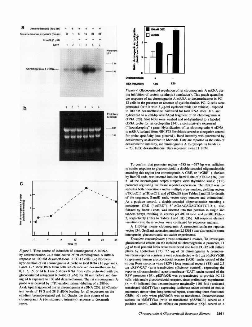
a Dexamethasone (100 nM) + + + + +
Dexamethasone exposure (hours) 0 1 5 15 24 24
RU-486 (1 pM) +
Lane 1 2 3 4 5 6
Northernblot
-25s
-18S
c
h
Ii£.E
Cyclohexdmide: +
DEXInduction: 1.95 2t30
1 2 3 4 5 6
Figure 4. Glucocorticoid regulation of rat chromogranin A mRNAdur-ing inhibition of protein synthesis (translation). This graph quantifiesthe response of rat chromogranin A mRNAto dexamethasone in PC-12 cells in the presence or absence of cycloheximide. PC-12 cells werepretreated for 6 h with 5 pg/ml cycloheximide (or vehicle), exposedto 100 nM dexamethasone, harvested for total RNAafter 18 h, and
Ethidium hybridized to a 288-bp Aval/Apal fragment of rat chromogranin Abromide
cDNA (20). Slot blots were washed and re-hybridized to a labeledcDNA probe for rat cyclophilin (34), a constitutively expressed
- 28S ("housekeeping") gene. Hybridization of rat chromogranin A cDNAto mRNAisolated from NIH 3T3 fibroblasts served as a negative control
18S for probe specificity (not pictured). Band intensity was quantitated bydensitometry as described in Methods. Data are reported as the ratio ofdensitometric intensity, rat chromogranin A to cyclophilin bands (n= 2). DEX, dexamethasone. Bars represent meant 1 SEM.
C
.4z
E: -
:0.
e
c..5
cg
3
2
A
e~~~~~~~~~~~~~~~~~~l.--
I I I I I0 4 a 12
Time (h)
Figure 3. Time course of induction of chromogrby dexamethasone. 24-h time course of rat chrorresponse to 100 nM dexamethasone in PC-12 cehybridization of rat chromogranin A probe to touLanes 1-5 show RNAfrom cells which receive0, 1, 5, 15, or 24 h. Lane 6 shows RNAfrom ceglucocorticoid antagonist RU-486 (1 pM) for 3(
ing 24 h exposure to 100 nM dexamethasone. TIprobe was derived by [32P] -random primer-labelAvaI/ApaI fragment of the rat chromogranin A cEtent levels of 18 S and 28 S rRNA loading for a
ethidium bromide-stained gel. (c) Graphs the tinchromogranin A (densitometric intensity) resporasone.
To confirm that promoter region -583 to -597 bp was sufficientto confer response to glucocorticoid, a double-stranded oligonucleotideencoding this region (rat chromogranin A GRE, or "rGRE"), flankedby BamHI ends, was inserted into the BamHI site of pTKluc (36), just5' of the heterologous herpes simplex virus thymidine kinase (TK)promoter regulating luciferase reporter expression. The rGRE was in-serted in both orientations and in multiple copy number, yielding vectorspTKlucC17, pTKlucCl9, and pTKlucE9 (see Tables I and HI for detailsof the sequence, BamHI ends, vector copy number and orientation).
v As a positive control, a double-stranded oligonucleotide encoding a
consensus GRE ("cGRE"; 5'-AGAACAGAGTGTTCT-3'), alsoflanked by BamHI ends, was inserted into this position in single andtandem arrays resulting in vectors pcGRETKluc- 1 and pcGRETKluc-2, respectively (refer to Tables I and III) (36). All response elementinsertions into these vectors were confirmed by sequence analysis.
A 1,133-bp mouse chromogranin A promoter/luciferase reportervector (34; GenBank accession number L3 1361 ) was also used in some
-J interspecies glucocorticoid activation experiments.16 20 24 Transient cotransfection (trans-activation) studies. To investigate
glucocorticoid effects on the isolated rat chromogranin A promoter, 11
,ug of total plasmid DNAwere transfected into 6-cm PC-12 cell cultureranin A mRNA plates by lipofection (37). 7.5 pg of rat chromogranin A promoter-mogranin A mRNA luciferase reporter constructs were cotransfected with 1 pg of pRSVhGRIlls. (a) Northern (expressing human glucocorticoid receptor [hGR] under control of theal RNA(10 pg/lane). strong Rous sarcoma virus [RSV] long terminal repeat) (38) and 2.5d dexamethasone for pg pRSV-CAT (as a transfection efficiency control), expressing thells pretreated with the reporter chloramphenicol acetyltransferase (CAT) under control of theD min before and dur- RSV promoter (39). pRSVhGRwas co-transfected to provide PC-12he rat chromogranin A cells with ample glucocorticoid receptor, since preliminary experimentsling of a 288-bp (n = 4) indicated that dexamethasone maximally (101-fold) activated)NA (20). (b) Consis- transfected pMMTVluc (expressing luciferase under control of mouse11 exposures on the mammary tumor virus long terminal repeat, containing three functionalne course of rat GREs; 40) only when pRSVhGRwas co-transfected. Dexamethasoneise to dexameth- actions on pMMTV1uc (with co-transfected pRSVhGR) served as a
positive control, while its effects on promoterless pXp2 served as a
Chromogranin A Glucocorticoid Response Element 2361
Chromogranin A mRNA->
b
14W!
a
0-
cz =.c 1.
ox sEos 0
. _
.1
4
3
21-
1
0 10 20Time of exposure to 100 nMdexamethasone (h)
Figure 5. Transcriptional activation of chromogranin A by glucocorti-coid. The graph quantitates the transcriptional response from n = 3nuclear runoff assays, with measurement of autoradiographic band inten-sity by densitometry. Nuclei from AtT-20 mouse anterior pituitary corti-cotrope cells were isolated after exposure to 100 nM dexamethasonefor 0, 8, or 24 h. Equivalent amounts of a- [32p] -UTP-labeled heteroge-neous nuclear RNA(hnRNA) were hybridized to slot blots containing5 ,ug of mouse chromogranin A genomic DNA(5.1 kb EcoRI/EcoRIfragment containing exons 1-3 and introns A-C) or pBluescriptKS -
DNA(negative control). hnRNA hybridized only to chromogranin Agenomic DNAin each experiment. Values given are relative intensity,normalized to basal rate of CgA transcription (that is, 0 h of exposure
to dexamethasone). * P < 0.01 (one-way ANOVA). Filled squares
with bars, mean+ SEM.
negative control. Each chromogranin A promoter/luciferase reportervector was evaluated in at least 2-3 separate transfections. For PCR-derived promoter fragments, two independently isolated amplificationproducts were tested. Luciferase activity was measured by luminometry(41), CATactivity was determined by '4C-acetylation of chlorampheni-col, with organic phase extraction (42), and cell protein was measuredby Coomassie blue dye-binding (43).
In some experiments, transfection results were normalized to thoseof pSV2ALA5 (wherein luciferase expression is driven by the SV40early promoter) (41 ).
Reagents. PC- 12 (rat adrenal chromaffin) cells, Cos (T antigen-transformed monkey kidney) cells, and AtT-20 (mouse anterior pituitarycorticotrope) cells were treated with the following reagents: the gluco-corticoid receptor agonist dexamethasone (100 nM; Sigma ChemicalCo., St. Louis, MO), the glucocorticoid receptor antagonist RU-486( I ,M; Roussel Uclaf, Paris, France), the mineralocorticoid receptorantagonist spironolactone (10 jiM; Sigma), or the protein synthesis(translation) inhibitor cycloheximide (5 jig/ml; Sigma).
Electrophoretic gel mobility shift studies. Nuclear extracts were pre-
pared from PC- 12 or Cos cells according to Dignam and Roeder (44).10-cm plates were transfected (37) with 10 jig pRSVhGRor controlDNAat 30% (Cos) or 50% (PC-12) confluence, then cultured in thepresence or absence of 100 nM dexamethasone, and harvested after 48hours. After phosphate buffered saline (PBS) washes at 0°C, cells were
pelleted for 5 min at 750 g at 4°C. Cell pellets were resuspended inthree volumes of modified Dignam solution A (10 mMHepes pH 7.9,1.5 mMMgCl2, 10 mMKCl, 0.5 mMDTT, 0.5 mMPMSF, with or
without 100 nMdexamethasone) and incubated for 15 min on ice. Cellswere lysed by passing the suspension five times through a 25 gauge
needle. Nuclei were isolated by a 30 second, 13,000 g micro-centrifuga-tion at 4°C. The pellet was resuspended on ice in one original volumeof modified Dignam C solution (10 mMHepes, pH 7.9, 25% glycerol,
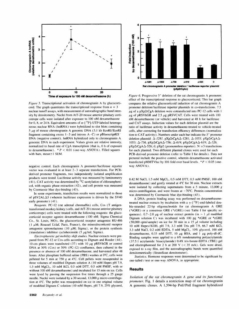
3
2
0
A-1281 A-1063 A-756 A-619 A-523 0
Rat chromogranin A promoter deletion / lucIfrase reporter plasmid(pXp2rCgAA)
Figure 6. Progressive 5' deletion of the rat chromogranin A promoter:effect of the transcriptional response to glucocorticoid. This bar graphcompares the relative glucocorticoid induction of rat chromogranin Apromoter deletion/luciferase reporter plasmids in co-transfections. 7.5,Ig of a pXp2rCgA deletion were cotransfected into PC-12 cells with I
,ug of pRSVhGRand 2.5 ,ug pRSVCAT. Cells were treated with 100nM dexamethasone (or vehicle) and harvested at 48 h for luciferaseand CAT assays. Induction values for each deletion plasmid are theratio of luciferase activity in dexamethasone-treated to vehicle-treatedcells, after correcting for transfection efficency differences (normaliza-tion to CATactivity). Numbers under each bar indicate the 5' promoterdeletion plasmid: A-1281, pXp2rCgAA-1281; A-1053, pXp2rCgAA\-1053; A\-756, pXp2rCgAA-756; A-619, pXp2rCgAZA-619; A-526,pXp2rCgAA-526; 0, pXp2 (promoterless reporter). N .3 transfectionsfor each plasmid. Two different plasmid clones were used for eachPCR-derived promoter deletion (refer to Table I for details). Data not
pictured include the positive control, wherein dexamethasone activatedtransfected pMMTVluc by 101-fold over basal levels. *P < 0.05 (one-way ANOVA).
0.42 MNaCl, 1.5 mMMgC92, 0.5 mMDTT, 0.5 mMPMSF, 100 nMdexamethasone) and gently rotated at 4°C for 30 min. Nuclear extractswere isolated by collecting supernatants from a 5 minute, 13,000 g
micro-centrifugation, and were frozen at -70°C. Protein concentrationwas determined by Coomassie blue dye-binding (43).
A DNA: protein binding assay was performed on dexamethasone-treated nuclear extracts by incubation with a y- [32P]-end-labeled dou-ble-stranded 22-bp oligonucleotide for rat chromogranin A GRE(*rGRE) or a consensus GRE(*cGRE) (see Table I for specific se-
quences). 0.7-2.0 ,ug of nuclear extract protein (in I [u1 modifiedDignam solution C) was incubated with 100 pg *rGRE or *cGRE(50,000 cpm/sample) on ice for 30 min in a 15 ,ul volume including10 mMHepes/KOH, pH 7.9, 3.3 mMTris pH 7.9, 66.7 mMKCl,3.3 mMNaCI, 0.2 mMEDTA, 5 mMMgCl2, 10% glycerol, 100 nMdexamethasone, 0.33 mMDTT, 10 jg BSA, and 1 jg poly-dI-dC.Binding samples were applied to a 6% nondenaturing polyacrylamide(37.5:1 acrylamide: bisacrylamide) 0.4X tris-borate-EDTA (TBE) geland electrophoresed for 2 h at 200 V (< 35 mA). Gels were dried,exposed to x-ray film, and the autoradiographic bands were quantifieddensitometrically (StratoScan densitometer).
Statistics. Hormone responses were determined to be significant byone-tailed t test or one-way ANOVA, as appropriate.
Results
Isolation of the rat chromogranin A gene and its functionalpromoter. Fig. 1 details a restriction map of rat chromograninA genomic clones. A 1,594-bp PstI/PstI fragment hybridized
2362 Rozansky et al.
* ~ ~--~~~~~~~~~~l-
/ Ns/ NNj
- ,. "T~I
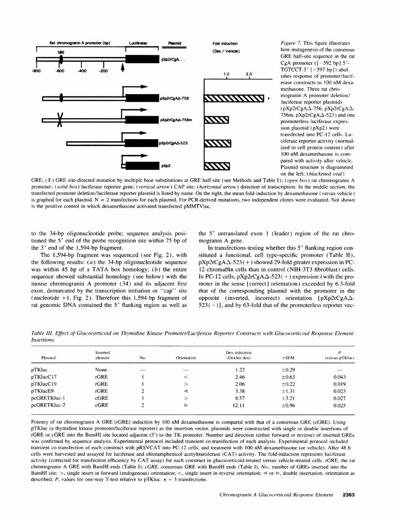
Rat chromogranin A promoter (bp) Luciferse Ptasmid Fol induction Figure 7. This figure illustrates
GRE I (Dex / Vehicie) how mutagenesis of the consensus
I * pXp2rCgA.. GREhalf-site sequence in the ratXr
2|* CgA promoter ([-592 bp] 5'-
-S0 -60 oo -200 TGTCCT-3' [-597 bp]) abol-1.0 2.0 ishes response of promoter/lucif-
erase constructs to 100 nM dexa-methasone. Three rat chro-
pXp2rCgA|-756 * mogranin A promoter deletion/luciferase reporter plasmids(pXp2rCgAz-756, pXp2rCgAz\-
/ 756m, pXp2rCgAz\-523) and onepXp2rCgA&-7S6m promoterless luciferase expres-
sion plasmid (pXp2) wereX4111 transfected into PC-12 cells. Lu-
pXP2rCgAA-523 ciferase reporter activity (normal-ized to cell protein content) after100 nM dexamethasone is com-
pared with activity after vehicle.pXp2 Plasmid structure is diagrammed
on the left: (blackened oval)GRE; (X) GREsite-directed mutation by multiple base substitutions at GREhalf-site (see Methods and Table I); (open box) rat chromogranin Apromoter; (solid box) luciferase reporter gene; (vertical arrow) CAP site; (horizontal arrow) direction of transcription. In the middle section, thetransfected promoter deletion/luciferase reporter plasmid is listed by name. On the right, the mean fold-induction by dexamethasone (versus vehicle)is graphed for each plasmid. N 2 2 transfections for each plasmid. For PCR-derived mutations, two independent clones were evaluated. Not shownis the positive control in which dexamethasone activated transfected pMMTVluc.
to the 34-bp oligonucleotide probe; sequence analysis posi-tioned the 5' end of the probe recognition site within 75 bp ofthe 3' end of the 1,594-bp fragment.
The 1,594-bp fragment was sequenced (see Fig. 2), withthe following results: (a) the 34-bp oligonucleotide sequencewas within 45 bp of a TATA box homology; (b) the entiresequence showed substantial homology (see below) with themouse chromogranin A promoter (34) and its adjacent firstexon, demarcated by the transcription initiation or "cap" site(nucleotide +1, Fig. 2). Therefore this 1,594-bp fragment ofrat genomic DNAcontained the 5' flanking region as well as
the 5' untranslated exon 1 (leader) region of the rat chro-mogranin A gene.
In transfections testing whether this 5' flanking region con-stituted a functional, cell type-specific promoter (Table II),pXp2rCgAzX-523 ( + ) showed 29-fold greater expression in PC-12 chromaffin cells than in control (NIH-3T3 fibroblast) cells.In PC- 12 cells, pXp2rCgAA-523 ( + ) expression (with the pro-moter in the sense [correct] orientation) exceeded by 6.3-foldthat of the corresponding plasmid with the promoter in theopposite (inverted, incorrect) orientation [pXp2rCgAA-523 ( -) ], and by 63-fold that of the promoterless reporter vec-
Table III. Effect of Glucocorticoid on Thymidine Kinase Promoter/Luciferase Reporter Constructs with Glucocorticoid Response ElementInsertions
Inserted Dex induction PPlasmid element No. Orientation (Dex/no dex) +SEM (versus pTKluc)
pTKluc None 1.22 10.29pTKlucC 17 rGRE I < 2.46 +0.63 0.043pTKlucCl9 rGRE 1 > 2.06 +0.22 0.019pTKlucE9 rGRE 2 < 3.38 +11.31 0.023pcGRETKluc- 1 cGRE I > 8.57 1 3.21 0.027pcGRETKluc-2 cGRE 2 > 12.11 10.96 0.025
Potency of rat chromogranin A GRE(rGRE) induction by 100 nM dexamethasone is compared with that of a consensus GRE(cGRE). UsingpTKluc (a thymidine kinase promoter/luciferase reporter) as the insertion vector, plasmids were constructed with single or double insertions ofrGRE or cGRE into the BamHI site located adjacent (5') to the TK promoter. Number and direction (either forward or reverse) of inserted GREswas confirmed by sequence analysis. Experimental protocol included transient co-transfection of each analysis. Experimental protocol includedtransient co-transfection of each construct with pRSVCATinto PC- 12 cells, and treatment with 100 nM dexamethasone (or vehicle). After 48 hcells were harvested and assayed for luciferase and chloramphenicol acetyltransferase (CAT) activity. The fold-induction represents luciferaseactivity (corrected for transfection efficiency by CAT assay) for each construct in glucocorticoid-treated versus vehicle-treated cells. rGRE, the ratchromogranin A GREwith BamHI ends (Table I); cGRE, consensus GREwith BamHI ends (Table I); No., number of GREs inserted into theBamHI site; >, single insert in forward (endogenous) orientation; <, single insert in reverse orientation; < or >, double insertation, orientation asdescribed; P, values for one-way T-test relative to pTKluc. n = 3 transfections.
Chromogranin A Glucocorticoid Respon.se Element 2363
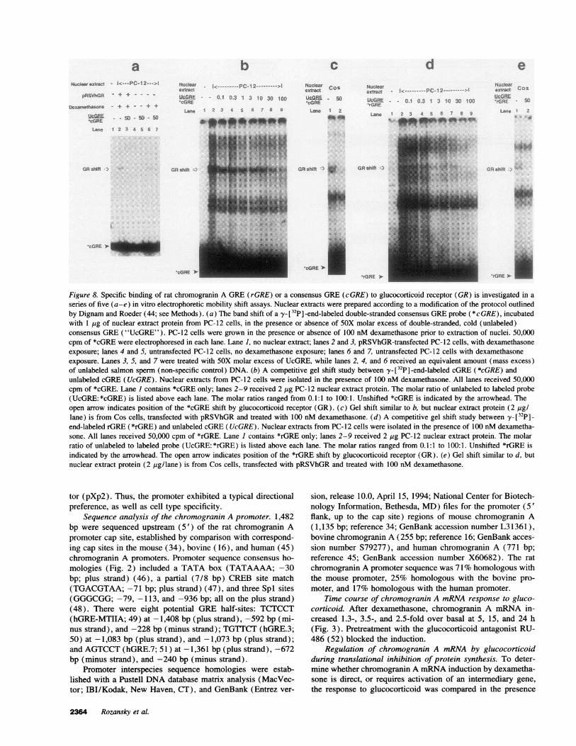
aNucleareatract - I<---PC-12 ---I
pRSVhGR - + + - - - -
Dexamethasone
'cGRE
Lane
- - 50 - 50 - 50
1 2 3 4 5 6 7
GRshitt )
bNuclear i< PC-12 - >1extract
cg-GRE - 0.1 0.3 1 3 10 30 100.cGRE
Lane I 2 3 4 5 6 7 8a
W-p .kJo...
, 4
GRshift )
CNuclear Cos
extract
UcRE 50'cGRE
Lane 1 2
GRshift ) w
IcGfRE _
dNuclearextract .c.- PC-12
-V-i3REE 0.1 0.3 1 3 10 30 100*rGRE
Lane 1 2 3 4 5 6 7 8 9
GR shift
cGRE IJ_
'cGRE 'I 'rGRE
Figure 8. Specific binding of rat chromogranin A GRE(rGRE) or a consensus GRE(cGRE) to glucocorticoid receptor (GR) is investigated in a
series of five (a-e) in vitro electrophoretic mobility shift assays. Nuclear extracts were prepared according to a modification of the protocol outlinedby Dignam and Roeder (44; see Methods). (a) The band shift of a y- [32P] -end-labeled double-stranded consensus GREprobe (*cGRE), incubatedwith 1 ,ug of nuclear extract protein from PC-12 cells, in the presence or absence of 5OX molar excess of double-stranded, cold (unlabeled)consensus GRE("UcGRE"). PC-12 cells were grown in the presence or absence of 100 nM dexamethasone prior to extraction of nuclei. 50,000cpm of *cGRE were electrophoresed in each lane. Lane 1, no nuclear extract; lanes 2 and 3, pRSVhGR-transfected PC-12 cells, with dexamethasoneexposure; lanes 4 and 5, untransfected PC-12 cells, no dexamethasone exposure; lanes 6 and 7, untransfected PC-12 cells with dexamethasoneexposure. Lanes 3, 5, and 7 were treated with 5OX molar excess of UcGRE, while lanes 2, 4, and 6 received an equivalent amount (mass excess)of unlabeled salmon sperm (non-specific control) DNA. (b) A competitive gel shift study between y- [32P] -end-labeled cGRE (*cGRE) andunlabeled cGRE(UcGRE). Nuclear extracts from PC-12 cells were isolated in the presence of 100 nM dexamethasone. All lanes received 50,000cpm of *cGRE. Lane I contains *cGRE only; lanes 2-9 received 2 Mg PC-12 nuclear extract protein. The molar ratio of unlabeled to labeled probe(UcGRE: *cGRE) is listed above each lane. The molar ratios ranged from 0.1:1 to 100:1. Unshifted *cGRE is indicated by the arrowhead. Theopen arrow indicates position of the *cGRE shift by glucocorticoid receptor (GR). (c) Gel shift similar to b, but nuclear extract protein (2 /sg/lane) is from Cos cells, transfected with pRSVhGRand treated with 100 nM dexamethasone. (d) A competitive gel shift study between y_ [32p] _
end-labeled rGRE (*rGRE) and unlabeled cGRE(UcGRE). Nuclear extracts from PC-12 cells were isolated in the presence of 100 nMdexametha-sone. All lanes received 50,000 cpm of *rGRE. Lane I contains *rGRE only; lanes 2-9 received 2 ,g PC-12 nuclear extract protein. The molarratio of unlabeled to labeled probe (UcGRE: *rGRE) is listed above each lane. The molar ratios ranged from 0.1:1 to 100:1. Unshifted *rGRE isindicated by the arrowhead. The open arrow indicates position of the *rGRE shift by glucocorticoid receptor (GR). (e) Gel shift similar to d, butnuclear extract protein (2 ,ug/lane) is from Cos cells, transfected with pRSVhGRand treated with 100 nM dexamethasone.
tor (pXp2). Thus, the promoter exhibited a typical directionalpreference, as well as cell type specificity.
Sequence analysis of the chromogranin A promoter. 1,482bp were sequenced upstream (5') of the rat chromogranin Apromoter cap site, established by comparison with correspond-ing cap sites in the mouse (34), bovine (16), and human (45)chromogranin A promoters. Promoter sequence consensus ho-mologies (Fig. 2) included a TATA box (TATAAAA; -30bp; plus strand) (46), a partial (7/8 bp) CREB site match(TGACGTAA; -71 bp; plus strand) (47), and three Spl sites(GGGCGG;-79, -113, and -936 bp; all on the plus strand)(48). There were eight potential GRE half-sites: TCTCCT(hGRE-MTIIA; 49) at -1,408 bp (plus strand), -592 bp (mi-nus strand), and -228 bp (minus strand); TGTTCT(hGRE.3;50) at -1,083 bp (plus strand), and -1,073 bp (plus strand);and AGTCCT(hGRE.7; 51) at -1,361 bp (plus strand), -672bp (minus strand), and -240 bp (minus strand).
Promoter interspecies sequence homologies were estab-lished with a Pustell DNAdatabase matrix analysis (MacVec-tor; IBI/Kodak, New Haven, CT), and GenBank (Entrez ver-
sion, release 10.0, April 15, 1994; National Center for Biotech-nology Information, Bethesda, MD) files for the promoter (5'flank, up to the cap site) regions of mouse chromogranin A(1,135 bp; reference 34; GenBank accession number L31361),bovine chromogranin A (255 bp; reference 16; GenBank acces-
sion number S79277), and human chromogranin A (771 bp;reference 45; GenBank accession number X60682). The rat
chromogranin A promoter sequence was 71 %homologous withthe mouse promoter, 25% homologous with the bovine pro-
moter, and 17% homologous with the human promoter.Time course of chromogranin A mRNAresponse to gluco-
corticoid. After dexamethasone, chromogranin A mRNAin-creased 1.3-, 3.5-, and 2.5-fold over basal at 5, 15, and 24 h(Fig. 3). Pretreatment with the glucocorticoid antagonist RU-486 (52) blocked the induction.
Regulation of chromogranin A mRNAby glucocorticoidduring translational inhibition of protein synthesis. To deter-mine whether chromogranin A mRNAinduction by dexametha-sone is direct, or requires activation of an intermediary gene,the response to glucocorticoid was compared in the presence
2364 Rozansky et al.
eNuclearextract COS
'rGRE O51Lane 2
*4iA
GRshitt l
I. ",
*. ....:f
.rGRE *
and absence of translation inhibition by cycloheximide (Fig. 4).Dexamethasone augmented chromogranin A gene expressionby 1.95-fold in the presence and 2.3-fold in the absence ofcycloheximide; therefore, the response to glucocorticoid did notrequire new protein synthesis.
Transcriptional (nuclear runoff) studies. To test whetherthe chromogranin A mRNAincrease (Fig. 5) is a transcriptionalresponse to glucocorticoid, three independent nuclear runoffstudies showed a significant increase in new chromogranin AhnRNA transcripts after 100 nM dexamethasone-by 3.3-foldat 8 h and 1.8-fold at 24 h.
Glucocorticoid effects on chromogranin Apromoter-lucifer-ase reporter constructs. To define the glucocorticoid-responsiveregion of the rat chromogranin A promoter, promoter deletion/luciferase reporter plasmids were transfected (Fig. 6). Reporterexpression was increased 2.6-3.1 -fold after glucocorticoid.There was a significant dropoff in glucocorticoid response be-tween positions -619 bp (plasmid pXp2rCgAL\-619) and -523bp (plasmid pXp2rCgAA-523).
A transfected mouse chromogranin A 1,133-bp promoter/luciferase reporter construct (34) (GenBank accession numberL31361) was also activated 2.52-fold (light units/mg protein;n = 4 replicates; P < 0.05) by 10-6 Mdexmethasone in PC12cells.
In a dose-response study from 10-12 to 10-5 Mdexametha-sone, transfected promoters of both chromogranin A and mousemammarytumor virus (pMMTVluc [40]) were each maximallyactivated after I0-7 Mdexamethasone.
Specificity of glucocorticoid action on the chromograninA promoter. Since dexamethasone is also a weak agonist atmineralocorticoid receptors (53), the response of thetransfected chromogranin A promoter (pXp2rCgAA-1281) todexamethasone (100 nM) was studied in the presence of gluco-corticoid receptor- and mineralocorticoid receptor-specific an-tagonists (added 30 min before agonist). The response to dexa-methasone was completely blocked by the glucocorticoid antag-onist RU-486 (1 iM), but was not affected by themineralocorticoid antagonist spironolactone (10 ,uM); in theabsence of agonist, neither of these antagonists affected pro-moter/reporter expression (data not shown).
Isolation of a glucocorticoid response element from the ratchromogranin A promoter. Sequence analysis of the rat pro-moter between -523 and -619 bp revealed a consensus matchfor a glucocorticoid receptor-binding half-site at (-597 bp) 5'-AGGACA-3' (-592 bp) (on the opposite strand: [-592 bp]5 '-TGTCCT-3' [-597 bp]). To test the function of this rGREmotif, PC-12 cells were transfected with pXp2rCgAA-756(which contains the wild-type rGRE motif) versuspXp2rCgAA-756m (with a substitution mutation at the rGREmotif, [-597 bp] 5'-gcGgtA-3' [-592 bp]; see Methods andTable I), and treated with dexamethasone or vehicle (Fig. 7).Only wild-type pXp2rCgA/\-756 responded significantly (2.44-fold, P = 0.005) to dexamethasone. No dexamethasone re-sponse was found for pXp2rCgAA-756m, or for the negativecontrols pXp2rCgALA-523 (which lacks sequence upstream of-523 bp) or the promoterless reporter pXp2.
A 15-bp sequence ([-597 bp] 5 '-AGGACACACTCATGT-3' [-583 bp] or "rGRE"; consensus half-site in bold; on theopposite strand: [-583 bp ] 5 '-ACATGAGTTGTCCT-3' [-597bp]), corresponding in length to the consensus 15-bp glucocor-ticoid receptor homodimer-binding GREmotif (49, 54, 55),was inserted into the heterologous promoter/luciferase reporter
--% \1 I 9~*cGRE*575
50-
25-
0
-1 0 1 2
log 2GREvGRE
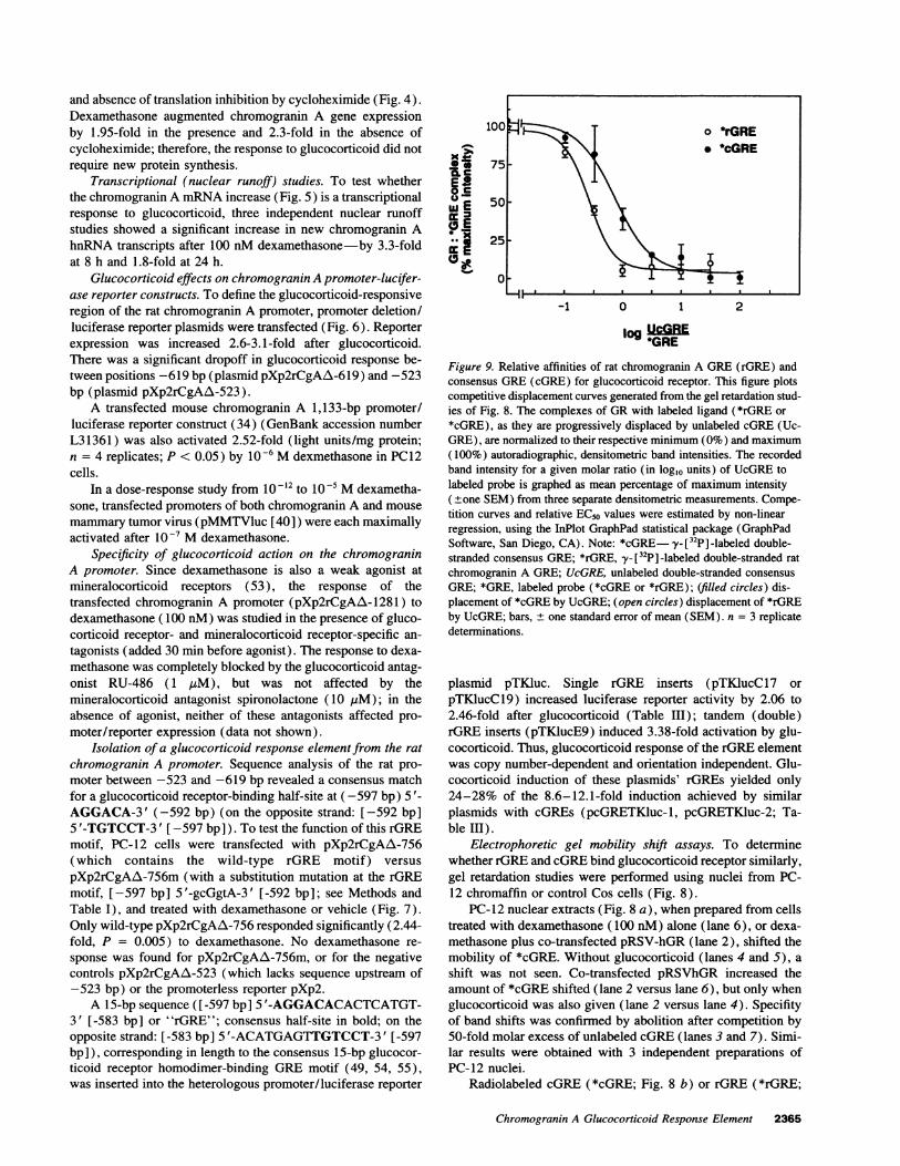
Figure 9. Relative affinities of rat chromogranin A GRE(rGRE) andconsensus GRE(cGRE) for glucocorticoid receptor. This figure plotscompetitive displacement curves generated from the gel retardation stud-ies of Fig. 8. The complexes of GRwith labeled ligand (*rGRE or*cGRE), as they are progressively displaced by unlabeled cGRE(Uc-GRE), are normalized to their respective minimum (0%) and maximum(100%) autoradiographic, densitometric band intensities. The recordedband intensity for a given molar ratio (in log10 units) of UcGREtolabeled probe is graphed as mean percentage of maximum intensity(±one SEM) from three separate densitometric measurements. Compe-tition curves and relative EC50 values were estimated by non-linearregression, using the InPlot GraphPad statistical package (GraphPadSoftware, San Diego, CA). Note: *cGRE y-_[32P] -labeled double-stranded consensus GRE; *rGRE, y-[32P] -labeled double-stranded ratchromogranin A GRE; UcGRE, unlabeled double-stranded consensusGRE; *GRE, labeled probe (*cGRE or *rGRE); (filled circles) dis-placement of *cGRE by UcGRE; (open circles) displacement of *rGREby UcGRE; bars, ± one standard error of mean (SEM). n = 3 replicatedeterminations.
plasmid pTKluc. Single rGRE inserts (pTKlucCl7 orpTKlucCl9) increased luciferase reporter activity by 2.06 to2.46-fold after glucocorticoid (Table III); tandem (double)rGRE inserts (pTKlucE9) induced 3.38-fold activation by glu-cocorticoid. Thus, glucocorticoid response of the rGRE elementwas copy number-dependent and orientation independent. Glu-cocorticoid induction of these plasmids' rGREs yielded only24-28% of the 8.6-12.1-fold induction achieved by similarplasmids with cGREs (pcGRETKluc-1, pcGRETKluc-2; Ta-ble III).
Electrophoretic gel mobility shift assays. To determinewhether rGRE and cGREbind glucocorticoid receptor similarly,gel retardation studies were performed using nuclei from PC-12 chromaffin or control Cos cells (Fig. 8).
PC-12 nuclear extracts (Fig. 8 a), when prepared from cellstreated with dexamethasone (100 nM) alone (lane 6), or dexa-methasone plus co-transfected pRSV-hGR (lane 2), shifted themobility of *cGRE. Without glucocorticoid (lanes 4 and 5), ashift was not seen. Co-transfected pRSVhGR increased theamount of *cGRE shifted (lane 2 versus lane 6), but only whenglucocorticoid was also given (lane 2 versus lane 4). Specifityof band shifts was confirmed by abolition after competition by50-fold molar excess of unlabeled cGRE(lanes 3 and 7). Simi-lar results were obtained with 3 independent preparations ofPC-12 nuclei.
Radiolabeled cGRE (*cGRE; Fig. 8 b) or rGRE (*rGRE;
Chromogranin A Glucocorticoid Response Element 2365
Table IV. Comparison of Rat Chromogranin A Glucocorticoid Response Element (GRE) to other GREs
Sequence
GREtype 5' bp 1 6 7 9 10 15 3' bp Homology Reference
Consensus AGAACA n n n TGTTCT (54)hGRE-MTIIA -263 GGTACA c t g TGTCCT -249 (49)Rat CgA GRE -583 AcAt gA g t g TGTCCT -597 9/12 Present workANFGREI -963 G c c t g t t t g TGTTCT -949 7/12 (60)ANFGREII -884 t c T c t g t a a TGTCCT -898 7/12 (60)PEPCKGREI -378 c a c a C A a a a TGTgCa -364 6/12 (61)PEPCKGREII -367 AGcAtA t g a a G T C C a -353 8/12 (61)
This table compares homology of GREs that show strong response (consensus, and human metalothionine-ILA [hGRE-MTIIA]) to glucocorticoid,versus GREs that show a more moderate (2-4-fold) response. Strong response GREs are shown in bold and aligned according to GREhalf-sitemotifs. They serve as indexes to which homology for less responsive GREscan be compared. Moderately responsive GREs from rat chromograninA (CgA), atrial natriuretic factor (ANF), and phosphoenolpyruvate carboxykinase (PEPCK) genes are aligned below the index GREsby placingthat gene's GREhalf-site motif with stronger homology to the sequences 5'-TGTTCT-3' or 5'-TGTCCT-3' at positions 10 through 15. Fraction ofhomology for moderate GREs is based on exact conservation (upper case lettering) with the base from either index (strongly responsive) GREforpositions 1 through 6 and 10 through 15. Negative numbers flanking GREs indicate position within each GRE's promoter. Among moderatelyresponsive GREs, rat CgA GREshows the highest homology (9/12) to the index GREs, while PEPCKand ANF require cooperativity between twoseparate 15-bp motifs (for example, ANFGREI plus ANFGREII) to achieve their moderate responses to glucocorticoid.
Fig. 8 d) each bound and was shifted by glucocorticoid receptorfrom PC-12 nuclei. The fractional band shift (shifted band mo-bility/free band mobility) in the same gel was identical for both*cGRE and *rGRE. Unlabeled cGRE(UcGRE) displaced both*cGRE (Fig. 8, b and c) and *rGRE (Fig. 8, d and e) fromglucocorticoid receptor, whether the receptor was from PC-12nuclei (Fig. 8, b and d) or (pRSVhGR-transfected-) Cos nuclei(Fig. 8, c and e). Unlabeled nonspecific (salmon sperm) DNA,at 50-fold mass excess, had no effect on the shifted *rGREband mobility.
As the molar ratio of UcGRE/*cGRE (Fig. 8 b) or UcGRE/*rGRE (Fig. 8 d) was progressively increased from 0.1 to100, *cGRE and *rGRE were competitively displaced fromglucocorticoid receptor in PC-12 nuclei. The relative affinities(EC5os) of *rGRE and *cGRE for glucocorticoid receptor wereestimated by nonlinear regression analysis (GraphPad InPlotcompetition curve; GraphPad Software, Inc., San Diego, CA)of the logl0 molar displacement curves (Fig. 9). *rGRE had2.75-fold (= antilog10 0.44) lower affinity for glucocorticoidreceptor than *cGRE.
Discussion
Isolation of a functional GREin the rat CgA gene. To determinethe mechanism by which glucocorticoids augment chro-mogranin A expression, we focused on transcriptional regula-tion of the gene, since glucocorticoid induction of chromograninA protein in vitro and in vivo parallels induction of chro-mogranin A mRNA(14-17). The investigation initially con-centrated its efforts on isolating the 5' regulatory region (pro-moter) of the rat chromogranin A gene (Figs. 1 and 2, andTable II), and determining the extent to which glucocorticoidsactivated chromogranin A expression at a transcriptional (pre-translational) level. We found that glucocorticoid activatedchromogranin A gene expression up to 3.5-fold (Fig. 3), thatthe response did not require new protein synthesis (Fig. 4),and involved a 3.3-fold increase in rate of initiation of newchromogranin A transcripts (Fig. 5).
A novel glucocorticoid response element ([-583 bp] 5'-ACATGAGTGTGTCCT-3'[-597 bp]) bound glucocorticoidreceptors (Fig. 8). Functional properties of this novel rGREincluded: (a) a 2.6- to 3.1-fold increment in promoter activityin response to glucocorticoid was lost after deletion of thisregion (Fig. 6); (b) transfer of the motif to a heterologouspromoter yielded 2.06-3.38-fold glucocorticoid induction (Ta-ble III); and (c) site-directed mutation of the motif abolishedthe glucocorticoid response (Fig. 7).
Evidence that this rGRE exerts its effects through a selectiveinteraction with ligand-activated glucocorticoid receptor ema-nated from two studies: dexamethasone induction was blockedby a glucocorticoid antagonist but not a mineralocorticoid an-tagonist, and the rGRE specifically bound ligand-activated glu-cocorticoid receptor in vitro (Fig. 8).
Glucocorticoid activation of the rat CgA gene. Glucocorti-coids trans-activate many genes at the level of transcription(56 and references therein). Ligand-activated glucocorticoidreceptor homodimers bind characteristic DNA response ele-ments (GREs). Mutagenesis of the glucocorticoid receptor (36)DNAbinding domain indicates that zinc finger motifs (com-posed of two sets of four cysteine residues per monomer) con-tain specific amino acids critical for DNAbinding specificityto a GRE. X-ray crystallography (57) further established thateach monomer in the homodimer interfaces with specific basesin the major groove of the double-stranded DNA, for high affin-ity binding.
Glucocorticoid receptor homodimers cooperatively bind fulllength, 15-bp GREs with at least 10-fold greater affinity thantheir attraction for GREhalf-sites (58, 59). Since the 15-bprGRE motif bound glucocorticoid receptor at only 2.75-foldlower affinity than the 15 bp cGRE(Fig. 9), even the degeneratehalf-site within the rGRE ([-583 bp] 5'-ACATGA-3' [-588bp ] ) may provide sufficient affinity to participate in cooperativebinding of the glucocorticoid receptor homodimer.
Functionally, rGRE mediated 2-3.5-fold increments in geneexpression after glucocorticoid, in several contexts (Figs. 3-6). In a direct comparison (Table III) of isolated cGREversus
2366 Rozansky et al.
rGRE effects, cGREcaused 3.6-4.2-fold greater glucocorticoidinduction than rGRE, a value consistent with the 2.75-fold af-finity differences of these motifs for glucocorticoid receptor(Fig. 9).
Inspection of the rGRE sequence ([-583 bp] 5 '-ACATGA-GTGTGTCCT-3' [-597 bp]; Table IV) reveals a consensus(49) GREhalf-site ([-592 bp] 5'-TGTCCT-3' [-597 bp]) anda degenerate half-site ([-588 bp] 5'-TCATGT-3' [-583 bp]).There are ample precedents for such imperfect (degenerate fromconsensus) GREswith preserved (though attenuated) responseto glucocorticoid. Atrial naturetic factor (ANF) and phospho-enolpyruvate carboxypeptidase (PEPCK) genes, each of whichdisplay 2-4-fold stimulation responses to glucocorticoid, haveGREs which are even more degenerate from consensus thanthe rGRE (60, 61; Table IV).
Functional GREshave not been isolated from other species'chromogranin A genes, although the transfected mouse chro-mogranin A 1133 bp promoter/luciferase reporter responded2.52-fold to glucocorticoid (see Results). In the region of themouse (34; GenBank accession number L31361) chromograninA promoter ([ -583 bp ] -5 '-ACATGGGTGGGTCCT-3' [ -597bp]) corresponding to the rGRE, 13/15 bp are identical (inbold) to those in the rGRE. The first 1,135 reported bp ofthe mouse chromogranin A promoter (34; GenBank accessionnumber L31361) also have another GREhalf-site (AGTCCT;hGRE.7; 51) match at position -679 bp (minus strand). Thefirst 255 reported bp of the bovine chromogranin A promoter(16; GenBank accession number S79277) contain one GREhalf-site match (AGTCCT; hGRE.7; 51) at position -230 bp(plus strand). The first 771 reported bp of the human chro-mogranin A promoter (45; GenBank accession numberX60682) contain no GREhalf-site matches.
Biological significance of the chromogranin A response toglucocorticoids. A similar degree of glucocorticoid activationof transfected rat and mouse chromogranin A promoters (2.6-to 3.1-fold, versus 2.52-fold), coupled with GREsequence ho-mologies in mouse, rat, and bovine chromogranin A promoters,suggest that chromogranin A GREs may be of general func-tional importance in mammalian species.
One function of chromogranin A is its action to complexor osmotically inactivate cations such as calcium and catechola-mines within the catecholamine storage vesicle core (8). Sinceglucocorticoid exposure also augments catecholamine storagein chromaffin cells by 2- to 4-fold (62-64), a parallel risein co-stored chromogranin A may provide additional bindingcapacity for glucocorticoid-stimulated increases in vesicularcatecholamine stores (8). Zhang et al. (65) have also shownthat the effects of glucocorticoid on the chromogranin A mRNAdepend on the prevailing extracellular calcium concentration.
Once secreted, chromogranin A proteolytic fragments areactive in the extracellular space, modulating further catechola-mine release from chromaffin cells (12). Thus, an incrementin chromogranin A may also provide a homeostatic or negative-feedback "brake" on release of steroid-augmented catechola-mine stores.
Acknowledgments
Weappreciate the technical assistance of Brian Strauss and Karen Shiao.This work was supported by the Department of Veterans Affairs,
the American Heart Association, and the National Institutes of Health.
References1. Anderson, D., and A. Mendelsohn. 1989. Role of glucocorticoids in the
chromaffin-neuron development decision. Int. J. Dev. Neurosci. 7:475-487.2. Stachowiak, M. K., J. S. Hong, and 0. H. Viveros. 1990. Coordinate
and differential regulation of phenylethanolamine-N-methyltransferase, tyrosinehydroxylase and proenkephalin mRNAsby neural and hormonal mechanisms incultured bovine adrenal medullary cells. Brain Res. 510:277-288.
3. Ross, M. E., M. J. Evinger, S. E. Hyman, J. M. Carroll, L. Mucke, M.Comb, D. J. Reis, T. H. Joh, and H. M. Goodman. 1990. Identification of a
functional glucocorticoid response element in the phenylethanolamine N-methyl-transferase promoter using fusion genes introduced into chromaffin cells in pri-mary culture. J. Neurosci. 10:520-530.
4. Lewis, E. J., C. A. Harrington, and D. M. Chikaraishi. 1987. Transcriptionalregulation of the tyrosine hydroxylase gene by glucocorticoid and cyclic AMP.Proc. Natl. Acad. Sci. USA. 84:3550-3554.
5. Tischler, A. S., R. L. Perlman, G. M. Morse, and B. E. Sheard. 1978.Glucocorticoids increase catecholamine synthesis and storage in PC- 12 pheochro-mocytoma cell cultures. J Neurochem. 40:364-370.
6. Winkler, H., and R. Fischer-Colbrie. 1992. The chromogranins A and B:the first 25 years and future perspectives. Neuroscience. 49:497-528.
7. Eiden, L., W. Huttner, J. Mallet, D. T. O'Connor, H. Winkler, and A.Zamini. 1987. Nomenclature proposal for the chromogranin secretogranin pro-
teins. Neuroscience. 21:1019-1021.8. Videen, J. S., M. S. Metzger, Y.-M. Chang, and D. T. O'Connor. 1992.
Calcium and catecholamine interactions with adrenal chromogranins: comparisonof driving forces in binding and aggregation. J. Biol. Chem. 267:3066-3073.
9. Reiffen, F. U., and M. Gratzl. 1986. Chromogranins, widespread in endo-crine and nervous tissue, bind calcium. FEBS (Fed. Eur. Biochem. Soc.) Lett.195:327-330.
10. Seidah, N. G., G. N. Hendy, J. Hamelin, J. Paquin, C. Lazure, K. M.Metters, J. Rossier, and M. Chretien. 1987. Chromogranin A can act as a reversibleprocessing enzyme inhibitor. Evidence form inhibition of the IRCM-serine prote-ase 1 cleavage of proenkephalin and ACTHat pairs of basic amino acids. FEBS(Fed. Eur. Biochem. Soc.) Lett. 211:144-150.
11. Iacangelo, A., R. Fischer-Colbrie, K. J. Koller, M. J. Brownstein, andL. E. Eiden. 1988. The sequence of porcine chromogranin A can serve as theprecursor for the biologically active hormone, pancreastatin. Endocrinology.122:2339-2341.
12. Simon, J.-P., M.-F. Bader, and D. Aunis. 1988. Secretion from chromaffincells is controlled by chromogranin A-derived peptides. Proc. Natl. Acad. Sci.USA. 85:1712-1716.
13. Barbosa, J. A., B. M. Gill, M. A. Takiyyuddin, and D. T. O'Connor. 1991.Chromogranin A: post-transtranslational modifications in secretory granules. En-docrinology. 128:174-180.
14. Sietzen, M., M. Schober, R. Fischer-Colbrie, D. Scherman, G. Sperk, andH. Winkler. 1987. Rat adrenal medulla: levels of chromogranins, dopamine-beta-hydroxylase and of the amine transporter are changed by nervous activity andhypophysectomy. Neuroscience. 22:131-139.
15. R. Fischer-Colbie, A. lacangelo, and L. Eiden. 1988. Neural and humoralfactors separately regulate neuropeptide Y, enkephalin, and chromogranin A andB mRNAlevels in rat adrenal medulla. Proc. Natl. Acad. Sci. USA. 85:3240-3244.
16. lacangelo, A., M. Grimes, and L. E. Eiden. 1991. The bovine chromograninA gene: structural basis for hormone regulation and generation of biologicallyactive peptides. Mol. Endocrinol. 5(11): 1651-1660.
17. Rausch, D. M., A. Iacangelo, and L. E. Eiden. 1988. Glucocorticoid andNGFinduced changes in chromogranin A expression define two different neuronalphenotypes in PC-12 cells. Mol. Endocrinol. 2:921-927.
18. Evans, G. A., K. Lewis, and B. E. Rothenberg. 1989. High efficencyvectors for cosmid microcloning and genomic microanalysis. Gene. 79:9-20.
19. Evans, G. A., and G. M. Wahl. 1987. Cosmid vectors for genomic walkingand rapid restriction mapping. Methods Enzymol. 152:604-610.
20. Parmer, R. J., A. H. Koop, M. T. Handa, and D. T. O'Connor. 1989.Molecular cloning of chromogranin A from rat pheochromocytoma cells. Hyper-tension. 14:435-444.
21. lacangelo, A., H. Okayama, and L. E. Eiden. 1988. Primary structure ofthe rat chromogranin A and distribution of its mRNA.FEBS (Fed. Eur. Biochem.Soc.) Lett. 227:115-121.
22. Sambrook, J., E. F. Fritsch, and T. Maniatas. 1989. Chorlamphenicolaminotransferase assays. In Molecular Cloning. A Laboratory Manual. ColdSpring Harbor Laboratory Press, Cold Spring Harbor, NY. 9.31-9.57, 7.37-7.52.
23. Sanger, F., S. Nicklen, and A. R. Coulson. 1977. DNAsequencing withchain-terminating inhibitors. Proc. Natl. Acad. Sci. USA. 74:5463-5467.
24. Davanloo, P., A. H. Rosenberg, J. J. Dunn, and F. W. Studier. 1984.Cloning and expression of the gene for bacteriophage T7 RNApolymerase. Proc.Natl. Acad. Sci. USA. 81:2035.
25. Greene, L. A., and A. S. Tischler. 1976. Establishment of a noradrenergicclonal line of rat adrenal pheochromocytoma cells which respond to nerve growthfactor. Proc. Natl. Acad. Sci. USA. 73:2424-2428.
Chromogranin A Glucocorticoid Response Element 2367
26. Dickerson, I. M., and R. E. Mains. 1990. Cell-type specific post-transla-tional processing of peptides by different pituitary cell lines. Endocrinology.127:133-140.
27. Copeland, N. G., A. D. Zelenetz, and G. M. Cooper. 1979. Transformationof NIH3T3 mouse cells by Rous sarcoma virus. Cell. 17:993-1002.
28. Gluzman, Y. 1981. SV-40 transformed Simian cells support the replicationof early SV-40 mutants. Cell. 23:175-182.
29. Samuels, H. H., F. Stanley, and J. Casanova. 1979. Depletion of L-3 '5 '3'-triiodothryonine and L-thyroxine in euthyroid calf serum for use in cell culturestudies on the action of thyroid receptor. Endocrinology. 105:80-85.
30. Chomczynski, P., and N. Sacchi. 1987. Single-step method of RNAinduc-tion by acid guanidinium thiocyanate-phenol-chloroform extraction. Anal. Bio-chem. 162:156-159.
31. Feinberg, A. P., and B. Vogelstein. 1983. A technique for radiolabelingDNArestriction endonuclease fragments to high specific activity. Anal. Biochem.132:6-13.
32. Lad, R. P., M. A. Smith, and D. C. Hilt. 1991. Molecular cloning andregional distribution of rat brain cyclophilin. Brain Res. 9:239-244.
33. Ausubel, F. M., R. Brent, R. E. Kingston, D. D. Moore, J. G. Seidman,J. A. Smith, and K. Struhl, editors. 1989. Nuclear runoff assays. Current Protocolsin Molecular Biology. John Wiley and Sons, NewYork. 10.18.1-10.18.6,4.10.1-4.10.4.
34. Wu, H. J., D. J. Rozansky, R. J. Parmer, B. M. Gill, and D. T. O'Connor.1991. Structure and function of the chromogranin A gene. Clues to evolution andtissue-specific expression. J. Biol. Chem. 266:13130-13134.
35. Nordeen, S. K.. 1988. Luciferase reporter gene vectors for analysis ofpromoters and enhancers. Biotechniques. 6(5):454-456.
36. Umesono, K., and R. M. Evans. 1989. Determinants of target gene speci-ficity for steroid/thyroid hormone receptors. Cell. 57:139-1146.
37. Muller, S. R., P. D. Sullivan, D. 0. Clegg, and S. C. Feinstein. 1990.Efficient transfection and expression of heterologous genes in PC-12 cells. DNACell Biol. 9(3):221-229.
38. Giguere, V., S. M. Hollenberg, M. G. Rosenfeld, and R. M. Evans. 1986.Functional domains of the human glucocorticoid receptor. Cell. 46:645-652.
39. Gorman, C. M., L. F. Moffat, and B. H. Howard. 1982. Recombinantgenomes which express chloramphenicol acetyltransferase in mammalian cells.Mol. Cell Biol. 2:1044-1051.
40. O'Connor, D. T., and S. Subramani. 1988. Do transcriptional enhancersalso augment DNAreplication? Nucleic Acids Res. 16:11207-11222.
41. De Wet, J. R., K. V. Wood, M. De Luca, D. R. Helsinki, and S. Subramani.1987. Firefly luciferase gene: structure and expression in mammalian cells. Mol.Cell Biol. 7:725-737.
42. Sambrook, J., E. F. Fritsch, and T. Maniatas. 1989. Chloramphenicolacetyltransferase assays. Molecular Cloning. A Laboratory Manual. Cold SpringHarbor Laboratory Press, Cold Spring Harbor, NY. 16.59-16.66.
43. Bradford, M. M. 1976. A rapid and sensitive method for the quantitationof microgram quantities of protein utilizing the principle of protein-dye binding.Anal. Biochem. 72:248-252.
44. Dignam, J. D., P. C. Martin, B. S. Shastry, and R. G. Roeder. 1983.Eukaryotic gene transcription with purified components. Methods Enzymol.101 :582-598.
45. Mouland, A. J., S. Bevan, J. H. White, and G. N. Hendy. 1994. Humanchromogranin A gene. J. Biol. Chem. 269:6918-6926.
46. Bucher, P., and E. Tritonov. 1986. Compilation and analysis of eukaryoticPOL II promoter sequences. Nucleic Acids Res. 14:10009-10026.
47. Montminy, M. R., K. A. Sevarino, J. A. Wagner, G. Mandel, and R. H.
Goodman. 1986. Identification of a cyclic-AMP responsive element within therat somatostatin gene. Proc. Natl. Acad. Sci. USA. 83:6682-6686.
48. Jones, K. A., and R. Tijan. 1985. SpI binds to promoter sequences andactivates herpes simplex virus 'immediate early' gene transcription in vitro. Nature(Lond.). 317:179-182.
49. Karin, M., A. Haslinger, H. Holtgrieve, R. I. Richards, P. Krauter, H. M.Westphal, and M. Beato. 1984. Characterization of DNAsequences through whichcadmium and glucocorticoid hormones induce human metallothionein-IIA gene.Nature (Lond.). 308:513-519.
50. Renkawitz, R., G. Schutz, D. von der Ahe, and M. Beato. 1984. Sequencesin the promoter region of the chicken lysozyme gene required for steroid regulationand receptor binding. Cell. 37:503-510.
51. Cato, A. C. B., S. Geisse, Z. M. Wenz, H. M. Westphal, and M. Beato.1984. The nucleotide sequences recognized by the glucocorticoid receptor in therabbit uteroglobin gene region are located far upstream from the initiation oftranscription. EMBO(Eur. Mol. Biol. Organ.) J. 3:2771-2778.
52. Baulieu, E.-E. 1991. The steroid hormone antagonist RU-486. Endocrinol.Metab. Clin. North Am. 20:873-891.
53. Arriza, J. L., R. B. Simerly, L. W. Swanson, and R. M. Evans. 1988. Theneuronal mineralocorticoid receptor as a mediator of glucocorticoid response.Neuron. 1:887-900.
54. Klock, G., U. Strahle, and G. Schutz. 1987. Oestrogen and glucocorticoidresponsive elements are closely related but distinct. Nature (Lond.). 329:734-36.
55. Wright, A. P., J. Zilliacus, I. J. McEwan, K. Dahlman-Wright, T. Almlof,J. Carlstedt-Duke, and J.-A. Gustafsson. 1993. Structure and function of theglucocorticoid receptor. J. Steroid Biochem. 47:11-19.
56. Evans, R. M. The steroid and thyroid hormone receptor superfamily.Science (Wash. DC). 240:889-895.
57. Luisi, B. F., W. X. Xu, Z. Otwinowski, L. P. Freedman, K. R. Yamamoto,and P. B. Sigler. 1991. Crystallographic analysis of the interaction of the glucocor-ticoid receptor with DNA. Nature (Lond.). 352:497-505.
58. Hard, T., K. Dahlman, J. Carlstedt-Duke, J.-A. Gustafsson, and R. Rigler.1990. Cooperativity and specificity in the interactions between DNA and theglucocorticoid receptor DNA-binding domain. Biochemistry. 29:5358-5364.
59. Alroy, I. and L. P. Freedman. 1992. DNAbinding analysis of glucocorti-coid receptor speificity mutants. Nucleic Acids Res. 20:1045-1052.
60. Argentin, S., Y. L. Sun, I. Lihrmann, T. J. Schmidt, J. Drouin, and M.Nemer. 1991. Distal cis-acting promoter sequences mediate glucocorticoid stimu-lation for cardiac atrial natriuretic factor gene transcription. J. Biol. Chem.266:23315-23322.
61. Imai, E., P. Stromstedt, P. G. Quinn, J. Carlstedt-Duke, J. Gustafsson, andD. K. Granner. 1990. Characterization of a complex glucocorticoid response unitin the phosphoenolpyruvate carboxykinase gene. Mol. Cell. Biol. 10:4712-4719.
62. Nawata, H., T. Yanase, K. Higuchi, K. Kato, and H. Ibayashi. 1985.Epinephrine and norepinephrine systhesis are regulated by a glucocorticoid recep-tor-mediated mechanism in the bovine adrenal medulla. Life Sci. 36:1957-1966.
63. Tischler, A. S., R. L. Perlman, G. Nunnemacher, G. M. Morse, R. A.DeLellis, H. J. Wolfe, and B. E. Sheard. 1982. Long-term effects of dexametha-sone and nerve growth factor on adrenal medullary cells cultured from youngadult rats. Cell Tissue Res. 225:535-542.
64. Tischler, A. S., R. L. Perlman, G. M. Morse, and B. E. Sheard. 1983.Glucocorticoids increase catecholamine synthesis and storage in PC12 pheochro-mocytoma cell cultures. J. Neurochem. 40:364-370.
65. Zhang, J. X., B. H. Fasciotto, and D. V. Cohn. 1993. Dexamethasone andcalcium interact in the regulation of parathormone and chromogranin-A secretionand messenger ribonucleic acid levels in patathyroid cells. Endocrinology.133:152-158.
2368 Rozansky et al.