Embed Size (px)
Citation preview
Glucocorticoid-induced Leucine Zipper 1 Stimulates theEpithelial Sodium Channel by Regulating Serum- andGlucocorticoid-induced Kinase 1 Stability and SubcellularLocalization*
Received for publication, July 3, 2010, and in revised form, September 20, 2010 Published, JBC Papers in Press, October 14, 2010, DOI 10.1074/jbc.M110.161133
Rama Soundararajan‡, Jian Wang‡, Daniel Melters§, and David Pearce‡¶1
From the ‡Division of Nephrology, Department of Medicine, University of California San Francisco, San Francisco, California 94143,the §Departments of Plant Biology and Molecular and Cellular Biology, Cell and Developmental Biology Graduate Group,University of California, Davis, California 95616, and the ¶Department of Cellular and Molecular Pharmacology, University ofCalifornia, San Francisco, California 94143
Serum- and glucocorticoid-induced kinase 1 (SGK1) is amultifunctional protein kinase that markedly influencesvarious cellular processes such as proliferation, apoptosis,glucose metabolism, and sodium (Na�) transport via theepithelial Na� channel, ENaC. SGK1 is a short-lived pro-tein, which is predominantly targeted to the endoplasmicreticulum (ER) to undergo rapid proteasome-mediated deg-radation through the ER-associated degradation (ERAD)system. We show here that the aldosterone-induced chaper-one, GILZ1 (glucocorticoid-induced leucine zipper pro-tein-1) selectively decreases SGK1 localization to ER as wellas its interaction with ER-associated E3 ubiquitin ligases,HRD1 and CHIP. GILZ1 inhibits SGK1 ubiquitinylation andsubsequent proteasome-mediated degradation, thereby pro-longing its half-life and increasing its steady-state expres-sion. Furthermore, comparison of the effect of GILZ1 withthat of proteasome inhibition (by MG-132) supports theidea that these effects of GILZ1 are secondary to physicalinteraction of GILZ1 with SGK1 and enhanced recruitmentof SGK1 to targets within an “ENaC regulatory complex,”thus making less SGK1 available to the ERAD machinery.Finally, effects of GILZ1 knockdown and overexpressionstrongly support the idea that these effects of GILZ1 arefunctionally important for ENaC regulation. These dataprovide new insight into how the manifold activities ofSGK1 are selectively deployed and strengthened throughmodulation of its molecular interactions, subcellular local-ization, and stability.
Serum- and glucocorticoid-induced kinase 1 (SGK1),2 aSer/Thr protein kinase activated by the PI3K pathway, is ex-pressed in multiple cell types and participates in the regula-
tion of a number of diverse cellular processes including iontransport, stress responses, proliferation, differentiation, andapoptosis (reviewed in Ref. 1). Dysregulation of SGK1 expres-sion and/or function has been implicated in numerous humanpathologies including diabetic nephropathy, hypertension,cancer, and cardiac fibrosis (1). An extensive body of litera-ture implicates SGK1 in the modulation of sodium (Na�)transport via the epithelial Na� channel, ENaC (reviewed inRefs. 1–4). Given the involvement of SGK1 in the modulationof such a wide variety of physiological functions, understand-ing the mechanistic basis for SGK1 specificity is extremelyimportant and could lead to new therapeutic strategies forcontrolling its function in disease states such as salt-sensitivehypertension and the metabolic syndrome.Unlike most other Ser/Thr kinases, SGK1 is under dual
control; both its expression level and specific activity aretightly regulated (reviewed in Refs. 5 and 6). Thus, in ion-transporting epithelia, insulin and other activators of the PI3Kpathway stimulate SGK1 activity, whereas its mRNA levels aretightly regulated by transcriptional activation in response tonumerous inputs including serum and glucocorticoids (henceits name), aldosterone, and cellular stress (reviewed in Ref. 1).Many tissues and cell types express abundant SGK1 mRNA;however, the protein is barely detectable because its half-lifeis short (�30 min) (7). SGK1 protein levels appear to be con-trolled through a rapid and highly regulated proteolytic pro-cess. Studies have shown that SGK1 is polyubiquitinylated invivo and rapidly degraded by the 26 S proteasome (7, 8). Bio-logical activity of SGK1 is tightly controlled through its con-stitutive degradation by the ubiquitin-proteasome pathway. Asix-amino acid hydrophobic motif, GMVAIL, within the Nterminus of SGK1 has been shown to be a key determinant ofits stability and is responsible for the rapid turnover of thisprotein (9). This sequence targets SGK1 to the endoplasmicreticulum (ER) where it undergoes ubiquitin modificationfollowed by degradation by the ER-associated degradationsystem (ERAD) (7, 9). Deletion of this motif disrupts its local-ization to the ER and prevents its rapid proteasomal degrada-tion. These studies suggest that transport to the ER is a keyearly step governing SGK1 protein stability. Interestingly, theN terminus of SGK1 (which includes the GMVAIL sequence)
* This work was supported by National Institutes of Health Grants K01-DK078679 (to R. S.), R01-DK56695 (to D. P.), and R01-DK85101 (to D. P.).
1 To whom correspondence should be addressed: 600 16th St., N274H, Box2140, Genentech Hall, Div. of Nephrology, Dept. of Medicine, Universityof California, San Francisco, CA 94143-2140. Tel.: 415-476-7015; Fax: 415-502-8644; E-mail: [email protected].
2 The abbreviations used are: SGK1, serum- and glucocorticoid-induced ki-nase-1; CCD, cortical collecting duct; ER, endoplasmic reticulum; ENaC,epithelial sodium channel; ERAD, ER-associated degradation; m, mouse.
THE JOURNAL OF BIOLOGICAL CHEMISTRY VOL. 285, NO. 51, pp. 39905–39913, December 17, 2010© 2010 by The American Society for Biochemistry and Molecular Biology, Inc. Printed in the U.S.A.
DECEMBER 17, 2010 • VOLUME 285 • NUMBER 51 JOURNAL OF BIOLOGICAL CHEMISTRY 39905
by guest on February 16, 2020http://w
ww
.jbc.org/D
ownloaded from
has been shown to be important for its ENaC stimulatory ac-tivities via Nedd4-2 (neural precursor cell expressed, develop-mentally downregulated protein) inhibition (10, 11): Althoughthe overall expression and kinase activity of SGK1 is greaterwhen the N terminus is deleted, its ability to stimulate ENaCis markedly reduced (10).Ubiquitinylation of proteins proceeds by the sequential ac-
tion of three types of enzymes (12): E1 (or the “ubiquitin-acti-vating enzyme”) first activates ubiquitin and “charges” a“ubiquitin-conjugating enzyme” or E2 with this ubiquitinmoiety. The third set of enzymes in this cascade is referred toas the E3 “ubiquitin ligases,” which provide specificity to thissystem. These enzymes, of which there are �500 (13), act asadaptors that bind both the target protein and the charged E2and facilitate the final covalent transfer of ubiquitin from E2onto the protein substrate. Independent studies have impli-cated two distinct ER-associated E3 ligases in the ubiquitinmodification and proteasome-dependent degradation ofSGK1: HRD1 (HMG-CoA reductase degradation protein-1)(7) and CHIP (C terminus of Hsc (heat-shock cognate pro-tein) 70-interacting protein) (14). HRD1 (or synoviolin) is atransmembrane RING finger type E3 ubiquitin ligase of theER and participates in the ERAD component of the ER pro-tein quality control process (15). CHIP is a chaperone-associ-ated U-box E3 ligase and has also been implicated in proteinquality control in association with the ER (16).In the distal nephron of the kidney, aldosterone is the most
notable stimulator of SGK1 expression. Its effects on ENaCare mediated, at least in part, through SGK1-dependent phos-phorylation and inhibition of the ENaC inhibitory E3 ligaseNedd4-2. Although Nedd4-2 may also ubiquitinylate SGK1,this does not influence ERAD-mediated degradation (17). Al-dosterone also coordinately stimulates the expression of anumber of other regulatory proteins, including GILZ1 (glu-cocorticoid-induced leucine zipper protein 1) (18–20). Thissmall chaperone has been shown to stimulate ENaC by physi-cally interacting with and inhibiting Raf-1 and Nedd4-2,which are physically associated with the channel in an ENaCregulatory complex (ERC) (21). In addition to direct inhibi-tion of Raf-1 and Nedd4-2, GILZ1 also physically interactswith SGK1, alters its subcellular localization and selectivelyrecruits it into the ERC (21). Thus, GILZ1 and SGK1 syner-gize to markedly stimulate ENaC surface expression, by an-tagonizing the effects of the inhibitory components (21). Inthe present study, we examine the effects of GILZ1 on SGK1protein stability, subcellular localization and interactions withCHIP and HRD1. We also establish the relevance of thesemolecular events to endogenous ENaC activity and endoge-nous SGK1 expression in a cortical collecting duct cell line.Our results strongly suggest that by interacting with SGK1and selectively targeting it to the ERC, GILZ1 relocalizesSGK1 away from the ER, and reduces its interactions with theER-associated E3 ubiquitin ligases HRD1 and CHIP. SGK1ubiquitinylation and subsequent proteasomal degradation arethereby inhibited, and its half-life is prolonged. Our datapoint to a key role for GILZ1 in controlling SGK1 protein sta-bility and availability in specific subcellular compartments,
thus selectively enhancing its regulation of a particular endpoint, in this case, Na� transport.
EXPERIMENTAL PROCEDURES
Co-immunoprecipitation/Immunoblotting Assays inHEK293T Cells—Human embryonic kidney (HEK 293T) cellswere regularly maintained in plastic tissue culture flasks at37 °C in DMEM supplemented with 10% fetal bovine serumand 100 units/ml penicillin/streptomycin. For co-immuno-precipitation experiments, cells were seeded on 10-cm dishes(3 � 106 cells/dish) and allowed to grow overnight in antibiot-ic-free medium. They were then transfected with 1.5 �g ofuntagged/HA-tagged mGILZ1, 2 �g of mSGK1/S422D-HA/FLAG and 500 ng of FLAG-mRaf-1 per dish as specified, us-ing Lipofectamine according to the manufacturer’s instruc-tions (Invitrogen). Following 48 h of transfection, cell lysateswere harvested for protein analysis. For certain experiments(see Figs. 1, 3, 4), cells were treated with cycloheximide (100�g/ml, Sigma) or MG-132 (Sigma) for specified time periodsprior to harvest. Immunoprecipitations were performed usingprotein A-agarose beads (Santa Cruz Biotechnology, SantaCruz, CA) and anti-GILZ antibody (described in Ref. 22) oranti-CHIP antibody (Cell Signaling Technology), immobilizedanti-FLAG (Sigma) or anti-HA beads (Roche Applied Sci-ence), and immunoblotting using our anti-GILZ antibody(22), anti-FLAG-HRP antibody (Sigma), anti-HA-HRP anti-body (Roche), anti-CHIP antibody, anti-HRD1 antibody(Novus Biologicals, Inc., Littleton, CO), or anti-ubiquitin anti-body (Covance, Emeryville, CA). Total GAPDH was used as aloading control. Anti-GAPDH antibody was purchased fromChemicon, Inc. (Temecula, CA). Each experiment was re-peated at least three independent times with similar results.For densitometric analyses of immunoblots, signal intensi-
ties of specified bands were determined using NIH ImageJsoftware and normalized to that of the internal control as de-scribed previously (21). Values so obtained were used to de-termine mean � S.E. for a graphical representation.Confocal Immunofluorescence Microscopy—HEK 293 cells
were grown on poly-L-lysine-coated coverslips in six-wellplates in DMEM supplemented with 10% FBS to optimal celldensity, followed by transfection with the following plasmidsin combinations as specified (see Fig. 2A): untagged mGILZ1(250 ng), mSGK1-FLAG (500 ng), pDsRed2-ER (300 ng; giftfrom Dr. Mark VonZastrow, University of California, SanFrancisco) and pMO (vector control) per well. Transfectionswere performed using Lipofectamine according to the manu-facturer’s instructions (Invitrogen). Following 24 h of trans-fection, cells were fixed with 3.7% formaldehyde � 3% sucroseand blocked for 1 h (in 5% nonfat dry milk, 0.1% TritonX-100). Cells were then washed in 1� Tris-buffered-salinesolution containing 1 mM CaCl2. The primary antibody usedwas mousemonoclonal anti-FLAGM2 (1:600 dilution, Sigma).The secondary antibody used was FITC-conjugated donkeyanti-mouse IgG (1:300 dilution, Jackson ImmunoResearchLaboratories), followed by nuclear co-staining with DAPI(Vectashield, Vector Laboratories). Cells were then examinedwith a Zeiss LSM 510 META confocal microscope (ConfocalMicroscopy Core Facility, University of California, San Fran-
GILZ1 Regulates SGK1 Cellular Availability
39906 JOURNAL OF BIOLOGICAL CHEMISTRY VOLUME 285 • NUMBER 51 • DECEMBER 17, 2010
by guest on February 16, 2020http://w
ww
.jbc.org/D
ownloaded from
cisco). These experiments were repeated five independenttimes with similar results.Biochemical Fractionation of Endoplasmic Reticulum (ER)-
enriched Microsomes—HEK 293T cells grown on 15-cmdishes were transfected with appropriate expression vectorsas specified in Fig. 2B (4 �g of mSGK1/S422D-HA with/with-out 2 �g of untagged-mGILZ1 per dish), using Lipofectamineaccording to the manufacturer’s instructions (Invitrogen).Following 48 h of transfection, the cell layers were exposed toa water-soluble membrane-permeable cross-linking agent,DTBP (dimethyl 3,3�-dithiobispropionimidate-2 HCl, 2 mM)(Thermo Fisher Scientific) for 30 min at room temperature.Calnexin-enriched ER-containing microsomes were then iso-lated from these cells using a commercially available kit(Sigma) according to the manufacturer’s instructions. Theisolation process was monitored by measuring the activity ofNAPDH cytochrome c reductase, an ER membrane enzymethat is commonly used as an ER marker, using a commerciallyavailable kit (Sigma) according to the manufacturer’s instruc-tions (data not shown). Anti-calnexin antibody for confirmingER-enrichment by immunoblotting was purchased from CellSignaling Technology. The ER marker calnexin was typicallyenriched �3.72-fold in isolated microsomes compared withwhole cell lysate. Each experiment was repeated at least threeindependent times with similar results.mpkCCDc14 Cell Culture, Transfection, and Electrophysi-
ological Measurements—mpkCCDc14 cells were regularlymaintained and grown on Transwell filters as described previ-ously (20, 22). Cells were transfected with appropriate expres-sion vectors (Myc/FLAG-mGILZ1 and mSGK1/S422D-HA)or the pMO vector control using a high efficiency electropo-ration protocol (nucleofection, Amaxa Biosystems, Inc.) asdescribed previously (20). For certain experiments (see Figs. 5and 6), cell monolayers were treated with aldosterone (10�7
or 10�6 M) for 2 h prior to electrophysiological measure-ments. All experiments were performed in parallel with ap-propriate vehicle controls (aldosterone was dissolved in etha-
nol). Transepithelial resistance and potential difference acrossthe cell monolayers were measured using a minivolt-ohmme-ter (MilliCell ERS, Millipore), and equivalent short-circuitcurrent (Ieq) calculated as described previously (20). All ex-periments were repeated at least three independent timeswith similar results.Generation of Recombinant Lentiviruses Harboring GILZ
shRNA and Knockdown of Endogenous GILZ in mpkCCDc14Cells—MISSION turbo-GFP-expressing plasmids harboringshRNA for GILZ and a scrambled nontarget sequence werepurchased from Sigma, along with the control plasmid ex-pressing turbo-GFP alone. This GILZ shRNA sequencetargets all known isoforms of GILZ (22). Recombinant len-tiviruses were generated by co-transfection of these indi-vidual plasmids with a mixture of packaging vectors(Sigma) into HEK 293T packaging cells according to themanufacturer’s instructions using Lipofectamine 2000. Vi-ral supernatants were harvested 48 h after transfection.Viral titer was determined as described previously (23). Aviral titer of 5 � 105/ml was routinely observed by examin-ing cells for turbo-GFP fluorescence. Turbo-GFP is an im-proved variant of the green fluorescent protein copGFPcloned from the copepoda Pontellina plumata. mpkCCDc14cells were infected in 10 cm dishes at a multiplicity of in-fection of 2. Infected cells were plated on Transwell filtersand allowed to grow to high resistance. ENaC-dependentNa� currents were measured upon aldosterone treatment(10�7 M) as described above. Aldosterone-induced SGK1expression was determined by immunoblotting using acommercial antibody purchased from Sigma. �-Tubulinwas used as a loading control.For densitometric analyses of immunoblots, signal intensi-
ties of specified bands were determined using NIH ImageJsoftware and normalized to that of the internal control as de-scribed previously (21). For quantitation of GILZ blots, signalintensities corresponding to both the detectable endogenousisoforms of GILZ, GILZ1, and GILZ2 (22) were added to pro-
- - + - + - + - + - + - + + + + + + + + + +
Untagged-mGILZ1mSGK1-S422D-HA
kDa 1 2 3 4 5 6 7 8 9 10 11
50
CHX treatment 0 hr 30 min 1 hr 2 hr 3 hr
20
37
IP: HA IB: HA
WCLIB: GILZ
WCLIB: GAPDH
0
20
40
60
80
100
120
0 1 2 3
Rel
ativ
e ra
te o
f deg
rada
tion
of
SGK
1 pr
otei
n (%
con
trol
)
-GILZ1 +GILZ1
FIGURE 1. GILZ1 protects SGK1 from rapid degradation. A, immunoblot assays showing total cellular expression of SGK1 protein in transfected HEK 293Tkidney epithelial cells treated with cycloheximide (CHX; 100 �g/ml) for varying time periods (as specified) in the presence and absence of GILZ1. Also de-picted are blots showing total cellular expression of GILZ1 and GAPDH. GAPDH was used as a loading control. IP, immunoprecipitation; IB, immunoblot;WCL, whole cell lysate. B, graphical representation of the above data showing relative rate of degradation of SGK1 protein in the presence and absence ofGILZ1 (n 4).
GILZ1 Regulates SGK1 Cellular Availability
DECEMBER 17, 2010 • VOLUME 285 • NUMBER 51 JOURNAL OF BIOLOGICAL CHEMISTRY 39907
by guest on February 16, 2020http://w
ww
.jbc.org/D
ownloaded from
vide one value. Values so obtained were used to determinemean � S.E. for a graphical representation relative to control.These experiments were repeated at least four independenttimes with similar results.Statistical Analyses—Data are represented as mean � S.E.
In all experiments involving statistical analyses, three to sixsamples were tested, and at least three independent experi-ments were performed with the same treatment protocol.Unless otherwise specified, all statistical comparisons wereevaluated using a Student’s unpaired two-tailed t test, andsignificance was defined as a p value � 0.05.
RESULTS
GILZ1 Protects SGK1 from Rapid Degradation—SGK1 isa short-lived protein and is degraded rapidly in cells (7, 8).Given that GILZ1 dramatically augments SGK1 function
with respect to stimulation of ENaC surface expression(21), we wanted to determine whether GILZ1 influencesSGK1 protein stability per se. We transfected HEK 293Tcells with epitope-tagged SGK1, treated them with the pro-tein synthesis inhibitor cycloheximide, and determined thestability of existing SGK1 protein in the presence and ab-sence of transfected GILZ1 (Fig. 1, A and B). As previouslyreported by others (7), we also observed a rapid loss inSGK1 steady-state protein levels in the presence of cyclo-heximide. Interestingly, however, in the presence of GILZ1,this rapid loss was dramatically prevented (Fig. 1, A and B),suggesting that GILZ1 protects SGK1 from rapiddegradation.GILZ1 Decreases SGK1 Accumulation in ER—SGK1 tar-
geting to the ER has been causally implicated in its rapiddegradation by the ER-associated degradation system (or
FIGURE 2. GILZ1 decreases SGK1 accumulation in the ER. A, confocal immunofluorescence images of HEK 293T kidney epithelial cells transfected withFLAG-SGK1 (green) and pDsRED2-ER (red) in the presence and absence of GILZ1, showing increased localization of FLAG-SGK1 (green) outside the ER in thepresence of GILZ1. Scale bars, 10 �m (n 5). B, immunoblot (IB) assays showing decreased SGK1 accumulation in ER-enriched microsome fractions isolatedfrom HEK 293T kidney epithelial cells transfected with HA-SGK1 in the presence of GILZ1 (n 4). C, graphical representation of the above immunoblot datadepicting extent of SGK1 redistribution away from the ER in the presence of GILZ1. Data are shown normalized to co-localization with calnexin, an ERmarker. ***, p � 0.001 compared with control (n 4). WCL, whole cell lysate. pmo, vector control.
GILZ1 Regulates SGK1 Cellular Availability
39908 JOURNAL OF BIOLOGICAL CHEMISTRY VOLUME 285 • NUMBER 51 • DECEMBER 17, 2010
by guest on February 16, 2020http://w
ww
.jbc.org/D
ownloaded from
ERAD) in both mammalian cells and yeast (7, 9, 14). Ourobservations thus far suggested that GILZ1 increases thehalf-life of SGK1 protein (Fig. 1), and we found previouslythat GILZ1 increases SGK1 colocalization with Raf-1 andNedd4-2 (21), neither of which is heavily localized to theER (24–26). We were therefore interested in determiningwhether GILZ1 alters the subcellular localization of SGK1.We performed immunofluorescence analyses to co-localizeSGK1 protein with the pDsRed2-ER marker in the presenceand absence of GILZ1. pDsRed2-ER contains the ER target-ing sequence of calreticulin as well as the ER retention se-quence KDEL (27, 28) and provides strong fluorescent la-beling of ER. Fig. 2A shows representative confocal imagesof HEK 293T cells transfected with expression vectors asshown. As can be seen in the merged images, in the ab-sence of GILZ1, SGK1 is strongly colocalized with the ERmarker (yellow signal), and indeed little SGK1 is visualizedin non-ER locations (green signal). In contrast, in GILZ1-expressing cells, there is a marked increase in SGK1 out-side the ER. As an independent confirmation, we also per-
formed Western blot analyses on calnexin-enriched ERmicrosomal fractions isolated from these cells. As shown inFig. 2B (representative immunoblots) and Fig. 2C (densito-metric quantitation), SGK1 expression in ER fractions ismarkedly reduced in the presence of GILZ1. These dataconfirm and extend previous observations that suggest thatSGK1 predominantly localizes to the ER under baselineconditions (7, 9, 14) and strongly support the idea thatGILZ1 causes a marked redistribution of SGK1 from theER to a non-ER compartment (Fig. 2, A–C). Importantly,we did not observe any reduction in the total cellular SGK1protein in the presence of GILZ1. In fact total SGK1 ex-pression was even increased in the presence of GILZ1 (Fig.2B, WCL-IB HA/SGK1; Fig. 3A, lanes 3 and 4, and SGK1expression; and Fig. 4, lanes 1 and 2, SGK1 expression). To-gether, these observations are consistent with the idea thatGILZ1 triggers the relocalization of SGK1 from the ER to acompartment rich in Nedd4-2 and Raf-1 (which are them-selves colocalized (21)) and consequently decreases itsavailability to the ERAD system.
1 2 3 4
- - + +- + + -HA-mGILZ1
mSGK1-S422D-Flag
SGK1 – CHIPinteraction
kDa
50 IP: CHIP IB: Flag
37
75
IP: Flag IB: CHIP
37
25
WCLIB: Flag
50SGK1expression
WCLIB: GAPDH37
25
IP: GILZ IB: HA
GILZ1expression
20
IP: Flag IB: Ubiquitin
50
SGK1polyubiquitinylation
20IP: Flag IB: GILZSGK1 – GILZ1
InteractionIP: GILZ IB: Flag
WCLIB: CHIP
3750
37
25
CHIPexpression
37
5075SGK1 – HRD1
interactionIP: Flag IB: HRD1
WCLIB: HRD1
HRD1expression 50
75
C
D
0
0.2
0.4
0.6
0.8
1
1.2
-GILZ1 +GILZ1
Nor
mal
ized
de
nsito
met
ric v
alue
s
SGK1-HRD1 interaction
*
0
0.2
0.4
0.6
0.8
1
1.2
-GILZ1 +GILZ1
Nor
mal
ized
de
nsito
met
ric v
alue
s
***
SGK1-CHIP interaction
0
0.2
0.4
0.6
0.8
1
1.2
-GILZ1 +GILZ1
Nor
mal
ized
de
nsito
met
ric v
alue
s
***
SGK1 ubiquitinylation
FIGURE 3. GILZ1 physically interacts with SGK1, markedly reduces its interaction with the ER-associated E3 ubiquitin ligases CHIP and HRD1, andprevents its subsequent polyubiquitinylation and proteasome-mediated degradation. A, co-immunoprecipitation assays in HEK 293T kidney epithe-lial cells demonstrating the ability of GILZ1 to physically interact with SGK1 and simultaneously decrease its interaction with endogenous ER-associated E3ubiquitin ligases CHIP and HRD1 and also decrease its ubiquitinylation status. Blots showing total cellular expression of the various proteins specified arealso depicted. GAPDH was used as a loading control. IP, immunoprecipitation; IB, immunoblot; WCL, whole cell lysate. Also shown are graphical representa-tions of the above data demonstrating decreased SGK1-HRD1 interaction (B), decreased SGK1-CHIP interaction (C), and decreased SGK1 ubiquitinylation (D)in the presence of GILZ1. *, p � 0.05; ***, p � 0.001 compared with respective controls (n 4).
GILZ1 Regulates SGK1 Cellular Availability
DECEMBER 17, 2010 • VOLUME 285 • NUMBER 51 JOURNAL OF BIOLOGICAL CHEMISTRY 39909
by guest on February 16, 2020http://w
ww
.jbc.org/D
ownloaded from

GILZ1 Physically Interacts with SGK1, Markedly ReducesSGK1 Interaction with ER-associated E3 Ubiquitin LigasesCHIP and HRD1, and also Prevents Subsequent Polyubiquitiny-lation and Proteasome-mediated Degradation of SGK1—Once targeted to the ER, SGK1 is polyubiquitinylated andrapidly degraded by the ER-associated ubiquitin-proteasomalmachinery (7, 14). Two major ER-associated E3 ubiquitin li-gases have been implicated in this process: HRD1 (7) andCHIP (14). Independent studies have shown that both CHIPand HRD1 physically interact with SGK1, result in its polyu-biquitinylation, and accelerate its subsequent proteasomaldegradation. With the above observations in mind, we nextasked whether GILZ1 influences the interaction of SGK1 withthese ER-associated E3 ubiquitin ligases, which are known toplay a key role in determining its steady-state expression lev-els. We performed a series of co-immunoprecipitation experi-ments using lysates prepared from HEK 293T cells trans-fected with expression vectors for SGK1 and its regulatorsand treated with the proteasome-inhibitor MG-132 (10 �M)for 16 h (Fig. 3).As reported previously (21), GILZ1 interacted strongly with
SGK1 (Fig. 3A). We also observed specific interactions ofSGK1 with endogenous HRD1 as well as CHIP, which were
markedly reduced by GILZ1 (Fig. 3, A–C). This was accompa-nied by an equally dramatic reduction in the ubiquitinylationstatus of SGK1 protein (Fig. 3, A and D), strongly suggestingthat GILZ1 blocks ubiquitinylation of SGK1 per se and that itseffect to increase SGK1 half-life is upstream of proteasome-mediated degradation. Interestingly, we were not able to de-tect any interaction of GILZ1 with either HRD1 or CHIP(data not shown).GILZ1 Recruitment of SGK1 into ERC May Account for
Markedly Reduced Ubiquitinylation and Subsequent Protea-some-mediated Degradation of SGK1—We next sought todirectly determine whether GILZ1 recruitment of SGK1 intothe ERC was the cause or consequence of reduced SGK1 deg-radation in the presence of GILZ1. To address these ques-tions, we compared the effects of GILZ1 and MG-132 on theinteraction of SGK1 with one of its target proteins, Raf-1.MG-132 markedly increases SGK1 levels by blocking its rapidproteasome-dependent degradation (7) and would be pre-dicted to increase SGK1-Raf-1 interaction by mass action. Weperformed a series of co-immunoprecipitation experimentsusing lysates prepared from HEK 293T cells transfected withexpression vectors as shown in Fig. 4, in the presence of in-creasing doses of MG-132. Our data reveal that lower concen-
50
37
SGK1expression
IP: HA IB: Flag
IP: HA IB: HA
37 WCLIB: GAPDH
25
Western blot analyses
SGK1 – Raf-1interaction 75
50
1 2 3 4 5 6 7 8
mSGK1-S422D-HAFlag-Raf-1
Untagged-mGILZ1
MG-132 0 0.5 2.5 5 µM
- + - + - + - ++ + + + + + + ++ + + + + + + +
SGK1polyubiquitinylation
IP: HA IB: Ubiquitin
50
0
0.2
0.4
0.6
0.8
1
1.2
1.4
0 0.5 2.5 5
SGK
1-R
af-1
inte
ract
ion
norm
aliz
ed to
SG
K1
expr
essi
on (a
rbitr
ary
dens
itom
etric
uni
ts)
-GILZ1+GILZ1
01234
56789
0 0.5 2.5 5
SGK
1 ub
iqui
tinyl
atio
nno
rmal
ized
to S
GK
1 ex
pres
sion
(arb
itrar
y de
nsito
met
ric u
nits
)
-GILZ1 +GILZ1
* * *
NS NS
**
*
FIGURE 4. GILZ1 recruits SGK1 into the ENaC regulatory complex while markedly reducing the ubiquitinylation and subsequent proteasome-medi-ated degradation of SGK1. HEK 293T kidney epithelial cells were transfected as shown and treated with increasing concentrations of the proteasome in-hibitor MG-132. A, co-immunoprecipitation assays demonstrating the ability of GILZ1 to induce the interaction of SGK1 with one of its key targets, Raf-1 (anintegral component of the ENaC regulatory complex), in the presence of increasing concentrations of MG-132 and simultaneously decreasing the ubiquiti-nylation status of SGK1. Blots showing total cellular expression of SGK1 and GAPDH are also depicted. GAPDH was used as a loading control. IP, immuno-precipitation; IB, immunoblot; WCL, whole cell lysate. Also shown are graphical representations of the above data demonstrating the relative ability ofGILZ1 and MG-132 in inducing SGK1-Raf-1 interaction (B) and decreasing SGK1 ubiquitinylation (C). Data are shown normalized to total SGK1 expression(n 4). *, p � 0.05; **, p � 0.01 compared with respective controls.
GILZ1 Regulates SGK1 Cellular Availability
39910 JOURNAL OF BIOLOGICAL CHEMISTRY VOLUME 285 • NUMBER 51 • DECEMBER 17, 2010
by guest on February 16, 2020http://w
ww
.jbc.org/D
ownloaded from
trations of MG-132 (while being sufficient to significantlyenhance cellular SGK1 protein levels) do not significantly en-hance the interaction of SGK1 with its target protein, Raf-1(Fig. 4, A and B). Only at high concentrations of MG-132 (5�M), which cause marked elevation in SGK1 expression, didwe observe any appreciable stimulatory effect on the SGK1-Raf-1 interaction. In contrast, GILZ1 significantly enhancedthe SGK1-Raf-1 interaction, even in the absence of MG-132(Fig. 4, A and B), when SGK1 expression was only modestlyincreased. Furthermore, in contrast to MG-132, which po-tently disrupts the proteolytic activity of the 26 S proteasomeand consequently increases the accumulation of ubiquitiny-lated forms of target proteins (29), GILZ1 markedly bluntedaccumulation of ubiquitinylated SGK1 (Fig. 4, A and C), sug-gesting that GILZ1 acts at or upstream of the ubiquitinylationstep. These data further support the conclusion that GILZ1increases SGK1 stability and function as a consequence ofphysically associating with SGK1 and enhancing its recruit-ment into the ERC, thereby restricting access to ER and asso-ciated ubiquitin ligases CHIP and HRD1.GILZ1 Synergizes with Aldosterone and SGK1 to Stimulate
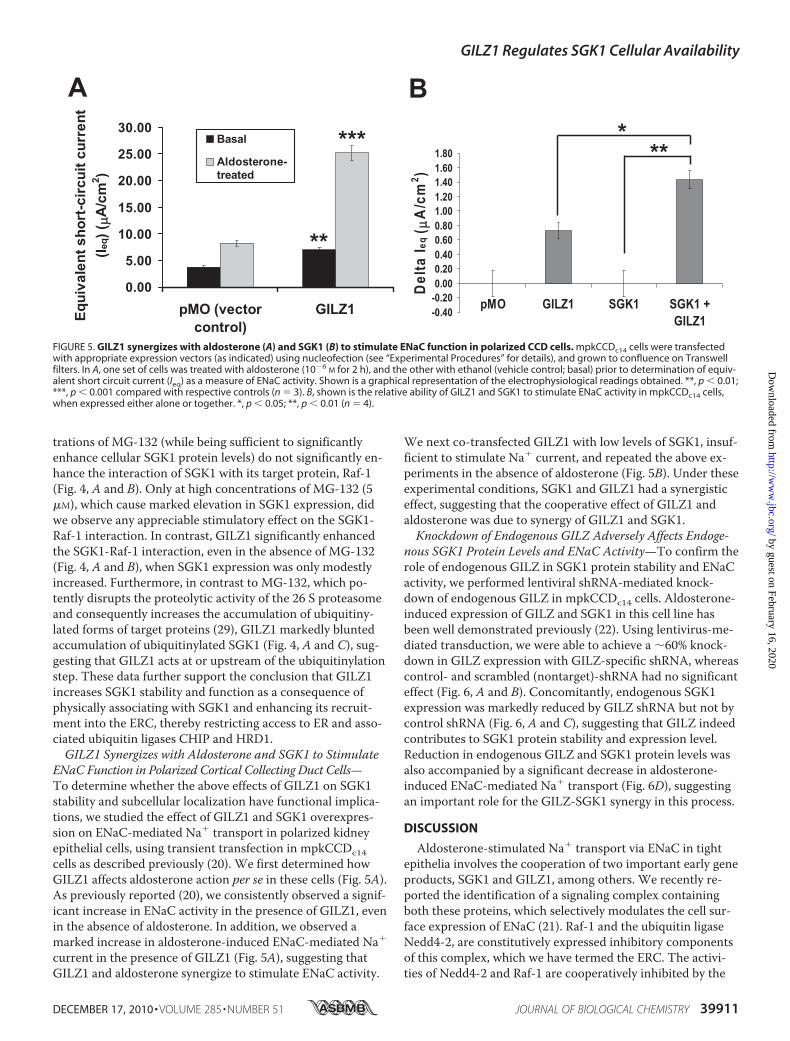
ENaC Function in Polarized Cortical Collecting Duct Cells—To determine whether the above effects of GILZ1 on SGK1stability and subcellular localization have functional implica-tions, we studied the effect of GILZ1 and SGK1 overexpres-sion on ENaC-mediated Na� transport in polarized kidneyepithelial cells, using transient transfection in mpkCCDc14cells as described previously (20). We first determined howGILZ1 affects aldosterone action per se in these cells (Fig. 5A).As previously reported (20), we consistently observed a signif-icant increase in ENaC activity in the presence of GILZ1, evenin the absence of aldosterone. In addition, we observed amarked increase in aldosterone-induced ENaC-mediated Na�
current in the presence of GILZ1 (Fig. 5A), suggesting thatGILZ1 and aldosterone synergize to stimulate ENaC activity.
We next co-transfected GILZ1 with low levels of SGK1, insuf-ficient to stimulate Na� current, and repeated the above ex-periments in the absence of aldosterone (Fig. 5B). Under theseexperimental conditions, SGK1 and GILZ1 had a synergisticeffect, suggesting that the cooperative effect of GILZ1 andaldosterone was due to synergy of GILZ1 and SGK1.Knockdown of Endogenous GILZ Adversely Affects Endoge-
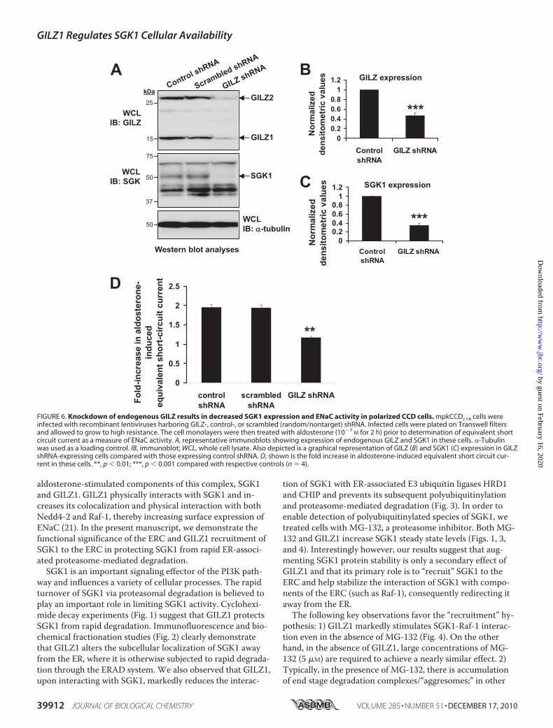
nous SGK1 Protein Levels and ENaC Activity—To confirm therole of endogenous GILZ in SGK1 protein stability and ENaCactivity, we performed lentiviral shRNA-mediated knock-down of endogenous GILZ in mpkCCDc14 cells. Aldosterone-induced expression of GILZ and SGK1 in this cell line hasbeen well demonstrated previously (22). Using lentivirus-me-diated transduction, we were able to achieve a �60% knock-down in GILZ expression with GILZ-specific shRNA, whereascontrol- and scrambled (nontarget)-shRNA had no significanteffect (Fig. 6, A and B). Concomitantly, endogenous SGK1expression was markedly reduced by GILZ shRNA but not bycontrol shRNA (Fig. 6, A and C), suggesting that GILZ indeedcontributes to SGK1 protein stability and expression level.Reduction in endogenous GILZ and SGK1 protein levels wasalso accompanied by a significant decrease in aldosterone-induced ENaC-mediated Na� transport (Fig. 6D), suggestingan important role for the GILZ-SGK1 synergy in this process.
DISCUSSION
Aldosterone-stimulated Na� transport via ENaC in tightepithelia involves the cooperation of two important early geneproducts, SGK1 and GILZ1, among others. We recently re-ported the identification of a signaling complex containingboth these proteins, which selectively modulates the cell sur-face expression of ENaC (21). Raf-1 and the ubiquitin ligaseNedd4-2, are constitutively expressed inhibitory componentsof this complex, which we have termed the ERC. The activi-ties of Nedd4-2 and Raf-1 are cooperatively inhibited by the
FIGURE 5. GILZ1 synergizes with aldosterone (A) and SGK1 (B) to stimulate ENaC function in polarized CCD cells. mpkCCDc14 cells were transfectedwith appropriate expression vectors (as indicated) using nucleofection (see “Experimental Procedures” for details), and grown to confluence on Transwellfilters. In A, one set of cells was treated with aldosterone (10�6
M for 2 h), and the other with ethanol (vehicle control; basal) prior to determination of equiv-alent short circuit current (Ieq) as a measure of ENaC activity. Shown is a graphical representation of the electrophysiological readings obtained. **, p � 0.01;***, p � 0.001 compared with respective controls (n 3). B, shown is the relative ability of GILZ1 and SGK1 to stimulate ENaC activity in mpkCCDc14 cells,when expressed either alone or together. *, p � 0.05; **, p � 0.01 (n 4).
GILZ1 Regulates SGK1 Cellular Availability
DECEMBER 17, 2010 • VOLUME 285 • NUMBER 51 JOURNAL OF BIOLOGICAL CHEMISTRY 39911
by guest on February 16, 2020http://w
ww
.jbc.org/D
ownloaded from
aldosterone-stimulated components of this complex, SGK1and GILZ1. GILZ1 physically interacts with SGK1 and in-creases its colocalization and physical interaction with bothNedd4-2 and Raf-1, thereby increasing surface expression ofENaC (21). In the present manuscript, we demonstrate thefunctional significance of the ERC and GILZ1 recruitment ofSGK1 to the ERC in protecting SGK1 from rapid ER-associ-ated proteasome-mediated degradation.SGK1 is an important signaling effector of the PI3K path-
way and influences a variety of cellular processes. The rapidturnover of SGK1 via proteasomal degradation is believed toplay an important role in limiting SGK1 activity. Cyclohexi-mide decay experiments (Fig. 1) suggest that GILZ1 protectsSGK1 from rapid degradation. Immunofluorescence and bio-chemical fractionation studies (Fig. 2) clearly demonstratethat GILZ1 alters the subcellular localization of SGK1 awayfrom the ER, where it is otherwise subjected to rapid degrada-tion through the ERAD system. We also observed that GILZ1,upon interacting with SGK1, markedly reduces the interac-
tion of SGK1 with ER-associated E3 ubiquitin ligases HRD1and CHIP and prevents its subsequent polyubiquitinylationand proteasome-mediated degradation (Fig. 3). In order toenable detection of polyubiquitinylated species of SGK1, wetreated cells with MG-132, a proteasome inhibitor. Both MG-132 and GILZ1 increase SGK1 steady state levels (Figs. 1, 3,and 4). Interestingly however, our results suggest that aug-menting SGK1 protein stability is only a secondary effect ofGILZ1 and that its primary role is to “recruit” SGK1 to theERC and help stabilize the interaction of SGK1 with compo-nents of the ERC (such as Raf-1), consequently redirecting itaway from the ER.The following key observations favor the “recruitment” hy-
pothesis: 1) GILZ1 markedly stimulates SGK1-Raf-1 interac-tion even in the absence of MG-132 (Fig. 4). On the otherhand, in the absence of GILZ1, large concentrations of MG-132 (5 �M) are required to achieve a nearly similar effect. 2)Typically, in the presence of MG-132, there is accumulationof end stage degradation complexes/“aggresomes;” in other
FIGURE 6. Knockdown of endogenous GILZ results in decreased SGK1 expression and ENaC activity in polarized CCD cells. mpkCCDc14 cells wereinfected with recombinant lentiviruses harboring GILZ-, control-, or scrambled (random/nontarget) shRNA. Infected cells were plated on Transwell filtersand allowed to grow to high resistance. The cell monolayers were then treated with aldosterone (10�7
M for 2 h) prior to determination of equivalent shortcircuit current as a measure of ENaC activity. A, representative immunoblots showing expression of endogenous GILZ and SGK1 in these cells. �-Tubulinwas used as a loading control. IB, immunoblot; WCL, whole cell lysate. Also depicted is a graphical representation of GILZ (B) and SGK1 (C) expression in GILZshRNA-expressing cells compared with those expressing control shRNA. D, shown is the fold increase in aldosterone-induced equivalent short circuit cur-rent in these cells. **, p � 0.01; ***, p � 0.001 compared with respective controls (n 4).
GILZ1 Regulates SGK1 Cellular Availability
39912 JOURNAL OF BIOLOGICAL CHEMISTRY VOLUME 285 • NUMBER 51 • DECEMBER 17, 2010
by guest on February 16, 2020http://w
ww
.jbc.org/D
ownloaded from
words, polyubiquitinylated species of target proteins, and endstage degradation of these already polyubiquitinylated speciesis prevented. Interestingly, in the presence of GILZ1, SGK1polyubiquitinylation is itself reduced (Figs. 3 and 4), suggest-ing that GILZ1 acts upstream of the proteasome to stabilizeSGK1. 3) GILZ1 does not affect the expression levels of eitherHRD1 or CHIP (Fig. 3A, HRD1 and CHIP expression). On theother hand, MG-132 uniformly increases the expression levelsof SGK1, CHIP, and HRD1 (data not shown), suggesting amore general inhibition of the common proteasome machin-ery. (Note that CHIP has been shown to undergo autoubiq-uitinylation (30) and is therefore itself a target for proteaso-mal degradation; GILZ1 has no appreciable effect on CHIPexpression.) 4) GILZ1 and SGK1 synergize to cooperativelystimulate ENaC activity in cortical collecting duct cells (Fig.5B). 5) Finally, knockdown of endogenous GILZ significantlyreduces aldosterone-induced SGK1 protein expression andENaC activity (Fig. 6), lending further support to the func-tional significance of these effects.Taken together with earlier work (20, 21), these data point
to GILZ1 as a key molecular determinant of intracellular sig-naling specificity governing SGK1 function and epithelial Na�
transport. In this regard, it is notable that an unrelated activ-ity of SGK1, inhibition of FOXO-mediated gene transcription,is unaffected by GILZ1 (21). The present findings lend sup-port to the growing view that subcellular localization is a criti-cal factor controlling SGK1 protein stability and further sug-gest that GILZ1 is a key determinant of where SGK1 localizeswithin the cell and with which proteins it interacts. Our cur-rent data strongly suggest that GILZ1 stabilizes SGK1 by re-cruiting it to the ERC. However, our experiments do notfirmly exclude the alternative possibility that SGK1 interac-tion with the ERC is increased because it is stabilized byGILZ1. Future studies will aim to address these issues in fur-ther detail. How exactly GILZ1 controls SGK1 localizationwill also be the subject of future investigation. The molecularidentities of players that facilitate this process and the specificsubcellular compartments wherein these interactions occuralso remain to be determined. Understanding the mechanisticdetails of this process will shed light on an important questionin cell biology, which is determining how cells achieve signal-ing specificity.
Acknowledgments—We thank Editha Setiawan for technical assis-tance and Vivek Bhalla for suggestions on the gene knockdown ex-periments in mpkCCD cells.
REFERENCES1. Lang, F., Artunc, F., and Vallon, V. (2009) Curr. Opin. Nephrol. Hyper-
tens. 18, 439–448
2. Verrey, F., Fakitsas, P., Adam, G., and Staub, O. (2008) Kidney Int. 73,691–696
3. Vallon, V., Wulff, P., Huang, D. Y., Loffing, J., Volkl, H., Kuhl, D., andLang, F. (2005) Am. J. Physiol. Regul. Integr. Comp. Physiol. 288, R4–10
4. McCormick, J. A., Bhalla, V., Pao, A. C., and Pearce, D. (2005) Physiology20, 134–139
5. Loffing, J., Flores, S. Y., and Staub, O. (2006) Annu. Rev. Physiol. 68,461–490
6. Bhargava, A., Wang, J., and Pearce, D. (2004)Mol. Cell. Endocrinol. 217,189–196
7. Arteaga, M. F., Wang, L., Ravid, T., Hochstrasser, M., and Canessa,C. M. (2006) Proc. Natl. Acad. Sci. U.S.A. 103, 11178–11183
8. Brickley, D. R., Mikosz, C. A., Hagan, C. R., and Conzen, S. D. (2002)J. Biol. Chem. 277, 43064–43070
9. Bogusz, A. M., Brickley, D. R., Pew, T., and Conzen, S. D. (2006) FEBS J.273, 2913–2928
10. Pao, A. C., McCormick, J. A., Li, H., Siu, J., Govaerts, C., Bhalla, V.,Soundararajan, R., and Pearce, D. (2007) Am. J. Physiol. 292,F1741–1750
11. Arteaga, M. F., Alvarez de la Rosa, D., Alvarez, J. A., and Canessa, C. M.(2007)Mol. Biol. Cell 18, 2072–2080
12. Paolino, M., and Penninger, J. M. (2009) Eur. J. Immunol. 39, 2337–234413. Petroski, M. D. (2008) BMC Biochem. 9, S714. Belova, L., Sharma, S., Brickley, D. R., Nicolarsen, J. R., Patterson, C., and
Conzen, S. D. (2006) Biochem. J. 400, 235–24415. Kaneko, M., Ishiguro, M., Niinuma, Y., Uesugi, M., and Nomura, Y.
(2002) FEBS Lett. 532, 147–15216. Meacham, G. C., Patterson, C., Zhang, W., Younger, J. M., and Cyr,
D. M. (2001) Nat. Cell Biol. 3, 100–10517. Zhou, R., and Snyder, P. M. (2005) J. Biol. Chem. 280, 4518–452318. Robert-Nicoud, M., Flahaut, M., Elalouf, J. M., Nicod, M., Salinas, M.,
Bens, M., Doucet, A., Wincker, P., Artiguenave, F., Horisberger, J. D.,Vandewalle, A., Rossier, B. C., and Firsov, D. (2001) Proc. Natl. Acad.Sci. U.S.A. 98, 2712–2716
19. Muller, O. G., Parnova, R. G., Centeno, G., Rossier, B. C., Firsov, D., andHorisberger, J. D. (2003) J. Am. Soc. Nephrol. 14, 1107–1115
20. Soundararajan, R., Zhang, T. T., Wang, J., Vandewalle, A., and Pearce, D.(2005) J. Biol. Chem. 280, 39970–39981
21. Soundararajan, R., Melters, D., Shih, I. C., Wang, J., and Pearce, D.(2009) Proc. Natl. Acad. Sci. U.S.A. 106, 7804–7809
22. Soundararajan, R., Wang, J., Melters, D., and Pearce, D. (2007) J. Biol.Chem. 282, 36303–36313
23. Lu, M., Wang, J., Jones, K. T., Ives, H. E., Feldman, M. E., Yao, L. J.,Shokat, K. M., Ashrafi, K., and Pearce, D. J. Am. Soc. Nephrol. 21,811–818
24. Morice, C., Nothias, F., Konig, S., Vernier, P., Baccarini, M., Vincent,J. D., and Barnier, J. V. (1999) Eur. J. Neurosci. 11, 1995–2006
25. Feng, L., Xie, X., Ding, Q., Luo, X., He, J., Fan, F., Liu, W., Wang, Z., andChen, Y. (2007) Proc. Natl. Acad. Sci. U.S.A. 104, 14348–14353
26. Lee, E. J., Hyun, S., Chun, J., Shin, S. H., and Kang, S. S. (2009) Exp. Mol.Med. 41, 555–568
27. Roderick, H. L., Campbell, A. K., and Llewellyn, D. H. (1997) FEBS Lett.405, 181–185
28. Kendall, J. M., Badminton, M. N., Dormer, R. L., and Campbell, A. K.(1994) Anal. Biochem. 221, 173–181
29. Kisselev, A. F., and Goldberg, A. L. (2001) Chem. Biol. 8, 739–75830. Xu, Z., Kohli, E., Devlin, K. I., Bold, M., Nix, J. C., and Misra, S. (2008)
BMC Struct. Biol. 8, 26
GILZ1 Regulates SGK1 Cellular Availability
DECEMBER 17, 2010 • VOLUME 285 • NUMBER 51 JOURNAL OF BIOLOGICAL CHEMISTRY 39913
by guest on February 16, 2020http://w
ww
.jbc.org/D
ownloaded from
Rama Soundararajan, Jian Wang, Daniël Melters and David PearceSubcellular Localization
Channel by Regulating Serum- and Glucocorticoid-induced Kinase 1 Stability and Glucocorticoid-induced Leucine Zipper 1 Stimulates the Epithelial Sodium
doi: 10.1074/jbc.M110.161133 originally published online October 14, 20102010, 285:39905-39913.J. Biol. Chem.
10.1074/jbc.M110.161133Access the most updated version of this article at doi:
Alerts:
When a correction for this article is posted•
When this article is cited•
to choose from all of JBC's e-mail alertsClick here
http://www.jbc.org/content/285/51/39905.full.html#ref-list-1
This article cites 29 references, 11 of which can be accessed free at
by guest on February 16, 2020http://w
ww
.jbc.org/D
ownloaded from


![Glucocorticoid-induced Cell Death Requires …...[CANCER RESEARCH 59, 1378–1385, March 15, 1999] Glucocorticoid-induced Cell Death Requires Autoinduction of Glucocorticoid Receptor](https://img.pdfslide.net/doc/110x75/5e5646d0314f24389e233453/glucocorticoid-induced-cell-death-requires-cancer-research-59-1378a1385.jpg)


![Endoplasmic reticulum[1]](https://img.pdfslide.net/doc/110x75/58ed5fc71a28aba1678b4611/endoplasmic-reticulum1.jpg)