Embed Size (px)
Citation preview
GLUCONEOGENESISStudent Edition 5/30/13 Version
Pharm. 304 Biochemistry
Fall 2014
Dr. Brad Chazotte 213 Maddox Hall
Web Site:
http://www.campbell.edu/faculty/chazotte
Original material only ©2000-14 B. Chazotte

Goals• Learn the enzymes and step of the gluconeogenesis pathway.
• Learn the similarities and difference the between gluconeogenesis and glycolysis pathways.
• Understand the principles for biosynthetic (anabolic) pathways vs catabolic.
• Understand how the gluconeogenesis pathway is regulated and how it is regulated vs glycolysis.
• Understand the concept and benefits of metabolic burden sharing
• Be aware the Cori cycle.
• Understand how substrate cycles can amplify metabolic signals and/or produce heat.

Biosynthetic (Anabolic) Pathways• Use chemical energy (ATP, NADH, NADPH) to
synthesize cellular components from simple precursor molecules.
• Generally reductive rather than oxidative.
• Anabolism and catabolism proceed simultaneously in a dynamic steady-state to maintain the “intricate orderliness of living cells”.
Lehninger 2000 p722

Organizing Principles of Biosynthesis 1
Lehninger 2000 p722
1. Molecules are synthesized and degraded by different pathways.
a. Two opposing anabolic and catabolic pathways may share many reversible reactions.
b. Each pathway has at least one unique and essentially irreversible reaction.
c. This insures that the carbon flow through these pathways is dictated by the cell’s changing requirements for energy, precursors, and macromolecules rather than simple mass action.

Organizing Principles of Biosynthesis 2
Lehninger 2000 p722
2. Corresponding anabolic and catabolic pathways are controlled by one or more reactions unique to each pathway.
a. Opposing pathways are regulated in a coordinated, reciprocal manner so that the stimulation of the anabolic pathway is accompanied by the inhibition of the catabolic one and vice versa.
b. Biosynthetic pathways are typically regulated at an early, exergonic step that commits intermediates to the pathway and does not waste energy by making unneeded intermediates.

Organizing Principles of Biosynthesis 3
Lehninger 2000 p722
3. Energy requiring biosynthetic processes are coupled to the breakdown of ATP in such a way that the overall process is essentially irreversible in vivo.
a. Thus the total amount of energy from ATP and NAD(P)H used always exceeds the minimum free energy needed to convert the precursor into the biosynthetic product.
b. Consequently the biosynthetic process is thermodynamically favorable (G < 0) even for low precursor concentrations.

Lehninger 2000 Fig 20.1Gluconeogenesis
Carbohydrate Biosyntheses from Simple PrecursorsThe pathway from PEP to glucose-6-P is common to the biosynthetic conversion of many different precursors in animals and plants.
Gluconeogenesis, meaning “formation of new sugar”, occurs in all animals, plants, fungi, and microorganisms. In most cases the reactions are essentially the same.

Importance of Gluconeogenesis
The brain depends on glucose as its primary fuel using ~120g/day out of ~160g/day for the typical adult human.
Red blood cells use only glucose as a fuel.
Only 20g are present in the body fluids and ~190 g are available from glycogen storage.
Therefore, total reserves of glucose are about a single day’s supply.
For longer periods of starvation, glucose must be formed from noncarbohydrate sources.

Noncarbohydrate Precursors for Gluconeogenesis
Any metabolite that can be converted to pyruvate or oxaloacetate can be a glucose precursor
Lactate is formed by active skeletal muscle when glycolysis > oxidative metabolism. Lactate is converted by lactate dehydrogenase to pyruvate.
Pyruvate
(Certain) Amino acids are derived from protein in the diet or from muscle protein catabolism during starvation. Carbon skeletons of most amino acids are catabolized to pyruvate or citric acid cycle intermediates
Glycerol is the result of a breakdown of triacylglycerols in fat cells. Fatty acids also result, but cannot be used by animals to make glucose. Glycerol enters glycolysis or gluconeogenesis at dihydroxyacetone phosphate
Pyruvate

Berg, Tymoczko & Stryer, 2012 Chap. 16 np.481Gluconeogenesis
Glycerol’s Entry in Gluconeogenesis or Glycolysis

The Gluconeogenesis Balanced Equation
2Pyruvate + 2NADH +4H+ + 4ATP +2GTP +6H2O
Glucose +2NAD+ + 4ADP + 2GDP + 6Pi
Gº = –16 kJ/ mol
Voet & Voet 1995 Chap 21 P.604Gluconeogenesis
Because of the presence of separate gluconeogenic enzymes at the three irreversible steps in the glycolytic pathway that converts glucose to pyruvate, glycolysis and gluconeogenesis both are rendered THERMODYNAMICALLY favorable.

Berg, Tymoczko & Stryer, 2012 Fig. 16.24Gluconeogenesis
Gluconeogenesis Pathway
Voet, Voet & Pratt 2013 Figure 16.15

Subcellular Location of Gluconeogenic Enzymes
• Gluconeogenesis enzymes are cytosolic except:
(1) Glucose 6-phosphatase (endoplasmic reticulum)
(2) Pyruvate carboxylase (mitochondria)
(3) PEPCK (cytosol and/or mitochondria)
Horton et al., 2000 Chap 13.6
Mitochondrial pyruvate can:1) be converted to citrate and used in the cytosol for the synthesis of fatty acids.2) be changed to acetyl CoA and enter the citric acid cycle.3) be converted by pyruvate carboxylase to oxaloacetate for gluconeogenesis.

Lehninger 2000 Table 20.1
G’s of Erythrocyte Glycolytic Rxs

Lehninger 2000 Table 20.2
Sequential Rx in Gluconeogenesis from Pyruvate

Berg, Tymoczko & Stryer, 2012 Fig. 16.24bGluconeogenesis
Gluconeogenesis Pathway I

Conversion of Pyruvate into Phosphoenolpyruvate
Pyruvate + CO2 + ATP + H2O oxaloacetate + ADP + Pi + 2H+
G’ = -2.1 kJ mol -1
Pyruvate carboxylase
Gluconeogenesis Voet, Voet & Pratt 2013 Fig 16.16
• The bypass of glycolysis’ pyruvate kinase reaction requires two separate reactions in gluconeogenesis.
• One of the anaplerotic reactions that is used to maintain the levels of intermediates in the citric acid cycle
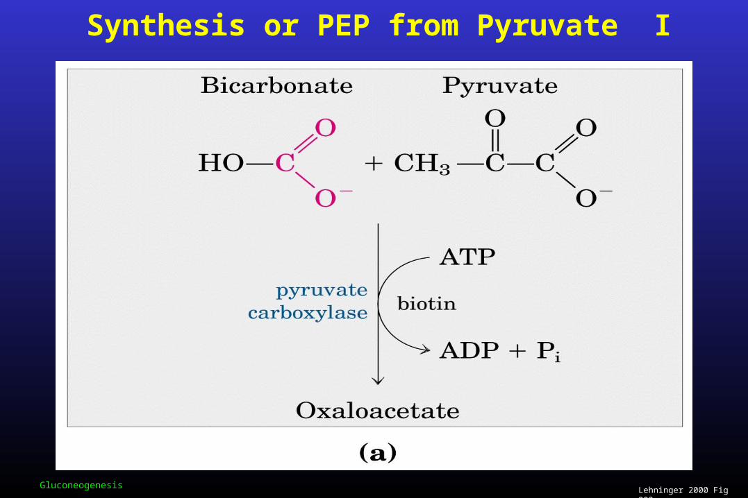
Lehninger 2000 Fig 203aGluconeogenesis
Synthesis or PEP from Pyruvate I

Pyruvate Carboxylase: Rx Mechanism
Voet , Voet & Pratt 2008 Fig 16.18Gluconeogenesis
Pyruvate Carboxylase catalyzes the ATP-driven synthesis of oxaloacetate from pyruvate and HCO3
-. This reaction occurs in two phases.
Phase I is a three step reaction sequence. Biotin is carboxylated at its N1’ position by a bicarbonate ion.
Phase II in this phase the activated carboxyl group is transferred to pyruvate from carboxybiotin in a three step reaction sequence to form oxaloacetate.

Berg, Tymoczko & Stryer, 2012 Fig. 16.25Gluconeogenesis
Domain Structure of Pyruvate Carboxylase
The ATP grasp domain activates HCO3- and transfers CO2 to the biotin-binding domain.
From there the CO2 is transferred to pyruvate generated in the central domain.

Berg, Tymoczko & Stryer, 2012 Fig. 16.26Gluconeogenesis
Pyruvate Carboxylase’s Biotin-binding Domain
Key feature: biotin is on a flexible tether, attached to the -amino group of lysine, allowing it to move between the ATP-bicarbonate site and the pyruvate site.

Voet, Voet & Pratt 2013 Fig 16.17bGluconeogenesis
Carboxybiotin
Berg, Tymoczko & Stryer, 2012 Fig. 16.27

Lehninger 2000 Fig 20.2bGluconeogenesis
Opposing Pathways of Gluconeogenesis and Glycolysis II

Conversion of Oxaloacetate into Phosphoenolpyruvate
Oxaloacetate + GTP Phosphoenolpyruvate + CO2 + GDP
G’ = 2.9 kJ mol -1
phosphoenolpyruvate carboxykinase
Gluconeogenesis

Gluconeogenesis
Synthesis or PEP from Pyruvate II
Voet, Voet & Pratt 2013 Figure 16.19
Oxaloacetate is converted to phosphoenolpyruvate in the cytosol by PEP carboxykinase in a reaction that requires Mg2+ and GTP as the phosphoryl donor.
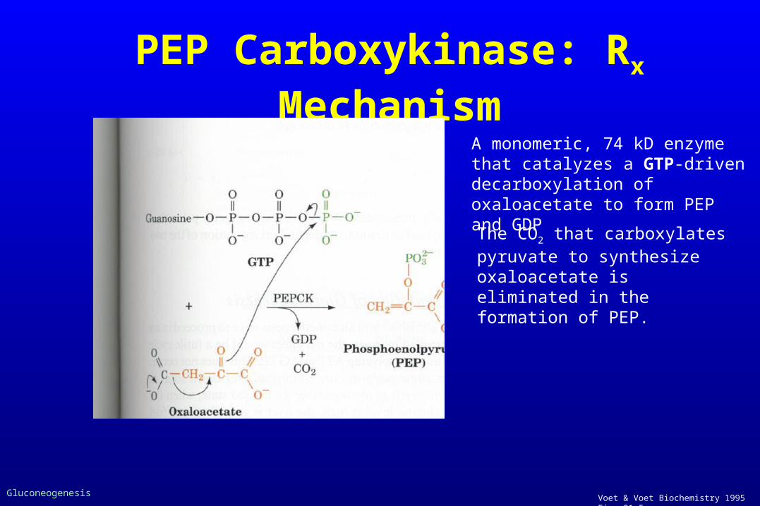
PEP Carboxykinase: Rx Mechanism
Voet & Voet Biochemistry 1995 Fig. 21.5Gluconeogenesis
A monomeric, 74 kD enzyme that catalyzes a GTP-driven decarboxylation of oxaloacetate to form PEP and GDP
The CO2 that carboxylates pyruvate to synthesize oxaloacetate is eliminated in the formation of PEP.

Berg, Tymoczko & Stryer, 2012 Fig. 16.28Gluconeogenesis
The Mitochondrion Supplies Malate made
from Pyruvate for use in Gluconeogenesis in the
Cytosol
Note: the starting molecule is pyruvate.
• Inside the mitochondrion oxaloacetate is reduced to malate in order to be transported outside the mitochondrion
• Once the malate is transported outside (via the malate - -ketoglutarate
shuttle) it is re-oxidized to
oxaloacetate

Lehninger 2000 Fig 20.4Gluconeogenesis
Alternate Paths from Pyruvate to PEP
Pathway predominant when the starting molecule is pyruvate When lactate is the
precursor this pyruvate to PEP bypass is predominant
This oxaloacetate is converted directly to PEP by a mitochondrial isozyme of PEP carboxykinase

PEP & Oxaloacetate Transport Cytosol Mitochondria
Voet, Voet & Pratt 2013 Figure 16.20
PEP is transported between the mitochondrion and the cell cytosol by a specific membrane transporter so it can move between the two compartments depending on the equilibrium.
Route 1 uses aspartate amino transferase. In this case oxaloacetate is converted to aspartate for transport.
PEP has a direct transporter, whereas oxaloacetate does not and must be converted
for transmembrane passage.Route 2 involves the conversion of oxaloacetate to malate AND involves NADH.

Lehninger 2000 Table 20.2
Gluconeogenesis
Sequential Reactions in Gluconeogenesis (Reprise)

Berg, Tymoczko & Stryer, 2012 Fig. 16.24aGluconeogenesis
Gluconeogenesis Pathway II

Conversion of Fructose 1,6-bisphosphate into Fructose -6P and PPi:
An Irreversible Step
G’ = -16.3 kJ mol -1
Fructose 1,6-bisphosphate + H2O fructose 6-phosphate + Pi
Gluconeogenesis
Fructose 1,6-biphosphatase

Conversion of Glucose-6-phosphate into Glucose: An Irreversible Step
G’ = -12.1 kJ mol -1
Glucose-6-phosphate + H2O Glucose + Pi
Gluconeogenesis
Glucose-6-phosphatase

Glucose-6-Phosphatase
This enzyme is found primarily in the endoplasmic reticulum of liver cells. – Why?

Metabolic Control of Glycolysis and Gluconeogenesis
In cells gluconeogenesis and glycolysis are coordinated such that one is mainly inactive while the other in highly active. (If both were highly active at the same time the result would be a FUTILE CYCLE consuming two ATP and two GTP per reaction cycle).
• The rate of glycolysis is typically controlled by the glucose concentration.
• The rate of gluconeogenesis is typically controlled by the concentrations of lactate and other glucose precursors.

Berg, Tymoczko & Stryer, 2012 Fig. 16.30Gluconeogenesis
Reciprocal Regulation of Gluconeogenesis and Glycolysis in Liver
Voet, Voet & Pratt 2013 Fig. 16.21; 16.23
IMPORTANT!!

Berg, Tymoczko & Stryer, 2012 Fig. 16.34Gluconeogenesis
“Subtrate Cycle” can amplify metabolic signals and produce heat.

Metabolic Burden SharingDifferent organs/tissues in the body can be metabolically linked.
Lactate produced by active skeletal muscle and erythrocytes is an energy source for other organs.
• Skeletal muscle during vigorous exercise produces pyruvate at a rate faster than oxidative metabolism via the citric acid cycle can use it.
• Also NADH production is more rapid than its conversion to NAD+ in aerobic metabolism.
• (Remember that glycolysis needs NAD+ for glyceraldehyde 3-P oxidation to proceed.) • The cell can oxidize NADH to NAD+ in a reaction that converts pyruvate to lactate via
lactate dehydrogenase. The problem is that lactate can not be further metabolized, only converted back to pyruvate.
Important: Muscle’s ability to reduce pyruvate to lactate is a way shift the metabolic burden under high stress to other organs, e.g. liver, heart. (Reaction also regenerates NAD+ in muscle)

Horton et al., 2002 Fig. 13.12Gluconeogenesis: Cori Cycle
Cori Cycle (an interorgan metabolic “pathway”)
Lactate & pyruvate diffuse out of active muscle into blood.
Excess lactate in the blood enters the liver where it is converted to pyruvate and then to glucose via gluconeogenesis.
Berg, Tymoczko & Stryer, 2012 Fig. 16.35

Berg, Tymoczko & Stryer, 2002 Fig. 16.34
Gluconeogenesis
Coordination of Glycolysis and Gluconeogenesis
In liver and kidney glucose is produced by gluconeogenesis and can go out into the blood to be used by other tissues such as muscle.
Muscles and red blood cells can produce lactate which, as we have seen in the Cori cycle can travel to the e.g. liver where it is metabolized to pyruvate, etc
In muscle alanine is formed by a transamination reaction from pyruvate. Whereas the reverse process occurs in the liver. This cycling helps to maintain the nitrogen balance
(Remember: alanine is also a major glucose precursor)
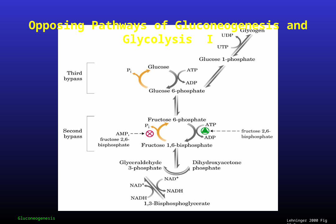
Lehninger 2000 Fig 20.2aGluconeogenesis
Opposing Pathways of Gluconeogenesis and Glycolysis I

End of Lectures


![Biochem [Gluconeogenesis]](https://img.pdfslide.net/doc/110x75/577c82b31a28abe054b1e4af/biochem-gluconeogenesis.jpg)