Embed Size (px)
Citation preview
University of Nebraska - LincolnDigitalCommons@University of Nebraska - Lincoln
John Janovy Publications Papers in the Biological Sciences
10-2007
Gregarina niphandrodes (Eugregarinorida:Septatorina): Oocyst Surface ArchitectureJohn J. Janovy Jr.University of Nebraska - Lincoln, [email protected]
Matthew G. BolekOklahoma State University, [email protected]
Jillian Tikka DetwilerPurdue University, [email protected]
Samana SchwankLondon School of Tropical Medicine, [email protected]
Alaine KnipesUniversity of Nebraska - Lincoln, [email protected]
See next page for additional authors
Follow this and additional works at: http://digitalcommons.unl.edu/bioscijanovy
Part of the Parasitology Commons
This Article is brought to you for free and open access by the Papers in the Biological Sciences at DigitalCommons@University of Nebraska - Lincoln.It has been accepted for inclusion in John Janovy Publications by an authorized administrator of DigitalCommons@University of Nebraska - Lincoln.
Janovy, John J. Jr.; Bolek, Matthew G.; Detwiler, Jillian Tikka; Schwank, Samana; Knipes, Alaine; and Langford, Gabriel J., "Gregarinaniphandrodes (Eugregarinorida: Septatorina): Oocyst Surface Architecture" (2007). John Janovy Publications. 57.http://digitalcommons.unl.edu/bioscijanovy/57

AuthorsJohn J. Janovy Jr., Matthew G. Bolek, Jillian Tikka Detwiler, Samana Schwank, Alaine Knipes, and Gabriel J.Langford
This article is available at DigitalCommons@University of Nebraska - Lincoln: http://digitalcommons.unl.edu/bioscijanovy/57
714 THE JOURNAL OF PARASITOLOGY, VOL. 93, NO.3, JUNE 2007
1. Parasitol., 93(3), 2007, pp. 714-716© American Society of Parasitologists 2007
Gregarina niphandrodes (Eugregarinorida: Septatorina): Oocyst Surface Architecture
J. Janovy, Jr., M. G. Bolek, J. Detwiler, S. Schwank, A. Knipes, and G. J. Langford, School of Biological Sciences, University of NebraskaLincoln, Lincoln, Nebraska 68588-0118. e-mail: [email protected]
ABSTRACT: The surface architecture of oocysts produced by Gregarinaniphandrodes (Eugregarinorida) from Tenebrio molitor adults (Coleoptera: Tenebrionidae) as revealed by scanning electron microscopy isreported. Gametocysts were allowed to dehisce on 15-mm, round coverglasses; the cover glasses with their oocysts chains were then mountedon stubs without further processing, and sputter-coated with 20-nmgold-palladium. Scanning electron microscopy was performed at lOIS kY with a Hitachi 3000N SEM. Oocysts retained their characteristicshapes as reported in the original species description but showed longitudinal ridges of relatively uniform height, width, and spacing, inseparate fields on either side of a central equatorial bulge in the oocysts.There was no ultrastructural evidence of an enclosing external sheathholding the oocysts in a chain. Oocyst ends were flared slightly, andthe chain itself was twisted, with adjacent oocysts offset slightly fromone another. This article now provides an additional set of structuralcharacters potentially useful in gregarine systematics.
The class Gregarinasina (Apicomplexa) is perhaps the most diverseeukaryotic taxon because its members parasitize invertebrates, especially annelids and arthropods, although gregarine species have beendescribed from most invertebrate phyla. Descriptive work, however, issomewhat hindered by a number of factors, such as gregarines' lack of
medical importance, their seeming paucity of structural features, andthe fact that so many potential hosts in interesting parts of the worldare relatively inaccessible to workers because of logistical constraints,political turmoil, and lack of funds. Nevertheless, in recent years, Clopton and various coworkers have established criteria by which new taxashould be described and have standardized terminology for shapes andproportions (Clopton et al., 1991, 1992; Clopton, 2004; Clopton et al.,2004). This body of published work shows clearly that oocyst shapeand size are highly stable characters of major taxonomic importance.
The present study was intended to increase the number of charactersavailable for gregarine systematics by examining surface architecture ofoocysts using scanning electron microscopy. Gregarina niphandrodesClopton, Percival, and Janovy, 1991, from adult Tenebrio molitor (Coleoptera: Tenebrionidae) was chosen as the material because both hostand parasite are common, well-studied species, and adult beetles aregenerally infected with only this single gregarine species (Clopton etal., 1992). In addition, G. niphandrodes gametocysts are relatively largeand easily handled, and oocyst production ("sporogeny") is easilyachieved in moist chambers (Clopton and Janovy, 1993).
Insects used in this study were from cultures maintained for manyyears at the University of Nebraska-Lincoln using wheat bran and potatoes as food. These are the same cultures from which the type hosts
Janovy, Bolek, Detwiler, Schwank, Knipes & Langford in Journal of Parasitology 93 (2007) Copyright 2007, American Society of Parasitologists. Used by permission.
RESEARCH NOTE 715
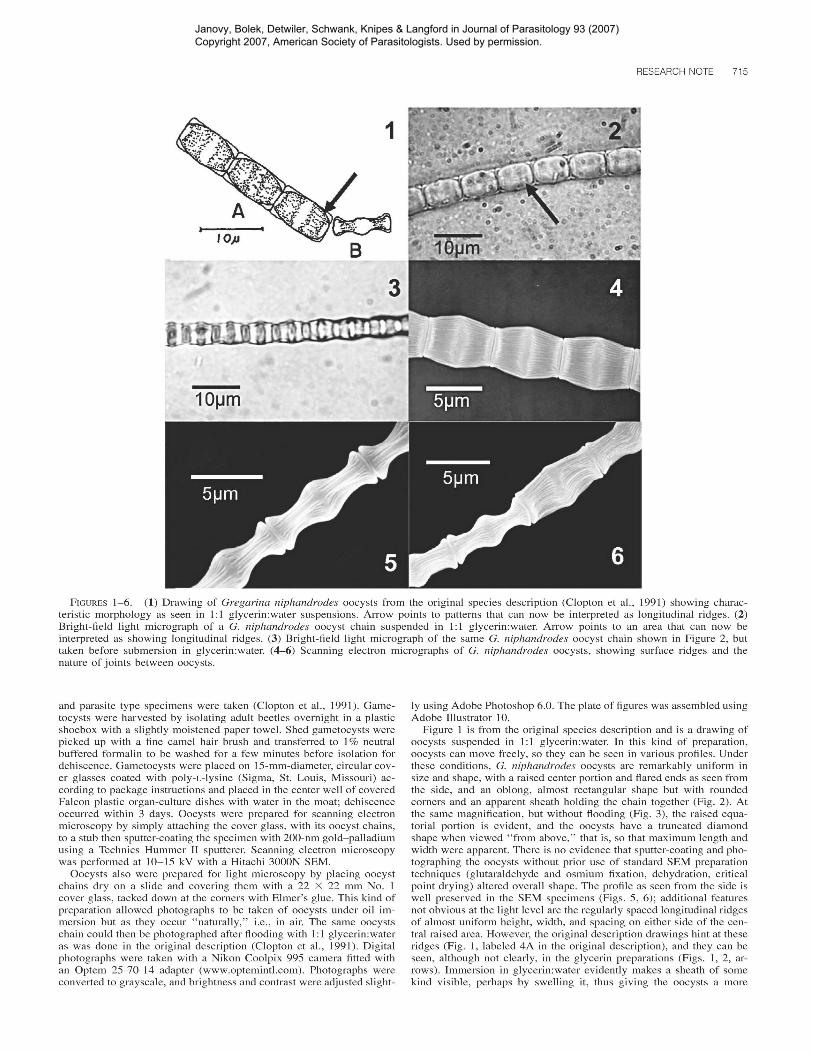
FIGURES 1-6. (1) Drawing of Gregarina niphandrodes oocysts from the original species description (Clopton et aI., 1991) showing characteristic morphology as seen in 1:1 glycerin:water suspensions. Arrow points to patterns that can now be interpreted as longitudinal ridges. (2)Bright-field light micrograph of a G. niphandrodes oocyst chain suspended in 1:1 glycerin:water. Arrow points to an area that can now beinterpreted as showing longitudinal ridges. (3) Bright-field light micrograph of the same G. niphandrodes oocyst chain shown in Figure 2, buttaken before submersion in glycerin:water. (4-6) Scanning electron micrographs of G. niphandrodes oocysts, showing surface ridges and thenature of joints between oocysts.
and parasite type specimens were taken (Clopton et aI., 1991). Gametocysts were harvested by isolating adult beetles overnight in a plasticshoebox with a slightly moistened paper towel. Shed gametocysts werepicked up with a fine camel hair brush and transferred to 1% neutralbuffered formalin to be washed for a few minutes before isolation fordehiscence. Gametocysts were placed on 15-mm-diameter, circular cover glasses coated with poly-L-Iysine (Sigma, St. Louis, Missouri) according to package instructions and placed in the center well of coveredFalcon plastic organ-culture dishes with water in the moat; dehiscenceoccurred within 3 days. Oocysts were prepared for scanning electronmicroscopy by simply attaching the cover glass, with its oocyst chains,to a stub then sputter-coating the specimen with 200-nm gold-palladiumusing a Technics Hummer II sputterer. Scanning electron microscopywas performed at 10-15 kV with a Hitachi 3000N SEM.
Oocysts also were prepared for light microscopy by placing oocystchains dry on a slide and covering them with a 22 X 22 mm No. 1cover glass, tacked down at the corners with Elmer's glue. This kind ofpreparation allowed photographs to be taken of oocysts under oil immersion but as they occur "naturally," i.e., in air. The same oocystschain could then be photographed after flooding with 1: 1 glycerin:wateras was done in the original description (Clopton et aI., 1991). Digitalphotographs were taken with a Nikon Coolpix 995 camera fitted withan Optem 25-70-14 adapter (www.optemintl.com). Photographs wereconverted to grayscale, and brightness and contrast were adjusted slight-
Iy using Adobe Photoshop 6.0. The plate of figures was assembled usingAdobe Illustrator 10.
Figure 1 is from the original species description and is a drawing ofoocysts suspended in 1: 1 glycerin:water. In this kind of preparation,oocysts can move freely, so they can be seen in various profiles. Underthese conditions, G. niphandrodes oocysts are remarkably uniform insize and shape, with a raised center portion and flared ends as seen fromthe side, and an oblong, almost rectangular shape but with roundedcorners and an apparent sheath holding the chain together (Fig. 2). Atthe same magnification, but without flooding (Fig. 3), the raised equatorial portion is evident, and the oocysts have a truncated diamondshape when viewed "from above," that is, so that maximum length andwidth were apparent. There is no evidence that sputter-coating and photographing the oocysts without prior use of standard SEM preparationtechniques (glutaraldehyde and osmium fixation, dehydration, criticalpoint drying) altered overall shape. The profile as seen from the side iswell preserved in the SEM specimens (Figs. 5, 6); additional featuresnot obvious at the light level are the regularly spaced longitudinal ridgesof almost uniform height, width, and spacing on either side of the central raised area. However, the original description drawings hint at theseridges (Fig. I, labeled 4A in the original description), and they can beseen, although not clearly, in the glycerin preparations (Figs. 1, 2, arrows). Immersion in glycerin:water evidently makes a sheath of somekind visible, perhaps by swelling it, thus giving the oocysts a more
Janovy, Bolek, Detwiler, Schwank, Knipes & Langford in Journal of Parasitology 93 (2007) Copyright 2007, American Society of Parasitologists. Used by permission.
716 THE JOURNAL OF PARASITOLOGY, VOL. 93, NO.3, JUNE 2007
rectangular appearance then when dry (cf. Figs. I, 2 vs. Fig. 3). Thebroad, truncated diamond shape seen in dry oocysts is also clearly seenin the SEMs (Figs. 3, 4). There was no ultrastructural evidence of anenclosing external sheath holding the oocysts in a chain. Oocyst endswere flared slightly, and the chain itself was twisted with adjacent 00
cysts offset slightly from one another.There are 2 main contributions of this study. First, the demonstration
that oocyst structure as seen under the light microscope is preservedthrough sputter-coating and scanning electron microscopy, even thoughstandard fixation, dehydration, and critical-point drying are not performed on the specimens. Second, there are distinct, fine folds or ridges,arranged in a distinctive pattern, on the oocysts. Although these structural features are potentially useful in future gregarine systematic work,such use requires comparative information on oocyst surface architecture from a variety of gregarine taxa.
The differences between oocyst structure as seen in glycerin:watersuspensions versus in air at oil immersion magnifications could be theresult of refraction of the light beam, although there is also a possibilitythat the cysts are contained within a sheath of some kind that is expanded in glycerin:water. These differences also emphasize the importance of reporting oocyst preparation methods in detail, for example, asexemplified by Clopton et al. (2004), when using such measurementsin taxonomic studies.
The authors wish to thank Kit Lee for assistance with the scanningelectron microscopy.
LITERATURE CITED
CLOPTON, R. E. 2004. Standard nomenclature and metrics of planeshapes for use in gregarine taxonomy. Comparative Parasitology71: 130-140.
---, T. J. COOK, AND J. L. COOK. 2004. Naiadocystis phykoterion n.gen., n. sp. (Apicomplexa: Eugregarinida: Hirmocystidae), from theMexican pygmy grasshopper, Paratettix mexicanus (Orthoptera:Tetrigidae), in the Texas Big Thicket with recognition of three previously described species of Naiadocystis. Journal of Parasitology90: 301-307.
---, AND J. JANOVY JR. 1993. Developmental niche structure in thegregarine assemblage parasitizing Tenebrio molitor. Journal of Parasitology 79: 701-709.
---, ---, AND T. J. PERCIVAL. 1992. Host stadium specificity inthe gregarine assemblage parasitizing Tenebrio molitor. Journal ofParasitology 78: 334-337.
---, T. J. PERCIVAL, AND J. JANOVY JR. 1991. Gregarina niphandrodes n. sp. (Apicomplexa: Eugregarinorida) from adult Tenebriomolitor (L.) with oocyst descriptions of other gregarine parasitesof the yellow mealworm. Journal of Protozoology 38: 472-479.
Janovy, Bolek, Detwiler, Schwank, Knipes & Langford in Journal of Parasitology 93 (2007) Copyright 2007, American Society of Parasitologists. Used by permission.