Embed Size (px)
Citation preview

357
GROWTH, DIFFERENTIATION, AND ULTRASTRUCTURE OF MICROSPORE CALLUS OF PICEA ABIES AS AFFECTED BY
NITROGENOUS SUPPLEMENTS AND LIGHT
LIISA KAARINA SIMOLA and OSSI HUHTINEN Department of Botany, University of Helsinki, SF-00170 Finland
(Received for publication 12 December 1986)
ABSTRACT
The growth of microspore callus cultures of Picea abies L. Karst, was stimulated by weak fluorescent and red light as opposed to darkness. Putrescine (0.1 mM) was able to enhance growth in the dark. The effects of spermidine and spermine on growth were rather similar but root differentiation was stimulated by spermidine in blue, red, and fluorescent light, by spermine only in red light. Glutamine (500 mg//) and a combination of it and casein hydrolysate (1000 mg//) retarded growth and root differentiation. The root had a weakly developed central cylinder with tracheids. The microspore callus cells had normal ultra-structure. Accumulation of starch, plastoglobuli, and some weakly developed grana were characteristic of plastids. Greening of cultures was not observed in red light. Accumulation of secondary metabolites was very prominent, especially in the vacuoles of some cells cultured in blue light. The plastids in those cells were usually rather small and contained very little or no starch.
Keywords: microspore callus; putrescine; spermidine; spermine; glutamine; plastid; Picea abies.
INTRODUCTION
In many cryptogams wholly or mainly haploid life cycles exist. In seed plants the haploid phase has been reduced so* much that the diploid phase occupies the main part of the life cycle. The conifers represent a category near the latter extreme in which the haploid phase is reduced to> the small female and male gametophytes.
The spontaneous generation of haploids among the angiosperms is not very rare and they have been produced experimentally in a large number of species. A haploid cultivar of Pelargonium has been cultured by vegetative propagation for at least 55 years (Daker 1967). Thus haploidy per se is not an overriding developmental constraint. It is, however, very rare among gymnosperms. Some haploid seedlings of Picea abies have been obtained from abnormal or polyembryonic seeds (lilies 1964). Several haploid and aneuploid embryos have been picked out using radiography on polyembryonic seeds (Simak et al. 1968), but the growth of these embryos was weak under normal germination conditions. For conifers the only report of a haploid specimen is for Thuja gigantea Nutt. cv. Gracilis in Denmark (Polheim 1968).
Callus tissue of trees has been cultured, e.g., from pollen of Ginkgo (Tulecke 1957), Thuja (Rao & Mehta 1969), and Betula (Simola & Koskimies-Soininen 1984). Cyto-
New Zealand Journal of Forestry Science 16(3): 357-68 (1986)
358 New Zealand Journal of Forestry Science 16(3)
differentiation of different cell types has been observed in megagametophyte callus of Ginkgo (Tulecke 1967) and Araucana araucana (Molina) K. Koch. (Cardemil & Jordan 1982) but organogenesis is usually very poor in callus lines deriving from haploid cells of gymnosperms (Huhtinen 1972; Bonga 1974). The small plantlets differentiated from megagametophyte callus of Picea abies could not grow to* bigger plants (Simola & Honkanen 1983).
The greatest advantage of working with haploids is the possibility of reducing the long inbreeding period of most forest trees. By using cross-breeding between homozygous lines the best results can be selected already in the Fi generation in agronomic crops and the same principle may be applied also in forestry.
The use of the haploid technique can save time because of the possibility of producing homozygotic lines by colchicine treatment. The haploid stage is very sensitive because lethal genes are expressed and lead to the death of poor genotypes.
The aim of this work has been to study the early stages of callus growth and differentiation in microspore-derived callus cultures of Picea abies, economically one of the most important forest trees in Finland. The very irregular and poor seed production of this tree in Northern Europe has resulted in an interest in vegetative propagation of the species. Because some cereals form albino haploids abundantly, special efforts have been made to investigate alsoi plastid development under different light and nutrient conditions.
MATERIAL AND METHODS Branches of P. abies (grafts cultivated in Ruotsinkyla, Tuusula, S-Finland) were
collected during winter and early spring (February-April). They were stored at + 4 ° C in the dark for a maximum of 4 weeks. The cut ends were put in tap water and male strobili developed at room temperature, usually in 7-10 days. The strobili were dipped in 94% ethanol and flamed. The microsporophylls were dissected when the microspores were uni-nuclear and placed in petri dishes (diameter 9 cm) containing a nutrient solution either according to Murashige & Skoog (1962) or N 7 (Simola 1985), supplemented with m-inositol (100 mg//) , casein hydrolysate (1000 mg//) , glutamine (500 g//) , and sucrose (20 g/t). 2,4-D (0.1 mg//) , IAA (25 mg//) , and BAP (1 mg//) were used as growth regulators. Other mineral nutrient media and combinations of growth regulators were also tested but without success.
The petri dishes were sealed with parafilm and placed in a rotary shaker (80 rpm) in the dark. The microsporophylls were removed after a couple of weeks, and the microspores dispersed in the medium formed small calluses 1-2 mm in diameter after 1 or 2 months. The calluses were placed on a solid agar medium which had the same composition as the liquid medium and the cultures were incubated in the dark.
Different combinations of organic supplements, especially polyamines and growth regulators, were tested in order to induce differentiation. The cultures were incubated in fluorescent light with an 18-h photoperiod. The possible effect of blue and red light was tested using Philips TL 15 and 18 lamps (40 W ) as light sources. The light intensity at the level of culture was c. 1000 lux.
Chromosome numbers of callus lines were studied using the lacto-propionic-dimethysulfoxide-carmine method (Bonga & Venketeswaran 1976). For electron
Simola & Huhtinen — Growth of microspore callus of Picea abies 359
microscope observations material was fixed with 3 % glutaraldehyde in 0.12 M K-phosphate buffer, pH 7.2, for 2 hours at room temperature. The material was rinsed with K-phosphate buffer, postfixed, and embedded for electron microscope observations according to Simola (1972). For light microscopy, roots differentiated from callus were fixed in FAA and embedded in paraffin; the sections were stained with fast green.
RESULTS AND DISCUSSION
Initiation of Callus
Microsporophylls of P. abies floating on liquid medium were able to produce small calluses which formed subculturable callus lines on semisolid medium. The cultures were first incubated in the dark but, because dim light had a favourable effect on growth, the main part of the culturing took place later in rhythmic dim light. Cultures turned pale green usually and the callus was friable.
Growth and Greening
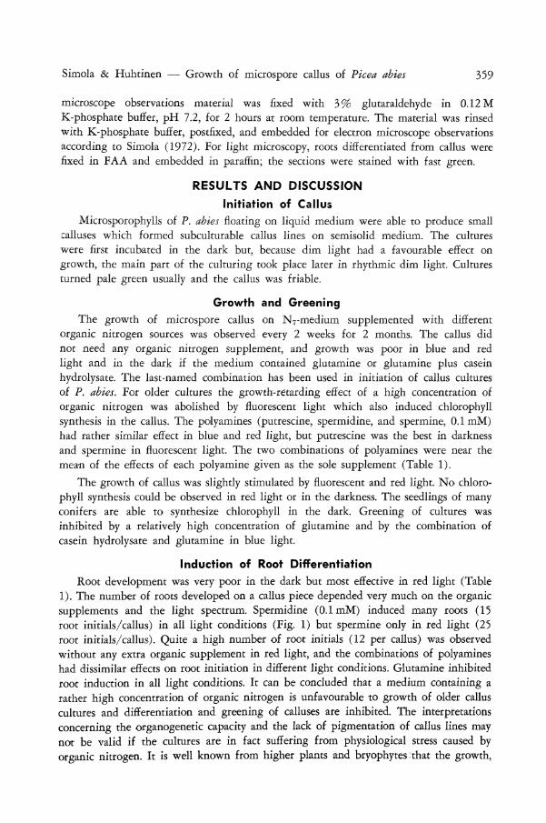
The growth of microspore callus on N7-medium supplemented with different organic nitrogen sources was observed every 2 weeks for 2 months. The callus did not need any organic nitrogen supplement, and growth was poor in blue and red light and in the dark if the medium contained glutamine or glutamine plus casein hydrolysate. The last-named combination has been used in initiation of callus cultures of P. abies. For older cultures the growth-retarding effect of a high concentration of organic nitrogen was abolished by fluorescent light which also induced chlorophyll synthesis in the callus. The polyamines (putrescine, spermidine, and spermine, 0.1 mM) had rather similar effect in blue and red light, but putrescine was the best in darkness and spermine in fluorescent light. The two combinations of polyamines were near the mean of the effects of each polyamine given as the sole supplement (Table 1).
The growth of callus was slightly stimulated by fluorescent and red light. N o chlorophyll synthesis could be observed in red light or in the darkness. The seedlings of many conifers are able to synthesize chlorophyll in the dark. Greening of cultures was inhibited by a relatively high concentration of glutamine and by the combination of casein hydrolysate and glutamine in blue light.
Induction of Root Differentiation
Root development was very poor in the dark but most effective in red light (Table 1). The number of roots developed on a callus piece depended very much on the organic supplements and the light spectrum. Spermidine (0.1 mM) induced many roots (15 root initials/callus) in all light conditions (Fig, 1) but spermine only in red light (25 root initials/callus). Quite a high number of root initials (12 per callus) was observed without any extra organic supplement in red light, and the combinations of polyamines had dissimilar effects on root initiation in different light conditions. Glutamine inhibited root induction in all light conditions. It can be concluded that a medium containing a rather high concentration of organic nitrogen is unfavourable to growth of older callus cultures and differentiation and greening of calluses are inhibited. The interpretations concerning the organogenetic capacity and the lack of pigmentation of callus lines may not be valid if the cultures are in fact suffering from physiological stress caused by organic nitrogen. It is well known from higher plants and bryophytes:that the growth,
v»
TABLE 1—Effect of organic supplements and light on growth and differentiation of microspore callus of Picea abies. Growth: 0 (no growth) to 5 (very good growth). Colour: green = g; white = w; number of roots per callus piece (mean of 20 pieces)
Medium Cone. Darkness Fluorescent light Blue light Red light
Growth Colour Roots Growth Colour Roots Growth Colour Roots Growth Colour Roots
2 w 1 2 g
Casein hydrolysate
+ glutamine
Glutamine
Putrescine
Spermidine
Spermine
Putrescine
+ spermidine
+ spermine
Putrescine
+ spermidine
-f- spermine
1000 mg
500 mg
500 mg
0.1 mM
0.1 mM
0.1 mM
0.25 mM
0.1 mM
0.1 mM
0.1 mM
0.1 mM
0.1 mM
1
1
5
3
2
3
3
w
w
w
w
w
w
w
0
0
3
3
0
6
3
3
3
3
4
5
4
4
g
g
g
g
g
g
g
ON
12
0
0
1
15
2
5
0
0
3
3
3
2
W
w
g
g
g
g
0
0
4
15
0
8
1
2
5
4
4
4
w
w
w
w
w
w
1
0
10
15
25
17
8 g &-o
I o
o
16 w

Simola & Huhtinen — Growth of microspore callus of Picea abies 361
FIG. 1—A root with root hairs differentiated from microspore culture of Picea abies (red light, medium supplemented with spermidine 0.1 mM). x 3
morphogenesis, and greening of cultures are very much affected by exogenously added amino acids (Waris 1967; Simola 1975). The real need for and right level of organic nitrogen sources must be tested if good callus growth and organogenesis are the aims of the study. It is obvious that a low intensity of fluorescent light is favourable to greening and growth. Callus cultures of trees usually need light but a low level of putrescine could compensate for the need for light for effective growth but not for good root differentiation in P. abies.
Light Microscope Observations
The microspore callus cells were round and some tracheids with reticulate cell wall thickenings could be observed under light microscopy. Many callus lines were mixoploid: haploid, diploid, triploid, and tetraploid cells were found in the same culture. The pollen-cultures of some other gymnosperms are karyologically more stable (Tulecke 1957). Spontaneous diploidisation in the megagametophyte calluses in P. abies is common (Huhtinen et al. 1982; Simola & Honkanen 1983). Diploidisation of megagametophyte callus tissue of Pinus lambertiana Dougl, is very rapid. Microspectrophoto-metric measurements showed that the relative D N A amounts of nuclei correspond to the C level only in newly proliferated callus tissue of original explants, whereas all nuclei undergo diploidisation during continued culture and few become tetraploid (Borchert 1968). The results were confirmed by chromosome counts. The callus cultures of gymnosperms deriving from somatic cells are karyologically more stable (Partanen 1963; D'Amato 1978) than those of angiosperms.

362 New Zealand Journal of Forestry Science 16(3)
infrastructure
The fine structure of microspore cultures of P. abies is rather similar to that seen in megagametophyte callus lines cultured in light (Simola & Honkanen 1983). Both types of cultures originating from haploid cells are able to form plastids with small grana and plastogjobuli. It has been found that corresponding cultures of Gmgko have very normal subcellular structure and the ability to form several cell types (Tulecke 1967).
The microspore callus cells of P. abies grown under dim light on a medium supplemented with casein hydrolysate and glutamine had two types of cells. Some cells stored much starch in compound amyloplasts (Simola 1987); some, more meristematic, have small proplastids containing very little or no starch (Fig. 2). In dim light the nucleus may be somewhat irregular in shape and the nucleolus containing proribosomes is very compact.
Dictyosomes are very rare in microspore cells of P. abies (Fig. 2). Irregular patterns of smooth and rough endoplasmic reticulum are seen near the plasmalemma which is relatively wavy in some cells with apparently effective cell wall synthesis (Fig. 3 and 4). Rather similar subcellular structure has been found in aseptically cultured protocorms of Dactylorhiza maculata (L.) Soo (Simola 1982a). Plasmodesmata are very rare (Fig. 5), as in megagametophyte callus of P. abies (Simola & Honkanen 1983).
Accumulation of secondary metabolites in the vacuoles was a characteristic feature of several microspore cultures of P. abies (Fig. 6 and 7). The process was not obviously related to light conditions or medium. Some neighbouring cells had quite dissimilar ultrastructure. The cytoplasm and stroma of plastids were very electron-dense in one cell and the cell may be degenerating; other cells had normal density of cytoplasm and stroma and no accumulation of secondary products was observed (Fig. 8).
In some cells of calluses cultured in blue light, synthesis of secondary products was very prominent and several electron-dense deposits were seen in the vacuoles (Fig. 7). The plastids store very little or no starch in these and neighbouring cells. It has been suggested that in ray parenchyma cells there is a trend for cells containing abundant phenolic compounds not to contain much starch (Wardrop & Crowshow 1962). Sometimes this type of relationship is found also in tissue cultures of conifers but not as a rule (Chafe & Durzan 1973).
The microspore callus cells developed in darkness showed several degenerative features. Ribosomes and mitochondria were few (Fig. 9), and there were some elongated patterns of endoplasmic reticulum near the nucleus and cell wall. The end of the cisternae may enlarge to form a vesicle. These vesicles develop into larger vacuoles. The plastids contained starch as very electron-dense deposits and the middle lamella of cell walls may be also quite electron-dense. No lobes of nucleus were observed but the nucleolus was clearly visible.
CONCLUSIONS
Microspore cells of P. abies are able to form all organelles of the normal plant cell and to undergo root differentiation (Fig. 1) and form small bud-like structures (cf. Fig. 2, Simola 1982b). It is, however, not known if the cells were still haploid in cultures
Simola & Huhtinen — Growth of microspore callus of Picea abies 363
> \H a u*>iv
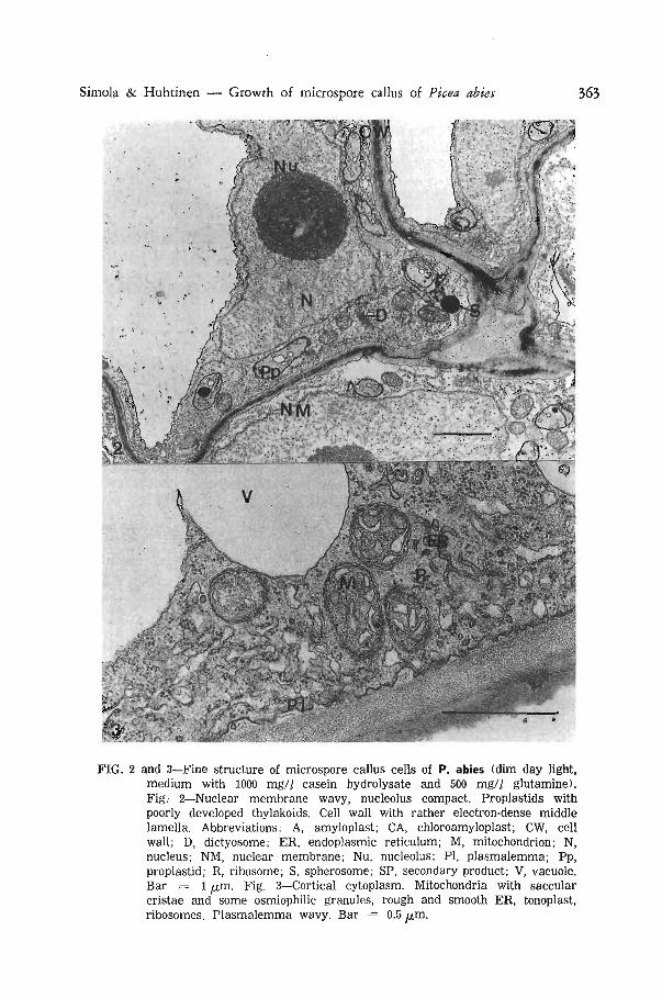
FIG. 2 and 3—Fine structure of microspore callus cells of P. abies (dim day light, medium with 1000 mg// casein hydrolysate and 500 mg/l glutamine). Fig. 2—Nuclear membrane wavy, nucleolus compact. Proplastids with poorly developed thylakoids. Cell wall with rather electron-dense middle lamella. Abbreviations: A, amyloplast; CA, chloroamyloplast; CW, cell wall; D, dictyosome; ER, endoplasmic reticulum; M, mitochondrion; N, nucleus; NM, nuclear membrane; Nu, nucleolus; PI, plasmalemma; Pp, proplastid; R, ribosome; S, spherosome; SP, secondary product; V, vacuole. Bar = 1 yu,m. Fig. 3—Cortical cytoplasm. Mitochondria with saccular cristae and some osmiophilic granules, rough and smooth ER, tonoplast, ribosomes. Plasmalemma wavy. Bar = 0.5 /xm.

364 New Zealand Journal of Forestry Science 16(3)
FIG. 4—Microspore callus cultivated in fluorescent light (no organic supplements in the medium). Chloroamyloplast with a large starch granule, small grana, and small plastoglobuli, mitochondria, secondary products in the vacuole. Bar = 1 ^m.
FIG. 5—Microspore callus cells cultivated in fluorescent light (medium supplemented with spermidine 0.1 mM). Ghloroamyloplasts abundant, no secondary metabolites. Bar = 5 ^m.
Simola & Huhtinen — Growth of microspore callus of Picea abies 365
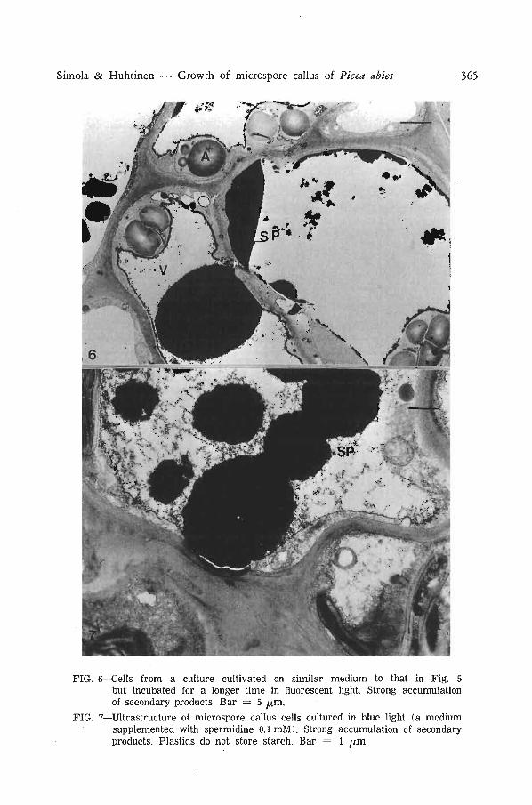
FIG. 6—Cells from a culture cultivated on similar medium to that in Fig. 5 but incubated for a longer time in fluorescent light. Strong accumulation of secondary products. Bar = 5 fx,m.
FIG. 7—Ultrastructure of microspore callus cells cultured in blue light (a medium supplemented with spermidine 0.1 mM). Strong accumulation of secondary products. Plastids do not store starch. Bar = 1 [im.
366 New Zealand Journal of Forestry Science 16(3)
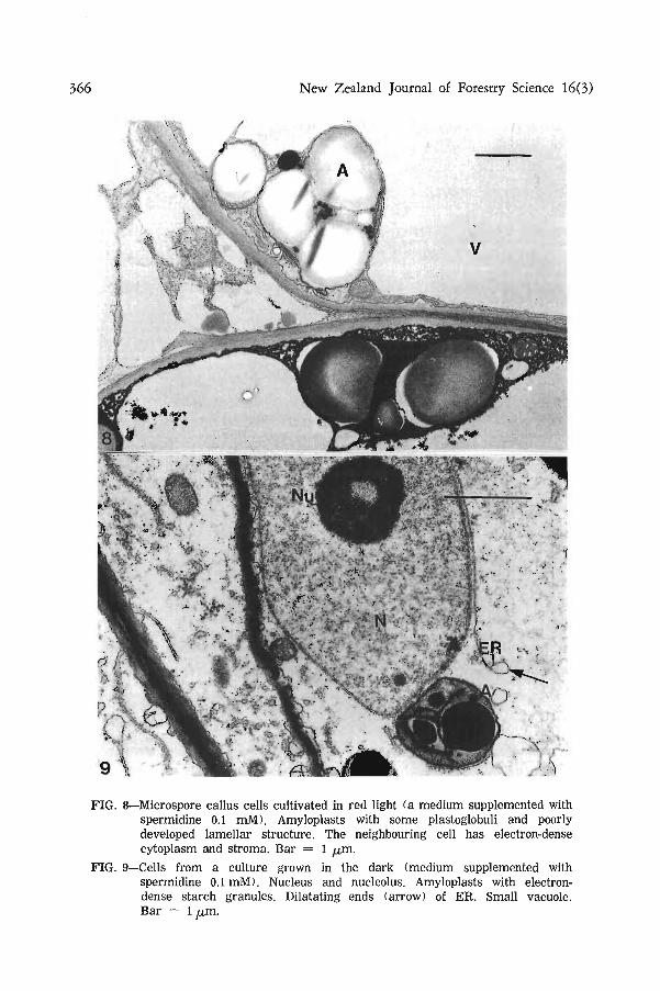
FIG. 8—Microspore callus cells cultivated in red light (a medium supplemented with spermidine 0.1 mM). Amyloplasts with some plastoglobuli and poorly developed lamellar structure. The neighbouring cell has electron-dense cytoplasm and stroma. Bar = 1 ^m.
FIG. 9—Cells from a culture grown in the dark (medium supplemented with spermidine 0.1 mM). Nucleus and nucleolus. Amyloplasts with electron-dense starch granules. Dilatating ends (arrow) of ER. Small vacuole. Bar = 1 /xm.
Simola & Huhtinen — Growth of microspore callus of Picea abies 367
from which the organogenesis began. Strong synthesis of starch indicates that the uptake of sucrose from the medium is effective, the regular structure of mitochondria ensures energy supply for biosynthetic processes, and the relatively high number of ribosomes ensures synthesis of structural and enzyme proteins. Light enhances both growth and organogenesis.
Leakage of growth-retarding phenolic compounds in the medium of tissue cultures of pine and spruce is a big problem. The strong browning of callus cultures of conifers due to synthesis of secondary metabolites seems to inhibit both cell division and differentiation and lead to death of the culture. On the other hand, it was found that rapidly growing white megagametophyte callus lines did not differentiate easily but strains with brownish areas showed also morphogenesis (Simola & Honkanen 1983). Secondary products may regulate the intracellular hormone balance and indirectly affect the triggering of the organogenetic programme. Accumulation of secondary products and strong synthesis of starch was observed in microspore callus of P. abies in fluorescent light (Fig. 6) on a medium supplemented with spermidine (0.1 mM). This culture forms only very few roots but in red light, when synthesis of secondary products is observed in some cells, roots were more abundant on the same medium.
The poor results obtained in callus cultures of conifers may be due partly to the lack of experiments concerning nitrogen supplements and light. The very low amount of starting material in the case of haploid callus lines makes the experiments time-consuming but without rather large experiments with homogeneous material very little progress can be made in this field.
ACKNOWLEDGMENTS Our thanks are due to Mrs Pirkko Lazanyi, Dept, of Electron Microscopy, University
of Helsinki, and Mrs Pirkko Einisto, M.Sc, for their kind technical assistance. The work has been supported by the Academy of Finland.
REFERENCES BORCHERT, R. 1968: Spontane Diploidisierung in Gewebekulturen des Megagametophyten
von Pinus lambertiana. Zeitschrift fiir Pflanzenphysiologie 59: 389-92.
BONGA, J. M. 1974: In vitro culture of microsporophyll and megagametophyte tissue of Pinus. In Vitro 9: 270-7.
BONGA, J. M.; VENKETESWARAN, S. 1976: Fixing and staining of conifer tissues with lacto-propiono-dimethylsulfoxide-carmine. Stain Technology 51: 197-9.
CARDEMIL, L.; JORDAN, M. 1982: Light and electron microscopic study in vitro cultured female gametophyte of Araucana araucana (Mol.) Koch. Zeitschrift fiir Pflanzenphysiologie 107: 329-38.
CHAFE, S. C; DURZAN, D. J. 1973: Tannin inclusions in cell suspension cultures of white spruce. Planta 113: 251-62.
DAKER, M. G. 1967: Cytological studies on a haploid cultivar of Pelargonium and its colchicine derivative. Chromosoma 21: 250-71.
D'AMATO, F. 1978: Chromosome number variation in cultured cells and regenerated plants. Pp. 287-95 in Thorpe, T. A. (Ed.) "Frontiers of Plant Tissue Culture". International Association for Plant Tissue Culture, University of Calgary Offset Printing Service, Calgary, Canada.
HUHTINEN, 0. 1972: Production and use of haploids in breeding conifers. IUFRO Genetics-Sabrao joint symposia, Tokyo, D-3 (I): 1-8.

368 New Zealand Journal of Forestry Science 16(3)
HUHTINEN, 0.; HONKANEN, J.; SIMOLA, L. K. 1982: Effects of genotype and nutrient media on callus production and differentiation of Norway spruce endosperms cultured in vitro. Pp. 307-11 in Colloque International sur Ia Culture "in vitro" des Essences Forestieres, IUFRO, Fontainebleau, 1981.
ILLIES, Z. M. 1964: Auftreten haploider Keimlinge bei Picea abies. Naturwissenschaften 51: 442.
MURASHIGE, T.; SKOOG, F. 1962: A revised medium for rapid growth and bioassays with tobacco tissue cultures. Physiologia Plantarum 15: 473-97.
PARTANEN, C. R. 1963: Plant tissue culture in relation to developmental cytology. International Review of Cytology 15: 215-43.
POHLHEIM, F. 1968: Thuja gigantea gracilis Beissn - ein Haplont under den Gymno-spermen. Biologisch Rundschau 6: 84-6.
RAO, N. M.; MEHTA, A. R. 1969: Callus tissue from the pollen of Thuja orientalis L. Indian Journal of Experimental Biology 7: 132-3.
SIMAK, M.; GUSTAFSSON, A.; CHING, K. 1968: Occurrence of mosaicaneuploid in poly-embryonic Norway spruce seed. Studia Forestalia Suecica 67: 1-8.
SIMOLA, L. K. 1972: Changes in the ultrastructure of cells of Atropa belladonna cv. lutea Doll, during growth and differentiation in suspension culture. Zeitschrift fur Pflan-zenphysiologie 68: 215-27.
1975: The effect of several protein amino acids and some inorganic nitrogen sources on the growth of Sphagnum nemoreum. Physiologia Plantarum 35: 194-9.
1982a: Electron microscope observations on the differentiation of protocorm cells of Dactylorhiza maculata. Nordic Journal of Botany 2: 125-30.
1982b: Ultrastructure of callus cultures from trees. Pp. 201-10 in Colloque International sur Ia Culture "in vitro" des Essences Forestieres, IUFRO, Fontainebleau, 1981.
1985 Propagation of plantlets from leaf callus of Betula pendula f. purpurea. Scientia Horticulturae 26: 77-85.
1987: Structure of cell organelles and cell wall in tissue cultures of trees. Pp. 389-418 in Bonga, J.; Durzan, D. (Ed.) 'Tissue Culture in Forestry" Vol. 1. Martinus Nijhoff/ Dr. W. Junk, The Hague.
SIMOLA, L. K.; HONKANEN, J. 1983: Organogenesis and fine structure in megagametophyte callus lines of Picea abies. Physiologia Plantarum 59: 551-61.
SIMOLA, L. K.; KOSKIMIES-SOININEN, K. 1984: Comparison of glycoUpids and plastids in callus cells and leaves of Alnus and Betula. Plant and Cell Physiology 25(8): 1329-40.
TULECKE, W. 1957: The pollen of Ginkgo biloba: In vitro culture and tissue formation. American Journal of Botany 44: 602-8.
1967: Studies on tissue cultures derived from Ginkgo biloba L. Phytomorphology 17: 381-6.
WARDKOP, A. B.; CROWSHOW, J. 1962: Formation of phenolic substances in the ray parenchyma of angiosperms. Nature 193: 90̂ -2.
WARIS, H. 1967: Morphological changes in seed plants induced with amino acids, purines and pyrimidines. Annales Academiae Scientiarum Fennicae Series A IV 106: 1-66.


![Practice For May: Cell Ultrastructure [114 marks]blogs.4j.lane.edu/.../2018/02/Cell-Ultrastructure-Test-1.pdfPractice For May: Cell Ultrastructure [114 marks]1. Which structure found](https://img.pdfslide.net/doc/110x75/5eda4db5b3745412b5711d9c/practice-for-may-cell-ultrastructure-114-marksblogs4jlaneedu201802cell-ultrastructure-test-1pdf.jpg)