Embed Size (px)
Citation preview

Growth Plate Injuries
Mo Mortazavi, MDDirector, SPARCC Sports Medicine ProgramAssistant Professor, University of ArizonaMedical Director, TUSD inter scholastics
Financial Disclosures
■ No relevant financial relationships

Objectives
■ Review anatomy of growth plates■ Review pathology of the apophysis■ Discuss clinical cases of apophysitis and avulsions■ Highlight management / Return to play
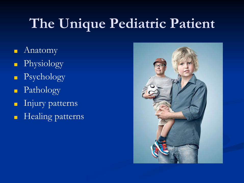
The Unique Pediatric Patient
■ Anatomy■ Physiology ■ Psychology■ Pathology■ Injury patterns■ Healing patterns

Growing Pediatric Bone
“E”piphysis vs “A”pophysis

Epiphysis Definition
■ Site of long bone longitudinal growth■ Epiphyseal plate is the primary center of
ossification

Epiphyseal Injuries
■ Salter Harris injuries■ Fracture/dislocation “slip” injuries■ Stress (overuse) injuries■ Age and physeal dependent■ Focus of next talk

Apophysis Definition
■ Secondary centers of ossification ■ Site of muscle-tendon unit insertion■ Provide contour and shape to growing bones
(without adding length)■ Age dependent closure of each apophysis

Apophysis Definition
■ Weak link in the musculoskeletal chain at given age ranges
■ Apophysis is less resistant to tensile forces than soft tissues surrounding that area
■ Chronic (overuse) injuries = Apophysitis■ Acute injuries = Avulsion fractures

ApophysitisDefinition
■ Chronic (overuse) injury resulting from ■ Traction of a tendon at its insertion ■ Microavulsions at the bone-cartilage junction
■ Common during periods of rapid growth
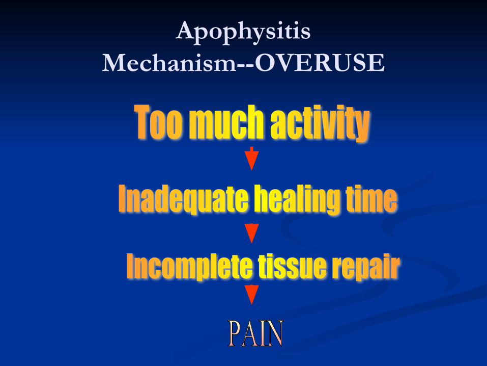
ApophysitisMechanism--OVERUSE

The 4 Stages of Overuse Injury
(1) Pain in the affected area after physical activity(2) Pain during the activity, without restricting
performance (3) Pain during the activity that restricts
performance (4) Chronic, unremitting pain even at rest
Brenner, JS. Overuse Injuries, Overtraining, and Burnout in Child and Adolescent Athletes. Pediatrics 2007;119: 1242.
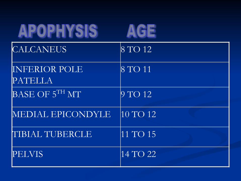
CALCANEUS 8 TO 12
INFERIOR POLE PATELLA
8 TO 11
BASE OF 5TH MT 9 TO 12
MEDIAL EPICONDYLE 10 TO 12
TIBIAL TUBERCLE 11 TO 15
PELVIS 14 TO 22

ApophysitisThe Athlete at Risk
■ Age dependent patterns■ Rapid increase in activity■ Elite, fit, driven■ Biomechanical deficits

ApophysitisManagement
■ Rest and Rehab (address bio-mechanics)■ Alleviating measures (Ice, OTC pain relief)■ Benefits of cross-training and varied sports
participation■ Reduce overuse injury (and potential for burnout)■ Improved performance in primary sport!

ApophysitisManagement
■ Relative Rest■ Pain free activities allowed
■ Correct muscle length/strength imbalances■ Correct “biomechanical” factors■ Consider orthotics, heel cups, bracing prn
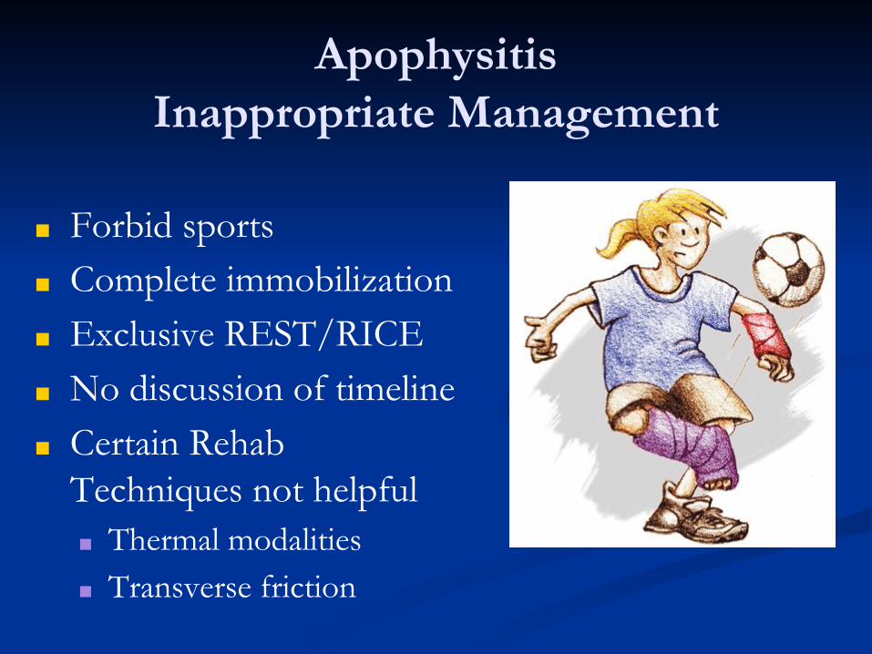
ApophysitisInappropriate Management
■ Forbid sports■ Complete immobilization■ Exclusive REST/RICE■ No discussion of timeline■ Certain Rehab
Techniques not helpful■ Thermal modalities ■ Transverse friction

Avulsion Fractures
■ Most commonly involve apophysis (weak link)
■ Acute MOI■ Pop, swelling, bruising,
severe disability■ MSK chain is “broken”■ Often preceded by
apophysitis

Not ALL Apophyses are created Equal!
■ Upper extremities■ LL elbow, coracoid, LE
■ Pelvis■ IT, AIIS, ASIS, LT, IC, AT
■ Lower extremity■ Osgood Schlatter’s, Sever’s,
SLJ, Islen’s
* Variable Avulsion and Arrest Risks

Avulsion fractureManagement
■ Non-displaced: immobization followed by PT
■ Displaced: may need surgical fixation
■ Varying degrees of acceptable displacement
■ Apophyseal dependent

Case 1 KNEE PAIN
■ 12 y.o. male, basketball ■ Intermittent L knee pain, no trauma■ Pain worse with jumping and kneeling

Knee Exam
■ Inspection- Enlarged tibial tuberosity ■ ROM-Normal, tight hamstrings ■ Palpation- Localized tenderness over tibial
tuberosity■ Neurovascular-Intact■ Special Maneuvers-no ligamentous laxity

Knee Exam
■ Popliteal Angle measures hamstring flexibility

Osgood-Schlatter Apophysitis

Osgood-Schlatter
■ Traction apophysitis of tibial tubercle
■ Forceful, repetitive contractions of quads■ Jumping, running, cutting
■ Athletes 11-15 years old, females younger
■ More common unilateral

Tibial Tuberosity Apophysitis Treatment
■ Active rest, RTP as tolerated■ Ice massage, OTC pain control■ Rehab-Stretch Hamstrings/Quads■ Cho-Pat Straps

Osgood-Schlatter gone badAvulsion fracture (acute) Heterotopic ossification (chronic)
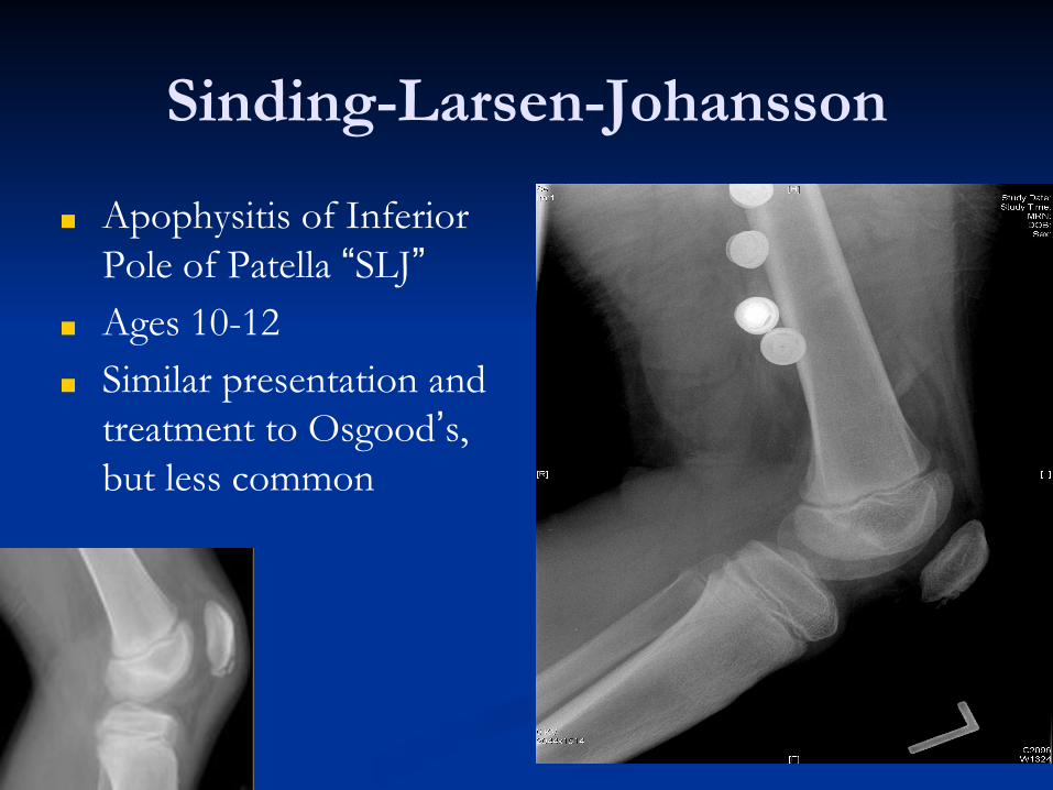
Sinding-Larsen-Johansson
■ Apophysitis of Inferior Pole of Patella “SLJ”
■ Ages 10-12 ■ Similar presentation and
treatment to Osgood’s, but less common

Case
■ 10 year old girl slipped and fell onto hyperflexed knee
■ Felt “pop”, immediate pain and large swelling■ Unable to walk

Exam
■ Inspection- Large effusion, high riding patella■ Palpation- Max pain over distal patellar pole w/
2cm gapping■ ROM- Guarding at 30 deg flexion■ Neurovascular- Unable to straight leg raise■ Special maneuvers- Deferred
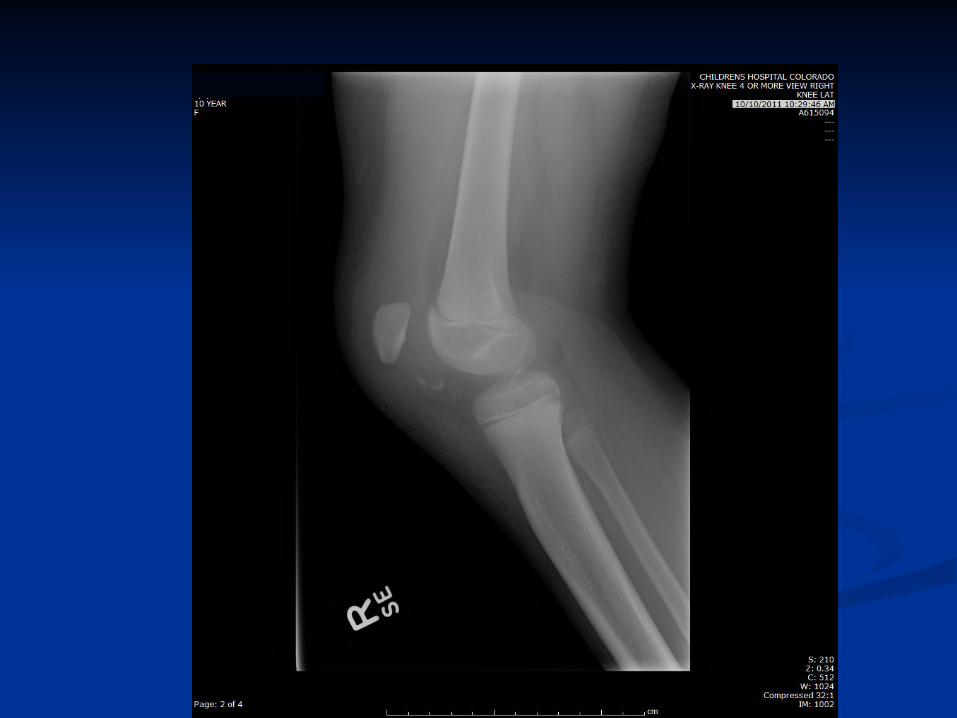


Patellar sleeve fracture
Treatment
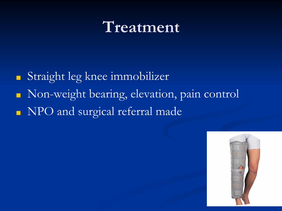
■ Straight leg knee immobilizer ■ Non-weight bearing, elevation, pain control■ NPO and surgical referral made

Post-Op films

Case HEEL PAIN
■ 11 y.o. boy, track athlete■ 2 month hx of R heel pain■ No injury, Pain activity related

Foot/Ankle Exam
■ Neurovascular-Intact■ Inspection- No swelling, normal arches ■ ROM-Normal, tight heel cord ■ Palpation- Localized tenderness over calcaneus■ Special Maneuvers-no ligamentous laxity,
positive calcaneal squeeze test

Sever’s DiseaseRadiographs
■ Normal ■ Varying degrees of sclerosis and fragmentation
of aphophysis

Sever’s Disease
■ Traction apophysitis of ossification center of calcaneus
■ Repetitive contractions of gastroc-soleus complex■ Impact sports especially when running and cleats or
without shoes■ Ages 9-12 yrs, earlier in girls■ Bilateral in 60%; Avulsion risk very low

SEVER’S DiseaseTreatment
■ Relative Rest, RTP as tolerated■ Gastrocnemius-soleus stretching and
eccentric strengthening■ Viscoelastic heel cups in all shoes

Case
• 12 yo twists R foot while playing soccer• Inversion type injury with immediate
swelling• Unable to put weight on his R foot• X-rays?
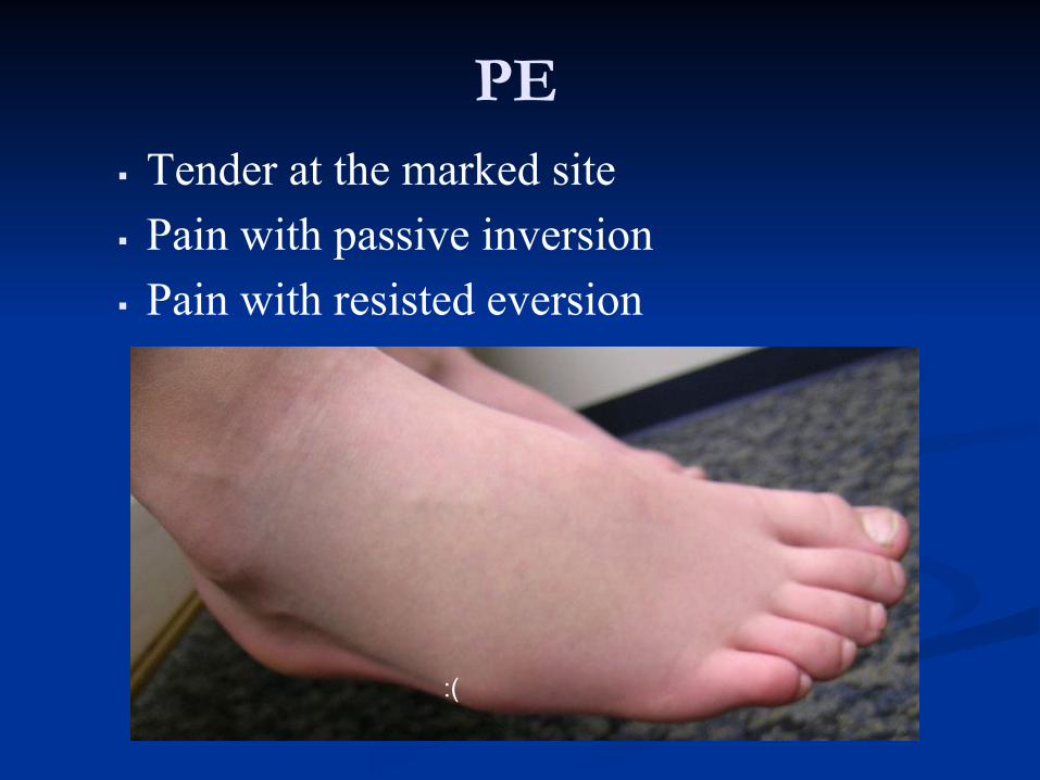
PE▪ Tender at the marked site▪ Pain with passive inversion▪ Pain with resisted eversion
:(

X-rays▪ Soft tissue swelling▪ Normal apophysis▪ Avulsion Fx of the base of
the 5th metatarsal

Pediatric base of 5th metatarsal fracture
■ Must be differentiated from a normal apophysis■ Tenderness■ Apophysis lies parallel to the long axis of MT■ Fracture is almost always transverse■ Comparison views in needed

Base of 5th metatarsal fracture
■ Treatment■ Non-surgical in most cases
with good outcome■ Short-leg walking cast or
boot for 3 weeks■ Ortho referral with
displacement >3 mm

Iselin’s Disease
■ Traction apopysitis at ossification center at base of 5th MT
■ Chronic contractions of peroneus brevis tendon■ Athletes 10-12 years old, females younger■ More common unilateral■ Xrays typically normal

Case
■ 15 yo female, Track runner■ Chronic lateral B/L hip pain■ Atraumatic, worse with running/hurdles■ No popping/mechanical symptoms

Hip Exam
■ Inspection- No swelling ■ ROM-Normal, symmetric IR/ER/AB, Pain with
resisted hip abduction and trunk rotation■ Palpation- Localized tenderness over iliac crest
bilaterally■ Neurovascular-Intact■ Special Maneuvers-negative FADIR

Iliac Crest Apophysitis
■ Traction apophysitis ossification center of iliac crest
■ Chronic contraction of abdominal muscles■ Running sports-arm
swing/trunk rotation■ Ages mid to late teens

Hip Apophysitis

Case
■ 15yo boy tries 50 yard FG attempt■ Suddenly feels a loud pop and falls to the ground
in severe pain■ Pain in the right anterior hip region■ Taken to the ED were x-rays were completed
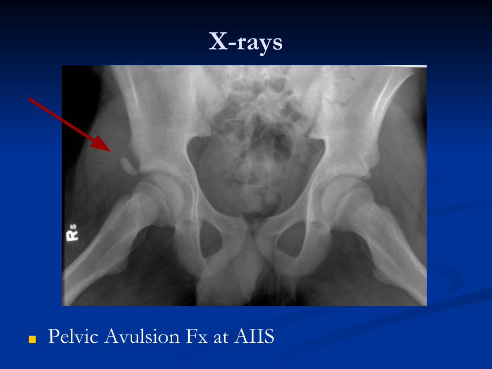
X-rays
■ Pelvic Avulsion Fx at AIIS

Pelvic Avulsion Fx
■ Acute presentation■ Tender apophysis after forceful contraction
and “pop”>>think avulsion fracture■ Pain and weakness elicited when activating
associated muscle

Treatment
■ Rest, Ice, protected movement, crutches 3-4 wks■ Begin early ROM when pain subsides.■ Formal physical therapy to regain ROM,
strength, and flexibility.■ Gradual return to play when pain free with
functional testing.

Pelvic Avulsion FractureIschial Tuberosity

Pelvic Avulsion FractureASIS

Pelvic Avulsion FractureIliac Crest
■

Normal?
■ 13 y/o felt pop in buttock during sprint in football
■ Pain, unable to continue running
■ X-rays read normal
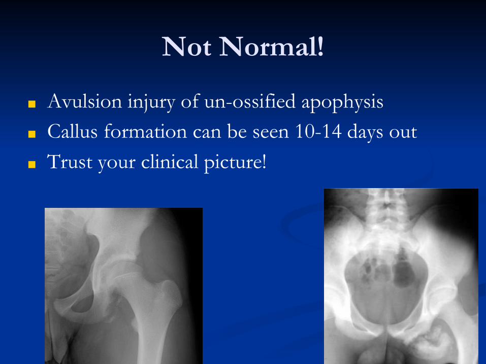
Not Normal!
■ Avulsion injury of un-ossified apophysis■ Callus formation can be seen 10-14 days out■ Trust your clinical picture!

Case 7ELBOW PAIN
■ 12 y.o. male, baseball pitcher, RHD■ 6 weeks R medial elbow pain, no injury■ No locking, catching, motion deficit■ Pain activity-related, no pain at rest

Elbow Exam
■ Inspection-No swelling, no atrophy■ ROM- Full, pain with wrist flexion■ Palpation- tenderness over medial epicondyle■ Neurovascular-intact■ Special Maneuvers-No instability

ELBOW XRAY

Elbow Anatomy
■ Multiple ossification centers (6)
■ CRITOE■ Appear every 2 years■ All close at different
times

Little Leaguer’s ElbowDefinition
■ Related to the stress of repetitive throwing in youth
■ Medial Epicondylar Apophysitis■ Medial epicondyle weakest structure in elbow, last to
close■ Stress reaction of medial epicondyle apophysis■ X-ray can be normal or slightly irregular

Little Leaguer ElbowMedial Epicondylar Apophysitis
■ Tenderness over medial epicondyle
■ Radiographs can show■ Separation/widening of
apophysis■ Fragmentation
■ MRI usually not necessary■ Clinical Diagnosis!

Elbow Pain in Throwers
■ Most common complaint in younger thrower ■ AGE IS IMPORTANT
RISK FACTOR■ Pitch counts, rest days are a
significant consideration

Forces on the elbow
■ Common site of injury■ Traction injuries -
medial side■ Compression – lateral
side

Medial elbow painA look at biomechanics
■ Large valgus loads with rapid elbow extension = high tensile stress on medial elbow■ Medial epicondyle apophysitis ■ Ulnar collateral ligament injury■ Flexor-pronator mass tendinitis■ Ulnar neuritis

Little Leaguer’s ElbowTreatment
■ NO THROWING until symptoms improve■ Usually 1 to 3 months
■ Rehabilitation, biomechanics, kinematics■ Interval Throwing Program--SLOW return,
consider switching position■ Pitch counts, types of pitches, rest days■ Never throw with a FATIGUED arm!!!

Acute Throwing Injury…

ME Avulsion Fracture
■ Often acute on chronic■ Preceded by MEA■ Pop, severe pain,
swelling, disability■ Surgical >3-5mm
displaced
AP and Stress views:
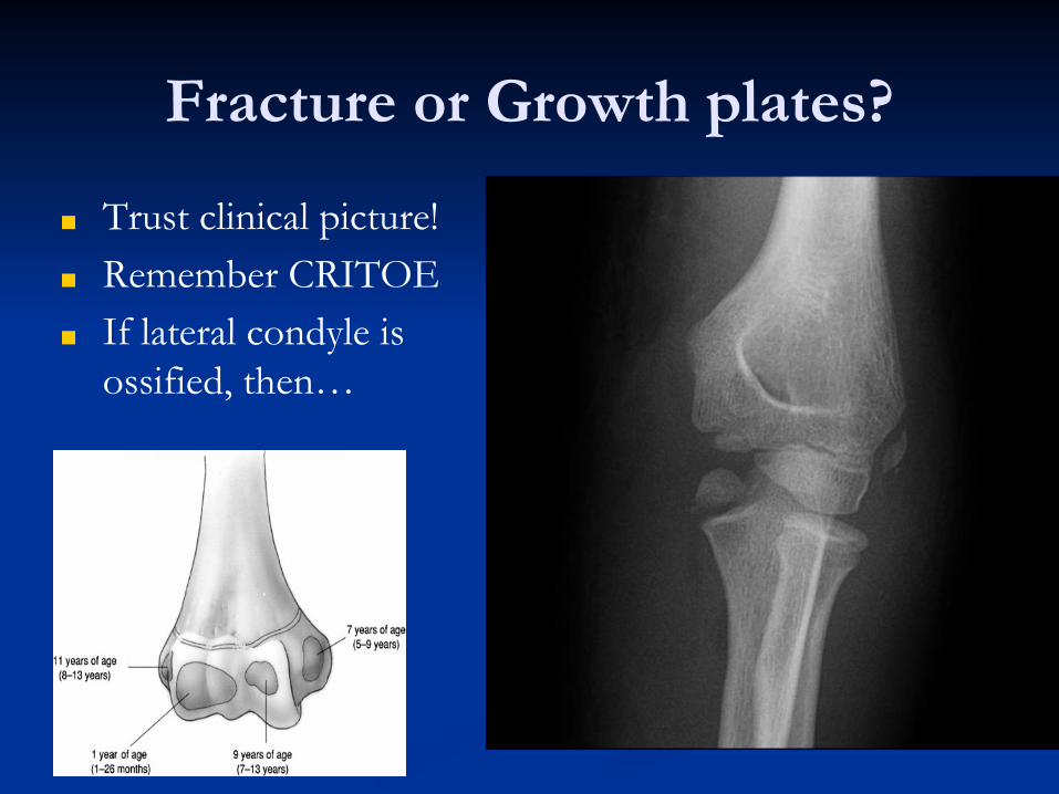
Fracture or Growth plates?
■ Trust clinical picture!■ Remember CRITOE■ If lateral condyle is
ossified, then…

Great PCSM References
http://www.wheelessonline.comhttp://www.radiologyassistant.nlwww.uahealth.com/pedssportsmed

■ Questions?


![FPM50b - SANUS VuePoint · VMPL50 / FPM50-Mount brackets to wall plate 002164.eps VMPL50 / FPM50-Mount brackets to wall plate [24] 2x [23] Avoid potential personal injuries or property](https://img.pdfslide.net/doc/110x75/5ffaad7c196dc924197817fc/fpm50b-sanus-vuepoint-vmpl50-fpm50-mount-brackets-to-wall-plate-002164eps-vmpl50.jpg)