Embed Size (px)
Citation preview

Guided surgery: accuracy andefficacyMARJOLEIN VERCRUYSSEN, MARGARETA HULTIN, NELE VAN ASSCHE,KRISTER SVENSSON, IGNACE NAERT & MARC QUIRYNEN
Preoperative three-dimensional planning has gainedpopularity because of the introduction of cone beamcomputed tomography. Different concepts of three-dimensional planning, such as computer-guided(static) surgery and computer-navigated (dynamic)surgery, have been proposed to transfer virtual digitalplanning from a personal computer to the surgicalfield (42). In computer-guided (static) surgery, a staticsurgical guide is used that transfers the virtualimplant position from computed tomography datatothe surgical site. These guides are produced by com-puter-aided design/computer-assisted manufacturetechnologies, such as stereolithography, or manuallyin a dental laboratory, using mechanical positioningdevices or drilling machines (42, 73, 78, 80). Duringcomputer-navigated surgery, the position of the sur-gical instruments in the surgical area is constantlydisplayed on a screen with a three-dimensional imageof the patient. In this way, the system allows real-timetransfer of the preoperative planning and visual feed-back on the screen (16, 67, 82). In the review of Junget al. (42), a statistically significant higher mean pre-cision was found in favor of dynamic systems com-pared with the static surgical guides. However thisdifference could be explained by the fact that thereare more preclinical studies on accuracy for thedynamic systems and more clinical studies for thestatic systems. In contrast to dynamic guidance, the‘static’ guidance via surgical templates does not allowchanges to be made to the surgical plan at the time ofsurgery. However, the bur sleeves of the templatespermit rigidly guided and highly controllable drilling,which may be an advantage in areas where irregularbone is present. Furthermore, the intraoperative set-up of a navigation system is not required, and thereare no time constraints and potential inconvenienceof intraoperative registration and tracking. Intraoper-ative optical navigation devices are more frequently
used in craniomaxillofacial surgery. Despite the factthat some clinical and accuracy studies are available,dynamic systems currently have a very limited indica-tion in implant dentistry and are not in widespread useas a result of the initial high costs. Computer-navigatedsurgery systems are not included in the current review.
Using three-dimensional planning software, thesurgeon can, after consulting with the dentist to pro-vide a template representing the planned prosthesis,properly position implants in a virtual reality. Whenthe planned prosthesis is incorporated into thesecomputed tomography images, the planning can takeinto account both the jawbone anatomy and theplanned superstructure. This should improve biome-chanics and esthetics. Moreover, it may optimize themutual interaction between the ‘surgical’ and theprosthetic teams. Precise preoperative planning hasmade it possible to implement immediate loading ina relatively predictive manner and hence reduce thetreatment time and increase comfort for the patient.Furthermore, when combined with flapless surgery, itis presumed that postoperative patient morbidity anddiscomfort may also be reduced. As a result, implantplacement may develop from difficult toward simplesurgery and from stress toward relative comfort, forboth the patient and the surgeon.
The limits of the use of static guided surgery are setby the maximum deviation observed between plan-ning and postoperative outcome. Deviations mayreflect the sum of all errors occurring from imaging tothe transformation of data into a guide, to the impro-per positioning of the latter during surgery. Thus, allerrors, although seldom occurring, can be cumula-tive. Much attention will be paid to the latter aspect.Indeed, when blind surgery is performed, as during aflapless approach, this is very relevant. Criticalanatomical structures, such as the mandibular canalor mental foramen, must be avoided at all costs to
228
Periodontology 2000, Vol. 66, 2014, 228–246 © 2014 John Wiley & Sons A/S. Published by John Wiley & Sons Ltd
Printed in Singapore. All rights reserved PERIODONTOLOGY 2000

prevent neurological complications. The preoperativeradiological determination of the distances betweenanatomical landmarks can lack precision (15), andthis constitutes a serious risk, especially in the case ofblind surgery. Significant variations can be observedwithin the systems working with surgical guides (e.g.for example, the guidance of the drills in the surgicaltemplates). Some use different templates with sleeveswith increasing diameter for one patient. Othersapply removable sleeves in one single template (withremovable sleeve inserts or sleeves on drills). Somesystems have specially designed drills or drill stops toallow depth control, whereas others have indicationlines on the drills. After preparation of the implant os-teotomy, some systems allow guided placement ofthe implant, whereas for other systems the templatehas to be removed before implant insertion. Theseare only some examples illustrating how difficult it isto interpret and compare individual studies. The sys-tematic reviews of Jung and co-workers (42) andSchneider and co-workers (67), who reviewed bothaccuracy and clinical efficacy, concluded that differinglevels and quantity of evidence were available for com-puter-assisted implant placement and that futureresearch should be directed to increase the number ofclinical studies with longer observation periods and toimprove the systems in terms of accuracy and efficacy.
This review aims to provide an overview of theaccuracy of the procedure and also to give an over-view of the efficacy of static guided surgery. The datafrom two recent systematic reviews (37, 73) arediscussed in this paper.
Accuracy
Definition
Accuracy is defined as matching the position of theplanned implant in the software with the actual posi-tion of the implant in the mouth of the patient. Theaccuracy of the implant or the osteotomy site ismostly expressed by four parameters (Fig. 1): deviationat the entry point; deviation at the apex; deviationof the long axis; and deviation in height/depth.Matching of the planned with the placed implantposition can be based on a second (cone beam) com-puted tomography scan (allowing matching betweenpreoperative planning and postoperative implantpositions) or via ‘model matching’ (by comparingpre- and postoperative models of the treated jaw)(43). The mean deviations for model and computedtomography matching are quite similar: respectively,
0.5 (range: 0.1–1.2) mm and 0.8 (range: 0.1–2.7) mmat the entry point and 0.5 (range: 0.1–1.3) mm and 1.1(range: 0.2–3.6) mm at the apex (46,59).
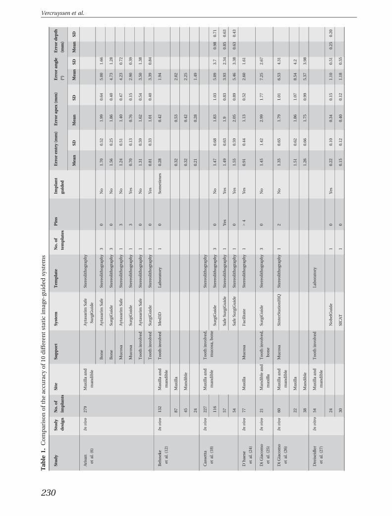
Findings
Data from a recent systematic review (73) revealed anoverall mean deviation, at the entry point, of 1.0 mm(standard error = 0.12 mm; 95% confidence interval:0.8–1.2); range: 0–6.5 mm. The corresponding data atthe apex were 1.2 mm (standard error = 0.1 mm; 95%confidence interval: 1.0–1.6); range: 0–6.9 mm. Theoverall mean angulation was 3.8° (standard error =0.3°, 95% confidence interval: 3.2–4.4); range: 0.0–24.9°. The overall mean vertical deviation (based onfive studies) was 0.5 mm (standard error = 0.1 mm,95% confidence interval: 0.2–0.7), with a maximumranging from 2.3 to 4.2 mm. This review included 19articles, which reported on accuracy. Of these studies,two were model based, five were on human cadaversand 12 were on patients. Four to 54 patients wereincluded in each study, giving a total of 279 patientsoverall. The accuracy of 10 different static image-guided systems has been reported (Table 1). Largedeviations were found to occur. The total deviation isthe cumulative number of deviations that can occurat each step (80, 82). These deviations may be consid-ered as very large, but an in-vivo randomized clinicaltrial comparing guided surgery with mental naviga-tion (with or without any type of surgical template) iscurrently not available. Two in-vitro studies on acrylicmodels (53, 65) compared deviations for mentalnavigation with deviations for guided surgery, and a
Fig. 1. Accuracy is expressed by the following parameters:a deviation at the entry point of the implant or cavity (indi-cated by letter a); deviation at the apex of the implant orcavity (indicated by the letter b); deviation of the axis ofthe cavity or implant (indicated by the symbol alpha);deviation in height/depth (indicated by the letter y) andthe horizontal/lateral deviation (x).
Guided surgery
229
Tab
le1.
Comparisonoftheac
curacy
of1
0differentstatic
imag
e-gu
ided
system
s
Study
Study
design
No.o
f
implants
Site
Support
System
Tem
plate
No.o
f
templates
Pins
Implant
guided
Erroren
try(m
m)
Errorap
ex(m
m)
Erroran
gle
(°)
Errordep
th
(mm)
Mea
nSD
Mea
nSD
Mea
nSD
Mea
nSD
Arisan
etal.(6)
Invivo
279
Maxillaan
d
man
dible
Aytasarim
Safe
SurgiG
uide
Stereo
lithograp
hy
Bone
Aytasarim
Safe
Stereo
lithograp
hy
30
No
1.70
0.52
1.99
0.64
5.00
1.66
Bone
SurgiG
uide
Stereo
lithograp
hy
30
No
1.56
0.25
1.86
0.40
4.73
1.28
Muco
saAytasarim
Safe
Stereo
lithograp
hy
13
No
1.24
0.51
1.40
0.47
4.23
0.72
Muco
saSu
rgiG
uide
Stereo
lithograp
hy
13
Yes
0.70
0.13
0.76
0.15
2.90
0.39
Tooth
invo
lved
Aytasarim
Safe
Stereo
lithograp
hy
10
No
1.31
0.59
1.62
0.54
3.50
1.38
Tooth
invo
lved
SurgiG
uide
Stereo
lithograp
hy
10
Yes
0.81
0.33
1.01
0.40
3.39
0.84
Beh
nek
e
etal.(12
)
Invivo
132
Maxillaan
d
man
dible
Tooth
invo
lved
Med
3DLa
boratory
10
Sometim
es0.28
0.42
1.94
87Maxilla
0.32
0.53
2.02
45Man
dible
0.32
0.42
2.25
240.21
0.28
1.49
Cassetta
etal.(18
)
Invivo
227
Maxillaan
d
man
dible
Tooth
invo
lved
,
muco
sa,b
one
Stereo
lithograp
hy
116
SurgiG
uide
Stereo
lithograp
hy
30
No
1.47
0.68
1.83
1.03
5.09
3.7
0.98
0.71
57Sa
feSu
rgiG
uide
Stereo
lithograp
hy
1Yes
Yes
1.49
0.63
1.9
0.83
3.93
2.34
0.85
0.63
54Sa
feSu
rgiG
uide
Stereo
lithograp
hy
10
Yes
1.55
0.59
2.05
0.89
5.46
3.38
0.63
0.43
D´hae
se
etal.(24
)
Invivo
77Maxilla
Muco
saFac
ilitate
Stereo
lithograp
hy
1>4
Yes
0.91
0.44
1.13
0.52
2.60
1.61
DiG
iaco
mo
etal.(25
)
Invivo
21Man
dible
and
maxilla
Tooth
invo
lved
,
bone
SurgiG
uide
Stereo
lithograp
hy
30
No
1.45
1.42
2.99
1.77
7.25
2.67
DiG
iaco
mo
etal.(26
)
Invivo
60Maxillaan
d
man
dible
Muco
saSinterStationHiQ
Stereo
lithograp
hy
12
No
1.35
0.65
1.79
1.01
6.53
4.31
22Maxilla
1.51
0.62
1.86
1.07
8.54
4.2
38Man
dible
1.26
0.66
1.75
0.99
5.37
3.98
Dreiseidler
etal.(27
)
Invitro
54Maxillaan
d
man
dible
Tooth
invo
lved
Laboratory
24NobelGuide
10
Yes
0.22
0.10
0.34
0.15
1.10
0.51
0.25
0.20
30SICAT
10
0.15
0.12
0.40
0.12
1.18
0.55
Vercruyssen et al.
230

Tab
le1.
(Con
tinued
)
Study
Study
design
No.o
f
implants
Site
Support
System
Tem
plate
No.o
f
templates
Pins
Implant
guided
Erroren
try(m
m)
Errorap
ex(m
m)
Erroran
gle
(°)
Errordep
th
(mm)
Mea
nSD
Mea
nSD
Mea
nSD
Mea
nSD
Ersoy
etal.(29
)
Invivo
94Maxillaan
d
man
dible
Ay-Design
Stereo
lithograp
hy
>1
Notap
plic
able
No
1.22
0.85
1.51
1.00
4.90
2.36
23Muco
sa1.10
0.70
1.70
1.00
4.90
2.20
45Bone
1.30
1.00
1.60
1.50
5.10
2.70
26Tooth
invo
lved
1.10
0.60
1.30
0.70
4.40
1.60
48Maxilla
1.04
0.56
1.57
0.97
5.31
0.36
46Man
dible
1.42
1.05
1.44
1.03
4.44
0.31
Ozan
etal.(56
)
Invivo
110
Maxillaan
d
man
dible
Tooth
invo
lved
,
muco
sa,b
one
Ay-Design
Stereo
lithograp
hy
>1
0No
1.10
0.70
1.41
0.90
4.10
2.30
58Maxilla
0.95
0.50
1.41
1.00
4.85
2.40
52Man
dible
1.28
0.90
1.40
0.90
3.32
1.90
30Tooth
invo
lved
0.87
0.40
0.95
0.60
2.91
1.30
50Bone
1.28
0.90
1.57
0.90
4.63
2.60
30Muco
sa1.06
0.60
1.60
1.00
4.51
2.10
Pettersson
etal.(58
)
Exvivo
145
Maxillaan
d
man
dible
Muco
saNobelGuide
Stereo
lithograp
hy
13–
5Yes
0.39
0.59
78Maxilla
0.83
0.57
0.96
0.50
2.02
0.66
67Man
dible
1.05
0.47
1.24
0.58
2.46
0.67
Pettersson
etal.(59
)
Invivo
139
Maxillaan
d
man
dible
Muco
saNobelGuide
Stereo
lithograp
hy
1Yes
Yes
0.80
1.09
2.26
�0.15
89Maxilla
0.80
1.05
2.31
�0.06
50Man
dible
0.80
1.15
2.16
�0.29
Ruppin
etal.(62
)
Exvivo
~60
Man
dible
Bone
SurgiG
uide
Stereo
lithograp
hy
30
No
1.50
0.80
NA
7.90
5.00
Sarm
ent
etal.(65
)
Invitro
50Man
dible
Epoxy
SurgiG
uide
Laboratory
3Osteo
tomies
0.90
0.50
1.00
0.60
4.50
2.00
Valen
te
etal.(70
)
Invivo
89Maxillaan
d
man
dible
Tooth
invo
lved
,
muco
sa,b
one
SurgiG
uide
Stereo
lithograp
hy
3Not
applic
able
No
1.40
1.30
1.60
1.20
7.90
4.70
1.00
1.00
Guided surgery
231
Tab
le1.
(Con
tinued
)
Study
Study
design
No.o
f
implants
Site
Support
System
Tem
plate
No.o
f
templates
Pins
Implant
guided
Erroren
try(m
m)
Errorap
ex(m
m)
Erroran
gle
(°)
Errordep
th
(mm)
Mea
nSD
Mea
nSD
Mea
nSD
Mea
nSD
Van
Assch
e
etal.(71
)
Exvivo
12Maxillaan
d
man
dible
Tooth
invo
lved
NobelGuide
Stereo
lithograp
hy
10or1
Yes
1.10
0.70
1.20
0.70
1.80
0.80
Van
Assch
e
etal.(72
)
Invivo
19Maxillaan
d
man
dible
Tooth
invo
lved
NobelGuide
Stereo
lithograp
hy
10or1
Yes
0.60
0.30
0.90
0.40
2.20
1.10
vanStee
nbergh
e
etal.(76
)
Exvivo
10Maxilla
Muco
saNobelGuide
Stereo
lithograp
hy
10
Yes
0.80
0.30
0.90
0.30
1.80
1.00
Vasak
etal.(77
)
Invivo
79Maxillaan
d
man
dible
Tooth
invo
lved
muco
sa
NobelGuide
Stereo
lithograp
hy
1Yes
Yes
0.46
BL
0.43
MD
0.35
BL,
0.32
MD
0.70
BL,
0.59
MD
0.49
BL,
0.44
MD
3.53
1.77
0.52
0.42
Muco
sa0.49
BL,
0.46
MD
0.64
BL,
0.62
MD
3.50
0.60
Tooth
invo
lved
0.37
BL,
0.35
MD
0.88
BL,
0.49
MD
3.70
0.37
Maxilla
0.47
BL,
0.45
MD
0.70
BL,
0.59
MD
3.55
0.57
Man
dible
0.41
BL,
0.36
MD
0.70
BL,
0.57
MD
3.68
0.34
Widman
n
etal.(81
)
Exvivo
51Maxillaan
d
man
dible
Threescrews
EasyTaxisAim
ing
Dev
ice
Laboratory
13
Yes
1.10
0.60
1.20
0.70
2.80
2.10
Thistable
isad
aptedfrom
thesystem
atic
review
ofV
anAssch
eet
al.(73
).Thefirstlin
eofe
achstudyrepresents
theove
ralldata.
Ifdataaremen
tion
edforsu
bgroups.they
arein
thelin
esbelow.P
ins,fixationpins;Sy
stem
,guidingsys-
tem.B
L,buco
lingu
al;M
D,m
esiodistal.
Vercruyssen et al.
232

significant improvement was observed in favor ofguided surgery for all deviations. The angular devia-tions were 4.5° and 8.0° (65) in the first study and 4.2°and 10.4° in the second, for guided surgery and men-tal navigation, respectively (53). An in-vivo pilot studyconfirmed the higher accuracy of guided surgery (79).
Possible sources of error
Radiographic technique. Preoperative planning canbe performed via multislice computed tomography orcone beam computed tomography (38, 39, 49, 57),with the latter offering imaging at low dose and rela-tively lower costs. Poeschl et al. (60) compared theaccuracy of multislice computed tomography withthat of cone beam computed tomography in image-guided surgery in an in-vitro model study. Acrylicmandibular models with four precise metal referencemarkers were scanned using multislice computedtomography and cone beam computed tomography.First of all, the distances between the fixed referencemarkers were measured using a three-axis drillingmachine; then, they were measured for multislicecomputed tomography and cone beam computedtomography, applying different software systems. Nostatistically significant difference was found betweenmultislice computed tomography and cone beamcomputed tomography. The difference between themean value overall and the reference was 0.4 mm formultislice computed tomography and 0.5 mm forcone beam computed tomography. Arisan et al. (5)compared the accuracy of multislice computedtomography with that of cone beam computedtomography in a clinical study. Similar deviation val-ues were found for multislice computed tomographyand cone beam computed tomography: respectively,0.8 (standard deviation = 0.3) mm and 0.8 (standarddeviation = 0.3) mm at the entry point, 0.8 (standarddeviation = 0.3) mm and 0.9 (standard deviation =0.3) mm at the apex and 3.3 (standard deviation = 0.4)°and 3.5 (standard deviation = 0.4)° for angulation.
Patient’s movement. The image quality of the (conebeam) computed tomography scan can impede thesystem’s accuracy if motion or metal artifacts arepresent (27). Metal artifacts can result from metal-dense tooth restorations, and motion artifacts mayresult from patient movement (owing to lack of com-pliance or inappropriate fixation during the radiologi-cal investigation) (Fig. 2). Pettersson et al. (59)observed, during the matching procedure, that insome cases the segmented implants from the follow-up cone beam computed tomography scan were no
longer cylindrical in shape. This could be explainedby minor movements during scanning. Pettersson et al.(59) emphasized that such movements are not alwaysvisible on the three-dimensional images. Furthermore,the automatic superimposing procedure of gutta-per-cha markers (visible on the patient’s cone beam com-puted tomography data and the prosthesis cone beamcomputed tomography data in the event that a dualscan had been performed) sometimes proceededwithout any notification of errors. The ‘movement’factor has a significant influence on the final accu-racy. However, this statistically significant differencemay not be clinically relevant.
Position of the scan prosthesis. The correct position-ing of the scan prosthesis, in particular in cases wherethe scan prosthesis is transferred into the surgicalguide, is extremely important. Therefore, an index isstrongly recommended to position and stabilize thetemplate in the mouth of the patient during the scan-ning process (Fig. 3). Optimal fit of the scan prosthesiswith the patient’s soft tissue is crucial. This can be con-trolled using the software to determine whether air isvisible between the scan prosthesis and the soft tissues(Fig. 4A). If the scan prosthesis does not fit well, thefollowing problems should be anticipated: incorrectposition of the teeth in relation to the jawbone; incor-rect planning of the implant positions; poor fit of thesurgical guide, resulting in instability of the guide; andincorrect position of the surgical guide, resulting ininaccuracy. Furthermore, it is also important that thescan prosthesis has sufficient thickness (Fig. 4B).
Surgical guide production. The production of thesurgical guide can be subdivided into two main
Fig. 2. Example of movement of the patient during thescan. The blue arrow on the three-dimensional model ofthe jaw shows a clear step, indicating that the patient hasmoved their head in a vertical manner.
Guided surgery
233

approaches: stereolithography; and laboratory produc-tion (for the latter the scan prosthesis is transferredinto a surgical guide) (78). The overall deviation duringthe production of a stereolithographic guide is<0.25 mm (Fig. 5) (14, 64, 69). This deviation mightoccur during one of the following three steps: the(cone beam) computed tomography scan for acquisi-tion of anatomical data of the patient; the image seg-mentation using dedicated software packagescombined with data processing; and the building ofthe model itself, using one of several available rapidprototyping technologies (68). Production of the guidein the laboratory can be executed manually with theaid of a coordinate transfer apparatus or with thecomputer numerical control milling machine (11, 27,28). The deviation of the latter is <0.5 mm (27). Thisoverall deviation is also the sum of three steps: imagequality of the (cone beam) computed tomographyscan; the production of the scan prosthesis; and theproduction accuracy of the device, which transfersthe planned implant positions to the correspondingdrill sleeve positions in the scan prosthesis.
Positioning and stabilization of the surgical tem-plate. The positioning and stabilization of the surgi-
cal template can also influence the inaccuracy(Fig. 6A). This is even more so when several consecu-tive guides are used for drills with increasing diameter(2, 4). Arisan et al. (4) reported that their consecutivebone-supported guides frequently moved spontane-ously away from the alveolar bone during drilling.This was seen especially in dense bone areas with athin alveolar crest. However, even when one guidewas used and fixed by fixation pins they occasionallyfound that fixation screws were loosened andrequired tightening. Therefore, one must checkwhether the guide remains stable in the correct posi-tion during the drilling process. Figure 6B shows anideal distribution of fixation pins, with the distal pinsbehind the most posterior implant position. Further-more, it is recommended that the most posterior pinsare tightened before the anterior pins; because of theundercutting of the jaw in the front region, there is arisk of tilting the surgical guide when the anterior pinsare tightened first. Another study (20) reported on amethod to enhance the stabilization of the guideusing a combination of bone–tooth supported guides.Via laser scanning, detailed dentition informationwas obtained, which is more accurate than the denti-tion information retrieved from the three-dimen-sional skull model reconstructed from computedtomography images. The laser-scanned dentitionmodel was then superimposed on the computedtomography model, to serve as the basis for a moreaccurate three-dimensional model and resulting ste-reolithographic guide, which is supported by bothtooth and bone. One publication (24) evaluated theinterimplant deviation within a patient to investigatewhether the deviation is related to malpositioning ofthe surgical guide or to individual malpositioning ofthe implants. They observed that the mean deviationwas substantially different from the interimplantdeviation (1.3 mm vs. 0.3 mm for apical inaccuracy).These results indicate that the inaccuracy is mainlydetermined by the mispositioning of the surgicalguide. Future studies should look to both aspects.
Fig. 3. Scan prosthesis with gutta-percha markers andindex to stabilize the guide during the scanning procedure.
A B
Fig. 4. (A) Cross-sectional image inthe planning software. The bluearrow indicates the air between theradiographic guide and the mucosa.(B) Three-dimensional model of thejaw and the scan prosthesis. Theblue arrows indicate insufficientthickness of the prosthesis.
Vercruyssen et al.
234

Tolerance of the drills. The tolerance of the drillswithin the drill guide and/or keys, as reported in twoin-vitro studies (47, 74), underlines the importance ofthe position of the drill within the guide. The maximaldeviation of the drill within the surgical guide canreach a maximum horizontal deviation of 1.3 mm atthe implant shoulder and 2.4 mm at the apex for a13-mm implant. A maximum deviation in angulationof 5.2° was observed (47). The latter is specific foreach guiding system. This can also explain a deviationof the implants to the right for right-handed surgeonsor to the mesial (especially for more distal implants).Data on these phenomena are limited. Di Giacomoet al. (26), as well as Vasak et al. (77), found signifi-cantly lower deviations for anterior implants com-pared with posterior implants. However, there are, ofcourse, other explanations for this deviation. Horwitzet al. (36) observed that attrition of sleeves and drills,after longer use, are a contributing factor.
Mucosal thickness. The mucosal thickness (dependingon the biotype or related to smoking) can influence theaccuracy of mucosa-supported templates (23, 77). For
example, the mean deviation at entry was 1.04 mm inthick mucosa (i.e. as seen in smokers) compared with0.80 mm in thin mucosa (i.e. as seen in nonsmokers)(23). Another study (77) observed that an increase of1 mm in the buccal mucosa thickness resulted in anincrease of the buccolingual deviation of 0.41 mm.
Learning curve. The literature is not consistent onwhether a learning curve is important; one clinicaltrial observed a learning curve (77), whereas twoother studies did not (18, 70).
Jaw position. There is an inconsistency in the obser-vations comparing the data of the maxilla with themandible. Some publications reported no differences(6, 11, 26, 29), whereas others observed less deviationfor the mandible (59, 77).
Computer-assisted implant system. Because of theheterogeneity in study designs included in the sys-tematic review (73), comparison of different staticcomputer-assisted implant systems (Ay-Design�,Aytasarim�, EasyTaxis�, SinterStationHiQ�, Surgi-Guide�, Safe SurgiGuide�, SICAT�, Med3D�, Nobel-Guide� and Facilitate�) was impossible. Each guidingsystem has its advantages and disadvantages. Morerandomized studies are needed, using the same studydesign in a large population of patients, in order tocalculate deviations for equivalent subgroups (samesurgeon, same guiding device, same scanning proce-dure and same matching procedure).
Recommendations
To postulate recommendations for increasing accu-racy, it is important to be aware that deviations reflectthe sum of all errors occurring from imaging to thetransformation of data into a guide, to the improperpositioning of the latter during surgery. As a first stepit is important to take a correct scan of an immobi-
Fig. 5. Example of a stereolithographic guide (courtesy ofMaterialise Dental�).
A B
Fig. 6. (A) Example of a surgical guide with the surgical index, which will stabilize the guide during fixation on the underly-ing bone. (B) Implant planning in software. Three fixation screws are planned (and are well distributed); one at the midlineand two posterior of the last implant position.
Guided surgery
235

lized patient with an optimally fitted scan prosthesis.During the surgical procedure it is essential to placeand fixate the surgical guide properly. For the latter itis strongly recommended to use fixation pins, and, ifpossible, to use one surgical guide in combinationwith sleeves of increasing internal diameter. Duringthe drilling process, one has to be aware that a certaintolerance of the drills exists and that one has tocheck that the correct direction is followed duringthe entire drilling sequence. Concerning the com-puter-assisted implant systems, no recommenda-tions can be given. In a randomized prospectivestudy from our center (79) no difference could befound between two guiding systems (MaterialiseUniversal� and FacilitateTM) in patients edentulousin the maxilla or mandible.
Efficacy
Definition
To determine the efficacy of guided implantplacement, the implant survival or success rate andthe prosthesis survival rate following guided place-ment should be compared with that following con-ventional implant placement. Furthermore, differentclinical protocols, such as flapless surgery, can alsocontribute to the efficacy of guided surgery.
Findings
Implant survival or success rate
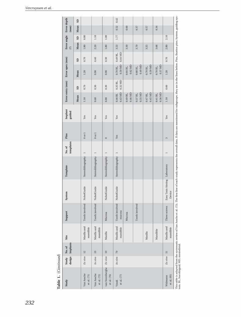
Several studies presenting prospective observationaldata on the clinical performance of guided implantplacement were identified (37). However, most ofthese studies had an observational period of <2 years(see Table 2) and only one study (63) had a follow-upperiod of up to 5 years. For these studies one canenvisage survival rates comparable with those for con-ventional implant treatment. Also, lower success rateshave been observed for smokers treated with guidedsurgery (3, 7, 8, 41). For example, a cohort study (63)reported cumulative survival rates of 81.2% and 98.9%for smokers and nonsmokers, respectively. The latterwas confirmed in a prospective clinical study of D’ha-ese et al. (22), in which patients were treated withflapless guided surgery in the maxilla (implant sur-vival = 69.2% in smokers vs. 98.7% in nonsmokers).
Prosthesis survival rates
The prosthesis survival rates ranged widely (from 62%to 100%) (see Table 3), probably as a result of several
factors – such as the definition of prosthesis survival,whether immediate or delayed loading was imple-mented and whether temporary or permanent pros-theses were evaluated – and hence direct comparisonwith the conventional technique can be difficult. Thecomputer-guided implant concept, in combinationwith immediate loading (Figs. 7A-D), is marketed aseasy, safe and predictable. However, several compli-cations or unexpected events were reported, asdescribed in Table 2, as were fracture of the surgicalguide (Fig. 8), dehiscences (31) and soft-tissue lacera-tion (26). Misfit of the temporary prosthesis was themost common prosthetic complication, caused byinaccurate placement of the implants (Fig. 9A). Afterplacement of the temporary prosthesis the mostcommon complication was prosthesis fracture(Fig. 9B). It seems obvious that guided surgery, espe-cially in combination with immediate loading, cannotbe regarded as easier than conventional techniques.
Clinical protocol
Flapless surgery has gained interest since severalarticles showed that raising a flap leads to boneresorption (30, 34, 83). Via a flapless approach theperiosteum and blood supply to the bone remainintact (10, 17) (Figs. 10A and 10B). Three studiescompared guided flapless surgery with conventionalopen flap surgery and reported on patient-centeredoutcomes (4, 32, 55). These studies demonstrated astatistically significant reduction in immediate post-operative pain, use of analgesics, swelling, edema,hematoma, hemorrhage and trismus, for flapless sur-gery. One of these studies (4) also compared guidedflapless surgery with guided open flap surgery anddemonstrated a consistently better outcome for theflapless approach. These results are supported by thegood scores for patient comfort and satisfactionreported by several observational studies on guidedflapless surgery (1, 54, 75). A prolonged oral surgicalintervention may increase postoperative pain anddiscomfort for the patient (66). One of the above-mentioned controlled studies reported that the dura-tion of the treatment with flapless guided surgery wasless than half (24 min) of that needed for open flapguided surgery and/or conventional surgery (4). Thisobservation is supported by Komiyama et al. (45)who reported that the duration of the flapless guidedsurgical intervention, including immediate recon-struction (Teeth-in-an-Hour concept; Nobel BiocareAB, Gothenburg, Sweden), took 30–45 min. Thus, thetime factor may indeed be part of the explanation ofwhy less pain and discomfort was reported bypatients after flapless guided surgery. Even if the
Vercruyssen et al.
236

Tab
le2.
Prosp
ective
observational
dataontheclinical
perform
ance
ofg
uided
implantplacemen
t
Study
Studydesign
Follow-u
pperiod
(months)
System
Complica
tionsat
guided
implantplacemen
tComplica
tionsaftergu
ided
placemen
t
Rea
son
No.o
fpro
sthetic
even
ts
Rea
son
No.o
fim
plant
failures
No.o
fpro
sthetic
even
ts
Rea
son
Abad
-Gallego
set
al.(1)
Retrosp
ective
observational
Not
reported
Nobel
Guide
Lack
ofp
rimary
stab
lility.
Limited
orala
perture
Notreported
Lack
ofp
assive
fit.Im
plant
pain.C
han
geto
angu
lated
abutm
ent
10Not
reported
Screw
loosening.
Fracture
of
prosthesisor
teeth
Arisanet
al.(4)
Prosp
ective
comparative*
2–4
Aytasarim
classic,
Simplant-SA
FE
Fracture
ofb
one-
supported
surgical
guides
Notap
plic
able
5Not
applic
able
Barter(9)
Prosp
ective
observational
Mea
n=49
coDiagn
ostiX
andGonyX
Notreported
1Not
reported
Berdougo
etal.(13
)Retrosp
ective
comparative*
12–4
8EasyG
uidean
dCAD
Implant
system
Notreported
10Not
reported
Cassetta
etal.(19
)Retrosp
ective
observational
Not
applic
able
SimPlantSa
feUnco
ntrolle
dremova
lofg
ingiva
.Alteration
ofe
xternal
hexag
on.
Laceration.T
emplate
break
age.
Limited
implantstab
ility
Notap
plic
able
Not
applic
able
Not
applic
able
Dan
zaet
al.(21
)Retrosp
ective
comparative*
1–41 (mea
n=14
)Im
plant3D
and
Ray
-Set
Notreported
0Not
reported
D´hae
se(22)
Prosp
ective
observational
12Astra
Fac
ilitate
Misplacemen
towing
tomisfabrica
tionof
surgical
guide
013
3Esthetic
reasons.
Prosthesis
frac
ture
DiG
iaco
mo
etal.(26
)Prosp
ective
observational
30Im
plantViewer
1.9an
dRhinoceros4.0
Pullingofsofttissue.
Insertionofw
ider
implants
than
planned
.Instab
ility.
Pain
1Midlin
edev
iation
11
Prosthesis
frac
ture
Fortin
etal.(32
)Ran
domized
controltrial*
Not
applic
able
CAD
implant
system
Notreported
Not
reported
Not
reported
Fortin
etal.(33
)Prosp
ective
observational
48EasyG
uide
Implantlost
before
load
ing
Notap
plic
able
0Not
reported
Guided surgery
237
Tab
le2.
(Con
tinued
)
Study
Studydesign
Follow-u
pperiod
(months)
System
Complica
tionsat
guided
implantplacemen
tComplica
tionsaftergu
ided
placemen
t
Rea
son
No.o
fpro
sthetic
even
ts
Rea
son
No.o
fim
plant
failures
No.o
fpro
sthetic
even
ts
Rea
son
Gillotet
al.(35
)Prosp
ective
observational
12–5
1Nobel
Guide
Guidedifficu
ltto
insert.A
bsence
of
primarystab
ility
1Majorocclusal
adjustmen
trequired
for
onepatient
411
Fracturesof
resin.P
rosthetic
screw
loosening
Johan
sson
etal.(40
)Prosp
ective
observational
12Nobel
Guide
Misfitofo
cclusal
index.M
isfitofthe
surgical
guide.
Problemsinstallin
gtheim
plants
15Problemsge
tting
theprosthesis
intheexac
tposition.M
ajor
occlusal
adjustmen
ts
21
Prosthesis
remad
eusing
stan
dard
abutm
ents
owingto
difficu
ltiesin
maintaining
adeq
uateoral
hyg
iene
Katsoulis
etal.(43
)Prosp
ective
comparative*
3Nobel
Guide
Notap
plic
able
Not
reported
Not
reported
Komiyam
aet
al.(45
)Prosp
ective
observational
6–44
(mea
n≥15
)Nobel
Guide
Fracture
ofsurgical
template
8Misfitof
prosthesis.
Majorocclusal
adjustmen
ts
19Three
prostheses
Prosthesishad
toberemove
dowingto
implantloss
Komiyam
aet
al.(44
)Prosp
ective
observational
>12 (mea
n=19
)Nobel
Guide
Notap
plic
able
Not
applic
able
Not
applic
able
Lindeb
oom
&va
nWijk
(48)
Ran
domized
controltrial
1Nobel
Guide
Notap
plic
able
Not
reported
Not
reported
Maloet
al.(50
)Prosp
ective
observational
6–21
(mea
n=13
)Nobel
Guide
Notreported
210
Melonie
tal.(51
)Retrosp
ective
observational
18Nobel
Guide
Fracture
ofsurgical
template
2Tem
porary
prosthesisdid
notfitat
time
ofp
lacemen
t
22
Fracture
ofthe
temporary
prosthesis
Vercruyssen et al.
238

Tab
le2.
(Con
tinued
)
Study
Studydesign
Follow-u
pperiod
(months)
System
Complica
tionsat
guided
implantplacemen
tComplica
tionsaftergu
ided
placemen
t
Rea
son
No.o
fpro
sthetic
even
ts
Rea
son
No.o
fim
plant
failures
No.o
fpro
sthetic
even
ts
Rea
son
Merliet
al.(52
)Prosp
ective
observational
8Nobel
Guide
Fracture
ofsurgical
guide.
Lost
implant
becau
seprimary
stab
ility
could
not
beac
hieve
d
4Prosthesisdid
notfitat
time
ofp
lacemen
t
25
Fracture
of
temporary
prosthesis.
Prosthetic
screw
loosening.
Fracture
of
porcelain
coatingof
perman
ent
prosthesis
Nikzad&
Azari(54)
Prosp
ective
observational
12Simplant,
SurgiG
uide
Notap
plic
able
22
Fixtureslost.N
oseatingof
prosthesis
Nke
nke
etal.(55
)Prosp
ective
comparative*
12NobelGuide
Notreported
0Notreported
Pomares
(61)
Retrosp
ective
observational
12NobelGuide
Fracture
ofsurgical
template
3Misfitof
temporary
prosthesis
48
Fracture
of
temporary
prosthesis
Sannaet
al.(63
)Prosp
ective
observational
6–60
(mea
n=26
)NobelGuide
Notreported
9Notreported
vanStee
nbergh
eet
al.(75
)Prosp
ective
observational
12NobelGuide
2Prosthetic
misfit.
Midlin
edev
iation
03
Occlusalm
aterial
frac
ture.
Prosthetic
screw
loosening
Yong&
Moy(84)
Prosp
ective
observational
Mea
n=27
NobelGuide
Toodee
pplacemen
tofo
neim
plant
whichwas
remove
d(failure)
2Inco
mplete
seatingof
prosthesis
owingto
bony
interferen
ce
712
Spee
chproblem.
Bila
teral
chee
kbiting.
Fracture
of
prosthesis.
Hea
vyocclusal
wea
r.Sc
rew
loosening
Thistable
was
adap
tedfrom
thesystem
atic
review
ofH
ultin
&Sv
ensson(37)
*Con
trolg
roupincluded
conve
ntional
open
flap
surgery.
Guided surgery
239

Tab
le3.
Prosthesissu
rvival
rates
Study
Survival
rate
Other
outcome
Immed
iate/D
elay
edload
ing
Implants
Pro
sthesis
Follow-u
p
period
(months)
Withgu
ided
placemen
t
Withoutgu
ided
placemen
t
Withgu
ided
placemen
t(%
)
Withoutgu
ided
placemen
t
Withgu
ided
placemen
t(%
)
Withoutgu
ided
placemen
t
Barter(9)
Notreported
Notreported
98Notap
plic
able
100
Notap
plic
able
Mea
n=49
Berdougo
etal.(13
)*Notreported
Notreported
9699
%Notreported
Notreported
12–4
8
Dan
zaet
al.(21)*
Immed
iate
load
ing/
Delay
edload
ing
Immed
iate
load
ing/
Delay
edload
ing
100
96%
Notreported
Notreported
1–41
(mea
n=14
)
D’hae
seet
al.(22
)Im
med
iate
load
ing/
Delay
edload
ing
Notap
plic
able
89Notap
plic
able
62†
Notap
plic
able
1299
%im
plantsu
rvival
rate
innonsm
oke
rsan
d74
%in
smoke
rs.S
mokingan
dim
med
iate
load
ingin
combinationin
eden
tulous
maxillae
increa
sedim
plantloss
DiG
iaco
mo
etal.(26
)Im
med
iate
load
ing
Notap
plic
able
96Notap
plic
able
92Notap
plic
able
30
Fortin
etal.(33
)Delay
edload
ing
Notap
plic
able
98Notap
plic
able
Notreported
Notap
plic
able
48
Gillotet
al.(35
)Im
med
iate
load
ing
Notap
plic
able
98Notap
plic
able
100
Notap
plic
able
12–5
1Rem
ova
landreplacemen
tof
adjustab
leab
utm
ents
usedin
thetemporary
prosthesiswas
unpleasan
tforthepatients
Johan
sson
etal.(40
)Im
med
iate
load
ing
Notap
plic
able
99Notap
plic
able
96Notap
plic
able
12Mea
nmarginal
boneloss
of
1.3mm;1
9%ofthesu
bjects
had
>2mm
boneloss;
muco
salinflam
mationwas
presentin
23%
ofp
robed
sites
Komiyam
aet
al.(45
)Im
med
iate
load
ing
Notap
plic
able
89Notap
plic
able
84Notap
plic
able
6–44
(mea
n≥15
)Bleed
ingonprobing=82
%(16–
100%
).Boneloss
was
more
commonwhen
pressure-likemuco
salu
lcers
weredetectedunder
the
prosthesis
Vercruyssen et al.
240

Tab
le3.
(Con
tinued
)
Study
Survival
rate
Other
outcome
Immed
iate/D
elay
edload
ing
Implants
Pro
sthesis
Follow-u
p
period
(months)
Withgu
ided
placemen
t
Withoutgu
ided
placemen
t
Withgu
ided
placemen
t(%
)
Withoutgu
ided
placemen
t
Withgu
ided
placemen
t(%
)
Withoutgu
ided
placemen
t
Maloet
al.(50
)Im
med
iate
load
ing
Notap
plic
able
98Notap
plic
able
Notreported
Notap
plic
able
6–21
(mea
n=13
)21
%ofa
llmea
suredsitesat
6monthsan
d28
%at
12monthshad
>2mm
radiograp
hic
boneloss
Melonie
tal.(51
)Im
med
iate
load
ing
Notap
plic
able
98Notap
plic
able
87‡
Notap
plic
able
18Mea
nmarginal
boneloss
of
1.6mm
after18
months
Nikzadet
al.(54
)Delay
edload
ing
Notap
plic
able
96Notap
plic
able
Notreported
Notap
plic
able
12Mea
npainscore
onvisu
alan
alogscaleat
follo
w-up
was
within
therange
for
littleornopain
Nke
nke
etal.(55
)*Im
med
iate
load
ing
Immed
iate
load
ing
100
100%
100
100%
12Guided
surgeryge
nerated
less
postoperativepain
andsw
ellin
gco
mpared
withopen
flap
surgery
Pomares
(61)
Immed
iate
load
ing
Notap
plic
able
98Notap
plic
able
100
Notap
plic
able
12
Sannaet
al.(63
)Im
med
iate
load
ing
Notap
plic
able
95Notap
plic
able
Notreported
Notap
plic
able
6–60
(mea
n=26
)Mea
nmarginal
boneloss
of2
.6mm
insm
oke
rsan
d1.2mm
innonsm
oke
rs
vanStee
nbergh
eet
al.(75
)Im
med
iate
load
ing
Notap
plic
able
100
Notap
plic
able
100
Notap
plic
able
12Mea
nmarginal
boneloss
of
1.2mm
mesiala
nd1.1mm
distal
Yongan
dMoy(84)
Immed
iate
load
ing
Notap
plic
able
91Notap
plic
able
Notreported
Notap
plic
able
Mea
n=27
Outcomewas
determined
instudiesusingstatic
guided
system
san
dwithamea
nfollo
w-upof≥
12months.Thistable
was
adap
tedfrom
thesystem
atic
review
ofH
ultin
andSv
ensson(37).
*Control
groupincluded
conve
ntional
open
flap
surgery.
†Su
rvival
rate
reported
ontemporary
prosthesisfortheim
med
iately
load
edca
ses.
‡Su
rvival
rate
reported
ontemporary
prostheses.
Guided surgery
241

duration of the surgical intervention is shorter withflapless guided surgery compared with conventionaltechniques, it seems that much more time has to beinvested in the preoperative planning. The flaplessguided implant placement technique allows the sur-geon to install the implants with minimal surgicaltrauma to the bone and associated soft tissues. Assuch, these techniques may be particularly attractive
for use in frail patients. However, again, very limitedinformation is available. Horwitz et al. (36) describedthe use of flapless guided implant placement in anirradiated cancer patient and showed good resultsafter 2 years. In the study by Barter (9), six patientswere treated with flapless guided surgery to avoid sec-ondary exposure of previously grafted sites. Theimplant survival rate was 98% and all prostheses werestill in use after 4 years.
Cost effectiveness
The cost effectiveness of different guided surgery pro-tocols is difficult to judge as no information on thisparameter could be found in the scientific literature.An interesting clinical question is whether these tech-niques can be used as an alternative to bone augmen-tation. Unfortunately, only one article addresses thisquestion. Fortin et al. (33) used the guided techniquein partially edentulous patients with severelyresorbed maxillae and reported a 98% implantsurvival rate after 4 years.
A B
C D
Fig. 7. (A–D) Clinical case of a patient treated with flapless guided surgery and immediately restored with a temporarypartial bridge.
Fig. 8. Example of a fracture of the surgical guide (cour-tesy of Prof. Bj€orn Klinge).
A B
Fig. 9. (A) Misfit of the prefabricatedprosthesis. (B) Radiographs showingthe misfit of the prefabricatedprosthesis.
Vercruyssen et al.
242

Conclusion
Different computer-assisted implant placement proce-dures are currently available. They differ in software,template manufacture, guiding device, stabilizationand fixation. The literature seems to indicate thatone has to accept a certain inaccuracy of �2.0 mm,which seems large initially but is clearly less thanfor nonguided surgery. A reduction of the accuracyto below 0.5 mm seems extremely difficult. A com-mon shortcoming identified in the studies includedfor this review was inconsistency in how clinicaldata and outcome variables were reported. Anotherlimitation was the small number of comparativeclinical studies. In order to find the best guiding sys-tem/most important parameters for optimal accu-racy, more randomized clinical trials, which alsoinclude information on cost-effectiveness, patient-centered evaluations (i.e. questionnaires and inter-views) and longer follow-up periods are necessary.Future research should consider the use of flaplessguided implant placement in special subgroupsof patients (for example those with severelyresorbed jaws and osteoporosis, and those treatedwith radiotherapy).
References
1. Abad-Gallegos M, G�omez-Santos L, S�anchez-Garc�es MA,Pi~nera-Penalva M, Freixes-Gil J, Castro-Garc�ıa A, Gay-Escoda C. Complications of guided surgery and immediateloading in oral implantology: a report of 12 cases. Med OralPatol Oral Cir Bucal 2011: 16: 220–224.
2. Al Harbi SA, Sun AY. Implant placement accuracy whenusing stereolithographic template as a surgical guide:preliminary results. Implant Dent 2009: 18: 46–56.
3. Alsaadi G, Quirynen M, Kom�arek A, van Steenberghe D.Impact of local and systemic factors on the incidence oforal implant failures, up to abutment connection. J ClinPeriodontol 2007: 34: 610–617.
4. Arisan V, Karabuda CZ, Ozdemir T. Implant surgery usingbone- and mucosa-supported stereolithographic guides intotally edentulous jaws: surgical and post-operative out-comes of computer-aided vs. standard techniques. ClinOral Implants Res 2010: 21: 980–988.
5. Arisan V, Karabuda ZC, Pis�kin B, Ozdemir T. Conven-tional multi-slice computed tomography (CT) and cone-beam CT (CBCT) for computer-aided implant placement.part II: reliability of mucosa-supported stereolitho-graphic guides. Clin Implant Dent Relat Res 2013: 15:907–917.
6. Arisan V, Karabuda Z.C, Ozdemir T. Accuracy of two stere-olithographic guide systems for computer-aided implantplacement: a computed tomography-based clinical com-parative study. J Periodontol 2010: 81: 43–51.
7. Aykent F, Inan O, Ozyesil AG, Alptekin NO. A 1- to 12-yearclinical evaluation of 106 endosseous implants supportingfixed and removable prostheses. Int J PeriodonticsRestorative Dent 2007: 27: 358–367.
8. Bain CA, Moy PK. The association between the failure ofdental implants and cigarette smoking. Int J Oral MaxillofacImplants 1993: 8: 609–615.
9. Barter S. Computer-aided implant placement in thereconstruction of a severely resorbed maxilla – a 5-yearclinical study. Int J Periodontics Restorative Dent 2010: 30:627–637.
10. Becker W, Goldstein M, Becker BE, Sennerby L, Kois D,Hujoel P. Minimally invasive flapless implant placement:follow-up results from a multicenter study. J Periodontol2009: 80: 347–352.
11. Behneke A, Burwinkel M, Behneke N. Factors influencingtransfer accuracy of cone beam CT-derived template-basedimplant placement. Clin Oral Implants Res 2012: 23: 416–423.
12. Behneke A, Burwinkel M, Knierim K, Behneke N. Accuracyassessment of cone beam computed tomography-derivedlaboratory-based surgical templates on partially edentulouspatients. Clin Oral Implants Res 2012: 23: 137–143.
13. Berdougo M, Fortin T, Blanchet E, Isidori M, Bosson JL.Flapless implant surgery using an image-guided system. A1-to 4-year retrospective multicenter comparative clinicalstudy. Clin Implant Dent Relat Res 2010: 12: 142–152.
14. Bianchi SD, Ramieri G, De Gioanni PP, Martinetto F,Berrone S. Validation of anatomical stereolithographicreplicas: personal experience and literature review. RadiolMed (Torino) 1997: 94: 503–510.
15. Bou Serhal C, Jacobs R, Flygare L, Quirynen M, vanSteenberghe D. Perioperative validation of localisation ofthe mental foramen. Dentomaxillofac Radiol 2002: 31:39–43.
16. Brief J, Edinger D, Hassfeld S, Eggers G. Accuracy ofimage-guided implantology. Clin Oral Implants Res 2005:16: 495–501.
17. Campelo LD, Camara JR. Flapless implant surgery: a10-year clinical retrospective analysis. Int J Oral MaxillofacImplants 2002: 17: 271–276.
18. Cassetta M, Giansanti M, Di Mambro A, Calasso S, BarbatoE. Accuracy of two stereolithographic surgical templates: a
A B
Fig. 10. Clinical picture of flaplesssurgery in the maxilla after removalof the guide (A) and after placementof the abutments (B).
Guided surgery
243

retrospective study. Clin Implant Dent Relat Res 2013: 15:448–459.
19. Cassetta M, Stefanelli LV, Giansanti M, Di Mambro A, Ca-lasso S. Depth deviation and occurrence of early surgicalcomplications or unexpected events using a single stereolitho-graphic surgi-guide. Int J Oral Maxillofac Surg 2011: 40:1377–1387.
20. Chen X, Yuan J, Wang C, Huang Y, Kang L. Modular preop-erative planning software for computer-aided oral implan-tology and the application of a novel stereolithographictemplate: a pilot study. Clin Implant Dent Relat Res 2010:12: 181–193.
21. Danza M, Zollin I, Carinci F. Comparison between implantsinserted with and without computer planning and custommodel coordination. J Craniofac Surg 2009: 20: 1086–1092.
22. D’haese J, Elaut L, Verbanck N, De Bruyn H. Clinical andradiographic outcome of dental implants placed usingstereolithographic guided surgery; a prospective monocen-ter study. Int J Oral Maxillofac Implants 2013: 28: 205–215.
23. D’haese J, De Bruyn H. Effect of smoking habits on accu-racy of implant placement using mucosally supported ste-reolithographic surgical guides. Clin Implant Dent Relat Res2013: 15: 402–411.
24. D’haese J, Van De Velde T, Elaut L, De Bruyn H. A prospectivestudy on the accuracy of mucosally supported stereolitho-graphic surgical guides in fully edentulous maxillae. ClinImplant Dent Relat Res 2012: 14: 293–303.
25. Di Giacomo GA, Cury PR, de Araujo NS, Sendyk WR, SendykCL. Clinical application of stereolithographic surgicalguides for implant placement: preliminary results.J Periodontol 2005: 76: 503–507.
26. Di Giacomo GD, Silva JVLd, da Silva AM, Paschoal GH, CuryPR, Szarf G. Accuracy and complications of com-puter-designed selective laser sintering surgical guides forflapless dental implant placement and immediate definitiveprosthesis installation. J Periodontol 2011: 83: 410–419.
27. Dreiseidler T, Neugebauer J, Ritter L, Lingohr T, RothamelD, Mischkowski RA, Zoller JE. Accuracy of a newly devel-oped integrated system for dental implant planning. ClinOral Implants Res 2009: 20: 1191–1199.
28. Eggers G, Patellis E, Muhling J. Accuracy of template-baseddental implant placement. Int J Oral Maxillofac Implants2009: 24: 447–454.
29. Ersoy AE, Turkyilmaz I, Ozan O, McGlumphy EA. Reliabilityof implant placement with stereolithographic surgicalguides generated from computed tomography: clinical datafrom 94 implants. J Periodontol 2008: 79: 1339–1345.
30. Fickl S, Zuhr O, Wachtel H, Bolz W, Huerzeler M. Tissuealterations after tooth extraction with and without surgicaltrauma: a volumetric study in the beagle dog. J Clin Period-ontol 2008: 35: 356–363.
31. Fortin T, Bosson JL, Coudert JL, Isidori M. Reliability of pre-operative planning of an image-guided system for oralimplant placement based on 3-dimensional images: an invivo study. Int J Oral Maxillofac Implants 2003: 18: 886–893.
32. Fortin T, Bosson JL, Isidori M, Blanchet E. Effect of flaplesssurgery on pain experienced in implant placement using animage-guided system. Int J Oral Maxillofac Implants 2006:21: 298–304.
33. Fortin T, Isidori M, Bouchet H. Placement of posterior max-illary implants in partially edentulous patients with severebone deficiency using CAD/CAM guidance to avoid sinus
grafting: a clinical report of procedure. Int J Oral MaxillofacImplants 2009: 24: 96–102.
34. Gargiulo MS, Frank MW, Orban B. Dimensions and rela-tions of the dentogingival junction in humans. J Periodontol1961: 32: 261–267.
35. Gillot L, Noharet R, Cannas B. Guided surgery and presurgi-cal prosthesis: preliminary results of 33 fully edentulousmaxillae treated in accordance with the NobelGuide proto-col. Clin Implant Dent Relat Res 2010: 12: 104–113.
36. Horwitz J, Zuabi O, Machtei EE. Accuracy of a computer-ized tomography-guided template-assisted implant place-ment system: an in vitro study. Clin Oral Implants Res 2009:20: 1156–1162.
37. Hultin M, Svensson K. Clinical advantages of computerguided implant placement: a systematic review. Clin OralImplants Res 2012: 23(Suppl. 6): 124–135.
38. Jacobs R, Quirynen M. Dental cone beam computed tomo-graphy: justification for use in planning oral implant place-ment. Periodontol 2000 2014: 66: 203–213.
39. Jacobs R. Dental cone beam CT and its justified use in oralhealth care. Belg J Radiol 2011: 94: 254–265.
40. Johansson B, Friberg B, Nilsson H. Digitally planned, imme-diately loaded dental implants with prefabricated prosthe-ses in the reconstruction of edentulous maxillae: a 1-yearprospective, multicenter study. Clin Implant Dent Relat Res2009: 11: 194–200.
41. Jones JD, Lupori J, Van Sickels JE, Gardner W. A 5-year com-parison of hydroxyapatite-coated titanium plasma-sprayedand titanium plasma-sprayed cylinder dental implants.Oral Surg Oral Med Oral Pathol Oral Radiol Endod 1999:87: 649–652.
42. Jung RE, Schneider D, Ganeles J, Wismeijer D, Zwahlen M,H€ammerle CH, Tahmaseb A. Computer technology applica-tions in surgical implant dentistry: a systematic review. Int JOral Maxillofac Implants 2009: 24: 92–109.
43. Katsoulis J, Avrampo M, Spycher C, Stipic M, Enkling N,Mericske-Stern R. Comparison of implant stability bymeans of resonance frequency analysis for flapless andconventionally inserted implants. Clin Implant Dent RelatRes 2012: 14: 915–923.
44. Komiyama A, Hultin M, N€asstr€om K, Benchimol D, KlingeB. Soft tissue conditions and marginal bone changesaround immediately loaded implants inserted in edentatejaws following computer guided treatment planning andflapless surgery: a≥1-year clinical follow-up study. ClinImplant Dent Relat Res 2012: 14: 157–169.
45. Komiyama A, Klinge B, Hultin M. Treatment outcome ofimmediately loaded implants installed in edentulous jawsfollowing computer-assisted virtual treatment planning andflapless surgery. Clin Oral Implants Res 2008: 19: 677–685.
46. Komiyama A, Pettersson A, Hultin M, Nasstrom K, Klinge B.Virtually planned and template-guided implant surgery: anexperimental model matching approach. Clin OralImplants Res 2011: 22: 308–313.
47. Koop R, Vercruyssen M, Vermeulen K, Quirynen M. Toler-ance within the sleeve inserts of different surgical guides forguided implant surgery. Clin Oral Implants Res 2013: 24:630–634.
48. Lindeboom JA, van Wijk AJ. A comparison of two implanttechniques on patient-based outcome measures: a reportof flapless vs. conventional flapped implant placement.Clin Oral Implants Res 2010: 21: 366–370.
Vercruyssen et al.
244

49. Loubele M, Bogaerts R, Van Dijck E, Pauwels R, Vanheus-den S, Suetens P, Marchal G, Sanderink G, Jacobs R. Com-parison between effective radiation dose of CBCT andMSCT scanners for dentomaxillofacial applications. Eur JRadiol 2009: 71: 461–468.
50. Malo P, de Araujo Nobre M, Lopes A. The use of com-puter-guided flapless implant surgery and four implantsplaced in immediate function to support a fixed denture:preliminary results after a mean follow-up period of thir-teen months. J Prosthet Dent 2007: 97: 26–34.
51. Meloni SM, De Riu G, Pisano M, Cattina G, Tullio A.Implant treatment software planning and guided flaplesssurgery with immediate provisional prosthesis delivery inthe fully edentulous maxilla. A retrospective analysis of 15consecutively treated patients. Eur J Oral Implantol 2010: 3:245–251.
52. Merli M, Bernardelli F, Esposito M. Computer-guided flap-less placement of immediately loaded dental implants inthe edentulous maxilla: a pilot prospective case series. Eur JOral Implantol 2008: 1: 61–69.
53. Nickenig HJ, Wichmann M, Hamel J, Schlegel KA, Eitner S.Evaluation of the difference in accuracy between implantplacement by virtual planning data and surgical guide tem-plates versus the conventional free-hand method–a com-bined in vivo - in vitro technique using cone-beam CT (PartII). J Craniomaxillofac Surg 2010: 38: 488–493.
54. Nikzad S, Azari A. Custom-made radiographic template,computed tomography, and computer-assisted flapless sur-gery for treatment planning in partially edentulouspatients: a prospective 12-month study. J Oral MaxillofacSurg 2010: 68: 1353–1359.
55. Nkenke E, Eitner S, Radespiel-Tr€oger M, Vairaktaris E, Neu-kam FW, Fenner M. Patient-centred outcomes comparingtransmucosal implant placement with an open approach inthe maxilla: a prospective, non-randomized pilot study.Clin Oral Implants Res 2007: 18: 197–203.
56. Ozan O, Turkyilmaz I, Yilmaz B. A preliminary report ofpatients treated with early loaded implants using comput-erized tomography-guided surgical stents: flapless versusconventional flapped surgery. J Oral Rehabil 2007: 34: 835–840.
57. Pauwels R, Beinsberger J, Collaert B, Theodorakou C, Rog-ers J, Walker A, Cockmartin L, Bosmans H, Jacobs R, Boga-erts R, Horner K. Effective dose range for dental cone beamcomputed tomography scanners. Eur J Radiol 2012: 81:267–271.
58. Pettersson A, Kero T, Gillot L, Cannas B, Faldt J, SoderbergR, Nasstrom K. Accuracy of CAD/CAM-guided surgical tem-plate implant surgery on human cadavers: Part I. J ProsthetDent 2010: 103: 334–342.
59. Pettersson A, Komiyama A, Hultin M, Nasstrom K, Klinge B.Accuracy of virtually planned and template guided implantsurgery on edentate patients. Clin Implant Dent Relat Res2012: 14: 527–537.
60. Poeschl PW, Schmidt N, Guevara-Rojas G, Seeman R, EwersR, Zipko HT, Schicho K. Comparison of cone-beam andconventional multislice computed tomography for imageguided dental implant planning. Clin Oral Investig 2013: 17:317–324.
61. Pomares C. A retrospective study of edentulous patientsrehabilitated according to the ‘all-on-four’ or the ‘all-on-six’immediate function concept using flapless com-
puter-guided implant surgery. Eur J Oral Implantol 2010: 3:155–163.
62. Ruppin J, Popovic A, Strauss M, Spuntrup E, Steiner A, StollC. Evaluation of the accuracy of three different com-puter-aided surgery systems in dental implantology: opticaltracking vs. stereolithographic splint systems. Clin OralImplants Res 2008: 19: 709–716.
63. Sanna A, Molly L, van Steenberghe D. Immediately loadedCAD-CAM manufactured fixed complete dentures usingflapless implant placement procedures: a cohort study ofconsecutive patients. J Prosthet Dent 2007: 97: 331–339.
64. Santler G, K€archer H, Gaggl A, Kern R. Stereolithographyversus milled three-dimensional models: comparison ofproduction method, indication, and accuracy. ComputAided Surg 1998: 3: 248–256.
65. Sarment DP, Sukovic P, Clinthorne N. Accuracy of implantplacement with a stereolithographic surgical guide. Int JOral Maxillofac Implants 2003: 18: 571–577.
66. Sato FR, Asprino L, de Ara�ujo DE, de Moraes M. Short termoutcome of postoperative patient recovery perception aftersurgical removal of third molars. J Oral Maxillofac Surg2009: 67: 1083–1091.
67. Schneider D, Marquardt P, Zwahlen M, Jung RE. A system-atic review on the accuracy and the clinical outcome ofcomputer-guided template-based implant dentistry. ClinOral Implants Res 2009: 20: 73–86.
68. Schneider J, Decker R, Kalender WA. Accuracy in medicinalmodelling. Phidias Newsl 2002: 8: 5–14.
69. Swaelens B, Kruth JP. Medical applications of rapid proto-typing techniques. Proceedings of the Fourth InternationalConference on Rapid Prototyping, Dayton, USA, 1993: 107–120.
70. Valente F, Schiroli G, Sbrenna A. Accuracy of computer-aidedoral implant surgery: a clinical and radiographic study. Int JOral Maxillofac Implants 2009: 24: 234–242.
71. Van Assche N, van Steenberghe D, Guerrero ME, Hirsch E,Schutyser F, Quirynen M, Jacobs R. Accuracy of implantplacement based on pre-surgical planning of three-dimen-sional cone-beam images: a pilot study. J Clin Periodontol2007: 34: 816–821.
72. Van Assche N, van Steenberghe D, Quirynen M, Jacobs R.Accuracy assessment of computer-assisted flapless implantplacement in partial edentulism. J Clin Periodontol 2010:37: 398–403.
73. Van Assche N, Vercruyssen M, Coucke W, Teughels W,Jacobs R, Quirynen M. Accuracy of computer aided implantplacement. Clin Oral Impl Res 2012: 23(Suppl. 6): 112–123.
74. Van Assche N, Quirynen M. Tolerance within a surgicalguide. Clin Oral Implants Res 2010: 21: 455–458.
75. van Steenberghe D, Glauser R, Blomb€ack U, AnderssonM, Schutyser F, Pettersson A, Wendelhag I. A computedtomographic scan-derived customized surgical templateand fixed prosthesis for flapless surgery and immediateloading of implants in fully edentulous maxillae: a pro-spective multicenter study. Clin Implant Dent Relat Res2005: 7: 111–120.
76. van Steenberghe D, Malevez C, Van Cleynenbreugel J, BouSerhal C, Dhoore E, Schutyser F, Suetens P, Jacobs R. Accu-racy of drilling guides for transfer from three-dimensionalCT-based planning to placement of zygoma implants inhuman cadavers. Clin Oral Implants Res 2003: 14: 131–136.
77. Vasak C, Watzak G, Gahleitner A, Strbac G, Schemper M,Zechner W. Computed tomography-based evaluation of
Guided surgery
245
template (NobelGuide)-guided implant positions: a pro-spective radiological study. Clin Oral Implants Res 2011: 22:1157–1163.
78. Vercruyssen M, Fortin T, Widmann G, Jacobs R, QuirynenM. Different techniques of static/dynamic guided: modal-ities and indications. Periodontol 2000 2014: 66: 214–227.
79. Vercruyssen M, Cox C, Jacobs R, Coucke W, Naert I, Quiry-nen M. An RCT comparing guided implant surgery (bone ormucosa supported) with mental navigation or the use of apilot-drill template. J Clin Periodontolgy 2014: 41: 717–723.
80. Vercruyssen M, Jacobs R, Van Assche N, van SteenbergheD. The use of CT scan based planning for oral rehabilitationby means of implants and its transfer to the surgical field: acritical review on accuracy. J Oral Rehabil 2008: 35: 454–474.
81. Widmann G, Zangerl A, Keiler M, Stoffner R, Bale R, Puel-acher W, Bale R. Flapless implant surgery in the edentulousjaw based on three fixed intra-oral reference points andimage-guided surgical templates: accuracy in humancadavers. Clin Oral Implants Res 2010: 21: 276–283.
82. Widmann G, Bale RJ. Accuracy in computer-aided implantsurgery: a review. Int J Oral Maxillofac Implants 2006: 21:305–313.
83. Wood DL, Hoag PM, Donnenfeld OW, Rosenfeld LD. Alveo-lar crest reduction following full and partial thickness flaps.J Periodontol 1972: 43: 141–144.
84. Yong LT, Moy PK. Complications of computer-aideddesign/computer-aided-machining-guided (NobelGuide (TM))surgical implant placement: an evaluation of early clinicalresults. Clin Implant Dent Relat Res 2008: 10: 123–127.
Vercruyssen et al.
246