Embed Size (px)
Citation preview
OKLAHOMA
Guidelines for Treatment of
The Lower Extremity (Knee, Ankle and Foot)
Developed and Adopted
by the
Physician Advisory Committee
Adopted by the Administrator
of the
Oklahoma Workers' Compensation Court
Effective September 1, 2007

2 Lower Extremity Guidelines
Lower Extremity Guidelines i
TABLE OF CONTENTS
INTRODUCTION..................................................................................................................................................4
BACKGROUND 4
DEVELOPMENT OF THE GUIDELINES 4
APPLICATION OF THE GUIDELINES 4
I. GENERAL GUIDELINE PRINCIPLES..................................................................................................4
A. Education 5
B. Treatment Parameter Duration 5
C. Active Interventions 5
D. Active Therapeutic Exercise Program 5
E. Positive Patient Response 5
F. Re-evaluate Treatment Every 2-4 Weeks 5
G. Surgical Interventions 5
H. Six-month Time Frame 5
I. Return to Work 6
J. Delayed Recovery 6
II. INITIAL DIAGNOSTIC PROCEDURES ...............................................................................................6
A. History Taking and Physical Examination (Hx & PE) 6
1. Knee - Significant History ...........................................................................................6
2. Knee - History Questions.............................................................................................7
3. Foot and Ankle - History .............................................................................................7
B. Physical Exam 7
1. Knee - Physical Exam..................................................................................................7
2. Foot and Ankle - Physical Exam..................................................................................8
C. Radiographic Imaging 8
D. Laboratory Tests 9
E. Inappropriate Initial Assessment Methods 9
1. Routine arthroscopic examination ...............................................................................9
2. MRI .............................................................................................................................9
3. CT................................................................................................................................9
III. FOLLOW-UP DIAGNOSTIC IMAGING AND TESTING PROCEDURES .........................................9
A. Imaging Studies 9
1. Computerized Axial Tomography and Lineal Tomography (CT)................................9
2. Magnetic Resonance Imaging (MRI)...........................................................................9
3. Bone Scanning.............................................................................................................9
4. Arthrography................................................................................................................9
5. Venography .................................................................................................................9
6. Doppler Ultrasonography/Plethysmography..............................................................10
7. Diagnostic Arthroscopy .............................................................................................10
8. Arteriography.............................................................................................................10
B. Other Diagnostic Tests 10
1. Thermography............................................................................................................10
2. Compartment Pressure Measurement ........................................................................10
3. Electro-Diagnostic Studies (Needle EMG/NCS) .......................................................10
4. Psychometric Testing Evaluations .............................................................................10
5. Personality/Psychological/Psychosocial Evaluations.................................................10
IV. THERAPEUTIC PROCEDURES..........................................................................................................10
A. Non-operative Treatment: 11
1. Initial Treatment ........................................................................................................11
2. Immobilization...........................................................................................................11
3. Medication.................................................................................................................12
ii Lower Extremity Guidelines
4. Therapeutic Injection .................................................................................................14
B. Operative Treatment 14
1. Manipulation Under Anesthesia.................................................................................14
2. Bursectomy ................................................................................................................14
3. Arthroscopy ...............................................................................................................14
4. Ligament Repair or Reconstruction ...........................................................................15
5. Arthrotomy.................................................................................................................15
6. Osteotomy..................................................................................................................15
7. Total Joint Replacement ............................................................................................15
8. Fusion ........................................................................................................................15
9. Amputation ................................................................................................................15
10. Hardware Removal ....................................................................................................15
C. Response Criteria 15
1. Good ..........................................................................................................................15
2. Partial.........................................................................................................................15
3. Poor............................................................................................................................16
V. PHYSICAL MEDICINE AND REHABILITATION ............................................................................16
A. Modalities 16
1. Thermal Agents..........................................................................................................16
2. Electrical Stimulation.................................................................................................18
3. Vasopneumatic Devices.............................................................................................18
B. Physical Therapy Procedures 18
1. Iontophoresis/Phonophoresis .....................................................................................18
2. Ultrasound..................................................................................................................18
3. Manual Electrical Stimulation....................................................................................18
4. Contrast Baths............................................................................................................19
5. Hubbard Tanks...........................................................................................................19
6. Massage .....................................................................................................................19
7. Gait Training - Simple and Complex .........................................................................19
8. Activities of Daily Living (ADL)...............................................................................20
9. Therapeutic Activities................................................................................................20
10. Therapeutic Exercise..................................................................................................20
11. Neuromuscular Re-Education ....................................................................................20
12. Work Hardening Programs ........................................................................................20
13. Joint Mobilization......................................................................................................21
14. Manipulation..............................................................................................................21
15. Orthotic Training (dynamic bracing, splinting)..........................................................21
16. Prosthetic Training.....................................................................................................21
17. Myofascial Release/Soft Tissue Mobilization............................................................22
18. Manual Traction.........................................................................................................22
19. Transcutaneous Electrical Nerve Stimulation (TENS) ..............................................22
20. Hyperbaric Chamber..................................................................................................22
C. Return to work 22
1. In most cases..............................................................................................................22
2. Communication..........................................................................................................23
3. Generally....................................................................................................................23
D. Special Tests 23
1. Work Conditioning Assessments/Screens..................................................................23
2. Functional Capacity Evaluation (FCE) ......................................................................23
3. Lift Analysis...............................................................................................................23
4. Mechanized/Computerized Strength Evaluations ......................................................23
VI. SPECIFIC JOINT INVOLVEMENT.....................................................................................................23
A. Lower Extremity - Outlying Diagnoses 24
1. Infections Requiring Intravenous Antibiotics ............................................................24
Lower Extremity Guidelines iii
2. Reflex Sympathetic Dystrophy (RSD) .......................................................................24
3. Hardware removal .....................................................................................................24
4. Contracture ................................................................................................................24
5. Severe Burns, Open Wounds, Vascular Injuries, Polytrauma, and Non-union
Fractures ....................................................................................................................24
B. Knee - Outlying Diagnoses 24
1. Severe Intra-Articular Fracture ..................................................................................24
2. Knee Fusion...............................................................................................................24
3. Total Knee Replacement............................................................................................24
4. Amputation ................................................................................................................24
5. Meniscectomy............................................................................................................24
6. Meniscal repair ..........................................................................................................24
7. Retropatellar Pain Syndrome .....................................................................................25
8. Patellar Subluxation...................................................................................................25
9. ACL/PCL Repair .......................................................................................................25
10. Automated Methods for Determining Cruciate Ligament Laxity (KT-1000, etc.).....25
11. Arthroscopy ...............................................................................................................25
12. Chondroplasties, Chondral defects and Cartilage Repairs .........................................25
C. Foot and Ankle Injuries 25
1. Background................................................................................................................25
2. Diagnostic Criteria.....................................................................................................26
3. Treatment...................................................................................................................26
4 Lower Extremity Guidelines
GUIDELINES FOR TREAMENT OF
THE LOWER EXTREMITY
(Knee, Ankle and Foot)
developed and adopted by the
PHYSICIAN ADVISORY COMMITTEE
Adopted by the Administrator of the Oklahoma Workers' Compensation Court
Effective September 1, 2007
INTRODUCTION
BACKGROUND: The Physician Advisory Committee (PAC), a statutorily created advisory body to the
Oklahoma Workers' Compensation Court, has been directed by Oklahoma Statute to develop and recommend
treatment guidelines for injured Oklahoma workers. The PAC is composed of nine members; three appointed
by the Governor, three appointed by the President Pro Tempore of the State Senate, and three appointed by the
Speaker of the Oklahoma House of Representatives. By statute, the Governor's appointees must include a
doctor of medicine and surgery, a family practitioner in a rural community of the state, and an osteopathic
physician; the President Pro Tempore's appointees must include a doctor of medicine and surgery, a doctor of
medicine or an osteopathic physician, and a podiatric physician; and the Speaker's appointees must include an
osteopathic physician, a doctor of medicine or an osteopathic physician, and a chiropractic physician.
DEVELOPMENT OF THE GUIDELINES: The Committee received input from a wide variety of sources
including employers, insurance carriers, and health care providers. Appropriate scientific literature has been
reviewed. The Occupational Medicine Practice Guidelines promulgated by the American College of
Occupational and Environmental Medicine and the Official Disability Guidelines published by the Work Loss
Data institute, and practice parameters of the American Academy of Orthopaedic Surgeons, Clinical
Guidelines on Ankle and Knee Injury and Knee Pain, were reviewed. Treatment protocols from Colorado,
Minnesota, California, Washington, Rhode Island, and West Virginia were also utilized.
APPLICATION OF THE GUIDELINES: These treatment guidelines should not be construed as including
all proper methods of care or excluding other acceptable methods of care which are based upon nationally
accepted practice standards.
For injury or illness treated under the Oklahoma Workers= Compensation Act, compliance with these treatment
guidelines is mandatory and an employer or insurer for an employer is not required to pay for treatment which
is not in compliance with the treatment guidelines, unless prior authorization is received. If prior authorization
is refused, independent review may be obtained under court procedures.
Authorization for treatment may not be denied on the sole basis the treatment is not addressed by these
guidelines if it is documented to be based upon nationally accepted practice standards.
These guidelines do not affect any determination of liability for an injury under the Oklahoma Workers'
Compensation Act, 85 O.S., Section 1, et seq., and are not intended to expand or restrict a health care
provider's scope of practice under any other statutes.
I. GENERAL GUIDELINE PRINCIPLES
The objective of the Lower Extremity Treatment Guidelines is to provide standards for prompt,
reasonable and appropriate treatment for work place injuries and to expedite optimum recovery and
return to work, while containing medical costs in the workers' compensation system.
The first step in achieving this objective requires that an employer report a compensable injury in a
Lower Extremity Guidelines 5
timely fashion to ensure there is no delay in the treatment of the compensable injury. It is important
that the employer work with the insurance carrier and health care providers to ensure the injured
worker is given the opportunity to return to work in either a modified or full duty status as quickly as
medically possible.
The principles summarized in this section are key to the intended implementation of these guidelines
and critical to the reader=s application of the guidelines in this document.
A. Education: Education of the patient as well as the employer, insurer, policy makers and the
community should be the primary emphasis in the treatment of lower extremity pain and
disability. Currently, practitioners often think of education last, after medications, manual
therapy and surgery. Practitioners must develop and implement an effective strategy and skills
to educate patients, employers, insurance systems, policy makers and the community as a
whole. An education-based paradigm should always start with inexpensive communication
providing reassuring information to the patient. More in depth education currently exists
within a treatment regime employing functional restorative and innovative programs of
prevention and rehabilitation. No treatment plan is complete without addressing issues of
individual and/or group patient education as a means of facilitating self-management of
symptoms and prevention.
B. Treatment Parameter Duration: Time frames for specific interventions commence once
treatments have been initiated, not on the date of injury. Obviously, duration will be
impacted by patient compliance, as well as availability of services. Clinical judgment may
substantiate the need to accelerate or decelerate the time frames discussed in this document.
C. Active Interventions: Interventions involving therapeutic exercise and emphasizing patient
responsibility are generally emphasized over passive modalities, especially as treatment
progresses. Generally, passive and palliative interventions are viewed as a means to facilitate
progress in an active rehabilitation program with concomitant attainment of objective
functional gains.
D. Active Therapeutic Exercise Program: An exercise program should contain elements of
improving patient strength, endurance, flexibility and education.
E. Positive Patient Response: Positive results are defined primarily as functional and/or
physiologic gains which can be objectively measured. Objective functional gains include, but
are not limited to, positional tolerances, strength, endurance, range of motion, decreased
muscle tension and efficiency/velocity measures which can be quantified. Subjective reports
of pain and function should be considered and given relative weight when the pain has
anatomic and physiologic correlation. Anatomic correlation must be based on objective
findings.
F. Re-evaluate Treatment Every 2-4 Weeks: If a given treatment is not producing positive
results within 2-4 weeks, the treatment should be either modified or discontinued.
Reconsideration of diagnosis should also occur in the event of poor response to a seemingly
rational intervention.
G. Surgical Interventions: Surgery should be contemplated within the context of expected
functional outcome and not purely for the purpose of pain relief. The concept of Acure@ with
respect to surgical treatment by itself is generally a misnomer. All operative interventions
must be based upon positive correlation of clinical findings, clinical course and diagnostic
tests. A comprehensive assimilation of these factors must lead to a specific diagnosis with
positive identification of pathologic condition(s).
H. Six-month Time Frame: Since the prognosis drops precipitously for returning an injured
worker to work once he/she has been temporarily totally disabled for more than six months,
the emphasis within these guidelines is to move patients along a continuum of care within a
six-month time frame, whenever possible. It is important to note that time frames may not be
pertinent to injuries which do not involve work-time loss or are not occupationally related.
6 Lower Extremity Guidelines
I. Return to Work: Even if there is residual chronic pain, return to work is not necessarily
contraindicated. Return to work may be therapeutic, assuming the work is not likely to
aggravate the basic problem . The practitioner must write detailed restrictions when returning
a patient to limited duty. At a minimum, the following functions should be considered and
modified as recommended: lifting, pushing, pulling, squatting, stooping, walking, using stairs,
bending at the waist, awkward and/or sustained postures, tolerance for sitting or standing, hot
and cold environments, data entry and other repetitive motion tasks, sustained grip, tool usage
and vibration factors. The patient should never be released to Alight duty@ without specific
physical limitations. The practitioner should understand all of the physical demands of the
patient=s job position before returning the patient to full duty and should request clarification
of the patient=s job duties, if necessary. Clarification should be obtained from the employer
or, if necessary, including, but not limited to, an occupational health nurse, occupational
therapist, physical therapist, vocational rehabilitation specialist, or an industrial hygienist.
The Physician Advisory Committee encourages employers to ensure an injured worker is
given the opportunity to return to work in either a modified or full duty status once it is
determined medically possible, and recommends that the injured worker=s full wages be paid
by the employer if such work is not provided.
J. Delayed Recovery: A psychological screen should be considered, as well as initiating
interdisciplinary rehabilitation treatment and vocational goal setting, for those patients who
are failing to make expected progress 6-12 weeks after an injury. The Physician Advisory
Committee recognizes that 3-10% of all industrially injured patients will not recover within
the time lines outlined in this document despite optimal care. Such individuals may require
treatment beyond the limits discussed within this document, but such treatment will require
clear documentation by the authorized treating practitioner focusing on objective functional
gains afforded by further treatment and impact upon prognosis.
The remainder of this document should be interpreted within the parameters of these guideline
principles which hopefully will lead to more optimal medical and functional outcomes for
injured workers.
II. INITIAL DIAGNOSTIC PROCEDURES
Standard procedures which should be utilized when initially diagnosing a work-related lower extremity
complaint are:
A. History Taking and Physical Examination (Hx & PE) are generally accepted, well-
established and widely used procedures which establish the foundation/basis for and dictates
all other following stages of diagnostic and therapeutic procedures. When findings of clinical
evaluation and those of other diagnostic procedures are not complementing each other, the
objective clinical findings should have preference.
The history and physical examination should include, but not be limited to, the following:
1. Knee - Significant History
a. Acute onset of pain within 72 hours of injury
b. Direct blow to medial or lateral aspect of knee
c. Prior knee surgery
d. Varus or valgus stress to knee
e. Twisting injury - painful popping and catching with delayed swelling
f. Direct blow to patella or hyperflexion
g. Osteoporotic risk factors or age>55 years
h. Audible pop and immediate swelling with twisting or forced hyperextension

Lower Extremity Guidelines 7
i. Direct blow to anterior tibia, forced hyperextension
2. Knee - History Questions
a. Where does it hurt-have patient point to specific location
b. Onset, history and location of pain
c. Any previous injuries or similar problem?
d. Factors that aggravate pain (activity, weight bearing, stairs, inclines, rest)
e. Factors that relieve pain (movement, stretching or resting)
f. Presence and location of swelling
g. Is the pain constant or intermittent?
h. Is the pain present at rest, with weight bearing only, or both?
i. Is stiffness present if you have rested for awhile?
j. Any similar symptoms in other joints?
k. Any fever, chills, or infection elsewhere?
l. Any change in sensation of muscle strength?
m. Thorough review of systems looking for rheumatologic, traumatic,
endocrine and other injuries
3. Foot and Ankle - History
a. Mechanism of injury - twisting, turning (internal or external)
b. Location of pain
c. Ability to bear weight (mild) or inability to bear weight (severe)
d. Age of patient (including skeletal Maturity)
e. Time of onset
f. Prior injury / recurrent injury, use of bracing devices
g. Thorough systems review looking for a history of rheumatologic, traumatic,
endocrine, or other injuries
B. Physical Exam
1. Knee - Physical Exam
a. Patellar tenderness or abnormal position
b. Inability to bear weight 4 steps without assistance
c. Joint line tenderness or positive McMurray=s test
d. Valgus or varus joint instability
e. Inability to perform straight-leg raising
f. Positive Lachman=s test
g. Effusion or acute swelling
h. Tenderness of lateral or medial aspect of knee or head of fibula
i. Positive posterior drawer sign
j. Inability to straighten knee or flex>90 degrees
8 Lower Extremity Guidelines
k. Visual inspection for abnormalities
l. Presence of warmth
m. Range of motion
n. Meniscal compression
o. Foot pulse
p. Presence of erythema
q. Crepitus
r. Hip pain abnormalities
s. Physical examination should include the accepted tests and examination
techniques applicable to the joint or area being examined including range of
motion, strength testing and joint stability.
t. Complete neurological examination should be performed, looking for
neurological deficits, muscular atrophy, and gait abnormalities.
u. Exclusionary diagnosis include limb ischemia, joint violation or penetrating
trauma, deep venous thrombosis or septic arthritis.
2. Foot and Ankle - Physical Exam
a. Swelling-location
b. Ecchymosis
c. Tenderness-location
d. Stability-laxity
e. Crepitus
f. Sensory changes
g. Presence or absence of pulses
h. Tendon/muscle dysfunction
i. Deformity
j. Range of motion studies
C. Radiographic Imaging of the lower extremity is a generally accepted, well-established and
widely used diagnostic procedure. Repeat radiographs for fracture follow-up and unexplained
pain are acceptable with appropriate documentation. When indicated by the history and
physical examination, adjacent joints may be evaluated radiographically. Radiographic stress
testing may be useful in assessing joint laxity, particularly in younger patients, or patients who
are too anxious to tolerate the clinical examination. Indications for radiographs are:
1. Obvious deformity.
2. Effusion or instability.
3. Tenderness in head of fibula.
4. Isolated patellar tenderness.
5. Inability to flex knee>90 degrees.
6. Osteoporosis risk factors or age>55 years.
7. Suspected lesion indicative of a systemic illness such as rheumatoid arthritis,
osteoarthritis, gout, pseudogout, and other systemic conditions.
Lower Extremity Guidelines 9
8. History and physical examination suggesting a pre-existing condition such as
Osgood-Schlatter Disease or previous surgery.
9. Unexplained or continued lower extremity pain of over two weeks duration.
D. Laboratory Tests are generally accepted, well-established and widely used procedures. They
are, however, rarely indicated at the time of initial evaluation. Laboratory tests, including, but
not limited to, the following can provide useful diagnostic information:
1. CBC with differential to detect infection, medication side effects, blood dyscrasias;
pre-operative situations.
2. Rheumatoid factor, ANA, HLA, CRP, sedimentation rate to detect connective tissue
disorder.
3. Serum calcium, phosphorus, uric acid, alkaline phosphatase and acid phosphatase to
detect metabolic bone disease.
4. Analysis of joint aspiration for bacteria, fat globules, crystalline birefringence and
chemistry to evaluate joint effusion.
The Physician Advisory Committee recommends the above diagnostic procedures be
considered, at least initially, the responsibility of the workers= compensation carrier to ensure
that an accurate diagnosis and treatment plan can be established.
E. Inappropriate Initial Assessment Methods
1. Routine arthroscopic examination: Arthroscopic examination is not necessary to
make a clinical diagnosis of torn ligaments.
In evaluation of acute injuries at the time of irrigation with arthroscopy, swelling of
the irrigated tissues with associated edema can make later repair more difficult. Thus
irrigation fluid with this extravasation may be a negative factor if surgical repair is
not planned immediately.
2. MRI: MRI generally is not routinely indicated if accurate diagnosis is established
with history, physical exam and radiographs. It is useful in identifying full thickness
meniscal tears, meniscus cysts, osteochondritic lesions, ligament injuries, and bone
contusions. False positives may occur and findings must be correlated with physical
examination.
3. CT: CT is not usually appropriate at the initial evaluation or within the first month.
III. FOLLOW-UP DIAGNOSTIC IMAGING AND TESTING PROCEDURES
A. Imaging Studies are generally accepted, well-established and widely used diagnostic
procedures. In addition to routine radiographic and laboratory studies, the following imaging
studies can be utilized for further evaluation of the lower extremity:
1. Computerized Axial Tomography and Lineal Tomography (CT) evaluates
fractures and masses not adequately demonstrated by routine radiographic evaluation.
2. Magnetic Resonance Imaging (MRI) enhances the investigation of traumatic or
degenerative injuries of the lower extremity.
3. Bone Scanning is useful for investigation of trauma, infection, stress fracture, reflex
sympathetic dystrophy and neoplastic conditions of the lower extremity.
4. Arthrography may be useful in the evaluation of internal derangement of a joint
when MRI or other tests are contraindicated or not necessary.
5. Venography is useful for investigation of vascular injuries or disorders, including
deep venous thrombosis.
10 Lower Extremity Guidelines
6. Doppler Ultrasonography/Plethysmography is useful in detecting arterial and
venous disease in an extremity.
7. Diagnostic Arthroscopy enables the diagnosis of conditions within the joint when
other diagnostic tests have failed to reveal the cause of symptoms. It is also
employed in patients who fail a reasonable regimen of conservative treatment when
the condition is amenable to arthroscopic repair.
8. Arteriography may be useful in determining vascular insufficiency in appropriate
patients.
B. Other Diagnostic Tests:
1. Thermography is rarely helpful as an adjunctive diagnostic tool in the diagnosis of
reflex sympathetic dystrophy, sympathetically maintained pain, autonomic
neuropathies and chronic neuropathic pain involving small-caliber sensory fiber
neuropathies.
2. Compartment Pressure Measurement devices, such as the Pressure Manometer,
are useful in the evaluation of patients who present with symptoms consistent with a
compartment syndrome.
3. Electro-Diagnostic Studies (Needle EMG/NCS) are generally accepted and well-
established for the evaluation of neurologic disease, including the evaluation of
muscle disease, nerve entrapment, radiculopathy and peripheral neuropathy. The
Physician Advisory Committee does not recommend use of surface EMGs for the
evaluation of neurologic disease.
4. Psychometric Testing Evaluations are generally accepted and well-established
diagnostic procedures with selected use in patients with lower extremity pain. These
procedures may be useful in patients with delayed recovery, chronic pain syndromes,
and recurrent painful conditions. They may be useful for pre-operative evaluations
and may have a predictive value for determining appropriate surgical candidates.
These evaluations may provide a better understanding of patients and allow for more
effective rehabilitation if a patient is not improving within 4-8 weeks or as soon as
the problem is identified.
5. Personality/Psychological/Psychosocial Evaluations are generally accepted and
well established diagnostic procedures with limited use in the acute lower extremity
disorders and more wide-spread use in sub-acute and chronic lower extremity
disorders. These procedures may be useful for patients with delayed recovery,
chronic pain, recurrent painful conditions, suspected concomitant closed head injury,
disability problems and for pre-operative evaluation, as well as a possible predictive
value for post-operative response. Results may provide clinicians with a better
understanding of the patient, thus allowing for more effective rehabilitation. Formal
psychological or psychosocial screening should be performed on patients not making
expected progress within 6-12 weeks following injury and whose subjective
symptoms do not correlate with objective signs and tests. This testing will determine
the need for further psychosocial interventions. Evaluations should be performed by
an individual with Ph.D., Psy.D., L.S.W. or Psychiatric M.D./D.O. credentials.
Initial psychological screening is generally completed within one hour. If
psychometric testing is indicated as a portion of the initial screening process, the time
for such testing should not exceed an additional two hours of professional time.
IV. THERAPEUTIC PROCEDURES
It is understood that patients undergoing the following therapeutic procedures may return to modified or
restricted duty during their rehabilitation at the earliest appropriate time. It is also understood that cessation
and/or review of the following treatment procedures should be undertaken when no further significant subjective
Lower Extremity Guidelines 11
or objective improvement in the patient=s condition is noted.
A. Non-operative Treatment:
1. Initial Treatment
a. Rest
b. Ice
c. Analgesics
d. Immobilization within the first 3-5 days prn
e. Crutches prn
f. Therapeutic exercises including straight-leg raising exercises
g. Active range-of-motion exercises within 3-5 days as pain allows
2. Immobilization usually involves splinting, casting, or non-weight-bearing. It can be
used early as a generic treatment for most injuries to the joint as part of management
pending referral to an appropriate specialist and/or the development of a formal
diagnostic and treatment plan. Immobilization has inherent side effects including
joint contracture, collagen tightness and rapid muscular deconditioning.
Initial immobilization is only temporary, should be re-evaluated within 1-3 weeks
and should be applied as part of the treatment plan with specific goals recognizing its
inherent side effects.
a. Specific Treatment: duration from 2-16 weeks at the practitioner=s
discretion. May or may not be associated with therapy techniques
depending upon the intensity of strict immobilization required. During
immobilization, a portable neuromuscular electrical stimulator may be
issued to the patient to minimize muscle atrophy and edema. As a result of
expected side effects, the following course of physical therapy may be
relatively long and extensive, spanning several months as range of motion,
joint stability, conditioning and endurance are re-established.
(1) Casting is indicated for those injuries associated with fracture
which can be reduced and maintained with casting techniques.
This procedure should give a reasonable result considering the
alternatives and natural history of casting techniques associated
with this injury.
(a) Optimal Duration: 3-16 weeks
(2) Bracing is considered when certain planes of motion should be
restricted while allowing
motion in other planes and
potentially limiting weight-
bearing.
(a) Optimal Duration: 4 weeks - permanent
(3) Continuous Passive Motion (CPM) is used in the post-operative
treatment phase of fracture repair or arthroplasty. CPM machines
should be monitored weekly and discontinued when no further
benefits are seen.
(a) Optimal Duration: 1-2 weeks
(4) Bone Stimulator is useful for improving fracture union in delayed
and non-union.
12 Lower Extremity Guidelines
(a) Optimal Duration 1-6 months
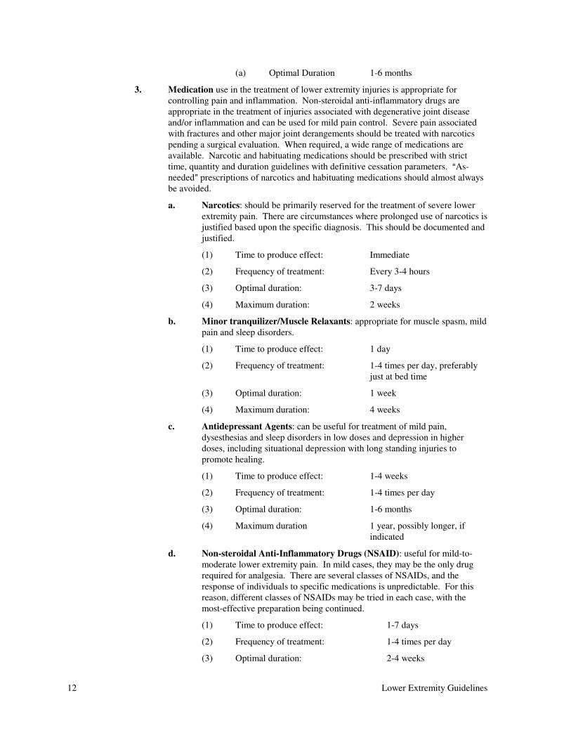
3. Medication use in the treatment of lower extremity injuries is appropriate for
controlling pain and inflammation. Non-steroidal anti-inflammatory drugs are
appropriate in the treatment of injuries associated with degenerative joint disease
and/or inflammation and can be used for mild pain control. Severe pain associated
with fractures and other major joint derangements should be treated with narcotics
pending a surgical evaluation. When required, a wide range of medications are
available. Narcotic and habituating medications should be prescribed with strict
time, quantity and duration guidelines with definitive cessation parameters. AAs-
needed@ prescriptions of narcotics and habituating medications should almost always
be avoided.
a. Narcotics: should be primarily reserved for the treatment of severe lower
extremity pain. There are circumstances where prolonged use of narcotics is
justified based upon the specific diagnosis. This should be documented and
justified.
(1) Time to produce effect: Immediate
(2) Frequency of treatment: Every 3-4 hours
(3) Optimal duration: 3-7 days
(4) Maximum duration: 2 weeks
b. Minor tranquilizer/Muscle Relaxants: appropriate for muscle spasm, mild
pain and sleep disorders.
(1) Time to produce effect: 1 day
(2) Frequency of treatment: 1-4 times per day, preferably
just at bed time
(3) Optimal duration: 1 week
(4) Maximum duration: 4 weeks
c. Antidepressant Agents: can be useful for treatment of mild pain,
dysesthesias and sleep disorders in low doses and depression in higher
doses, including situational depression with long standing injuries to
promote healing.
(1) Time to produce effect: 1-4 weeks
(2) Frequency of treatment: 1-4 times per day
(3) Optimal duration: 1-6 months
(4) Maximum duration 1 year, possibly longer, if
indicated
d. Non-steroidal Anti-Inflammatory Drugs (NSAID): useful for mild-to-
moderate lower extremity pain. In mild cases, they may be the only drug
required for analgesia. There are several classes of NSAIDs, and the
response of individuals to specific medications is unpredictable. For this
reason, different classes of NSAIDs may be tried in each case, with the
most-effective preparation being continued.
(1) Time to produce effect: 1-7 days
(2) Frequency of treatment: 1-4 times per day
(3) Optimal duration: 2-4 weeks
Lower Extremity Guidelines 13
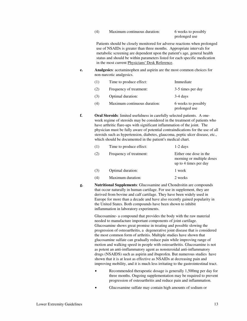
(4) Maximum continuous duration: 6 weeks to possibly
prolonged use
Patients should be closely monitored for adverse reactions when prolonged
use of NSAIDs is greater than three months. Appropriate intervals for
metabolic screening are dependent upon the patient=s age, general health
status and should be within parameters listed for each specific medication
in the most current Physicians= Desk Reference.
e. Analgesics: acetaminophen and aspirin are the most common choices for
non-narcotic analgesics.
(1) Time to produce effect: Immediate
(2) Frequency of treatment: 3-5 times per day
(3) Optimal duration: 3-4 days
(4) Maximum continuous duration: 6 weeks to possibly
prolonged use
f. Oral Steroids: limited usefulness in carefully selected patients. A one-
week regime of steroids may be considered in the treatment of patients who
have arthritic flare-ups with significant inflammation of the joint. The
physician must be fully aware of potential contraindications for the use of all
steroids such as hypertension, diabetes, glaucoma, peptic ulcer disease, etc.,
which should be documented in the patient=s medical chart.
(1) Time to produce effect: 1-2 days
(2) Frequency of treatment: Either one dose in the
morning or multiple doses
up to 4 times per day
(3) Optimal duration: 1 week
(4) Maximum duration: 2 weeks
g. Nutritional Supplements: Glucosamine and Chondroitin are compounds
that occur naturally in human cartilage. For use in supplement, they are
derived from bovine and calf cartilage. They have been widely used in
Europe for more than a decade and have also recently gained popularity in
the United States. Both compounds have been shown to inhibit
inflammation in laboratory experiments.
Glucosamine- a compound that provides the body with the raw material
needed to manufacture important components of joint cartilage.
Glucosamine shows great promise in treating and possible slowing the
progression of osteoarthritis, a degenerative joint disease that is considered
the most common form of arthritis. Multiple studies have shown that
glucosamine sulfate can gradually reduce pain while improving range of
motion and walking speed in people with osteoarthritis. Glucosamine is not
as potent an anti-imflammatory agent as nonsteroidal anti-inflammatory
drugs (NSAIDS) such as aspirin and ibuprofen. But numerous studies have
shown that it is at least as effective as NSAIDs at decreasing pain and
improving mobility, and it is much less irritating to the gastrointestinal tract.
$ Recommended therapeutic dosage is generally 1,500mg per day for
three months. Ongoing supplementation may be required to prevent
progression of osteoarthritis and reduce pain and inflammation.
$ Glucosamine sulfate may contain high amounts of sodium or

14 Lower Extremity Guidelines
potassium, so individuals on a restricted diet or taking potassium-
sparing diuretics should carefully check the label before taking it.
People with diabetes should have their blood sugar checked
regularly, since glucosamine may raise insulin resistance.
Individuals with shellfish allergies should check with a health care
professional before taking glucosamine.
4. Therapeutic Injection:
a. Joint Injections can be performed as analgesic (pain relieving) or anti-
inflammatory procedures. All techniques should include sterile technique as
appropriate. Soft tissue regions such as bursae may also be injected
utilizing the same criteria and medications. Viscol supplementation
injections should be utilized to decrease pain and improve function if
traditional injections have failed. Care should be utilized to avoid tendon
insertions which may result in loss of integrity or stability.
(1) Frequency of treatment: Not more than 3-4
times/annually
b. Trigger Point Injections are appropriate in selected cases where there are a
few defined points of muscular tenderness associated with pressure-induced,
radiating pain. Injection of trigger points with a local anesthetic followed
by physical therapy treatment may be appropriate.
(1) Time to produce effect: Immediate
(2) Frequency of treatment: 3-4 injections in same site
assuming favorable results
after each injection
c. Sclero/Prolotherapy has no proven value in the management of lower
extremity injuries via well controlled double-blind studies and may have
harmful effects. It has been advocated by some practitioners for the
treatment of unstable ligaments or joint capsules to stabilize the knee.
Proponents of these techniques should present supporting evidence to the
Physician Advisory Committee for future consideration.
B. Operative Treatment:
1. Manipulation Under Anesthesia should be considered if routine non-operative
therapeutic procedures such as physical therapy and/or dynamic bracing do not
restore the degree of motion expected after a reasonable period of time, usually at
least twelve weeks.
a. Time to produce effect: Immediate
b. Frequency of treatment: 1 time
2. Bursectomy is indicated for recurrent symptomatic conditions of a bursa which are
uncontrolled by conservative modalities.
a. Time to produce effect: Immediate
b. Frequency of treatment: 1 time
3. Arthroscopy is indicated when an intra-articular derangement is confirmed by
radiographic and/or physical examination. Arthroscopy is also considered when
resolution of signs and symptoms does not occur within an expected period of time
with appropriate non-invasive techniques or if the injury is such that it is not
expected to heal with conservative treatment.
Lower Extremity Guidelines 15
Reference Section VI, ASpecific Joint Involvement@, Item B., AKnee - Outlying
Diagnoses@.
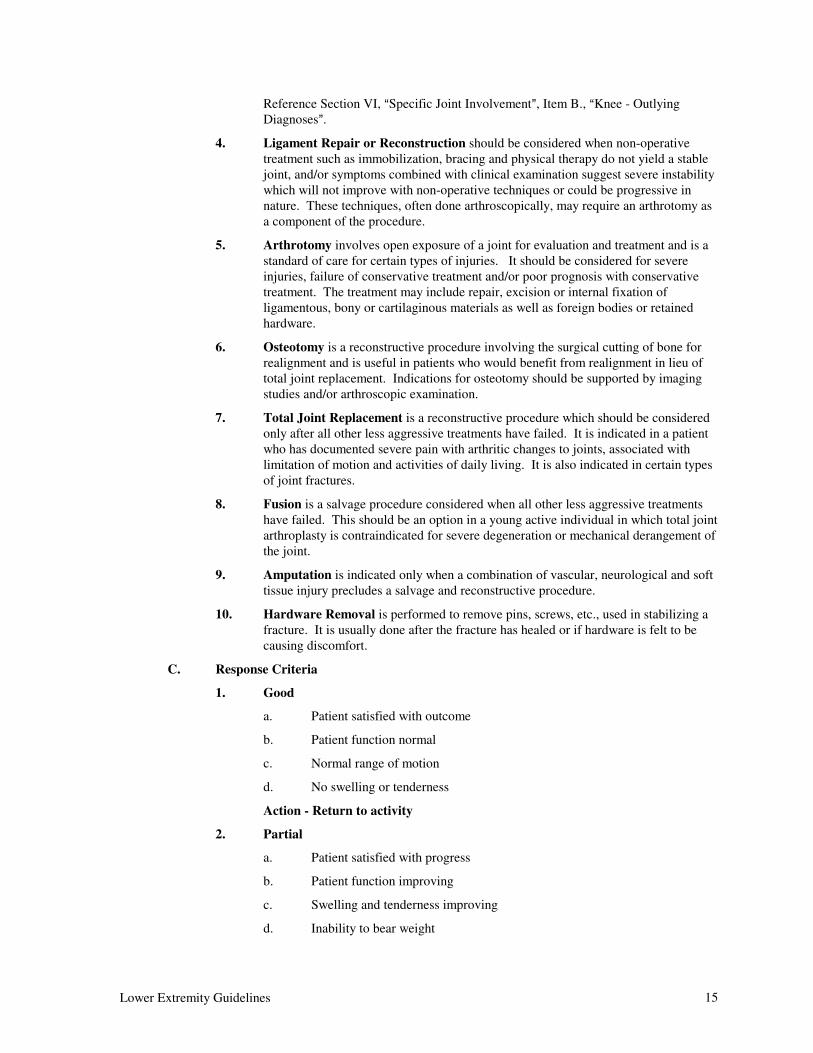
4. Ligament Repair or Reconstruction should be considered when non-operative
treatment such as immobilization, bracing and physical therapy do not yield a stable
joint, and/or symptoms combined with clinical examination suggest severe instability
which will not improve with non-operative techniques or could be progressive in
nature. These techniques, often done arthroscopically, may require an arthrotomy as
a component of the procedure.
5. Arthrotomy involves open exposure of a joint for evaluation and treatment and is a
standard of care for certain types of injuries. It should be considered for severe
injuries, failure of conservative treatment and/or poor prognosis with conservative
treatment. The treatment may include repair, excision or internal fixation of
ligamentous, bony or cartilaginous materials as well as foreign bodies or retained
hardware.
6. Osteotomy is a reconstructive procedure involving the surgical cutting of bone for
realignment and is useful in patients who would benefit from realignment in lieu of
total joint replacement. Indications for osteotomy should be supported by imaging
studies and/or arthroscopic examination.
7. Total Joint Replacement is a reconstructive procedure which should be considered
only after all other less aggressive treatments have failed. It is indicated in a patient
who has documented severe pain with arthritic changes to joints, associated with
limitation of motion and activities of daily living. It is also indicated in certain types
of joint fractures.
8. Fusion is a salvage procedure considered when all other less aggressive treatments
have failed. This should be an option in a young active individual in which total joint
arthroplasty is contraindicated for severe degeneration or mechanical derangement of
the joint.
9. Amputation is indicated only when a combination of vascular, neurological and soft
tissue injury precludes a salvage and reconstructive procedure.
10. Hardware Removal is performed to remove pins, screws, etc., used in stabilizing a
fracture. It is usually done after the fracture has healed or if hardware is felt to be
causing discomfort.
C. Response Criteria
1. Good
a. Patient satisfied with outcome
b. Patient function normal
c. Normal range of motion
d. No swelling or tenderness
Action - Return to activity
2. Partial
a. Patient satisfied with progress
b. Patient function improving
c. Swelling and tenderness improving
d. Inability to bear weight
16 Lower Extremity Guidelines
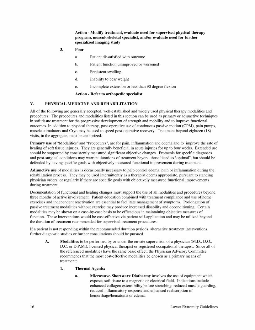
Action - Modify treatment, evaluate need for supervised physical therapy
program, musculoskeletal specialist, and/or evaluate need for further
specialized imaging study
3. Poor
a. Patient dissatisfied with outcome
b. Patient function unimproved or worsened
c. Persistent swelling
d. Inability to bear weight
e. Incomplete extension or less than 90 degree flexion
Action - Refer to orthopedic specialist
V. PHYSICAL MEDICINE AND REHABILITATION
All of the following are generally accepted, well-established and widely used physical therapy modalities and
procedures. The procedures and modalities listed in this section can be used as primary or adjunctive techniques
in soft tissue treatment for the progressive development of strength and mobility and to improve functional
outcomes. In addition to physical therapy, post-operative use of continuous passive motion (CPM), pain pumps,
muscle stimulators and Cryo may be used to speed post-operative recovery. Treatment beyond eighteen (18)
visits, in the aggregate, must be authorized.
Primary use of AModalities@ and AProcedures@, are for pain, inflammation and edema and to improve the rate of
healing of soft tissue injuries. They are generally beneficial in acute injuries for up to four weeks. Extended use
should be supported by consistently measured significant objective changes. Protocols for specific diagnoses
and post-surgical conditions may warrant durations of treatment beyond those listed as Aoptimal@, but should be
defended by having specific goals with objectively measured functional improvement during treatment.
Adjunctive use of modalities is occasionally necessary to help control edema, pain or inflammation during the
rehabilitation process. They may be used intermittently as a therapist deems appropriate, pursuant to standing
physician orders, or regularly if there are specific goals with objectively measured functional improvements
during treatment.
Documentation of functional and healing changes must support the use of all modalities and procedures beyond
three months of active involvement. Patient education combined with treatment compliance and use of home
exercises and independent reactivation are essential to facilitate management of symptoms. Prolongation of
passive treatment modalities without exercise may produce increased disability and deconditioning. Certain
modalities may be shown on a case-by-case basis to be efficacious in maintaining objective measures of
function. These interventions would be cost-effective via patient self-application and may be utilized beyond
the duration of treatment recommended for supervised treatment procedures.
If a patient is not responding within the recommended duration periods, alternative treatment interventions,
further diagnostic studies or further consultations should be pursued.
A. Modalities to be performed by or under the on-site supervision of a physician (M.D., D.O.,
D.C. or D.P.M.), licensed physical therapist or registered occupational therapist. Since all of
the referenced modalities have the same basic effect, the Physician Advisory Committee
recommends that the most cost-effective modalities be chosen as a primary means of
treatment:
1. Thermal Agents:
a. Microwave-Shortwave Diathermy involves the use of equipment which
exposes soft tissue to a magnetic or electrical field. Indications include
enhanced collagen extensibility before stretching, reduced muscle guarding,
reduced inflammatory response and enhanced reabsorption of
hemorrhage/hematoma or edema.
Lower Extremity Guidelines 17
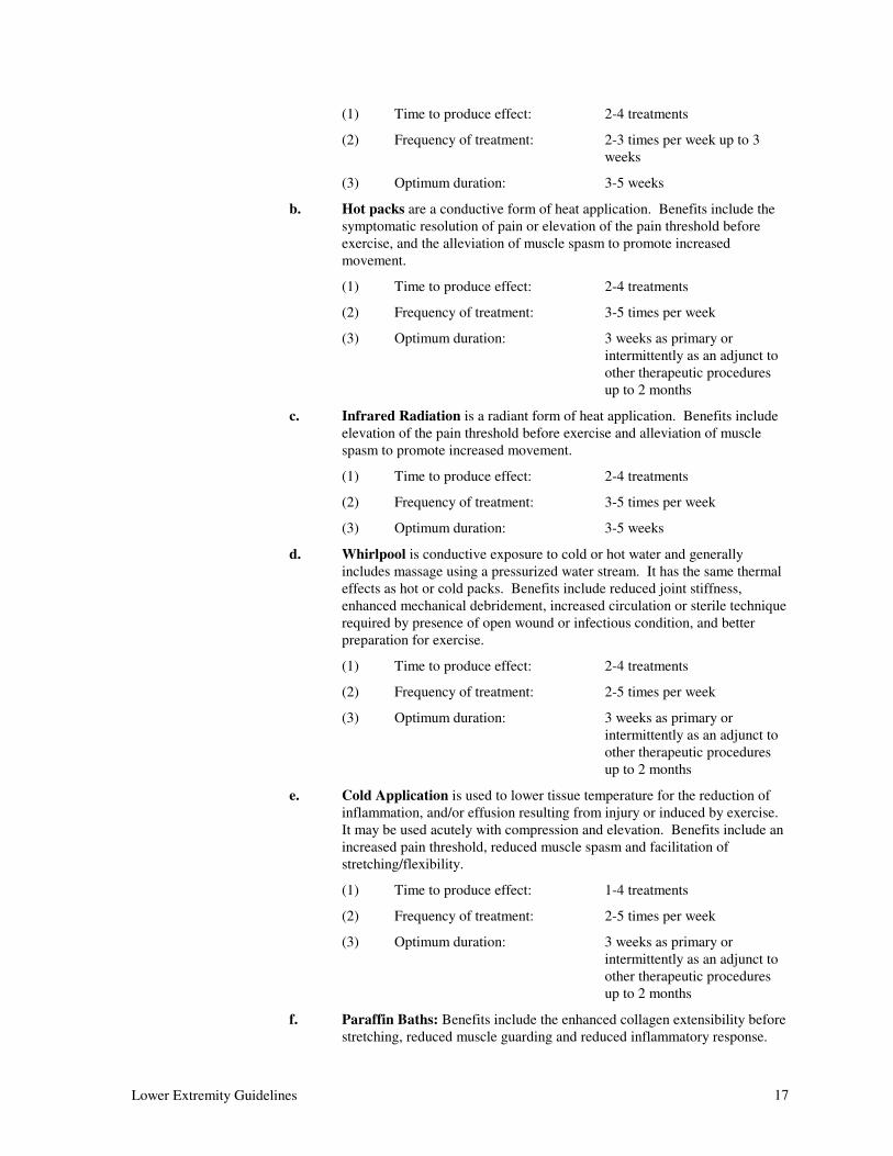
(1) Time to produce effect: 2-4 treatments
(2) Frequency of treatment: 2-3 times per week up to 3
weeks
(3) Optimum duration: 3-5 weeks
b. Hot packs are a conductive form of heat application. Benefits include the
symptomatic resolution of pain or elevation of the pain threshold before
exercise, and the alleviation of muscle spasm to promote increased
movement.
(1) Time to produce effect: 2-4 treatments
(2) Frequency of treatment: 3-5 times per week
(3) Optimum duration: 3 weeks as primary or
intermittently as an adjunct to
other therapeutic procedures
up to 2 months
c. Infrared Radiation is a radiant form of heat application. Benefits include
elevation of the pain threshold before exercise and alleviation of muscle
spasm to promote increased movement.
(1) Time to produce effect: 2-4 treatments
(2) Frequency of treatment: 3-5 times per week
(3) Optimum duration: 3-5 weeks
d. Whirlpool is conductive exposure to cold or hot water and generally
includes massage using a pressurized water stream. It has the same thermal
effects as hot or cold packs. Benefits include reduced joint stiffness,
enhanced mechanical debridement, increased circulation or sterile technique
required by presence of open wound or infectious condition, and better
preparation for exercise.
(1) Time to produce effect: 2-4 treatments
(2) Frequency of treatment: 2-5 times per week
(3) Optimum duration: 3 weeks as primary or
intermittently as an adjunct to
other therapeutic procedures
up to 2 months
e. Cold Application is used to lower tissue temperature for the reduction of
inflammation, and/or effusion resulting from injury or induced by exercise.
It may be used acutely with compression and elevation. Benefits include an
increased pain threshold, reduced muscle spasm and facilitation of
stretching/flexibility.
(1) Time to produce effect: 1-4 treatments
(2) Frequency of treatment: 2-5 times per week
(3) Optimum duration: 3 weeks as primary or
intermittently as an adjunct to
other therapeutic procedures
up to 2 months
f. Paraffin Baths: Benefits include the enhanced collagen extensibility before
stretching, reduced muscle guarding and reduced inflammatory response.
18 Lower Extremity Guidelines
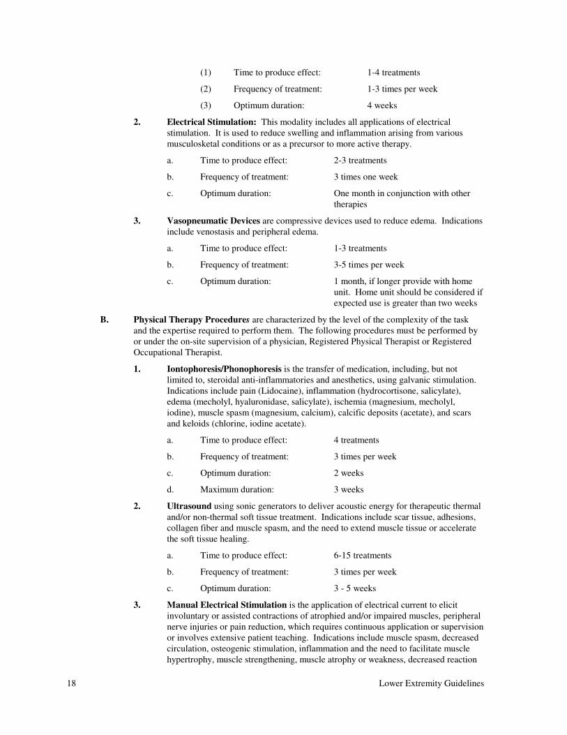
(1) Time to produce effect: 1-4 treatments
(2) Frequency of treatment: 1-3 times per week
(3) Optimum duration: 4 weeks
2. Electrical Stimulation: This modality includes all applications of electrical
stimulation. It is used to reduce swelling and inflammation arising from various
musculosketal conditions or as a precursor to more active therapy.
a. Time to produce effect: 2-3 treatments
b. Frequency of treatment: 3 times one week
c. Optimum duration: One month in conjunction with other
therapies
3. Vasopneumatic Devices are compressive devices used to reduce edema. Indications
include venostasis and peripheral edema.
a. Time to produce effect: 1-3 treatments
b. Frequency of treatment: 3-5 times per week
c. Optimum duration: 1 month, if longer provide with home
unit. Home unit should be considered if
expected use is greater than two weeks
B. Physical Therapy Procedures are characterized by the level of the complexity of the task
and the expertise required to perform them. The following procedures must be performed by
or under the on-site supervision of a physician, Registered Physical Therapist or Registered
Occupational Therapist.
1. Iontophoresis/Phonophoresis is the transfer of medication, including, but not
limited to, steroidal anti-inflammatories and anesthetics, using galvanic stimulation.
Indications include pain (Lidocaine), inflammation (hydrocortisone, salicylate),
edema (mecholyl, hyaluronidase, salicylate), ischemia (magnesium, mecholyl,
iodine), muscle spasm (magnesium, calcium), calcific deposits (acetate), and scars
and keloids (chlorine, iodine acetate).
a. Time to produce effect: 4 treatments
b. Frequency of treatment: 3 times per week
c. Optimum duration: 2 weeks
d. Maximum duration: 3 weeks
2. Ultrasound using sonic generators to deliver acoustic energy for therapeutic thermal
and/or non-thermal soft tissue treatment. Indications include scar tissue, adhesions,
collagen fiber and muscle spasm, and the need to extend muscle tissue or accelerate
the soft tissue healing.
a. Time to produce effect: 6-15 treatments
b. Frequency of treatment: 3 times per week
c. Optimum duration: 3 - 5 weeks
3. Manual Electrical Stimulation is the application of electrical current to elicit
involuntary or assisted contractions of atrophied and/or impaired muscles, peripheral
nerve injuries or pain reduction, which requires continuous application or supervision
or involves extensive patient teaching. Indications include muscle spasm, decreased
circulation, osteogenic stimulation, inflammation and the need to facilitate muscle
hypertrophy, muscle strengthening, muscle atrophy or weakness, decreased reaction
Lower Extremity Guidelines 19
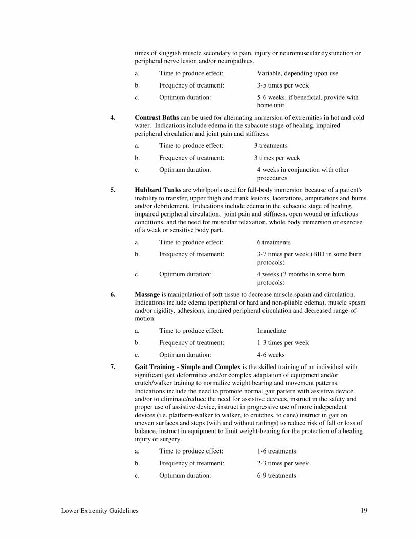
times of sluggish muscle secondary to pain, injury or neuromuscular dysfunction or
peripheral nerve lesion and/or neuropathies.
a. Time to produce effect: Variable, depending upon use
b. Frequency of treatment: 3-5 times per week
c. Optimum duration: 5-6 weeks, if beneficial, provide with
home unit
4. Contrast Baths can be used for alternating immersion of extremities in hot and cold
water. Indications include edema in the subacute stage of healing, impaired
peripheral circulation and joint pain and stiffness.
a. Time to produce effect: 3 treatments
b. Frequency of treatment: 3 times per week
c. Optimum duration: 4 weeks in conjunction with other
procedures
5. Hubbard Tanks are whirlpools used for full-body immersion because of a patient=s
inability to transfer, upper thigh and trunk lesions, lacerations, amputations and burns
and/or debridement. Indications include edema in the subacute stage of healing,
impaired peripheral circulation, joint pain and stiffness, open wound or infectious
conditions, and the need for muscular relaxation, whole body immersion or exercise
of a weak or sensitive body part.
a. Time to produce effect: 6 treatments
b. Frequency of treatment: 3-7 times per week (BID in some burn
protocols)
c. Optimum duration: 4 weeks (3 months in some burn
protocols)
6. Massage is manipulation of soft tissue to decrease muscle spasm and circulation.
Indications include edema (peripheral or hard and non-pliable edema), muscle spasm
and/or rigidity, adhesions, impaired peripheral circulation and decreased range-of-
motion.
a. Time to produce effect: Immediate
b. Frequency of treatment: 1-3 times per week
c. Optimum duration: 4-6 weeks
7. Gait Training - Simple and Complex is the skilled training of an individual with
significant gait deformities and/or complex adaptation of equipment and/or
crutch/walker training to normalize weight bearing and movement patterns.
Indications include the need to promote normal gait pattern with assistive device
and/or to eliminate/reduce the need for assistive devices, instruct in the safety and
proper use of assistive device, instruct in progressive use of more independent
devices (i.e. platform-walker to walker, to crutches, to cane) instruct in gait on
uneven surfaces and steps (with and without railings) to reduce risk of fall or loss of
balance, instruct in equipment to limit weight-bearing for the protection of a healing
injury or surgery.
a. Time to produce effect: 1-6 treatments
b. Frequency of treatment: 2-3 times per week
c. Optimum duration: 6-9 treatments
20 Lower Extremity Guidelines
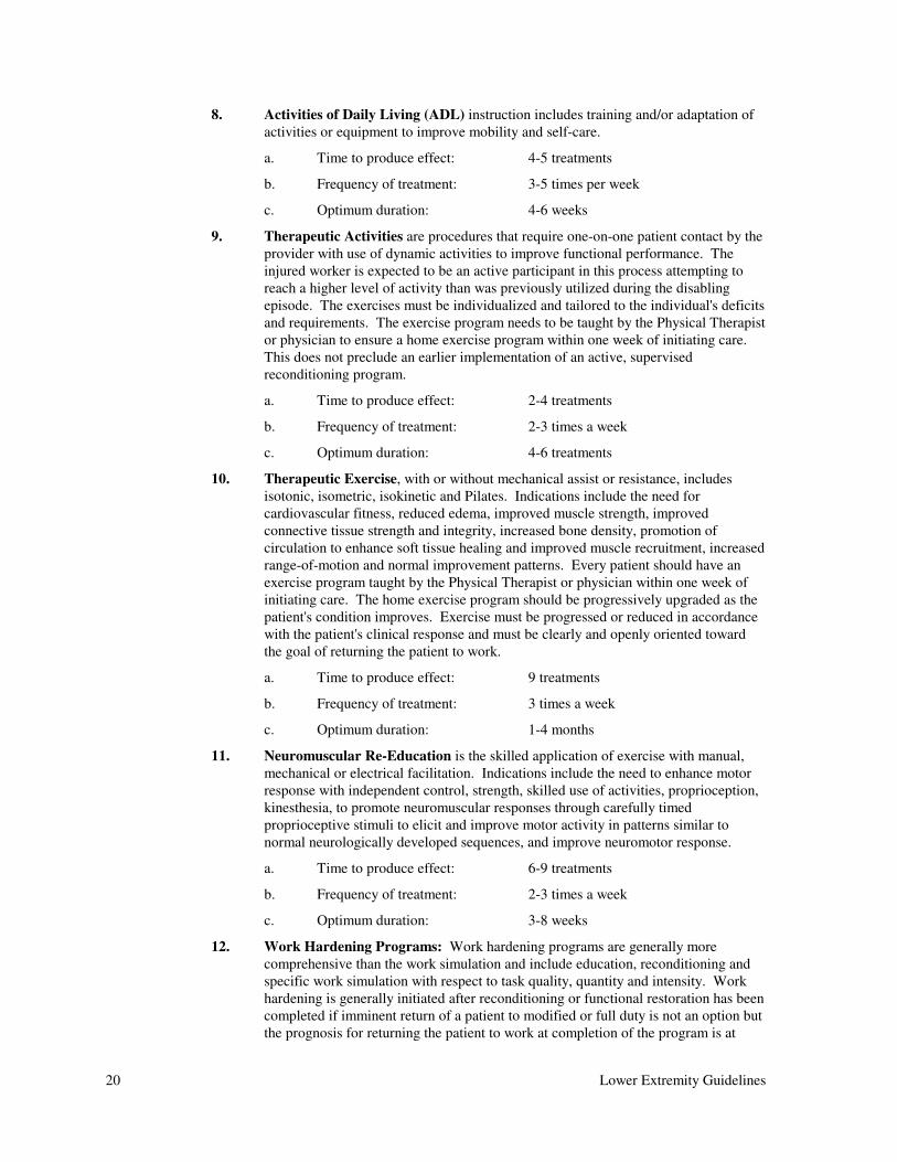
8. Activities of Daily Living (ADL) instruction includes training and/or adaptation of
activities or equipment to improve mobility and self-care.
a. Time to produce effect: 4-5 treatments
b. Frequency of treatment: 3-5 times per week
c. Optimum duration: 4-6 weeks
9. Therapeutic Activities are procedures that require one-on-one patient contact by the
provider with use of dynamic activities to improve functional performance. The
injured worker is expected to be an active participant in this process attempting to
reach a higher level of activity than was previously utilized during the disabling
episode. The exercises must be individualized and tailored to the individual's deficits
and requirements. The exercise program needs to be taught by the Physical Therapist
or physician to ensure a home exercise program within one week of initiating care.
This does not preclude an earlier implementation of an active, supervised
reconditioning program.
a. Time to produce effect: 2-4 treatments
b. Frequency of treatment: 2-3 times a week
c. Optimum duration: 4-6 treatments
10. Therapeutic Exercise, with or without mechanical assist or resistance, includes
isotonic, isometric, isokinetic and Pilates. Indications include the need for
cardiovascular fitness, reduced edema, improved muscle strength, improved
connective tissue strength and integrity, increased bone density, promotion of
circulation to enhance soft tissue healing and improved muscle recruitment, increased
range-of-motion and normal improvement patterns. Every patient should have an
exercise program taught by the Physical Therapist or physician within one week of
initiating care. The home exercise program should be progressively upgraded as the
patient's condition improves. Exercise must be progressed or reduced in accordance
with the patient's clinical response and must be clearly and openly oriented toward
the goal of returning the patient to work.
a. Time to produce effect: 9 treatments
b. Frequency of treatment: 3 times a week
c. Optimum duration: 1-4 months
11. Neuromuscular Re-Education is the skilled application of exercise with manual,
mechanical or electrical facilitation. Indications include the need to enhance motor
response with independent control, strength, skilled use of activities, proprioception,
kinesthesia, to promote neuromuscular responses through carefully timed
proprioceptive stimuli to elicit and improve motor activity in patterns similar to
normal neurologically developed sequences, and improve neuromotor response.
a. Time to produce effect: 6-9 treatments
b. Frequency of treatment: 2-3 times a week
c. Optimum duration: 3-8 weeks
12. Work Hardening Programs: Work hardening programs are generally more
comprehensive than the work simulation and include education, reconditioning and
specific work simulation with respect to task quality, quantity and intensity. Work
hardening is generally initiated after reconditioning or functional restoration has been
completed if imminent return of a patient to modified or full duty is not an option but
the prognosis for returning the patient to work at completion of the program is at
Lower Extremity Guidelines 21
least fair to good. As discussed in this section, identification of realistic vocational
goals is essential for the successful completion of a work hardening program.
Generally, work hardening programs entail a progressive increase in the number of
hours per day that a patient completes work simulation tasks until the patient can
tolerate a full work day.
a. Time to produce effect: 2-4 weeks
b. Frequency of treatment: 2-5 times per week
c. Optimum duration: 4-6 weeks
d. Maximum duration: 2-3 months
13. Joint Mobilization techniques are passive movements applied to a joint in a specific
manner to restore the full, free, painless range of motion of a joint in the extremities.
Indications for the use of mobilization techniques include joints that are painful,
hypomobile or involve mechanical motion dysfunctions. Gentle mobilization can
also promote healing of injured tissues. A muscle cannot be fully rehabilitated if the
underlying joints are not free to move, and conversely, a muscle cannot move a joint
that is not free to move.
a. Time to produce effect: 6-9 treatments
b. Frequency of treatment: 3 times per week
c. Optimum duration: 6 weeks
14. Manipulation is manual therapy that moves a joint beyond the physiologic range of
motion but not beyond the anatomic range-of-motion. It is indicated for pain and
adhesions.
a. Time to produce effect: Immediate - 10 treatments
b. Frequency of treatment: 1-5 times per week as indicated by the
severity of involvement and the desired
effect
c. Optimum duration: 10 treatments
15. Orthotic Training (dynamic bracing, splinting) is the skilled instruction of the
proper use of orthotics, bracing, and/or splinting. Benefits include normalization of
weight-bearing, facilitation of motion, stabilization of joints with insufficient muscle
and proprioceptive/reflex competencies, protection of post-operative or sub-acute
conditions to fit the individual as needed during movement, correction of
biomechanical problems, control of neurological and orthopedic injuries for reduced
stress during functional activities and modification of tasks through instruction in the
use of a device or physical modification of a device which reduces stress on the
injury. Equipment should improve safety and reduce risk of re-injury.
a. Time to produce effect: 1-3 treatments
b. Frequency of treatment: As indicated to establish independent
use (1-3 sessions)
c. Optimum duration: 4 treatments
16. Prosthetic Training is the skilled instruction in the proper use of prosthetic limbs
including stump preparation, donning and doffing limbs, gait and transfer training
and prosthesis maintenance training. Indication for training is the need for prothesis
use.
a. Time to produce effect: 9 sessions
22 Lower Extremity Guidelines
b. Frequency of treatment: 3 times per week
c. Optimum duration: 2-4 months
17. Myofascial Release/Soft Tissue Mobilization: Myofascial Release is a form of soft
tissue mobilization based upon neuroreflexive responses that reduce tissue tension.
The net result is a relaxation of tissue tension and subsequent decrease in myofascial
tightness. It is a safe, effective method to normalize myofascial activity, regain tissue
extensibility and reduce pain. Normalization of myofascial tissue allows for
improved joint mobility.
Soft Tissue Mobilization is aimed at enhancing muscle tone and/or extensibility in
soft tissues. Restoration of soft tissue extensibility and/or inhibition of hyperactive
musculature helps promote motion function which in turn leads to a reduction in pain.
Soft tissue mobilization can be used during acute, sub-acute, and chronic
musculosketal conditions. Soft tissue mobilization can be used as a preparatory
procedure to decrease muscle guarding so that joint mobilization is effective in
improving extremity and/or spinal joint mobility.
a. Time to produce effect: 6-9 treatments
b. Frequency of treatment: 3-5 times a week
c. Optimum duration: 6-8 weeks
18. Manual Traction is an integral part of manual manipulation or joint mobilization.
Indications include decreased joint space, muscle spasm around joints, and the need
for increased synovial nutrition and response.
a. Time to produce effect: 1-3 sessions
b. Frequency of treatment: 2-3 times per week
c. Optimum duration: 30 days
19. Transcutaneous Electrical Nerve Stimulation (TENS) should be prescribed within
a supervised setting in order to assure proper electrode placement and patient
education. TENS can be used for muscle spasm or rigidity, atrophy, decreased
circulation and pain. If the response to three treatments is beneficial, it may be
continued for 1-3 months and for intermittent unsupervised use thereafter if it
facilitates objective functional gains. The Physician Advisory Committee
recommends rental of a TENS unit with reassessment after 30 days.
20. Hyperbaric Chamber: The Physician Advisory Committee recommends
documented use of a hyperbaric chamber on a case-by-case basis.
C. Return to work: Given the poor return to work prognosis for the injured worker after having
been out of work for more than six months, early return to work should be a prime goal in
treating occupational injuries. When attempting to return a patient to work after a specific
injury, it is understood that an accurate job description is essential to the physician in making
return to work recommendations.
Due to the large spectrum of injuries of varying severity and varying physical demands in the
work place, it is not possible to make specific return to work guidelines for each injury.
Therefore, the Physician Advisory Committee recommends the following:
1. In most cases of musculoskeletal injury to the lower extremity the patient should be
able to return to work in some capacity within two weeks unless there are extenuating
circumstances. Injuries which require more than two weeks off-work are listed in the
Section VI, ASpecific Joint Involvement@.
Lower Extremity Guidelines 23
2. Communication between the patient, employer and physician to determine
appropriate restrictions and return to work dates. A worksite evaluation may be
necessary and should be performed by a qualified specialist, such as an occupational
health nurse, occupational therapist, physical therapist, vocational rehabilitation
specialist, or an industrial hygienist. The adjuster should be notified of all return to
work orders.
3. Generally, if a patient has been out-of-work for more than two weeks, it is the
responsibility of the employer or adjuster to contact the patient and physician to
determine why they are unable to return to work.
Working or attaining a return to work status should not interfere with necessary medical care
or rehabilitation. See Section VI, ASpecific Joint Involvement@, for specific diagnostics
related to return to work situations.
D. Special Tests are performed as part of a skilled assessment of the patient=s capacity to return
to work or strength capacities, physical work demand classifications and tolerances. They
include:
1. Work Conditioning Assessments/Screens are functional assessments and work
tolerance assessments and/or any individualized evaluation tests and/or procedures
required to specifically identify and quantify work-relevant cardiovascular and
neuromuscular fitness and to address ergonomic issues affecting the participant=s
return to work potential.
2. Functional Capacity Evaluation (FCE) is a series of tests performed to determine
physical ability to perform work related tasks with consideration of pertinent medical
and behavioral improvements. The data derived from this evaluation will determine
the person=s ability to match job demands. Components of this evaluation may
include:
a. Musculoskeletal screen
b. Cardiovascular assessment
c. Coordination simulation
d. Assessment of fine motor tasks
e. Work simulated endurance testing
f. Reliability and validity of testing
g. Lift task analysis
3. Lift Analysis indications include the need to return to work or identify physical
restrictions in a particular job.
4. Mechanized/Computerized Strength Evaluations are isotonic, isometric and/or
isokinetic. Indications include the need to measure lower extremity strength and
monitor rehabilitation.
a. Frequency of treatment: 1 time for evaluation, can monitor
improvements in strength every 3-4
weeks up to a total of 6 evaluations
VI. SPECIFIC JOINT INVOLVEMENT
The diagnostic approach and treatment of lower extremity musculoskeletal conditions is similar in most of the
joints. In order to avoid repetition, this section covers the joints individually and contains material which is
applicable only to that specific joint. There are certain diagnoses or situations which may require extended
periods off work and must be tailored to individual situations. The physician should provide the patient with
24 Lower Extremity Guidelines
applicable restrictions during the various stages of recovery and rehabilitation. These recommendations are to
be viewed as general guidelines and may vary depending upon individual injury and job site.
A. Lower Extremity - Outlying Diagnoses
1. Infections Requiring Intravenous Antibiotics:
a. Return to work: When the infection is controlled, usually
with restrictions, 1-2 weeks
2. Reflex Sympathetic Dystrophy (RSD) may require extended modified duty.
3. Hardware removal:
a. Return to work: With restrictions, 1 week
b. Return to work: Full duty, 2-8 weeks
4. Contracture may require physical therapy up to 3 months. An aggressive home
stretching program is strongly encouraged. Occasional surgical release may be
required.
5. Severe Burns, Open Wounds, Vascular Injuries, Polytrauma, and Non-union
Fractures - case by case.
B. Knee - Outlying Diagnoses:
1. Severe Intra-Articular Fracture:
a. Non-Weight-Bearing: 8-12 weeks
b. Return to work: With restrictions, 4-6 weeks
2. Knee Fusion:
a. Non-Weight-Bearing: 0-20 weeks
b. Return to work: With restrictions, 4-8 weeks
Return to work: Full duty, at physician=s discretion
3. Total Knee Replacement:
a. Non-Weight-Bearing: 0-4 weeks, depending on the type of
replacement
b. Return to work: With restrictions, 4-12 weeks
c. Return to work: Full duty, at physician=s discretion
4. Amputation: 4 weeks wound healing; 12 weeks for prosthetic construction and
training.
5. Meniscectomy is the removal of the meniscus either by arthrotomy or arthroscopy.
Indications include documented symptomatic tear of the meniscus (diagnostic
arthroscopy, arthrogram, MRI).
a. Return to work: With restrictions, 3 weeks
b. Return to work: Full duty, 6-8 weeks
6. Meniscal repair:
a. Non-Weight-Bearing: 2-4 weeks
b. Return to work: With restrictions, 2-4 weeks
c. Return to work: Full duty, 4-6 weeks
Lower Extremity Guidelines 25
7. Retropatellar Pain Syndrome is pain in the anterior part of the knee for which there
are many causes. Treatment should be conservative for at least 3-6 months; if pain
persists despite adequate rehabilitation, certain surgical procedures can be indicated
such as arthroscopic lateral release, synovial shaving, and plica removal. More
invasive procedures should require a second opinion.
a. Non-Surgical Treatment:
Return to work: Can work with restrictions
b. Surgical Treatment:
Return to work: With restrictions, 3 weeks
Return to work: Full duty, 3-6 months
8. Patellar Subluxation is the recurrent dislocation or subluxation of the knee; surgical
indications include giving-way, pain, locking, catching, unresponsiveness to
conservative care with aggressive quadriceps strengthening program.
a. Return to work: With restrictions, 3 weeks
b. Return to work: Full duty, 3-6 months
9. ACL/PCL Repair indications include torn ACL/PCL with instability if the
instability interferes with activities or work.
a. Return to work: With restrictions, 3-6 weeks, bracing
may be required
b. Return to work: Full Duty, 3-9 months, bracing may be
required.
10. Automated Methods for Determining Cruciate Ligament Laxity (KT-1000, etc.)
are indicated for objective measurement of knee joint laxity to monitor success of
surgery or progress of rehabilitation of ACL/PCL injuries.
Frequency of use: Pre-operative, then total of three more
for follow-up
11. Arthroscopy may be diagnostic or therapeutic. Indications include: 1) abnormal
physical exam which demonstrates locking or instability or effusion over two weeks;
2) failure of conservative treatment with accurate diagnosis; or 3) retropatellar pain
lasting over three months which interferes with activities of daily living, and is
unresponsive to conservative treatment.
a. Return to work: With restrictions, 1 week
b. Return to work: Full duty, 2 weeks for diagnostic
arthroscopy without findings
Return to work recommendations vary with particular arthroscopic surgery
performed.
12. Chondroplasties, Chondral defects and Cartilage Repairs:
a. Return to work: With restrictions, 2-10 weeks
b. Return to work: Full duty, 10-12 weeks
C. Foot and Ankle Injuries
1. Background: Injuries to the foot and ankle usually relate to a specific traumatic
event and have a predictable clinical course depending on the severity index of the
26 Lower Extremity Guidelines
initial injury. For simplicity, injuries will be discussed relative to the anatomic
region of the foot and ankle (Ankle, hindfoot, midfoot, forefoot or phalanges).
2. Diagnostic Criteria
a. Pertinent historical and physical findings
(1) Onset of pain and/or swelling is related to a single event, either a
twisting injury, fall or direct blunt trauma. The degree of the injury
can be judged quickly by determining which one can bear weight
and the degree of initial swelling. The more severe injuries will
have greater swelling, ecchymosis, inability to bear weight, and
may have obvious deformity.
b. Appropriate diagnostic tests and examinations
(1) If differentiation between a soft tissue ligamentous injury and a
fracture is required, X-rays in several planes will be required in
ALL cases.
(2) CT Scans may be indicated in hindfoot injuries to define subtle
fractures, tarsal coalitions or the degree of displacement in three
planes in acute injuries.
(3) Bone scans are occasionally indicated in long standing pain
problems to rule out stress fractures or inflammatory causes of foot
pain (after four weeks of pain with normal X-rays).
(4) MRI rarely indicated - may require specialty consultation.
(5) EMG and Vascular studies (non-invasive arterial perfusion or
arteriography at the request of the specialist).
c. Inappropriate diagnostic tests
(1) Thermogram.
d. Indications for Specialty referral
(1) Displaced fractures
(2) Neuro Vascular Compromise
(3) Pain and Swelling longer than three weeks
3. Treatment
a. Non-operative
(1) Sprains (No fracture seen on X-ray)
(a) RICE, (Rest, Ice, Compression, Elevation)
(b) Crutches and splinting (one through three days).
(c) Early mobilization as pain allows. This may involve
active supervised physical therapy.
(d) Usual course, several days to three weeks.
(e) Referral to Specialist required if no improvement by three
weeks.
(2) Fractures
(a) Simple Non-displaced
Lower Extremity Guidelines 27
i. Ankle - Specialty referral - Will require special
splinting or casting for three to six weeks; may
require an additional two to four weeks of
physical therapy.
ii. Hindfoot - Same as ankle.
iii. Midfoot - Same as ankle but course is usually
two to four weeks shorter.
iv. Forefoot - Specialty referral not required.
Special shoe or cast may be necessary. Usually
resolved in three to six weeks.
v. Phalanges - Same as forefoot, simple taping
and/or modified shoe usually sufficient.
vi. Hallux fractures - May require casts in certain
instances.
(b) Displaced fractures - Specialty referral is mandatory.
Non-operative treatment requires casting for three to six
weeks followed by up to four weeks of rehabilitation.
b. Operative - All operative decisions require specialty referral
(1) Sprains - Indicated when there is complete dislocation/subluxation
without a fracture anywhere in the ankle, hindfoot, or midfoot.
May be indicated in the forefoot.
(2) Severe instability unresponsive after 2-4 weeks of conservative
treatment.
(3) Fractures
(a) Simple - May be indicated in the ankle
(b) Displaced - Usually indicated in the ankle, hindfoot,
midfoot, and forefoot. Displaced phalange fractures can
sometimes be treated non-operatively.