Embed Size (px)
Citation preview

Risk factors for lower extremity injuries among contemporary dance students
Christine van Seters, MD1,2
; Rogier M van Rijn, PhD1; Marienke van Middelkoop, PhD
2; Janine H Stubbe, Phd
1,3
1 Codarts, University of the Arts, Rotterdam, The Netherlands
2 Department of General practice, Erasmus MC University Medical Center, Rotterdam, The Netherlands
3 Amsterdam University of Applied Sciences, Centre for Applied Research in Sports and Nutrition, Amsterdam, The
Netherlands
Corresponding author: Rogier M van Rijn, Codarts University of the Arts, Rotterdam, The Netherlands,
Kruisplein 26,3012 CC, Rotterdam, The Netherlands, [email protected]
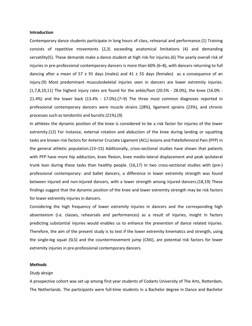
Abstract
This prospective cohort study was conducted to assess if student characteristics, lower extremity
kinematics and strength are risk factors for sustaining lower extremity injuries in pre-professional
contemporary dancers. At the start of the academic year (2015/2016) a total of 45 first year students of
Bachelor Dance and Bachelor Dance Teacher received a questionnaire containing items on injury history
(only lower extremity) and student characteristics (age, gender, educational program); height and
weight measurements (BMI); and functional tests (single-leg squat (SLS) and countermovement jump).
During the academic year injuries were recorded using a monthly questionnaire. Substantial lower
extremity injuries during the academic year (main outcome) were defined as any problems leading to
moderate or severe reductions in training volume or in performance, or complete inability to participate
in dance at least once during follow-up as measured with the Oslo Sports Trauma Research Center
(OSTRC) Questionnaire on Health Problems. Analyses on leg level were performed using Generalized
Estimating Equations to test the associations between substantial lower extremity injuries and potential
risk factors. The one-year prevalence of lower extremity injuries was 82.2%. Of these 51.4% was a
substantial lower extremity injury. Multivariate analyses identified that ankle dorsiflexion during the SLS
(OR 1.25; 95%CI 1.03–1.52) was a risk factors for a substantial lower extremity injury. The findings
indicate that contemporary dance students are at high risk for lower extremity injuries. Therefore, the
identified risk factor should be considered for prevention purposes.
Keywords: dance, pre-professional, injury, lower extremity, risk factors, prospective cohort study
Introduction
Contemporary dance students participate in long hours of class, rehearsal and performance.(1) Training
consists of repetitive movements (2,3) exceeding anatomical limitations (4) and demanding
versatility(5). These demands make a dance student at high risk for injuries.(6) The yearly overall risk of
injuries in pre-professional contemporary dancers is more than 60% (6–8), with dancers returning to full
dancing after a mean of 57 ± 91 days (males) and 41 ± 55 days (females) as a consequence of an
injury.(9) Most predominant musculoskeletal injuries seen in dancers are lower extremity injuries.
(1,7,8,10,11) The highest injury rates are found for the ankle/foot (20.5% - 28.0%), the knee (16.0% -
21.4%) and the lower back (13.4% - 17.0%).(7–9) The three most common diagnoses reported in
professional contemporary dancers were muscle strains (28%), ligament sprains (23%), and chronic
processes such as tendonitis and bursitis (21%).(9)
In athletes the dynamic position of the knee is considered to be a risk factor for injuries of the lower
extremity.(12) For instance, external rotation and abduction of the knee during landing or squatting
tasks are known risk factors for Anterior Cruciate Ligament (ACL) lesions and Patellofemoral Pain (PFP) in
the general athletic population.(13–15) Additionally, cross-sectional studies have shown that patients
with PFP have more hip adduction, knee flexion, knee medio-lateral displacement and peak ipsilateral
trunk lean during these tasks than healthy people. (16,17) In two cross-sectional studies with (pre-)
professional contemporary- and ballet dancers, a difference in lower extremity strength was found
between injured and non-injured dancers, with a lower strength among injured dancers.(18,19) These
findings suggest that the dynamic position of the knee and lower extremity strength may be risk factors
for lower extremity injuries in dancers.
Considering the high frequency of lower extremity injuries in dancers and the corresponding high
absenteeism (i.e. classes, rehearsals and performances) as a result of injuries, insight in factors
predicting substantial injuries would enables us to enhance the prevention of dance related injuries.
Therefore, the aim of the present study is to test if the lower extremity kinematics and strength, using
the single-leg squat (SLS) and the countermovement jump (CMJ), are potential risk factors for lower
extremity injuries in pre-professional contemporary dancers.
Methods
Study design
A prospective cohort was set up among first year students of Codarts University of The Arts, Rotterdam,
The Netherlands. The participants were full-time students in a Bachelor degree in Dance and Bachelor
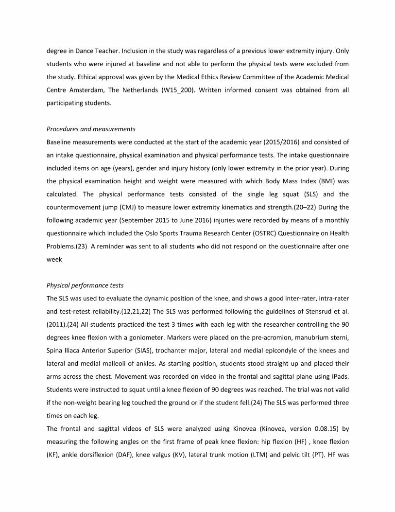
degree in Dance Teacher. Inclusion in the study was regardless of a previous lower extremity injury. Only
students who were injured at baseline and not able to perform the physical tests were excluded from
the study. Ethical approval was given by the Medical Ethics Review Committee of the Academic Medical
Centre Amsterdam, The Netherlands (W15_200). Written informed consent was obtained from all
participating students.
Procedures and measurements
Baseline measurements were conducted at the start of the academic year (2015/2016) and consisted of
an intake questionnaire, physical examination and physical performance tests. The intake questionnaire
included items on age (years), gender and injury history (only lower extremity in the prior year). During
the physical examination height and weight were measured with which Body Mass Index (BMI) was
calculated. The physical performance tests consisted of the single leg squat (SLS) and the
countermovement jump (CMJ) to measure lower extremity kinematics and strength.(20–22) During the
following academic year (September 2015 to June 2016) injuries were recorded by means of a monthly
questionnaire which included the Oslo Sports Trauma Research Center (OSTRC) Questionnaire on Health
Problems.(23) A reminder was sent to all students who did not respond on the questionnaire after one
week
Physical performance tests
The SLS was used to evaluate the dynamic position of the knee, and shows a good inter-rater, intra-rater
and test-retest reliability.(12,21,22) The SLS was performed following the guidelines of Stensrud et al.
(2011).(24) All students practiced the test 3 times with each leg with the researcher controlling the 90
degrees knee flexion with a goniometer. Markers were placed on the pre-acromion, manubrium sterni,
Spina Iliaca Anterior Superior (SIAS), trochanter major, lateral and medial epicondyle of the knees and
lateral and medial malleoli of ankles. As starting position, students stood straight up and placed their
arms across the chest. Movement was recorded on video in the frontal and sagittal plane using IPads.
Students were instructed to squat until a knee flexion of 90 degrees was reached. The trial was not valid
if the non-weight bearing leg touched the ground or if the student fell.(24) The SLS was performed three
times on each leg.
The frontal and sagittal videos of SLS were analyzed using Kinovea (Kinovea, version 0.08.15) by
measuring the following angles on the first frame of peak knee flexion: hip flexion (HF) , knee flexion
(KF), ankle dorsiflexion (DAF), knee valgus (KV), lateral trunk motion (LTM) and pelvic tilt (PT). HF was
defined as the angle between the line formed by pre-acromion and trochanter major and the line
between the lateral knee epicondyle and trochanter major. KF was defined as the angle between the
line formed by trochanter major and lateral epicondyle and the line between lateral knee epicondyle
and lateral malleolus. DAF was the angle between the line formed by lateral epicondyle and calcaneus
through lateral malleolus and the line between the fifth toe and calcaneus, with a larger dorsiflexion
indicating limited ankle dorsiflexion. PT was the angle between the line formed by ipsilateral and
contralateral SIAS and the horizontal line starting in the ipsilateral SIAS. The KV and LTM were measured
accordingly to Dingenen et al. (2014).(25) The average angle of three trials was calculated for both legs
separately.
The CMJ test was used as a measure for strength of the lower extremity, and shows a good inter-rater,
intra-rater and test-retest reliability.(20,26) Students were instructed to stand on the electronic timing
plate(Fusion Sport) with their hands on the hips. The plate detects flight time and converts this in jump
height (cm). Students were instructed to squat as deep as preferred and consequently jump as high as
possible without flexion of the knees during the jump or removing the hands from the hips. The trial was
rejected if an arm swing or knee bending occurred or if the student fell or lost balance while performing
the CMJ. Students were instructed to land on the plate at exactly the same place as the starting point.
The CMJ test was performed three times for jumping with both legs, and three times on the left and
right leg separately.(27) Consequently, the average jump height of these three trails from the different
jumping tasks was computed.
Prior to the physical performance tests the students performed a standardized warming-up consisting of
bipodal squats (2x8 repetitions), bipodal jumps (2x5 repetitions) and stretching of the calf muscle with
straight and bended knees.(24)
Injury registration
The monthly questionnaire consisted of four key questions on the consequences of health problems on
participation, training volume, and performance as well as the degree to which the student perceived
symptoms (OSTRC Questionnaire on Health Problems). Each question of the OSTRC was scored with a
four or five point scale, ranging from 0 (respectively: no problem, no reduction, no effect and no
symptoms) to 25 (cannot participate at all or severe symptoms). The severity of a health problem was
calculated on a scale of 0 (no health problem) – 100 (cannot participate at all due to sever health
problems) by summing the score of the four questions, according to the method proposed by Clarsen et
al..(28,29) If the severity score was 0, the questionnaire was finished for that month. However, if a
symptom was reported, the students were asked whether they referred to a physical injury, mental
problem or an illness. For physical injuries, the student was automatically directed to an injury
registration form based on an international consensus statement on injury surveillance methodology for
football to collect further details (e.g. location, history and acute or overuse onset).(30–33)
Lower extremity injuries are defined as injuries at the lower back, pelvis, leg, knee and foot. Students
were defined to be substantial injured at their lower extremity if they reported problems leading to
moderate or severe reductions (value ≥13 on question 2 or 3 of the OSTRC) in training volume, or
moderate or severe reductions in performance or complete inability to participate in dance at least once
during follow-up.(23)
Statistical methods
Statistical analyses were conducted using SPSS (SPSS, V21.0) and statistical significance level was set at
an alpha level >0.05. Descriptive statistics were used to describe baseline characteristics of all
participants using means and standard deviation (SD) or number and percentages (%). The one year
prevalence of all lower extremity injuries and substantial lower extremity injuries was calculated by
dividing the number of students that reported at least 1 lower extremity injury during the academic year
by the number of respondents
To examine potential risk factors for lower extremity injuries, univariate and multivariate regression
models were applied on leg level using Generalized Estimating Equations (GEE), taking into account the
association between two legs within one person. Potential risk factors included age (years), gender
(male), BMI (kg/m2), educational program (Bachelor Dance Teacher versus Bachelor Dance), injury
history in the prior year (only lower extremity injuries), all measured angles from the SLS (°) and jump
height from the CMJ (cm) for jumping with both legs and single leg. First, univariate associations
between the potential risk factors and the dichotomized outcome: substantially injured at the lower
extremity during follow-up (yes/no) were assessed. Secondly, multivariate regression modeling using
GEE was performed including all potential risk factors and the outcome of interest. The results of the
regression analyses were expressed in odds ratio’s (OR) with corresponding 95% confidence interval
(95% CI).
Results
Participants
All approached students (n=45) agreed to participate and were consequently included in the present
study. Four of these did not perform the CMJ and one student was not able to execute the SLS on the
right leg. The cohort comprised 28 females (62.2%), the mean age was 18.6 years (SD 1.1), mean BMI
was 20.7 kg/m2 (SD 1.6) and 17 students had a lower extremity injury history (37.8%). Twenty eight
(62%) were students enrolled in the Bachelor degree Dance and 17 (38 %) in the Bachelor Dance
Teacher (Table 1). The monthly response rate of the follow-up questionnaires ranged from 88.9% up to
100%.
Injuries
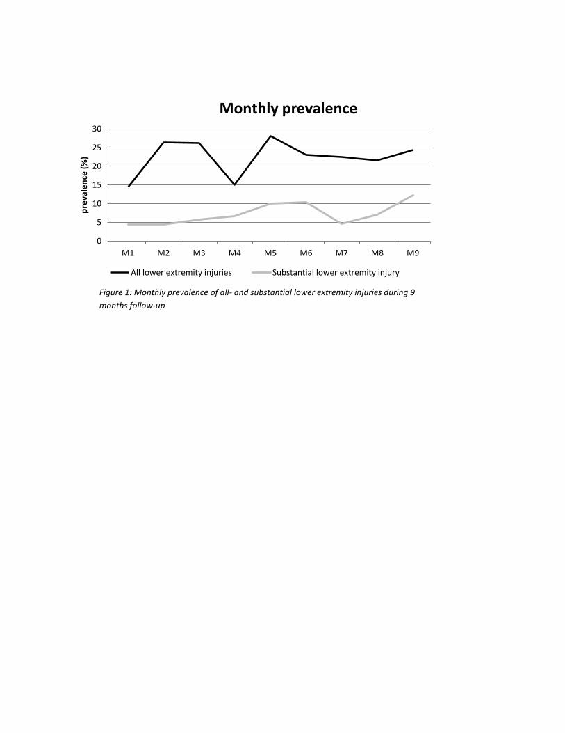
During the academic year a total of 37 (82.2%) students reported a lower extremity injury of which 19
(51.4%) were categorized as substantial. The monthly prevalence of all lower extremity injuries ranged
from 14.5% to 28.0% and from 4.4% to 12.2% for substantial lower extremity injuries (Figure 1).
Risk factors for lower extremity injuries
The univariate analyses showed a significant association between a limited dorsiflexion of the ankle (OR
1.11; 95% CI 1.02 – 1.20) and substantial lower extremity injuries during follow-up (Table 2). None of the
other tested variables were univariately associated with the outcome of interest.
The multivariate analysis also showed a significant association between limited dorsiflexion of the ankle
(OR 1.25; 95% CI 1.03 – 1.52) and the occurrence of substantial injuries. None of the other potential risk
factors were associated with the outcome in the multivariate analysis.
Discussion
This is the first prospective cohort study investigating risk factors for lower extremity injuries among
contemporary dance students. We found a one year prevalence of lower extremity injuries of 82.2%. Of
these, 51.4% were substantial injuries meaning that the students were not able to participate at all or
had a moderate or severe reduction in training volume or performance due to a lower extremity injury.
Results of the multivariate analysis showed that students with a limited ankle dorsiflexion (OR 1.25; 95%
CI 1.03 – 1.52) had a higher risk of sustaining a substantial lower extremity injury during the academic
year.
The monthly prevalence of all lower extremity injuries ranged from 14.5% to 28.0% and from 4.4% to
12.2% for substantial lower extremity injuries. In contrast to our findings, a retrospective cohort study
of contemporary dance students found a one year prevalence of lower extremity injuries of 64%.(8)
Besides, injury prevalence in professional contemporary dancers ranged from 24% to 74% in
literature.(7,9,34) Differences in reported injury prevalence may be due to differences in injury
registration and associated injury definitions. Therefore, there is a need for one universal injury case
definition in dance medicine.(6,35) Liederbach et al. (2012) made a first attempt for standardized testing
and reporting methodology in dance medicine and science research.(36) Their recommendation is to
define an injury as an anatomic tissue-level impairment diagnosed by a health care practitioner that
results in full time loss of activity for one or more days beyond the day of onset. However, this time-loss
definition would be inadequate if the focus is on early detection. Therefore, the OSTC Overuse Injury
Questionnaire as used in the current study seems to be a good instrument for this population, in
addition to the recommendation of Liederbach et al. (2012), as it registries all health problems (injuries,
illness and mental health problems) with standardized questions resulting in a summary severity score
providing a measure of the impact of health problems. Using this questionnaire, we found injury rates
higher previously described in literature.
To our knowledge, up to now no prospective studies have been performed to determine if lower
extremity kinematics (SLS) and strength (CMJ) can predict lower extremity injuries in contemporary
dance students. The current study showed that limited ankle dorsiflexion was associated with a higher
risk on lower extremity injuries during 9 months follow-up. None of the other potential risk factors
measured with the SLS were associated with lower extremity injuries. In other sport disciplines
prospective studies were performed to determine risk factors for injuries using functional tests. Bayne et
al (2016) conducted a prospective injury study in cricket fast bowlers and found that an increased knee
valgus angle during the single leg decline squat was associated with a higher low back injury risk during
the season.(37) In elite Olympic class sailors left-sided single-leg-decline squat performance was
associated with overall injury status, with better performing athletes recording fewer injuries.(38)
Furthermore some cross-sectional studies have found significant associations between injury rates and
SLS performances.(17,39) However, due to the cross-sectional design of these studies, conclusions on
causation cannot be drawn.
The contribution of limited ankle dorsiflexion to accumulation of lower extremity injuries remains
unclear from the current study. From different studies it is known, for example, that in individuals with
a history of ankle sprains and/or functional ankle instability a limited ankle dorsiflexion is associated
with impaired balance.(40,41) This indicates that changes in ankle motion may negatively influence
dynamic postural control and may contribute to the occurrence of lower extremity injuries . To unravel
the influence of limited ankle dorsiflexion on sustaining lower extremity injuries it is necessary to assess
the association of ankle dorsiflexion with injuries of specific lower extremity regions (e.g. ankle, knee or
hip) or with specific lower extremity injuries (e.g. ankle sprain, patellofemoral pain syndrome). More
large-scale prospective studies are needed to gain insight into the association between limited ankle
dorsiflexion and specific lower extremity regions/injuries.
The outcomes of the CMJ in the present study were not associated with a higher risk on lower extremity
injuries. In contrast to our findings, Henry et al. (2016) found that poorer lower limb power output
measured with the vertical jump was associated with an increased risk of noncontact ankle injuries
among amateur soccer players.(42) Likewise, a retrospective study among police recruits found a
significant correlation between vertical jump height and reported injuries.(43) The difference in
outcomes between our study and these latter studies could be due a different study population (dance
students vs. police recruits and soccer players) or a different outcome of interest (lower extremity
injuries vs. ankle injuries and upper- and lower extremity injuries).
It is notable that students enrolled in the Bachelor Dance Teacher showed generally higher ORs,
although not significant, for sustaining a lower extremity injury during the academic year compared to
students enrolled in the Bachelor Dance. The fact that Dance Teacher students are possibly more
susceptible for lower extremity injuries may be due to differences in the structure of the educational
program and/or their physical fitness. Due to the differences in the educational program Dance Teacher
students might be exposed to higher physical strain and have less time to recover from their training,
rehearsals and performances. Combined with a lower physical fitness makes a Dance Teacher student
at higher risk for injuries. However, in the present study the balance between exposure and recovery,
and physical fitness have not been measured. To understand why these students might be at higher risk
for sustaining a lower extremity injury more research is needed to get insight in the relation between
exposure, recovery and physical fitness.
Strengths and limits
The major strength of the current study is the prospective study design with a monthly follow-up,
resulting in low interference of recall bias. Additionally, the response rate to the monthly questionnaires
was high (89% to 100%). Although it is recommended to register injuries on a weekly basis with the
OSTC Overuse Injury Questionnaire, the frequency of injury registration once a month does not
influence the average prevalence and severity scores.(23)
However, there are some limitations. First, because of the small sample size (N=45, 90 legs) it was not
possible to adhere to the ‘rule of 10’ (14 potential risk factors, 31 events), resulting in overfitting of the
final model.(44) This causes us to be cautious to draw firm conclusions. Second, all injuries were self-
reported, which lead to a lack of detailed diagnostic information on each case. This limits us to
distinguish between diagnoses of different lower extremity injuries. Third, the knee flexion of the dance
students while performing the SLS was on average 120.5° to 125.2° for the non-injured and injured leg
respectively. From the literature is known that a knee flexion of 75°-90° is needed to differentiate
between genders.(12,45,46) Therefore, it can be expected that the relatively small knee flexion was not
sufficient to show relevant discrepancies in all measured angles in order to predict lower extremity
injuries. This might have influenced our study outcomes. Finally, Stensrud et al. (2011) found that 50% of
the participants with poor knee control were not detected when only one test, like the SLS, would be
used.(24) Poor knee control was defined as having lateral tilt of the pelvis and/or moving the knee in
valgus position and/or clear medial/lateral side-to-side movements of the knee.(24) Since we may not
have identified all students with poor knee control, it may have limited us in recording an association
between the SLS and lower extremity injuries in this population. Combining information from several
tests may improve sensitivity identifying participants with poor knee control. However, in a recent
critical review of Bahr (2016) it becomes clear that there is no screening test with adequate test
properties to predict sports injuries, and that evidence in support for screening injury risk is lacking.(47)
Therefore, screening tests to predict and prevent dance related injuries should be developed according
to the three steps proposed by Bahr (2016).(47)
Implications for future research
Although this is the first prospective cohort study investigating risk factors for lower extremity injuries
among contemporary dance students, more prospective research with larger sample sizes is needed.
These studies will allow us to draw stronger conclusions about risk factors for lower extremity injuries
and compare different dance populations. Besides, it will enables us to identify risk factors for specific
injuries. Insight in factors predicting substantial injuries enables us to enhance the prevention of dance
related injuries in the future by developing preventive strategies.
Conclusion
The present study aimed to identify risk factors, i.e. lower extremity kinematics and strength for lower
extremity injuries in contemporary dance students during the academic year. The results show that
students with a limited ankle dorsiflexion during the SLS are at higher risk for lower extremity injuries
during the academic year. Since the results of the present study are based on a small population the
conclusions should be interpreted with some caution. Therefore, further research is needed in order to
gain more insight in risk factors for this high risk population in order to develop preventive strategies.
Perspectives
Thus far, research to identify risk factors for lower extremity injuries by using functional tests was mainly
done in other disciplines than dance.(37,38) This is the first prospective cohort study investigating risk
factors for lower extremity injuries among contemporary dance students. The findings that the risk of
sustaining a lower extremity injury increases if you have a limited ankle dorsiflexion provides us
essential information to enhance the prevention of dance related injuries.
Acknowledgements
The authors would like to thank Angelo Richardson, Diana van Winden, Stephanie Keizer - Hulsebosch
and Suze Steemers for their help in administering the monthly questionnaires and their help during the
physical performance tests.
References
1. Kenny SJ, Whittaker JL, Emery CA. Risk factors for musculoskeletal injury in preprofessional dancers: a systematic review. Br J Sports Med. 2016;50(16):997–1003.
2. Gamboa JM, Roberts L a, Maring J, Fergus A. Injury patterns in elite preprofessional ballet dancers and the utility of screening programs to identify risk characteristics. J Orthop Sports Phys Ther. 2008;38(3):126–36.
3. Ekegren CL, Quested R, Brodrick A. Injuries in pre-professional ballet dancers: Incidence, characteristics and consequences. J Sci Med Sport. 2014;17(3):271–5.
4. Luke AC, Kinney SA, D’Hemecourt PA, Baum J, Owen M, Micheli LJ. Determinants of injuries in young dancers. Med Probl Perform Art. 2002;17(3):105–12.
5. Weigert BJ, Erickson M. Incidence of injuries in female university-level modern dancers and the effectiveness of a screening program in altering injury patterns. Med Probl Perform Art. 2007;22:52–7.
6. Hincapié CA, Morton EJ, Cassidy JD. Musculoskeletal Injuries and Pain in Dancers: A Systematic Review. Vol. 89, Archives of Physical Medicine and Rehabilitation. 2008;89(9):1819-29.
7. Campoy FAS, Coelho LRDO, Bastos FN, Netto Júnior J, Vanderlei LCM, Monteiro HL, et al. Investigation of risk factors and characteristics of dance injuries. Clin J Sport Med. 2011;21(6):493–8.
8. Baker J, Scott D, Watkins K, Keegan-Turcotte S, Wyon M. Self-reported and reported injury patterns in contemporary dance students. Med Probl Perform Art. 2010;25(1):10–5.
9. Shah S, Weiss DS, Burchette RJ. Injuries in professional modern dancers: incidence, risk factors, and management. J Dance Med Sci. 2012;16(1):17–25.
10. Echegoyen S, Acuña E, Rodríguez C. Injuries in students of three different dance techniques. Med Probl Perform Art. 2010;25:72–4.
11. Anand Prakash A. Medical attention seeking dance injuries: systematic review of case reports. Phys Sport. 2017;45(1):64–74.
12. Zeller BL, McCrory JL, Kibler WB, Uhl TL. Differences in kinematics and electromyographic activity between men and women during the single-legged squat. Am J Sport Med. 2003;31(3):449–56.
13. Myer GD, Ford KR, Barber Foss KD, Goodman A, Ceasar A, Rauh MJ, et al. The incidence and potential pathomechanics of patellofemoral pain in female athletes. Clin Biomech. 2010;25(7):700–7.
14. Myer G, Ford K, Di Stasi S, Foss K, Micheli L, Hewett T. High knee abduction moments are common risk factors for patellofemoral pain (PFP) and anterior cruciate ligament (ACL) injury in girls: is PFP itself a predictor for subsequent ACL injury? Br J Sports Med. 2015;49(2):118–22.
15. Hewett TE, Myer GD, Ford KR, Heidt RS, Colosimo AJ, McLean SG, et al. Biomechanical measures of neuromuscular control and valgus loading of the knee predict anterior cruciate ligament injury risk in female athletes: a prospective study. Am J Sports Med. 2005;33(4):492–501.
16. Herrington L. Knee valgus angle during single leg squat and landing in patellofemoral pain patients and controls. Knee. 2014;21(2):514–7.
17. Nakagawa TH, Maciel CD, Serrão FV. Trunk biomechanics and its association with hip and knee kinematics in patients with and without patellofemoral pain. Man Ther. 2015;20(1):189–93.
18. Angioi M, Metsios G. Physical fitness and severity of injuries in contemporary dance. Med Probl. 2009;26–9.
19. Koutedakis Y, Khaloula M, Pacy PJ, Murphy M, Dunbar GMJ. Thigh Peak Torques and Lower-Body Injuries in Dancers. J Danc Med Sci. 1997;1(1):12–5.
20. Markovic G, Dizdar D, Jukic I, Cardinale M. Reliability and factorial validity of squat and countermovement jump tests. J Strength Cond Res. 2004;18(3):551–5.
21. Weeks BK, Carty CP, Horan SA. Kinematic predictors of single-leg squat performance: a comparison of experienced physiotherapists and student physiotherapists. BMC Musculoskelet Disord. 2012;13:207.
22. Whatman C, Hume P, Hing W. Kinematics during lower extremity functional screening tests in young athletes - Are they reliable and valid? Phys Ther Sport. 2013;14(2):87–93.
23. Clarsen B, Myklebust G, Bahr R. Development and validation of a new method for the registration of overuse injuries in sports injury epidemiology: the Oslo Sports Trauma Research Centre (OSTRC) overuse injury questionnaire. Br J Sports Med. 2013;47(8):495–502.
24. Stensrud S, Myklebust G, Kristianslund E, Bahr R, Krosshaug T. Correlation between two-dimensional video analysis and subjective assessment in evaluating knee control among elite female team handball players. Br J Sports Med. 2011;45(7):589–95.
25. Dingenen B, Malfait B, Vanrenterghem J, Verschueren SMP, Staes FF. The reliability and validity of the measurement of lateral trunk motion in two-dimensional video analysis during unipodal functional screening tests in elite female athletes. Phys Ther Sport. 2014;15(2):117–23.
26. Cormack SJ, Newton RU, McGulgan MR, Doyle TLA. Reliability of measures obtained during single and repeated countermovement jumps. Int J Sports Physiol Perform. 2008;3(2):131–44.
27. Vescovi JD, VanHeest JL. Effects of an anterior cruciate ligament injury prevention program on performance in adolescent female soccer players. Scand J Med Sci Sport. 2010;20(3):394–402.
28. Clarsen B, Rønsen O, Myklebust G, Flørenes TW, Bahr R. The Oslo Sports Trauma Research Center questionnaire on health problems: a new approach to prospective monitoring of illness and injury in elite athletes. Br J Sports Med. 2014;48(9):754–60.
29. Pluim BM, Loeffen FGJ, Clarsen B, Bahr R, Verhagen EALM. A one-season prospective study of injuries and illness in elite junior tennis. Scand J Med Sci Sport. 2016;26(5):564–71.
30. Pluim BM, Fuller CW, Batt ME, Chase L, Hainline B, Miller S, et al. Consensus statement on epidemiological studies of medical conditions in tennis, April 2009. Br J Sports Med. 2009;43(12):893–7.
31. Fuller CW, Ekstrand J, Junge A, Andersen TE, Bahr R, Dvorak J, et al. Consensus statement on
injury definitions and data collection procedures in studies of football (soccer) injuries. Scand J Med Sci Sport. 2006;16:83–92.
32. Fuller CW, Bahr R, Dick RW, Meeuwisse WH. A framework for recording recurrences, reinjuries, and exacerbations in injury surveillance. Clin J Sport Med. 2007;17(3):197–200.
33. Fuller C, Molloy M, Bagate C, Bahr R, Brooks J, Donson H, et al. Consensus statement on injury definitions and data collection procedures for studies of injuries in rugby union. Br J Sports Med. 2007;41(5):328–31.
34. Jacobs CL, Cassidy JD, Cote P, Boyle E, Ramel E, Ammendolia C, et al. Musculoskeletal Injury in Professional Dancers: Prevalence and Associated Factors: An International Cross-Sectional Study. Clin J Sport Med Off J Can Acad Sport Med. 2016.
35. Smith PJ, Gerrie BJ, Varner KE, McCulloch PC, Lintner DM, Harris JD. Incidence and Prevalence of Musculoskeletal Injury in Ballet: A Systematic Review. Orthop J Sport Med. 2015;3(7):1-9.
36. Liederbach M, Hagins M, Gamboa JM, Welsh TM. Assessing and Reporting Dancer Capacities, Risk Factors, and Injuries: Recommendations from the IADMS Standard Measures Consensus Initiative. J Dance Med Sci. 2012;16(4):139–53.
37. Bayne H, Elliott B, Campbell A, Alderson J. Lumbar load in adolescent fast bowlers: A prospective injury study. J Sci Med Sport. 2016;19(2):117–22.
38. Schultz AB, Taaffe DR, Blackburn M, Logan P, White D, Drew M, et al. Musculoskeletal screening as a predictor of seasonal injury in elite Olympic class sailors. J Sci Med Sport. 2016;19(11):903–9.
39. Yamazaki J, Muneta T, Ju YJ, Sekiya I. Differences in kinematics of single leg squatting between anterior cruciate ligament-injured patients and healthy controls. Knee Surgery, Sport Traumatol Arthrosc. 2010;18(1):56–63.
40. Hoch MC, Staton GS, Medina McKeon JM, Mattacola CG, McKeon PO. Dorsiflexion and dynamic postural control deficits are present in those with chronic ankle instability. J Sci Med Sport. 2012;15(6):574–9.
41. Terada M, Harkey MS, Wells AM, Pietrosimone BG, Gribble PA. The influence of ankle dorsiflexion and self-reported patient outcomes on dynamic postural control in participants with chronic ankle instability. Gait Posture. 2014;40(1):193–7.
42. Henry T, Evans K, Snodgrass SJ, Miller A, Callister R. Risk Factors for Noncontact Ankle Injuries in Amateur Male Soccer Players: A Prospective Cohort Study. Clin J Sport Med. 2016;26(3):251–8.
43. Orr R, Pope R, Peterson S, Hinton B, Stierli M. Leg power as an indicator of risk of injury or illness in police recruits. Int J Environ Res Public Health. 2016;13(2).
44. Peduzzi P, Concato J, Kemper E, Holford TR, Feinstein AR, Concato J, et al. A simulation study of the number of events per variable in logistic regression analysis. J Clin Epidemiol. 1996;49(12):1373–9.
45. Graci V, Van Dillen LR, Salsich GB. Gender differences in trunk, pelvis and lower limb kinematics during a single leg squat. Gait Posture. 2012;36(3):461–6.

46. Claiborne TL, Armstrong CW, Gandhi V, Pincivero DM. Relationship between hip and knee strength and knee valgus during a single leg squat. J Appl Biomech. 2006;22(1):41–50.
47. Bahr R. Why screening tests to predict injury do not work-and probably never will…: a critical review. Br J Sports Med. 2016;50:776–80.
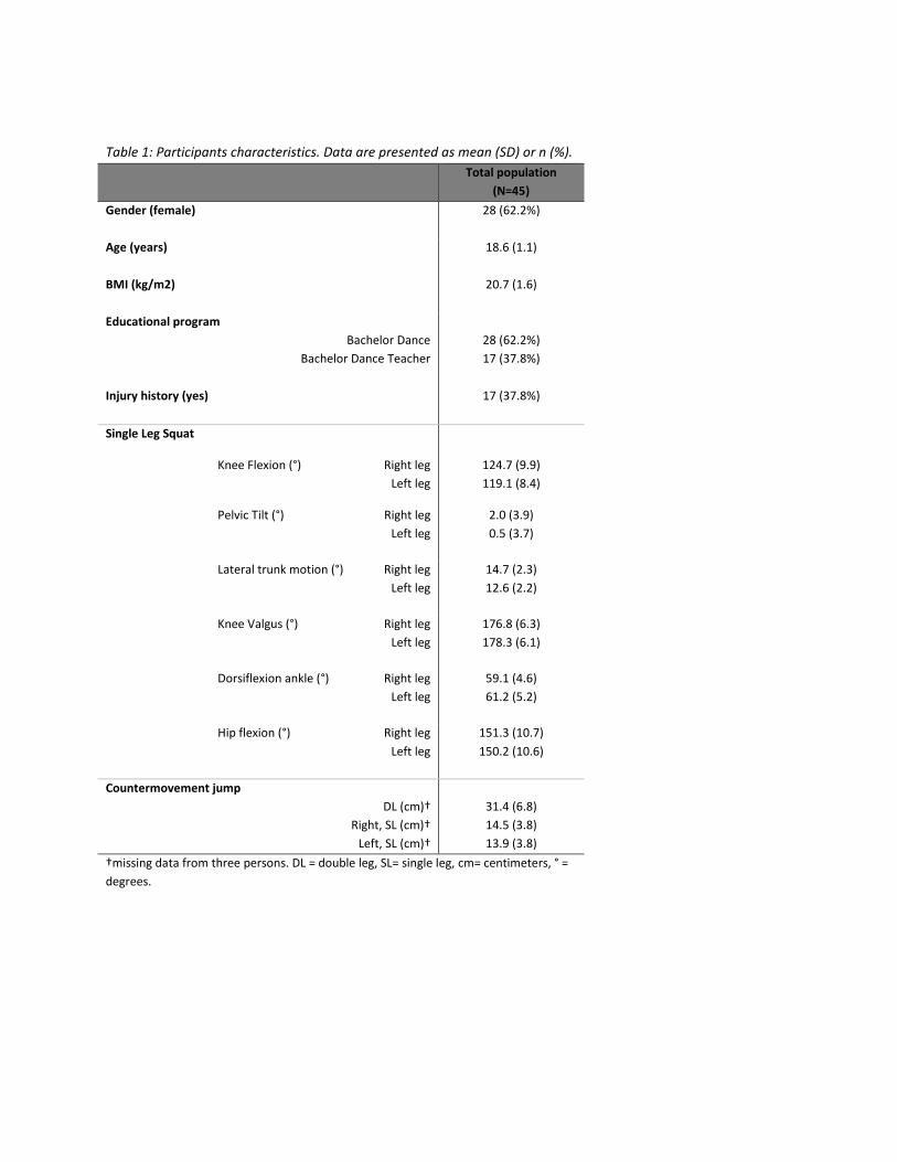
Table 1: Participants characteristics. Data are presented as mean (SD) or n (%).
Total population
(N=45)
Gender (female) 28 (62.2%)
Age (years) 18.6 (1.1)
BMI (kg/m2) 20.7 (1.6)
Educational program
Bachelor Dance
Bachelor Dance Teacher
28 (62.2%)
17 (37.8%)
Injury history (yes)
17 (37.8%)
Single Leg Squat
Knee Flexion (°)
Right leg
Left leg
124.7 (9.9)
119.1 (8.4)
Pelvic Tilt (°) Right leg
Left leg
2.0 (3.9)
0.5 (3.7)
Lateral trunk motion (°) Right leg
Left leg
14.7 (2.3)
12.6 (2.2)
Knee Valgus (°) Right leg
Left leg
176.8 (6.3)
178.3 (6.1)
Dorsiflexion ankle (°) Right leg
Left leg
59.1 (4.6)
61.2 (5.2)
Hip flexion (°) Right leg
Left leg
151.3 (10.7)
150.2 (10.6)
Countermovement jump
DL (cm)†
Right, SL (cm)†
Left, SL (cm)†
31.4 (6.8)
14.5 (3.8)
13.9 (3.8)
†missing data from three persons. DL = double leg, SL= single leg, cm= centimeters, ° =
degrees.
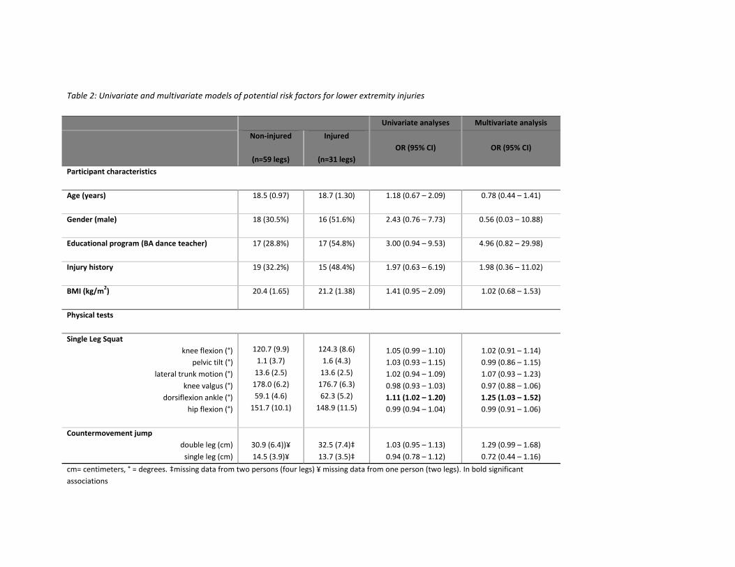
Table 2: Univariate and multivariate models of potential risk factors for lower extremity injuries
Univariate analyses Multivariate analysis
Non-injured
(n=59 legs)
Injured
(n=31 legs)
OR (95% CI) OR (95% CI)
Participant characteristics
Age (years)
18.5 (0.97) 18.7 (1.30) 1.18 (0.67 – 2.09) 0.78 (0.44 – 1.41)
Gender (male)
18 (30.5%) 16 (51.6%) 2.43 (0.76 – 7.73)
0.56 (0.03 – 10.88)
Educational program (BA dance teacher)
17 (28.8%) 17 (54.8%) 3.00 (0.94 – 9.53)
4.96 (0.82 – 29.98)
Injury history
19 (32.2%) 15 (48.4%) 1.97 (0.63 – 6.19) 1.98 (0.36 – 11.02)
BMI (kg/m2)
20.4 (1.65) 21.2 (1.38) 1.41 (0.95 – 2.09) 1.02 (0.68 – 1.53)
Physical tests
Single Leg Squat
knee flexion (°)
pelvic tilt (°)
lateral trunk motion (°)
knee valgus (°)
dorsiflexion ankle (°)
hip flexion (°)
120.7 (9.9)
1.1 (3.7)
13.6 (2.5)
178.0 (6.2)
59.1 (4.6)
151.7 (10.1)
124.3 (8.6)
1.6 (4.3)
13.6 (2.5)
176.7 (6.3)
62.3 (5.2)
148.9 (11.5)
1.05 (0.99 – 1.10)
1.03 (0.93 – 1.15)
1.02 (0.94 – 1.09)
0.98 (0.93 – 1.03)
1.11 (1.02 – 1.20)
0.99 (0.94 – 1.04)
1.02 (0.91 – 1.14)
0.99 (0.86 – 1.15)
1.07 (0.93 – 1.23)
0.97 (0.88 – 1.06)
1.25 (1.03 – 1.52)
0.99 (0.91 – 1.06)
Countermovement jump
double leg (cm)
single leg (cm)
30.9 (6.4))¥
14.5 (3.9)¥
32.5 (7.4)‡
13.7 (3.5)‡
1.03 (0.95 – 1.13)
0.94 (0.78 – 1.12)
1.29 (0.99 – 1.68)
0.72 (0.44 – 1.16)
cm= centimeters, ° = degrees. ‡missing data from two persons (four legs) ¥ missing data from one person (two legs). In bold significant
associations
0
5
10
15
20
25
30
M1 M2 M3 M4 M5 M6 M7 M8 M9
pre
vale
nce
(%
)
Monthly prevalence
All lower extremity injuries Substantial lower extremity injury
Figure 1: Monthly prevalence of all- and substantial lower extremity injuries during 9
months follow-up