Embed Size (px)
Citation preview

ORIGINAL ARTICLE
Gut symbiotic microbes imprint intestinal immunecells with the innate receptor SLAMF4 whichcontributes to gut immune protection againstenteric pathogensAllison Cabinian,1 Daniel Sinsimer,1 May Tang,1 Youngsoon Jang,2 Bongkum Choi,2
Yasmina Laouar,2 Amale Laouar1
▸ Additional material ispublished online only. To viewplease visit the journal online(http://dx.doi.org/10.1136/gutjnl-2016-313214).
1The Child Health Institute ofNew Jersey, Robert WoodJohnson Medical School,Rutgers University, NewBrunswick, New Jersey, USA2Department of Microbiologyand Immunology, University ofMichigan School of Medicine,Ann Arbor, Michigan, USA
Correspondence toDr Amale Laouar, The ChildHealth Institute of New Jersey,Robert Wood Johnson MedicalSchool, Rutgers University, 89French Street, New Brunswick,NJ 08901, USA;[email protected]
AC and DS contributed equally.
Received 20 October 2016Revised 27 February 2017Accepted 4 March 2017
To cite: Cabinian A,Sinsimer D, Tang M, et al.Gut Published Online First:[please include Day MonthYear] doi:10.1136/gutjnl-2016-313214
ABSTRACTBackground Interactions between host immune cellsand gut microbiota are crucial for the integrity andfunction of the intestine. How these interactions regulateimmune cell responses in the intestine remains a majorgap in the field.Aim We have identified the signalling lymphocyteactivation molecule family member 4 (SLAMF4) as animmunomodulator of the intestinal immunity. The aim isto determine how SLAMF4 is acquired in the gut andwhat its contribution to intestinal immunity is.Methods Expression of SLAMF4 was assessed in miceand humans. The mechanism of induction was studiedusing GFPtg bone marrow chimaera mice, lymphotoxin αand TNLG8A-deficient mice, as well as gnotobiotic mice.Role in immune protection was revealed using oralinfection with Listeria monocytogenes and Cytobacterrodentium.Results SLAMF4 is a selective marker of intestinalimmune cells of mice and humans. SLAMF4 inductionoccurs directly in the intestinal mucosa without theinvolvement of the gut-associated lymphoid tissue. Gutbacterial products, particularly those of gut anaerobes,and gut-resident antigen-presenting cell (APC)TNLG8A arekey contributors of SLAMF4 induction in the intestine.Importantly, lack of SLAMF4 expression leads theincreased susceptibility of mice to infection by oralpathogens culminating in their premature death.Conclusions SLAMF4 is a marker of intestinal immunecells which contributes to the protection against entericpathogens and whose expression is dependent on thepresence of the gut microbiota. This discovery provides apossible mechanism for answering the long-standingquestion of how the intertwining of the host and gutmicrobial biology regulates immune cell responses in the gut.
INTRODUCTIONGut microbes comprise more than 800 species that,as a whole, constitute the gut microbiota.1 In theintestinal tract, the microbiota contribute to thedigestion of food, the provision of essential nutri-ents and to preventing the invasion of pathogens,as it represents the most frequent site of infec-tion.1 2 To maintain this beneficial relationship, themucosal immune system is likely to exert the meansfor tolerogenic regulation by inducing inhibitorymolecules for immune signalling. On the other
Significance of this study
What is already known on this subject?▸ The constant changes imposed on the intestinal
tract require different dynamics from theimmune system at this site.
▸ Signalling lymphocytic activation molecule(SLAM) receptors have an important role in thedevelopment of immune responses.
▸ SLAMF4 is expressed by a small fraction (<8%)of haematopoietic-derived cells in the periphery,and most of its functions have been examinedon natural killer (NK) cells.
What are the new findings?▸ SLAMF4 is expressed in the intestinal mucosa by
conventional and natural T lymphocyte subsets,B cells and professional APCs (Mϕ and DCs), inaddition to NK cells and innate lymphoid celltypes.
▸ Gut bacterial products are efficient at increasingSLAMF4 expression on APCs but require thepresence of gut-resident APCTNLG8A for SLAMF4induction on lymphocytes.
▸ SLAMF4 contributes to the regulation of gutimmunity by promoting the production ofproinflammatory cytokines during enteric infection.
How might it impact on clinical practice inthe foreseeable future?▸ SLAMF4 is expressed by gut innate and adaptive
immune cells involved in GI pathologies, andhence, this identification may expand the currentlist of targets that can facilitate the developmentof new intestinal mucosa-targeted therapeutics.
▸ Our finding further supports the importance of abalanced gut microflora biodiversity in hostimmune homeostasis and suggests thatprescribing oral antibiotics to patients, particularlythose who are immunocompromised, has to becarefully weighed.
▸ These findings suggest that phenotypical andfunctional analysis of SLAMF4 is warranted inhuman patients with immune-related intestinaldiseases and may also lead to a betterunderstanding of immune cell regulationmechanisms in human intestine.
Cabinian A, et al. Gut 2017;0:1–13. doi:10.1136/gutjnl-2016-313214 1
Intestinal inflammation Gut Online First, published on May 9, 2017 as 10.1136/gutjnl-2016-313214
Copyright Article author (or their employer) 2017. Produced by BMJ Publishing Group Ltd (& BSG) under licence.
on 8 July 2018 by guest. Protected by copyright.
http://gut.bmj.com
/G
ut: first published as 10.1136/gutjnl-2016-313214 on 24 March 2017. D
ownloaded from

hand, because the gut is exposed to the environment, the risk ofinfection with exogenous pathogenic microorganisms is con-stant. Therefore, the mucosal immune system is likely to remainguarded and poised to turn on a quick attack on invasive patho-gens by inducing activating molecules for immune signalling.However, the signalling molecules by which the gut immunesystem generates these simultaneously activating and inhibitorypathways, to switch between homeostatic, often immunosup-pressive and barrier-protective, function and potent activeimmunity are not fully understood. In this regard, the mostcommonly accepted view is that such a dual function may occuras a result of the interactions between host immune cells andthe gut microbiota.1 2
Natural killer cell receptors (NKR) are membrane proteinsthat provide specificity to NK cell responses in either an activat-ing or inhibitory fashion.3 There are two major families ofNKRs: NKRs that share homology with C-type lectins andkiller cell Ig-like receptors, which include the signalling lympho-cyte activation molecule family member 4, termed SLAMF4(also known as CD244 and 2B4).3 4 The natural ligand forSLAMF4 is CD48, and in vitro engagement of SLAMF4 byCD48 induces cytotoxicity and cytokine secretion by humanand mouse NK cells.5 6 The slamf4 gene can be alternatelyspliced into two protein products, differing in their intracellulardomains, with affinities for adaptor molecules that initiate orinhibit signalling.7–10 One splice variant has a shorter intracellu-lar domain and is activating, while the variant with the longerintracellular domain was shown to be inhibitory.7 8 Since thereare two isoforms of SLAMF4 that differ in their signalling cap-acities, the relative amounts of these isoforms could dictate cellresponsiveness to SLAMF4 ligation.7 8
Under normal physiological conditions, SLAMF4 is expressedby murine and human NK cells, but it is absent from most naïveCD4 and CD8T cells, B lymphocytes and neutrophils.7 11 12
However, other cell types such as mast cells, dendritic cells, skinγδ T cells, eosinophils and some activated CD8T cell subsets areSLAMF4+.11 13–16 In humans and mice, CD8+ T cells expres-sing SLAMF4 are absent from cord blood, and expression ofSLAMF4 can be induced on only a small fraction of CD8+ Tcells after in vitro activation or in vivo antigen challenge.10 17
Previously, we and others reported that in the steady-state condi-tion, the vast majority (>95%) of conventional CD8+ T cells inthe gut mucosa are SLAMF4+,18 19 while their peripheral coun-terparts are SLAMF4−.18 Because the intestine provides aunique environment for the development and function ofimmune cells,20–23 we sought to determine whether SLAMF4expression in the gut mucosa extends to other immune cellpopulations, and if so, how it is acquired in the intestine andwhat its contribution to gut immunity is.
RESULTSSLAMF4 is a marker for intestinal immune cellsIn the murine intestine, over 60% of haematopoietic-derived(CD45+) cells express SLAMF4, while this expression in otherorgans was <5% (figure 1A and see online supplementaryfigure S1a, b). In addition, over 80% of gut CD45+ cells werealso positive for CD48, the only known ligand for SLAMF47 8
(figure 1B). SLAMF4 was highly expressed in both the intrae-pithelial (IEL) compartment and lamina propria (LP) of thesmall intestine and colon, but this expression was generallymore enhanced among haematopoietic-derived cells of the IEL(∼70%) compartment than among those of the LP (∼50%)(figure 1C,D, and see online supplementary figures S2 and S3).In concordance with the murine data, SLAMF4 was found to be
expressed at high levels in both human small intestine (n=10)and human colon (n=7) (figure 1E–G, and see onlinesupplementary figure S4), suggesting that SLAMF4 can be usedas a marker for intestinal immune cells in mice and humans.
The finding that SLAMF4 is induced on most intestinal CD45+cells raised a key question: which cell types express SLAMF4? Toanswer this question, we first assessed the expression of SLAMF4on gut NK cell types to serve as positive control.7 As anticipated,we found that virtually all conventional (c) NK (NKp46+RORγt−) cells expressed SLAMF4 (figure 2A). Interestingly,while most NK-22 cells (NKp46+RORγt+) expressed SLAMF4,only a few LTi (NKp46−RORγt+) cells were positive for SLAMF4expression (figure 2A). Thus, in conformity with the classificationof SLAMF4 as an NK receptor,3 7 most cNK cells expressSLAMF4. However, this expression was divergent among gutinnate lymphoid cell subsets.
Next, we examined the expression of SLAMF4 on T lympho-cyte populations. Confirming our previous report,18 the vastmajority (>80%) of γδTCR+, CD8αα+ and CD8αβ+ intestinalT cells were SLAMF4+ (figure 2B,C). In addition, SLAMF4 wasalso significantly expressed by gut mucosal CD4+ T cells, andthis expression was eightfold to tenfold higher than the expres-sion of SLAMF4 on peripheral CD4+ lymphocytes (figure 2D).Because T cells and NK cells alone cannot account for the highpercentage of CD45+ cells expressing SLAMF4 in the intestinalmucosa, we examined whether other cell populations expressSLAMF4. B lymphocytes and plasmacytoid dendritic cells(pDCs) are crucial for defence against bacterial and viral patho-gens,24 25 and the gut is known to be a major reservoir in thebody for these cell types.25 Previous studies showed that periph-eral B cells lack SLAMF4 expression.11 12 Our phenotypic ana-lysis of splenic B cells confirmed this observation (figure 2E).Strikingly, a significant portion of B cells in the gut mucosaexpressed SLAMF4 (figure 2E). In addition, most intestinalpDCs were SLAMF4+ while their peripheral counterparts wereSLAMF4− (figure 2E). Furthermore, over 60% of dendriticcells (DCs, CD11c+CD19−CD3−F4/80−) in the small intestinewere SLAMF4+, and in the colon this expression was increasedto >90% (figure 2F). Similarly, we found that some macro-phages (Mϕ, F4/80+CD11b+CD3−CD19−) expressedSLAMF4 in lymphoid organs (∼10%), and this expression wasapproximately threefold higher on intestinal macrophages(figure 2F and data not shown).
Together, these data show that SLAMF4 is expressed by amultitude of T lymphocyte subsets in the gut mucosa, in additionto NK and NK22 cells. In the periphery, NK cells expressSLAMF4, but CD4 and CD8T cell expression of SLAMF4 isrestricted to the intestine. We also show that professional APCs(Mϕ and DCs) express higher levels of SLAMF4 in the gutmucosa when compared with those in the periphery, whileSLAMF4 expression on B cells and pDCs is restricted to theintestinal mucosa. Therefore, while previous studies showedrestricted SLAMF4 expression to a few cell types, the novelty ofthese findings is that SLAMF4 expression extends to most innateand adaptive immune cell populations of the intestinal mucosa.
Induction of SLAMF4 occurs directly in the intestinal mucosaHigh expression of SLAMF4 among immune cells is a uniquefeature of the intestine (figures 1 and 2). Thus, we speculatedthat SLAMF4 might be induced on immune cells directly in theintestinal mucosa. To test this hypothesis, we generated bonemarrow (BM) chimaeras using donor UBC-GFP transgenic(GFPtg) mice and sublethally irradiated major histocompatibilitycomplex (MHC)-matched C57BL/6 recipients. Of note, GFPtg
2 Cabinian A, et al. Gut 2017;0:1–13. doi:10.1136/gutjnl-2016-313214
Intestinal inflammation on 8 July 2018 by guest. P
rotected by copyright.http://gut.bm
j.com/
Gut: first published as 10.1136/gutjnl-2016-313214 on 24 M
arch 2017. Dow
nloaded from
mice express GFP under the ubiquitin C promoter, and allhaematopoietic-derived cells are GFP+26 (figure 3A). We foundthat GFP+ cells derived from donor cells expressed SLAMF4 athigh frequencies in the intestinal mucosa (∼70%), but not inother tissues (<5%) (figure 3B, C). In addition, the GFP− hostcells, expected to survive in sublethally irradiated recipientanimals, also expressed SLAMF4 only at high frequencies in theintestinal mucosa (figure 3B, D), suggesting that the inductionof SLAMF4 on intestinal CD45+ cells occurs directly in the gutmucosa.
GFP+ cells migrating to the gut mucosa clearly becomeSLAMF4+ (figure 3A–D). However, it was unclear whetherthese cells were induced with SLAMF4 in the gut-associatedlymphoid tissue (GALT) and mesenteric lymph nodes (MLN),and then migrated to the gut mucosa, or whether they wereimprinted directly in the mucosa. To clarify this issue, we gener-ated GFPtg BM chimaeras using sublethally irradiatedMHC-matched C57BL/6 recipients lacking GALT and MLN,
known as lymphotoxin-α (Ltα)-deficient mice.27 Our datashowed no significant difference in the percentage ofSLAMF4-expressing CD45+ cells of Ltαko mice when com-pared with their wild-type (WT) counterparts (figure 3E, F).Moreover, similar to GFP− host cells, GFP+ cells derived fromGFPtg donor cells also expressed high levels of SLAMF4 in theintestinal mucosa of Ltαko recipients (figure 3G). Together,these data show that SLAMF4 expression on intestinal CD45+cells occurs directly in the gut mucosa, and that GALT andMLN are largely dispensable for this process.
Intestinal microflora drive SLAMF4 expressionThe intestine houses the largest population of commensal micro-organisms in the body.1 2 We speculated that the differencenoted in SLAMF4 expression between the gut mucosa andother organs might be related to the vast difference in numbersof commensal microbes available. Therefore, we compared theexpression of SLAMF4 on CD45+ cells in the gut mucosa of
Figure 1 Signalling lymphocyte activation molecule family member 4 (SLAMF4) is a marker of intestinal immune cells. (A) SLAMF4 expression wasassessed on haematopoietic-derived (CD45+) cells in the Peyer’s patches (PP) and caecal patch (CP) which belong to the gut-associated lymphoidtissue (GALT), mesenteric lymph nodes (MLN), spleen (SPL), peripheral lymph nodes (PLN), as well as other organs as indicated. Bar graphsummarises SLAMF4 expression as means of % CD45 cells that are SLAMF4+. (Inset) Dot plots show SLAMF4 expression by CD45+ cells in differenttissues. (B) Gut mucosal CD45+ cells express both SLAMF4 and its ligand CD48. Data are presented as means of % SLAMF4+CD45+ cells that areCD48+. Data shown in (A, B) are from ten experiments using two to three mice per experiment for intestinal tissues and four experiments using twoto three mice per experiment for other organs. (C) Dot plots show SLAMF4 expression by CD45+ cells in the intraepithelial (IEL) compartment ofdifferent segments of the small intestine and colon. Numbers indicate % SLAMF4+CD45+ cells. (D) Murine IEL and lamina propria (LP) fractionswere prepared from the small intestine and colon and stained for flow cytometry. Bar graphs show SLAMF4 expression as means of % CD45+ cellsthat are SLAMF4+ in the IEL compartment versus those in the LP. Data shown in (C, D) are from n=6. (E) SLAMF4 expression was assessed onhuman small intestine (n=7) and colon (n=10) samples. Dot plots show SLAMF4 expression on human CD45+ cells in the IEL and LP compartments.(F) Data are shown as means of % human CD45+ cells expressing SLAMF4 and (G) relationship between IEL and LP for each tissue sample. Errorbars represent SEM. A two-tailed Student’s t-test distribution with paired groups of samples was evaluated for statistical significance. A value ofp>0.05 is considered not significant (NS); *p<0.05, **p<0.005, ***p<0.005, ****p<0.00005.
Cabinian A, et al. Gut 2017;0:1–13. doi:10.1136/gutjnl-2016-313214 3
Intestinal inflammation on 8 July 2018 by guest. P
rotected by copyright.http://gut.bm
j.com/
Gut: first published as 10.1136/gutjnl-2016-313214 on 24 M
arch 2017. Dow
nloaded from

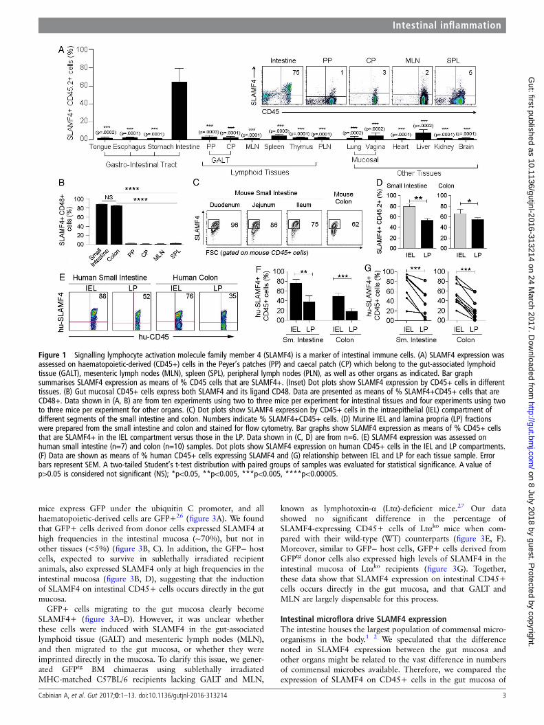
conventionally (Cv) raised BALB/c mice to that of germ-free(GF) animals (figure 4A and28). SLAMF4 expression onhaematopoietic-derived cells of the Peyer’s patches (PP), MLNand spleen (SPL) was not significantly different between GF andCv animals (figure 4B, C). Strikingly, SLAMF4 expression wasalmost completely abolished on intestinal CD45+ cells of GFmice (figure 4B, C), except on a small fraction (∼20%) consti-tuted mainly by natural CD8αα T cells that remain SLAMF4+(see online supplementary figure S5A).
Next, we wanted to test whether SLAMF4 expression in thegut is plastic or developmental in nature. In the first set ofexperiments, we conventionalised GF mice to determine ifSLAMF4 expression reappears in the gut mucosa. Our datarevealed that recolonising the GF bowel with commensal micro-flora significantly increased SLAMF4 expression on intestinalCD45+ cells (figure 4D). However, the percentage of SLAMF4+ cells in conventionalised (Cvz) animals was still less than thepercentage found in the gut of Cv animals. This is probably due
to the fact that transferring faecal contents only partiallyreplenishes GF mice with the total composition of the micro-flora, which is a known limitation of current procedures of gutmicrobiota transplantation.
In the second set of experiments, we tested whether alteringthe gut microflora of Cv mice through antibiotic (ATB) treatmentcould alter SLAMF4 expression in the gut mucosa (figure 4E–G,see online supplementary figures S5b and S6a). To this end, wetreated Cv mice with a cocktail of four ATB (ampicillin, neomy-cin, metronidazole and vancomycin) for 4 weeks.29 While thistreatment resulted in the ablation of most bacteria (figure 4E),the remaining bacteria were over-represented by the Firmicutes(a group consisting of Staphylococcus and Clostridium speciesamong others)1 29 30 (figure 4F). Importantly, SLAMF4+ cellswere significantly less frequent in the intestine of treated thanuntreated animals (figure 4G, see online supplementary figureS5b), suggesting that SLAMF4 expression can be altered by per-turbation of normal microflora composition.
Figure 2 Signalling lymphocyte activation molecule family member 4 (SLAMF4) is expressed by different immune cell types in the intestinalmucosa. (A) Lamina propria cells were first gated on CD45+CD3− cells. Dot plots show three NK cell subtypes identified based on the expression ofNKp46 and the transcriptional factor RORγt. Numbers indicate the % of each cell subset (left) as well as % SLAMF4+ cells (right). Bar graphsummarises % SLAMF4+ cells gated on each subtype. Data are from four experiments using three mice per experiment. (B) SLAMF4 expression wasassessed on CD8αα+, CD8αβ+TCRαβ+ and TCRγδ+ T cells in the intestine, mesenteric lymph nodes (MLN) and spleen (SPL). Numbers indicate % ofSLAMF4+ cells. (C) Summary data are shown as means of % gut CD8 T cell subsets that are SLAMF4+. Data are from four experiments using twomice per experiment. (D) Bar graph shows SLAMF4 expression on CD4+ cells in the intestine versus periphery. Data are presented as means of %CD4+ (CD3+CD4+CD8α−CD8β−) cells that are SLAMF4+. Data are from three experiments using three mice per experiment. (E) SLAMF4 expressionon B cells (CD45+CD3-B220+CD19+) and plasmacytoid dendritic cells (pDCs, CD45+PDCA1+CD11c+). Below, bar graphs summarise FACS data. (F)Bar graphs show SLAMF4 expression on dendritic cells (DC, CD11c+CD19−CD3−F4/80−, left) and macrophages (Mϕ, CD11c−CD11b+F4/80+,right). Data are shown as means of % DCs and Mϕ that are SLAMF4+. Data shown in (E, F) are from three experiments using three to four mice perexperiment. Data are shown as means±SEM. Error bars represent SEM. A two-tailed Student’s t-test distribution with paired groups of samples wasevaluated for statistical significance. *p<0.05, **p<0.005, ***p<0.0005.
4 Cabinian A, et al. Gut 2017;0:1–13. doi:10.1136/gutjnl-2016-313214
Intestinal inflammation on 8 July 2018 by guest. P
rotected by copyright.http://gut.bm
j.com/
Gut: first published as 10.1136/gutjnl-2016-313214 on 24 M
arch 2017. Dow
nloaded from

In the third set of experiments, we sought to investigatewhether haematopoietic cells, recently developed in the gutmucosa and induced with SLAMF4, can also lose SLAMF4expression when the gut microflora composition is disturbed. Tothis end, we treated GFPtg BM chimaera mice with ATB as indi-cated above (figure 4H). Our data revealed a significant reduc-tion in SLAMF4+CD45+ cells among both the GFP+ donorcells and GFP− host cells in ATB-treated versus control chi-maera mice (figure 4I, J). Together, these findings show thatSLAMF4 expression on intestinal CD45+ cells depends on thecontinued presence of the normal gut microflora.
Role of TNLG8A costimulation in SLAMF4 inductionPreviously, we showed that SLAMF4 expression on conventionalCD8αβ+ T cells in the intestinal mucosa can be significantlydiminished by injecting a blocking TNLG8A antibody.18
TNLG8A, the tumour necrosis factor ligand 8A, also known asCD70, is a costimulatory molecule that is constitutively expressedby gut-resident APCs.31 32 Thus, we hypothesised that SLAMF4induction on conventional CD8T lymphocytes and potentiallyother cell types may require gut bacterial antigens to be presentedto them in the context of TNLG8A costimulation. To test this
possibility, we generated a TNLG8A-deficient mouse line usingES clones from the KOMP repository (figure 5A). Since differ-ences in intestinal microbiota composition contribute to pheno-typical and functional differences in mice,33 age-matched andsex-matched TNLG8Ako mice and WT littermates werecohoused to prevent potential intestinal microbiota changes.Overall analysis showed no significant difference in intestinal cellnumbers between TNLG8Ako and WT littermates (figure 5Btop). However, there was a significant decrease in SLAMF4expression on intestinal cell populations of TNLG8Ako whencompared with their WT counterparts (figure 5B bottom, C).Specifically, a lack of TNLG8A significantly reduced SLAMF4expression on inducible CD8T cells (CD3+CD8β+TCRαβ+),CD4T cells (CD3+CD4+CD8α−TCRαβ+) and B cells(CD3-CD19+NKp46−), but had little or no effect on DC/Mϕ(MHCII+CD19−CD3−) and NK/NK22 cells (CD3−CD19−NKp46+) (figure 5D). Still, the observed defect was onlypartial and did not fully recapitulate the phenotype observed inGF mice (figure 5E, left). Therefore, we tested whether disrup-tion of the normal gut microbiota would have synergistic effectswith TNLG8A gene deficiency. Following ATB treatment, thealready decreased SLAMF4 expression in TNLG8Ako mice was
Figure 3 Signalling lymphocyte activation molecule family member 4 (SLAMF4) induction occurs directly in the gut mucosa and independently ofthe gut-associated lymphoid tissue (GALT). (A) B6.GFPtg bone marrow cells were adoptively transferred into sublethally irradiated B6 mice. Plotsshow GFP expression on donor (left) and host (right) CD45+ cells. Data are representative of n=10. (B) Eight weeks later, mice were euthanised toisolate cells from the gut mucosa, Peyer’s patch (PP), caecal patch (CP), mesenteric lymph nodes (MLN) and spleen (SPL). Numbers indicate % ofhost (GFP−, top left) and donor (GFP+, top right) SLAMF4+ cells. Summary of data displayed in (B) are shown in (C) and (D), respectively. (E)Expression of SLAMF4 in the intestinal mucosa of B6.WT and B6.Ltαko mice. Data are representative of n=9. (F) Summary of data indicating SLAMF4expression as means of % CD45+ cells in the intestine and spleen. (G) B6.GFPtg bone marrow cells were adoptively transferred into sublethallyirradiated B6.Ltαko recipients as described in (A). Data are shown as means of % host (GFP−Ltα−/−) and donor (GFP+Ltα+/+) CD45.2+ cells thatare SLAMF4+. Data are from three experiments using two (C,D,F) and three (g) mice per experiment. Data are shown as means±SEM. Error barsrepresent SEM. A two-tailed Student’s t-test distribution with paired groups of samples was evaluated for statistical significance. p>0.05 isconsidered not significant (NS); *p<0.05, ***p<0.0005, ****p<0.00005. WT, wild type.
Cabinian A, et al. Gut 2017;0:1–13. doi:10.1136/gutjnl-2016-313214 5
Intestinal inflammation on 8 July 2018 by guest. P
rotected by copyright.http://gut.bm
j.com/
Gut: first published as 10.1136/gutjnl-2016-313214 on 24 M
arch 2017. Dow
nloaded from
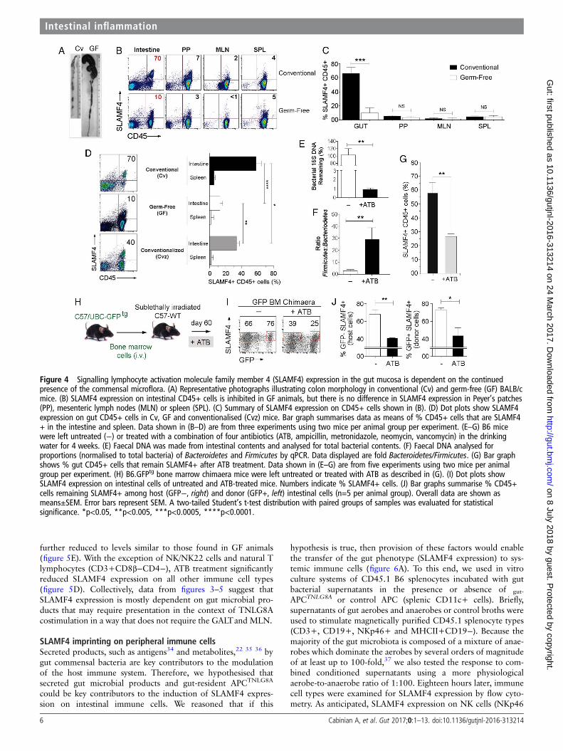
further reduced to levels similar to those found in GF animals(figure 5E). With the exception of NK/NK22 cells and natural Tlymphocytes (CD3+CD8β−CD4−), ATB treatment significantlyreduced SLAMF4 expression on all other immune cell types(figure 5D). Collectively, data from figures 3–5 suggest thatSLAMF4 expression is mostly dependent on gut microbial pro-ducts that may require presentation in the context of TNLG8Acostimulation in a way that does not require the GALTand MLN.
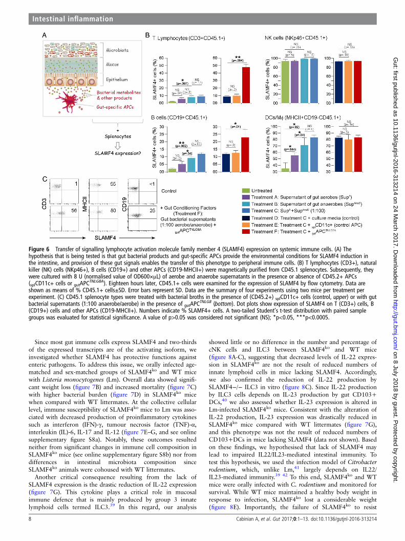
SLAMF4 imprinting on peripheral immune cellsSecreted products, such as antigens34 and metabolites,22 35 36 bygut commensal bacteria are key contributors to the modulationof the host immune system. Therefore, we hypothesised thatsecreted gut microbial products and gut-resident APCTNLG8A
could be key contributors to the induction of SLAMF4 expres-sion on intestinal immune cells. We reasoned that if this
hypothesis is true, then provision of these factors would enablethe transfer of the gut phenotype (SLAMF4 expression) to sys-temic immune cells (figure 6A). To this end, we used in vitroculture systems of CD45.1 B6 splenocytes incubated with gutbacterial supernatants in the presence or absence of gut-
APCTNLG8A or control APC (splenic CD11c+ cells). Briefly,supernatants of gut aerobes and anaerobes or control broths wereused to stimulate magnetically purified CD45.1 splenocyte types(CD3+, CD19+, NKp46+ and MHCII+CD19−). Because themajority of the gut microbiota is composed of a mixture of anae-robes which dominate the aerobes by several orders of magnitudeof at least up to 100-fold,37 we also tested the response to com-bined conditioned supernatants using a more physiologicalaerobe-to-anaerobe ratio of 1:100. Eighteen hours later, immunecell types were examined for SLAMF4 expression by flow cyto-metry. As anticipated, SLAMF4 expression on NK cells (NKp46
Figure 4 Signalling lymphocyte activation molecule family member 4 (SLAMF4) expression in the gut mucosa is dependent on the continuedpresence of the commensal microflora. (A) Representative photographs illustrating colon morphology in conventional (Cv) and germ-free (GF) BALB/cmice. (B) SLAMF4 expression on intestinal CD45+ cells is inhibited in GF animals, but there is no difference in SLAMF4 expression in Peyer’s patches(PP), mesenteric lymph nodes (MLN) or spleen (SPL). (C) Summary of SLAMF4 expression on CD45+ cells shown in (B). (D) Dot plots show SLAMF4expression on gut CD45+ cells in Cv, GF and conventionalised (Cvz) mice. Bar graph summarises data as means of % CD45+ cells that are SLAMF4+ in the intestine and spleen. Data shown in (B–D) are from three experiments using two mice per animal group per experiment. (E–G) B6 micewere left untreated (−) or treated with a combination of four antibiotics (ATB, ampicillin, metronidazole, neomycin, vancomycin) in the drinkingwater for 4 weeks. (E) Faecal DNA was made from intestinal contents and analysed for total bacterial contents. (F) Faecal DNA analysed forproportions (normalised to total bacteria) of Bacteroidetes and Firmicutes by qPCR. Data displayed are fold Bacteroidetes/Firmicutes. (G) Bar graphshows % gut CD45+ cells that remain SLAMF4+ after ATB treatment. Data shown in (E–G) are from five experiments using two mice per animalgroup per experiment. (H) B6.GFPtg bone marrow chimaera mice were left untreated or treated with ATB as described in (G). (I) Dot plots showSLAMF4 expression on intestinal cells of untreated and ATB-treated mice. Numbers indicate % SLAMF4+ cells. (J) Bar graphs summarise % CD45+cells remaining SLAMF4+ among host (GFP−, right) and donor (GFP+, left) intestinal cells (n=5 per animal group). Overall data are shown asmeans±SEM. Error bars represent SEM. A two-tailed Student’s t-test distribution with paired groups of samples was evaluated for statisticalsignificance. *p<0.05, **p<0.005, ***p<0.0005, ****p<0.0001.
6 Cabinian A, et al. Gut 2017;0:1–13. doi:10.1136/gutjnl-2016-313214
Intestinal inflammation on 8 July 2018 by guest. P
rotected by copyright.http://gut.bm
j.com/
Gut: first published as 10.1136/gutjnl-2016-313214 on 24 M
arch 2017. Dow
nloaded from
+) was affected neither by stimulation with bacterial supernatantsnor by the presence of APCs (figure 6B). In addition, stimulationwith control broths exerted no detrimental effects on the cell via-bility and had no effect on SLAMF4 induction (see onlinesupplementary figure S7). Importantly, we found that superna-tants of anaerobes were more potent at inducing SLAMF4 thanaerobic supernatants, and this phenotype was somewhatenhanced when a physiological aerobe-to-anaerobe ratio of1:100 was used (figure 6B). Notably, SLAMF4 expression wasfound to be significantly increased on APCs (DCs and MΦ;MHCII+CD19−) when compared with T (CD3+) and B (CD19+) lymphocytes (figure 6B). However, provision ofgutAPC
TNLG8A drastically enhanced SLAMF4 expression onlymphocyte subsets, particularly Tcells, but had little or no effecton APCs (figure 6B, C). Together, these data suggest that gut bac-terial products, particularly those derived from gut anaerobes,are efficient at increasing SLAMF4 expression on APCs but
require the presence of gutAPCTNLG8A for SLAMF4 induction on
lymphocytes.
Lack of SLAMF4 renders mice more susceptible to entericinfectionsThere are two distinct SLAMF4 proteins which are generated bythe long (L) and short (S) isoforms.7–9 SLAMF4S is reported tobe an activating receptor6 7 38 while SLAMF4L is known to beinhibitory.4 7 38 Due to the high levels of SLAMF4 expressionin the gut, we were interested to know which isoform dominatesin the intestine. To this end, we extracted RNA from magnetic-ally purified intestinal CD45+ cells and performed QPCR forboth splice variants. In the gut mucosa, approximatelytwo-thirds of the expressed SLAMF4 transcripts are of the shortisoform (figure 7A), suggesting that SLAMF4 expression in theintestine favours the activating splice variant of SLAMF4.
Figure 5 Role of TNLG8A in signalling lymphocyte activation molecule family member 4 (SLAMF4) induction. (A) Lack of TNLG8A gene expressionin knockout mice. Amplification of two PCR products, TNLG8A (824bp) and lacZ (270bp), revealed the deletion of the TNLG8A gene in knockoutmice (top), and absence of the TNLG8A product (3.6 kb) by Southern blot analysis confirmed this gene deletion (bottom). (B) Pooled IEL and LPcells from wild-type (WT) and TNLG8Ako mice were analysed by flow cytometry. Bar graphs summarise SLAMF4 expression as means of numbers(top) and % (bottom) of total leucocytes (CD45+), T cells (CD3+MHCII−) and APCs (MHCII+CD3−) that are SLAMF4+. (C) Dot plots show SLAMF4expression in the intestinal mucosa of WT and TNLG8Ako mice. Numbers indicate % SLAMF4− cells. Data shown in (A–C) are from threeexperiments using three mice per animal group per experiment. (D) WT and TNLG8Ako mice were left untreated or treated with ATB as described infigure 4E. Graphs summarise % SLAMF4+ cells in the intestinal mucosa of untreated WT (control) versus untreated and ATB-treated TNLG8Ako
animals. Data show significant reduction of SLAMF4 expression on CD4T cells (CD3+CD4+CD8α−), inducible CD8T cells (CD3+CD8β+TCRαβ+), Bcells (CD3−CD19+NKp46−) and DC/MΦ (CD3−CD19−MHCII+), but little or no decrease on natural CD8T cells (CD3+CD8α+CD8β−CD4−) and NK/NK22 cells (CD3−CD19−NKp46+). (E) Dot plots show SLAMF4 expression on gut leucocytes in untreated and ATB-treated TNLG8Ako animals. Bargraph summarises SLAMF4 expression as means of % intestinal CD45+ cells that are SLAMF4+ in untreated (−) and ATB-treated WT and TNLG8Ako
mice in comparison with germ-free (GF) animals. Data are from three experiments using two (untreated) and three (ATB-treated) mice per animalgroup per experiment. Data are shown as means ±SD. Error bars represent SD. A two-tailed Student’s t-test distribution with paired sample groupswas evaluated for statistical significance. Value of p>0.05 was considered not significant (NS); *p<0.05, **p<0.005, ***p<0.0005, ****p<0.0001.
Cabinian A, et al. Gut 2017;0:1–13. doi:10.1136/gutjnl-2016-313214 7
Intestinal inflammation on 8 July 2018 by guest. P
rotected by copyright.http://gut.bm
j.com/
Gut: first published as 10.1136/gutjnl-2016-313214 on 24 M
arch 2017. Dow
nloaded from
Since most gut immune cells express SLAMF4 and two-thirdsof the expressed transcripts are of the activating isoform, weinvestigated whether SLAMF4 has protective functions againstenteric pathogens. To address this issue, we orally infected age-matched and sex-matched groups of SLAMF4ko and WT micewith Listeria monocytogenes (Lm). Overall data showed signifi-cant weight loss (figure 7B) and increased mortality (figure 7C)with higher bacterial burden (figure 7D) in SLAMF4ko micewhen compared with WT littermates. At the collective cellularlevel, immune susceptibility of SLAMF4ko mice to Lm was asso-ciated with decreased production of proinflammatory cytokinessuch as interferon (IFN)-γ, tumour necrosis factor (TNF)-α,interleukin (IL)-6, IL-17 and IL-12 (figure 7E–G, and see onlinesupplementary figure S8a). Notably, these outcomes resultedneither from significant changes in immune cell composition inSLAMF4ko mice (see online supplementary figure S8b) nor fromdifferences in intestinal microbiota composition sinceSLAMF4ko animals were cohoused with WT littermates.
Another critical consequence resulting from the lack ofSLAMF4 expression is the drastic reduction of IL-22 expression(figure 7G). This cytokine plays a critical role in mucosalimmune defence that is mainly produced by group 3 innatelymphoid cells termed ILC3.39 In this regard, our analysis
showed little or no difference in the number and percentage ofcNK cells and ILC3 between SLAMF4ko and WT mice(figure 8A-C), suggesting that decreased levels of IL-22 expres-sion in SLAMF4ko are not the result of reduced numbers ofinnate lymphoid cells in mice lacking SLAMF4. Accordingly,we also confirmed the reduction of IL-22 production bySLAMF4−/− ILC3 in vitro (figure 8C). Since IL-22 productionby ILC3 cells depends on IL-23 production by gut CD103+DCs,40 we also assessed whether IL-23 expression is altered inLm-infected SLAMF4ko mice. Consistent with the alteration ofIL-22 production, IL-23 expression was drastically reduced inSLAMF4ko mice compared with WT littermates (figure 7G),and this phenotype was not the result of reduced numbers ofCD103+DCs in mice lacking SLAMF4 (data not shown). Basedon these findings, we hypothesised that lack of SLAMF4 maylead to impaired IL22/IL23-mediated intestinal immunity. Totest this hypothesis, we used the infection model of Citrobacterrodentium, which, unlike Lm,41 largely depends on IL22/IL23-mediated immunity.39 42 To this end, SLAMF4ko and WTmice were orally infected with C. rodentium and monitored forsurvival. While WT mice maintained a healthy body weight inresponse to infection, SLAMF4ko lost a considerable weight(figure 8E). Importantly, the failure of SLAMF4ko to resist
Figure 6 Transfer of signalling lymphocyte activation molecule family member 4 (SLAMF4) expression on systemic immune cells. (A) Thehypothesis that is being tested is that gut bacterial products and gut-specific APCs provide the environmental conditions for SLAMF4 induction inthe intestine, and provision of these gut signals enables the transfer of this phenotype to peripheral immune cells. (B) T lymphocytes (CD3+), naturalkiller (NK) cells (NKp46+), B cells (CD19+) and other APCs (CD19-MHCII+) were magnetically purified from CD45.1 splenocytes. Subsequently, theywere cultured with 8 U (normalised value of OD600×μL) of aerobe and anaerobe supernatants in the presence or absence of CD45.2+ APCs(splCD11c+ cells or gutAPC
TNLG8A). Eighteen hours later, CD45.1+ cells were examined for the expression of SLAMF4 by flow cytometry. Data areshown as means of % CD45.1+ cells±SD. Error bars represent SD. Data are the summary of four experiments using two mice per treatment perexperiment. (C) CD45.1 splenocyte types were treated with bacterial broths in the presence of (CD45.2+) splCD11c+ cells (control, upper) or with gutbacterial supernatants (1:100 anaerobe/aerobe) in the presence of gutAPC
TNLG8 (bottom). Dot plots show expression of SLAMF4 on T (CD3+) cells, B(CD19+) cells and other APCs (CD19-MHCII+). Numbers indicate % SLAMF4+ cells. A two-tailed Student’s t-test distribution with paired samplegroups was evaluated for statistical significance. A value of p>0.05 was considered not significant (NS); *p<0.05, ***p<0.0005.
8 Cabinian A, et al. Gut 2017;0:1–13. doi:10.1136/gutjnl-2016-313214
Intestinal inflammation on 8 July 2018 by guest. P
rotected by copyright.http://gut.bm
j.com/
Gut: first published as 10.1136/gutjnl-2016-313214 on 24 M
arch 2017. Dow
nloaded from

C. rodentium resulted in premature death, as indicated by 100%in response to 2×10¹⁰ colony-forming unit (CFU) of the patho-gen, while all WT animals efficiently resisted C. rodentium andsurvived this dose of infection (figure 8F).
DISCUSSIONGut microbes are critical for inducing SLAMF4 on intestinalimmune cells, and their continued presence is required for themaintenance of this expression. We came to this conclusion whenwe compared the expression of SLAMF4 on CD45+ cells in thegut mucosa of conventionally raised and GF mice. Interestingly,GF animals exhibit a drastic but not a complete decline inSLAMF4 expression among gut immune cells. Notably, CD8αα+CD8β−CD4− T lymphocytes remain SLAMF4+ in the intes-tinal mucosa of GF animals. These cells, often referred to as
natural or unconventional IELs, are induced in the thymus withgut-homing molecules to migrate directly from the thymus to thegut epithelium.43 Interestingly, thymic natural CD8αα, but notinducible CD8αβ, are SLAMF4+.44 Therefore, it appears that theacquisition of gut-specific markers in the thymus is not limited tothe induction of gut-homing receptors but extends to the induc-tion of SLAMF4. Whether SLAMF4 plays a role in the thymicdevelopment of natural IELs and/or their post-thymic maturationand transition to the gut epithelium is completely unknown andrequires further investigation.
Ablation of the gut microflora, either through GF husbandryor extensive antibiotic treatment is efficient at reducing SLAMF4expression in the gut mucosa. These examples are extreme as it isimpossible for any individual to be GF, and most individuals willnever be subjected to such a stringent ATB regimen, while most
Figure 7 Lack of signalling lymphocyte activation molecule family member 4 (SLAMF4) renders mice susceptible to Listeria monocytogenes (Lm)infection. (A) RNA was isolated from murine gut CD45+ cells. Subsequently, cDNA was generated, and qPCR was performed using primers for theactivating (SLAMF4S) and inhibitory (SLAMF4L) isoform and normalised to β-actin expression. Each sample was performed in triplicate. (B–F)Wild-type (WT) and SLAMF4ko mice were infected orally with 0.46–1.2×10¹⁰ CFU of Lm and monitored for 12 days post infection. Results show (B)body weight change, (C) % survival and (D) bacterial load in the spleen and liver on day 6 post infection with 1.2×10¹⁰ CFU Lm. (E–G) InfectedSLAMF4ko mice and WT littermates were euthanised on day 4.5 post infection. (E) Pooled intraepithelial and lamina propria suspensions wererestimulated with Lm and incubated in the presence of protein transporter inhibitor GolgiStop overnight. Dot plots show cytokine-producing CD45+cells. (F) Data are presented as means of % cytokine-producing CD45 cells±SD. Error bars represent SD. Data shown in (B–F) are from fiveexperiments using 4–6 (SLAMF4ko mice) and 2–4 (WT littermates) per group per experiment. (g) Intestines were harvested and homogenised toobtain cDNA for the quantitative gene expression analysis of cytokines and transcriptional factors. Results are shown as the fold of change of eachgene expression in Lm-infected SLAM4ko mice versus Lm-infected WT littermates. Data are from n=9 per animal group and expressed as means±SD.Error bars represent SD. Statistical significance was evaluated using a two-tailed Student’s t-test (A) and Mann-Whitney test (B–D) distribution withpaired groups. *p<0.05 was considered significant, and **p<0.005. IFN, interferon; IL, interleukin; TGF, transforming growth factor; TNF, tumournecrosis factor.
Cabinian A, et al. Gut 2017;0:1–13. doi:10.1136/gutjnl-2016-313214 9
Intestinal inflammation on 8 July 2018 by guest. P
rotected by copyright.http://gut.bm
j.com/
Gut: first published as 10.1136/gutjnl-2016-313214 on 24 M
arch 2017. Dow
nloaded from

will take a course of a single ATB at some point in their lives. Inthis context, our findings showed that even subtle gut microflorachanges, such as those caused by antibiotic monotherapies andweight gain, can alter SLAMF4 expression (see onlinesupplementary figure S6). From our standpoint, these findingspoint to an intriguing observation that the overall balance in thecomposition of the gut microbial community, and probably thepresence or absence of cues from key species capable of affectingSLAMF4 expression, is important for ensuring optimal inductionof SLAMF4 in immune cells or alteration thereof at the intestine.Cues from gut microbes are known to modulate the type ofimmune response, with major consequences for immune cellphenotype and function in the intestine22 35 36 and at sitesdistant from the gut mucosa such as the brain34 and joints.45
These are striking examples of a transfer of information from thegut microbiota to the intestine and from the intestine to distantsites. It appears, however, that SLAMF4 induction in gut
immune cells by the microbiota is a phenomenon that is confinedto the intestine, and data suggested that this immune phenotypeis not exported to distant organs. This is probably due to the factthat SLAMF4 expression requires cues from intestinal microbes(eg, more than one gut microbial component) as well asgut-resident APCs, and migrating cells from the intestinal mucosato distant tissues may be unable to export this ensemble of signalsto transfer the gut phenotype.
Several features distinguish intestinal immune cells from theirperipheral counterparts. For instance, systemic administration ofsoluble proteins without adjuvant induces differentiated T cellsin the mucosa, but anergy in peripheral tissues.20 In addition,while B cell maturation to immunoglobulin (Ig)-producing cellsoccurs in the germinal centres of lymphoid organs, gut mucosalB cells expressing AID (activation-induced cytidine deaminaserequired for IgA class-switch recombination) may mature toIgA-producing plasma cells in the LP outside the germinal
Figure 8 Lack of signalling lymphocyte activation molecule family member 4 (SLAMF4) leads to impaired IL-22-mediated gut immunity. (A) Dotplots of lamina propria cells show three NK cell subtypes identified based on the expression of NKp46 and the transcriptional factor RORγt.Numbers indicate % of each cell type (left) and % SLAMF4+ cells (right) in wild-type (WT) and SLAMF4ko mice. (B) Bar graph summarises % andnumber of cells. (C) Dot plots of splenocytes harvested from WT and SLAMF4ko show conventional (C) NK cells (NKp46+CD3−) that are expressingSLAMF4. Bar graphs summarise % (left) and total number (right) of cells gated on cNK cells. Data are shown as means of % SLAMF4+ cNK cells±SD. Error bars represent SD. Data shown in (A–C) are from n=8 (WT) and n=6 (SLAMF4ko) mice. (D) IL-22 production by ILC3 was assessed by invitro stimulation of lamina propria cells with or without 10 ng/mL of IL-23. Dot plots show IL-22 production by NKp46+ILC3 (NKP46+RORγt+) ofWT and SLAMF4ko mice. Data are representative of three experiments using two mice per strain per experiment. (E,F) WT and SLAMF4ko mice wereinfected orally with 2×10¹⁰ CFU of Citrobacter rodentium and monitored for 12 days post infection. Results show (E) body weight change and (F) %survival of SLAMF4ko versus WT mice. Data were obtained from two independent experiments using five (WT) to eight (SLAMF4ko) mice per animalgroup per experiment. Data are shown as means±SD. Error bars represent SD. A two-tailed Student’s t-test distribution with paired groups wasevaluated for statistical significance. Statistical significance was evaluated using a two-tailed Student’s t-test (A–C) and Mann-Whitney test (E, F)distribution with paired groups. A value of p>0.05 was considered not significant (NS); *p<0.05, **p<0.005.
10 Cabinian A, et al. Gut 2017;0:1–13. doi:10.1136/gutjnl-2016-313214
Intestinal inflammation on 8 July 2018 by guest. P
rotected by copyright.http://gut.bm
j.com/
Gut: first published as 10.1136/gutjnl-2016-313214 on 24 M
arch 2017. Dow
nloaded from

centres of the GALT.21 Moreover, while macrophages provide astimulatory environment in the periphery, these innate cells gen-erally provide an inhibitory milieu in the gut mucosa.22
Furthermore, despite the apparent suppressive gut environment,T lymphocyte responses to infections are more robust and moreprolonged in the gut mucosa than in the periphery.23 Given thatSLAMF4 is expressed by gut lymphoid and myeloid cells, it willbe important to understand the possible role of this gene in themaintenance of gut immune homeostasis. Many studies ofSLAMF4 function in peripheral NK cells have characterised thereceptor as being inhibitory.4 In humans, SLAMF4 seems tohave a role supportive of activation.5 There is a paucity ofreports detailing expression of SLAMF4 and its splice variantson other cell types.11 13 14 17 Such studies found that the acti-vating SLAMF4 splice variant is to be preferred by activatedCD8+ T cells.10 17 These results are similar to our findingsshowing that expression of the activating splice variant isfavoured in the intestine. In this regard, the high-level expres-sion of the activating splice variant in the gut mucosa could be amechanism by which intestinal immune cells counterbalance thesuppressive gut environment to fight oral pathogens. Indeed,our studies suggest that SLAMF4 licenses gut mucosal immunecells to mount inflammatory responses against enteric pathogensby promoting proinflammatory cytokines production. Since abi-directional signalling can occur through SLAMF4 andCD4817 46 47 and over 60% of gut immune cells are positive forboth CD48 and SLAMF4, different scenarios of directional sig-nalling may occur in the gut mucosa. For instance, SLAMF4may (1) deliver signals to modulate the immune functions ofneighbouring CD48+ intestinal cells, which as a whole consti-tute 86% of haematopoietic-derived cells of the gut mucosa, (2)receive signals from neighbouring CD48+ intestinal cells tostimulate SLAMF4+ immune cells or (3) simultaneously receiveand deliver signals to modulate immune responses at an individ-ual cell level and cell-type level, and at a cell community level topromote overall gut homoeostasis. Nevertheless, our under-standing of the function of SLAMF4 is far more complex asother factors, in addition to the expression of splice variants,can determine whether the activating or inhibitory form cantake a minor role. Such factors include the density and glycosy-lation changes of SLAMF4,48 the phosphorylation of the intra-cellular domain17 and the availability of signalling adaptors atthe cell-type level.6 8 9 17 Currently, the status of these factors inthe intestine is completely unknown.
In summary, the discovery that gut symbiotic bacteria induceintestinal immune cells with the SLAMF4 receptor exemplifieshost–microbe communication within the intestine and furthersupports the importance of a balanced gut microflora biodiver-sity in host immune functions. There are, however, gaps in ourknowledge that require greater understanding of the mechan-isms upstream SLAMF4 through which gut microbial productsact. We also do not know the extent to which SLAMF4 is sig-nificant in the competence of individual gut immune lineages(eg, whether SLAMF4 expression in only certain gut immunecell types is important for intestinal immune defence) as well asthe extent to which the observed splice variant bias is function-ally significant in intestinal immune functions. Still, the identifi-cation of SLAMF4 on most gut innate and adaptive immunecells involved in GI pathologies is an important discovery as itmay expand the current list of targets that can facilitate thedevelopment of new intestinal mucosa-targeted therapeutics. Inaddition, the identification of SLAMF4 on human gut immunecells suggests that phenotypical and functional analysis ofSLAMF4 is warranted in human patients with immune-related
intestinal diseases and may lead to a better understanding ofimmune cell regulation mechanisms in human gut.
METHODSMiceC57BL/6 (B6), B6.CD45.1, BALB/c, UBC-GFPtg, Ltαko anddiet-induced obesity (DIO) mice were purchased from JacksonLaboratory. SLAMF4ko mice were kindly provided to us by DrDorothy Yuan, UT Southwestern Medical Center at Dallas. TheRutgers Transgenic Core provided the service of the rederivationof this strain. Age-matched and sex-matched SLAMF4ko andWT mice were cohoused to prevent potential intestinal micro-biota changes. All mice were maintained in microisolator cagesunder specific pathogen-free conditions and used when theywere 7–12 weeks of age. Germ-free BALB/c mice were pur-chased from the Medical University of South CarolinaGnotobiotic Animal Research Core Facility. All animals werehoused in temperature-controlled, water-controlled andhumidity-controlled cages that alternated between 12-hour lightand dark cycles. All animal experiments were reviewed andapproved by the Institutional Animal Care and Use Committee(IACUC) of Rutgers University and Michigan University.
Generation of TNLG8A-deficient miceThe experimental set-up is described in detail in the onlinesupplementary methods.
Generation of GFP BM chimaera miceThe experimental set-up is described in detail in the onlinesupplementary methods.
Isolation of haematopoietic cells from mucosal tissuesThe experimental set-up is described in detail in the onlinesupplementary methods.
Isolation of haematopoietic cells from human intestinesThe experimental set-up is described in detail in the onlinesupplementary methods.
Isolation of haematopoietic cells from other tissuesWe employed purification techniques described in Refs.49–51
The experimental set-up is described in the onlinesupplementary methods.
Flow cytometryClones of SLAMF4 antibodies, B6 Alloantigen (BD Biosciences)and eBio244F4 (eBioscience) for C57BL-6 mice, and C9.1 forBALB-c mice were used in this study. The experimental set-up isdescribed in detail in the online supplementary methods.
Gating strategyLeucocyte types were identified based on the methods ofFaucher et al by using similar orientating and specific gates aspreviously described.52 Details are described in the onlinesupplementary methods.
Quantitative gene expressionQPCR reactions for the long and short splice variants ofSLAMF4 were performed as described in Ref. 38. QPCR reac-tions for cytokines and transcriptional factors were performedusing primers described in online supplementary table S1. Theexperimental set-up is described in detail in the onlinesupplementary methods.
Cabinian A, et al. Gut 2017;0:1–13. doi:10.1136/gutjnl-2016-313214 11
Intestinal inflammation on 8 July 2018 by guest. P
rotected by copyright.http://gut.bm
j.com/
Gut: first published as 10.1136/gutjnl-2016-313214 on 24 M
arch 2017. Dow
nloaded from
Oral infection with Listeria monocytogenes and CytobacterrodentiumThe experimental set-up is described in detail in the onlinesupplementary methods.
Survival assaysThe experimental set-up is described in detail in the onlinesupplementary methods.
Direct intracellular stainingWe employed the in vivo approach described in Ref. 53. Theexperimental set-up is described in detail in the onlinesupplementary methods.
Antibiotic treatmentWe employed the in vivo approach described in Ref. 29. Theexperimental set-up is described in detail in the onlinesupplementary methods.
Conventionalisation of germ-free mice with normal gutmicrofloraThe experimental set-up is described in detail in the onlinesupplementary methods.
DNA isolation from mouse faecal material and QPCRQPCR primers and conditions for Firmicutes, Bacteroidetes andall bacteria were described previously.30 The experimentalset-up is described in detail in the online supplementarymethods.
Culture of gut bacteriaMicrobial supernatants were prepared as previously described54
using the oxygen scavenger OxyRase AnaSelect (OxyRase,Ohio) for anaerobic cultures.55–57 The experimental set-up isdescribed in detail in the online supplementary methods.
Isolation of APCs and stimulation of splenocytesTo purify gut-resident APCTNLG8A, we employed the approachdescribed in Ref. 31. Stimulation with gut bacterial supernatantswas carried out as described in Ref. 54. The experimental set-upis described in detail in the online supplementary methods.
In vitro stimulation of LP cellsThe experimental set-up is described in detail in the onlinesupplementary methods.
STATISTICSSoftware programs Microsoft Excel for PC and Prism for Mac(GraphPad, La Jolla, California, USA) were used to calculate thearithmetic mean, SEM and/or SD for each group of mice.Statistical significance was evaluated using a two-tailed Student’st-test or Mann-Whitney test distribution with paired groups ofsamples. A value of p<0.05 was considered significant.
STUDY APPROVALAnimal handling and procedures were conducted according toanimal protocols approved by the review committee at theRutgers University and the facility used by the Child HealthInstitute to house mice as well as the review committee at theMichigan University. Testing of human intestinal samples wasconducted according to IRB protocol approved by the reviewcommittee at Rutgers University and the Robert Wood Johnson
Medical School. Written informed consent was excluded sincethe human samples were discarded after bowel surgery.
Acknowledgements The authors thank Dr Yufang Shi (Child Health Institute ofNJ, Rutgers University, New Brunswick, New Jersey, USA) for his critical reading ofthe manuscript; Dr Tae Joon Won (University of Michigan, Ann Arbor, Michigan,USA) for help with intestinal phenotype; Amira Esseghir, Mariel Watkins and GauravSingh for technical assistance with faecal QPCR and FCAP analysis. The authorsthank Dr Dorothy Yuan (UT Southwestern Medical Center at Dallas, Texas, USA) forkindly providing SLAMF4ko mice which were originally given to her by Dr MichaelBennett (The University of Texas Southwestern Medical Center at Dallas, Dallas,Texas). The authors also thank Dr Bhaumik for rederiving this mouse strain and helpwith generating TNLG8Ako mice. They also acknowledge the support of theNIH-R01AI083642 to YL, the Robert Wood Johnson Foundation (grant # 67038) forthe Child Health Institute of NJ and the Robert Wood Johnson Foundation (grant #581534) to AL.
Contributors AC, DS, MT, YJ, BC and AL performed the experiments. AL, DS, AC,YS, YL and BC analysed the data. YL provided reagents. AL and YL designed theexperiments. AL, DS and AC wrote the manuscript.
Funding NIH (R01AI083642 to YL, The Robert Wood Johnson Foundation_SG(grant # 581534 to AL), The Robert Wood Johnson Foundation (grant # 67038 forthe Child Health Institute of NJ).
Competing interests None declared.
Ethics approval The review committee at Rutgers University and the Robert WoodJohnson Medical School.
Provenance and peer review Not commissioned; externally peer reviewed.
Data sharing statement We do not have unpublished data from the study.However, as soon as the paper is accepted, we would wish to make our resultsavailable to the scientific community. In addition, we would welcome collaborationwith others. Our plan includes also presentations at national and internationalscientific meetings. In addition, important data might be posted on our institutionalwebsite and published through our institutional newsletter.
Open Access This is an Open Access article distributed in accordance with theCreative Commons Attribution Non Commercial (CC BY-NC 4.0) license, whichpermits others to distribute, remix, adapt, build upon this work non-commercially,and license their derivative works on different terms, provided the original work isproperly cited and the use is non-commercial. See: http://creativecommons.org/licenses/by-nc/4.0/
REFERENCES1 Hooper LV, Macpherson AJ. Immune adaptations that maintain homeostasis with
the intestinal microbiota. Nat Rev Immunol 2010;10:159–69.2 Round JL, Mazmanian SK. The gut microbiota shapes intestinal immune responses
during health and disease. Nat Rev Immunol 2009;9:313–23.3 Lanier LL. Up on the tightrope: natural killer cell activation and inhibition. Nat
Immunol 2008;9:495–502.4 Lee KM, McNerney ME, Stepp SE, et al. 2B4 acts as a non-major histocompatibility
complex binding inhibitory receptor on mouse natural killer cells. J Exp Med2004;199:1245–54.
5 Nakajima H, Colonna M. 2B4: an NK cell activating receptor with unique specificityand signal transduction mechanism. Hum Immunol 2000;61:39–43.
6 Bida AT, Upshaw Neff JL, Dick CJ, et al. 2B4 utilizes ITAM-containing receptorcomplexes to initiate intracellular signaling and cytolysis. Mol Immunol2011;48:1149–59.
7 Schatzle JD, Sheu S, Stepp SE, et al. Characterization of inhibitory and stimulatoryforms of the murine natural killer cell receptor 2B4. Proc Natl Acad Sci USA1999;96:3870–5.
8 Chlewicki LK, Velikovsky CA, Balakrishnan V, et al. Molecular basis of the dualfunctions of 2B4 (CD244). J Immunol 2008;180:8159–67.
9 Wang N, Calpe S, Westcott J, et al. Cutting edge: the adapters EAT-2A and -2B arepositive regulators of CD244- and CD84-dependent NK cell functions in the C57BL/6 mouse. J Immunol 2010;185:5683–7.
10 McNerney ME, Lee KM, Kumar V. 2B4 (CD244) is a non-MHC binding receptorwith multiple functions on natural killer cells and CD8+ T cells. Mol Immunol2005;42:489–94.
11 Romero X, Benitez D, March S, et al. Differential expression of SAP andEAT-2-binding leukocyte cell-surface molecules CD84, CD150 (SLAM), CD229 (Ly9)and CD244 (2B4). Tissue Antigens 2004;64:132–44.
12 De Salort J, Sintes J, Llinàs L, et al. Expression of SLAM (CD150) cell-surface receptorson human B-cell subsets: from pro-B to plasma cells. Immunol Lett 2011;134:129–36.
13 Munitz A, Bachelet I, Fraenkel S, et al. 2B4 (CD244) is expressed and functional onhuman eosinophils. J Immunol 2005;174:110–18.
14 Schuhmachers G, Ariizumi K, Mathew PA, et al. 2B4, a new member of theimmunoglobulin gene superfamily, is expressed on murine dendritic epidermal
12 Cabinian A, et al. Gut 2017;0:1–13. doi:10.1136/gutjnl-2016-313214
Intestinal inflammation on 8 July 2018 by guest. P
rotected by copyright.http://gut.bm
j.com/
Gut: first published as 10.1136/gutjnl-2016-313214 on 24 M
arch 2017. Dow
nloaded from

T cells and plays a functional role in their killing of skin tumors. J Invest Dermatol1995;105:592–6.
15 Georgoudaki AM, Khodabandeh S, Puiac S, et al. CD244 is expressed on dendriticcells and regulates their functions. Immunol Cell Biol 2015;93:581–90.
16 Elishmereni M, Fyhrquist N, Singh Gangwar R, et al. Complex 2B4 regulation ofmast cells and eosinophils in murine allergic inflammation. J Invest Dermatol2014;134:2928–37.
17 Waggoner SN, Kumar V. Evolving role of 2B4/CD244 in T and NK cell responsesduring virus infection. Front Immunol 2012;3:377.
18 Laouar A, Manocha M, Wan M, et al. Cutting edge: distinct NK receptor profilesare imprinted on CD8T cells in the mucosa and periphery during the same antigenchallenge: role of tissue-specific factors. J Immunol 2007;178:652–6.
19 O’Keeffe MS, Song JH, Liao G, et al. SLAMF4 is a negative regulator of expansionof cytotoxic intraepithelial CD8+ T cells that maintains homeostasis in the smallintestine. Gastroenterology 2015;148:991–1001.e1004.
20 Kim SK, Reed DS, Olson S, et al. Generation of mucosal cytotoxic T cells againstsoluble protein by tissue-specific environmental and costimulatory signals. Proc NatlAcad Sci USA 1998;95:10814–19.
21 Fagarasan S, Kinoshita K, Muramatsu M, et al. In situ class switching anddifferentiation to IgA-producing cells in the gut lamina propria. Nature2001;413:639–43.
22 Chang PV, Hao L, Offermanns S, et al. The microbial metabolite butyrate regulatesintestinal macrophage function via histone deacetylase inhibition. Proc Natl Acad SciUSA 2014;111:2247–52.
23 Pope C, Kim SK, Marzo A, et al. Organ-specific regulation of the CD8T cellresponse to Listeria monocytogenes infection. J Immunol 2001;166:3402–9.
24 Pabst O. New concepts in the generation and functions of IgA. Nat Rev Immunol2012;12:821–32.
25 Tezuka H, Abe Y, Asano J, et al. Prominent role for plasmacytoid dendritic cells inmucosal T cell-independent IgA induction. Immunity 2011;34:247–57.
26 Schaefer BC, Schaefer ML, Kappler JW, et al. Observation of antigen-dependentCD8+ T-cell/ dendritic cell interactions in vivo. Cell Immunol 2001;214:110–22.
27 De Togni P, Goellner J, Ruddle NH, et al. Abnormal development of peripherallymphoid organs in mice deficient in lymphotoxin. Science 1994, 264:703–7.
28 Fox J, Barthold S, Davisson M, et al. The mouse in biomedical research In: TheMouse of Biomedical Research. Fox J, ed. Vol. 3, 2nd edn. San Diego, CA: ElsevierAcademic Press, 2006.
29 Ismail AS, Severson KM, Vaishnava S, et al. Gammadelta intraepithelial lymphocytesare essential mediators of host-microbial homeostasis at the intestinal mucosalsurface. Proc Natl Acad Sci USA 2011;108:8743–8.
30 Guo X, Xia X, Tang R, et al. Development of a real-time PCR method for Firmicutesand Bacteroidetes in faeces and its application to quantify intestinal population ofobese and lean pigs. Lett Appl Microbiol 2008;47:367–73.
31 Laouar A, Haridas V, Vargas D, et al. CD70+ antigen-presenting cells control theproliferation and differentiation of T cells in the intestinal mucosa. Nat Immunol2005;6:698–706.
32 Atarashi K, Nishimura J, Shima T, et al. ATP drives lamina propria T(H)17 celldifferentiation. Nature 2008;455:808–12.
33 Ivanov II, Atarashi K, Manel N, et al. Induction of intestinal Th17 cells bysegmented filamentous bacteria. Cell 2009;139:485–98.
34 Ochoa-Repáraz J, Mielcarz DW, Wang Y, et al. A polysaccharide from the humancommensal Bacteroides fragilis protects against CNS demyelinating disease. MucosalImmunol 2010;3:487–95.
35 Furusawa Y, Obata Y, Fukuda S, et al. Commensal microbe-derived butyrate inducesthe differentiation of colonic regulatory T cells. Nature 2013;504:446–50.
36 Rooks MG, Garrett WS. Gut microbiota, metabolites and host immunity. Nat RevImmunol 2016;16:341–52.
37 Harris MA, Reddy CA, Carter GR. Anaerobic bacteria from the large intestine ofmice. Appl Environ Microbiol 1976;31:907–12.
38 Kambayashi T, Assarsson E, Chambers BJ, et al. Cutting edge: regulation ofCD8(+) T cell proliferation by 2B4/CD48 interactions. J Immunol2001;167:6706–10.
39 Zheng Y, Valdez PA, Danilenko DM, et al. Interleukin-22 mediates early hostdefense against attaching and effacing bacterial pathogens. Nat Med2008;14:282–9.
40 Kinnebrew MA, Buffie CG, Diehl GE, et al. Interleukin 23 production by intestinalCD103(+)CD11b(+) dendritic cells in response to bacterial flagellin enhancesmucosal innate immune defense. Immunity 2012;36:276–87.
41 Graham AC, Carr KD, Sieve AN, et al. IL-22 production is regulated by IL-23 duringListeria monocytogenes infection but is not required for bacterial clearance or tissueprotection. PLoS ONE 2011;6:e17171.
42 Killig M, Glatzer T, Romagnani C. Recognition strategies of group 3 innate lymphoidcells. Front Immunol 2014:5:142.
43 Smith P, MacDonald T, Blumberg, R, eds. Principles of mucosal immunology. 1stedn. New York, NY: Garland Science, 2012.
44 Yamagata T, Mathis D, Benoist C. Self-reactivity in thymic double-positive cellscommits cells to a CD8 alpha alpha lineage with characteristics of innate immunecells. Nat Immunol 2004;5:597–605.
45 Wu HJ, Ivanov, II, Darce J, et al. Gut-residing segmented filamentous bacteria driveautoimmune arthritis via T helper 17 cells. Immunity 2010;32:815–27.
46 Assarsson E, Kambayashi T, Schatzle JD, et al. NK cells stimulate proliferation of Tand NK cells through 2B4/CD48 interactions. J Immunol 2004;173:174–80.
47 Lee KM, Bhawan S, Majima T, et al. Cutting edge: the NK cell receptor 2B4augments antigen-specific T cell cytotoxicity through CD48 ligation on neighboringT cells. J Immunol 2003;170:4881–5.
48 Margraf-Schönfeld S, Böhm C, Watzl C. Glycosylation affects ligand binding andfunction of the activating natural killer cell receptor 2B4 (CD244) protein. J BiolChem 2011;286:24142–9.
49 Fischer HG, Reichmann G. Brain dendritic cells and macrophages/microglia in centralnervous system inflammation. J Immunol 2001;166:2717–26.
50 Austyn JM, Hankins DF, Larsen CP, et al. Isolation and characterization of dendriticcells from mouse heart and kidney. J Immunol 1994;152:2401–10.
51 Pillarisetty VG, Miller G, Shah AB, et al. GM-CSF expands dendritic cells and theirprogenitors in mouse liver. Hepatology 2003;37:641–52.
52 Faucher JL, Lacronique-Gazaille C, Fré, et al. ‘6 markers/5 colors’ extended whiteblood cell differential by flow cytometry. Cytometry A 2007;71:934–44.
53 Liu F, Whitton JL. Cutting edge: re-evaluating the in vivo cytokine responses of CD8+ T cells during primary and secondary viral infections. J Immunol2005;174:5936–40.
54 Sinsimer D, Esseghir A, Tang M, et al. The common prophylactic therapy for bowelsurgery is ineffective for clearing Bacteroidetes, the primary inducers of systemicinflammation, and causes faster death in response to intestinal barrier damage inmice. BMJ Open Gastroenterol 2014;1:e000009.
55 Joseph JK, Bunnachak D, Burke TJ, et al. A novel method of inducing and assuringtotal anoxia during in vitro studies of O2 deprivation injury. J Am Soc Nephrol1990;1:837–40.
56 Wiggs LS, Cavallaro JJ, Miller JM. Evaluation of the oxyrase OxyPlate anaerobeincubation system. J Clin Microbiol 2000;38:499–507.
57 Gannon C, Copeland JC. An anaerobic microbiology system for the clinicallaboratory. Am Clin Lab 1999;18:18–19.
Cabinian A, et al. Gut 2017;0:1–13. doi:10.1136/gutjnl-2016-313214 13
Intestinal inflammation on 8 July 2018 by guest. P
rotected by copyright.http://gut.bm
j.com/
Gut: first published as 10.1136/gutjnl-2016-313214 on 24 M
arch 2017. Dow
nloaded from