Embed Size (px)
Citation preview
9/20/2016
1
Frank Guyette MD, MPHAssociate Professor of Emergency MedicineMedical Director, STAT MedEvac
Shock Recognition and Treatment
• Employment:UPMC– ROC: Pittsburgh Resuscitation Network
– DoD Prehospital AdMinistration of Plasma (PAMPer)
– DoD TXA in hemorrhagic Shock (STAAMP)
– MedEvac Foundation
– PEMF- Remote Ischemic Conditioning
Disclosure
• Describe how to identify shock • Delineate the categories of shock• Discuss potential strategies for management• Review current controversies in Shock
Learning Objective

9/20/2016
2
• Shock is a “reduction in tissue perfusion leading to cellular organ dysfunction and death.”
• “The rude unhinging of the machinery of life”- Gross• Inadequate delivery of oxygen to tissues• Early recognition of shock may be complicated by patient
decompensation, medications, or premorbid conditions.
Shock
� � Pulse Pressure (� CO)
• Cardiogenic– ACS
– HF– Myocarditis
• Obstructive– Tamponade
– Pneumothorax
– Pulmonary Embolus
• Hypovolemic– Hemorrhage– Dehydration
� � Pulse Pressue (�SVR)
• Distributive Shock– Sepsis, Anaphylaxis
• Vasodilation (Pipes)• Tachycardia
– Neurogenic Shock
• Vasodilation• Sympathectomy
– Spinal Shock
Mechanisms of Shock
• Hypovolemic - (Hemorrhage) Flat neck veins, tachycardia, pallor
• Obstructive - Distended neck veins, tachycardia– Tension PTX-unilateral breath sounds, SQ emphysema
– Pulmonary Embolus- Tachycardia, tachypnea, chest pain
• Cardiogenic - Distended neck veins, tachycardia and cyanosis
• Distributive - Flat neck veins, tachycardia, pallor– Sepsis- Fever
– Neurogenic- pink skin and bradycardia
– Anaphylaxis- Rash, exposure
Presentation of Shock

9/20/2016
3
• SBP <90 mmHg*– <110mmHg (Elderly)
– 70+2(Age) in Children<10
• HR > 120 BPM• SI (HR/SBP)> 0.9• Lactate ≥4 mmol/L• Findings of Decreased Perfusion
– AMS
– Skin pallor, mottling, or cyanosis
– Cap refill >2 sec.
– Urine output <30 ml/hr.
Recognition of Shock
How is Shock Recognized?
• 82 y/o male, h/o HTN, unrestrained driver of car that struck guardrails. Immobilized; Head with 2 cm laceration; bleeding controlled; GCS= 15 with a patent airway. The patient has a severe laceration to the left arm. A bulky dressing is placed.
• Vitals: HR: 81 (NSR) RR: 20 BP: 109/74 SpO2: 100% on 15 L NRBM
• Is this patient in shock?• What prehospital treatment would you order?
How is Shock Recognized?
• Patient was taken to the OR for operative management of extremity trauma.
• Found to have a Left PTX, Left 5-9 rib fxs.
• C2 and C7 fractures.
• Patient required admission to the ICU for 26 days. Total LOS was 31 days.
9/20/2016
4
Occult or Cryptic Shock
• Inadequate delivery of oxygen or nutrients to meet the metabolic needs of tissues with abnormalities in vital signs.
• What are abnormal vital signs?• What adjunctive technologies can help us identify occult
shock?• What treatment should be initiated once shock is
recognized?
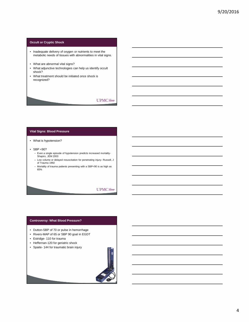
Vital Signs: Blood Pressure
• What is hypotension?
• SBP <90?– Even a single episode of hypotension predicts increased mortality-
Shapiro, JEM 2003
– Low volume or delayed resuscitation for penetrating injury- Russell, J of Trauma 1992
– Mortality of trauma patients presenting with a SBP<90 is as high as 65%
Controversy: What Blood Pressure?
• Dutton-SBP of 70 or pulse in hemorrhage• Rivers-MAP of 65 or SBP 90 goal in EGDT• Estridge- 110 for trauma• Heffernan-120 for geriatric shock• Spaite- 144 for traumatic brain injury

9/20/2016
5
• SV varies as a function of Preload, Afterload and Contractility
• Preload- Hypovolemia or � SVR• Afterload- Obstruction or � SVR• Contractility- Pump failure
CO= SV x HR
BP=CO x SVR
Treatment of Hypotension

9/20/2016
6
Vital Signs: Heart Rate
• What is Tachycardia?– ATLS guideline: >100
• What conditions effect a tachycardic response?• Does Heart Rate predict outcomes?
– Bleeding? McGee, JAMA 1999
– Injury Severity? Brasel, J of Trauma 2007
– Mortality in hypotensive patients? Demetriades, J of Trauma 1998
• Elevated HR– Hypoxia– ↑ WOB
– Hypotension/Shock
– Hypoglycemia– Anemia
– ↑ DO2 (fever, thyroid storm, exercise, etc)
– Medications (Intoxication & Withdrawal)
– Pain/Anxiety
• Low HR– Hypoxia– Meds
– AMI (post, right)
– Vasovagal– Hypothermia
Abnormal HR
Vital Signs: Respiratory Rate
• Commonly used in validated triage scales.– Revised Trauma Score (RTS)
– Simple Triage And Rapid Transport (START)
• May be particularly useful when other tools are limited or not availalble. – Respiratory rate > 25 breaths/min is a useful triage tool.
• Husum, J of Trauma 2003
9/20/2016
7
Vital Signs: End Tidal CO2
• Can be used to assess perfusion during cardiac arrest.
• End Tidal CO2 less than 15 during CPR probably indicates ineffective CPR.
Vital Signs: Others?
• Shock Index (SI)– HR/SBP 0.5-0.7 Normal 0.9 or > is predictive of severe illness.
(Rady, Ann of E Med 1994)– SI may provide a means to monitor acute hypovolemia and
circulatory failure. (Rady, Resuscitation 1992_
• Pulse Pressure– Narrowed pulse pressure is an early sign of hypovolemic shock
– Widened pulse pressure is an early sign of septic shock
Adjunctive Tools: Lactate
• Serum lactate is marker of organ oxygen supply/demand mismatch, and is directly related to mortality in patients with sepsis, myocardial infarction, and trauma.
• Prehospital lactate identifies a cohort of patients with normal initial vital signs who required intensive resuscitation during the first 24 hours of hospitalization

9/20/2016
8
Lactate
Base Deficit
• Base deficit is a marker of impaired oxygen utilization.• The amount of acid that must be removed to return the body
to a normal pH.• >6 is considered markedly abnormal• >8 is associated with a 25% chance of mortality – Rutherford,
J of Trauma 1992
Tissue Oximetry
• Near infra-red spectroscopy may be able to determine oxygen consumption and delivery in peripheral tissue.
• Prehospital use of a vascular occlusion test (VOT) with tissue oximetry can predict mortality and the need for in-patient resources.
www.Hutchinson.com

9/20/2016
9
Tissue Oximetry
• The sensitivity of StO2 is increased by performing a regional stress test
Tissue Oximetry
Heart Rate Variability (HRV)
• A measure of beat to beat variation which is associated with changes in the autonomic nervous system.
• As compensatory mechanisms are depleted heart rate variability decreases.
• Decreased HRV represents cardiac uncoupling and is associated with shock.
• Cardiac uncoupling is an independent predictor of death throughout the ICU stay and appears to increase in response to inflammation, infection, and multiple organ failure – Norris et al., Ann Surg 2006

9/20/2016
10
Heart Rate Variability (HRV)
• HRV measures can be used to predict which septic patients in the emergency department (ED) will progress to septic shock. -Chan and Kuo, Accad Emerg Med 2007
• HRV in trauma patients is a better predictor of survival than standard physiologic measurements (Vitals signs and GCS).-Cooke et al., J of Trauma 2006
Ultrasound
• Useful for the recognition of shock and for directing therapy.
• FAST is used for termination of care- Eckstein, PEC 2005
• FAST is used in the field as an adjunct to triage- Sztajnkrycer et al., PEC 2006
• FAST is used in a helicopter to direct treatment- Melanson et al., PEC 2001
• Systematic Review of Prehospital US in Trauma-O’Dochartaigh and Douma, Injury 2015
• Five Year Retrospective-O’Dochartaigh and Douma PEC, 2016
• You are consulted for a patient who struck a guardrail with his motorcycle. The crew notes that there is a large amount of blood running down the side of the gurney from the patient’s left thigh area. They have placed a tourniquet and the bleeding has slowed but not stopped.
• The patient is awake but not alert GCS 11, he has a BP of 68/32, HR 128, SpO2 not obtainable due to perfusion. The crew has placed bilateral IO’s and given 1L of NS.
Scenario

9/20/2016
11
• Control Blood Loss• Hemorrhage� Transfuse*• Dehydration� NS (2L)• If SBP<90 mmHg� Levophed 0.05mcg/Kg/min• If SBP<70 mmHg� Epinephrine 100mcg IVP
Treatment of Shock: Hypovolemic
• Source Control– Tourniquets, T-POD, Splints
– Hemostatic Agents (Quickclot, Combat Gauze)
– Transexamic Acid
– Look for concomitant shock states
• Obstructive (Tamponade, PTX)
• Distributive Shock (Spinal, Neurogenic)
• Volume Resuscitation– Crystalloid is BAD, NS is Evil
– Resuscitate to SBP 90* in penetrating injury
– Hemorrhage- FFP first then PRBCs
Hypovolemic Shock for EMS
• Vasopressors– Rapidly titrate Levophed to 0.3 mcg/kg/min
– If ineffective consider 2nd Agent
• Vasopressin 0.04 u/min- acidosis, GI hemorrhage (may have to increase dose)
• Epinephrine 0.05-0.15 mcg/kg/min- bradycardia, if in extremis there is no max dose
• Adjunctive Therapy– Ketamine (Sedation RSI)
– Bicarbonate (pH <7.1)
– Calcium (if giving more than 5U of any blood product)
Hypovolemic Shock for EMS

9/20/2016
12
• Chest Wall Trauma, COPD, or Airway Manipulation plus any of the following:– Difficulty ventilating
– Hypotension
– Subcutaneous emphysema
– JVD
– Tracheal Deviation
• Manage the Airway • Needle Decompress at the 2nd Intercostal space mid-
clavicular line or 4th intercostal space mid-axillary line • Repeat as necessary
Treatment of Shock: Obstructive
Secondary Injury
Address the abnormal vital signs• Hypoxia• Hypotension• Hyper or hypocarbia• Hypothermia
Controversy: Treatment of Shock
• ABCs or CABs?• Prevent secondary injury• Determine the etiology of the shock state• Resuscitation• Deliver the patient to definitive care
36

9/20/2016
13
ABC vs. CAB
• Approach may vary based on the presentation• Patients who are peri-arrest benefit from CAB
– Cardiac Arrest: Compressions first
– Exsanguinating hemorrhage- control bleeding
• Patients with primary respiratory issues still require ABC– Even in these circumstances it may be beneficial to delay definitive
airway management
Controversies
• Volume for Hemorrhagic Shock– Evidence for resuscitation to SBP of 70-90
– Large volume flids may result in:
• Dislodged clot
• Hypothermia
• Hyperchloremic Acidosis (NS)
• Dilutional Coagulopathy
• Disruption of endothelium and inflammation
• Interventions vs. Transport to Definitive Care– Delay for IV access or therapy may outweigh benefit
– Blood products are superior to fluids as initial resuscitative fluid
38
• Large Volume Crystalloids – Increase mortality
– Worsen coagulopathy of trauma and TBI
• Hypotensive Resuscitation with Blood– Expensive, limited availability and storage
– Patients remain coagulopathic and hypothermic
• Plasma or plasma derivitives– Treats coagulopathy
– Increases survival as part of Damage Control Resuscitation
Fluid Resuscitation:

9/20/2016
14
Controversies in Hemorrhage?
• STOP THE BLEEDING– External
• Extremity-Direct Pressure, Tourniquet
• Junctional-External Compression Device
• Cavity- Direct pressure, hemostatic dressing
– Internal
• Reverse Coagulopathy
• TXA
• OR
Vasopressors
Vasopressors should be initiated if the patient has not responded to the initial volume challenge
– Distributive (Needed to increase SVR)– Obstructive (only adjunctive)– Cardiogenic (may be harmful 2/2 afterload)
– Hypovolemic (in conjunction with volume)
• Prospective, randomized controlled trial• Over 20,000 patients• TXA significantly reduced all causes mortality
from 16.0% to 14.5%• TXA significantly reduced death from bleeding
from 5.7% to 4.9%
TXA – CRASH 2
42

9/20/2016
15
CRASH-2: Timing of TXA
• Subgroup analysis of 20,211 trauma patients based on time of administration of TXA
• Timing; only deaths due to bleeding• Risk of death due to bleeding reduced (5.3% vs
7.7%) if TXA given within 1 hour of injury. At 1-3 hrs after injury, also significant (4.8 vs 6.1%)
Take-Home Points
• Early identification and treatment of shock reduces mortality
• Normal vital signs does not = normal perfusion
• Treatment of shock varies by etiology (category)
• Treatment in the field should not delay definitive care
44
Take-Home Points
• Have an understanding of the diagnostic limitations and the use of fluids and vasopressors.– Clinical aspects of EMS = 40% of tests items
• Take home points – Cause of shock state is difficult to assess so standardized approach
is needed
– Volume resuscitation is dependent on etiology
– Vasopressor options are limited
– Accurate dosing of vasopressors is challenge for EMS
45

9/20/2016
16
• Christian Martin-Gill MD, MPH• Chris Schott MD, MS
Anknowledgement


![SHOCK[1] - Hypovolemic Shock](https://img.pdfslide.net/doc/110x75/58edc1bc1a28abae538b4711/shock1-hypovolemic-shock.jpg)