Embed Size (px)
Citation preview
Guyon's Canal Revisited: An Anatomic Study of the Carpal
Ulnar Neurovascular Space
Tyson K. Cobb, MD, Stephen W. Carmichaei, PhD, William P. Cooney, MD, Rochester, MN
The boundaries of the space through which the ulnar neurovascular bundle crosses the wrist have been reinvestigated. Using gross dissections, transverse and sagittal sections, and histo- logic study, we determined that the roof of Guyon's canal, the "carpal ulnar neurovascular space," does not directly connect to the hamate bone, as is currently accepted. The roof of this space extends radially to the hook of hamate and attaches to the flexor retinaculum. This anatomic arrangement allows the ulnar artery and sensory component of the ulnar nerve to course radially to the hook of hamate, where they lie on the flexor retinaculum (transverse carpal ligament). The roof and radial border have three segments: (1) a proximal segment that begins near the pisiform and extends distally to the level of the hook of hamate but does not attach directly to it, (2) a central segment that contains only adipose tissue, and (3) a dis- tal fascial layer that includes the palmaris brevis muscle. The floor of the space consists of the muscles of the hypothenar eminence, their fibers of origin, and the flexor retinaculum (transverse carpal ligament). Guyon accurately described the proximal portion of the carpal ulnar neurovascular space, but his description has been misinterpreted; the hook of hamate does not serve as the radial boundary of Guyon's canal. The anatomic relationships of the "carpal ulnar neurovascular space" need to be appreciated to avoid complications during carpal tunnel surgery. (J Hand Surg 1996;21A:861-869.)
In 1861, Guyon 1 described a "loge" (space), now known as Guyon 's canal, as: . . . an intra-aponeurotic compartment, because its ante- rior wall seemed to be dependent with the local fibrous layer; its posterior wall is formed by the anterior carpal ligament. One cannot say that there are lateral walls; nonetheless, the pisiform forms medially (ulnarly) a sort of wall coated with aponeurotical tissue; superiorly, infe- riorly, and laterally, the anterior wall blends with the aponeurosis, of which it is only a split part . . . . Laterally (radially), the limit is more or less at the middle of the
From the Department of Orthopedics and the Department of Anatomy, Mayo Clinic and Mayo Foundation, Rochester, MN.
Received for publication Sept. 28, 1994; accepted in revised form Feb. 28, 1996.
No benefits in any form have been received or will be received from a commercial party related directly or indirectly to the subject of this article
Reprint requests: William R Cooney, MD, Mayo Clinic, 200 First Street SW, Rochester, MN 55905.
anterior carpal ligament, which means this small com- partment belongs only to the medial side . . . . An inter- esting p o i n t . . , is the presence of the cubital artery and nerve lying on the posterior wall, and that is the anterior carpal l igament. . .
During dissection of the carpal tunnel, we observed, as did Guyon, that the radial boundary of Guyon 's
canal extends radial to the hook of hamate. This allows the ulnar artery and possibly the sensory com- ponent of the ulnar nerve to course radial to the hook of hamate in some specimens (Fig. 1). Furthermore, we noted that the floor of Guyon ' s canal appeared to be intimately involved with the ulnar portion of
the roof of the carpal tunnel. This is in contrast to the commonly accepted belief that the hook of hamate serves as the radial (lateral) border of this space. 2-13 The purpose of this study was to reinvesti- gate the anatomic boundaries of the ulnar neurovas- cular space.
The Journal of Hand Surgery 861

862 Cobb et al. / Guyon's Canal Revisited
. . . . .
Flexor retiL _ _ ~ Flexor retinaculum Ulnar a. & n.
Figure 1. Diagram of relationship of ulnar neurovascular bundle with the carpal tunnel and hand. Arrows mark the levels of the two axial sections. The roof of the carpal ulnar neurovascular space (CUNS) has been elevated from the ulnar side. The thenar fascia has been elevated from the radial side to demonstrate its relationship to the roof of CUNS. Note the fatty tissue separating the proximal and distal segments of the CUNS roof. FCR, flexor carpi radialis tendon; H, hypothenar muscles; P, pisiform; T, thenar muscle. (By permission of Mayo Foundation.)
Materials and Methods
Twenty-five fresh frozen and thawed unembalmed specimens were prepared for this study. Eight speci- mens were used for cross-section anatomic studies: 6 in the transverse plane and 2 in the sagittal plane. Five of these 8 specimens were also used for histo- logic examination. All 8 specimens were obtained as above-elbow amputations and immediately frozen at -70~ in liquid nitrogen. The specimens were then sectioned with a band saw into 4- to 5-ram-thick sec- tions, beginning at the distal metacarpals and pro- ceeding proximally to the distal radius. The cross- section specimens were placed serially into rows and
examined grossly and with loupe magnification to determine the relationship of the ulnar nerve (sen- sory component) and artery and the radial attach- ment of the roof of Guyon's canal to the hook of hamate. The two specimens sectioned in the sagittal plane were aligned serially from medial to lateral to examine the changing relationship of the location of the ulnar artery and nerve and palmar fascia with respect to the hook of hamate. We looked for anatomic changes primarily in a proximal to distal direction.
In the 17 other specimens, dissections were con- ducted with loupe magnification to delineate the characteristics of the superficial fascia (roof of
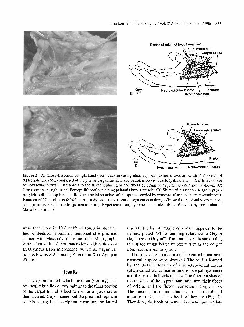
Guyon's canal) that constrain the ulnar neurovascular bundle on the palmar and ulnar aspects of the flexor retinaculum. After the skin of the distal forearm and palm was removed by sharp dissection, leaving the superficial fascia, the ulnar neurovascular bundle was exposed proximal in the forearm and traced distally. Through an ulnar approach, the ulnar nerve and artery were exposed at Guyon's canal by sharply transsecting the subcutaneous tissue, palmaris brevis muscle, and fascia over the pisiform and distally along the anteromedial border of the hypothenar emi- nence. A radial-based flap (roof of canal) containing subcutaneous tissue, fascia, and the palmaris brevis muscle was created, exposing the ulnar neurovascular bundle from the ulnar side (Fig. 2). Three distinct segments of this flap (roof and radial border of canal) were noted: (1) a proximal fascial segment, (2) a cen- tral segment containing only adipose tissue, and (3) a distal fascia segment containing the palmaris brevis muscle (Fig. 2). The proximodistal length of these segments was measured and recorded (Table 1). The measurements were made with a ruler on the radial aspect of the carpal ulnar neurovascular space and the hook of hamate, where the fascial roof of Guyon's canal joins the flexor retinaculum.
Five specimens were prepared for histologic examination. Three specimens were cut transversely, and two were cut sagittally. The gross specimens
The Journal of Hand Surgery/Vol. 21A No. 5 September 1996 863
B MAYO
Tendon of origin of hypothenar r a m .
laris br. m. Carpal tunnel
Neurovascular bundle Pisiform Hypothenar r a m .
Palmaris br. m.
tlum
orm
Hypothenar mm. I~eurovascu,ur uuudle
Figure 2, (A) Gross dissection of right hand (fresh cadaver) using ulnar approach to neurovascular bundle. (B) Sketch of dissection. The roof, comprised of the palmar carpal ligament and palmaris brevis muscle (palmaris br. m.), is lifted off the neurovascular bundle. Attachment to the flexor retinaculum and fibers of origin of hypothenar eminence is shown. (C) Gross specimen; right hand. Forceps lift roof containing palmaris brevis muscle. (D) Sketch of dissection. Right is proxi- mal; left is distal. Top is radial. Roof and radial boundary of the space occupied by neurovascular bundle are discontinuous. Fourteen of 17 specimens (82%) in this study had an open central segment containing adipose tissue. Distal segment con- rains palmaris brevis muscle (palmaris br. m.). Hypothenar mm, hypothenar muscles. (Figs. B and D by permission of Mayo Foundation.)
were then fixed in 10% buffered formalin, decalci- fied, embedded in paraffin, sectioned at 6 gin, and stained with Masson's trichrome stain. Micrographs were taken with a Canon macro lens with bellows or an Olympus BH-2 microscope, with final magnifica- tion as low as x 2.5, using Panatomic-X or Agfapan 25 film.
Results
The region through which the ulnar (sensory) neu- rovascular bundle courses palmar to the ulnar portion of the carpal tunnel is best defined as a space rather than a canal. Guyon described the proximal segment of this space; his description regarding the lateral
(radial) border of "Guyon's canal" appears to be misinterpreted. While retaining reference to Guyon (ie, "loge de Guyon"), from an anatomic standpoint, this space might better be referred to as the carpal ulnar" neurovascular space.
The following boundaries of the carpal ulnar neu- rovascular space were observed. The roof is formed by the distal extension of the antebrachial fascia (often called the palmar or anterior carpal ligament) and the palmaris brevis muscle. The floor consists of the muscles of the hypothenar eminence, their fibers of origin, and the flexor retinaculum (Figs. 3-7). The flexor retinaculum attaches to the radial and anterior surfaces of the hook of hamate (Fig. 4). Therefore, the hook of hamate is dorsal and not lat-
864 Cobb et al. / Guyon's Canal Revisited
Table 1. Length of Each of the Three Regions of the Carpal Ulnar Neurovascular Space
Proximal Middle Distal Segment* Segment~- Segmentr
Specimen (mm) (Open) (mm) (ram)
1 5 15 10 2 4 15 15 3 5 15 15 4 10 11 18 5 8 10 18 6 10 10 t2 7w - - - - - - 8 10 15 17 9 14 6 15 10 15 5 20 l l 10 6 12 12 10 10 12 13 7 10 10 14w - - - - - -
15 14 l0 18 16 6 l0 15 1 7 w - - - - - -
Mean 9.14 10 14.79 Standard deviation 3.40 3.84 3.08 Variance 11.55 14.71 9.45
*Extends from region of pisiform to region of flexor retinacu- lum proximal to hook of hamate.
Hs essentially open; contains adipose tissue. :)Longest and thickest of the three segments; contains palmaris
brevis muscle. w were not observed because the border was continuous.
eral (radial) to the carpal ulnar neurovascular space. The radial boundary is formed by the junction of the roof (patmar carpal ligament), including the pal-
marls brevis muscle, to the flexor ret inaculum and tendons of origin of the thenar muscles. The junc- tion between the roof and the flexor ret inaculum occurs radial to the hook of hamate in the central aspect of the flexor retinaculum, roughly midway between the ulnar and radial borders of the flexor ret inaculum (Figs. 3-7). The ulnar boundary is formed by the junction of the fascial roof, with the fascia covering the hypothenar eminence distally and the pis iform bone proximally.
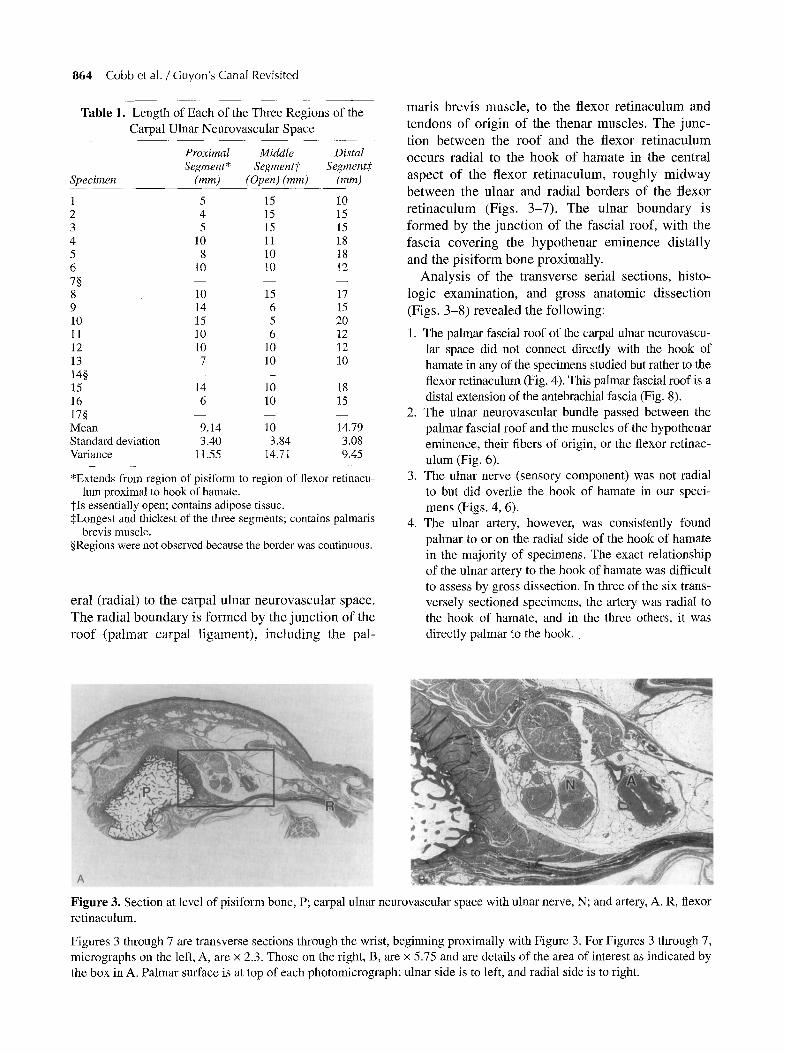
Analysis of the transverse serial sections, histo- logic examination, and gross anatomic dissection (Figs. 3-8) revealed the following:
1. The palmar fascial roof of the carpal ulnar neurovascu- lar space did not connect directly with the hook of hamate in any of the specimens studied but rather to the flexor retinaculum (Fig. 4). This palmar fascial roof is a distal extension of the antebrachial fascia (Fig. 8).
2. The ulnar neurovascular bundle passed between the palmar fascial roof and the muscles of the hypothenar eminence, their fibers of origin, or the flexor retinac- ulum (Fig. 6).
3. The ulnar nerve (sensory component) was not radial to but did overlie the hook of hamate in our speci- mens (Figs. 4, 6).
4. The ulnar artery, however, was consistently found palmar to or on the radial side of the hook of hamate in the majority of specimens. The exact relationship of the ulnar artery to the hook of hamate was difficult to assess by gross dissection. In three of the six trans- versely sectioned specimens, the artery was radial to the hook of hamate, and in the three others, it was directly palmar to the hook.
Figure 3. Section at level of pisiform bone, P; carpal ulnar neurovascular space with ulnar nerve, N; and artery, A. R, flexor retinaculum.
Figures 3 through 7 are transverse sections through the wrist, beginning proximally with Figure 3. For Figures 3 through 7, micrographs on the left, A, are x 2.3. Those on the right, B, are x 5.75 and are details of the area of interest as indicated by the box in A. Palmar surface is at top of each photomicrograph; ulnar side is to left, and radial side is to right.
The Journal of Hand Surgery / Vol. 21A No. 5 September 1996 865
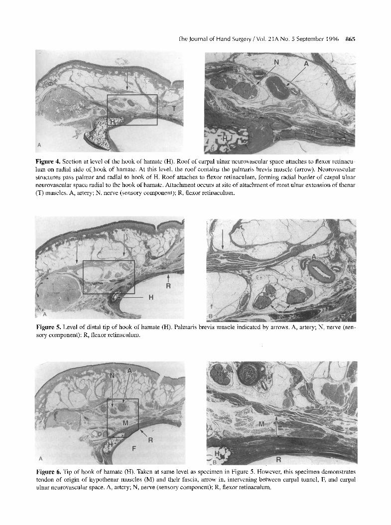
Figure 4. Section at level of the hook of hamate (H). Roof of carpal ulnar neurovascular space attaches to flexor retinacu- lure on radial side of hook of hamate. At this level, the roof contains the palmaris brevis muscle (arrow). Neurovascular structures pass palmar and radial to hook of H. Roof attaches to flexor retinaculum, forming radial border of carpal ulnar neurovascular space radial to the hook of hamate. Attachment occurs at site of attachment of most ulnar extension of thenar (T) muscles. A, artery; N, nerve (sensory component); R, flexor retinaculum.
Figure 5. Level of distal tip of hook of hamate (H). Palmaris brevis muscle indicated by arrows. A, artery; N, nerve (sen- sory component); R, flexor retinaculum.
Figure 6. Tip of hook of hamate (H). Taken at same level as specimen in Figure 5. However, this specimen demonstrates tendon of origin of hypothenar muscles (M) and their fascia, arrow in, intervening between carpal tunnel, F, and carpal ulnar neurovascular space. A, artery; N, nerve (sensory component); R, flexor retinaculum.
866 Cobb et al. / Guyon's Canal Revisited
Figure 7. (A) Taken through distal aponeurotic portion of flexor retinaculum, distal to carpal bones. Note distal extension of flexor retinaculum, R, and flexor tendons and lumbrical muscles, F, in distal carpal tunnel. (B) Ulnar nerve (sensory component) is outlined (box) palmar to R. Tendon of origin of hypothenar muscles, arrow, intervenes between carpal ulnar neurovascular space and R.
In all specimens, the fascial roof of the carpal ulnar neurovascular space extended in a radial direc- tion from the ulnar border of the palm to form the radial boundary of the space occupied by the ulnar neurovascular bundle by attaching to the flexor reti- naculum (Fig. 1). There did not appear to be an attachment or insertion to the hook of hamate; rather, in all specimens, this attachment was radial to the hook of hamate. The roof is contiguous with the fas- cia of the hypothenar eminence on the ulnar aspect of this space. Within this space, the ulnar neurovas- cular bundle was surrounded by adipose tissue. The crotch created by the opposing surfaces of the fascial roof, and the flexor retinaculum was occupied by adipose tissue (Fig. 4).
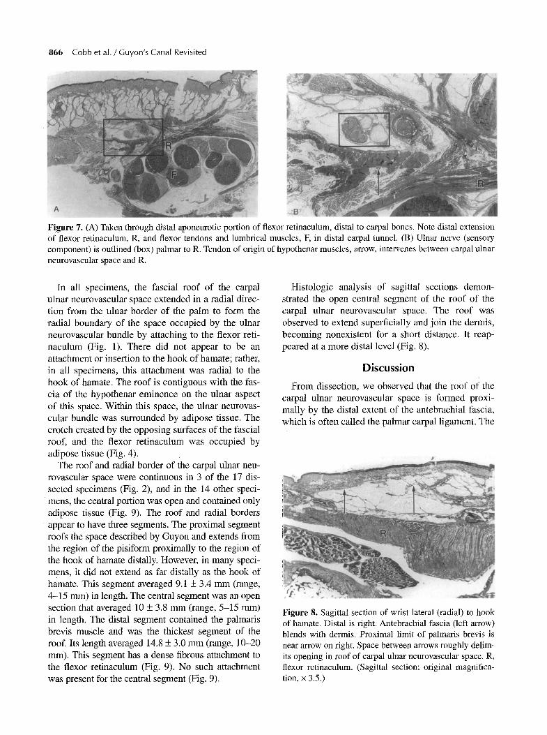
The roof and radial border of the carpal ulnar neu- rovascular space were continuous in 3 of the 17 dis- sected specimens (Fig. 2), and in the 14 other speci- mens, the central portion was open and contained only adipose tissue (Fig. 9). The roof and radial borders appear to have three segments. The proximal segment roofs the space described by Guyon and extends from the region of the pisiform proximally to the region of the hook of hamate distally. However, in many speci- mens, it did not extend as fro distally as the hook of hamate. This segment averaged 9.1 _+ 3.4 mm (range, 4-15 mm) in length. The central segment was an open section that averaged 10 _+ 3.8 mm (range, 5-15 ram) in length. The distal segment contained the palmaris brevis muscle and was the thickest segment of the roof. Its length averaged 14.8 _+ 3.0 mm (range, 10-20 ram). This segment has a dense fibrous attachment to the flexor retinaculum (Fig. 9). No such attachment was present for the central segment (Fig. 9).
Histologic analysis of sagittal sections demon- strated the open central segment of the roof of the carpal ulnar neurovascular space. The roof was observed to extend superficially and join the dermis, becoming nonexistent for a short distance. It reap- peared at a more distal level (Fig. 8).
Discussion
From dissection, we observed that the roof of the carpal ulnar neurovascular space is formed proxi- mally by the distal extent of the antebrachial fascia, which is often called the palmar carpal ligament. The
Figure 8. Sagittal section of wrist lateral (radial) to hook of hamate. Distal is right. Antebrachial fascia (left arrow) blends with dermis. Proximal limit of palmaris brevis is near arrow on right. Space between arrows roughly delim- its opening in roof of carpal ulnar neurovascular space. R, flexor retinaculum. (Sagittal section; original magnifica- tion, • 3.5.)
The Journal of Hand Surgery/Vol. 21A No. 5 September 1996 867
Carpal ulnar neurovascular space - - - - -~
(Guyon's canal) Distal ' /
~ ~ 4 U O r Carpal ~ ~ J tunnel
f
~ Ulnar a.
~ ' ; 2 ~ / / ~ ~ Ulnar n.
Right hand
Proximal
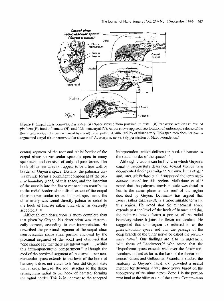
Figure 9. Carpal ulnar neurovascular space. (A) Space viewed from proximal to distal. (B) transverse sections at level of pisiform (P), hook of hamate (H), and fifth metacarpal (V). Arrow shows approximate location of endoscopic release of the flexor retinaculum (transverse carpal ligament). Note potential vulnerability of ulnar artery. This specimen does not have a segmented carpal ulnar neurovascular space roof. A, artery; n, nerve. (By permission of Mayo Foundation.)
central segment of the roof and radial border of the carpal ulnar neurovascular space is open in many specimens and consists of only adipose tissue. The hook of hamate does not appear to be a true wall or border of Guyon's space. Distally, the palmaris bre- vis muscle forms a prominent component of the pal- mar boundary (roof) of this space, and the insertion of the muscle into the flexor retinaculum contributes to the radial border of the distal extent of the carpal ulnar neurovascular space. In most specimens, the ulnar artery was found directly palmar or radial to the hook of hamate rather than ulnar, as currently accepted.14-16
Although our description is more complete than that given by Guyon, his description was anatomi- cally correct, according to our interpretation. He described the proximal segment of the carpal ulnar neurovascular space (that portion enclosed by the proximal segment of the roof) and observed that "one cannot say that there are lateral wa l l s . . , within this intra-aponeurotic compartment." Although the roof of the proximal segment of the carpal ulnar neu- rovascular space extends to the level of the hook of hamate; it does not attach to it (nor did Guyon state that it did). Instead, the roof attaches to the flexor retinaculum radial to the hook of hamate, forming the radial border. This is in contrast to the accepted
interpretation, which defines the hook of hamate as the radial border of the space. 2-13
Although citations can be found in which Guyon's canal is inaccurately described, several studies have documented findings similar to our own. Enna et al. 17 and, later, McFarlane et aL as suggested the term piso- hamate tunnel for this region. McFarlane et al. 18 noted that the palmaris brevis muscle was distal to but in the same plane as the roof of the region described by Guyon. Denman 19 has argued that space, rather than canal, is a more suitable term for this region. He noted that the ulnacarpal space extends past the level of the hook of hamate and that the palmaris brevis forms a portion of the radial boundary where it joins the flexor retinaculum. He suggested that this region be referred to as the pisoretinacular space and that the passage of the deep branch of the ulnar nerve be called the pisoha- mate tunnel. Our findings are also in agreement with those of Landsmeer, 2~ who stated that the "hypothenar space extends well over the flexor reti- naculum, indeed as far as the base of the thenar emi- nence." Gross and Gelberman u carefully studied the anatomy of Guyon's canal and provided a useful method for dividing it into three zones based on the topography of the ulnar nerve. Zone 1 is the portion proximal to the bifurcation of the nerve. Compression
868 Cobb et al. / Guyon's Canal Revisited
neuropathies in this region are expected to cause a combination of motor and sensory deficits. The prox- imal extent of this zone is identical (Fig. 3) to the findings in this study and, in a previous description, to the anatomy of the flexor retinaculum (see Fig. 4A and B in Cobb et al.Zt). Zones II and III, described by Gross and Gelberman, are the regions that contain the deep motor branch and the superficial sensory branch of the ulnar nerve, respectively. The three divisions described in our study do not correlate with the three zones of Gross and Gelberman, because their study was based on ulnar nerve topography and ours is based on purely anatomic boundaries. The relation- ship of the two segments of the roof separated by a central open segment is important from a surgical perspective because the distal segment could poten- tially be overlooked during surgical procedures per- formed to release the ulnar nerve.
Until recently, the distal extent of the flexor reti- naculum (transverse carpal ligament) was considered to be at the level of the hook of hamate and the ridge of the trapezium.3,6,18, 2a We now know that these landmarks define the junction between the middle and distal segments of the flexor retinaculum. 2t The distal segment of the flexor retinaculum is the apo- neurosis between the thenar and hypothenar mus- cles, which extend approximately 1 cm distal to the hook of hamate. 23,24
In a similar fashion, the distal extent of the carpal ulnar neurovascular space should be extended from the previously defined hook of hamatetO, 25,26 to the distal extent of the flexor retinaculum. Denman~9 and Landsmeer 20 have documented similar findings. As pointed out by Gross and Gelberman, 11 this portion of the canal is less rigid and, thus, it is less likely for the ulnar nerve to be compressed at this level.
The hook of hamate does not serve as the radial boundary of the space but rather as a portion of the floor. This is important clinically because of the potential for trauma to the ulnar neurovascular bundle, particularly the ulnar artery, which courses palmar to this structure. 27,28 Conn et al. 29 coined the phrase hypothenar hammer syndrome to describe injury to the ulnar artery resulting from blunt trauma against the hook of hamate. In 50% of the specimens that were sectioned transversely, the ulnar artery was directly palmar to the hook of hamate. Therefore, our findings provide an anatomic explanation for the development of this syndrome.
The finding of the ulnar artery and occasionally the sensory component of the ulnar nerve radial or pal- mar to the hook of hamate is also of clinical signifi-
cance with respect to both open and endoscopic carpal tunnel release. These procedures are per- formed on the ulnar aspect of the carpal tunnel, usu- ally near the hook of hamate, to avoid the median nerve. 30 The ulnar border of the carpal tunnel angles in a radial direction from the pisiform to the hook of hamate. 2~ ff the ulnar neurovascular bundle were con- tained in a space between the pisiform and the hook of hamate, the bundle would be safe during the surgi- cal procedure. However, we have demonstrated that this is not the case. We have observed that the ulnar artery may fall into the interval created by release of the flexor retinaculum during endoscopic carpal tun- nel procedures. 31 The fact that the ulnar artery is not commonly injured during endoscopic carpal tunnel release is likely related to protective adipose tissue and mobility of the neurovascular bundle. In some hands, well-developed fibers of the hypothenar emi- nence extend over the flexor retinaculum and separate it from the ulnar artery (Fig. 6). To some extent, these fibers serve as a protective barrier. 23
It is important to note that by releasing the flexor retinaculum (transverse carpal ligament) during carpal tunnel surgery, the floor of the carpal ulnar neurovascular space is also released. The ulnar nerve can occasionally be entrapped in this region, causing nerve compression. 2 However, release of the flexor retinaculum for median nerve entrapment appears to release simultaneously the compartment that con- tains the ulnar neurovascular bundle 23 (Figs. 3-5).
We suggest the term carpal ulnar neurovascular space to anatomically define what has been called Guyon's canal. We note that the ulnar artery and pos- sibly the sensory division of the ulnar nerve can be directly palmar to the flexor retinaculum and may be at risk during both open and endoscopic carpal tun- nel release.
We thank Dean R. Fisher, Department of Anatomy, for help with cadaveric specimens, and Duane M. Ilstrup, MS, and Steven L. Wall- richs, BS, Section of Biostatistics, for help with statistics.
References 1. Guyon F. Note sur une disposition anatomique propre a la
face anttrieure de la rtgion du poignet et non encore ddcrite. Bull Soc Anat Paris (2nd series) 1861;6:184-186.
2. Eversmann Jr WW. Entrapment and compression neu- ropathies. In: Green DR ed. Operative hand surgery. 3rd ed. New York: Churchill Livingstone, 1993:1365.
3. Hoppenfeld S, deBoer R Surgical exposures in ortho- paedics: the anatomic approach. Philadelphia: JB Lippin- cot, 1984: 165.
4. Nigst H. Nerve compression syndromes in the upper limb. In: Nigst H, Buck-Gramcko D, Millesi H, Lister GD, eds.
The Journal of Hand Surgery / Vol. 21A No. 5 September 1996 869
Hand surgery. Vol. 1: General aspects, elective surgery. Stuttgart: Georg Thieme Verlag, 1988: 17-20.
5. Stern PJ, Vice M. Compression of the deep branch of the ulnar nerve: a case report. J Hand Surg 1983;8:72-74.
6. American Society for Surgery of the Hand. The hand: examination and diagnosis. 3rd ed. New York: Churchill Livingstone, 1990: 11-47.
7. Whitaker JH, Richardson GA. Compressive neuropathies. In: Strickland JW, Rettig AC, eds. Hand injuries in ath- letes. Philadelphia: WB Saunders, 1992: 212.
8. Coyle Jr MP. Nerve entrapment syndromes in the upper extremity. In: Dee R, Mango E, Hurst LC, eds. Principles of orthopaedic practice. New York: McGraw-Hill, 1988: 680.
9. Snell RS, Smith MS. Clinical anatomy for emergency medicine. St. Louis: Mosby-Year Book, 1993: 643.
10. Greene MH, Hadied AM. Bipartite hamulus with ulnar tunnel syndrome: case report and literature review. J Hand Surg 1981;6:605-609.
11. Gross MS, Gelberman RH. The anatomy of the distal ulnar tunnel. Clin Orthop 1985;196:238-247.
12. Lister G. The hand: diagnosis and indications. 3rd ed. New York: Churchill Livingstone, 1993: 286.
13. Spinner M. Kaplan's functional and surgical anatomy of the hand. 3rd ed. Philadelphia: JB Lippincott, 1984: 123-130.
14. Akesson EJ, Loeb JA, Wilson-Pauwels L. Thompson's core textbook of anatomy. 2nd ed. Philadelphia: JB Lip- pincott 1990: 505.
15. Hall-Craggs ECB. Anatomy as a basis for clinical medicine. 2nd ed. Baltimore: Urban & Schwarzenberg, 1990: 158.
16. Rogers AW. Textbook of anatomy. Edinburgh: Churchill Livingstone, 1992: 396.
17. Enna CD, Berghtholdt HT, Stockwell E A study of surface and deep temperatures along the course of the ulnar nerve in the pisohamate tunnel. Int J Leprosy 1974;42:43-47.
18. McFarlane RM, Mayer JR, Hugill JV. Further observations on the anatomy of the ulnar nerve at the wrist. Hand 1976; 8:115-117.
19. Denman EE. The anatomy of the space of Guyon. Hand 1978;10:69-76.
20. Landsmeer JME Atlas of anatomy of the hand. Edinburgh: Churchill Livingstone, 1976: 147.
21. Cobb TK, Dalley BK, Posteraro RH, Lewis RC. Anatomy of the flexor retinaculum. J Hand Surg 1993;18A:91-99.
22. Robbins H. Anatomical study of the median nerve in the carpal tunnel and etiologies of the carpal-tunnel syndrome. J Bone Joint Surg 1963;45A:953-966.
23. Cobb TK, Carmichael SW, Cooney WR The ulnar nan- rovascular bundle at the wrist: a technical note on endo- scopic carpal tunnel release. J Hand Surg 1994;19B:24-26.
24. Cobb TK, Cooney WR An K-N. Relationship of deep structures of the hand and wrist to topographical land- marks. Clin Anat 1993;6:300-307.
25. Brown FE. Compression neuropathies of the upper extremity. In: Jupiter JB, ed. Flynn's hand surgery. 4th ed. Baltimore: Williams & Wilkins, 1991: 526.
26. Moneim MS. Ulnar nerve compression at the wrist: ulnar tunnel syndrome. Hand Clin 1992;8:337-344.
27. Shea JD, McClain EJ. Ulnar-nerve compression syn- dromes at and below the wrist. J Bone Joint Surg 1969; 51A:1095-1103.
28, Howard FM. Ulnar-nerve palsy in wrist fractures. J Bone Joint Surg 1961;43:1197-1201.
29. Conn J Jr, Bergan JJ, Bell JL. Hypothenar hammer syn- drome: posttraumatic digital ischemia. Surgery 1970;68: 1122-1128.
30. Agee JM, McCarroll Jr HR, Tortosa RD et al. Endoscopic release of the carpal tunnel: a randomized prospective mul- ticenter study. J Hand Surg 1992;17A:987-995.
31. Cobb TK, Knudson GA, Cooney WE The use of topograph- ical landmarks to improve the outcome of Agee endoscopic carpal tunnel release. Arthroscopy 1995;11:165-172.