Embed Size (px)
Citation preview
19. S. Y. M. Lau et al., J. Biol. Chem. 259, 13253 (1984); R. S. Hodges et al., 256,1214 (1981).
20. H. Edelhoch, Biochemistry 6, 1948 (1967).21. L. G. Presta and G. D. Rose, Scietnce 240, 1591 (1988).22. B. V. Prasad and P. Balaram, CRC Crit. Rev. Biochem. 16, 307 (1989); C. Toniolo
and E. Benedetti, ISI Atlas of Science, p. 225 (1988).23. H. R. Wolfe and R. Wilke, Peptide Res. 2, 352 (1989).24. J. Schneider and S. B. H. Kent, Cell 54, 363 (1988); W. F. DeGrado and J. D.
Lear, Biopolymers 29, 205 (1990).25. Y.-H. Chen, J. T. Yang, K. H. Chau, Biochemistry 13, 3350 (1974); N. Greenfield
and G. D. Fasman, ibid. 8, 4108 (1969).26. R. J. Pollet, B. A. Haase, M. L. Standaert, J. Biol. Chem. 254, 30 (1979).27. C. N. Pace, Methods Enzymol. 131, 266 (1986); J. U. Bowie and R. T. Sauer,
Biochemistry 28, 7139 (1989).28. These free energy differences refer to the values expected at 300°C, based on an
examination of the van't Hoff plots in (9).29. R. W. Williams, A. Chang, D. Juretic, S. Loughran, Biochem. Biophys. Acta. 916,
200 (1987).30. P. Y. Chou and G. D. Fasman, J. Mol. Biol. 74, 263 (1973); Biochemistry 13, 211
(1974).31. M. H. Hecht, J. M. Sturtevant, R. T. Sauer, Proteins 1, 43 (1986).32. S. Dao-Pin, W. A. Baase, B. W. Matthews, ibid. 7, 198 (1990).
33. M. Matsumura, M. Becktel, B. W. Matthews, J. Biol. Chem. 264, 16059 (1989); J.T. Kellis, Jr., K. Nyberg, A. R. Fersht, Biochemistry 28, 4914 (1989).
34. The difference between AAG,,, for residues in the center of the helices versus innonhelical conformations was calculated from the equation y = IL4f(i)(AAGt.)] -,Lfn(i)(AAG<,)], where Y. refers to the sum over all 20 naturally occurring amino
acids, and f,(i) and fn,,,(i) refer to the probability of finding a given type of aminoacid in the center of a helix, or a nonhelical conformation, respectively. Thequantities f .(i) and fn,,(i) for all 20 amino acids were calculated from the datacompiled in (4).
35. J. U. Bowie, J. F. Reidhaar-Olson, W. A. Lim, R. T. Sauer, Science 247, 1306(1990); R. Kauzmann, Adv. Prot. Chem. 14, 1 (1959).
36. L. Piela, G. N. Nemethy, H. A. Scheraga, Biopolymers 26, 1587 (1987).37. , ibid., p. 1273; M. J. McGregor, S. A. Islam, M. J. E. Sternberg, J. Mol.
Biol. 198, 295 (1987); J. Janin, S. Wodak, M. Levitt, B. Maigret, ibid. 125, 357(1978).
38. J. Wojcik, K.-H. Altmann, H. A. Scheraga, Biopolymers 30, 12 (1990).39. We gratefully acknowledge J. Bryson for his preliminary work on this project. We
thank H. A. Scheraga for sharing unpublished data, R. Houghton for peptidesynthesis, T. Devine for peptide purification, L. Janvier for amino acid analysis, andJ. Lazaar for mass spectrometry.
1 May 1990; accepted 5 September 1990
Protein Splicing Converts the Yeast TEPI GeneProduct to the 69-kD Subunit of the Vacuolar
H+-Adenosine Triphosphatase
PATRICiA M. KANE, CARL T. YAMASHIRO, DAVID F. WOLCZYK, NoRMA NEFF,MARK GOEBL, TOM H. STEVENS*
The TFPI gene of the yeast Saccharomyces cerevisiaeencodes two proteins: the 69-kilodalton (kD) catalyticsubunit of the vacuolar proton-translocating adenosinetriphosphatase (H+-ATPase) and a 50-kD protein. The69-kD subunit is encoded by the 5' and 3' thirds of theTFPI coding region, whereas the 50-kD protein is en-coded by the central third. Evidence is presented thatboth the 69-kD and 50-kD proteins are obtained from asingle translation product that is cleaved to release the 50-kD protein and spliced to form the 69-kD subunit.
V T ACUOLAR-TYPE PROTON-TRANSLOCATING ADENOSINE TRI-phosphatases (H+-ATPases), which acidify certain intracel-lular compartments in eukaryotic cells (1, 2), have been
purified from various sources and show an overall structural similar-ity (1). All of the enzymes purified are multisubunit complexescontaining at least two peripheral membrane subunits with molecu-
P. M. Kane, C. T. Yamashiro, D. F. Wolczyk, and T. H. Stevens are at the Institute ofMolecular Biology, University of Oregon, Eugene, OR 97403. P. M. Kane is now inthe Department of Chemistry, College of William and Mary, Williamsburg, VA 23185and C. T. Yamashiro is in the Department of Biological Sciences, Stanford University,Stanford, CA 94305. N. Neff is at the Memorial Sloan-Kettering Cancer Center, NewYork, NY 10021. M. Goebl was in the Department of Genetics, University ofWashington, Seattle, WA 98195, but is now in the Department of Biochemistry,Indiana University School of Medicine, Indianapolis, IN 46202.
*To whom correspondence should be addressed.
lar masses of -70 and 60 kD and at least one integral membranesubunit of -15 kD. The 70-kD subunit contains the catalyticadenosine triphosphate (ATP) binding site. The vacuolar H+-ATPase of the yeast Saccharomyces cerevisiae consists ofeight differentsubunits, including a 69-kD catalytic subunit and 60- and 17-kDsubunits similar to those found in other cells (3, 4). Genes encodingthe 70- and 60-kD subunits have been cloned and characterizedfrom a number of different species, and the sequences of bothsubunits from plant, animal, and fungal sources are remarkablyconserved (5-7). The predicted amino acid sequence ofthe yeast 60-kD subunit is 82 percent identical to that ofthe Neurospora crassa 57-kD subunit and 74 percent identical to that of the human 60-kDsubunit (8). The functional roles of the yeast vacuolar H+-ATPasehave been assessed by disrupting the gene for the 60-kD subunit,which is termed VAT2 (8). Cells lacking VAT2 grow more slowlythan wild-type cells, fail to grow at neutral pH, and lack the abilityto acidify their vacuoles. Isolated vacuoles from these cells lackATPase activity.We now present evidence that the 69-kD subunit of the yeast
vacuolar H+-ATPase is one of two proteins encoded by thepreviously identified TFP1 gene (9). The vacuolar H+-ATPasesubunit is specified by the two ends of TFP1, and the central one-third encodes a 50-kD "spacer" protein. Our results indicate that the69- and 50-kD proteins are formed from a single translation productby post-translational cleavage and splicing.Encoding of the catalytic subunit of the yeast vacuolar H+-
ATPase by TFP1. A dominant allele of TFP1 was previouslyisolated from a mutant yeast strain resistant to the drug trifluopera-
RESEARCH ARTICLES 6512 NOVEMBER 1990
on
Sep
tem
ber
17, 2
009
ww
w.s
cien
cem
ag.o
rgD
ownl
oade
d fr
om
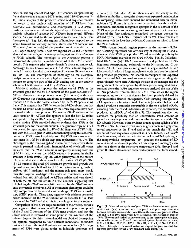
zine (9). The sequence of wild-type TFP1 contains an open readingframe that encodes a putative 1071-amino acid, 1 19-kD protein (9-11). Initial analysis of the predicted amino acid sequence revealedhomology to the catalytic (p) subunits of H+-ATPases fromEscherichia coli, mitochondria, and chloroplasts (FIFO-ATPases);further analysis uncovered a much higher degree of identity with thecatalytic subunits of vacuolar H+-ATPases from several differentspecies. As illustrated by the comparison to the vma-1 gene fromNeurospora (5) (Fig. IA), the regions of homology mapped to theamino-terminal and carboxyl-terminal thirds (the "N domain" and"C domain," respectively) of the putative protein encoded by theTFP1 open reading frame. These two regions are 73 and 77 percentidentical, respectively, to the corresponding regions of the Neurospo-ra 70-kD subunit. The homology to the Neurospora protein isinterrupted abruptly by the middle one-third of the TFP1-encodedprotein. This segment (the "spacer domain") shows no amino acidhomology to any known vacuolar H+-ATPase subunit sequence,
but is 31 percent identical to one region ofthe yeast HO endonucle-ase (10, 12). The interruption of homology to the Neurosporacatalytic subunit occurs in a very highly conserved sequence that isthought to comprise part of the ATP binding site in the catalyticsubunits of both vacuolar and F1FO ATPases (5, 7).
Additional evidence supports the assignment of TFP1 as thestructural gene for the 69-kD subunit of the yeast vacuolar H+-ATPase. Amino-terminal protein sequence (17 residues) ofthe yeast69-kD subunit was obtained and found to correspond to amino acidresidues 13 to 29 of the protein encoded by the TFP1 open readingframe. This suggests that TFP1 encodes the 69-kD subunit, but thatthe first 12 amino acids predicted by the DNA sequence have beenremoved in the mature protein. [The mature 60-kD subunit of theyeast vacuolar H+-ATPase also appears to lack the first 12 aminoacids predicted by its DNA sequence (8).] Analysis of mutant yeaststrains lacking TFP1 provided further evidence that TFP1 is thestructural gene for the 69-kD subunit. The genomic copy of TFP1was deleted by replacing the Eco RV-Sph I fragment of TFP1 (Fig.1B) with the LEU2 gene in vitro and then integrating this construc-tion at the TFP1 locus ofhaploid yeast cells [TFP1 is not an essentialyeast gene (9, 10)] by a single-step gene replacement (13, 14). Thephenotypes of the resulting tfp1-A8 mutant were compared with theisogenic parental haploid strain. Immunoblots of whole cell lysatesindicated that the 69-kD subunit is completely missing from thetfp1-A8 strain, whereas the 60-kD subunit is present in similaramounts in both strains (Fig. 2). Other phenotypes of the mutantcells were identical to those seen for cells lacking VAT2 (8). Thetfp1-8 mutants displayed pH-dependent growth (cell growth was
optimal in medium buffered to pH 5 in contrast to no growth inbuffered pH 7 medium), and the mutant cells grew more slowlythan the isogenic wild-type cells under all conditions. Vacuolesisolated from tp1-A8 cells had no ATPase activity and were devoidof both the 60- and the 69-kD ATPase subunits, suggesting thatcells lacking the 69-kD subunit failed to assemble the 60-kD subunitonto the vacuole membrane. All of the mutant phenotypes could befully complemented by introducing wild-type TFP1 on a single-copy (CEN) plasmid. These data, together with evidence presentedbelow, indicate that the 69-kD subunit of the vacuolar H+-ATPaseis encoded by TFP1 and that this is the sole gene for this subunit.Comparison ofthe TFP1 sequence to that ofthe Neurospora vma-1
gene suggested that the mature 69-kD subunit in yeast is composedof the N and C domains of the TFP1 gene product, and that thespacer domain is removed at some point in the synthesis of thesubunit. Support for this structural model was obtained by mappingthe epitopes recognized by four different monoclonal antibodiesthat reacted with the 69-kD subunit on immunoblots (15). Frag-ments of TFP1 were placed under an inducible promoter and
652
expressed in Escherichia coli. We then assessed the ability of thedifferent antibodies to recognize the proteins expressed on inductionby comparing lysates from induced and uninduced cells on immu-noblots (16). From this analysis, we determined that three of themonoclonal antibodies recognized epitopes in the N domain of theTFP1 gene product and one recognized an epitope in the C domain.None of the four antibodies recognized the spacer domain (asdefined by the Kpn I-Nae I fragment of TFP1). These results areconsistent with the idea that the N and C domains are joined to formthe 69-kD subunit.TFPI spacer domain region present in the mature mRNA.
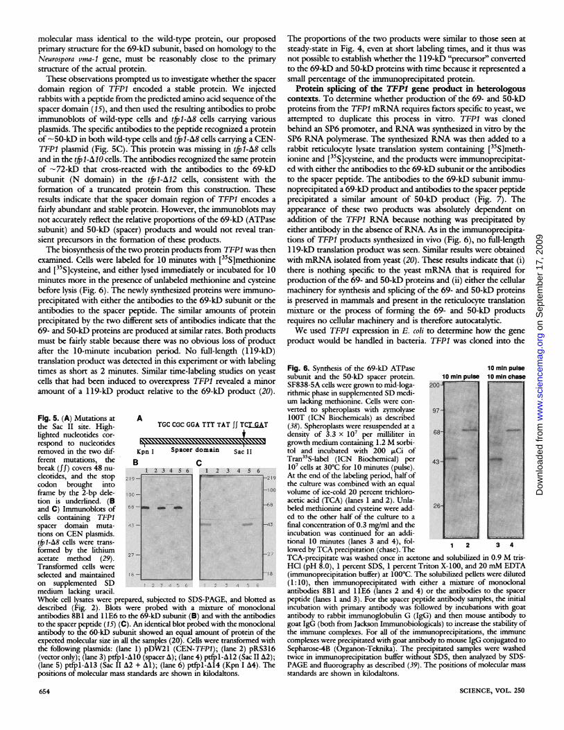
RNA splicing represents one obvious way of joining the N and Cdomains ofthe TFP1 gene product. We addressed this possibility byRNA (Northern) analysis of the TFP1 mRNA (Fig. 3). Polyadeny-lated RNA [poly(A)+ RNA] was isolated and probed with DNAfragments corresponding exclusively to the N, spacer, and C do-mains. All of these probes recognized a single mRNA of 3.7kilobases (kb), which is large enough to encode the three domains ofthe predicted polypeptide. No specific transcripts of the expectedsize for an mRNA processed to remove the region encoding thespacer domain were seen. Although the size of the message and therecognition of the same species by all three probes suggested that itcontains the entire TFP1 sequence, we also analyzed the size of themRNA produced from an allele of TFP1 from which the regioncorresponding to the spacer domain had been precisely deleted byoligonucleotide-directed mutagenesis. Cells carrying this (f1-AlOallele synthesize a functional 69-kD subunit (described below) andshould produce a transcript comparable in size to a spliced mRNAencoding only the N and C domains. As expected, this constructionproduced an mRNA of -2.4 kb (17). This experiment does noteliminate the possibility that an undetectably small amount ofspliced message is present and is responsible for synthesis of the 69-kD subunit. However, other evidence argues against splicing of theTFP1 mRNA. Introns in yeast nuclear genes contain highly con-
served sequences at the 5' end and at the branch site (18), andneither of these sequences is present in TFP1. Indeed, rna2ts rna8tSdouble mutants, which are defective in nuclear mRNA splicing atthe nonpermissive temperature (19), produce the normal 69-kDsubunit (and no aberrant products from unspliced message) even
after long times at the restrictive temperature (20). Group I andgroup II introns also contain conserved sequences that form second-
Aa.a. 1 273 608
Neurospora vma- I
Yeast TFP1 L
B
Ia.a. 1 284 738
N domain Spacer domain C domain
1071
N SX, B KI
..---
ATG Stop
Fig. 1. (A) Schematic comparison ofyeast TFPI and Neurospora vma-l genes.Amino acid (a.a.) sequences were compared and percentage identitiesbetween predicted amino acids 1 to 608 from the Neurospora gene and 20 to284 and 738 to 1071 from yeast TFP1 are shown. (B) Restriction map ofTFP1. The open and slashed boxes correspond to the same regions as in (A);the dotted boxes are the 5' and 3' untranslated regions. Restriction enzymes:X, Xba I; B, Bam HI; E, Eco RV; H, Hind III; K, Kpn I; N, Nae I; P, Pst I;S, Sac II; Sp, Sph I. The overall restriction map of the gene matched thatreported previously for the TFP1 dominant allele exactly (9).
SCIENCE, VOL. 250
2a -....................... ....... .:j
I I73 Fo 7 7 Po
.\\\\\A o
n S
epte
mbe
r 17
, 200
9 w
ww
.sci
ence
mag
.org
Dow
nloa
ded
from
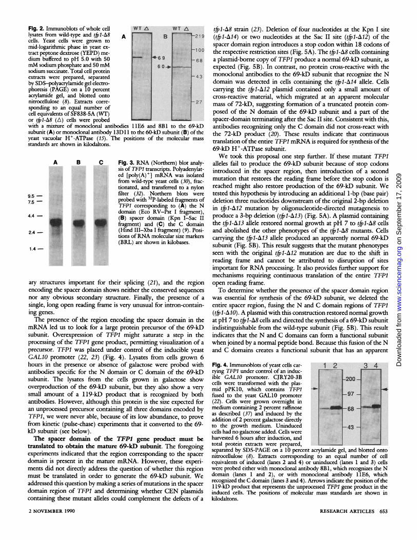
Fig. 2. Immunoblots of whole celllysates from wild-type and tfp1-A8 Acells. Yeast cells were grown tomid-logarithmic phase in yeast ex-tract peptone dextrose (YEPD) me-dium buffered to pH 5.0 with 50mM sodium phosphate and 50 mMsodium succinate. Total cell proteinextracts were prepared, separatedby SDS-polyacrylamide gel electro-phoresis (PAGE) on a 10 percentacrylamide gel, and blotted ontonitrocellulose (8). Extracts corre-sponding to an equal number ofcell equivalents of SF838-5A (WT)or tfjl-A8 (A) cells were probedwith a mixture of monoclonal antibodies
WT AI_
B
-6 96 0O
11E6 and 8B1
WT A-219
-1 00
68'V -
43
27
to the 69-kDsubunit (A) or monoclonal antibody 13D1 1 to the 60-kD subunit (B) of theyeast vacuolar H+-ATPase (15). The positions of the molecular massstandards are shown in kilodaltons.
B C
,
::
__. _-. ._S
0a_j- iE: _EFt
A¢ii i :|0 U> ,BE SC >EQe
0> ;| rS e gys
0'Rt! 04 0Q''YtlE';t'%SS'W''{073:7:
Fig. 3. RNA (Northern) blot analy-sis of TEP1 transcripts. Polyadenylat-ed [poly(A)+] mRNA was isolatedfrom wild-type yeast cells (30), frac-tionated, and transferred to a nylonfilter (32). Northern blots wereprobed with 32P-labeled fragments ofTFPI corresponding to (A) the Ndomain (Eco RV-Pst I fragment),(B) spacer domain (Kpn I-Sac IIfragment) and (C) the C domain(Hind III-Xba I fragment) (9). Posi-tions ofRNA molecular size markers(BRL) are shown in kilobases.
ary structures important for their splicing (21), and the regionencoding the spacer domain shows neither the conserved sequencesnor any obvious secondary structure. Finally, the presence of a
single, long open reading frame is very unusual for intron-contain-ing genes.
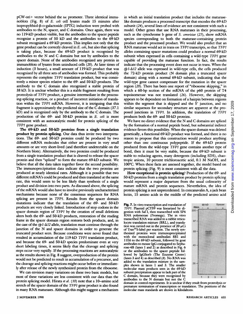
The presence of the region encoding the spacer domain in themRNA led us to look for a large protein precursor of the 69-kDsubunit. Overexpression of TFP1 might saturate a step in theprocessing of the TFP1 gene product, permitting visualization of a
precursor. TFP1 was placed under control of the inducible yeastGAL10 promoter (22, 23) (Fig. 4). Lysates from cells grown 6hours in the presence or absence of galactose were probed withantibodies specific for the N domain or C domain of the 69-kDsubunit. The lysates from the cells grown in galactose showoverproduction of the 69-kD subunit, but they also show a verysmall amount of a 119-kD product that is recognized by bothantibodies. However, although this protein is the size expected foran unprocessed precursor containing all three domains encoded byTFP1, we were never able, because of its low abundance, to prove
from kinetic (pulse-chase) experiments that it converted to the 69-kD subunit (see below).The spacer domain of the TFP1 gene product must be
translated to obtain the mature 69-kD subunit. The foregoingexperiments indicated that the region corresponding to the spacerdomain is present in the mature mRNA. However, these experi-ments did not directly address the question of whether this regionmust be translated in order to generate the 69-kD subunit. Weaddressed this question by making a series ofmutations in the spacer
domain region of TFP1 and determining whether CEN plasmidscontaining these mutant alleles could complement the defects of a
2 NOVEMBER 1990
tfrl-A8 strain (23). Deletion of four nucleotides at the Kpn I site(Qfp1-A4) or two nucleotides at the Sac II site (Qfp1-A12) of thespacer domain region introduces a stop codon within 18 codons ofthe respective restriction sites (Fig. 5A). The tXfrl-A8 cells containinga plasmid-borne copy of TFP1 produce a normal 69-kD subunit, asexpected (Fig. 5B). In contrast, no protein cross-reactive with themonoclonal antibodies to the 69-kD subunit that recognize the Ndomain was detected in cells containing the t(fl-A14 allele. Cellscarrying the tfl-A12 plasmid contained only a small amount ofcross-reactive material, which migrated at an apparent molecularmass of 72-kD, suggesting formation of a truncated protein com-posed of the N domain of the 69-kD subunit and a part of thespacer-domain terminating after the Sac II site. Consistent with this,antibodies recognizing only the C domain did not cross-react withthe 72-kD product (20). These results indicate that continuoustranslation ofthe entire TFP1 mRNA is required for synthesis ofthe69-kD H+-ATPase subunit.We took this proposal one step further. If these mutant TFP1
alleles fail to produce the 69-kD subunit because of stop codonsintroduced in the spacer region, then introduction of a secondmutation that restores the reading frame before the stop codon isreached might also restore production of the 69-kD subunit. Wetested this hypothesis by introducing an additional 1-bp (base pair)deletion three nucleotides downstream of the original 2-bp deletionin tfl-A12 mutation by oligonucleotide-directed mutagenesis toproduce a 3-bp deletion (Qfp1-A13) (Fig. 5A). A plasmid containingthe tf[pl-A13 allele restored normal growth at pH 7 to tf[il-A8 celsand abolished the other phenotypes of the t([il-A8 mutants. Cellscarrying the t([il-A13 allele produced an apparently normal 69-kDsubunit (Fig. 5B). This result suggests that the mutant phenotypesseen with the original t([pl-A12 mutation are due to the shift inreading frame and cannot be attributed to disruption of sitesimportant for RNA processing. It also provides further support formechanisms requiring continuous translation of the entire TFP1open reading frame.To determine whether the presence of the spacer domain region
was essential for synthesis of the 69-kD subunit, we deleted theentire spacer region, fusing the N and C domain regions of TFP1(t[1-Al0). A plasmid with this construction restored normal growthat pH 7 to t(p1-A8 cells and directed the synthesis ofa 69-kD subunitindistinguishable from the wild-type subunit (Fig. 5B). This resultindicates that the N and C domains can form a fimctional subunitwhen joined by a normal peptide bond. Because this fusion of the Nand C domains creates a functional subunit that has an apparent
Fig. 4. Immunoblots of yeast cells car- 1 2 3 4rying TFP1 under control of an induc-ible GAL10 promoter. CJRY20-3Bcells were transformed with the plas-mid pPK1O, which contains TFP1fused to the yeast GALIO promoter 97(22). Cells were grown overnight inmedium containing 2 percent raffinoseas described (37) and induced by theaddition of 2 percent galactose directlyto the growth medium. Uninduced -43cells had no galactose added. Cells wereharvested 6 hours after induction, andtotal protein extracts were prepared,separated by SDS-PAGE on a 10 percent acrylamide gel, and blotted ontonitrocellulose (8). Extracts corresponding to an equal number of cellequivalents of induced (lanes 2 and 4) or uninduced (lanes 1 and 3) cellswere probed either with monoclonal antibody 8B1, which recognizes the Ndomain (lanes 1 and 2), or with monoclonal antibody 11E6, whichrecognized the C domain (lanes 3 and 4). Arrows indicate the position ofthe1 19-kD product that represents the unprocessed TFP1 gene product in theinduced cells. The positions of molecular mass standards are shown inkilodaltons.
RESEARCH ARTICLES 653
A
9.5 -7.5 -
4. -
2-
2.4 -
1.4 -
X,i..
on
Sep
tem
ber
17, 2
009
ww
w.s
cien
cem
ag.o
rgD
ownl
oade
d fr
om
molecular mass identical to the wild-type protein, our proposedprimary structure for the 69-kD subunit, based on homology to theNeurospora vma-1 gene, must be reasonably close to the primarystructure of the actual protein.These observations prompted us to investigate whether the spacer
domain region of TFP1 encoded a stable protein. We injectedrabbits with a peptide from the predicted amino acid sequence ofthespacer domain (15), and then used the resulting antibodies to probeimmunoblots of wild-type cells and (f[pl-A8 cells carrying variousplasmids. The specific antibodies to the peptide recognized a proteinof -50-kD in both wild-type cells and tf[pl-A8 cells carrying a CEN-TFP1 plasmid (Fig. 5C). This protein was missing in tffil-A8 cellsand in the tff1-A0 cells. The antibodies recognized the same proteinof -72-kD that cross-reacted with the antibodies to the 69-kDsubunit (N domain) in the tfpl-A12 cells, consistent with theformation of a truncated protein from this construction. Theseresults indicate that the spacer domain region of TFP1 encodes afairly abundant and stable protein. However, the immunoblots maynot accurately reflect the relative proportions of the 69-kD (ATPasesubunit) and 50-kD (spacer) products and would not reveal tran-sient precursors in the formation of these products.The biosynthesis of the two protein products from TFP1 was then
examined. Cells were labeled for 10 minutes with [35S]methionineand [35S]cysteine, and either lysed immediately or incubated for 10minutes more in the presence of unlabeled methionine and cysteinebefore lysis (Fig. 6). The newly synthesized proteins were immuno-precipitated with either the antibodies to the 69-kD subunit or theantibodies to the spacer peptide. The similar amounts of proteinprecipitated by the two different sets of antibodies indicate that the69- and 50-kD proteins are produced at similar rates. Both productsmust be fairly stable because there was no obvious loss of productafter the 10-minute incubation period. No full-length (1 19-kD)translation product was detected in this experiment or with labelingtimes as short as 2 minutes. Similar time-labeling studies on yeastcells that had been induced to overexpress TFP1 revealed a minoramount of a 119-kD product relative to the 69-kD product (20).
Fig. 5. (A) Mutations at Athe Sac II site. High- TGC CGC GGA TTT TAT JJ TCT ATlighted nucleotides cor-respond to nucleotides
Saedo i
removed in the two dif- Kpn I Spacer domain Sac IIferent mutations, the B cbreak (ff) covers 48 nu- 1 2 3 4 5 6 1 2 3 4 5 6cleotides, and the stop 219- 219codon brought intoframe by the 2-bp dele- loo- l-100tion is underlined. (Band C) Immunoblots of 68N_ _ _.cells containing TFPIspacer domain muta- 43 - 3tions on CEN plasmids.(jp1-A8 cells were trans-formed by the lithium 2 -27acetate method (29).Transformed cells wereselected and maintained 18- -18on supplemented SD 1 2 3 4 5 6 2 3 4 5 6medium lacking uracil.Whole cell lysates were prepared, subjected to SDS-PAGE, and blotted asdescribed (Fig. 2). Blots were probed with a mixture of monoclonalantibodies 8B1 and 11E6 to the 69-kD subunit (B) and with the antibodiesto the spacer peptide (15) (C). An identical blot probed with the monoclonalantibody to the 60-kD subunit showed an equal amount of protein of theexpected molecular size in all the samples (20). Cells were transformed withthe following plasmids: (lane 1) pDW21 (CEN-TFP1); (lane 2) pRS316(vector only); (lane 3) ptfpl-AIO (spacer A); (lane 4) ptfpl-A12 (Sac II A2);(lane 5) ptfpl-A13 (Sac II A2 + Al); (lane 6) ptfpl-A14 (Kpn I A4). Thepositions of molecular mass standards are shown in kilodaltons.
654
The proportions of the two products were similar to those seen atsteady-state in Fig. 4, even at short labeling times, and it thus wasnot possible to establish whether the 1 19-kD "precursor" convertedto the 69-kD and 50-kD proteins with time because it represented asmall percentage of the immunoprecipitated protein.
Protein splicing of the TFPI gene product in heterologouscontexts. To determine whether production of the 69- and 50-kDproteins from the TFP1 mRNA requires factors specific to yeast, weattempted to duplicate this process in vitro. TFP1 was clonedbehind an SP6 promoter, and RNA was synthesized in vitro by theSP6 RNA polymerase. The synthesized RNA was then added to arabbit reticulocyte lysate translation system containing [35S]meth-ionine and [35S]cysteine, and the products were immunoprecipitat-ed with either the antibodies to the 69-kD subunit or the antibodiesto the spacer peptide. The antibodies to the 69-kD subunit immu-noprecipitated a 69-kD product and antibodies to the spacer peptideprecipitated a similar amount of 50-kD product (Fig. 7). Theappearance of these two products was absolutely dependent onaddition of the TFPI RNA because nothing was precipitated byeither antibody in the absence ofRNA. As in the immunoprecipita-tions of TFP1 products synthesized in vivo (Fig. 6), no full-length119-kD translation product was seen. Similar results were obtainedwith mRNA isolated from yeast (20). These results indicate that (i)there is nothing specific to the yeast mRNA that is required forproduction of the 69- and 50-kD proteins and (ii) either the cellularmachinery for synthesis and splicing of the 69- and 50-kD proteinsis preserved in mammals and present in the reticulocyte translationmixture or the process of forming the 69- and 50-kD productsrequires no cellular machinery and is therefore autocatalytic.We used TFP1 expression in E. coli to determine how the gene
product would be handled in bacteria. TFP1 was cloned into the
Fig. 6. Synthesis of the 69-kD ATPase 10 min pulsesubunit and the 50-kD spacer protein. 10minpulse 10minchaseSF838-5A cells were grown to mid-loga- 200rithmic phase in supplemented SD medi-um lacking methionine. Cells were con-verted to spheroplasts with zymolyase 97LOOT (ICN Biochemicals) as described(38). Spheroplasts were resuspended at adensity of 3.3 x 10' per milliliter in 68growth medium containing 1.2 M sorbi- 4tol and incubated with 200* Ci ofTran -5S-label (ICN Biochemical) per107 cells at 30°C for 10 minutes (pulse).
43
At the end of the labeling period, half ofthe culture was combined with an equalvolume of ice-cold 20 percent trichloro-acetic acid (TCA) (lanes 1 and 2). Unla-
2
beled methionine and cysteine were add- 26ed to the other half of the culture to afinal concentration of 0.3 mg/mi and theincubation was continued for an addi-tional 10 minutes (lanes 3 and 4), fol- 2 3 4lowed by TCA precipitation (chase). TheTCA-precipitate was washed once in acetone and solubilized in 0.9 M tris-HCI (pH 8.0), 1 percent SDS, 1 percent Triton X-100, and 20 mM EDTA(immunoprecipitation buffer) at 100°C. The solubilized pellets were diluted(1:10), then immunoprecipitated with either a mixture of monoclonalantibodies 8B1 and 11E6 (lanes 2 and 4) or the antibodies to the spacerpeptide (lanes 1 and 3). For the spacer peptide antibody samples, the initialincubation with primary antibody was followed by incubations with goatantibody to rabbit immunoglobulin G (IgG) and then mouse antibody togoat IgG (both from Jackson Immunobiologicals) to increase the stability ofthe immune complexes. For all of the immunoprecipitations, the immunecomplexes were precipitated with goat antibody to mouse IgG conjugated toSepharose-4B (Organon-Teknika). The precipitated samples were washedtwice in immunoprecipitation buffer without SDS, then analyzed by SDS-PAGE and fluorography as described (39). The positions of molecular massstandards are shown in kilodaltons.
SCIENCE, VOL. 250
on
Sep
tem
ber
17, 2
009
ww
w.s
cien
cem
ag.o
rgD
ownl
oade
d fr
om
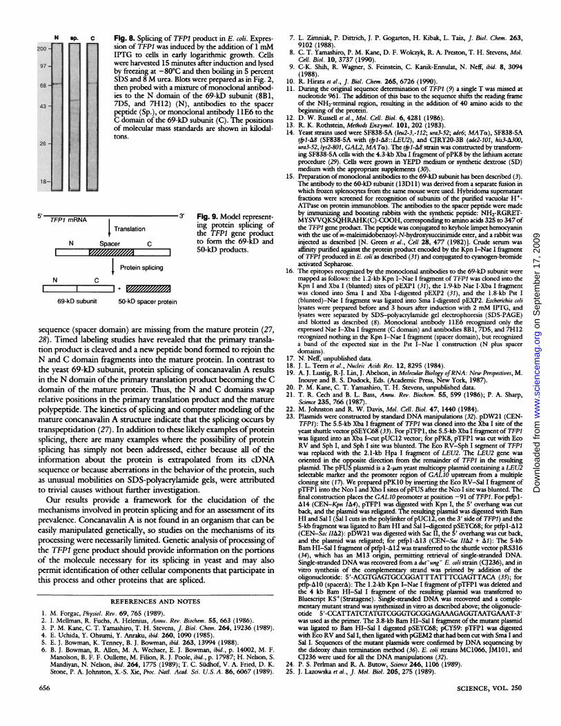
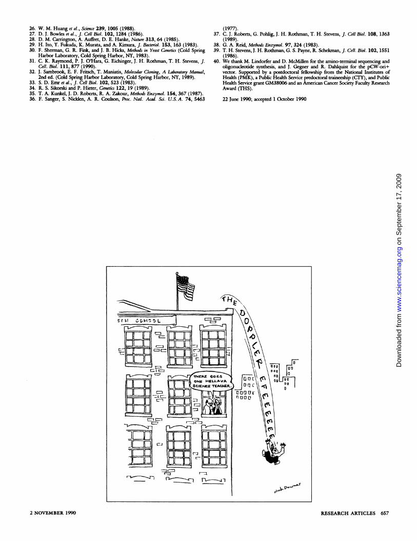
pCW-ori+ vector behind the tac promoter. Three identical immu-noblots (Fig. 8) of E. coli cell lysates made 15 minutes afterisopropylthiol-o-D-galactoside (IPTG) induction were probed withantibodies to the N, spacer, and C domains. Once again, there wasno 1 19-kD product visible, but the antibodies to the spacer peptiderecognize a protein of 50 kD and the antibodies to the 69-kDsubunit recognized a 69-kD protein. This implies not only that thegene product can be correctly cleaved in E. coli, but also that splicingis taking place, because the 69-kD product is recognized byantibodies to the N and C domains but not by antibodies to thespacer domain. None of the antibodies recognized any protein inimmunoblots of lysates from uninduced cells (20). At later times ofinduction (3 hours), a small amount of 119-kD product that wasrecognized by all three sets of antibodies was formed. This probablyrepresents the complete TFP1 translation product, but was consis-tently a minor species relative to the 69- and 50-kD products. Theantibody to the C domain also recognized a stable protein of38 kD. It is unclear whether this is a stable fragment resulting fromproteolysis of TFP1 product, a genuine intermediate in the process-ing-splicing reaction, or a product of incorrect initiation of transla-tion within the TFP1 mRNA. However, it is intriguing that thisfragment is approximately the predicted size of the C domain (37.1kD) and is recognized only by the antibody to the C domain. Theproduction of the 69- and 50-kD proteins in E. coli is moreconsistent with an autocatalytic model for protein splicing of theTFPI gene product.The 69-kD and 50-kD proteins from a single translation
product by protein splicing. Our data thus invite two interpreta-tions. The 69- and 50-kD proteins could be produced by twodifferent mRNA molecules that either are present in very smallamounts or are very short-lived (and therefore undetectable on theNorthern blots). Altematively, the two proteins could be producedfrom a single translation product that is cleaved to release the spacerprotein and then "spliced" to form the mature 69-kD subunit. Webelieve that all the data taken together favor the second possibility.The immunoprecipitation data indicate that the two proteins areproduced at nearly identical rates. Although it is possible that twodifferent mRNA's could be produced and then translated at the samerate, this would seem to be less likely than synthesis of a singleproduct and division into two parts. As discussed above, the splicingofthe mRNA would also have to involve previously uncharacterizedmechanisms because none of the consensus sequences for RNAsplicing are present in TFP1. Results from the spacer domainmutations indicate that the translation of the 69- and 50-kDproducts are very closely linked. Introduction of stop codons in thespacer domain region of TFP1 by the creation of small deletionsalters both the 69- and 50-kD products, restoration of the readingframe in the spacer domain region restores both products, and, inthe case ofthe tfpl-A12 allele, translation must continue through thejunction of the N and spacer domains in order to generate thetruncated product seen. Because conditions were never found thatresulted in accumulation of the 119-kD TFP1 translation product,and because the 69- and 50-kD species predominate even at veryshort labeling times, it seems likely that the cleavage and splicingsteps occur very rapidly. If the processing reactions are autocatalyticas the results shown in Fig. 8 suggest, overproduction of the proteinwould not be predicted to result in accumulation ofa precursor, andthe cleavage and splicing reactions might occur before or immediate-ly after release of the newly synthesized protein from the ribosome.We can envision many variations on these two basic models, but
most of these variations are less consistent with our data than theprotein splicing model. Hirata et al. (10) note that a 10-amino acidstretch of the spacer domain ofthe TFP1 gene product is also foundin many RNA maturases. Although this might suggest a mechanism
in which an initial translation product that includes the maturase-like domain produces a processed transcript that encodes the 69-kDsubunit (24), several lines of evidence are not consistent with such amodel. Other genes that use RNA maturases in their processing,such as the cytochrome b gene of S. cerevisiae (25), show mRNAspecies corresponding to both the maturase-containing proteinproduct and the processed product. We would also predict that anRNA maturase would act in trans on TFPI transcripts, so that TFP1alleles containing spacer mutations could produce a normal 69-kDsubunit when expressed in cells containing a wild-type TFP1 genecapable of providing the maturase function. In fact, the resultsindicate that the processing event does not occur in trans. When the!fpl-A12 allele was expressed in wild-type cels, the cells producedthe 72-kD protein product (N domain plus a truncated spacerdomain) along with a normal 69-kD subunit, indicating that thewild-type gene cannot compensate for the mutation in the spacerregion (20). There has been one report of "ribosome skipping," inwhich a 60-bp section of the mRNA of the p60 protein of T4topoisomerase was not translated (26). However, the skippingappears to be dependent on formation of a hairpin between regionswithin the segment that is skipped and the 5' junction, and nosimilar sequences for secondary structure are apparent at the pro-posed junctions in TFP1. In addition, the translation of TFPIproduces both the 69- and 50-kD proteins.We have no direct evidence that the N and C domains are spliced
by the formation of a normal peptide bond, but substantial indirectevidence favors this possibility. When the spacer domain was deletedgenetically, a functional 69-kD product was formed, and there is noreason to propose that this construction would produce anythingother than one continuous polypeptide. If the 69-kD proteinproduced from the wild-type TFP1 gene contains another type ofbond, then it must be very stable, because the 69-kD subunit isstable to reducing agents, many detergents (including SDS), chao-tropic anions, 10 percent trichloroacetic acid, 0.1 M NaOH, and100°C. When these facts are assessed together, the model based onprotein splicing (Fig. 9) is most consistent with all the data.How exceptional is protein splicing? Production ofthe 69- and
50-kD proteins from a single translation product by protein splicingwould represent a major departure from the usual colinearity ofmature mRNA and protein sequences. Nevertheless, the idea ofprotein splicing is not unprecedented. In concanavalin A, a jack beanlectin, 15 amino acids in the middle of the predicted amino acid
Fig. 7. In vitro transcription and translation of 69 kD SpacerTFP1. Plasmid pCY59 was linearized by di- RNA: - + +gestion with Sal I, then transcribed with SP6RNA polymerase (Promega). The in vitrotranscribed RNA was added to a rabbit reticu-locyte translation mixture (BRL), and transla-tion was carried out in the presence of20 RCiof Tran35S-label per reaction. The newly syn-n68thesized proteins were immunoprecipitatedwith the monoclonal antibodies 8B1 and11E6 to the 69-kD subunit, followed by goatantibodies to mouse IgG conjugated to Sepha- _rose-4B (lanes 1 and 2) as described in Fig. 6or the antibodies to the spacer peptide fol-lowed by IgGSorb (The Enzyme Center)(lanes 3 and 4) as described (8). No RNA wasadded to the translation mixture in the sam-ples shown in lanes 1 and 3. The smallermolecular mass products seen in the 69-kDsubunit precipitation appear to lack part oftheC domain, because they were recognized by 1 2 3 4antibodies to the N domain but not the Cdomain in control experiments. It is unclear if they result from proteolysis orpremature termination of transcription or translation. The positions of themolecular mass standards are shown in kilodaltons.
RESEARCH ARTICLES 6552 NOVEMBER 1990
on
Sep
tem
ber
17, 2
009
ww
w.s
cien
cem
ag.o
rgD
ownl
oade
d fr
om
sph:v
::D
-
:::
:: :::
:s.
aDES: Vi
zi
.LS:jzS: ':?
.,l: .,i'....
!:.:,:' :,'::'X: -y%:
'..i.'th.g
:'
di.D:
:E::
i:':
_
c Fig. 8. Splicing of TFP1 product in E. coli. Expres-sion of TFP1 was induced by the addition of 1 mMIPTG to cells in early logarithmic growth. Cellswere harvested 15 minutes after induction and lysedby freezing at -80°C and then boiling in 5 percentSDS and 8 M urea. Blots were prepared as in Fig. 2,then probed with a mixture ofmonoclonal antibod-ies to the N domain of the 69-kD subunit (8B1,7D5, and 7H12) (N), antibodies to the spacerpeptide (Sp.), or monoclonal antibody 1 1E6 to theC domain of the 69-kD subunit (C). The positionsof molecular mass standards are shown in kilodal-tons.
TFPI mRNAT Translation
N Spacer CI VY//M /S//// I
j Protein splicing
N C
- 3' Fig. 9. Model represent-ing protein splicing ofthe TFP1 gene productto form the 69-kD and50-kD products.
I ~ + IYKO//M/fMS69-kD subunit 50-kD spacer protein
sequence (spacer domain) are missing from the mature protein (27,28). Timed labeling studies have revealed that the primary transla-tion product is cleaved and a new peptide bond formed to rejoin theN and C domain fragments into the mature protein. In contrast tothe yeast 69-kD subunit, protein splicing of concanavalin A resultsin the N domain of the primary translation product becoming the Cdomain of the mature protein. Thus, the N and C domains swap
relative positions in the primary translation product and the mature
polypeptide. The kinetics of splicing and computer modeling of themature concanavalin A structure indicate that the splicing occurs bytranspeptidation (27). In addition to these likely examples of proteinsplicing, there are many examples where the possibility of proteinsplicing has simply not been addressed, either because all of theinformation about the protein is extrapolated from its cDNAsequence or because aberrations in the behavior of the protein, suchas unusual mobilities on SDS-polyacrylamide gels, were attributedto trivial causes without further investigation.Our results provide a framework for the elucidation of the
mechanisms involved in protein splicing and for an assessment of itsprevalence. Concanavalin A is not found in an organism that can beeasily manipulated genetically, so studies on the mechanisms of itsprocessing were necessarily limited. Genetic analysis ofprocessing ofthe TFP1 gene product should provide information on the portionsof the molecule necessary for its splicing in yeast and may alsopermit identification ofother cellular components that participate inthis process and other proteins that are spliced.
REFERENCES AND NOTES
1. M. Forgac, Physiol. Rev. 69, 765 (1989).2. I. Mellman, R. Fuchs, A. Helenius, Annu. Rev. Biochem. 55, 663 (1986).3. P. M. Kane, C. T. Yamashiro, T. H. Stevens, J. Biol. Chem. 264, 19236 (1989).4. E. Uchida, Y. Ohsumi, Y. Anraku, ibid. 260, 1090 (1985).5. E. J. Bowman, K. Tenney, B. J. Bowman, ibid. 263, 13994 (1988).6. B. J. Bowman, R. Allen, M. A. Wechser, E. J. Bowman, ibid., p. 14002, M. F.
Manolson, B. F. F. Oullette, M. Filion, R. J. Poole, ibid., p. 17987; H. Nelson, S.Mandiyan, N. Nelson, ibid. 264, 1775 (1989); T. C. Sudhof, V. A. Fried, D. K.Stone, P. A. Johnston, X.-S. Xie, Proc. Natl. Acad. Sci. U.S.A. 86, 6067 (1989).
7. L. Zimnniak, P. Dittrich, J. P. Gogarten, H. Kibak, L. Taiz, J. Biol. Chem. 263,9102 (1988).
8. C. T. Yamashiro, P. M. Kane, D. F. Wokzyk, R. A. Preston, T. H. Stevens, Mol.Cell. Biol. 10, 3737 (1990).
9. C-K. Shih, R. Wagner, S. Feinstein, C. Kanik-Ennulat, N. Neff, ibid. 8, 3094(1988).
10. R. Hirata et al., J. Biol. Chem. 265, 6726 (1990).11. During the original sequence determination of TFPI (9) a single T was missed at
nucleotide 961. The addition of this base to the sequence shifts the reading frameof the NH2-terminal region, resulting in the addition of 40 amino acids to thebeginning of the protein.
12. D. W. Russell et al., Mol. Cell. Biol. 6, 4281 (1986).13. R. K. Rothstein, Methods Enzymol. 101, 202 (1983).14. Yeast strains used were SF838-5A (leu2-3,-112; ura3-52; ade6; MATa), SF838-5A
tfp1-,&8 (SF838-SA with (f41-A8::LEU2), and CJRY2O-3B (ade2-101, his3-A300,ura3-52, lys2-801, GAL2, MATa). The !fp1-A8 strain was constructed by transform-ing SF838-5A cells with the 4.3-kb Xba I fragment ofpPK8 by the lithium acetateprocedure (29). Cells were grown in YEPD medium or synthetic dextrose (SD)medium with the appropriate supplements (30).
15. Preparation of monoclonal antibodies to the 69-kD subunit has been described (3).The antibody to the 60-kD subunit (13D11) was derived from a separate fusion inwhich frozen splenocytes from the same mouse were used. Hybridoma supematantfractions were screened for recognition of subunits of the purified vacuolar H+-ATPase on protein immunoblots. The antibodies to the spacer peptide were madeby inunizing and boosting rabbits with the synthetic peptide: NH2-RGRET-MYSVVQKSQHRAHK(C)-COOH, corresponding to amino acids 325 to 347 ofthe TFP1 gene product. The peptide was conjugated to keyhole limpet hemocyaninwith the use of m-maleimidobenzoyl-N-hydroxysuccinimide ester, and a rabbit wasinjected as described [N. Green et al., Cell 28, 477 (1982)]. Crude serum wasaffinity purified against the protein product encoded by the Kpn I-Nae I fragmentof TFP1 produced in E. coli as described (31) and conjugated to cyanogen-bromideactivated Sepharose.
16. The epitopes recognized by the monoclonal antibodies to the 69-kD subunit weremapped as follows: the 1.2-kb Kpn I-Nae I fragment of TFP1 was cloned into theKpn I and Xba I (blunted) sites of pEXP1 (31), the 1.9-kb Nae I-Xba I fragmentwas cloned into Sma I and Xba I-digested pEXP2 (31), and the 1.8-kb Pst I(blunted)-Nae I fragment was ligated into Sma I-digested pEXP2. Escherichia colilysates were prepared before and 3 hours after induction with 2 mM IPTG, andlysates were separated by SDS-polyacrylamide gel electrophoresis (SDS-PAGE)and blotted as described (8). Monoclonal antibody 11E6 recognized only theexpressed Nae I-Xba I fragment (C domain) and antibodies 8B1, 7D5, and 7H12recognized nothing in the Kpn I-Nae I fragment (spacer domain), but recognizeda band of the expected size in the Pst I-Nae I construction (N plus spacerdomains).
17. N. Neff, unpublished data.18. J. L. Teem et al., Nucleic Acids Res. 12, 8295 (1984).19. A. J. Lustig, R-J. Lin, J. Abelson, in Molecular Biology ofRNA: New Perspectives, M.
Inouye and B. S. Dudock, Eds. (Academic Press, New York, 1987).20. P. M. Kane, C. T. Yarnashiro, T. H. Stevens, unpublished data.21. T. R. Cech and B. L. Bass, Annu. Rev. Biochem. 55, 599 (1986); P. A. Sharp,
Science 235, 766 (1987).22. M. Johnston and R. W. Davis, Mol. Cell. Biol. 47, 1440 (1984).23. Plasmids were constructed by standard DNA manipulations (32). pDW21 (CEN-
TFPI): The 5.5-kb Xba I fragment of TFPI was doned into the Xba I site of theyeast shuttle vector pSEYC68 (33). For pTFP1, the 5.5-kb Xba I fragment of TFP1was ligated into an Xba I-cut pUC12 vector; for pPK8, pTFP1 was cut with EcoRV and Sph I, and Sph I site was blunted. The Eco RV-Sph I segment of TFPIwas replaced with the 2.1-kb Hpa I fragment of LEU2. The LEU2 gene wasoriented in the opposite direction from the remainder of TFPI in the resultingplasmid. The pFUS plasmid is a 2-pLm yeast multicopy plasmid containing a LEU2selectable marker and the promoter region of GAL1O upstream from a multiplecloning site (17). We prepared pPK10 by inserting the Eco RV-Sal I fragment ofpTFP1 into the Nco Iand Xho I sites of pFUS after the Nco I site was blunted. Thefinal construction places the GAL10 promoter at position -91 of TFPI. For ptfp1-A14 (CEN-Kpn 1A4), pTFP1 was digested with Kpn I, the 5' overhang was cutback, and the plasmid was religated. The resulting plasmid was digested with BamHI and Sal I (Sal I cuts in the polylinker ofpUC12, on the 3' side of TFPI) and the5-kb fragment was ligated to Bam HI and Sal I-digested pSEYC68; for ptfpl-A12(CEN-Sac 1IA2): pDW21 was digested with Sac II, the 5' overhang was cut back,and the plasmid was religated; for ptfpl-A13 (CEN-Sac IIA2 + Al): The 5-kbBarn HI-Sal I fragment ofptfpl-A12 was transferred to the shuttle vector pRS316(34), which has an M13 origin, permitting retrieval of single-stranded DNA.Single-stranded DNA was recovered from a dut-ungj E. coli strain (CJ236), and invitro synthesis of the complemcntary strand was primed by addition of theoligonucleotide: 5'-ACGTGAGTGCCGGATTTATTTCGAGTTACA (35); forptfp-A1O (spacer/v): The 1.2-kb Kpn I-Nae I fragment of pTFP1 was deleted andthe 4 kb Bam HI-Sal I fragment of the resulting plasmid was transferred toBluescript KS+(Stratagene). Single-stranded DNA was recovered and a comple-mentary mutant strand was synthesized in vitro as described above; the oligonucle-otide 5'-CCATTATCIATGTCGGGTGCGGAGAAAGAGGTAATGAAAT-3'was used as the primer. The 3.8-kb Bam HI-Sal I fragment of the mutant plasmidwas ligated to Bam HI-Sal I digested pSEYC68; pCY59: pTFP1 was digestedwith Eco RV and Sal I, then ligated with pGEM2 that had been cut with Sma IandSal I. Sequences of the mutant plasmids were confirmed by DNA sequencing bythe dideoxy chain termination method (36). E. coli strains MC1066, JM1IO, andCJ236 were used for all the DNA manipulations (32).
24. P. S. Perlnan and R. A. Butow, Science 246, 1106 (1989).25. J. Lazowska et al., J. Mol. Biol. 205, 275 (1989).
SCIENCE,
200 -
97 -
68 -
43.
26-
18-
N
656
on
Sep
tem
ber
17, 2
009
ww
w.s
cien
cem
ag.o
rgD
ownl
oade
d fr
om
26. W. M. Huang et al., Science 239, 1005 (1988).27. D. J. Bowles et al., J. Cell Biol. 102, 1284 (1986).28. D. M. Carrington, A. Auffret, D. E. Hanke, Nature 313, 64 (1985).29. H. Ito, Y. Fukuda, K. Murata, and A. Kimura, J. Bacteriol. 153, 163 (1983).30. F. Sherman, G. R. Fink, and J. B. Hicks, Methods in Yeast Genetics (Cold Spring
Harbor Laboratory, Cold Spring Harbor, NY, 1983).31. C. K. Raymond, P. J. O'Hara, G. Eichinger, J. H. Rothman, T. H. Stevens, J.
Cell. Biol. 111, 877 (1990).32. J. Sambrook, E. F. Fritsch, T. Maniatis, Molecular Cloning, A Laboratory Manual,
2nd ed. (Cold Spring Harbor Laboratory, Cold Spring Harbor, NY, 1989).33. S. D. Emr et al., J. Cell Biol. 102, 523 (1983).34. R. S. Sikorski and P. Hieter, Genetics 122, 19 (1989).35. T. A. Kunkel, J. D. Roberts, R. A. Zakour, Methods Enzymol. 154, 367 (1987).36. F. Sanger, S. Nicklen, A. R. Coulson, Proc. Natl. Acad. Sd. U.S.A. 74, 5463
(1977).37. C. J. Roberts, G. Pohlig, J. H. Rothman, T. H. Stevens, J. Cell Biol. 108, 1363
(1989).38. G. A. Reid, Methods Enzymol. 97, 324 (1983).39. T. H. Stevens, J. H. Rothman, G. S. Payne, R. Schekunan, J. Cell. Biol. 102, 1551
(1986).40. We thank M. Lindorfer and D. McMillen for the amino-termiinal sequencing and
oligonucleotide synthesis, and J. Gegner and R. Dahlquist for the pCW-ori+vector. Supported by a postdoctoral fellowship from the National Institutes ofHealth (PMK), a Public Health Service predoctoral traineeship (CIY), and PublicHealth Service grant GM38006 and an American Cancer Society Faculty ResearchAward (THS).
22 June 1990; accepted 1 October 1990
RESEARCH ARTICLES 6572 NOVEMBER 1990
on
Sep
tem
ber
17, 2
009
ww
w.s
cien
cem
ag.o
rgD
ownl
oade
d fr
om