Embed Size (px)
Citation preview
Sports Related Injuries of the Hand, Wrist and Elbow
Melissa Nayak, M.D.Department of OrthopaedicsDivision of Sports Medicine

Injury triage• History, mechanism of injury (MOI)• Assess extent of swelling, pain, deformity,
functional deficits & neurovascular status– Determine if xray needed (fracture)– Determine if stabilization needed (dislocation,
deformity)• Disposition
– Can you safely treat and manage?– Is ER evaluation needed?
• open fracture, deformity, dislocation, neurovascular compromise
• sling/splint and refer
Sprains/Strains
• Can be managed on site• R.I.C.E.• NSAIDs, analgesics• Short-term reassessment • Most progressively improve w/in 1-3 wks• Home rehab exercise program
Be prepared
• Supplies – finger splints (stax), aluminum foam splints– athletic tape, coban (self-adherent wrap),
elastic bandage – wrist braces (regular & thumb spica)– slings– misc: trauma shears (scissors), ring cutter
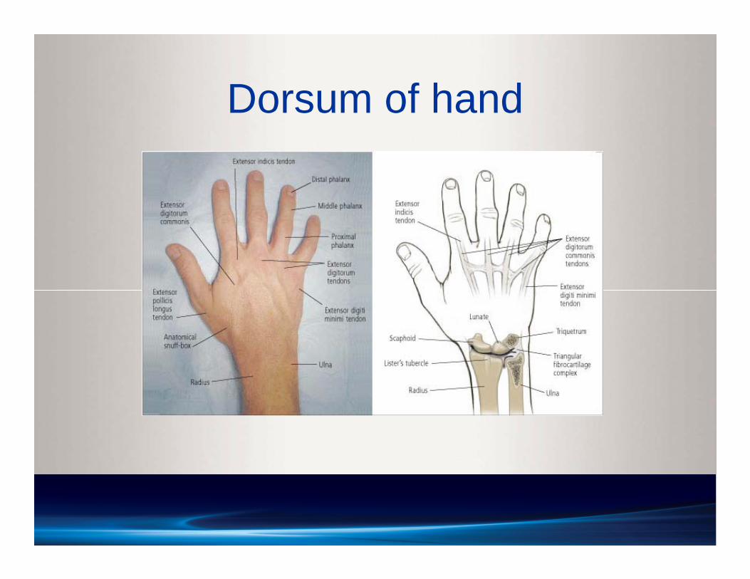
Dorsum of hand
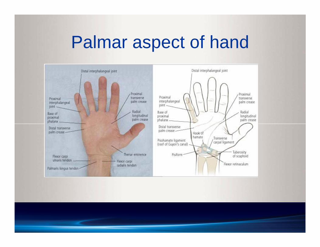
Palmar aspect of hand
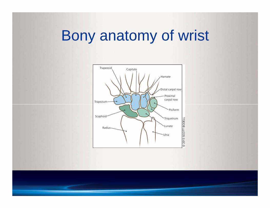
Bony anatomy of wrist
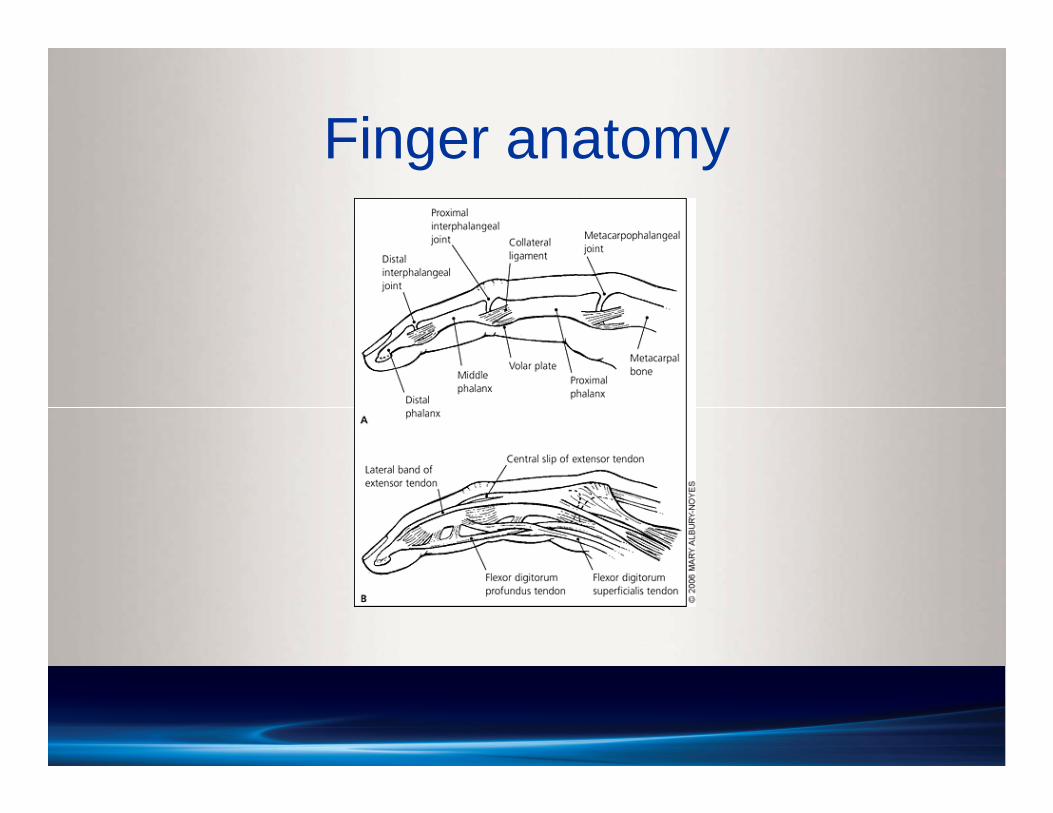
Finger anatomy
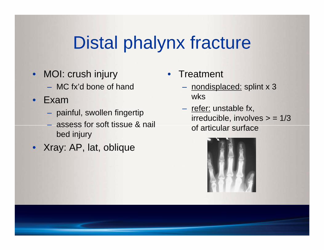
Distal phalynx fracture• MOI: crush injury
– MC fx’d bone of hand
• Exam– painful, swollen fingertip– assess for soft tissue & nail
bed injury
• Xray: AP, lat, oblique
• Treatment– nondisplaced: splint x 3
wks– refer: unstable fx,
irreducible, involves > = 1/3 of articular surface
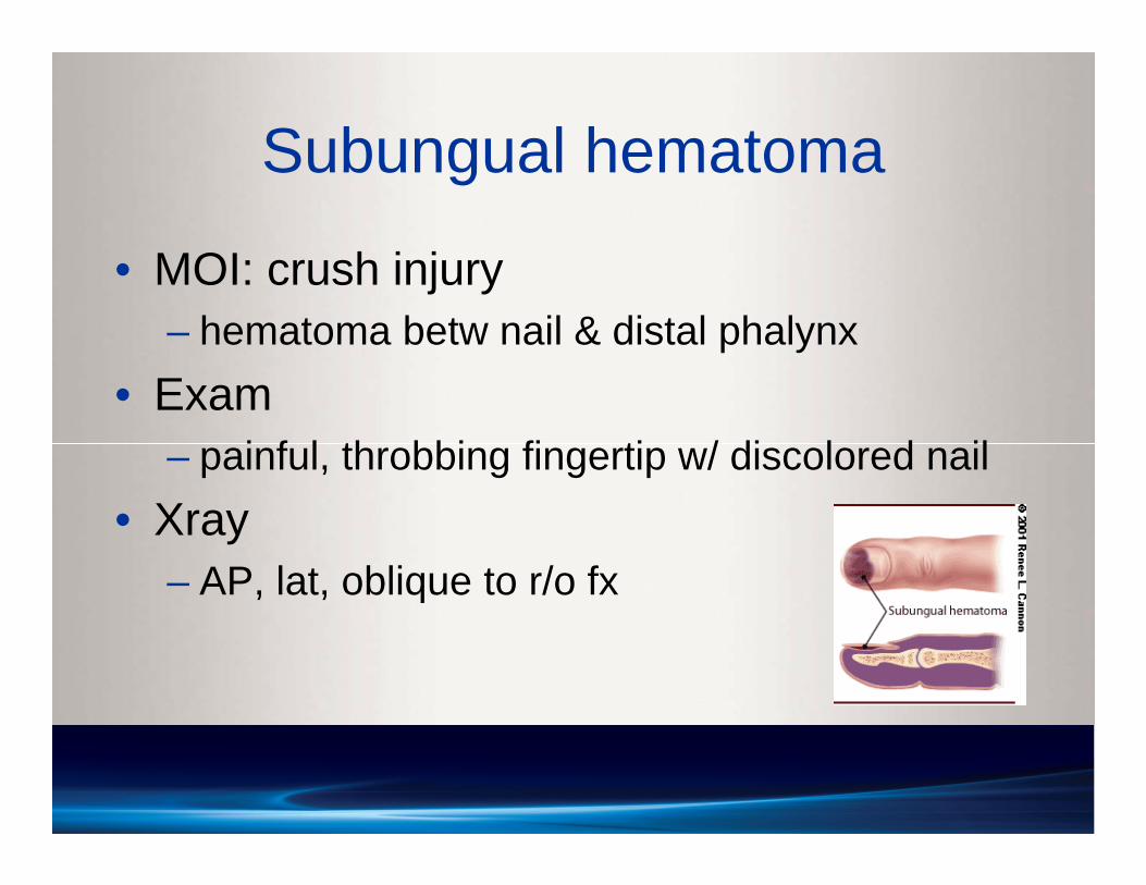
Subungual hematoma
• MOI: crush injury– hematoma betw nail & distal phalynx
• Exam– painful, throbbing fingertip w/ discolored nail
• Xray– AP, lat, oblique to r/o fx
Treatment: Subungual hematoma
• Subungual decompression– drainage via 2-3 small holes in nail w/ cautery
tip or heated paperclip • Splint until tenderness resolves
– large hematoma (>= 50%) likely nail bed laceration
• may need nail removal/nail bed suturing• full growth of new nail ~5-6 months
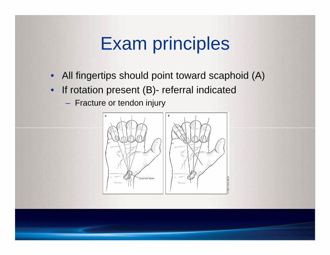
Exam principles• All fingertips should point toward scaphoid (A)• If rotation present (B)- referral indicated
– Fracture or tendon injury
Collateral ligament injuries
• MOI: jammed finger– forced ulnar or radial deviation at any IP joint
• Exam– pain at ligament– w/ MCPJ flexed at 90o, apply varus/valgus
stress in 30o flexion • compare laxity w/ unaffected finger
• Xray: AP, lat, oblique– normal, or avulsion fx at ligament insertion
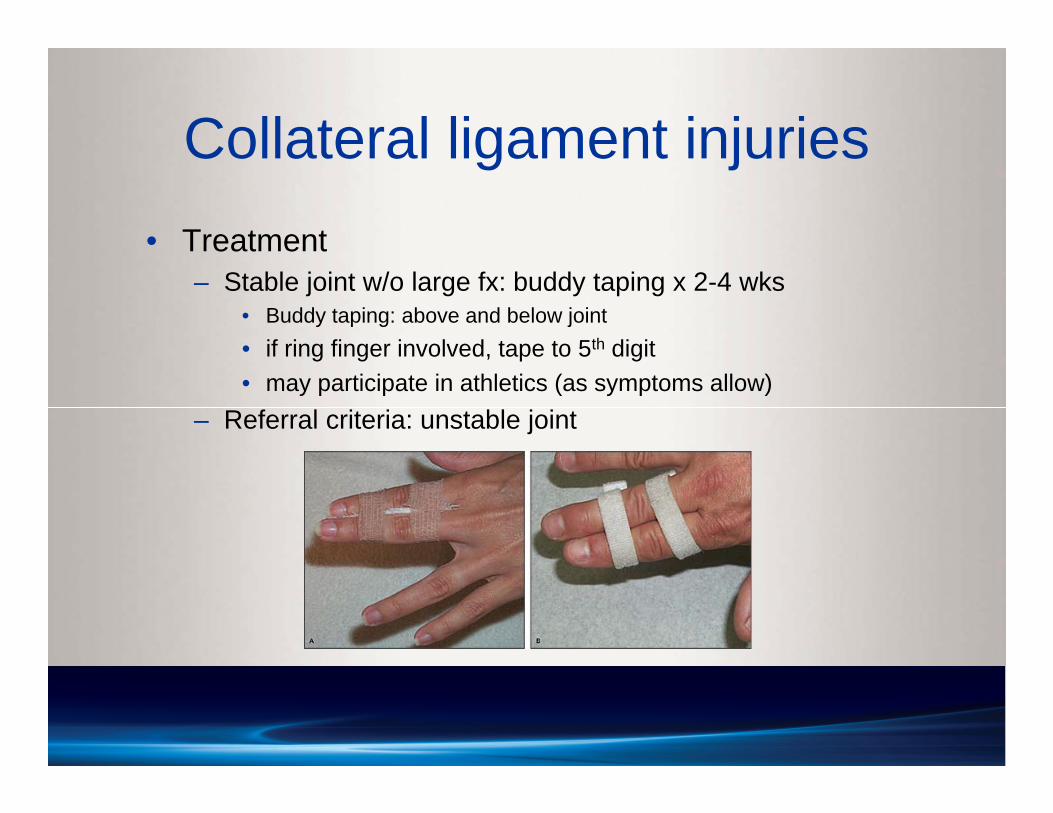
Collateral ligament injuries• Treatment
– Stable joint w/o large fx: buddy taping x 2-4 wks• Buddy taping: above and below joint• if ring finger involved, tape to 5th digit• may participate in athletics (as symptoms allow)
– Referral criteria: unstable joint
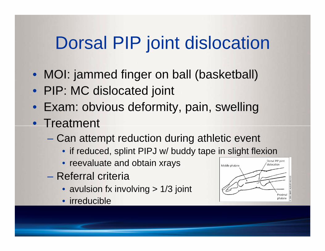
Dorsal PIP joint dislocation• MOI: jammed finger on ball (basketball)• PIP: MC dislocated joint • Exam: obvious deformity, pain, swelling• Treatment
– Can attempt reduction during athletic event • if reduced, splint PIPJ w/ buddy tape in slight flexion• reevaluate and obtain xrays
– Referral criteria• avulsion fx involving > 1/3 joint• irreducible
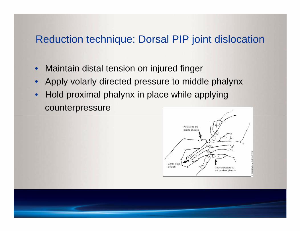
Reduction technique: Dorsal PIP joint dislocation
• Maintain distal tension on injured finger • Apply volarly directed pressure to middle phalynx• Hold proximal phalynx in place while applying
counterpressure
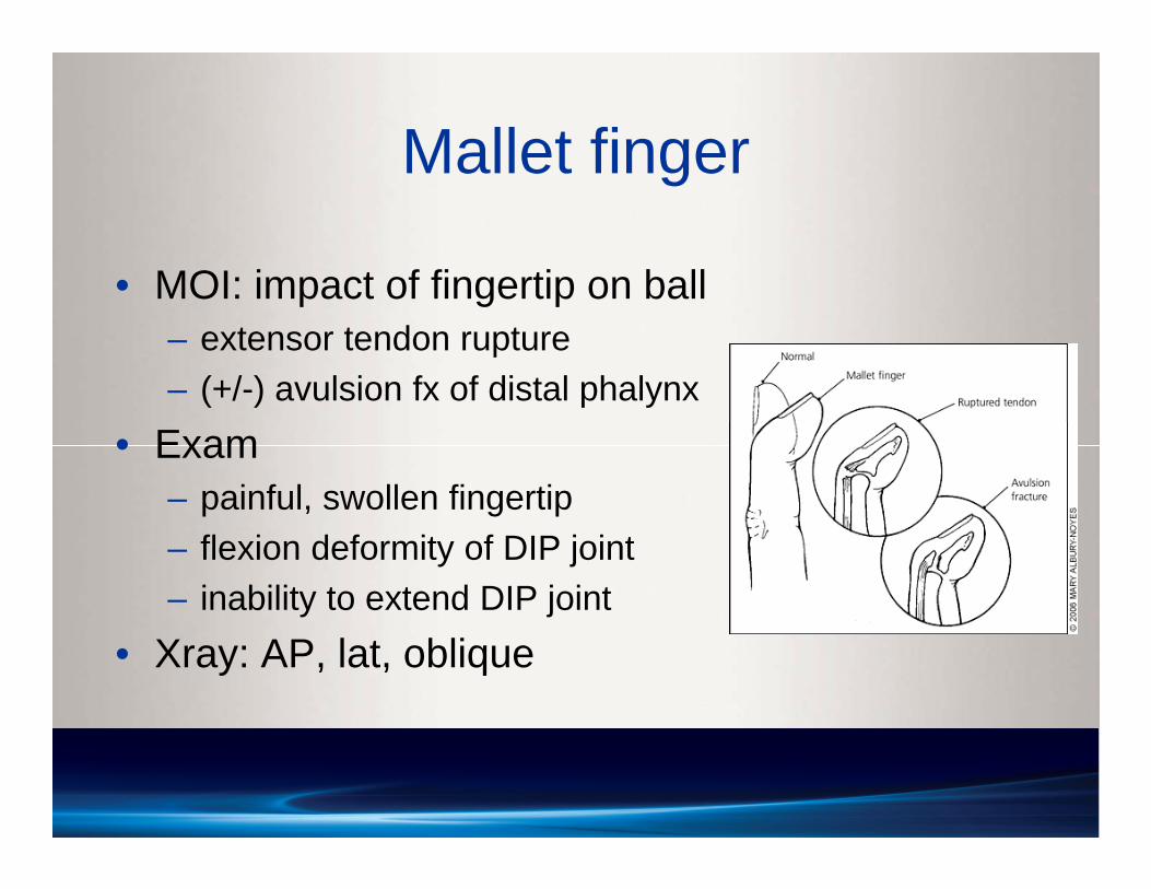
Mallet finger
• MOI: impact of fingertip on ball– extensor tendon rupture– (+/-) avulsion fx of distal phalynx
• Exam– painful, swollen fingertip– flexion deformity of DIP joint– inability to extend DIP joint
• Xray: AP, lat, oblique
Mallet finger• Treatment
– apply dorsal padded splint to DIP joint x 6-8 wks incontinuous extension
– keep PIP joint free– f/u q 2 wks, assess compliance
• Referral criteria– avulsion fracture involving >30% joint– inability to achieve full passive extension
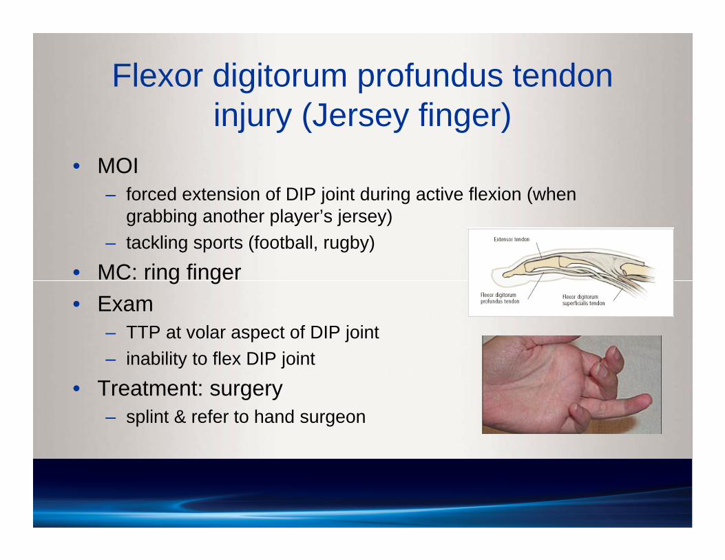
Flexor digitorum profundus tendon injury (Jersey finger)
• MOI– forced extension of DIP joint during active flexion (when
grabbing another player’s jersey)– tackling sports (football, rugby)
• MC: ring finger• Exam
– TTP at volar aspect of DIP joint – inability to flex DIP joint
• Treatment: surgery– splint & refer to hand surgeon
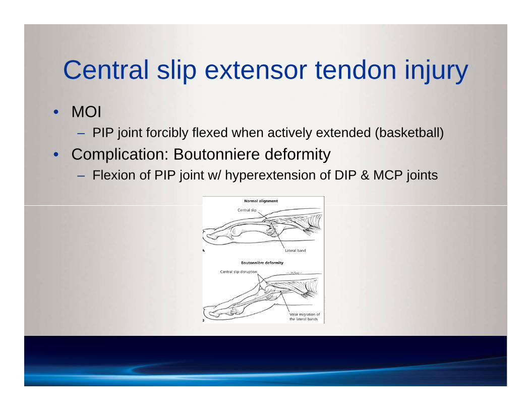
Central slip extensor tendon injury• MOI
– PIP joint forcibly flexed when actively extended (basketball)
• Complication: Boutonniere deformity– Flexion of PIP joint w/ hyperextension of DIP & MCP joints
Central slip extensor tendon injury• Exam
– TTP dorsal aspect PIP joint– inability to actively extend PIP joint – examiner can passively extend joint
• Treatment– XR: AP, lateral, oblique– Splint PIPJ in full extension x 6 wks
• (if no avulsion fx, or fx < 1/3 joint)
– Referral criteria • inability of full passive extension • avulsion fracture involving >30% of joint
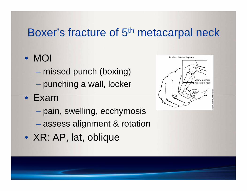
Boxer’s fracture of 5th metacarpal neck
• MOI– missed punch (boxing)– punching a wall, locker
• Exam– pain, swelling, ecchymosis– assess alignment & rotation
• XR: AP, lat, oblique
Boxer’s fracture
• Treatment– ulnar gutter splint/cast x 6 wks– f/u xrays to assess healing & alignment– Referral criteria:
• Rotation/angulation (>70o)• Reduction may be attempted (upto 40o angulation
can be tolerated)
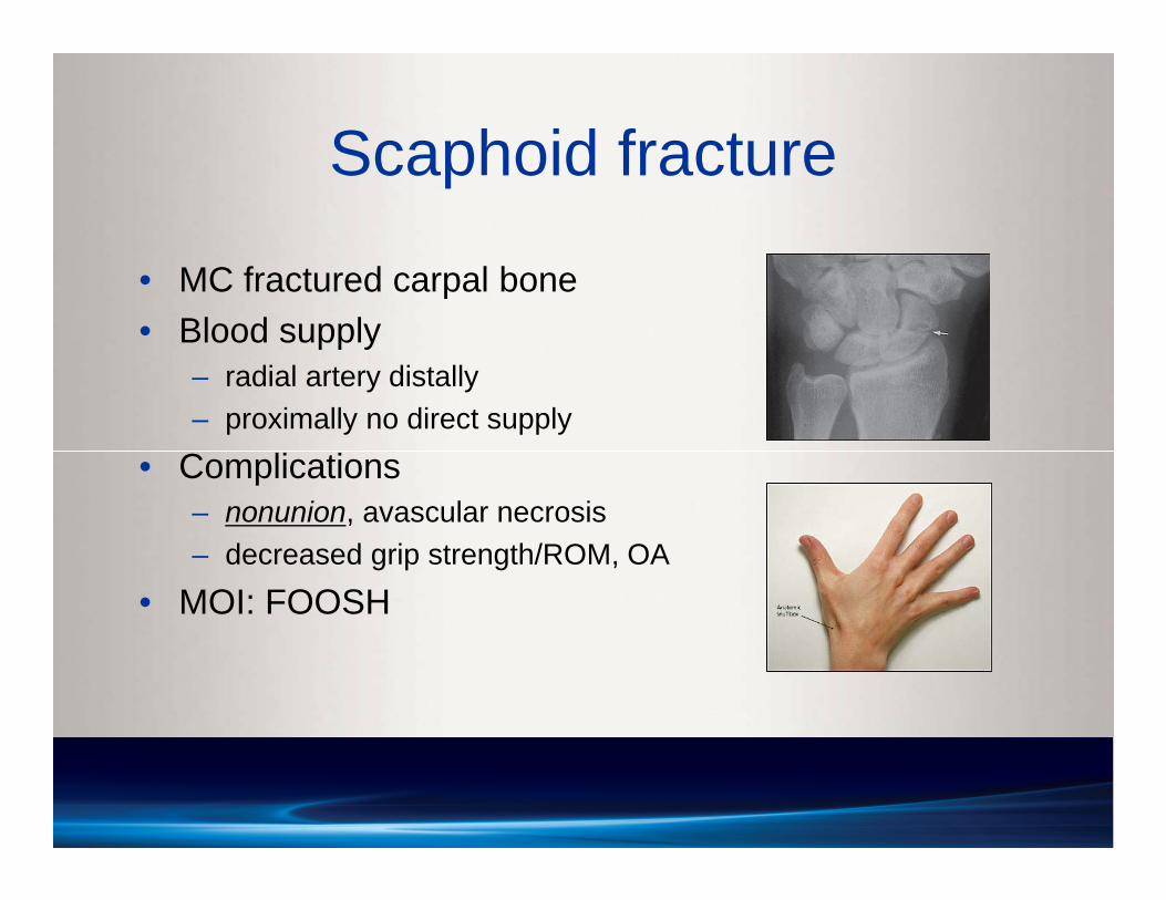
Scaphoid fracture
• MC fractured carpal bone• Blood supply
– radial artery distally– proximally no direct supply
• Complications– nonunion, avascular necrosis– decreased grip strength/ROM, OA
• MOI: FOOSH
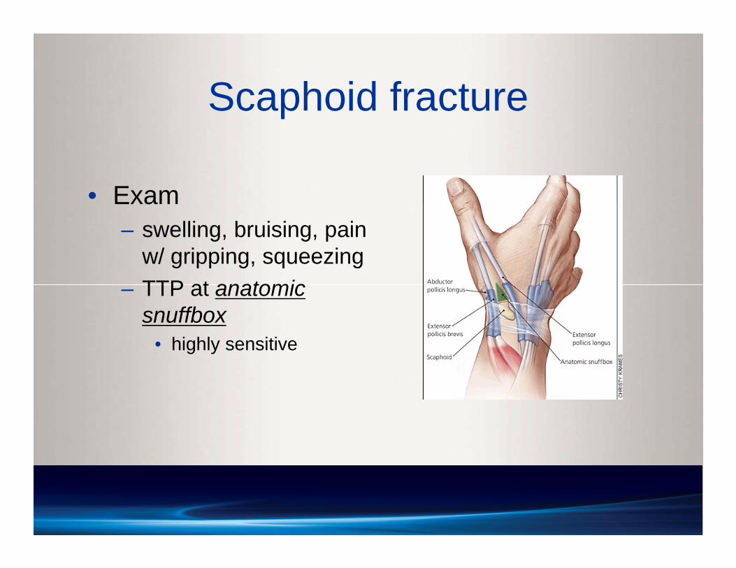
Scaphoid fracture
• Exam– swelling, bruising, pain
w/ gripping, squeezing– TTP at anatomic
snuffbox • highly sensitive
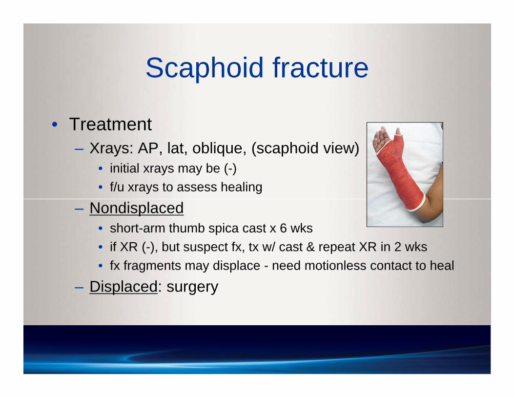
Scaphoid fracture
• Treatment– Xrays: AP, lat, oblique, (scaphoid view)
• initial xrays may be (-)• f/u xrays to assess healing
– Nondisplaced• short-arm thumb spica cast x 6 wks• if XR (-), but suspect fx, tx w/ cast & repeat XR in 2 wks• fx fragments may displace - need motionless contact to heal
– Displaced: surgery
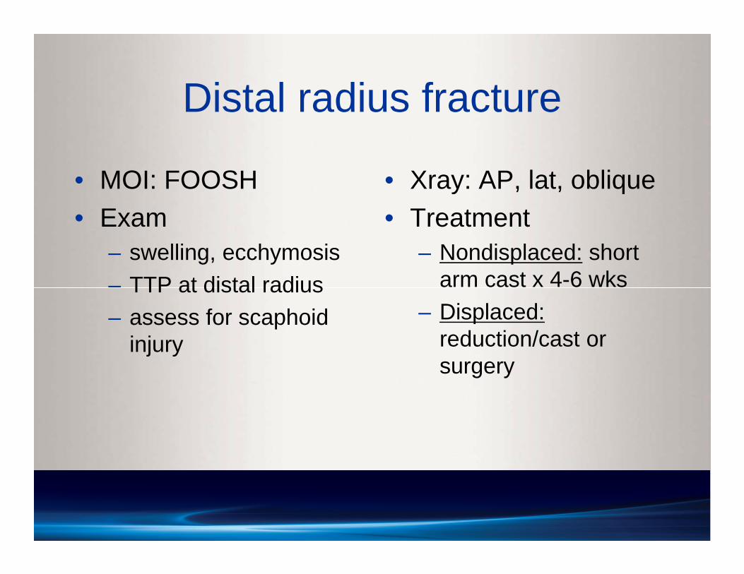
Distal radius fracture
• MOI: FOOSH• Exam
– swelling, ecchymosis– TTP at distal radius– assess for scaphoid
injury
• Xray: AP, lat, oblique• Treatment
– Nondisplaced: short arm cast x 4-6 wks
– Displaced:reduction/cast or surgery
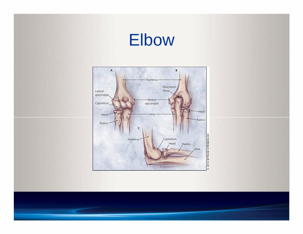
Elbow
Elbow injury
• History, mechanism of injury• If swelling, guarding, not using arm
– place sling, obtain xrays• fat pad sign suggest fracture (even if appears neg)• continue sling, repeat xrays in 7-10 days• limit sling use to 7-10 days (for comfort)
• ER: dislocation; gross deformity; neurovascular compromise
Little Leaguer’s elbow
• Medial epicondyle apophysitis due to overuse– MC baseball, softball– ~age 9-12– medial elbow pain w/ throwing*– may have decreased pitch velocity/control
• Throwing– tension forces medial elbow, compression forces
lateral elbow
Little Leaguer’s elbow• History taking
– Position– Change in technique– Increase in
amount/intensity of throwing
– Pitch counts– Number of teams
plays on– Time off from throwing
during year
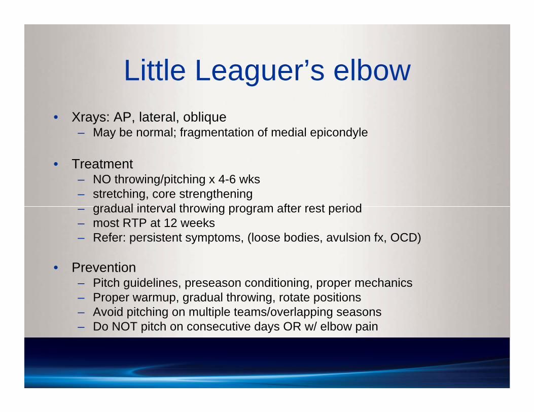
Little Leaguer’s elbow• Xrays: AP, lateral, oblique
– May be normal; fragmentation of medial epicondyle
• Treatment– NO throwing/pitching x 4-6 wks– stretching, core strengthening– gradual interval throwing program after rest period– most RTP at 12 weeks– Refer: persistent symptoms, (loose bodies, avulsion fx, OCD)
• Prevention– Pitch guidelines, preseason conditioning, proper mechanics– Proper warmup, gradual throwing, rotate positions– Avoid pitching on multiple teams/overlapping seasons– Do NOT pitch on consecutive days OR w/ elbow pain
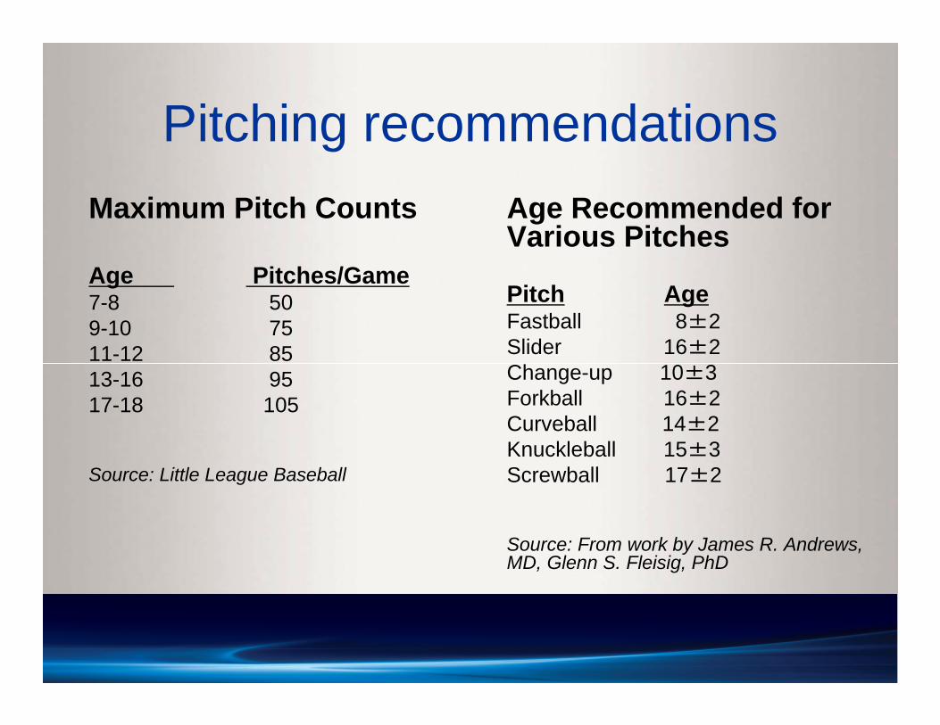
Pitching recommendationsMaximum Pitch Counts
Age Pitches/Game7-8 509-10 7511-12 8513-16 9517-18 105
Source: Little League Baseball
Age Recommended for Various Pitches
Pitch AgeFastball 8±2Slider 16±2Change-up 10±3Forkball 16±2Curveball 14±2Knuckleball 15±3Screwball 17±2
Source: From work by James R. Andrews, MD, Glenn S. Fleisig, PhD
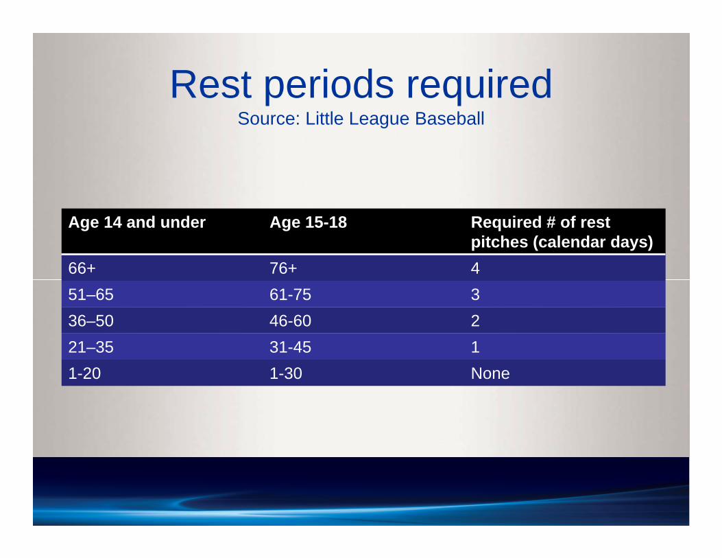
Rest periods requiredSource: Little League Baseball
Age 14 and under Age 15-18 Required # of rest pitches (calendar days)
66+ 76+ 4 51–65 61-75 336–50 46-60 221–35 31-45 11-20 1-30 None
Summary
• Assess injury and determine disposition– Treat or Refer?
• Stabilize (for safety & comfort) and refer
• Referral criteria– Xray needed? – ER: dislocation (elbow), gross deformity, open
fracture, neurovascular compromise– Pain out of proportion– Anything you are NOT comfortable treating
Return to sports/activity considerations
• Short-term periodic reevaluation to monitor progress
• Asymptomatic– Pain-free– No swelling– Normal ROM and strength– No functional deficits
Resources
• AAFP: American Family Physician Journal– www.aafp.org
• AAOS: www.stopsportsinjuries.org
• Fracture Management for Primary Care (Eiff, et. al.)
• The 5-Minute Sports Medicine Consult (2nd ed)