Embed Size (px)
Citation preview

HARDWARE IMPLEMENTATION FOR CARDIAC ELECTRICAL
EXCITATION AND CONDUCTION USING AN FPGA
NUR ATIQAH BINTI ADON
A thesis submitted in partial
fulfillment of the requirement for the award of the
Degree of Master of Electrical Engineering
Faculty of Electrical and Electronic Engineering
Universiti Tun Hussein Onn Malaysia
JANUARY 2017

iii
For my beloved husband Zainul Idlan Bin Komar,
my adorable son Zainul Zafran and my beautiful daughter Zainur Zafrah,
all my family and to everyone who supports me, it just begins…

iv
ACKNOWLEDGEMENT
First of all, I would like to thank the Almighty ALLAH for His power and His
blessing to me to complete my Master research study.
My highest gratitude goes to my supervisor, Dr. Farhanahani Binti Mahmud
for her ideas, advices and providing unceasing guidance throughout my Master
work. I would also like to thank Dr. Mohamad Hairol Bin Jabbar as my co-
supervisor for insightful guidance of my research studies.
I wish to thank my sponsors, Universiti Tun Hussein Onn Malaysia
(UTHM) and Ministry of Higher Education Malaysia for funding this research work
through the Fundamental Research Grant Scheme (FRGS) Vote 1053.
v
ABSTRACT
Contraction of the heart is controlled by electrical excitations of cardiac cell
membranes. The electrical excitations of the cells and their propagation in the heart
tissue provide a basis of the physiological function of the heart through the cardiac
excitation-conduction mechanism. One way to understand normal and abnormal
dynamics of the heart is to simulate a comprehensive mathematical model of the
cardiac excitation in order to study underlying mechanisms of the heart electrical
system. However, simulating the dynamics of large numbers of a cellular model to
form a tissue model requires an immense amount of computational time. In order to
reduce the computational time required for the simulation, a hardware
implementation of cardiac electrical excitation-conduction analysis system has been
developed based on FitzHugh-Nagumo (FHN) model for a mammalian cardiac
ventricular cell. In this research, one dimensional (1D) ring-shaped cable model
with 80 compartments of the cell model designed using MATLAB Simulink blocks
is able to be converted into synthesizable VHSIC (Very High Speed Integrated
Circuit) of Hardware Description Language (VHDL) code by using an FPGA-based
rapid-prototyping approach of MATLAB HDL Coder in order to simulate an action
potential signal and its conduction through a hardware-implemented Field
Programmable Gate Array (FPGA). Then, the VHDL design is functionally verified
on an FPGA Xilinx Virtex-6 board using MATLAB HDL Verifier through FPGA-
in-the-Loop (FIL) simulation approach. Simulations of cardiac cellular processes
and reentrant arrhythmia are successfully conducted on Xilinx Chipscope Pro. High
accuracy results have been obtained from the FPGA-on-board simulation compared
to a software-based computer simulation with Percentage Error (PE) of 1.28% and
1.56% in performing the simulations of reentrant initiation and annihilation,
respectively. The simulations are also capable to run in real time.
vi
ABSTRAK
Kontraksi jantung dikawal oleh eksitasi elektrik pada membran sel jantung.
Pengujaan elektrik pada sel dan perambakan sel-sel dalam tisu jantung memberikan
asas fungsi fisiologi jantung melalui mekanisme eksitasi-kontraksi. Salah satu cara
memahami dinamik normal dan tidak normal pada jantung adalah mensimulasikan
satu model matematik yang komprehensif untuk mengkaji mekanisme sistem
jantung. Walaubagaimanapun, simulasi dinamik model sel dengan jumlah yang
banyak bagi membentuk tisu memerlukan tempoh masa pengiraan yang lama.
Dalam usaha mengurangkan masa pengiraan simulasi, pelaksanaan perkakasan
jantung elektrik bagi sistem analisis eksitasi-kontraksi telah dihasilkan berdasarkan
model Fitzhugh-Nagumo (FHN) untuk sel ventrikel jantung pada mamalia. Dalam
kajian ini, kabel bentuk cecincin satu dimensi (1D) dengan 80 sel direka
menggunakan blok MATLAB Simulink seterusnya ditukar secara automatik kepada
bahasa perihal peralatan litar bersepadu berkelajuan tinggi (VHDL) boleh sintesis
menggunakan kaedah protataip-pantas MATLAB HDL Coder untuk
mensimulasikan isyarat keupayaan tindakan dan konduksinya melalui pelaksanaan
peranti tatasusunan get boleh aturcara (FPGA). Kemudian, fungsi reka bentuk VHDL
itu disahkan pada satu papan tunggal FPGA Xilinx Virtex-6 menggunakan Pengesah
HDL melalui simulasi gelung dalam FPGA (FIL). Simulasi proses sel jantung dan
aritmia berjaya dikendalikan pada penganalisis logik terbenam Xilinx Chipscope
Pro. Keputusan dengan ketepatan yang tinggi telah diperolehi daripada simulasi atas
papan FPGA berbanding dengan simulasi komputer berasaskan perisian dengan peratus
ralat (PE) 1.28% dan 1.56%, masing-masing dalam melaksanakan simulasi
penghasilan dan penghapusan masuk-semula. Simulasi ini juga mampu beroperasi
dalam masa nyata.
vii
CONTENTS
TITLE i
DECLARATION ii
DEDICATION iii
ACKNOWLEDGEMENT iv
ABSTRACT v
ABSTRAK vi
CONTENTS vii
LIST OF PUBLICATIONS xi
LIST OF TABLES xiii
LIST OF FIGURES xiv
LIST OF ABBREVIATIONS xvii
CHAPTER 1 INTRODUCTION 1
1.1 Background of the research 1
1.2 Problem statement 5
1.3 Aim and research objectives 6
1.4 Scope of the research 6
1.5 Overall contributions 7
1.6 Thesis organisations 8
CHAPTER 2 LITERATURE REVIEW 9
2.1 Overview 9
2.2 Electrical system of the heart 10

viii
2.2.1 Propagation of electrical activity in cardiac tissue 12
2.2.2 Mechanism of cardiac arrhythmia 14
2.2.2.1 Anatomical circus movement reentry 15
2.3 Approaches in cardiac electrophysiological analysis 18
2.3.1 Experimental approach 18
2.3.2 Clinical approach 19
2.3.3 Model simulation approach 20
2.3.3.1 Computer simulation 21
2.3.3.2 Hardware implementation 21
2.3.4 Comparison between experimental, clinical and
model simulations approaches 22
2.4 Mathematical models for cardiac electrical activity 23
2.4.1 FitzHugh-Nagumo (FHN) model 25
2.4.2 Phase Plane Analysis 27
2.5 Real-time hardware implementation of cardiac model 28
2.5.1 Application Specific Integrated Circuit (ASIC) 28
2.5.2 Digital Signal Processing (DSP) 29
2.5.3 Field Programmable Analog Array (FPAA) 30
2.5.4 Field Programmable Gate Array (FPGA) 32
2.5.4.1 FPGA Platform 34
2.6 FPGA development methods 37
2.6.1 Traditional method 38
2.6.2 FPGA rapid-prototyping method 40
2.7 FPGA in solving Ordinary Differential Equations (ODE) 44
2.8 Limitation of existing works and research opportunities 46
2.9 Potential application: Cardiac catheters based analysis tool
for education and training 50
CHAPTER 3 RESEARCH METHODOLOGY 52
3.1 Overview 52
3.2 Rapid VHDL coding using MATLAB HDL Coder 53
3.2.1 Floating-point data-type system design 55
3.2.2 Fixed-point data-type system design 56
3.2.3 System design optimisation in fixed-point
ix
data-type 59
3.2.4 VHDL code generation and FIL verification 61
3.2.5 Design of a cardiac conduction simulation-based
analysis system 62
3.2.6 FIL verification for cardiac conduction simulation-
based analysis system 66
3.3 FPGA Programming: Implementation on Xilinx
Virtex-6 FPGA target board 67
3.3.1 Synthesis and implementation: Xilinx
Integrated Software Environments (ISE) 14.6 69
3.3.2 Simulation testing and verification: Xilinx
Integrated Software Environments Simulator
(ISim) 14.6 70
3.3.3 FPGA-on-board simulation: Xilinx Chipscope
Pro Analyzer 14.6 71
3.4 Evaluation of the FPGA implemented cardiac electrical
excitation and conduction simulation-based analysis
system: Reentrant mechanism 74
3.4.1 Initiation and annihilation of reentrant in 1D ring-
shaped model 74
3.4.2 Phase Resetting Curve (PRC) 75
CHAPTER 4 RESULT AND ANALYSIS 76
4.1 Overview 76
4.2 Rapid-prototyping design for cardiac excitation and
conduction analysis system based on the FitzHugh-Nagumo
(FHN) model 77
4.2.1 Design of floating-point data-type system for single
cell cardiac excitation 77
4.2.2 Design of fixed-point data-type system for single cell
cardiac excitation 79
4.2.3 Fixed-point optimisation using MATLAB HDL Coder 80
4.2.4 FPGA-in-the-Loop (FIL) verification 82
4.2.5 Hardware performance analysis of the cardiac

x
excitation-conduction 86
4.3 Execution of cardiac excitation simulation on FPGA board 88
4.3.1 Verification of the single-cell cardiac excitation
simulation using Xilinx ISE Simulator (ISim) 88
4.3.2 Single-cell cardiac based cardiac analysis system
execution on Xilinx Virtex-6 FPGA board through on-
board simulation using Xilinx Chipscope Pro Analyzer 89
4.4 Evaluation of the FPGA implemented cardiac electrical
excitation and conduction simulation-based analysis system 90
4.4.1 Accuracy evaluation: Simulation and analysis studies
of the cardiac reentrant mechanism 91
4.4.1.1 Initiation of reentrant in the 1D ring-shaped
cable models 91
4.4.1.2 Annihilation of reentrant in the 1D
ring-shaped cable models 92
4.4.1.3 Phase Resetting Curve (PRC) 94
4.4.2 Timing performance evaluation based simulation
studies of the reentrant mechanism 96
CHAPTER 5 CONCLUSIONS AND FUTURE WORKS 97
5.1 Overview 97
5.2 Achievements 98
5.3 Limitations 99
5.4 Future works 99
REFERENCES 101
APPENDIX A 112
APPENDIX B 116
xi
LIST OF PUBLICATIONS
1. N.A. Adon, F. Mahmud, M.H. Jabbar & N. Othman. (2015). Optimization in
MATLAB for Cardiac Excitation Modeling Towards FPGA Standalone
Simulation Tools. Applied Mechanics and Materials Trans Tech Publications,
Switzerland, Vol. 773-774, pp. 761-765. ISSN: 1662-7482.
2. N.A. Adon, M.H. Jabbar & F. Mahmud. (2015). FPGA Implementation for
Cardiac Excitation-Conduction Simulation based on FitzHugh-Nagumo Model.
5th International Conference on Biomedical Engineering in Vietnam (IFMBE
Proceedings), Vol. 46, pp. 117-120. Springer International Publishing. ISBN:
978-3-319-11775-1.
3. N.A. Adon, F. Mahmud, M.H. Jabbar & N. Othman. (2014). FPGA-in-the-
Loop Co-simulation of Reentrant Arrhythmia Mechanism in One Dimensional
(1D) Ring-Shaped based on FitzHugh-Nagumo Model. 2014 IEEE
International Conference on Control System, Computing and Engineering, pp.
239-244. ISBN: 978-1-4799-5685-2.
4. N.A. Adon & F. Mahmud. (2013). Simulation of Reentrant Arrhythmia
Mechanism in One Dimension Ring-Shaped Model using Simulink. Prosiding
Seminar Kebangsaan Aplikasi Sains Dan Matematik (SKASM), Jilid 3: pp. 9-
20. Penerbit UTHM. ISBN: 978-9-670-46850-1.
5. N.A. Adon & F. Mahmud. (2013). Cardiac Excitation-Conduction Modeling
using MATLAB/Simulink for Real Time FPGA Implementation. Proceeding
of Microelectronics & Nanotechnology (PMiNT), pp. 1-3. Penerbit UTHM.
ISBN: 978-9-670-76400-9.
xii
6. N. Othman, F. Mahmud, A.K. Mahamad, M.H. Jabbar &. N.A. Adon. (2014).
Cardiac Excitation Modeling: HDL Coder Optimization Towards FPGA Stand-
Alone Implementation. 2014 IEEE International Conference on Control
System, Computing and Engineering, pp. 507-511. ISBN: 978-1-4799-5685-2.
7. N. Othman, F. Mahmud, A.K. Mahamad, M.H. Jabbar &. N.A. Adon. (2016).
Voltage-Clamp Simulation of Cardiac Excitation: Field Programmable Gate
Array (FPGA) Implementation. ARPN Journal of Engineering and Applied
Sceinces 2016. Vol. 11, no. 24, pp. 14056-14064. ISSN: 1819-6608.
8. N. Othman, N.A. Adon & F. Mahmud. (2017). FPGA in-the-loop Simulations
of Cardiac Excitation Model under Voltage Clamp Conditions. International
Conference on Engineering, Science and Nanotechnology (ICESNANO) 2016,
Vol. 1788, pp. 030105-(1-7). American Institute of Physics (AIP) Conference
Proceeding. ISBN: 978-0-7354-1452-5.

xiii
LIST OF TABLES
2.1 Comparison of experimental, computer simulation and hardware
implementation
22
2.2 Comparison of specifications based on the type of hardware solutions 28
2.3 ASIC versus FPGA 29
2.4 DSP versus FPGA 30
2.5 FPAA versus FPGA 31
2.6 Specifications of FPGA Virtex-6 ML605 Board 36
2.7 FPGA as an ODE solver 46
2.8 Survey for electrophysiology analysis approaches 49
4.1 Hardware performance and statistical analysis results of a single-cell
FHN model for a single-cell cardiac excitation simulation according to
three proposed fixed-point values
83
4.2 Hardware performance results of a single-cell and 80 cells of the FHN
model
87
xiv
LIST OF FIGURES
1.1 The ten leading causes of death in the world 1
2.1 Overall of related literature review 9
2.2 The heart system component 10
2.3 Cardiac action potential phases 11
2.4 The cardiac muscle 12
2.5 An equivalent circuit for a single fiber of cardiac tissue model 13
2.6 Normal and abnormal heart rate 14
2.7 Unidirectional block and circus movement reentry 17
2.8 Rat model of myocardial infarction 18
2.9 Circuit diagram of the nerve model 25
2.10 Phase plane analysis of FHN model 27
2.11 Architecture of FPAA 30
2.12 Platform and tools for Altera family 32
2.13 Platform and tools for Xilinx family 33
2.14 FPGA market share 33
2.15 Virtex-6 XC6VLX240T features locator diagram 35
2.16 Architecture of generic FPGA 37
2.17 FPGA development methods 37
2.18 Block diagram of FPGA system development process using traditional
method
39
2.19 Block diagram of FPGA system development process using rapid-
prototyping method
40
2.20 Comparison of time spent on FPGA implementation between rapid-
prototyping method by MATLAB HDL Coder and manual HDL coding
42
2.21 Timeline survey for electrophysiology analysis approaches 48
2.22 Cardiac catheter ablation procedures 50
3.1 Block diagram of the overall process for the development of FPGA 53
xv
implemented cardiac model simulation-based analysis system
3.2 Workflow of rapid VHDL coding using MATLAB HDL Coder 54
3.3 A floating-point data-type design of the FHN model for a single-cell
cardiac excitation simulation using MATLAB Simulink
56
3.4 MATLAB function code of the external stimulation current, I 56
3.5 Among examples block needs to be changed to fulfill the requirements
of fixed-point data-type
57
3.6 A fixed-point data-type design of FHN model for a single-cell cardiac
excitation simulation using MATLAB Simulink
58
3.7 MATLAB function codes with fixed-point setting 59
3.8 FPGA-in-the-Loop (FIL) verification 61
3.9 Generated block for comparison of FIL verification and MATLAB
Simulink simulation
62
3.10 A ring-shaped cable model. The ring model consists of N cell models
and gap junction resistance, Rd
62
3.11 The overall MATLAB Simulink block diagram for 80 cells of FHN
model in ring-shaped cable model
63
3.12 Layer 1 of FHN model in 1D ring-shaped cable model 64
3.13 Layer 2 of FHN model in 1D ring-shaped cable model 65
3.14 Layer 3 of FHN model in 1D ring-shaped cable model 65
3.15 FIL verification for 80 cells of FHN model in 1D ring-shaped cable
model
66
3.16 Workflow of the hardware programming for FPGA-on-board simulation 68
3.17 Setup of ChipScope Pro system for realisation of FPGA-on-board
simulation
72
3.18 VHDL code segment for ILA and ICON core components 73
4.1 Action potential and recovery state waveforms of single-cell produced
by the FHN model in floating-point data-type design
78
4.2 Action potential and recovery state waveforms of single-cell produced
by the FHN model in fixed-point data-type design with (WL, FL) of
(24,22)
79
4.3 Comparison of simulated action potential waveforms for different
optimum WL and FL values used in the optimisation process
81
xvi
4.4 Floor plans mapping of a single-cell FHN model for a single-cell cardiac
excitation simulation
84
4.5 Result of the FIL for the single-cell cardiac excitation simulation 85
4.6 Results of the FIL for the cardiac conduction simulation in 80 cells 1D
ring-shaped cable model
86
4.7 A simulation result of the cardiac electrical conduction in 80 cells FHN
1D ring-shaped cable model verified using MATLAB
87
4.8 Simulation using ISim for single-cell FHN model 88
4.9 Waveform window in Chipscope Pro of the FPGA on-board simulation
for the single-cell cardiac excitation
89
4.10 Bus plot window displayed by Chipscope Pro 90
4.11 A space-time diagram showing membrane voltage as a function of time
and position around the ring-shaped cable models
92
4.12 Representation of the termination by single impulsive stimulations that
responses from the reentry dynamics
93
4.13 Representation of the phase resetting curves (PRCs) that showing the
amount of phase reset, Δφ against the stimulation phase, φ
95

xvii
LIST OF ABBREVIATIONS
1D One Dimensional
2D Two Dimensional
3D Three Dimensional
AIDS Acquired Immune Deficiency Syndrome
ASIC Application Specific Integrated Circuit
BER Bit Error Rate
CAB Configurable Analog Blocks
CLB Configurable Logic Blocks
CoreGen Core Generator
DDR Double Data Rate
DEPE Differential Equation Processing Element
DSP Digital Signal Processing
FHN FitzHugh-Nagumo
FIL FPGA-In-the-Loop
FL Fraction-Length
FMC FPGA Mezzanine Cards
FPAA Field Programmable Analog Array
FPGA Field Programmable Gate Array
GPU Graphics Processing Unit
HDL Hardware Description Language
HIV Human Immunodeficiency Virus
ICON Integrated Controller
ILA Integrated Logic Analyzer
IOB Input Output Blocks
ISE Integrated Software Environment
ISim ISE Simulator
xviii
JTAG Joint Test Action Group
LUT Lookup Table
MAC Multiply Accumulate Operations
MMCM Mixed Mode Clock Managers
MSE Mean Squared Error
NCD Native Circuit Description
NGD Native Generic Database
ODE Ordinary Differential Equation
PAR Place And Route
PC Personal Computer
PCI
PCR
Payment Card Industry
Polymerase Chain Reaction
PDSP
PE
Programmable Digital Signal Processors
Percentage Error
PRC Phase Resetting Curve
RAM Random Access Memory
RTL Register Transfer Language
SNR Signal to Noise Ratio
SOC System On Chip
SVPWM Space Vector Pulse Width Modulation
VHDL VHSIC Hardware Description Language
VHM Virtual Heart Model
VHSIC Very High Speed Integrated Circuit
VLSI Very Large Scale Integration
VSI Voltage Source Inverter
WHO World Health Organization
WL Word-Length
XSG Xilinx System Generator
CHAPTER 1
INTRODUCTION
1.1 Background of the research
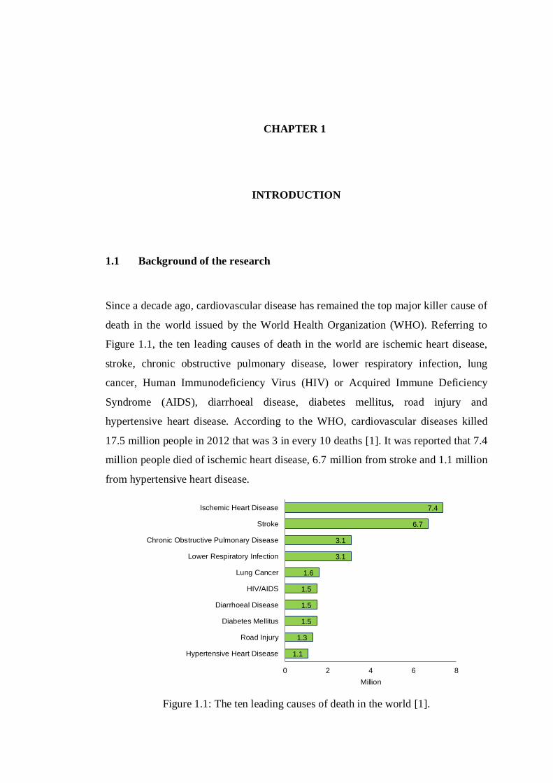
Since a decade ago, cardiovascular disease has remained the top major killer cause of
death in the world issued by the World Health Organization (WHO). Referring to
Figure 1.1, the ten leading causes of death in the world are ischemic heart disease,
stroke, chronic obstructive pulmonary disease, lower respiratory infection, lung
cancer, Human Immunodeficiency Virus (HIV) or Acquired Immune Deficiency
Syndrome (AIDS), diarrhoeal disease, diabetes mellitus, road injury and
hypertensive heart disease. According to the WHO, cardiovascular diseases killed
17.5 million people in 2012 that was 3 in every 10 deaths [1]. It was reported that 7.4
million people died of ischemic heart disease, 6.7 million from stroke and 1.1 million
from hypertensive heart disease.
Figure 1.1: The ten leading causes of death in the world [1].
1.1
1.3
1.5
1.5
1.5
1.6
3.1
3.1
6.7
7.4
0 2 4 6 8
Hypertensive Heart Disease
Road Injury
Diabetes Mellitus
Diarrhoeal Disease
HIV/AIDS
Lung Cancer
Lower Respiratory Infection
Chronic Obstructive Pulmonary Disease
Stroke
Ischemic Heart Disease
Million
2
Ischemic heart disease caused by decreased blood flow and reduced oxygen
supply to the heart muscle [2]. The disease can interrupt the electrophysiology
function of the ion channels responsible for the cellular electrical activity of the
heart. Changes in the intracellular and extracellular occur during ischemic and can
alter the electrophysiology of several species of ionic channels are in fact related to
disturbances in the cellular activity of cardiac myocytes [3]. Thus, until now, many
studies have been done to elucidate the causes and interpret the underlying
mechanisms in cardiac electrophysiological which are through experimental [4], [5]
clinical measurement [6], [7] and computational model simulation [8], [9], [10]
studies.
Although, experimental study is generally preferable, but it has several
restrictions such as requires high variables quantity for monitoring, expensive and
high-resolution data in investigating larger preparations [11], [12]. Meanwhile,
clinical tools have become widely used in cardiac electrophysiological studies, often
indispensable in evaluating patients with specific cardiac electrical activity [13].
Clinical data are used to validate computational modeling which allow integration of
previous findings, quantitative assessments of the models and the projection across
relevant spatial and temporal scales [14]. However, clinical tools have several
drawbacks which are risk assessment tools, potential harms to patients and the
effectiveness or difficulty in comparing results across studies [15], [16]. In contrast, a
model simulation technique is not associated with such problems and also has the
capability to increase study parameters through mathematical representations and
decreasing the time responsible for investigating cardiac dynamics [17], [18].
There are numerous mathematical model had been represented in excitable
media for the simulation studies such as Hodgkin–Huxley [19], FitzHugh-Nagumo
(FHN) model [20], [21], continued with the Noble model [22], [23], Beeler and
Reuter model [24], [25], Luo-Rudy model [26], [27] and details models in order to
show different regions of the heart. However, advancement of the computational
technique has initiated cardiac cell models to become more complicated as variable
parameters in the mathematical descriptions are increased in order to present the
cellular process in more details [28], [29]. Thus, a large number of the cell models in
forming a tissue model cause drawback in the amount of computational processing
which would increase a time required to run the model simulations [30].

3
According to Prof. Yoshihiko Nakamura (2012), two second motion
according of the neuromusculoskeletal model of human takes one hour to compute
reduction of computation cost. These situations can be concluded that a real-time
model simulation technique has been difficult to achieve especially in the
electrophysiological that involves high level dimensional models and simulation
conditions [31], [32]. However, a real-time model simulation system is very
important in order to diagnose cardiac abnormalities due to heart failure since it
simulate more realistically cardiac work and it would be a very useful and convenient
system to be applied in medical education and training in cardiac surgical planning
such as permanent pacemaker insertion, catheter-based intervention and invasive
cardiovascular surgeries [33], [34], [35].
In order to overcome this computational challenge, high performance and low
power consumption hardware-based implementation can provide valuable tools for
real-time simulation analysis in electrophysiological studies applications such as in
the medical and educational field [36], [37]. Currently, researchers have moved
forward to use hardware-implemented of analysis tool for cardiac electrophysiology
considering their several advantages of extremely fast and parallel mode execution,
low power usage, reconfigurable, development ease and low cost [38], [39], [40].
Hence, various types of hardware had been used to simulate the electrical potentials
exist across the membrane of cells using hardware such as analog-digital circuit [41],
microcontroller [42], Graphic Processing Unit (GPU) [30], [43] and Field
Programmable Analog Array (FPAA) [44], [45]. However, these previous studies
have shown limitations due to their power consumption and inefficient regarding
rapid calculations in performing the real-time operation [46].
Therefore, reconfigurable hardware in the form of Field Programmable Gate
Array (FPGA) has appeared as a viable system solution with complex chips in the
construction of high performance systems at an economical price [47], [48]. FPGA
technology is now considered very useful by an increasing number of designers in
various fields of application as it offers reconfigurable hardware, programmable
circuit architecture, execute in parallel mode with million gate counts and a low
power consumption [47], [49]. Moreover, it is also capable of solving higher orders
of Ordinary Differential Equations (ODEs) describing the electrical behavior of the
cell membrane [38].

4
In recent literature, a large number of studies using FPGA for biomedical
application are reported [50], [51], [52]. As regards, these have given motivation to
implement the FPGA to perform real-time simulations, primarily responsible for the
cardiac abnormal activity. This research will emphasise the simulation of reentrant
excitation-conduction of cardiac cells realised by coupling 80 active circuits in one
dimensional (1D) ring-shaped based on FHN [53] model. In this research, the 1D
ring-shaped cable model is constructed using MATLAB Simulink based FPGA
rapid-prototyping method towards a real-time simulation in producing an analysis
tool to study the underlying mechanism of the heart through understandings of non-
linear dynamics in cardiac excitation.
The FPGA configuration is generally specified using a Hardware Description
Language (HDL), therefore for rapid design and faster development, the HDL code
is generated using MATLAB HDL Coder that is capable to convert the designed
MATLAB Simulink model to Very High Speed Integrated Circuit (VHSIC)
Hardware Description Language (VHDL) code. Furthermore, the FPGA-based
model simulation system which is designed through MATLAB HDL Coder is
verified using an FPGA-in-the-Loop (FIL) approach. Towards the hardware
implementation in real-time model simulation analysis tool, the design system is then
implemented on Xilinx Virtex-6 XC6VLX240T FPGA development board and
simulated through embedded logic analyser Xilinx Chipscope Pro. The dynamics of
the FHN model simulation using FPGA board are compared to those obtained from
the conventional software-based computer simulation technique to evaluate the
accuracy and performance of the simulation-based analysis system in order to
demonstrate that the FPGA model can be utilised for simulating large scale cellular
network in real-time as an alternative to the software-based computer simulation
technique in the future.

5
1.2 Problem statement
As mentioned earlier in the introduction, due to a number of challenges in
experimental and clinical investigations of the cardiac electrical behavior [54],
mathematical models of cardiac tissue have been developed and analysed by
simulating conduction of action potentials in a variety of conditions [18]. However, it
is inevitable for those models to become large scale in the number of dynamical
variables, requiring immense amounts of computational time for their dynamic
simulations. This could cause difficulties in performing in-depth analysis on cardiac
electrical functions since many hours of time is required to run dynamic simulations
of electrical conduction in tissue or organ level on a conventional computer station.
Although a high performance supercomputer is commonly used in conducting fast
speed computations for the analysis, it usually requires high installation cost and high
energy. Therefore, FPGA based hardware implementation of electrical excitation and
conduction of a cardiac cell model simulation system is developed to overcome the
challenges. The simulation of 1D ring-shaped cable model is conducted through this
research work based on FHN model, a typical mathematical model of a cardiac cell.

6
1.3 Aim and research objectives
The aim of this research is to develop an analysis system in order to perform a real-
time simulation of a cellular excitation reaction in tissue level based on a
mathematical model by using FPGA hardware implementation. The proposed
implementation can be deployed in a biomedical field for understanding and
analysing the mechanism of abnormalities cardiac cycle and will function in the real
FPGA-on-board application. To enable this, the thesis is presented in three objectives
as follows:
Objective I: To construct a FHN model algorithm based on MATLAB HDL Coder
for an FPGA implementation.
Objective II: To develop FPGA-based model simulation system for cardiac
excitation and conduction towards real-time analysis tool implementation.
Objective III: To evaluate the technique with conventional simulation method in
terms of its accuracy and performance based on simulation studies of the reentrant
mechanism in a cardiac excitation-conduction.
1.4 Scope of the research
This research covers a study related to a mathematical model of the action potential
conduction in a ventricular cardiac cell, which focuses on the FHN model for
developing a new simulation-based analysis technique in cardiac excitation and
conduction studies using FPGA. The FPGA-based hardware implemented real-time
model simulation system will be developed by solving ODEs of the model based on
a rapid-prototyping design flow through MATLAB HDL Coder. This technique
accelerates the FPGA design process through automatic generation the VHDL code
at a certain level in developing the system and faster optimisation. The rapid-
prototyping method does not directly involve on a hardware architecture design of
the FPGA by the developer which has more to do with an FPGA traditional design
technique.
The reason for choosing the MATLAB HDL Coder as rapid-prototyping tool
are because the MATLAB HDL Coder offers and keeps updating many advanced
and latest functions for the FPGA design systems, and rapidly assembles system

7
models usually using only existing blocks of MATLAB Simulink compare to other
tools such as Xilinx System Generator (XSG), Digital Signal Processing (DSP)
Builder and Labview. Besides, the MATLAB HDL Coder also provides FIL
approach to verify the designed system based on various types of FPGA
development boards from different manufacturers. Lastly, the generated code by
MATLAB HDL Coder is modified, synthesised and implemented by using Xilinx
ISE software on the FPGA. The results are displayed on the Xilinx Chipscope Pro as
it is able to log data for further analysis.
The FHN model will be used to understand of reentrant mechanisms by
performing 1D ring-shaped cable model in cardiac tissue. A 1D ring-topology-
network of 80 compartments of the cell model is constructed through interconnection
of gap junction resistances for exhibiting the reentrant action potential conduction.
The number of the cell models presented is relied on the FPGA specification used
and 80 cells is an appropriate number to perform the simulation of the cardiac
excitation-conduction in FHN model-based 1D ring-topology-network. However
based on the design and type of model, the number could be different. For example,
the design of a two dimensional (2D) model might require more number of cells. The
simulation results from the FPGA-based model will be compared to those obtained
numerically MATLAB software-based computer simulations of the cardiac
excitation-conduction activity using the FHN model to verify the accuracy according
to an acceptable error simulation of not more than 1.5% [55] and the timing
performance of the simulation system.
1.5 Overall contributions
This project of developing a cardiac reentrant excitation-conduction simulation
system has two significant contributions. Firstly, a new approach of FPGA system
design based rapid-prototyping that can provide a high performance system and high
accuracy result has demonstrated a significant contribution. Through this rapid-
prototyping approach, various types of parameters are involved to analyse and
optimise the system performances such as area, maximum frequency and power
consumption in a much more convenient way. Secondly, the realisation of a real-time
simulation of FHN model based cardiac action potential through FPGA

8
implementation itself has contributed in introducing a new technique of conducting a
hardware-based model simulation for real-time performance in cardiac
electrophysiological studies.
1.6 Thesis organisation
The thesis organisations are as follows:
Chapter 2 (Literature Review): The second chapter of the thesis will review on the
technique of cardiac electrophysiological analysis, which takes a closer look at the
most recent studies related to mathematical models focusing on the FHN
mathematical model. Besides, topics related to real-time hardware implementation
approaches of cardiac model for cardiac excitation simulation and analysis, and a
potential application related to the analysis system also being will be discussed in
this chapter.
Chapter 3 (Methodology): The third chapter will present the method and design
strategies for developing the proposed FPGA hardware implemented model
simulation-based analysis system. The workflows of the FPGA development
methods used in this research which include rapid-prototyping method for VHDL
coding using MATLAB HDL Coder and FPGA programming method for on-board
implementation will be concisely explained.
Chapter 4 (Results and Analysis): The fourth chapter will present the results
obtained throughout the development of FPGA implemented cardiac simulation-
based analysis system using the rapid-prototyping method by MATLAB HDL Coder.
The FPGA-on-board simulation and software-based computer simulation results of
cardiac excitation-conduction relating to the reentrant mechanism will be presented
and a comparative study of the FPGA implementation results for the FHN model for
both simulations will also be discussed.
Chapter 5 (Conclusions): The last chapter will summarise the conclusion drawn
from the results acquired in this thesis based on the achievements and limitations
discovered from this research. Several recommendations for potential future works
for further improvement in the development system will also be discussed.
CHAPTER 2
LITERATURE REVIEW
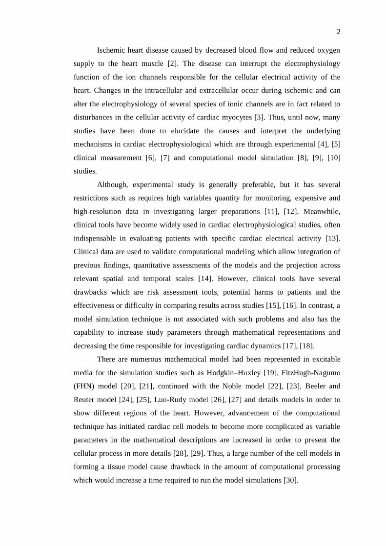
2.1 Overview
In this research, several related topics from previous researches have been studied in
order to further understand the situation and the relevance in developing a model
simulation-based analysis system implemented on FPGA hardware for real-time
simulations of cardiac electrophysiological mechanism. The summarised topic of
review in this chapter is illustrated in Figure 2.1. They are identified based on five
main topics which are electrical system of the heart, approaches in cardiac
electrophysiological analysis, cardiac mathematical models, real-time hardware
implementation and its potential application. The oval shape shows the approaches
used in this research and selected based on the previous studies according to the
advantages and disadvantages that have been identified and discussed in section 2.5
of this chapter.
Literature Review
Mathematical models for
cardiac electrical activity
Real-time hardware
implementation of cardiac model
Propagation of
electrical activity in
cardiac tissue
Techniques in cardiac
electrophysiological analysis
Experimental
Computer simulation
Application Specific
Integrated Circuit (ASIC)
Cardiac catheter procedure
(as training tool)
Education/Clinical
Electrical system of
the heart
Mechanism of
cardiac arrhythmia
Clinical
Model simulation
Hardware implementation
FitzHugh-Nagumo (FHN)
Digital Signal Processing
(DSP)
Graphical Processing Unit
(GPU)
Field Programmable
Analog Array (FPAA)
Field Programmable Gate
Array (FPGA)
Advantages/Disadvantages
Advantages/Disadvantages
Figure 2.1: Overall of related literature review.

10
2.2 Electrical system of the heart
The heart is responsible for pumping blood through the blood vessels by an electrical
conduction system that coordinates the contraction of the various chambers of the
heart. The conduction system is based on a regularly generated electrical impulse by
a sinoatrial (SA) node located in the right atrium chamber of the heart and it travels
down through the conduction pathway which causes the cardiac cells to excite and
the heart to contract in order to allow the heart pumps out blood to the entire body.
The electrical impulse travels from the SA node to the atrioventricular (AV)
node. There, impulses are slowed down for a very short period and continue down
the conduction pathway via the Bundle of His into the ventricles as shown in Figure
2.2. The Bundle of His divides into right and left pathways to provide electrical
stimulation to the right and left ventricles. Normally at rest, as the electrical impulse
moves through the heart, the heart contracts about 60 to 100 times a minute,
depending on a person's age. Each contraction of the ventricles represents one
heartbeat. The atria contract a fraction of a second before the ventricles so their blood
empties into the ventricles before the ventricles contract, in other words, the right and
left atria are stimulated first and contract for a short period of time before the right
and left ventricles.
Pulmonary artery
(to lungs)
Aorta
(to body)
Left atrium
Bundle of His
Left bundle branch
Left ventricle
SeptumRight ventricle
Right bundle branch
Right atrium
Atrioventricular (AV) node
Sinoatrial (SA) node
Figure 2.2: The heart system component [56].

11
Contraction of the heart is controlled by electrical excitations of cardiac cell
membranes. The electrical excitations of the cells and their propagation in the heart
tissue provide a basis of the physiological function of the heart through the cardiac
excitation-contraction mechanism [57]. The initiating event in cardiac excitation-
contraction coupling is finely controlled by the influx and efflux of transmembrane
currents through various types of ion channels permeable to specific kinds of ions.
This event is also known as called action potential.
Most generally, cardiac action potential waveform is defined by five phases
as shown in Figure 2.3. The cardiac action potential is often generated in responses
where stimulus current from adjacent myocytes (muscle cells) to a threshold value
such that fast inward Na+ current cause rapid depolarisation and transient outward
current due to movement of Cl- and K
+ (phase 0). Then, initial rapid repolarization
occurs when closure of the fast inward Na+ current and outward of K+ (phase 1),
sustained by the balance between the slow inward movement of Ca2+ and outward
movement of K+ current for producing plateaus action potential (phase 2). A final
repolarization then takes place, when K+ current remain active to build up outside the
cell and K+ current will inactivate when the membrane voltage reaches a certain level
(phase 3). Finally, the membrane voltage in resting phase (phase 4), when the cardiac
cell has at rest remained in until stimulated [3].
4
0
1 2
3
4
100
0
-100200ms
Time [ms]
Me
mb
ran
e V
olt
ag
e [
mV
]
Figure 2.3: Cardiac action potential phases [30].

12
2.2.1 Propagation of electrical activity in cardiac tissue
The important feature of cardiac cellular interaction is the propagation of action
potential waves through interconnected cells in a complex network [58]. Among
other factors, the flow of various ions throughout the cardiac tissue is responsible for
the propagation of electrical waves through heart tissue. The cells that constitute
cardiac muscle, known as myocytes are coupled to each other by intercalated discs,
referred to as gap junctions as illustrates in Figure 2.4, allowing the inward current in
a single-cell to depolarise another cell and causing repolarization to be synchronised
between cells [59] to accommodate the electrophysiological function of the heart
through the cardiac excitation-contraction mechanism [57].
Intercalated discNucleus
Myocyte
Figure 2.4: The cardiac muscle [11].
By coupling membrane models together, it is possible to take these
interactions into account and to create tissue models in which propagating activation
can be simulated [29]. Thus, it may be possible to identify the underlying
mechanisms that are primarily responsible for the abnormal activity in excitable
systems such as cardiac arrhythmias [60], [61]. As for the heart dynamics, some
cardiac arrhythmias are perpetuated by reentrant mechanisms. To model the action
potential propagation, the cardiac tissue is often represented by a 1D cable model, as
a design modeling for a single fiber [62]. By assuming that the effect of the
extracellular potential is negligible and it is therefore can be considered as the ground
[11] as shown in Figure 2.5.

13
Rd Rd Rd
Cm Cm Cm CmRion Rion Rion Rion
Δx
intracellular
extracellular
Figure 2.5: An equivalent circuit for a single fiber of cardiac tissue model [11].
The parameters are as follows; Rd is the intracellular resistance which also
known as the gap junction resistance, Rion is the membrane resistance referring to
specific ionic channels and Cm is the membrane capacitance. This continuous
structure described as the limit of an infinite number of resistor and capacitor
elements found by subdividing the continuum into segments of length ∆x, and every
∆x also can be referred as a spatial of one cardiac cell model. As ∆x → 0, the discrete
representation approaches a continuous presentation [11], [63].
Propagation of action potentials in an excitable tissue is often modeled by
using significantly simplified quantitative method that can be represented by the 1D
cable model. It is necessary to specify the currents resulting from the intercellular
coupling, which can usually be approximated by a monodomain reaction-diffusion
and can be described as Equation 2.1 [53].
mC
mImV2D
tmV
(2.1)
Where,
mV : Cardiac cell membrane voltage
D : Conductivity tensor (10-3
cm2/ms)
mC : Diffusion coefficient (1µF/cm2)
mI : Time and space dependent injected current
14
2.2.2 Mechanism of cardiac arrhythmia
In a normal dynamics of the cardiac cycle, the electrical excitation wave dies when it
reaches a complete activation of myocardium because of a refractoriness effect of the
cardiac tissue according to a previous electrical excitation event. Nevertheless, under
abnormal conditions generally due to an abnormal excitation generation or
propagation, the propagating wave does not die out completely but re-excite the
myocardium that has recovered from the refractoriness which disrupt the mechanical
functioning of the heart from supplying sufficient blood to the body [64]. The
abnormalities of cardiac excitation basically will cause an irregular heartbeat or
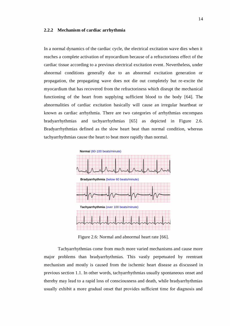
known as cardiac arrhythmia. There are two categories of arrhythmias encompass
bradyarrhythmias and tachyarrhythmias [65] as depicted in Figure 2.6.
Bradyarrhythmias defined as the slow heart beat than normal condition, whereas
tachyarrhythmias cause the heart to beat more rapidly than normal.
Normal (60-100 beats/minute)
Bradyarrhythmia (below 60 beats/minute)
Tachyarrhythmia (over 100 beats/minute)
Figure 2.6: Normal and abnormal heart rate [66].
Tachyarrhythmias come from much more varied mechanisms and cause more
major problems than bradyarrhythmias. This vastly perpetuated by reentrant
mechanism and mostly is caused from the ischemic heart disease as discussed in
previous section 1.1. In other words, tachyarrhythmias usually spontaneous onset and
thereby may lead to a rapid loss of consciousness and death, while bradyarrhythmias
usually exhibit a more gradual onset that provides sufficient time for diagnosis and
15
therapy, thereby leading to lower levels of death [66]. Therefore, this research will
focus on the simulation and analysis aspects of tachyarrhythmias related to the
reentrant mechanism.
Reentrant is a mechanism that occurs when cardiac tissue is excited
repeatedly by the action potential wave that keeps reentering the same anatomical
region [67]. Such reentry could lead to ventricular tachycardia that causes extremely
rapid excitation of the heart and even more dangerous cause ventricular fibrillation
(VF) [68]. This potentially causes a fatal risk of the heart’s ability to efficiently pump
blood throughout the body which could lead to a sudden death [61]. The reactivation
occurs indefinitely until the excitability of the tissue in the reentrant circuit is
somehow affected. In many situations, the cessation of reentrant activity occurs
through the interaction of the reentrant activation wave with an activation wave
originating from some other part of the heart [11].
2.2.2.1 Anatomical circus movement reentry
By far, the most common type of reentry is circus movement reentry [69], [70]. The
name refers to the circulation of an action potential wavefront around an anatomical
obstacle, which leads to repetitive activation of the tissue at a frequency that is
dependent on the velocity at which the wavefront conducts around the obstacle, and
the length of the path around the obstacle [67].
The simplest model of circus movement reentry is the closed ring, where the
activation wavefront rotates around an anatomical obstacle. Reentry around a closed
ring can typically be identified by the following characteristics: (i) the activation
wavefront moves around an anatomically distinct pathway, returning to its origin and
then following the same path again; (ii) the activation wavefront moves in one
direction only around the ring as a result of the unidirectional conduction block when
the reentry is initiated; and (iii) interruption of the reentrant circuit at any point along
its path terminates the reentry [71], [72].
For a given closed circuit to form a reentrant ring, the rotation time around
the ring must also be longer than the recovery period of all segments of the circuit,
which is another way of saying that the front and back of the action potential wave
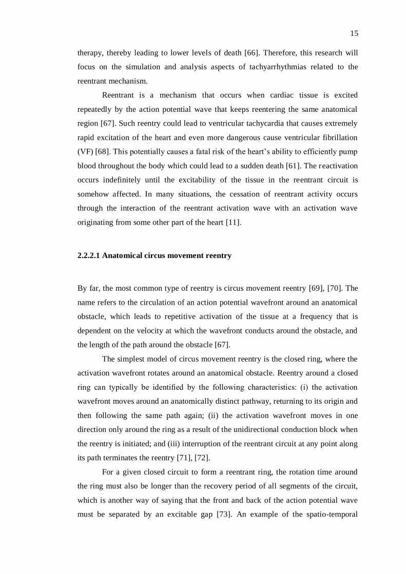
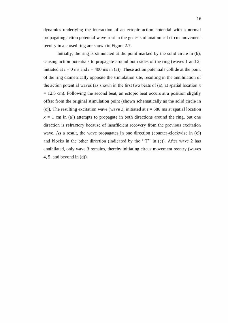
must be separated by an excitable gap [73]. An example of the spatio-temporal
16
dynamics underlying the interaction of an ectopic action potential with a normal
propagating action potential wavefront in the genesis of anatomical circus movement
reentry in a closed ring are shown in Figure 2.7.
Initially, the ring is stimulated at the point marked by the solid circle in (b),
causing action potentials to propagate around both sides of the ring (waves 1 and 2,
initiated at t = 0 ms and t = 400 ms in (a)). These action potentials collide at the point
of the ring diametrically opposite the stimulation site, resulting in the annihilation of
the action potential waves (as shown in the first two beats of (a), at spatial location x
= 12.5 cm). Following the second beat, an ectopic beat occurs at a position slightly
offset from the original stimulation point (shown schematically as the solid circle in
(c)). The resulting excitation wave (wave 3, initiated at t = 680 ms at spatial location
x = 1 cm in (a)) attempts to propagate in both directions around the ring, but one
direction is refractory because of insufficient recovery from the previous excitation
wave. As a result, the wave propagates in one direction (counter-clockwise in (c))
and blocks in the other direction (indicated by the ‘‘T’’ in (c)). After wave 2 has
annihilated, only wave 3 remains, thereby initiating circus movement reentry (waves
4, 5, and beyond in (d)).
17
Figure 2.7: Unidirectional block and circus movement reentry. (a) A space-time
diagram showing trans-membrane voltage as a function of time and position around
the ring (b) - (d) Schematic diagrams of the scenario depicted in panel (a), The
numbers in the panel (b) - (d) correspond to the wave number in the panel (a) [74]

18
2.3 Approaches in cardiac electrophysiological analysis
Previously mentioned in section 1.1 research backgrounds, there are three types of
analysis tools to explore and interpret the underlying mechanisms in cardiac
electrophysiological which are experimental, clinical and computational model
simulation. This subtopic will discuss further details of these three methods.
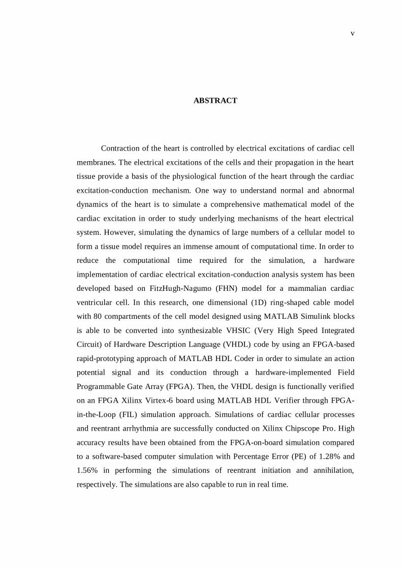
2.3.1 Experimental approach
Experimental approach is used to reveal the underlying mechanisms in the electrical
state of the heart based on a lot of experimental researches performed on the heart of
mammalian animal such as rat, guinea pig, rabbit and dog as illustrated in Figure 2.8.
However, due to the limited procedures and study parameters of this invasive
analysis approach, discovering underlying mechanisms of the heart primarily the
cardiac arrhythmias is quite challenging [75], [76], [77].
Coronary artery
ligated inducing
an infarct
Infarct
Figure 2.8: Rat model of myocardial infarction [78].
Another major limitation of any experimental study of ventricular
arrhythmias is that patterns of excitation can be recorded with reasonable resolution
only from the surface of the heart, whereas the underlying excitation patterns are
three dimensional (3D). Moreover, the spatial resolution of 3D current measurement
technique used in the experimental study is insufficient to identify the reentrant
sources of arrhythmias and to study their dynamics [79]. Thus, computational model
19
simulation, especially detailed quantitative modeling of the human heart, can play an
important role in overcoming these types of limitations [80].
2.3.2 Clinical approach
At its most basic, a clinical measurement electrophysiology study requires equipment
to allow recording of cardiac activity and delivery of electrical stimulation to the
human heart. Besides, clinical cardiac electrophysiology approach, involving
intracardiac recording and electrical stimulation, have been a major importance in
elucidating the mechanisms of cardiac arrhythmias. Therefore, such simple
procedures are now rarely performed and modern clinical electrophysiology
equipment is designed to undertake more complex studies, with recordings from
multiple intracardiac electrode catheters, and programmed electrical stimulation for
the induction and investigation of tachyarrhythmias. The induction of ventricular
fibrillation (VF) by electrical currents which associated with abnormal wave
propagation caused by reentrant sources of excitation, was demonstrated as early as
1899 by Prevost and Battelli [81], but the electrical induction and termination of
arrhythmias essentially started in the early 1950s. Reentry as a mechanism for
tachyarrhythmias is proposed initially by Mines [82] and the presence of potential
substrate for reentry was identified by Moe [83].
Although the clinical studies are generally favourable, investigating the
cardiac electrical behavior clinically poses a number of challenges such as various
risk assessment procedures to the patient, limited sources of data and uncertainty or
less informative with respect to the long-term performance of the device [84], [85].
In this case, the procedures of clinical testing may significantly influence to elucidate
the underlying the mechanism in cardiac electrophysiological determination while
independent of the clinical findings. Therefore, the use of model simulation is to
avoid the limitations of clinical study and improve the knowledge of arrhythmia
mechanism. These aspects provide a better understanding of abnormal cardiac
electrical activity at various levels such as in the ion channels, cells, tissues and
organ.
20
2.3.3 Model simulation approach
The availability of precise information on the formation and transmission of cardiac
impulses, under both normal and pathological conditions, has helped elucidate the
underlying mechanisms of cardiac arrhythmia. Several relevant topics have been
considered in a number of experimental and clinical studies in the field of cardiac
electrophysiology. These include analyses of the structure and function of the ionic
channels that determine the action potential of cardiac cells, as well as the factors
that regulate transmembrane ionic currents, such as voltage, time elapsed since
activation, frequency, and ion concentrations [86]. However, the understanding of
underlying mechanism of the cardiac electrophysiology is still incomplete in many
aspects, and there are limitations to some of the procedures used to treat various
cardiac arrhythmias for instance fibrillation processes.
The creation of models for an example, theoretical simulations of the
electrophysiological phenomena based on mathematical model, forms a part of the
efforts aimed at enhancing understanding of these phenomena and predicting
behavior in various normal and abnormal mechanisms. The development of such
models has been driven by several factors, including; i) Precise experimental and
clinical data collection. This has been essential to constructing models based on real
data and verifying how well such models work. ii) Advances in information
processing and compilation capabilities through the use of faster, increasingly more
complex computers. iii) The use of the models and simulations themselves to
improve our understanding of the underlying cardiac arrhythmia mechanism and to
predict responses under conditions that is sometimes hard to reproduce in
experimental preparations. A look at the historical evolution of the mathematical
models will be discussed afterwards in section 2.4.
The technique in the model simulation can be divided into two types which
are a software-based simulation and a hardware-based simulation. The detail reviews
regarding both methods are discussed in the section 2.3.3.1 and 2.3.3.2.

21
2.3.3.1 Software-based simulation
Computer simulations referred to the cardiac simulations that are performed on
computer software such as Visual Studio software by using the C++ Programming
Language, MATLAB Simulink by using graphical programming blocks, and etc.
Recent advancements in computational science and the development of high
performance computers have increased the usage of the computer simulation
approaches in order to study the underlying mechanism of the cardiac. The computer
simulation is the most favourable approach as it enabled the creation of multi-scale
simulation by using the cardiac models. However, the computer simulations require
huge computational resources, thus computational efficiency becomes a prime
concern [87].
The computer simulations based on various types of mathematical models
used to study cardiac arrhythmias show that the available information of the models
has grown in complexity and advances in computational techniques have been made
in the capacity to process it rapidly and efficiently. Parallel to the development
parameters and variables of the mathematical models to study the formation of the
action potential of cardiac cells, fast computational speed has been essential in order
to analyse the data which contributed to the usage of supercomputer for analysis
[88]. However, supercomputer is expensive and sizable which require a large room
to store them [89]. Therefore, a new solution needs to be introduced to elucidate the
mechanism of the heart such as hardware implementation.
2.3.3.2 Hardware-based simulation
Hardware implementation represents here is the cardiac mathematical model that is
adapted into the hardware to perform real-time simulations such as by using FPGA,
FPAA, GPU, DSP, Application Specific Integrated Circuit (ASIC) and etc. Recently,
the hardware is widely chosen by researchers as the computational tool to study the
underlying mechanism of the cardiac since it provides faster execution time compare
to computer simulation according to its real-time and parallel mode execution.
Furthermore, certain types of the hardware platform require very low power
consumption in their operation.
22
2.3.4 Comparison between experimental, clinical and model simulations
approaches
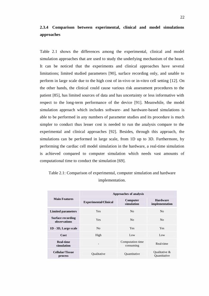
Table 2.1 shows the differences among the experimental, clinical and model
simulation approaches that are used to study the underlying mechanism of the heart.
It can be noticed that the experiments and clinical approaches have several
limitations; limited studied parameters [90], surface recording only, and unable to
perform in large scale due to the high cost of in-vivo or in-vitro cell setting [12]. On
the other hands, the clinical could cause various risk assessment procedures to the
patient [85], has limited sources of data and has uncertainty or less informative with
respect to the long-term performance of the device [91]. Meanwhile, the model
simulation approach which includes software- and hardware-based simulations is
able to be performed in any numbers of parameter studies and its procedure is much
simpler to conduct thus lesser cost is needed to run the analysis compare to the
experimental and clinical approaches [92]. Besides, through this approach, the
simulations can be performed in large scale, from 1D up to 3D. Furthermore, by
performing the cardiac cell model simulation in the hardware, a real-time simulation
is achieved compared to computer simulation which needs vast amounts of
computational time to conduct the simulation [69].
Table 2.1: Comparison of experimental, computer simulation and hardware
implementation.
Main Features
Approaches of analysis
Experimental/Clinical Computer
simulation
Hardware
implementation
Limited parameters Yes No No
Surface recording
observations Yes No No
1D - 3D, Large scale No Yes Yes
Cost High Low Low
Real-time
simulation -
Computation time
consuming Real-time
Cellular/Tissue
process Qualitative Quantitative
Qualitative & Quantitative
23
2.4 Mathematical models for cardiac electrical activity
Cardiac electrophysiology concerns on the studies of the electrical activity of the
heart under both normal and abnormal conditions. A cardiac excitation occurs when
ions flowing through ion channels in and out of the plasma membrane that generates
currents and causes changes in membrane potential from resting to an action
potential. In a definition, an action potential is the electrical signal of cell that passes
through the cardiac during the cell is excites. Commonly, the action potential is
triggered by a voltage spike from the action potential of its neighboring tissue or
from an artificial pacing signal [93].
Action potential models have been very useful in investigating different
features of cardiac physiology, from action potential generation in a single-cell to
action potential conduction in a multidimensional structure of cardiac tissues [94]. In
which, the action potential models of cardiac cells are defined by mathematical
descriptions of electrical events at the cellular level that give rise to cardiac action
potentials. The models that describe action potentials and the ion transport across the
cell membrane are also referred to as ionic models [66].
Most the ionic models of cardiac cells are based on the Hodgkin-Huxley
formulation, which first that intend to formulate mathematically the cellular
processes to lead the generation of the action potential in the squid nerve axon [95].
The model has been built in 1952 which are proposed an ion model that is
specialized by three types of ion channel currents that are involved in the generation
of the action potential to represent the sodium, potassium and chloride (leakage or
background) through the membrane of squid axon [19]. The currents are
characterised as the product of conductance and the differences of the driving forces
for ions, namely chemical gradient and electrical gradient. The conductance of
sodium and potassium are time-dependent and voltage-dependent, and modeled by
using gating variables, which are formulated by first-order ODEs. The Hodgkin-
Huxley model described the way in which ionic currents vary with membrane
potential and time. Its structure forms a basis for almost all models of excitable
membrane behavior [96].
FitzHugh and Nagumo, are two of the first researchers reduce ODEs in the
Hodgkin-Huxley model from four state variables of gates in the potassium, sodium

24
and leakage conductance which describe the ionic channel currents to two state
variables of excitation membrane voltage and refractory period [97]. FHN model
used a single mathematical process to represent multiple channel properties and it
preserved the essential behavior of the membrane while offering a lower
computational cost compared to other models, it has been frequently used as the
excitability component of cardiac action potential propagation [98]. Consequently,
the FHN model is applied in this study in developing a new simulation-based
analysis technique in cardiac excitation and conduction studies.
In 1962, Noble published one of the first mathematical models of a cardiac
cell applicable for long lasting action and pacemaker potentials of the Purkinje fibers
of the heart [23]. The Noble model adopted all the gating variables from Hodgkin-
Huxley, and is also the four-variable differential system [22]. Then, continued by
Beeler-Reuter model issued in 1977, is a pioneering effort to describe the cardiac
ventricular action potential [25]. The experimental data used in the Beeler-Reuter
model is subjected to limitation in available voltage clamp techniques and their
application to multicellular preparations of cardiac muscle [99].
The Luo-Rudy I model, a system of eight variable ODEs for describing six
types of ion channel currents (IK1, IKp, Ib, INa, IK, Isi) to present the flow of sodium,
potassium, calcium and chloride currents is based on the data derived from the new
measurement techniques. The Luo-Rudy I model is published in 1991 [26] and Luo-
Rudy II model further developed in 1994 which described the electrophysiology of
guinea pig ventricular cells [27]. The Luo-Rudy II model a system of fourteen
variable ODEs. This model is also based on guinea pig ventricular cells from a
single-cell experimental data [100]. It provides a framework for future development
of models of the excitation-contraction coupling process in cardiac cells.
Since then, the mathematical models turn out numerous onward, but
complicated from year to year as variable parameters in the mathematical
descriptions are increased in order to represent the cellular processes in more detail.
Progress in mathematical modeling has facilitated simulations as a tool for
investigating cardiac dynamics. However, simulating the dynamics of large numbers
of cellular models forming a tissue model requires an immense amount of
computational time, while, hardware implementation could provide a high
performance simulation system for the electrophysiological analysis.
101
REFERENCES
[1] World Health Organization (2016). The top 10 causes of death. Retrieved on
November 21, 2016, from http://www.who.int/mediacentre/factsheets
[2] Tunstall-Pedoe, H. Coronary Heart Disease. British Medical Journal. 1991.
303(6804): 701–704.
[3] Eick, H. H. R., Robert, E. T. and David, W. W. Connections : Heart Disease,
Cellular Electrophysiology, and Ion Channels. Federation of American
Societies for Experimental Biology. 1996. 6(8): 2568–2580.
[4] Glass, L., Nagai, Y., Hall, K., Talajic, M. and Nattel, S. Predicting the
Entrainment of Reentrant Cardiac Waves Using Phase Resetting Curves. The
American Physical Society. 2002. 65(2): 1–10.
[5] Xu, B., Binczak, S., Jacquir, S., Pont, O. and Yahia, H. Complexity Analysis
of Experimental Cardiac Arrhythmia. IEEE Region 10 Symposium. Kuala
Lumpur, Malaysia. 2014. pp. 23–28.
[6] Niederer, S., Mitchell, L., Smith, N. and Plank, G. Simulating Human Cardiac
Electrophysiology on Clinical Time-Scales. Frontiers in Physiology. 2011.
2(14): 1–7
[7] John Camm, A. Heart Rate Variability. European Heart Journal. 1996. 17:
54–381.
[8] Priebe, L. and Beuckelmann, D. J. Simulation Study of Cellular Electric
Properties in Heart Failure. Circulation Research: American Heart
Association. 1998. 82: 1206–1223.
[9] Li-ping, C., Li, L., Lin, Y., Yin-bin, J. and Hong, Z. Effects of Low [K]o on
Cardiac Excitation Conduction and Membrane Currents in the Vicinity of
Vulnerable Window : A Computer Simulation Study. IEEE 5th Int. Conf. on
Bioinformatics and Biomedical Engineering (iCBBE). Wuhan, China. 2011.
pp. 1–4.
[10] Qi, J., Chen, M., Huo, Y., Yu, J.,. Zhang, M., Chen, M., Huo, Y., Yu, J. and
Zhang, M. Cardiac Arrhythmia in Sepsis-A Simulation Study. Experimental
and Clinical Cardiology. 2014. 20(6): 4017–4045.
[11] Mahmud, F. Real-Time Simulation and Control of Spatio-Temporal Cardiac
Excitation Using an Analog-Digital Hybrid Circuit Model. Ph.D. Thesis.
Osaka University; 2011.
[12] Carusi, A., Burrage, K. and Rodriguez, B. Bridging Experiments, Models and

102
Simulations: An Integrative Approach to Validation in Computational Cardiac
Electrophysiology. American Journal of Physiology. 2012. 303(2): 144–155.
[13] Ritchie, J. L. Guidelines for Clinical Intracardiac Electrophysiological and
Catheter Ablation Procedures. Journal of the American College of Cardiology,
and the Journal of Cardiovascular Electrophysiology. 1995. 92: 673–691.
[14] Quinn, T. A. and Kohl, P. Combining Wet and Dry Research: Experience with
Model Development for Cardiac Mechano-electric Structure-function Studies.
Cardiovascular Research, 2013. 97(4): 601–611.
[15] Glancy, G. D. and Chaimowitz, G. The Clinical Use of Risk Assessment.
2015. Canadian Journal of Psychiatry. 2005. 50(1): 12–17.
[16] Travaglia, J. and Debono, D. Clinical Audit : A Comprehensive Review of the
Literature. Centre for Clinical Governance Research in Health, Faculty of
Medicine, University of New South Wales, Sydney, Australia. 2009.
[17] Vigmond, E. J., Hughes, M., Plank, G. and Leon, L.J. Computational Tools
for Modeling Electrical Activity in Cardiac Tissue. Journal of
Electrocardiology. 2003. 36: 69–74.
[18] Clayton, R.H., Bernus, O., Cherry, E. M., Dierckx, H., Fenton, F. H.,
Mirabella, L., Pan, A. V., Sachse, F. B., Seemann, G. and Zhang, H. Models
of Cardiac Tissue Electrophysiology : Progress, Challenges and Open
Questions. Progress in Biophysics and Molecular Biology. 2011. 104: 22–48.
[19] Hodgkin, A. L. and Huxley, A. F. A Quantitative Description of Membrane
Current and Its Application to Conduction and Excitation in Nerve.
Physiology Journal. 1952. 117: 500–544.
[20] Fitzhugh, R. Thresholds and Plateaus in the Hodgkin-Huxley Nerve
Equations. The Journal of General Physiology. 1960. 43: 867–896.
[21] Nagumo, J., Arimoto, S. and Yoshizawa, S. An Active Pulse Transmission
Line Simulating Nerve Axon. Proc. IRE. 1962. 50(10): 2061–2071.
[22] Noble, D. Cardiac Action and Pacemaker Potentials Based on the Hodgkin-
Huxley Equation. Nature. 1960. 188: 495–497.
[23] Noble, D. A Modification of the Hodgkin-Huxley Equations Applicable to
Purkinje Fibre Action and Pacemaker Potentials. J. Physiol., 1962. 160: 317–
352.
[24] Reuter, H. Divalent Cations as Charge Carriers in Excitable Membranes.
Progress in Biophysics & Molecular Biology. 1973. 26: 1–43.
[25] Beeler, G. W. and Reuter, H. Reconstruction of the Action Potential of
Ventricular Myocardial Fibres. J. Physiol. 1977. 268: 177–210.
[26] Luo, C. H. and Rudy, Y. A Model of the Ventricular Cardiac Action Potential.
Depolarization, Reolarization, and their Interaction. Circulation Research.
1991. 68(6): 1501–1526.

103
[27] Luo, C. and Rudy, Y. A Dynamic Model of the Cardiac Ventricular Action
Potential. I. Simulations of Ionic Currents and Concentration Changes.
Journal of the American Heart Association. 1994. 74: 1071–109.
[28] Mahmud, F. Real-time Simulations for Resetting and Annihilation of
Reentrant Activity Using Hardware-implemented Cardiac Excitation
Modeling. IEEE Int. Conf. on EMBS Biomedical Engineering and Sciences
(IECBES). Langkawi, Malaysia. 2012. pp. 321–325.
[29] Potse, M. Mathematical Modeling and Simulation of Ventricular Activation
Sequences: Implications for Cardiac Resynchronization Therapy. Journal of
Cardiovascular Translational Research. 2012. 5(2): 146–158.
[30] Yu, D., Du, D., Yang, H. and Tu, Y. Parallel Computing Simulation of
Electrical Excitation and Conduction in the 3D Human Heart. 36th Annual Int.
Conf. of the IEEE Engineering in Medicine and Biology Society (EMBC).
Chicago, Illinois, USA. 2014. pp. 4315–4319.
[31] Vigmond, E. J., Boyle, P. M., Leon, L. J. and Plank, G. Near-Real-Time
Simulations of Biolelectric Activity in Small Mammalian Hearts using
Graphical Processing Units. 31st Annual Int. Conf. of the IEEE Engineering in
Medicine and Biology Society (EMBC). Minneapolis, Minnesota, USA. 2009.
pp. 3290–3293.
[32] Huang, C., Vahid, F. and Givargis, T. A Custom FPGA Processor for Physical
Model Ordinary Differential Equation Solving. IEEE Embeded System Letter.
2011. 3(4): 113–116.
[33] Hu, S., Wei, H., Chen, Y. and Tan, J. A Real-Time Cardiac Arrhythmia
Classification System with Wearable Sensor Networks. Sensors Journal.
2012. 12: 12844–12869.
[34] Rajiah, P. and Schoenhagen, P. The Role of Computed Tomography in Pre-
procedural Planning of Cardiovascular Surgery and Intervention. Insights
Imaging. 2013. 4(5): 671–689.
[35] Sorensen, T. S., Therkildsen, S. V., Makowski, P., Knudsen, J. L. and
Pedersen, E. M. A New Virtual Reality Approach for Planning of Cardiac
Interventions. Artificial Intelligence in Medicine. 2011. 22(3): 193–214.
[36] Maeda, Y., Yagi, E. and Makino, H. Synchronization with Low Power
Consumption of Hardware Models of Cardiac Cells. BioSystems. 2005. 79(1–
3): 125–131.
[37] Charles, G., Gordon, C. and Alexander, W. E. An Implementation of a
Biological Neural Model using Analog-Digital Integrated Circuits. IEEE Int.
Conf. Behavioral Modeling and Simulation Workshop (BMAS). San Jose,
USA. 2008. pp. 78–83.
[38] Chen, H., Sun, S., Aliprantis, D. C. and Zambreno, J. Dynamic Simulation of
Electric Machines on FPGA Board. IEEE Int. Electric Machines and Drives
Conf. (IEMDC). Miami, Florida, USA. 2009. pp. 1523–1528.
[39] Fox, P. J. Massively Parallel Neural Computation. University of Cambridge

104
Computer Laboratory, United Kingdom. 2013.
[40] Hassan, E., Mimouni, E. and Karim, M. A MicroBlaze-Based Multiprocessor
System on Chip for Real-Time Cardiac Monitoring. IEEE Int. Conf.
Multimedia Computing and Systems (ICMCS). Marrakesh, Morocco. 2014. pp.
331–336.
[41] Mahmud, F., Sakuhana, T., Shiozawa, N. and Nomura, T. An Analog-Digital
Hybrid Model of Electrical Excitation in a Cardiac Ventricular Cell.
Transactions of Japanese Society for Medical and Biological Engineering.
2009. 47(5): 428–435.
[42] Petrovas, A., Lisauskas, S. and Slepikas, A. Investigation of Microcontroller
Based Model of FitzHugh-Nagumo Neuron. 15th IEEE Int. Conf.
MECHATHRONIKA. Prague, Czech Republic. 2012. pp. 1–4.
[43] Amorim, R. M., Rocha, B. M., Campos, F. O. and Dos Santos, R. W.
Automatic Code Generation for Solvers of Cardiac Cellular Membrane
Dynamics in GPUs. 32nd Annual IEEE Int. Conf of the IEEE Engineering in
Medicine and Biology Society (EMBS). Buenos Aires, Argentina. 2010. pp.
2666–2669.
[44] Zhao, J. and Kim, Y. Circuit Implementation of FitzHugh-Nagumo Neuron
Model Using Field Programmable Analog Arrays. 50th IEEE Midwest
Symposium, Circuits and Systems (MWSCAS). Montreal, Canada. 2007. pp.
772–775.
[45] Korkmaz, N., Ozturk, I. and Kilic, R. Multiple Perspectives on the Hardware
Implementations of Biological Neuron Models and Programmable Design
Aspects. Turkish Journal of Electrical Engineering & Computer Sciences.
2016. 24: 1729–1746.
[46] Kadam, M. and Sawarkar, K. An Overview of Reconfigurable Hardware for
Efficient Implementation of DSP Algorithms. International Organization of
Scientific Research Journal of Engineering (IOSRJEN). 2014. 4(2): 34–43.
[47] Weinstein, R. K. and Lee, R. H. Architectures for High-Performance FPGA
Implementations of Neural Models. Journal of Neural Engineering. 2006. 3:
21–34.
[48] Dinechin, F., Detrey, J., Cret, O. and Tudoran, R. When FPGAs are Better at
Floating-Point Than Microprocessor. Proc. of the 16th International
ACM/SIGDA Symposium on Field Programmable Gate Arrays. Monterey,
California, USA. 2008. pp. 1–13.
[49] Yang, S. A Biologically Plausible Real-time Spiking Neuron Simulation
Environment Based on a Multiple-FPGA Platform. ACM SIGARCH Computer
Architecture News. 2011. 39(4): 78–81.
[50] Desai, V. Electrocardiogram (ECG/EKG ) using FPGA. Master’s Thesis. San
Jose State, University. 2012.
[51] Tze Weng, O. W, Chia, W. C., Bakhteri, R. and Hau, Y. W. SoC-based
Design of Arrhythmia Detector. 2nd IEEE Int. Conf. on Electronic Design

105
(ICED). Penang, Malaysia. 2014. pp. 42–46.
[52] Zairi, H, Talha, M. K., Benouar, S. and Amer, A. A. Intelligent System for
Detecting Cardiac Arrhythmia on FPGA,” 5th IEEE Int. Conf. on Information
and Communication Systems (ICICS) Intelligent. Irbid, Jordan. 2014. pp. 1-5.
[53] Nomura, T and Glass, L. Entrainment and Termination of Reentrant Wave
Propagation in a Periodically Stimulated Ring of Excitable Media. The
American Physical Society. 1996. 53(6): 6353–6360.
[54] Panfilov, A. V., Keldermann, R. H. and Nash, M. P. Drift and Breakup of
Spiral Waves in Reaction-Diffusion-Mechanics Systems. Proc. of the National
Academy of Science. 2007. 104(19): 7922–7926.
[55] Siwakoti, Y. P. and Town, G. E. Design of FPGA-Controlled Power
Electronics and Drives Using MATLAB Simulink. IEEE Int. Conf. Asia
Downunder. Melbourne, Australia, 2013. pp. 571–577.
[56] Joseph, S. (2016). Electrocardiodiagrams. Retrieved on December 14, 2016,
from http://www.primedix.net/service/electrocardigrams
[57] Usyk, T. P., Belik, M. E., Michailova, A. and Mcculloch, A. D. Three-
Dimensional Model of Cardiac Electromechanics: Cell to Organ. Proc. of the
Second Joint EMBS/BMES Conference. Houston, USA. 2002. pp. 1220–1221.
[58] Wu, X. D., Shen, Y. L., Bao, J. L., Cao, C. M., Xu, W. H. and Xia, Q. A
Model of Gap Junction Conductance and Ventricular Tachyarrhythmia. Proc.
23rd Annual Int. Conf. IEEE EMBS. Istanbul, Turkey. 2001. pp. 28–31.
[59] Yan, G. X., Shimizu, W. and Antzelevitch, C. Characteristics and Distribution
of M Cells in Arterially Perfused Canine Left Ventricular Wedge Preparations.
Circulation Research: American Heart Association. 1998. 98(18): 1921–1927.
[60] Grant, A. O. Cardiac Ion Channels. Circulation: Arrhythmia and
Electrophysiology. 2009. 2: 185–194.
[61] Fenton, F. H., Cherry, E. M., Karma, A. and Rappel, W. J. Modeling Wave
Propagation in Realistic Heart Geometries Using The Phase-Field Method.
Chaos. 2005. 15(1): 21–23.
[62] Wang, S., Xie, Y. and Qu, Z. Coupled Iterated Map Models of Action
Potential Dynamics in a One-Dimensional Cable of Cardiac Cells. New
Journal of Physics. 2008. 10(5): 55001–55024.
[63] Jaye, D. A., Xiao, Y. and Sigg, D. C. Basic Cardiac Electrophysiology :
Excitable Membranes. In: Cardiac Electrophysiology Methods and Models.
University of Minnesota: Minneapolis, USA pp; 41–52; 2010.
[64] Veeraraghavan, R., Gourdie, R. D. and Poelzing, S. Mechanisms of Cardiac
Conduction: A History of Revisions. American Journal Physiology
Heart.2014. 306(38): 619–627.
[65] Sampath, V., Sarode, S. and Mangharam, R. Heart-on-a-Chip: A Closed-loop
Testing Platform for Implantable Pacemakers. Real-Time and Embedded
106
Systems Lab (mLAB), University of Pennsylvania. 2013.
[66] Bartocci, E., Cherry, E. M., Glimm, J., Grosu, R., Smolka, S. A. and Fenton,
H. Toward Real-Time Simulation of Cardiac Dynamics. Proc. 9th ACM Int.
Conf. Comput. Methods Syst. Biol. Paris, France. 2011. pp. 103–110.
[67] Marchlinski, M. E. and Betensky, B. P. Cardiac Arrhythmias. Rev Esp
Cardiol. Elsevier. 2012. 65(2): 174–185.
[68] Nanthakumar, K., Walcott, G. P., Melnick, S., Rogers, J. M., Kay, M. W.,
Smith, W. M., Ideker, R. E. and Holman, W. Epicardial Organization of
Human Ventricular Fibrillation. Hear. Rhythm Soc. 2004 1(1): 14–23.
[69] Mahmud, F. Real-time Simulation of Cardiac Excitation Using Hardware-
implemented Cardiac Excitation Modeling. International Journal of
Integrated Engineering. 2012. 4(3): 13–18.
[70] Cimponeriu, A., Frank Starmer, C. and Anastasios, B. Ischemic Modulation of
Vulnerable Period and the Effects of Pharmacological Treatment of Ischemia-
Induced Arrhythmias: A Simulation Study. IEEE Trans. On Biomedical
Engineering. 2003. 50(2): 168–177.
[71] Gray, R. A. and Chattipakorn, N. Termination of Spiral Waves During
Cardiac Fibrillation via Shock-Induced Phase Resetting. Proc. Natl. Acad. Sci.
U. S. A. 2005. 102(13): 4672–4677.
[72] Krogh-Madsen, T. and Christini, D. J. Resetting and Termination of Reentry
in a Loop-and-Tail Cardiac Model. Phys. Rev. E - Stat. Nonlinear, Soft Matter
Phys.. 2008. 77(1): 1–5.
[73] Cherry, M., Fenton, F. H. and Gilmour, R. F. Mechanisms of Ventricular
Arrhythmias : A Dynamical Systems-Based Perspective. Am J Physiol Hear.
Circ Physiol. 2012. 302(12): 2451–2463.
[74] Jordan, P. and Christini, D. Cardiac Arrhythmia. First Edition. Wiley Encycl.
Biomed. Eng. 2006.
[75] Clayton, R. H., Murray, A. and Campbell, R. W. Objective Features of the
Surface Electrocardiogram During Ventricular Tachyarrhythmias. Eur. Heart
J.. 1995. 16(8): 1115–1119.
[76] Valderrabano, M. Frequency Analysis of Ventricular Fibrillation in Swine
Ventricles. Circ. Res.. 2002. 90(2): 213–222.
[77] Jalife, J. Ventricular Fibrillation: Mechanism of Initiation and Maintanance.
Annu. Rev. Physiol.. 2000. 62(1): 25–50.
[78] National Institute of Health, (2016). Can Stem Cells Repair a Damaged
Heart?. Retrieved on October 15, 2016, from
https://stemcells.nih.gov/info/2001report/chapter9/htm
107
[79] Trayanova, N., Constantino, J., Ashihara, T. and Plank, G. Modeling
Defibrillation of the Heart: Approaches and Insights. IEEE Rev. Biomed. Eng..
2011. 4(1): 89–102.
[80] Tentusscher, K., Bernus, O., Hren, R. and Panfilov, A. Comparison of
Electrophysiological Models for Human Ventricular Cells and Tissues. Prog.
Biophys. Mol. Biol.. 2006. 90(1–3): 326–345.
[81] Dreifuss, J.-J. Electric Countershock and External Cardiac Massage. Rev.
Med. Suisse Romande. 2011. 7(1): 511–512.
[82] Mines, G. R. On Dynamic Equilibrium in the Heart. J. Physiol.. 1913. 46(4–
5): 349–383.
[83] Moe, G. K., Preston, J. B. and H. Burlington. Physiologic Evidence for a Dual
A-V Transmission System. Circ. Res.. 4(4): 357–375.
[84] Rankin, A. C., Quinn, F. R. and Rae, A. P. Clinical Cardiac
Electrophysiology. American College of Physicians. 2012. 2(118): 1–17.
[85] Ehrlich, J. R., Hohnloser, S. H. and Nattel, S. Role of Angiotensin System and
Effects of Its Inhibition in Atrial Fibrillation: Clinical and Experimental
Evidence. Eur. Heart J.. 2006. 27(5): 512–518.
[86] Chorro Gascó, F. J. Mathematical Modeling and Simulations to Study Cardiac
Arrhythmias. Rev. Española Cardiol.. 2005. 58(1): 6–9.
[87] Sugiura, S., Washion, T., Hatano, A., Okada, J., Watanabe, H. and Hisada, T.
Multi-scale Simulations of Cardiac Electrophysiology and Mechanics using
the University of Tokyo Heart Simulator. Elsevier Ltd., 2012. 110(2–3): 380–
389.
[88] Noble, D., Garny, A., and Noble, P. J. How the Hodgkin-Huxley equations
inspired the Cardiac Physiome Project. J. Physiol.. 2012. 590(11): 2613–28.
[89] Wang, W., Huang, H., Kay, M. and Cavazos, J. GPGPU Accelerated Cardiac
Arrhythmia Simulations. 33rd Annu. Int. Conf. IEEE EMBS. Boston, MA,
USA. 2011. pp. 724–727.
[90] Cherry, E. M. and Fenton, F. H. Visualization of Spiral and Scroll Waves in
Simulated and Experimental Cardiac Tissue. New J. Phys.. 2008. 10(1): 1–43.
[91] Zannoli, R., Bianchini, D. and Corazza, I. A Medical Instrumentation
Laboratary Dedicated to Cardiovascular Training. Nurse Educ. Today. 2015.
35(8): 1–5.
[92] Wang, C. C., Shi, C., Brodersen, R. W. and Markovi, D. An Automated Fixed-
Point Optimization Tool in MATLAB XSG / SynDSP Environment. Int. Sch.
Res. Netw.. 2011. 2011(1): 1–17.
[93] Shih, H.-T. Anatomy of the Action Potential in the Heart. Texas Heart
Institute Journal. 1994. 21(1): 30–41.
[94] Wilders, R., Verkerk, A. O., Verheijck, E. E., Van Ginneken, A. C. G.,
108
Kumar, R., Wagner, M. B., Golod, D. A., Goolsby, W. N., Joyner, R. W. and
Jongsma, H. J. Model clamp: A computer tool to probe action potential
transfer between cardiac cells. Annu. Reports Res. React. Institute, Kyoto
Univ.. 2001. 4(1): 4036–4039.
[95] Bonabi, S. Y., Asgharian, H., Bakhtiari, R., Safari, S. and Ahmadabadi, M. N.
FPGA Implementation of a Cortical Network Based on the Hodgkin-Huxley
Neuron Model. Front. Neurosci.. 2012. 8(1): 1–12.
[96] Horng, T. L. and Huang, M. W. Spontaneous Oscillations in Hodgkin-Huxley
Model. J. Med. Biol. Eng.. 2006. 26(4): 161–168.
[97] Xu, B., Binczak, S., Jacquir, S., Pont, O. and Yahia, H. Parameters Analysis of
FitzHugh-Nagumo Model for a Reliable Simulation. 36th Annu. Int. Conf.
IEEE Eng. Med. Biol. Soc.. 26-30 Aug 2014. Chicago, IL, USA. 2014. pp.
4334–4337.
[98] Sundnes, J., Nielsen, B. F., Mardal, K. A., Cai, X., Lines, G. T. and Tveito, A.
On the Computational Complexity of the Bidomain and the Monodomain
Models of Electrophysiology. Ann. Biomed. Eng.. 2006. 34(7): 1088–1097.
[99] Wan, L. VLSI Design of Heart Model. Ph.D. Thesis. Maryland University;
2007.
[100] Rubin, L. J., Keller, R. S., Parker, J. L., Adams, H. R., Wu, X. D., Shen, Y. L.,
Bao, J. L., Cao, C. M., Xu, W. H. and Xia, Q. Contractile Dysfunction of
Ventricular Myotes Isolated from Endotoxemic Guinea Pigs. Shock Soc..
1994. 2(2): 113–120.
[101] Wedge, N. A., Branicky, M. S. and Cavusoglu, M. C. Computationally
Efficient Cardiac Bioelectricity Models Toward Whole-Heart Simulation. Int.
Conf. IEEE Eng. Med. Biol. Soc.. 2004. 4(1): 3027–3030.
[102] Goktepe, S. and Kuhl, E. Computational Modeling of Cardiac
Electrophysiology : A Novel Finite Element Approach. Int. J. Numer. Methods
Eng.. 2009. 79(2): 156-178.
[103] Izhikevich, E. and FitzHugh, R. FitzHugh-Nagumo model. Scholarpedia.
2006. 1(9): 1349.
[104] Rosolen, A. M., Ordas, S. and Frangi, A. F. Numerical Schemes for the
Simulation of Three-Dimensional Cardiac Electrical Propagation in Patient-
Specific Ventricular Geometries. Eur. Conf. Comput. Fluid Dyn.. 5-8 Sept
2006. Netherlands. 2006. pp. 1–15.
[105] Nouri, M., Karimi, G. R., Ahmadi, A. and Abbott, D. Digital Multiplierless
Implementation of the Biological FitzHugh – Nagumo Model.
Neurocomputing. 2015. 165(1): 1–9.
[106] Sadoudi, S., Azzaz, M. S., Djeddou, M. and Benssalah, M. An FPGA Real-
time Implementation of the Chen’s Chaotic System for Securing Chaotic
Communications. Int. J. Nonlinear Sci.. 2009. 7(4): 467–474.
[107] Kuon, I. and Rose, J. Measuring the Gap Between FPGAs and ASICs. Int.
109
Conf. IEEE Trans. Comput. Des. Integr. Circuits Syst.. 2007. 26(2):203–215.
[108] Titri, S., Larbes,C. and Toumi, K. Y. Rapid Prototyping of PVS into FPGA :
From Model Based Design to FPGA / ASICs Implementation. 9th Int. Des.
Test Symp.. 16-18 December 2014. Algiers, Algeria. 2014. pp. 162–167.
[109] Altera Corporation. Accelerating High-Performance Computing With FPGAs.
San Jose (USA): Innovation Drive. 2007.
[110] Shah, S., Hasler, J., Kim, S., Lal, I., Kagle, M. and Collins, M. A Remote
FPAA System for Research and Education. Proc.- IEEE Int. Symp. Circuits
Syst.. 2016–July. Montreal, QC. 2016. pp. 141–144.
[111] Deese A. S. and Nwankpa, C. O. Design and Testing of Custom FPAA
Hardware with Improved Scalability for Emulation of Smart Grids. IEEE
Trans. Smart Grid. 2014. 5(3): 1369–1378.
[112] Xu, F., Chen, H., Jin, W. and Xu, Y. FPGA Implementation of Nonlinear
Model Predictive Control. Control Decis. Conf.. 2014. 108(1): 108–113.
[113] Kehtarnavaz, N. and Mahotra, S. FPGA Implementation Made Easy for
Applied Digital Signal Processing Courses. Int. Conf. IEEE ICASSP Acoust.
Speech Signal Process. - Proc. Montreal, QC. 2011. pp. 2892–2895.
[114] George, S., Kim, S., Shah, S., Hasler, J., Collins, M., Adil, F., Wunderlich, R.,
Nease, S. and Ramakrishnan, S. A Programmable and Configurable Mixed-
Mode FPAA SoC. IEEE Trans. Very Large Scale Integr. Syst.. 2016. 24(6):
2253–2261.
[115] Wong, J. L. Flexible ASIC : Shared Masking for Multiple Media Processors.
Des. Autom. Conf. 2005. Proceedings. 42nd. Anaheim, CA. 2005. pp. 900–914.
[116] Farooq, U. FPGA Architectures : An Overview. In: Tree-Based
Heterogeneous FPGA Architectures. New York: Springer. pp; 7–148; 2012.
[117] Butt, S. A., Lavagno, L. and Torino, P. Model-Based Rapid Prototyping of
Multi Rate Digital Signal Processing Algorithms. IEEE Nordic
Microelectronics. Norchip, Lund. 2011. pp. 1–4.
[118] Eyre, J. and Bier, J. The Evolution of DSP Processors. IEEE Signal Process.
Mag.. 2000. 17(2): 43–50.
[119] Maslennikow, O. and Sergiyenko, A. Mapping DSP Algorithms into FPGA.
Proc. Int. Symp. Parallel Comput. Electr. Eng.. Bialystok. 2006. pp. 208–213.
[120] Hall, T. S. Field-Programmable Analog Arrays A Floating Gate Approach.
Ph.D. Thesis. Georgia Institute of Technology; 2004.
[121] Selow, R., Lopes, H. S. and Lima, C. R. E. A Comparison of FPGA and
FPAA Technologies for a Signal Processing Application. IEEE Field
Programmable Logic and Applications. Prague. 2009. pp. 230–235.
[122] Scekic, O. FPGA Comparative Analysis. University of Belgrade, Serbia. p.
48, 2005.

110
[123] SourceTech (2016). Top FPGA Companies for 2013. Retrieved on November
22, 2016, from http://sourcetech411.com/2013/04/top-fpga-companies-for-
2013/
[124] Xilinx All Programmable. Virtex-6 FPGA ML605 Evaluation Kit. San Jose
(USA): FPGA Design Platform. 2012.
[125] Mehta, N. Xilinx Redefines Power, Performance, and Design Productivity
with Three Innovative 28 nm FPGA Families : Virtex-7 , Kintex-7 , and Artix-
7 Devices. Xilinx. 2012. 373(1): 1–10.
[126] Monmasson, E., Member, S., Idkhajine, L., Cirstea, M. N., Member, S., Bahri,
I., Member, S. and Tisan, A. FPGAs in Industrial Control Applications. Ind.
Informatics, IEEE Trans.. 2011. 7(2): 224–243.
[127] Van Beek, S., Sharma, S. and Prakash, S. Four Best Practices for Prototyping
MATLAB and Simulink Algorithms on FPGAs. Electronic Engineering
Journal. 2012. 2(1): 49–53.
[128] Leue, S. and Johanna, T. Scenarios: Models, Transformations and Tools. New
York: Springer. p. 123, 2003.
[129] Jiang, Z., Pajic, M., Connoly, A., Dixit, S. and Mangharam, R. Real-time
Heart Model for Implantable Cardiac Device Validation and Verification.
22nd Euromicro Conf. Real-Time Syst.. Brussels. 2010. pp. 239–248.
[130] The MathWorks Inc. Model-Based Design with Simulink , HDL Coder, and
Xilinx System Generator for DSP. Natick, Massachusetts, USA: Matlab. 2012.
[131] The MathWorks Inc. HDL Coder TM Getting Started Guide. Natick,
Massachusetts, USA: Matlab. 2013.
[132] The MathWorks Inc. From MATLAB to HDL Implementation and Functional
Verification in 5 Short Steps. Natick, Massachusetts, USA: Matlab. 2014.
[133] The MathWorks Inc. Generating , Optimizing and Verifying HDL Code with
MATLAB and Simulink. Natick, Massachusetts, USA: Matlab. 2012.
[134] Maclennan, B. J. A Review of Analog Computing. New York: Springer. pp. 1–
45, 2007.
[135] Fasih, A., Trong, T. D., Chedjou, J. C. and Kyamakya, K. New
Computational Modeling for Solving Higher Order ODE Based on FPGA. 2nd
Int. Work. Nonlinear Dyn. Synchronization. Klagenfurt. 2009. pp. 49–53.
[136] Kheir, N. System Modeling and Computer Simulation. 2nd Edition. New
York: Printed in the United States of America, 1995.
[137] Cowan, G. E. R., Melville, R. C. and Tsividis, Y. P. A VLSI Analog
Computer/Digital Computer Accelerator. IEEE J. Solid-State Circuits. 2006.
41(1): 42–53.
[138] Matsubara, T. and Torikai, H. Asynchronous Cellular Automaton-Based
Neuron: Theoretical Analysis and On-FPGA Learning. IEEE Trans. Neural

111
Networks Learn. Syst.. 2013. 24(5): 736–748.
[139] Zhao, J., Jin, Y., Ma, L. and Corless, R. M. A Highly Efficient and Accurate
Algorithm for Solving the Partial Differential Equation in Cardiac Tissue
Models. Proc. 2006 WSEAS Int. Conf. Math. Biol. Ecol.. Miami, Florida,
USA. 2006. pp. 81–86.
[140] Shuaiby, S. M., Hassan, M. A., Sharkawy, A. and Gad, A. M. M. A Finite
Element Model for the Electrical Activity in Human Cardiac Tissues. J. Ecol.
Heal. Environ. 2013. 1(1): 25–33.
[141] Rachmuth, G. and Poon, C.-S. Transistor Analogs of Emergent Iono-Neuronal
Dynamics. HFSP J.. 2008. 2(3): 156–166.
[142] McRury, I. D. and Haines, D. E. Ablation for the Treatment. Proc. IEEE.
1996. 84(3): 404–416.
[143] Fallavollita, P., Savard, P. and Sierra, G. Fluoroscopic Navigation to Guide
RF Catheter Ablation of Cardiac Arrhythmias. Proc. 28th Annu. Int. Conf.
IEEE EMBS. 2004. 3(1): 1929–1932.
[144] Short, K. L. VHDL for Engineers. London: Pearson Education Ltd. pp. 169,
2009.
[145] Pellerin, D. An Introduction to VHDL for Synthesis and Simulation. Salt Lake
City, UT: Accolade Design Automation Inc. pp. 1–7, 1995.
[146] Menard, D., Chillet, D., Charot, F. and Sentieys, O. Automatic Floating-point
to Fixed-point Conversion for DSP Code Generation. Int. Conf. IEEE on
Automation Science and Engineering. Grenoble, France. 2002. pp. 1–7.
[147] Jha, S. and Seshia, S. A. Synthesis of Optimal Fixed-Point Implementation of
Numerical Software Routines. 6th International Workshop on Numerical
Software Verification. Berkeley, California. 2009. pp. 230–235.
[148] XILINX Chipscope Pro. Lower Verification Times by up to 50%. San Jose,
USA: Matlab. 2008. pp. 78–83.
[149] Khan, M. I. and Shah, S. M. A. Implementation of Advance Encryption
Standard Algorithm on FPGA for the Protection of Remote Sensing Satellite.
Proc. - 3rd IEEE Int. Conf. Comput. Sci. Inf. Technol.. 2010. 5(1): 147–151.
[150] Purohit, G., Chaubey, V. K., Raju, K. S. and Reddy, P. V. FPGA Based
Implementation and Testing of OVSF Code. International Conference on
Advanced Electronic Systems. Pilani, India. 2013. pp. 88–92.
[151] Arshak, K., Jafer, E. and Ibala, C. Testing FPGA Based Digital System using
XILINX ChipScope Logic Analyzer. 29th International Spring Seminar on
Electronics Technology (ISSE). St Marienthal, Germany. 2006. pp. 355–360.