Embed Size (px)
Citation preview
Review ArticleImplications of Oxidative Stress and Cellular Senescence inAge-Related Thymus Involution
Alexandra Barbouti ,1 Panagiotis V. S. Vasileiou ,2 Konstantinos Evangelou,2
Konstantinos G. Vlasis,3 Alexandra Papoudou-Bai ,4 Vassilis G. Gorgoulis,2,5,6
and Panagiotis Kanavaros1
1Department of Anatomy-Histology-Embryology, Medical School, University of Ioannina, Ioannina, Greece2Molecular Carcinogenesis Group, Department of Histology and Embryology, Medical School, National and Kapodistrian Universityof Athens, Athens, Greece3Department of Anatomy, Medical School, National and Kapodistrian University of Athens, Athens, Greece4Department of Pathology, Medical School, University of Ioannina, Ioannina, Greece5Faculty Institute for Cancer Sciences, Manchester Academic Health Sciences Centre, University of Manchester, Manchester, UK6Biomedical Research Foundation, Academy of Athens, Athens, Greece
Correspondence should be addressed to Alexandra Barbouti; [email protected]
Received 15 November 2019; Revised 20 January 2020; Accepted 23 January 2020; Published 5 February 2020
Academic Editor: Cinzia Domenicotti
Copyright © 2020 Alexandra Barbouti et al. This is an open access article distributed under the Creative Commons AttributionLicense, which permits unrestricted use, distribution, and reproduction in any medium, provided the original work isproperly cited.
The human thymus is a primary lymphoepithelial organ which supports the production of self-tolerant T cells with competentand regulatory functions. Paradoxically, despite the crucial role that it exerts in T cell-mediated immunity and prevention ofsystemic autoimmunity, the thymus is the first organ of the body that exhibits age-associated degeneration/regression, termed“thymic involution.” A hallmark of this early phenomenon is a progressive decline of thymic mass as well as a decreasedoutput of naïve T cells, thus resulting in impaired immune response. Importantly, thymic involution has been recently linkedwith cellular senescence which is a stress response induced by various stimuli. Accumulation of senescent cells in tissues hasbeen implicated in aging and a plethora of age-related diseases. In addition, several lines of evidence indicate that oxidativestress, a well-established trigger of senescence, is also involved in thymic involution, thus highlighting a possible interplaybetween oxidative stress, senescence, and thymic involution.
1. Introduction
The thymus is a central lymphoepithelial organ of theimmune system. Its primary function is to provide a uniquemicroenvironment in which T cell precursors (thymocytes),derived from hematopoietic stem cells, migrate and undergoselection, activation, clonal expansion, and differentiationinto self-tolerant, immunocompetent T cells that are releasedto the periphery [1, 2]. Proper T cell development requiresthe interaction of thymocytes with critical cellular popula-tions of the cortical and/or the medullary regions of thethymus, especially thymic epithelial cells (TECs) and den-dritic cells (DCs) [1, 3–6], which regulate thymopoiesis
through cell to cell contacts and production of soluble fac-tors (e.g., chemokines, cytokines, and extracellular matrixcomponents) [2, 7–11].
Despite the fundamental requirement for lifelong estab-lishment and maintenance of an overall effective and ade-quate defense against pathogens, the function of theimmune system deteriorates with age, affecting both innateand adaptive immune responses (immunosenescence) [12, 13].Surprisingly, the thymus is the first organ of the body thatexhibits age-associated changes, known as thymus regressionor involution, a biological event that takes place in almost allvertebrates, suggesting that this is an evolutionary ancientand conserved process [14, 15]. Although normal (disease-
HindawiOxidative Medicine and Cellular LongevityVolume 2020, Article ID 7986071, 14 pageshttps://doi.org/10.1155/2020/7986071
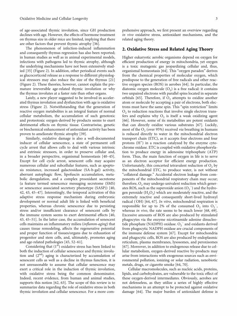
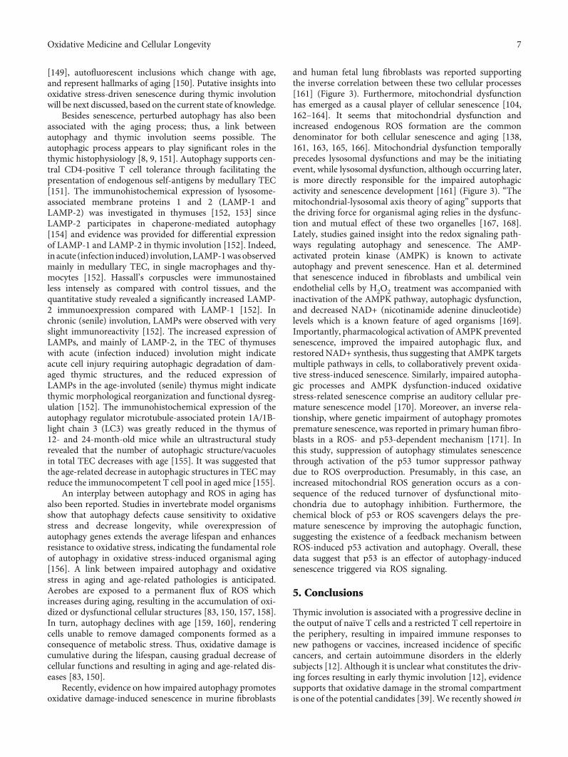
free) aging has a significant effect in the mass of specific high-metabolic-rate organs (for example, the brain, kidneys, liver,and spleen) [16], and muscle mass decreases with aging [17],these changes appear slowly and over a long period. On thecontrary, the thymus, which reaches its maximal size and Tcell output during early postnatal life, exhibits early thymicinvolution, a phenomenon that becomes even more promi-nent with advancing age [12, 13, 18–21]. Although the sizeof the human thymus seems to remain unchanged throughoutlife under normal conditions [22], in other vertebrates, itdeclines during aging [23]. Nevertheless, in almost all verte-brates having a thymus, thymic cellularity is progressivelydecreased and replaced by adipose tissue over time, resultingin perturbation of the normal tissue architecture [14, 21, 24,25] (Figure 1). Since T cell production is proportional to thy-mic epithelial tissue mass [26], thymic involution results insignificant loss of its capability for de novo generation ofimmunocompetent T cells (Figure 1). The net outcome is adecline in frequency and function of naïve T cells, leading toa restricted T cell repertoire in the periphery [12, 27]. Thesechanges may be at least in part responsible for the enhancedsusceptibility and severity of infections, poor responsivenessto vaccination, and increased propensity for cancers and auto-immune diseases in the elderly [12, 28–32].
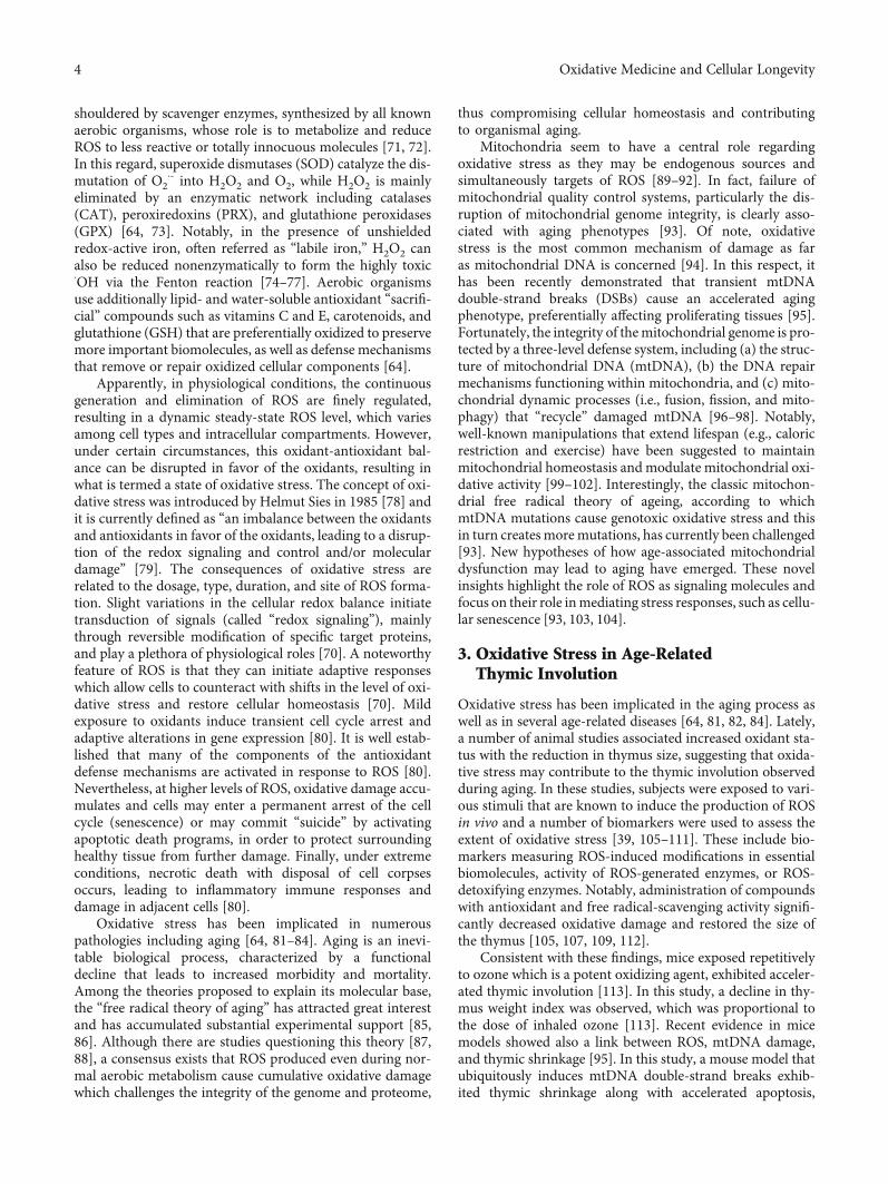
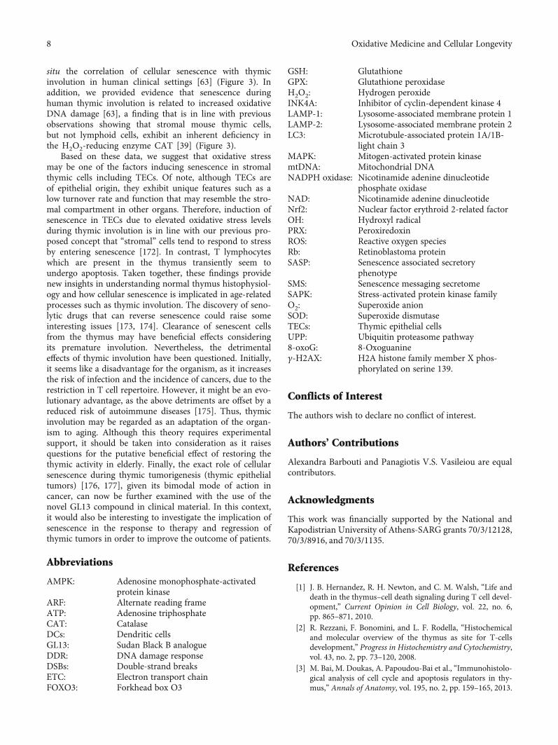
Even though age-associated thymic regression representsone of the most recognizable features of the aging immunesystem, the underlying mechanisms are not well understood[33]. Several candidates have been proposed, suggesting thatthymic regression involves the interplay of various and differ-ent mechanisms (Figure 2); interestingly, there are lines ofevidence that in this complex process, the thymic stromaand especially the TECS are the most sensitive compartment[12, 23, 27, 34]. A number of studies reported that sex steroidhormones, and especially androgens, contribute to age-associated thymic involution [12, 23, 27, 35] (Figure 2). Thisnotion was based on the observations (a) that thymic involu-tion, although beginning in early postnatal life, is more pro-nounced with the onset of puberty when sex steroid levelsincrease and (b) that high doses of sex steroid administrationcause degeneration of the thymus (reviewed in [23]). More-over, androgen impairment or ablation reduces thymic atro-
phy, while castration induces robust regeneration of theatrophied thymus; in the latter case, however, althoughandrogen reduction is permanent, thymus rebound is onlya transient response [12, 23, 27]. This observation, combinedwith the finding that thymus involution begins soon afterbirth, supports the notion that although the thymus isextremely sensitive to sex steroids, these hormones are notthe predominant factors that induce thymus involution[23]. In addition, it cannot be explained why the thymusinvolutes at a faster rate than other tissues.
Numerous studies have also implicated the growth hor-mone- (GH-) insulin-like growth factor- (IGF-) I axis in thy-mus regression [23, 36, 37] (Figure 2). Both hormonespromote thymic growth, and lately, GH has been used as analternative strategy to rejuvenate the thymus in certainimmunodeficiency disorders associated with thymic atrophy[38]. GH and IGF-I have been also considered as regulators
Histopathological findings
(i) Thymic size(ii) Thymic cellularity
(iii) Adipose tissue deposition(iv) Loss of normal tissue
architecture(v) Output of naïve T
lymphocytes-mature Tcells
Clinical features
(i) Severity and susceptibility ininfections
(ii) Responsiveness to vaccinations
(iii) Incidence of cancers(iv) Incidence of autoimmune
diseases
Thymic involution
↓↓
↓↑↑
↓
↑
Figure 1: Summary of key histopathological findings and clinical manifestations of thymic involution.
Thymic involution
Sex steroidhormones
GH-IGF1axis
Infections/pathogens
Glucocorticoidrelease
Oxidativestress
Figure 2: Proposed mechanisms involved in the pathophysiology ofthymic involution. It must be highlighted that none of them canthoroughly explain this well-conserved biological phenomenon.
2 Oxidative Medicine and Cellular Longevity
of age-associated thymic involution, since GH productiondeclines with age. However, the effects of hormone treatmenton thymus size in older mice are limited, implying that thereare other factors that prevent thymic atrophy [36].
The phenomenon of infection-induced inflammationand consequently thymus regression has also been reported;in human studies as well as in animal experimental models,infections with pathogens led to thymic atrophy, althoughthe underlying mechanisms have not been extensively stud-ied [35] (Figure 2). In addition, other periodical events suchas glucocorticoid release as a response to different physiolog-ical stressors may also reduce the size of the thymus [21](Figure 2). These theories, however, cannot explain the pre-mature irreversible age-related thymic involution or whythe thymus involutes at a faster rate than other organs.
Lately, a new player suggested to be involved in acceler-ated thymus involution and dysfunction with age is oxidativestress (Figure 2). Notwithstanding that the generation ofreactive oxygen metabolites is an integral feature of normalcellular metabolism, the accumulation of such genotoxicand proteotoxic oxygen-derived by-products seems to exertdetrimental effects on thymic tissue. Contrariwise, geneticor biochemical enhancement of antioxidant activity has beenproven to ameliorate thymic atrophy [39].
Similarly, oxidative damage is also a well-documentedinducer of cellular senescence, a state of permanent cellcycle arrest that allows cells to deal with various intrinsicand extrinsic stressors, in order to preserve cellular and,in a broader perspective, organismal homeostasis [40–45].Except for cell cycle arrest, senescent cells may acquirenumerous cellular and subcellular features, such as apopto-sis resistance, increased galactosidase (SA-b-gal) activity,aberrant autophagic flow, lipofuscin accumulation, meta-bolic deregulation, and a complex prooxidant secretome(a feature termed senescence messaging secretome (SMS)or senescence associated secretory phenotype (SASP)) [40,42, 43, 45–47]. Interestingly, the temporal activation of thisadaptive stress response mechanism during embryonicdevelopment or normal adult life is linked with beneficialproperties, whereas chronic senescence due to persistingstress and/or insufficient clearance of senescent cells bythe immune system seems to exert detrimental effects [40,43, 45–51]. In the latter case, the accumulation of senescentcells maintains an inflammatory milieu (inflamm-aging) thatcauses tissue remodeling, affects the regenerative potentialand proper function of tissues/organs due to exhaustion ofprogenitor and stem cells, and, ultimately, promotes agingand age-related pathologies [45, 52–61].
Considering that (1st) oxidative stress has been linked toboth the induction of cellular senescence and thymic involu-tion and (2nd) aging is characterized by accumulation ofsenescent cells as well as a decline in thymus function, it isnot unreasonable to assume that cellular senescence mayexert a critical role in the induction of thymic involution,with oxidative stress being the common denominator.Indeed, recent evidence, from human and animal studies,supports this notion [62, 63]. The scope of this review is tosummarize data regarding the role of oxidative stress in boththymic involution and cellular senescence. For a more com-
prehensive approach, we first present an overview regardingin vivo oxidative stress, antioxidant mechanisms, and therelated aging theory.
2. Oxidative Stress and Related Aging Theory
Higher eukaryotic aerobic organisms depend on oxygen forefficient production of energy in mitochondria, yet oxygenis a toxic mutagenic gas jeopardizing cellular and, thus,organismal homeostasis [64]. This “oxygen paradox” derivesfrom the chemical properties of molecular oxygen, whichpredispose to the generation of free radicals and other reac-tive oxygen species (ROS) in aerobes [64]. In particular, thediatomic oxygen molecule (O2) is a free radical: it containstwo unpaired electrons with parallel spins located in separateorbitals [65]. Therefore, if O2 attempts to oxidize anotheratom or molecule by accepting a pair of electrons, both elec-trons must have the same spin. This “spin restriction” limitsO2 to reduction reactions that involve single electron trans-fers and explains why O2 is itself a weak oxidizing agent[66]. However, some of its metabolites are potent oxidantsthat can directly oxidize various substrates [67]. Luckily,most of the O2 (over 95%) received via breathing in humansis reduced directly to water in the mitochondrial electrontransport chain (ETC), as it accepts four electrons and fourprotons (H+) in a reaction catalyzed by the enzyme cyto-chrome oxidase. ETC is coupled with oxidative phosphoryla-tion to produce energy in adenosine triphosphate (ATP)form. Thus, the main function of oxygen in life is to serveas an electron acceptor for efficient energy production.Unfortunately, this concerted tetravalent reduction of O2 bythe mitochondrial ETC, to produce water, is not without“collateral damage.” Accidental electron leakage from com-ponents of the mitochondrial respiratory chain may occur;therefore, O2 may undergo univalent reduction which gener-ates ROS, such as the superoxide anion (O2
⋅-) and the hydro-gen peroxide (H2O2) which are moderately reactive, and theextremely reactive and consequently short-lived hydroxylradical (⋅OH) [64, 67]. In vitro, mitochondrial respiration isresponsible for up to 2% of the consumed O2 into O2
⋅-,whereas in vivo, the rate seems to be much lower [68, 69].Excessive amounts of ROS are also produced by stimulatedphagocytes via the enzyme nicotinamide adenine dinucleo-tide phosphate (NADPH) oxidase. O2
⋅- and oxidants derivedfrom phagocytic NADPH oxidase are crucial components ofthe immune defense system [67]. Except for mitochondriaand phagocytic cells, ROS are also produced by endoplasmicreticulum, plasma membranes, lysosomes, and peroxisomes[67]. Moreover, in addition to endogenous release due to cel-lular metabolism, oxygen-derived reactive by-products mayarise from interactions with exogenous sources such as envi-ronmental pollution, ionizing or solar radiation, xenobioticmetals, drugs, or cigarette smoke [64, 70].
Cellular macromolecules, such as nucleic acids, proteins,lipids, and carbohydrates, are vulnerable to the toxic effect ofthese oxygen-derived intermediates. Obviously, aerobes arenot defenseless, as they utilize a series of highly effectivemechanisms in an attempt to be protected against oxidativedamage [64]. The major burden of antioxidant defense is
3Oxidative Medicine and Cellular Longevity
shouldered by scavenger enzymes, synthesized by all knownaerobic organisms, whose role is to metabolize and reduceROS to less reactive or totally innocuous molecules [71, 72].In this regard, superoxide dismutases (SOD) catalyze the dis-mutation of O2
⋅- into H2O2 and O2, while H2O2 is mainlyeliminated by an enzymatic network including catalases(CAT), peroxiredoxins (PRX), and glutathione peroxidases(GPX) [64, 73]. Notably, in the presence of unshieldedredox-active iron, often referred as “labile iron,” H2O2 canalso be reduced nonenzymatically to form the highly toxic⋅OH via the Fenton reaction [74–77]. Aerobic organismsuse additionally lipid- and water-soluble antioxidant “sacrifi-cial” compounds such as vitamins C and E, carotenoids, andglutathione (GSH) that are preferentially oxidized to preservemore important biomolecules, as well as defense mechanismsthat remove or repair oxidized cellular components [64].
Apparently, in physiological conditions, the continuousgeneration and elimination of ROS are finely regulated,resulting in a dynamic steady-state ROS level, which variesamong cell types and intracellular compartments. However,under certain circumstances, this oxidant-antioxidant bal-ance can be disrupted in favor of the oxidants, resulting inwhat is termed a state of oxidative stress. The concept of oxi-dative stress was introduced by Helmut Sies in 1985 [78] andit is currently defined as “an imbalance between the oxidantsand antioxidants in favor of the oxidants, leading to a disrup-tion of the redox signaling and control and/or moleculardamage” [79]. The consequences of oxidative stress arerelated to the dosage, type, duration, and site of ROS forma-tion. Slight variations in the cellular redox balance initiatetransduction of signals (called “redox signaling”), mainlythrough reversible modification of specific target proteins,and play a plethora of physiological roles [70]. A noteworthyfeature of ROS is that they can initiate adaptive responseswhich allow cells to counteract with shifts in the level of oxi-dative stress and restore cellular homeostasis [70]. Mildexposure to oxidants induce transient cell cycle arrest andadaptive alterations in gene expression [80]. It is well estab-lished that many of the components of the antioxidantdefense mechanisms are activated in response to ROS [80].Nevertheless, at higher levels of ROS, oxidative damage accu-mulates and cells may enter a permanent arrest of the cellcycle (senescence) or may commit “suicide” by activatingapoptotic death programs, in order to protect surroundinghealthy tissue from further damage. Finally, under extremeconditions, necrotic death with disposal of cell corpsesoccurs, leading to inflammatory immune responses anddamage in adjacent cells [80].
Oxidative stress has been implicated in numerouspathologies including aging [64, 81–84]. Aging is an inevi-table biological process, characterized by a functionaldecline that leads to increased morbidity and mortality.Among the theories proposed to explain its molecular base,the “free radical theory of aging” has attracted great interestand has accumulated substantial experimental support [85,86]. Although there are studies questioning this theory [87,88], a consensus exists that ROS produced even during nor-mal aerobic metabolism cause cumulative oxidative damagewhich challenges the integrity of the genome and proteome,
thus compromising cellular homeostasis and contributingto organismal aging.
Mitochondria seem to have a central role regardingoxidative stress as they may be endogenous sources andsimultaneously targets of ROS [89–92]. In fact, failure ofmitochondrial quality control systems, particularly the dis-ruption of mitochondrial genome integrity, is clearly asso-ciated with aging phenotypes [93]. Of note, oxidativestress is the most common mechanism of damage as faras mitochondrial DNA is concerned [94]. In this respect, ithas been recently demonstrated that transient mtDNAdouble-strand breaks (DSBs) cause an accelerated agingphenotype, preferentially affecting proliferating tissues [95].Fortunately, the integrity of themitochondrial genome is pro-tected by a three-level defense system, including (a) the struc-ture of mitochondrial DNA (mtDNA), (b) the DNA repairmechanisms functioning within mitochondria, and (c) mito-chondrial dynamic processes (i.e., fusion, fission, and mito-phagy) that “recycle” damaged mtDNA [96–98]. Notably,well-known manipulations that extend lifespan (e.g., caloricrestriction and exercise) have been suggested to maintainmitochondrial homeostasis and modulate mitochondrial oxi-dative activity [99–102]. Interestingly, the classic mitochon-drial free radical theory of ageing, according to whichmtDNA mutations cause genotoxic oxidative stress and thisin turn createsmoremutations, has currently been challenged[93]. New hypotheses of how age-associated mitochondrialdysfunction may lead to aging have emerged. These novelinsights highlight the role of ROS as signaling molecules andfocus on their role inmediating stress responses, such as cellu-lar senescence [93, 103, 104].
3. Oxidative Stress in Age-RelatedThymic Involution
Oxidative stress has been implicated in the aging process aswell as in several age-related diseases [64, 81, 82, 84]. Lately,a number of animal studies associated increased oxidant sta-tus with the reduction in thymus size, suggesting that oxida-tive stress may contribute to the thymic involution observedduring aging. In these studies, subjects were exposed to vari-ous stimuli that are known to induce the production of ROSin vivo and a number of biomarkers were used to assess theextent of oxidative stress [39, 105–111]. These include bio-markers measuring ROS-induced modifications in essentialbiomolecules, activity of ROS-generated enzymes, or ROS-detoxifying enzymes. Notably, administration of compoundswith antioxidant and free radical-scavenging activity signifi-cantly decreased oxidative damage and restored the size ofthe thymus [105, 107, 109, 112].
Consistent with these findings, mice exposed repetitivelyto ozone which is a potent oxidizing agent, exhibited acceler-ated thymic involution [113]. In this study, a decline in thy-mus weight index was observed, which was proportional tothe dose of inhaled ozone [113]. Recent evidence in micemodels showed also a link between ROS, mtDNA damage,and thymic shrinkage [95]. In this study, a mouse model thatubiquitously induces mtDNA double-strand breaks exhib-ited thymic shrinkage along with accelerated apoptosis,
4 Oxidative Medicine and Cellular Longevity
senescence, and adipose tissue differentiation, mimickingage-related thymic involution. This phenotype was alsoaccompanied by increased production of H2O2 and activa-tion of cell cycle arrest proteins which were reverted bytreatment with MitoQ (a mitochondrial-targeted antioxi-dant) and n-acetylcysteine (an antioxidant and free radicalscavenger) [95]. Similarly, in an aging rat model employedby D-galactose treatment, both thymic atrophy and ele-vated oxidative stress levels were reported [114]. Again,saponin supplementation (an antioxidant derivative fromthe plant Aralia taibaiensis) attenuated oxidative stress-induced aging traits, including thymus shrinkage. Notably,both the FOXO3a (Forkhead box O3) pathway and theNrf2 (nuclear factor erythroid 2-related factor 2) pathwaywere involved in the protective process [114]. Moreover,a mitochondria-targeted antioxidant has been shown todelay thymic atrophy in both normal and senescence-prone rats [115]. OXYS rats which are fast senescence-prone rats show shortened lifespan, early development ofage-associated phenotypes including premature thymicinvolution, and decreased function of T cell-dependentimmunity. Moreover, they often exhibit higher levels of oxi-dative damage. The mitochondria-targeted antioxidantSkQ1 (plastoquinonyl decyltriphenyl phosphonium) delayedage-related processes as well as age-dependent thymic invo-lution in both normal rats and OXYS rats.
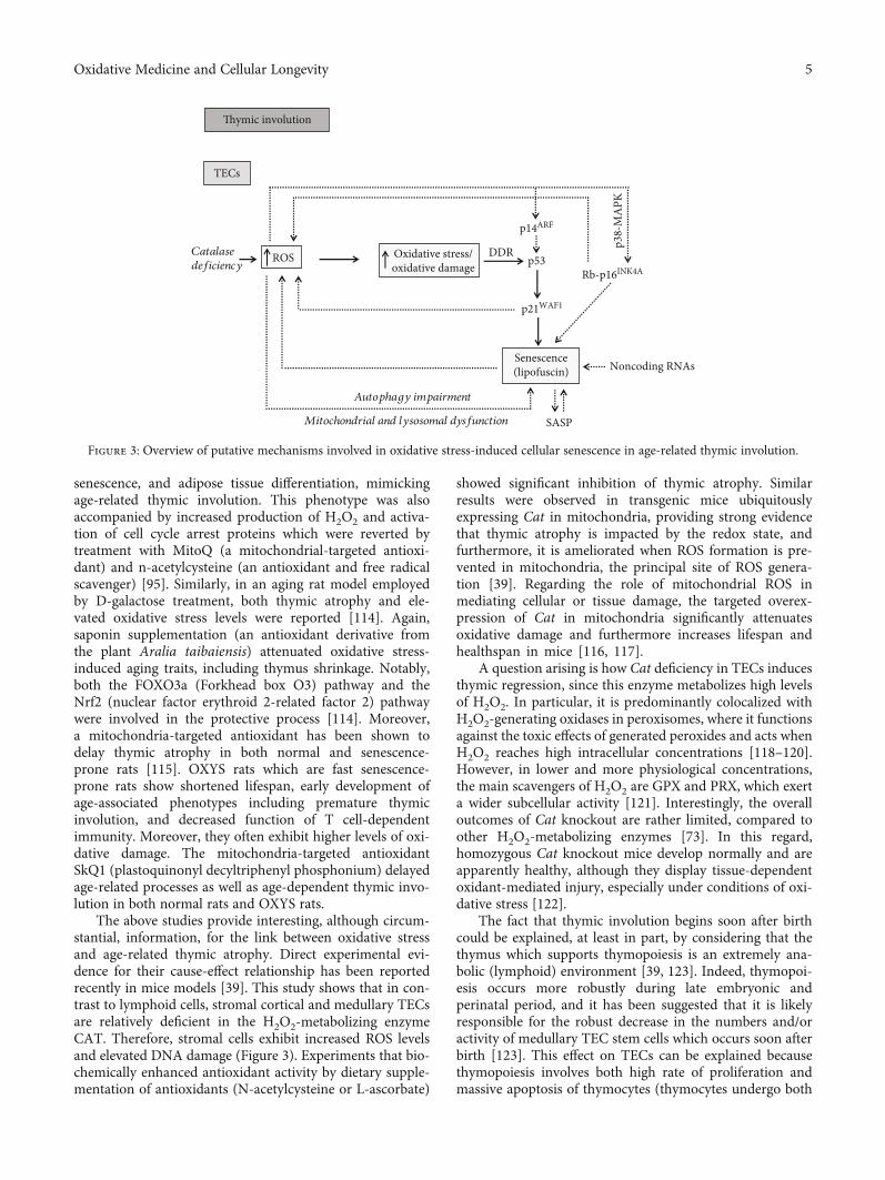
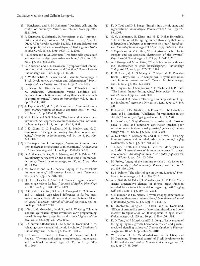
The above studies provide interesting, although circum-stantial, information, for the link between oxidative stressand age-related thymic atrophy. Direct experimental evi-dence for their cause-effect relationship has been reportedrecently in mice models [39]. This study shows that in con-trast to lymphoid cells, stromal cortical and medullary TECsare relatively deficient in the H2O2-metabolizing enzymeCAT. Therefore, stromal cells exhibit increased ROS levelsand elevated DNA damage (Figure 3). Experiments that bio-chemically enhanced antioxidant activity by dietary supple-mentation of antioxidants (N-acetylcysteine or L-ascorbate)
showed significant inhibition of thymic atrophy. Similarresults were observed in transgenic mice ubiquitouslyexpressing Cat in mitochondria, providing strong evidencethat thymic atrophy is impacted by the redox state, andfurthermore, it is ameliorated when ROS formation is pre-vented in mitochondria, the principal site of ROS genera-tion [39]. Regarding the role of mitochondrial ROS inmediating cellular or tissue damage, the targeted overex-pression of Cat in mitochondria significantly attenuatesoxidative damage and furthermore increases lifespan andhealthspan in mice [116, 117].
A question arising is how Cat deficiency in TECs inducesthymic regression, since this enzyme metabolizes high levelsof H2O2. In particular, it is predominantly colocalized withH2O2-generating oxidases in peroxisomes, where it functionsagainst the toxic effects of generated peroxides and acts whenH2O2 reaches high intracellular concentrations [118–120].However, in lower and more physiological concentrations,the main scavengers of H2O2 are GPX and PRX, which exerta wider subcellular activity [121]. Interestingly, the overalloutcomes of Cat knockout are rather limited, compared toother H2O2-metabolizing enzymes [73]. In this regard,homozygous Cat knockout mice develop normally and areapparently healthy, although they display tissue-dependentoxidant-mediated injury, especially under conditions of oxi-dative stress [122].
The fact that thymic involution begins soon after birthcould be explained, at least in part, by considering that thethymus which supports thymopoiesis is an extremely ana-bolic (lymphoid) environment [39, 123]. Indeed, thymopoi-esis occurs more robustly during late embryonic andperinatal period, and it has been suggested that it is likelyresponsible for the robust decrease in the numbers and/oractivity of medullary TEC stem cells which occurs soon afterbirth [123]. This effect on TECs can be explained becausethymopoiesis involves both high rate of proliferation andmassive apoptosis of thymocytes (thymocytes undergo both
Oxidative stress/oxidative damage
DDRp53
p21WAF1
Senescence(lipofuscin)
SASP
Rb-p16INK4A
Mitochondrial and lysosomal dysfunction
ROSCatalasedeficiency
TECs
Autophagy impairment
Thymic involution
p14ARF
p38-
MA
PK
Noncoding RNAs
Figure 3: Overview of putative mechanisms involved in oxidative stress-induced cellular senescence in age-related thymic involution.
5Oxidative Medicine and Cellular Longevity
positive and negative selection within the thymus) resultingin the accumulation of potentially damaging factors suchas ROS which may affect TECs [123]. In this context ofrobust thymopoiesis, an inherent deficiency of TECs inCAT may render these cells more vulnerable to ROS andcould contribute to early thymic atrophy (Figure 1), therebyproviding evidence to explain, at least in part, why the thy-mus is affected more rapidly in comparison to other organs[39]. Relevant to the above analysis is our previous study,in which we showed for the first time in human thymusesthat TECs but not lymphoid cells exhibit oxidative DNAdamage in samples from aged individuals [63]. These find-ings provide in situ evidence the TECs but not lymphoidcells are sensitive to ROS [63].
Yet, despite their effect in thymic atrophy, preliminarydata address the possibility that ROS are required for special-ized functions of stromal cells in mice and thus play physio-logical roles. In particular, ROS produced by thymic stromalcells seem to be necessary for autophagy which is essential fornegative selection and thus for the prevention of autoim-mune diseases [124]. Probably, ROS play a dual role in thethymus: they may be involved in the establishment of centralT cell tolerance in the young steady-state thymus, while theresulting accumulated oxidative damage can eventually leadto thymic atrophy during aging.
Among several different types of ROS, H2O2 has emergedas a central hub in both oxidant-mediated damage andredox-signaling networks [125] although the underlyingmolecular mechanisms remain unclear. Lately, it has beenreported that “labile iron,” a small cellular fraction of poorlycharacterized unshielded redox-active iron, determinesdiverse H2O2-mediated effects [126–129]. It seems that thelabile iron pool plays a major role not only in H2O2-inducedtoxicity but also in specific redox signaling pathways [75, 76].Of note, iron accumulates in aged tissues and the dysregula-tion of iron homeostasis has been implicated in the develop-ment of aging phenotypes [83, 130–135]. Nevertheless, thepotential contribution of iron in thymic involution has notyet been examined.
4. Cellular Senescence in Age-RelatedThymic Involution
Oxidative damage is a well-known trigger of cellular senes-cence. Internal elevated ROS levels can directly damage cel-lular components through the oxidation of biomolecules orcan act as second messengers to modulate specific signalingcascades [136]. The direct damaging of DNA induces theDNA damage response (DDR) pathway that in turn acti-vates p53 and its downstream effector p21WAF1, leading toone of the following alternative outcomes in an attempt torepair these lesions: reversible cell cycle arrest, senescence,or apoptosis [137] (Figure 3). Additionally, p21WAF1 activa-tion promotes a steady increase in ROS generation that fur-ther fuels DNA damage. This positive feedback seems to beboth necessary and sufficient to maintain cell cycle arrestduring establishment of irreversible senescence [138]. Inaddition, senescence can be induced upon DNA damagevia the Rb- (retinoblastoma protein-) p16INK4A axis
(Figure 3). Of note, it has been suggested that while p53and p21WAF1 act to initiate the senescence response,p16INK4A functions in the maintenance of this state. Inter-estingly, p16INK4A can be activated in response to oxidativestress through the p38-MAPK (mitogen-activated proteinkinase) pathway [139] (Figure 3). p38 belongs to thestress-activated protein kinase family (SAPK) that respondsto a variety of physiologic stresses, including oxidativestress [140]. Moreover, the p16INK4a/Rb pathway is impli-cated in a ROS-dependent positive feedback loop, whichreinforces the irreversible cell cycle arrest in senescent cells[141]. Moreover, p14ARF, the alternate reading frame pro-tein product of the INK4A (inhibitor of cyclin-dependentkinase 4)/ARF (alternate reading frame) locus, has alsobeen shown to play an important role in the long-term sta-bilization of p53 following DNA damage. As previouslyshown, this protein acts as a sensor of oxidative stress[142] and plays an important role, under circumstances,in mediating senescence. Importantly, p14ARF also inter-plays with the DNA damage response pathway, as previ-ously shown, in order to exert p53-independent antitumorfunctions [143].
Of great importance, lipofuscin, a nondegradable mate-rial that accumulates in the cytoplasm of senescent cells,mediates the release of excessive amounts of ROS andimpairs the ubiquitin proteasome pathway (UPP), the princi-pal mechanism for catabolism of dysfunctional proteins inthe mammalian cytosol and nucleus. [144, 145]. In this man-ner, a damaging feedback loop that fuels oxidative stress andpromotes aging occurs.
Given that oxidative stress can induce senescence andsenescent cells accumulate in tissues as a consequence ofaging [57], we investigated whether senescence is presentduring age-related thymic involution in human archival(formalin fixed and paraffin embedded) material [63]. Forthis purpose, we used a novel, biotinylated Sudan Black Banalogue termed GL13 (SenTraGor™) that specificallyreacts with lipofuscin, a hallmark of senescent cells [41,44, 63, 146, 147]. Indeed, a number of TECs were detectedmainly within the medulla and especially in the vicinity ofHassall’s corpuscles that exhibited dense cytoplasmicGL13 positivity in the majority of aged samples whereasstaining in sections from infants was negative [63](Figure 3). The GL13-positive TECs showed simultaneouslynuclear p21WAF1 immunopositivity in double-stainingexperiments, further confirming their senescent phenotype[63] (Figure 3). As previously shown, these cells undergocell cycle arrest (p16INK4A and p21WAF1 positive), whileproliferation and apoptotic markers showed low expressionor were negative [3, 148]. Interestingly, medullary TECsexhibited also 8-oxoG and γ-H2AX (H2A histone familymember X phosphorylated on serine 139) immunoreactiv-ity, suggesting that a relationship between cellular senes-cence and oxidative DNA damage exists in the agedhuman thymus [63]. Similar findings in mouse modelsdemonstrated increased β-galactosidase activity and strongγ-H2AX immunopositivity in TECs [62]. In addition, itwas lately shown that in aged mice, the subcapsular spaceof the thymus exhibited high density of pigmented bodies
6 Oxidative Medicine and Cellular Longevity
[149], autofluorescent inclusions which change with age,and represent hallmarks of aging [150]. Putative insights intooxidative stress-driven senescence during thymic involutionwill be next discussed, based on the current state of knowledge.
Besides senescence, perturbed autophagy has also beenassociated with the aging process; thus, a link betweenautophagy and thymic involution seems possible. Theautophagic process appears to play significant roles in thethymic histophysiology [8, 9, 151]. Autophagy supports cen-tral CD4-positive T cell tolerance through facilitating thepresentation of endogenous self-antigens by medullary TEC[151]. The immunohistochemical expression of lysosome-associated membrane proteins 1 and 2 (LAMP-1 andLAMP-2) was investigated in thymuses [152, 153] sinceLAMP-2 participates in chaperone-mediated autophagy[154] and evidence was provided for differential expressionof LAMP-1 and LAMP-2 in thymic involution [152]. Indeed,inacute (infection induced) involution,LAMP-1wasobservedmainly in medullary TEC, in single macrophages and thy-mocytes [152]. Hassall’s corpuscles were immunostainedless intensely as compared with control tissues, and thequantitative study revealed a significantly increased LAMP-2 immunoexpression compared with LAMP-1 [152]. Inchronic (senile) involution, LAMPs were observed with veryslight immunoreactivity [152]. The increased expression ofLAMPs, and mainly of LAMP-2, in the TEC of thymuseswith acute (infection induced) involution might indicateacute cell injury requiring autophagic degradation of dam-aged thymic structures, and the reduced expression ofLAMPs in the age-involuted (senile) thymus might indicatethymic morphological reorganization and functional dysreg-ulation [152]. The immunohistochemical expression of theautophagy regulator microtubule-associated protein 1A/1B-light chain 3 (LC3) was greatly reduced in the thymus of12- and 24-month-old mice while an ultrastructural studyrevealed that the number of autophagic structure/vacuolesin total TEC decreases with age [155]. It was suggested thatthe age-related decrease in autophagic structures in TECmayreduce the immunocompetent T cell pool in aged mice [155].
An interplay between autophagy and ROS in aging hasalso been reported. Studies in invertebrate model organismsshow that autophagy defects cause sensitivity to oxidativestress and decrease longevity, while overexpression ofautophagy genes extends the average lifespan and enhancesresistance to oxidative stress, indicating the fundamental roleof autophagy in oxidative stress-induced organismal aging[156]. A link between impaired autophagy and oxidativestress in aging and age-related pathologies is anticipated.Aerobes are exposed to a permanent flux of ROS whichincreases during aging, resulting in the accumulation of oxi-dized or dysfunctional cellular structures [83, 150, 157, 158].In turn, autophagy declines with age [159, 160], renderingcells unable to remove damaged components formed as aconsequence of metabolic stress. Thus, oxidative damage iscumulative during the lifespan, causing gradual decrease ofcellular functions and resulting in aging and age-related dis-eases [83, 150].
Recently, evidence on how impaired autophagy promotesoxidative damage-induced senescence in murine fibroblasts
and human fetal lung fibroblasts was reported supportingthe inverse correlation between these two cellular processes[161] (Figure 3). Furthermore, mitochondrial dysfunctionhas emerged as a causal player of cellular senescence [104,162–164]. It seems that mitochondrial dysfunction andincreased endogenous ROS formation are the commondenominator for both cellular senescence and aging [138,161, 163, 165, 166]. Mitochondrial dysfunction temporallyprecedes lysosomal dysfunctions and may be the initiatingevent, while lysosomal dysfunction, although occurring later,is more directly responsible for the impaired autophagicactivity and senescence development [161] (Figure 3). “Themitochondrial-lysosomal axis theory of aging” supports thatthe driving force for organismal aging relies in the dysfunc-tion and mutual effect of these two organelles [167, 168].Lately, studies gained insight into the redox signaling path-ways regulating autophagy and senescence. The AMP-activated protein kinase (AMPK) is known to activateautophagy and prevent senescence. Han et al. determinedthat senescence induced in fibroblasts and umbilical veinendothelial cells by H2O2 treatment was accompanied withinactivation of the AMPK pathway, autophagic dysfunction,and decreased NAD+ (nicotinamide adenine dinucleotide)levels which is a known feature of aged organisms [169].Importantly, pharmacological activation of AMPK preventedsenescence, improved the impaired autophagic flux, andrestored NAD+ synthesis, thus suggesting that AMPK targetsmultiple pathways in cells, to collaboratively prevent oxida-tive stress-induced senescence. Similarly, impaired autopha-gic processes and AMPK dysfunction-induced oxidativestress-related senescence comprise an auditory cellular pre-mature senescence model [170]. Moreover, an inverse rela-tionship, where genetic impairment of autophagy promotespremature senescence, was reported in primary human fibro-blasts in a ROS- and p53-dependent mechanism [171]. Inthis study, suppression of autophagy stimulates senescencethrough activation of the p53 tumor suppressor pathwaydue to ROS overproduction. Presumably, in this case, anincreased mitochondrial ROS generation occurs as a con-sequence of the reduced turnover of dysfunctional mito-chondria due to autophagy inhibition. Furthermore, thechemical block of p53 or ROS scavengers delays the pre-mature senescence by improving the autophagic function,suggesting the existence of a feedback mechanism betweenROS-induced p53 activation and autophagy. Overall, thesedata suggest that p53 is an effector of autophagy-inducedsenescence triggered via ROS signaling.
5. Conclusions
Thymic involution is associated with a progressive decline inthe output of naïve T cells and a restricted T cell repertoire inthe periphery, resulting in impaired immune responses tonew pathogens or vaccines, increased incidence of specificcancers, and certain autoimmune disorders in the elderlysubjects [12]. Although it is unclear what constitutes the driv-ing forces resulting in early thymic involution [12], evidencesupports that oxidative damage in the stromal compartmentis one of the potential candidates [39]. We recently showed in
7Oxidative Medicine and Cellular Longevity
situ the correlation of cellular senescence with thymicinvolution in human clinical settings [63] (Figure 3). Inaddition, we provided evidence that senescence duringhuman thymic involution is related to increased oxidativeDNA damage [63], a finding that is in line with previousobservations showing that stromal mouse thymic cells,but not lymphoid cells, exhibit an inherent deficiency inthe H2O2-reducing enzyme CAT [39] (Figure 3).
Based on these data, we suggest that oxidative stressmay be one of the factors inducing senescence in stromalthymic cells including TECs. Of note, although TECs areof epithelial origin, they exhibit unique features such as alow turnover rate and function that may resemble the stro-mal compartment in other organs. Therefore, induction ofsenescence in TECs due to elevated oxidative stress levelsduring thymic involution is in line with our previous pro-posed concept that “stromal” cells tend to respond to stressby entering senescence [172]. In contrast, T lymphocyteswhich are present in the thymus transiently seem toundergo apoptosis. Taken together, these findings providenew insights in understanding normal thymus histophysiol-ogy and how cellular senescence is implicated in age-relatedprocesses such as thymic involution. The discovery of seno-lytic drugs that can reverse senescence could raise someinteresting issues [173, 174]. Clearance of senescent cellsfrom the thymus may have beneficial effects consideringits premature involution. Nevertheless, the detrimentaleffects of thymic involution have been questioned. Initially,it seems like a disadvantage for the organism, as it increasesthe risk of infection and the incidence of cancers, due to therestriction in T cell repertoire. However, it might be an evo-lutionary advantage, as the above detriments are offset by areduced risk of autoimmune diseases [175]. Thus, thymicinvolution may be regarded as an adaptation of the organ-ism to aging. Although this theory requires experimentalsupport, it should be taken into consideration as it raisesquestions for the putative beneficial effect of restoring thethymic activity in elderly. Finally, the exact role of cellularsenescence during thymic tumorigenesis (thymic epithelialtumors) [176, 177], given its bimodal mode of action incancer, can now be further examined with the use of thenovel GL13 compound in clinical material. In this context,it would also be interesting to investigate the implication ofsenescence in the response to therapy and regression ofthymic tumors in order to improve the outcome of patients.
Abbreviations
AMPK: Adenosine monophosphate-activatedprotein kinase
ARF: Alternate reading frameATP: Adenosine triphosphateCAT: CatalaseDCs: Dendritic cellsGL13: Sudan Black B analogueDDR: DNA damage responseDSBs: Double-strand breaksETC: Electron transport chainFOXO3: Forkhead box O3
GSH: GlutathioneGPX: Glutathione peroxidaseH2O2: Hydrogen peroxideINK4A: Inhibitor of cyclin-dependent kinase 4LAMP-1: Lysosome-associated membrane protein 1LAMP-2: Lysosome-associated membrane protein 2LC3: Microtubule-associated protein 1A/1B-
light chain 3MAPK: Mitogen-activated protein kinasemtDNA: Mitochondrial DNANADPH oxidase: Nicotinamide adenine dinucleotide
phosphate oxidaseNAD: Nicotinamide adenine dinucleotideNrf2: Nuclear factor erythroid 2-related factorOH: Hydroxyl radicalPRX: PeroxiredoxinROS: Reactive oxygen speciesRb: Retinoblastoma proteinSASP: Senescence associated secretory
phenotypeSMS: Senescence messaging secretomeSAPK: Stress-activated protein kinase familyO2: Superoxide anionSOD: Superoxide dismutaseTECs: Thymic epithelial cellsUPP: Ubiquitin proteasome pathway8-oxoG: 8-Oxoguanineγ-H2AX: H2A histone family member X phos-
phorylated on serine 139.
Conflicts of Interest
The authors wish to declare no conflict of interest.
Authors’ Contributions
Alexandra Barbouti and Panagiotis V.S. Vasileiou are equalcontributors.
Acknowledgments
This work was financially supported by the National andKapodistrian University of Athens-SARG grants 70/3/12128,70/3/8916, and 70/3/1135.
References
[1] J. B. Hernandez, R. H. Newton, and C. M. Walsh, “Life anddeath in the thymus–cell death signaling during T cell devel-opment,” Current Opinion in Cell Biology, vol. 22, no. 6,pp. 865–871, 2010.
[2] R. Rezzani, F. Bonomini, and L. F. Rodella, “Histochemicaland molecular overview of the thymus as site for T-cellsdevelopment,” Progress in Histochemistry and Cytochemistry,vol. 43, no. 2, pp. 73–120, 2008.
[3] M. Bai, M. Doukas, A. Papoudou-Bai et al., “Immunohistolo-gical analysis of cell cycle and apoptosis regulators in thy-mus,” Annals of Anatomy, vol. 195, no. 2, pp. 159–165, 2013.
8 Oxidative Medicine and Cellular Longevity
[4] J. Banchereau and R. M. Steinman, “Dendritic cells and thecontrol of immunity,” Nature, vol. 392, no. 6673, pp. 245–252, 1998.
[5] P. Kanavaros, K. Stefanaki, D. Rontogianni et al., “Immuno-histochemical expression of p53, p21/waf1, Rb, p16, cyclinD1, p27, Ki67, cyclin A, cyclin B1, Bcl2, Bax and Bak proteinsand apoptotic index in normal thymus,” Histology and Histo-pathology, vol. 16, no. 4, pp. 1005–1012, 2001.
[6] I. Mellman and R. M. Steinman, “Dendritic cells: specializedand regulated antigen processing machines,” Cell, vol. 106,no. 3, pp. 255–258, 2001.
[7] G. Anderson and E. J. Jenkinson, “Lymphostromal interac-tions in thymic development and function,” Nature ReviewsImmunology, vol. 1, no. 1, pp. 31–40, 2001.
[8] A. W. Bronietzki, M. Schuster, and I. Schmitz, “Autophagy inT-cell development, activation and differentiation,” Immu-nology and Cell Biology, vol. 93, no. 1, pp. 25–34, 2015.
[9] L. Klein, M. Hinterberger, J. von Rohrscheidt, andM. Aichinger, “Autonomous versus dendritic cell-dependent contributions of medullary thymic epithelial cellsto central tolerance,” Trends in Immunology, vol. 32, no. 5,pp. 188–193, 2011.
[10] A. Papoudou-Bai, M. Bai, M. Doukas et al., “Immunohistolo-gical characterization of thymic dendritic cells,” In Vivo,vol. 26, no. 6, pp. 985–992, 2012.
[11] M. A. Ritter and D. B. Palmer, “The human thymic microen-vironment: new approaches to functional analysis,” Seminarsin Immunology, vol. 11, no. 1, pp. 13–21, 1999.
[12] I. K. Chinn, C. C. Blackburn, N. R. Manley, and G. D.Sempowski, “Changes in primary lymphoid organs withaging,” Seminars in Immunology, vol. 24, no. 5, pp. 309–320, 2012.
[13] S. Ponnappan and U. Ponnappan, “Aging and immune func-tion: molecular mechanisms to interventions,” Antioxidants& Redox Signaling, vol. 14, no. 8, pp. 1551–1585, 2011.
[14] D. P. Shanley, D. Aw, N. R. Manley, and D. B. Palmer, “Anevolutionary perspective on the mechanisms of immunose-nescence,” Trends in Immunology, vol. 30, no. 7, pp. 374–381, 2009.
[15] M. Torroba and A. G. Zapata, “Aging of the vertebrateimmune system,” Microscopy Research and Technique,vol. 62, no. 6, pp. 477–481, 2003.
[16] Q. He, S. Heshka, J. Albu et al., “Smaller organ mass withgreater age, except for heart,” Journal of Applied Physiology,vol. 106, no. 6, pp. 1780–1784, 2009.
[17] U. G. Kyle, L. Genton, D. Hans, L. Karsegard, D. O. Slosman,and C. Pichard, “Age-related differences in fat-free mass,skeletal muscle, body cell mass and fat mass between 18 and94 years,” European Journal of Clinical Nutrition, vol. 55,no. 8, pp. 663–672, 2001.
[18] J. Gui, L. M. Mustachio, D.-M. Su, and R. W. Craig, “Thymussize and age-related thymic involution: early programming,sexual dimorphism, progenitors and stroma,” Aging and Dis-ease, vol. 3, no. 3, pp. 280–290, 2012.
[19] E. Montecino-Rodriquez, H. Min, and K. Dorshkind, “Ree-valuating current models of thymic involution,” Seminars inImmunology, vol. 17, no. 5, pp. 356–361, 2005.
[20] R. Rezzani, L. Nardo, G. Favero, M. Peroni, and L. F.Rodella, “Thymus and aging: morphological, radiological,and functional overview,” Age, vol. 36, no. 1, pp. 313–351, 2014.
[21] D. D. Taub and D. L. Longo, “Insights into thymic aging andregeneration,” Immunological Reviews, vol. 205, no. 1, pp. 72–93, 2005.
[22] G. G. Steinmann, B. Klaus, and H. K. Müller-Hermelink,“The involution of the ageing human thymic epithelium isindependent of puberty. A morphometric study,” Scandina-vian Journal of Immunology, vol. 22, no. 5, pp. 563–575, 1985.
[23] S. Cepeda and A. V. Griffith, “Thymic stromal cells: roles inatrophy and age-associated dysfunction of the thymus,”Experimental Gerontology, vol. 105, pp. 113–117, 2018.
[24] A. J. George and M. A. Ritter, “Thymic involution with age-ing: obsolescence or good housekeeping?,” ImmunologyToday, vol. 17, no. 6, pp. 267–272, 1996.
[25] H. E. Lynch, G. L. Goldberg, A. Chidgey, M. R. Van denBrink, R. Boyd, and G. D. Sempowski, “Thymic involutionand immune reconstitution,” Trends in Immunology,vol. 30, no. 7, pp. 366–373, 2009.
[26] B. F. Haynes, G. D. Sempowski, A. F. Wells, and L. P. Hale,“The human thymus during aging,” Immunologic Research,vol. 22, no. 2-3, pp. 253–261, 2000.
[27] D. Aw and D. B. Palmer, “The origin and implication of thy-mic involution,” Aging and Disease, vol. 2, no. 5, pp. 437–443,2011.
[28] R. Aspinall, G. Del Giudice, R. B. Effros, B. Grubeck-Loeben-stein, and S. Sambhara, “Challenges for vaccination in theelderly,” Immunity & Ageing, vol. 4, no. 1, p. 9, 2007.
[29] L. Čičin-Šain, S. Smyk-Paerson, N. Currier et al., “Loss ofnaive T cells and repertoire constriction predict poorresponse to vaccination in old primates,” Journal of Immu-nology, vol. 184, no. 12, pp. 6739–6745, 2010.
[30] A. D. Foster, A. Sivarapatna, and R. E. Gress, “The agingimmune system and its relationship with cancer,” AgingHealth, vol. 7, no. 5, pp. 707–718, 2011.
[31] T. Fulop, R. Kotb, C. F. Fortin, G. Pawelec, F. de Angelis, andA. Larbi, “Potential role of immunosenescence in cancerdevelopment,” Annals of the New York Academy of Sciences,vol. 1197, no. 1, pp. 158–165, 2010.
[32] M. Prelog, “Aging of the immune system: a risk factor forautoimmunity?,” Autoimmunity Reviews, vol. 5, no. 2,pp. 136–139, 2006.
[33] D. B. Palmer, “The effect of age on thymic function,” Fron-tiers in Immunology, vol. 4, p. 316, 2013.
[34] A. V. Griffith, M. Fallahi, T. Venables, and H. T. Petrie, “Per-sistent degenerative changes in thymic organ functionrevealed by an inducible model of organ regrowth,” AgingCell, vol. 11, no. 1, pp. 169–177, 2012.
[35] S. Majumdar and D. Nandi, “Thymic atrophy: experimentalstudies and therapeutic interventions,” Scandinavian Journalof Immunology, vol. 87, no. 1, pp. 4–14, 2018.
[36] E. Montecino-Rodriguez, R. Clark, and K. Dorshkind,“Effects of insulin-like growth factor administration and bonemarrow transplantation on thymopoiesis in aged mice,”Endocrinology, vol. 139, no. 10, pp. 4120–4126, 1998.
[37] D. D. Taub, W. J. Murphy, and D. L. Longo, “Rejuvenation ofthe aging thymus: growth hormone-mediated and ghrelin-mediated signaling pathways,” Current Opinion in Pharma-cology, vol. 10, no. 4, pp. 408–424, 2010.
[38] W. Savino, D. A. Mendes-da-Cruz, A. Lepletier, andM. Dardenne, “Hormonal control of T-cell development inhealth and disease,” Nature Reviews Endocrinology, vol. 12,no. 2, pp. 77–89, 2016.
9Oxidative Medicine and Cellular Longevity
[39] A. V. Griffith, T. Venables, J. Shi et al., “Metabolic damageand premature thymus aging caused by stromal catalase defi-ciency,” Cell Reports, vol. 12, no. 7, pp. 1071–1079, 2015.
[40] D. G. A. Burton and V. Krizhanovsky, “Physiological andpathological consequences of cellular senescence,” Cellularand Molecular Life Sciences, vol. 71, no. 22, pp. 4373–4386,2014.
[41] E. A. Georgakopoulou, K. Tsimaratou, K. Evangelou et al.,“Specific lipofuscin staining as a novel biomarker to detectreplicative and stress-induced senescence. A method applica-ble in cryo-preserved and archival tissues,” Aging, vol. 5,no. 1, pp. 37–50, 2013.
[42] V. Gorgoulis, P. D. Adams, A. Alimonti et al., “Cellularsenescence: defining a path forward,” Cell, vol. 179, no. 4,pp. 813–827, 2019.
[43] V. G. Gorgoulis and T. D. Halazonetis, “Oncogene-inducedsenescence: the bright and dark side of the response,” CurrentOpinion in Cell Biology, vol. 22, no. 6, pp. 816–827, 2010.
[44] E. Liakou, E. Mavrogonatou, H. Pratsinis et al., “Ionizingradiation-mediated premature senescence and paracrineinteractions with cancer cells enhance the expression of syn-decan 1 in human breast stromal fibroblasts: the role of TGF-β,” Aging, vol. 8, no. 8, pp. 1650–1669, 2016.
[45] D. Munoz-Espin and M. Serrano, “Cellular senescence: fromphysiology to pathology,” Nature Reviews Molecular CellBiology, vol. 15, no. 7, pp. 482–496, 2014.
[46] F. Rodier and J. Campisi, “Four faces of cellular senes-cence,” The Journal of Cell Biology, vol. 192, no. 4,pp. 547–556, 2011.
[47] R. Salama, M. Sadaie, M. Hoare, and M. Narita, “Cellularsenescence and its effector programs,” Genes & Development,vol. 28, no. 2, pp. 99–114, 2014.
[48] J. Bartkova, N. Rezaei, M. Liontos et al., “Oncogene-inducedsenescence is part of the tumorigenesis barrier imposed byDNA damage checkpoints,” Nature, vol. 444, no. 7119,pp. 633–637, 2006.
[49] T. D. Halazonetis, V. G. Gorgoulis, and J. Bartek, “Anoncogene-induced DNA damage model for cancer develop-ment,” Science, vol. 319, no. 5868, pp. 1352–1355, 2008.
[50] M. Liontos, M. Koutsami, M. Sideridou et al., “Deregulatedoverexpression of hCdt1 and hCdc6 promotes malignantbehavior,” Cancer Research, vol. 67, no. 22, pp. 10899–10909, 2007.
[51] M. Liontos, K. Niforou, G. Velimezi et al., “Modulation of theE2F1-driven cancer cell fate by the DNA damage responsemachinery and potential novel E2F1 targets in osteosarco-mas,” The American Journal of Pathology, vol. 175, no. 1,pp. 376–391, 2009.
[52] J. Campisi and F. d'Adda di Fagagna, “Cellular senescence:when bad things happen to good cells,” Nature ReviewsMolecular Cell Biology, vol. 8, no. 9, pp. 729–740, 2007.
[53] J. P. Coppe, P. Y. Desprez, A. Krtolica, and J. Campisi, “Thesenescence-associated secretory phenotype: the dark side oftumor suppression,” Annual Review of Pathology, vol. 5,no. 1, pp. 99–118, 2010.
[54] V. G. Gorgoulis, H. Pratsinis, P. Zacharatos et al., “p53-dependent ICAM-1 overexpression in senescent human cellsidentified in atherosclerotic lesions,” Laboratory Investiga-tion, vol. 85, no. 4, pp. 502–511, 2005.
[55] V. G. Gorgoulis, P. Zacharatos, A. Kotsinas et al., “p53 acti-vates ICAM-1 (CD54) expression in an NF-κB-independent
manner,” The EMBO Journal, vol. 22, no. 7, pp. 1567–1578,2003.
[56] A. Helman, D. Avrahami, A. Klochendler et al., “Effects ofageing and senescence on pancreatic β-cell function,” Diabe-tes, Obesity & Metabolism, vol. 18, pp. 58–62, 2016.
[57] U. Herbig, M. Ferreira, L. Condel, D. Carey, and J. M. Sedivy,“Cellular senescence in aging primates,” Science, vol. 311,no. 5765, article 1257, 2006.
[58] A. McShea, P. L. Harris, K. R. Webster, A. F. Wahl, and M. A.Smith, “Abnormal expression of the cell cycle regulators P16and CDK4 in Alzheimer's disease,” The American Journal ofPathology, vol. 150, no. 6, pp. 1933–1939, 1997.
[59] J. S. Price, J. G. Waters, C. Darrah et al., “The role of chondro-cyte senescence in osteoarthritis,” Aging Cell, vol. 1, no. 1,pp. 57–65, 2002.
[60] I. Sturmlechner, M. Durik, C. J. Sieben, D. J. Baker, and J. M.van Deursen, “Cellular senescence in renal ageing and dis-ease,” Nature Reviews Nephrology, vol. 13, no. 2, pp. 77–89,2017.
[61] E. Vasile, Y. Tomita, L. F. Brown, O. Kocher, and H. F.Dvorak, “Differential expression of thymosin β-10 by earlypassage and senescent vascular endothelium is modulatedby VPF/VEGF: evidence for senescent endothelial cellsin vivo at sites of atherosclerosis,” The FASEB Journal,vol. 15, no. 2, pp. 458–466, 2001.
[62] D. Aw, A. B. Silva, M. Maddick, T. von Zglinicki, and D. B.Palmer, “Architectural changes in the thymus of aging mice,”Aging Cell, vol. 7, no. 2, pp. 158–167, 2008.
[63] A. Barbouti, K. Evangelou, I. S. Pateras et al., “_In situ_ evi-dence of cellular senescence in Thymic Epithelial Cells(TECs) during human thymic involution,” Mechanisms ofAgeing and Development, vol. 177, pp. 88–90, 2019.
[64] J. M. S. Davies, J. Cillard, B. Friguet et al., “The oxygen para-dox, the French paradox, and age-related diseases,” Gero Sci-ence, vol. 39, no. 5-6, pp. 499–550, 2017.
[65] J. M. C. Gutteridge and B. Halliwell, “Mini-review: oxidativestress, redox stress or redox success?,” Biochemical and Bio-physical Research Communications, vol. 502, no. 2, pp. 183–186, 2018.
[66] H. Taube, “Mechanisms of oxidation with oxygen,” Journal ofGeneral Physiology, vol. 49, no. 1, pp. 29–50, 1965.
[67] S. Di Meo, T. T. Reed, P. Venditti, and V. M. Victor, “Role ofROS and RNS sources in physiological and pathological con-ditions,” Oxidative Medicine and Cellular Longevity,vol. 2016, Article ID 1245049, 44 pages, 2016.
[68] M. P. Murphy, “How mitochondria produce reactive oxygenspecies,” The Biochemical Journal, vol. 417, no. 1, pp. 1–13,2009.
[69] J. F. Turrens, “Mitochondrial formation of reactive oxygenspecies,” The Journal of Physiology, vol. 552, no. 2, pp. 335–344, 2003.
[70] H. Sies, “On the history of oxidative stress: concept and someaspects of current development,” Current Opinion in Toxicol-ogy, vol. 7, pp. 122–126, 2018.
[71] H. Sies, “Oxidative stress: a concept in redox biology andmedicine,” Redox Biology, vol. 4, pp. 180–183, 2015.
[72] H. Sies, “Oxidative stress: impact in redox biology and med-icine,” Archives of Medical and Biomedical Research, vol. 2,no. 4, pp. 146–150, 2015.
[73] X. G. Lei, J. H. Zhu, W. H. Cheng et al., “Paradoxical roles ofantioxidant enzymes: basic mechanisms and health
10 Oxidative Medicine and Cellular Longevity
implications,” Physiological Reviews, vol. 96, no. 1, pp. 307–364, 2016.
[74] Z. I. Cabantchik, “Labile iron in cells and body fluids: physi-ology, pathology, and pharmacology,” Frontiers in Pharma-cology, vol. 5, p. 45, 2014.
[75] D. Galaris and K. Pantopoulos, “Oxidative stress and ironhomeostasis: mechanistic and health aspects,” CriticalReviews in Clinical Laboratory Sciences, vol. 45, no. 1,pp. 1–23, 2008.
[76] D. Galaris, A. Barbouti, and K. Pantopoulos, “Iron homeosta-sis and oxidative stress: an intimate relationship,” Biochimicaet Biophysica Acta (BBA) - Molecular Cell Research, vol. 1866,no. 12, article 118535, 2019.
[77] M. Valko, K. Jomova, C. J. Rhodes, K. Kuča, and K. Musílek,“Redox- and non-redox-metal-induced formation of freeradicals and their role in human disease,” Archives of Toxicol-ogy, vol. 90, no. 1, pp. 1–37, 2016.
[78] H. Sies, “Oxidative stress: introductory remarks,” in Oxida-tive Stress, H. Sies, Ed., pp. 1–8, Academic Press, London,1985.
[79] H. Sies and D. P. Jones, “Oxidative stress,” in Encyclopedia ofstress, G. Fink, Ed., pp. 45–48, Elsevier, Amsterdam, 2nd edi-tion, 2007.
[80] A. M. Pickering, L. Vojtovich, J. Tower, and K. J. A. Davies,“Oxidative stress adaptation with acute, chronic, andrepeated stress,” Free Radical Biology & Medicine, vol. 55,pp. 109–118, 2013.
[81] P. Davalli, T. Mitic, A. Caporali, A. Lauriola, andD. D’Arca, “ROS, cell senescence, and novel molecularmechanisms in aging and age-related diseases,” OxidativeMedicine and Cellular Longevity, vol. 2016, Article ID3565127, 18 pages, 2016.
[82] T. Finkel and N. J. Holbrook, “Oxidants, oxidative stress andthe biology of ageing,” Nature, vol. 408, no. 6809, pp. 239–247, 2000.
[83] D. Galaris, M. Mantzaris, and C. Amorgianiotis, “Oxidativestress and aging: the potential role of iron,” Hormones,vol. 7, no. 2, pp. 114–122, 2008.
[84] L. C. D. Pomatto and K. J. A. Davies, “Adaptive homeostasisand the free radical theory of ageing,” Free Radical Biology &Medicine, vol. 124, pp. 420–430, 2018.
[85] D. F. Dai, Y. A. Chiao, D. J. Marcinek, H. Szeto, and P. S.Rabinovitch, “Mitochondrial oxidative stress in aging andhealthspan,” Longevity & Healthspan, vol. 3, no. 1, p. 6, 2014.
[86] K. C. Kregel and H. J. Zhang, “An integrated view of oxidativestress in aging: basic mechanisms, functional effects, andpathological considerations,” American Journal of Physiol-ogy-Regulatory, Integrative and Comparative Physiology,vol. 292, no. 1, pp. R18–R36, 2007.
[87] Y. H. Edrey and A. B. Salmon, “Revisiting an age-old questionregarding oxidative stress,” Free Radical Biology & Medicine,vol. 71, pp. 368–378, 2014.
[88] S. I. Liochev, “Reactive oxygen species and the free radicaltheory of aging,” Free Radical Biology & Medicine, vol. 60,pp. 1–4, 2013.
[89] G. Barja, “The mitochondrial free radical theory of aging,”Progress in Molecular Biology and Translational Science,vol. 127, pp. 1–27, 2014.
[90] G. López-Lluch, C. Santos-Ocaña, J. A. Sánchez-Alcázaret al., “Mitochondrial responsibility in ageing process: inno-
cent, suspect or guilty,” Biogerontology, vol. 16, no. 5,pp. 599–620, 2015.
[91] F. L. Muller, M. S. Lustgarten, Y. Jang, A. Richardson, andH. Van Remmen, “Trends in oxidative aging theories,”Free Radical Biology & Medicine, vol. 43, no. 4, pp. 477–503, 2007.
[92] R. S. Sohal and R. Weindruch, “Oxidative stress, caloricrestriction, and aging,” Science, vol. 273, no. 5271, pp. 59–63, 1996.
[93] M. Pinto and C. T. Moraes, “Mechanisms linking mtDNAdamage and aging,” Free Radical Biology & Medicine,vol. 85, pp. 250–258, 2015.
[94] F. M. Yakes and B. Van Houten, “Mitochondrial DNA dam-age is more extensive and persists longer than nuclear DNAdamage in human cells following oxidative stress,” Proceed-ings of the National Academy of Sciences of the United Statesof America, vol. 94, no. 2, pp. 514–519, 1997.
[95] M. Pinto, A. M. Pickrell, X. Wang et al., “Transient mito-chondrial DNA double strand breaks in mice cause acceler-ated aging phenotypes in a ROS-dependent but p53/p21-independent manner,” Cell Death Differ, vol. 24, no. 2,pp. 288–299, 2017.
[96] M. Alexeyev, I. Shokolenko, G. Wilson, and S. LeDoux, “Themaintenance of mitochondrial DNA integrity–critical analy-sis and update,” Cold Spring Harbor Perspectives in Biology,vol. 5, no. 5, article a012641, 2013.
[97] L. Kazak, A. Reyes, and I. J. Holt, “Minimizing the damage:repair pathways keep mitochondrial DNA intact,” NatureReviews Molecular Cell Biology, vol. 13, no. 10, pp. 659–671,2012.
[98] P. V. S. Vasileiou, I. Mourouzis, and C. Pantos, “Principalaspects regarding the maintenance of mammalian mitochon-drial genome integrity,” International Journal of MolecularSciences, vol. 18, no. 8, article 1821, 2017.
[99] L. Fontana and L. Partridge, “Promoting health and longevitythrough diet: from model organisms to humans,” Cell,vol. 161, no. 1, pp. 106–118, 2015.
[100] G. López-Lluch and P. Navas, “Calorie restriction as an inter-vention in ageing,” The Journal of Physiology, vol. 594, no. 8,pp. 2043–2060, 2016.
[101] A. Raffaello and R. Rizzuto, “Mitochondrial longevity path-ways,” Biochimica et Biophysica Acta (BBA) - Molecular CellResearch, vol. 1813, no. 1, pp. 260–268, 2011.
[102] M. H. Vendelbo and K. S. Nair, “Mitochondrial longevitypathways,” Biochimica et Biophysica Acta (BBA) - MolecularCell Research, vol. 1813, no. 4, pp. 634–644, 2011.
[103] P. D. Ray, B.-W. Huang, and Y. Tsuji, “Reactive oxygen spe-cies (ROS) homeostasis and redox regulation in cellular sig-naling,” Cellular Signalling, vol. 24, no. 5, pp. 981–990, 2012.
[104] P. V. S. Vasileiou, K. Evangelou, K. Vlasis et al., “Mitochon-drial homeostasis and cellular senescence,” Cells, vol. 8,no. 7, p. 686, 2019.
[105] M. K. Singh, S. S. Yadav, V. Gupta, and S. Khattri, “Immuno-modulatory role of Emblica officinalis in arsenic induced oxi-dative damage and apoptosis in thymocytes of mice,” BMCComplementary and Alternative Medicine, vol. 13, no. 1,p. 193, 2013.
[106] Y. Wang, L. Jiang, Y. Li, X. Luo, and J. He, “Effect of differentselenium supplementation levels on oxidative stress, cyto-kines, and immunotoxicity in chicken thymus,” BiologicalTrace Element Research, vol. 172, no. 2, pp. 488–495, 2016.
11Oxidative Medicine and Cellular Longevity
[107] X. Wei, F. Su, X. Su, T. Hu, and S. Hu, “Stereospecific antiox-idant effects of ginsenoside Rg3 on oxidative stress inducedby cyclophosphamide in mice,” Fitoterapia, vol. 83, no. 4,pp. 636–642, 2012.
[108] D. Yu, Z. Zhang, H. Yao, S. Li, and S.W. Xu, “The role of sele-noprotein W in inflammatory injury in chicken immune tis-sues and cultured splenic lymphocyte,” Biometals, vol. 28,no. 1, pp. 75–87, 2015.
[109] J. Yu, Y. Chen, L. Zhai et al., “Antioxidative effect of gin-seng stem-leaf saponins on oxidative stress induced bycyclophosphamide in chickens,” Poultry Science, vol. 94,no. 5, pp. 927–933, 2015.
[110] Z. W. Zhang, Q. H. Wang, J. L. Zhang, S. Li, X. L. Wang,and S. W. Xu, “Effects of oxidative stress on immunosup-pression induced by selenium deficiency in chickens,” Bio-logical Trace Element Research, vol. 149, no. 3, pp. 352–361,2012.
[111] Z. Zhang, J. L. Zhang, Y. H. Zhang et al., “Effect of oxygen freeradicals and nitric oxide on apoptosis of immune organinduced by selenium deficiency in chickens,” Biometals,vol. 26, no. 2, pp. 355–365, 2013.
[112] R. Uchio, Y. Hirose, S. Murosaki, Y. Yamamoto, andA. Ishigami, “High dietary intake of vitamin C suppressesage-related thymic atrophy and contributes to the mainte-nance of immune cells in vitamin C-deficient senescencemarker protein-30 knockout mice,” British Journal of Nutri-tion, vol. 113, no. 4, pp. 603–609, 2015.
[113] R. Feng, W. He, and H. Ochi, “A new murine oxidative stressmodel associated with senescence,” Mechanisms of Ageingand Development, vol. 122, no. 6, pp. 547–559, 2001.
[114] Y. N. Li, Y. Guo, M. M. Xi et al., “Saponins from Aralia tai-baiensis attenuate D-galactose-induced aging in rats by acti-vating FOXO3a and Nrf2 pathways,” Oxidative Medicineand Cellular Longevity, vol. 2014, Article ID 320513, 13 pages,2014.
[115] L. A. Obukhova, V. P. Skulachev, and N. G. Kolosova, “Mito-chondria-targeted antioxidant SkQ1 inhibits age-dependentinvolution of the thymus in normal and senescence-pronerats,” Aging, vol. 1, no. 4, pp. 389–401, 2009.
[116] D. F. Dai, Y. A. Chiao, G. M. Martin et al., “Mitochondrial-targeted catalase: extended longevity and the roles in variousdisease models,” Progress in Molecular Biology and Transla-tional Science, vol. 146, pp. 203–241, 2017.
[117] S. E. Schriner, N. J. Linford, G. M. Martin et al., “Extension ofmurine life span by overexpression of catalase targeted tomitochondria,” Science, vol. 308, no. 5730, pp. 1909–1911,2005.
[118] M. Schrader and H. D. Fahimi, “Peroxisomes and oxidativestress,” Biochimica et Biophysica Acta (BBA) - Molecular CellResearch, vol. 1763, no. 12, pp. 1755–1766, 2006.
[119] M. Shingu, K. Yoshioka, M. Nobunaga, and K. Yoshida,“Human vascular smooth muscle cells and endothelial cellslack catalase activity and are susceptible to hydrogen perox-ide,” Inflammation, vol. 9, no. 3, pp. 309–320, 1985.
[120] H. van den Bosch, R. B. H. Schutgens, R. J. A. Wanders, andJ. M. Tager, “Biochemistry of peroxisomes,” Annual Reviewof Biochemistry, vol. 61, no. 1, pp. 157–197, 1992.
[121] N. Makino, Y. Mochizuki, S. Bannai, and Y. Sugita, “Kineticstudies on the removal of extracellular hydrogen peroxideby cultured fibroblasts,” The Journal of Biological Chemistry,vol. 269, no. 2, pp. 1020–1025, 1994.
[122] Y. S. Ho, Y. Xiong, W. Ma, A. Spector, and D. S. Ho,“Mice lacking catalase develop normally but show differ-ential sensitivity to oxidant tissue injury,” The Journal ofBiological Chemistry, vol. 279, no. 31, pp. 32804–32812,2004.
[123] Y. Hamazaki, M. Sekai, and N. Minato, “Medullary thymicepithelial stem cells: role in thymic epithelial cell mainte-nance and thymic involution,” Immunological Reviews,vol. 271, no. 1, pp. 38–55, 2016.
[124] M. K. Semwal, A. K. Hester, Y. Xiao, T. Venables, andA. Griffith, “Redox regulation of autophagy in thymic stromalcell function,” The Journal of Immunology, vol. 202, article65.15, 2019.
[125] H. Sies, “Role of metabolic H2O2 generation: redox signalingand oxidative stress,” The Journal of Biological Chemistry,vol. 289, no. 13, pp. 8735–8741, 2014.
[126] A. Barbouti, C. Amorgianiotis, E. Kolettas, P. Kanavaros, andD. Galaris, “Hydrogen peroxide inhibits caspase-dependentapoptosis by inactivating procaspase-9 in an iron-dependent manner,” Free Radical Biology & Medicine,vol. 43, no. 10, pp. 1377–1387, 2007.
[127] P. Gerogianni, M. Chatziathanasiadou, D. Diamantis,A. Tzakos, and D. Galaris, “Lipophilic ester and amide deriv-atives of rosmarinic acid protect cells against H2O2-inducedDNA damage and apoptosis: the potential role of intracellularaccumulation and labile iron chelation,” Redox Biology,vol. 15, pp. 548–556, 2018.
[128] N. Kitsati, M. D. Mantzaris, and D. Galaris, “Hydroxytyrosolinhibits hydrogen peroxide-induced apoptotic signaling vialabile iron chelation,” Redox Biology, vol. 10, pp. 233–242,2016.
[129] M. D. Mantzaris, S. Bellou, V. Skiada, N. Kitsati, T. Fotsis,and D. Galaris, “Intracellular labile iron determines H2O2-induced apoptotic signaling via sustained activation ofASK1/JNK-p38 axis,” Free Radical Biology & Medicine,vol. 97, pp. 454–465, 2016.
[130] A. R. Bogdan, M. Miyazawa, K. Hashimoto, and Y. Tsuji,“Regulators of iron homeostasis: new players in metabolism,cell death, and disease,” Trends in Biochemical Sciences,vol. 41, no. 3, pp. 274–286, 2016.
[131] R. Gozzelino, “The pathophysiology of heme in the brain,”Current Alzheimer Research, vol. 13, no. 2, pp. 174–184,2016.
[132] R. Gozzelino and P. Arosio, “The importance of iron in path-ophysiologic conditions,” Frontiers in Pharmacology, vol. 6,p. 26, 2015.
[133] R. Gozzelino and P. Arosio, “Iron homeostasis in health anddisease,” International Journal of Molecular Sciences, vol. 17,no. 1, p. 130, 2016.
[134] A. S. Polla, L. L. Polla, and B. S. Polla, “Iron as the malignantspirit in successful ageing,” Ageing Research Reviews, vol. 2,no. 1, pp. 25–37, 2003.
[135] Z. Yarjanli, K. Ghaedi, A. Esmaeili, S. Rahgozar, andA. Zarrabi, “Iron oxide nanoparticles may damage to the neu-ral tissue through iron accumulation, oxidative stress, andprotein aggregation,” BMC Neuroscience, vol. 18, no. 1,p. 51, 2017.
[136] J. Navarro-Yepes, M. Burns, A. Anandhan et al., “Oxidativestress, redox signaling, and autophagy: cell death versus sur-vival,” Antioxidants & Redox Signaling, vol. 21, no. 1,pp. 66–85, 2014.
12 Oxidative Medicine and Cellular Longevity
[137] Y. Qian and X. Chen, “Senescence regulation by the p53 pro-tein family,” Methods in Molecular Biology, vol. 965, pp. 37–61, 2013.
[138] J. F. Passos, G. Nelson, C. Wang et al., “Feedback betweenp21 and reactive oxygen production is necessary for cellsenescence,” Molecular Systems Biology, vol. 6, no. 1,p. 347, 2010.
[139] H. Iwasa, J. Han, and F. Ishikawa, “Mitogen-activated proteinkinase p38 defines the common senescence-signalling path-way,” Genes to Cells, vol. 8, no. 2, pp. 131–144, 2003.
[140] J. M. Kyriakis and J. Avruch, “Mammalian mitogen-activatedprotein kinase signal transduction pathways activated bystress and inflammation,” Physiological Reviews, vol. 81,no. 2, pp. 807–869, 2001.
[141] A. Takahashi, N. Ohtani, K. Yamakoshi et al., “Mitogenic sig-nalling and the p16INK4a-Rb pathway cooperate to enforceirreversible cellular senescence,” Nature Cell Biology, vol. 8,no. 11, pp. 1291–1297, 2006.
[142] M. Liontos, I. S. Pateras, K. Evangelou, and V. G. Gorgoulis,“The tumor suppressor gene ARF as a sensor of oxidativestress,” Current Molecular Medicine, vol. 12, no. 6, pp. 704–715, 2012.
[143] B. Eymin, P. Claverie, C. Salon et al., “p14ARF activates aTip60-dependent and p53-Independent ATM/ATR/CHKpathway in response to genotoxic stress,”Molecular and Cel-lular Biology, vol. 26, no. 11, pp. 4339–4350, 2006.
[144] A. Hohn, T. Jung, S. Grimm, and T. Grune, “Lipofuscin-bound iron is a major intracellular source of oxidants: rolein senescent cells,” Free Radical Biology & Medicine, vol. 48,no. 8, pp. 1100–1108, 2010.
[145] N. Sitte, M. Huber, T. Grune et al., “Proteasome inhibition bylipofuscin/ceroid during postmitotic aging of fibroblasts,”The FASEB Journal, vol. 14, no. 11, pp. 1490–1498, 2000.
[146] K. Evangelou, N. Lougiakis, S. V. Rizou et al., “Robust,universal biomarker assay to detect senescent cells in bio-logical specimens,” Aging Cell, vol. 16, no. 1, pp. 192–197,2017.
[147] K. Evangelou and V. G. Gorgoulis, “Sudan Black B, the spe-cific histochemical stain for lipofuscin: a novel method todetect senescent cells,” Methods in Molecular Biology,vol. 1534, pp. 111–119, 2017.
[148] P. Kanavaros, K. Stefanaki, J. Vlachonikolis et al., “Expressionof p53, p21/waf1, bcl-2, Bax, Rb and Ki67 proteins in Hodg-kin's lymphomas,” Histology and Histopathology, vol. 15,no. 2, pp. 445–453, 2000.
[149] T. Venables, A. V. Griffith, A. DeAraujo, and H. T. Petrie,“Dynamic changes in epithelial cell morphology control thy-mic organ size during atrophy and regeneration,” NatureCommunications, vol. 10, no. 1, article 4402, 2019.
[150] A. Terman and U. T. Brunk, “Oxidative stress, accumulationof biological 'garbage', and aging,” Antioxidants & Redox Sig-naling, vol. 8, no. 1-2, pp. 197–204, 2006.
[151] M. Aichinger, C. Wu, J. Nedjic, and L. Klein, “Macroauto-phagy substrates are loaded onto MHC class II of medul-lary thymic epithelial cells for central tolerance,” TheJournal of Experimental Medicine, vol. 210, no. 2,pp. 287–300, 2013.
[152] V. S. Sarafian and T. T. Marinova, “Lysosomal membrane-associated glycoproteins are differentially expressed in acuteand chronic human thymic involution,” Acta Biologica Hun-garica, vol. 57, no. 3, pp. 315–322, 2006.
[153] V. S. Sarafian, T. T. Marinova, and M. V. Gulubova, “Differ-ential expression of LAMPs and ubiquitin in human thy-mus,” APMIS, vol. 117, no. 4, pp. 248–252, 2009.
[154] J. F. Dice, “Chaperone-mediated autophagy,” Autophagy,vol. 3, no. 4, pp. 295–299, 2007.
[155] M. N. Uddin, N. Nishio, S. Ito, H. Suzuki, and K. Isobe,“Autophagic activity in thymus and liver during aging,”Age, vol. 34, no. 1, article 9221, pp. 75–85, 2012.
[156] E. Lionaki, M. Markaki, and N. Tavernarakis, “Autophagyand ageing: insights from invertebrate model organisms,”Ageing Research Reviews, vol. 12, no. 1, pp. 413–428, 2013.
[157] A. Hohn and T. Grune, “Lipofuscin: formation, effects androle of macroautophagy,” Redox Biology, vol. 1, no. 1,pp. 140–144, 2013.
[158] C. Ott and T. Grune, “Protein oxidation and proteolytic sig-nalling in aging,” Current Pharmaceutical Design, vol. 20,no. 18, pp. 3040–3051, 2014.
[159] B. Levine and G. Kroemer, “Autophagy in the pathogenesis ofdisease,” Cell, vol. 132, no. 1, pp. 27–42, 2008.
[160] D. C. Rubinsztein, G. Marino, and G. Kroemer, “Autophagyand Aging,” Cell, vol. 146, no. 5, pp. 682–695, 2011.
[161] H. Tai, Z. Wang, H. Gong et al., “Autophagy impairmentwith lysosomal and mitochondrial dysfunction is an impor-tant characteristic of oxidative stress-induced senescence,”Autophagy, vol. 13, no. 1, pp. 99–113, 2017.
[162] C. Correia-Melo, F. D. M. Marques, R. Anderson et al.,“Mitochondria are required for pro-ageing features of thesenescent phenotype,” The EMBO Journal, vol. 35, no. 7,pp. 724–742, 2016.
[163] C. Correia-Melo and J. F. Passos, “Mitochondria: are theycausal players in cellular senescence?,” Biochimica et Biophy-sica Acta (BBA) - Bioenergetics, vol. 1847, no. 11, pp. 1373–1379, 2015.
[164] C. Lopez-Otin, M. A. Blasco, L. Partridge, M. Serrano, andG. Kroemer, “The hallmarks of aging,” Cell, vol. 153, no. 6,pp. 1194–1217, 2013.
[165] C. Lawless, D. Jurk, C. S. Gillespie et al., “A stochastic stepmodel of replicative senescence explains ROS production ratein ageing cell populations,” PLoS One, vol. 7, no. 2, articlee32117, 2012.
[166] J. F. Passos, G. Saretzki, S. Ahmed et al., “Mitochondrial dys-function accounts for the stochastic heterogeneity intelomere-dependent senescence,” PLoS Biology, vol. 5, no. 5,article e110, 2007.
[167] U. T. Brunk and A. Terman, “The mitochondrial-lysosomal axis theory of aging: accumulation of damagedmitochondria as a result of imperfect autophagocytosis,”European Journal of Biochemistry, vol. 269, no. 8,pp. 1996–2002, 2002.
[168] A. Terman, T. Kurz, M. Navratil, E. A. Arriaga, and U. T.Brunk, “Mitochondrial turnover and aging of long-livedpostmitotic cells: the mitochondrial-lysosomal axis theoryof aging,” Antioxidants & Redox Signaling, vol. 12, no. 4,pp. 503–535, 2010.
[169] X. Han, H. Tai, X. Wang et al., “AMPK activation protectscells from oxidative stress-induced senescence via autophagicflux restoration and intracellular NAD+ elevation,” AgingCell, vol. 15, no. 3, pp. 416–427, 2016.
[170] N. A. Tsuchihashi, K. Hayashi, K. Dan et al., “Autophagythrough 4EBP1 and AMPK regulates oxidative stress-
13Oxidative Medicine and Cellular Longevity
induced premature senescence in auditory cells,” Oncotarget,vol. 6, no. 6, pp. 3644–3655, 2015.
[171] H. T. Kang, K. B. Lee, S. Y. Kim, H. R. Choi, and S. C. Park,“Autophagy impairment induces premature senescence inprimary human fibroblasts,” PLoS One, vol. 6, no. 8, articlee23367, 2011.
[172] E. Georgakopoulou, K. Evangelou, S. Havaki, P. Townsend,P. Kanavaros, and V. G. Gorgoulis, “Apoptosis or senes-cence? Which exit route do epithelial cells and fibroblastspreferentially follow?,” Mechanisms of Ageing and Develop-ment, vol. 156, pp. 17–24, 2016.
[173] V. Myrianthopoulos, K. Evangelou, P. V. S. Vasileiou et al.,“Senescence and senotherapeutics: a new field in cancer ther-apy,” Pharmacology & Therapeutics, vol. 193, pp. 31–49,2019.
[174] Y. Ovadya and V. Krizhanovsky, “Senescent cell death bringshopes to life,” Cell Cycle, vol. 16, no. 1, pp. 9-10, 2017.
[175] M. Aronson, “Hypothesis: involution of the thymus withaging–programmed and beneficial,” Thymus, vol. 18, no. 1,pp. 7–13, 1991.
[176] A. Papoudou-Bai, A. Barbouti, V. Galani, K. Stefanaki,D. Rontogianni, and P. Kanavaros, “Expression of cell cycleand apoptosis regulators in thymus and thymic epithelialtumors,” Clinical and Experimental Medicine, vol. 16, no. 2,pp. 147–159, 2016.
[177] K. Stefanaki, D. Rontogianni, C. H. Kouvidou et al., “Expres-sion of p53, mdm2, p21/waf1 and bcl-2 proteins in thymo-mas,” Histopathology, vol. 30, no. 6, pp. 549–555, 1997.
14 Oxidative Medicine and Cellular Longevity
![$1RYHO2SWLRQ &KDSWHU $ORN6KDUPD +HPDQJL6DQH … · 1 1 1 1 1 1 1 ¢1 1 1 1 1 ¢ 1 1 1 1 1 1 1w1¼1wv]1 1 1 1 1 1 1 1 1 1 1 1 1 ï1 ð1 1 1 1 1 3](https://img.pdfslide.net/doc/110x75/5f3ff1245bf7aa711f5af641/1ryho2swlrq-kdswhu-orn6kdupd-hpdqjl6dqh-1-1-1-1-1-1-1-1-1-1-1-1-1-1.jpg)

![HDOMC 8870656 1.Ophiopogon japonicus (Thunb.) is a well-known tradi-tional Chinese medicine used to treat cardiovascular and chronic inflammatory diseases [9, 10]. Although it is](https://img.pdfslide.net/doc/110x75/60ac978547f9156b231ba2b3/hdomc-8870656-1-ophiopogon-japonicus-thunb-is-a-well-known-tradi-tional-chinese.jpg)


![1 ¢ Ù 1 £¢ 1 £ £¢ 1 - Narodowy Bank Polski · 1 à 1 1 1 1 \ 1 1 1 1 ¢ 1 1 £ 1 £ £¢ 1 ¢ 1 ¢ Ù 1 à 1 1 1 ¢ à 1 1 £ ï 1 1. £¿ï° 1 ¢ 1 £ 1 1 1 1 ] 1 1 1 1 ¢](https://img.pdfslide.net/doc/110x75/5fc6757af26c7e63a70a621e/1-1-1-1-narodowy-bank-polski-1-1-1-1-1-1-1-1-1-1-1.jpg)