Embed Size (px)
Citation preview

What’s inside
Ankle Arthritis – Fusion or Joint Replacement?Page 4 >
Area: Otolaryngology Article written by: Mr Simon Robinson, Otolaryngologist, phone (04) 381 8120
Nasal Polyposis: New Ways of Managing a Common Problem > 1 & 3
Chief Executive’s Message > 2
Ankle Arthritis – Fusion or Joint Replacement > 4
Coblation Technology and its use in Otolaryngology > 6
New Consultants > 8
Update on Pelvic Mesh in 2012 > 10
Wakefield Health Annual GP Conference 2012 > 14
Bariatrics: – A Balanced Surgical Approach > 16 – Patient’s Story > 17 – Behind the Face > 19 – Nutritional Support > 20
Enhanced Recovery After Surgery (ERAS) Coming to a Hospital Near You > 22
Age Related Muscular Degeneration > 23
Septorhinoplasty Changing the Face of ENT > 24
Balloon Sinuplasty > 26
An Overview of Malignancies Lymphoproliferative > 27
Chronic rhinosinusitis (CRS) associated with nasal polyposis is a common condition. In the general population, the prevalence of nasal polyps (NP) is four percent.
The reason why polyps develop in some patients and not in others remains unknown. Although far from being completely understood, pathomechanisms in NP are better understood today and begin to allow us to differentiate this disease via its cytokine profile, pattern of inflammation as well as remodeling processes, see figure 1.
The significant advance in the management of nasal polyposis, is the realisation that rather than aiming for symptomatic relief we can now achieve disease curation and symptomatic relief in the majority of patients presenting with nasal polyposis.
Historically symptomatic relief was achieved with debulking of polyps (often referred to as nasal polypectomy) and pulsed courses of oral steroids. Advances in the surgical techniques and post-operative medical management has allowed us to re-think the aims of intervention, which is now disease eradication. Continued on page 2
Nasal Polyposis: New Ways of Managing a Common Problem
Update on Pelvic Mesh in 2012Page 10 >
Bariatrics:A Balanced Surgical Approach Page 16 >
Health Group Limited
Your patients our focus
Issue 5 – Spring 2012
Health Matters
Health Group Limited
Figure 1. Nasal polyposis demonstrating eosinophilic mucin
Formerly Wakefield Health Ltd
Mr Alistair DrayMr Andrew Kennedy-Smith
Wakefield Hospital

Welcome to this fifth edition of Health Matters, a magazine we produce especially for General Practitioners. This publication would not be possible without the enlightening contributions provided by the
highly respected consultants who work in our hospitals. I take this opportunity to thank each of those who have taken the time to write informative and interesting articles. We hope that the knowledge these specialists are sharing will assist General Practitioners in deciding the best options for care of their patients.
At the Annual Shareholders Meeting of Wakefield Health Limited held on 3 August 2012, a resolution was approved to change the company’s name to Acurity Health Group Limited. As Wakefield Health had grown from a single hospital company based at Wakefield Hospital in Newtown, Wellington to the owner and operator of three private hospitals (Wakefield and Bowen Hospitals in Wellington and Royston Hospital in Hastings) and become significant investors in other hospitals including Grace Hospital, Tauranga; Boulcott Hospital, Lower Hutt and Endoscopy and Laparoscopy in Auckland. The decision was made to change the name of the company to allow the Wakefield brand to be solely associated with the original Wakefield Hospital.
In choosing the name Acurity it is intended to evoke accuracy, security, cure, professionalism and stability; qualities that are common to all of the company’s activities. In a corporate sense, it is also suggestive of the sharpness of vision, innovation and growth required as the company evolves. For General Practitioners, the change of the company name will have little impact as the strong brands for each of our hospitals, and the specialists to whom you may refer your patients, remain unchanged as a result.
To those of you who attended the annual GP Conference1 held earlier this year in Wellington, I thank you for your support. I hope you found it of educational value and you took the opportunity to network with other General Practitioners and specialist consultants. Planning for the 2013 annual GP Conference has begun. Once again it will be held in Wellington and it is shaping up to be a conference you will not want to miss. More information will be provided in future publications of Health Matters and on our website www.acurity.co.nz
Kind regards
Andrew Blair Chief Executive
1 http://tinyurl.com/957hapg
2. 3.
Medical Therapy
The role of medical therapy in the management of nasal polyposis is two-fold. Firstly, reduction of symptoms through the use of pulsed oral steroids and ancillary treatments such as topical saline irrigations and steroids. There is no role for the use of oral antibiotics, aside from in their role in treating associated bacterial super-infection. So primary medical therapy should be thought of as symptomatic relief, rather than disease eradication.
The real change in medical therapy has been in the post-operative treatment of patients. Probably the most important advance
has been in the understanding of bacterial biofilms and their role in nasal polyposis. A biofilm is a multicellular community of bacteria that are embedded in a self produced exo-polysaccharide matrix and irreversibly attached to a surface. Eighty percent of patients with NP are biofilm positive. Topical application of mupirocin via nasal irrigation successfully eliminates staph aureus biofilms present on the sinus mucosa of patients with CRS with nasal polyposis. It should be noted that mupirocin douches are only able to be used after patients have undergone sinus surgery; otherwise it is not possible for topical treatments to enter the sinus cavities.
Chief Executive’s Message
Andrew Blair, Chief Executive, Acurity Health Group Limited, P: (04) 381 8100, E:[email protected]
Surgical Intervention
The aim of surgical intervention in nasal polyposis is the meticulous removal of all area of polyposis, eosinophilic-fungal mucin, and biofilm laden mucin plus the wide ventilation of all paranasal sinuses.
Two more complex endoscopic sinus surgical procedures have been developed over recent years that have significantly improved the results achieved with standard Endoscopic Sinus Surgery (ESS). These are the Endoscopic Modified Lothrop and Modified Medial Maxillectomy.
1. Endoscopic Modified Lothrop (EML)
Endoscopic Modified Lothrop (EML) is the term used to describe an operation, the aim of which is to co-join the natural drainage pathways of the frontal sinuses into one large neo-ostium. Originally this was described using an external cut on the face. This was modified recently to be performed instead through the nostrils using an endoscope for visualisation, thereby avoiding incisions on the face. In patients with severe nasal polyps there is a requirement to widely provide aeration into the frontal sinuses, either as a primary or secondary operation for the application of topical antibiotics and steroids, see figure 2.
2. Modified Medial Maxillectomy (MMM)
Managing the maxillary sinus requires a graduated array of techniques. For most patients with nasal polyposis it is imperative that all disease from this large sinus is removed. The most common initial intervention is through naturally enlarging the maxillary sinus ostium, and utilising angled 120° micro-debrider blades. However, recurrence of disease, primarily dominated by bio-film activity, leads to the requirement of more significant ventilation of the maxillary sinus. The next step is an endoscopic MMM. This procedure results in the removal of the medial wall of the maxillary sinus, but preserving the lacrimal duct and anterior end of the inferior turbinate that would usually be removed in a full medial maxillectomy.
Continued from page 1 Mr Simon Robinson
Figure 2. Endoscopic view of an Endoscopic Modified Lothrop
Mr Robinson is an Otolaryngologist who consults from the Wakefield Specialist Medical Centre, Wakefield Hospital, Newtown, Wellington. Mr Robinson specialises in endoscopic sinus surgery including revision sinus surgery, management of complex frontal sinus disease, endoscopic DCR (dacryocystorhinostomy) and endoscopic management of sinus and skull base tumours. For further information contact Mr Robinson, P: (04) 381 8120.
Results
Our unit recently analysed the long term results of patients with severe nasal polyposis. Over 86% of patients with nasal polyposis were disease and symptom free at 41 months follow-up. For those patients with severe disease, there was a 27% chance of needing an EML, and 14% chance of needing a MMM within two years of their initial ESS, to achieve this goal.
Summary
Our understanding of the aetiology, immunology and microbiology of nasal polyposis, along with advances in surgical techniques has allowed us to develop an integrated approach to managing nasal polyposis. These surgical techniques, backed by complimentary medical therapy, allows us to achieve disease eradication in a significant number of patients with nasal polyposis.
Health Group Limited
Nasal Polyposis: New Ways of Managing a Common Problem
Advances in the surgical techniques and post-operative medical management has allowed us to re-think the aims of intervention, which is now disease eradication.

Management
Following clinical and x-ray assessment of the patient’s ankle, an intra-articular injection of local anaesthetic and steroid is often helpful in confirming the ankle as the source of pain, especially if there is concern pain may be from adjacent joints. A CT may be indicated to assess suspected bone cysts or adjacent-joint arthritis.
The decision regarding TAR versus fusion then needs to be made with the patient. Inclusion criteria for TAR are relative, but include1 weight < 90kg2, relatively low demand lifestyle, preferably retired3, no major deformity (mild deformity may be acceptable if it is correctable). For example, a 50 year old 105kg farmer would not be a good candidate for TAR, as the prosthesis may fail early, he would be more reliably served by an arthrodesis.
I discuss the relative merits of fusion versus TAR with my patients: arthrodesis has a similar expectation for pain relief, but a risk of subsequent subtalar arthritis, whereas TAR maintains mobility and protects against adjacent joint arthritis, but may require revision in the long-term if it fails.
Many patients are initially unkeen on arthrodesis, understandably concerned about having a stiff foot. I use a short video showing a patient’s movement three months post-fusion to demonstrate how well the other joints move and compensate.
This usually reassures those patients unsuitable for TAR that fusion is a good alternative for them.
Post-operatively, TAR patients are usually managed in a backslab for 10 – 14 days until the wound is healed, then a further month in a moonboot to allow early osseo-integration of the uncemented prosthesis. Ankle fusion patients need six weeks in a backslab then cast, followed by a further six weeks in a moonboot. For either operation, it is usually six months until the patient is reasonably happy with the outcome.
In summary, TAR is a viable alternative to fusion, with equivalent pain relief and better function. However, patient selection is important, as unsuitable patients may have a higher risk of medium to long-term prosthetic failure compared to knee and hip replacement groups.
References
1. Saltzman, CL; Mann, RA; Ahrens, JE; et al.: Prospective controlled trial of STAR total ankle replacement versus ankle fusion: initial results. Foot Ankle Int, 30(7):579-96, 2009
2. Rippstein PF, Huber M, Coetzee JC, Naal FD. Total ankle replacement with use of a new three component implant. J Bone Joint Surg Am. 2011 Aug 3;93(15):1426-35
3. Haddad SL, Coetzee JC, Estok R, Fahrbach K, Banel D, Nalysnyk L. Intermediate and long-term outcomes of total ankle arthroplasty and ankle arthrodesis. A systematic review of the literature. J Bone Joint Surg Am. 2007 Sep;89(9):1899-905. Review.
4.
Joint replacement is an option in the management of ankle arthritis, as in the hip or knee. However, unlike in the hip and knee, ankle fusion (arthrodesis) is a comparable alternative to total ankle replacement (TAR).Early ankle replacements in the 1970s had disappointing results, and the procedure lost favour to arthrodesis. The downside of arthrodesis is overload and potential arthritis of adjacent joints (typically the subtalar joint), this may necessitate further arthrodesis and increased hind-foot stiffness.
There has been a resurgence of newer TAR designs which more closely approximate ankle anatomy and associated biomechanics. These are typically three component designs – metal tibial and talar components sandwiching a polyethylene bearing. This bearing may be mobile, or fixed to the tibial component.
TAR is performed much less commonly than hip or knee arthroplasty. The New Zealand Joint Registry data for 2010 records 125 TARs, vs 6100 TKJRs (Total Knee Joint Replacement) and 7300 THJRs (Total Hip Joint Replacement).
TAR – for suitable patients – is a viable alternative to fusion, with equivalent pain relief and better function.
Ankle Arthritis – Fusion or Joint Replacement?
Figure 1. ’Mobility’ TAR AP view
Figure 2. ‘Mobility’ TAR lateral view
TAR gives equivalent pain relief to arthrodesis but better function1, with the major advantage of maintaining mobility. Visual analogue pain scores improved from an average 7.7 to 1.7 following TAR at mean follow-up of 32 months2.
Mean American Orthopaedic Foot and Ankle Society (AOFAS) hind-foot scores improved from 48 to 84 after TAR (maximum score = 100) in one study2. In another study comparing TAR with fusion, mean AOFAS hind-foot scores were 78.2 for TAR and 75.6 for fusion,
the five year implant survival rate was 78% and the ten year survival rate was 77%3. However, large, long-term studies of current TAR designs do not exist; the available medium term studies are small and often authored by prosthesis design surgeons.
Mr Alistair Dray
5.
Area: Orthopaedics Article written by: Mr Alistair Dray, Orthopaedic Surgeon, phone (06) 873 8806
Royston Hospital
6. 7.
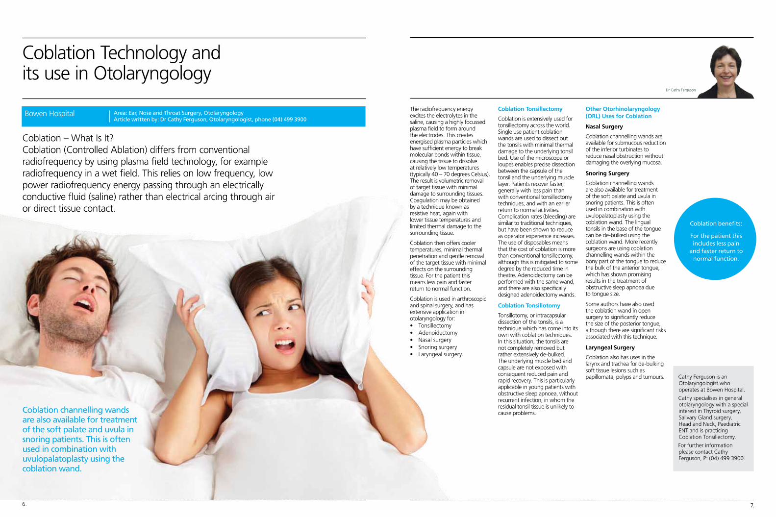
The radiofrequency energy excites the electrolytes in the saline, causing a highly focussed plasma field to form around the electrodes. This creates energised plasma particles which have sufficient energy to break molecular bonds within tissue, causing the tissue to dissolve at relatively low temperatures (typically 40 – 70 degrees Celsius). The result is volumetric removal of target tissue with minimal damage to surrounding tissues. Coagulation may be obtained by a technique known as resistive heat, again with lower tissue temperatures and limited thermal damage to the surrounding tissue.
Coblation then offers cooler temperatures, minimal thermal penetration and gentle removal of the target tissue with minimal effects on the surrounding tissue. For the patient this means less pain and faster return to normal function.
Coblation is used in arthroscopic and spinal surgery, and has extensive application in otolaryngology for:• Tonsillectomy• Adenoidectomy• Nasalsurgery• Snoringsurgery• Laryngealsurgery.
Coblation Tonsillectomy
Coblation is extensively used for tonsillectomy across the world. Single use patient coblation wands are used to dissect out the tonsils with minimal thermal damage to the underlying tonsil bed. Use of the microscope or loupes enables precise dissection between the capsule of the tonsil and the underlying muscle layer. Patients recover faster, generally with less pain than with conventional tonsillectomy techniques, and with an earlier return to normal activities. Complication rates (bleeding) are similar to traditional techniques, but have been shown to reduce as operator experience increases. The use of disposables means that the cost of coblation is more than conventional tonsillectomy, although this is mitigated to some degree by the reduced time in theatre. Adenoidectomy can be performed with the same wand, and there are also specifically designed adenoidectomy wands.
Coblation Tonsillotomy
Tonsillotomy, or intracapsular dissection of the tonsils, is a technique which has come into its own with coblation techniques. In this situation, the tonsils are not completely removed but rather extensively de-bulked. The underlying muscle bed and capsule are not exposed with consequent reduced pain and rapid recovery. This is particularly applicable in young patients with obstructive sleep apnoea, without recurrent infection, in whom the residual tonsil tissue is unlikely to cause problems.
Coblation Technology and its use in Otolaryngology
Coblation – What Is It? Coblation (Controlled Ablation) differs from conventional radiofrequency by using plasma field technology, for example radiofrequency in a wet field. This relies on low frequency, low power radiofrequency energy passing through an electrically conductive fluid (saline) rather than electrical arcing through air or direct tissue contact.
Other Otorhinolaryngology (ORL) Uses for Coblation
Nasal Surgery
Coblation channelling wands are available for submucous reduction of the inferior turbinates to reduce nasal obstruction without damaging the overlying mucosa.
Snoring Surgery
Coblation channelling wands are also available for treatment of the soft palate and uvula in snoring patients. This is often used in combination with uvulopalatoplasty using the coblation wand. The lingual tonsils in the base of the tongue can be de-bulked using the coblation wand. More recently surgeons are using coblation channelling wands within the bony part of the tongue to reduce the bulk of the anterior tongue, which has shown promising results in the treatment of obstructive sleep apnoea due to tongue size.
Some authors have also used the coblation wand in open surgery to significantly reduce the size of the posterior tongue, although there are significant risks associated with this technique.
Laryngeal Surgery
Coblation also has uses in the larynx and trachea for de-bulking soft tissue lesions such as papillomata, polyps and tumours. Cathy Ferguson is an
Otolaryngologist who operates at Bowen Hospital.
Cathy specialises in general otolaryngology with a special interest in Thyroid surgery, Salivary Gland surgery, Head and Neck, Paediatric ENT and is practicing Coblation Tonsillectomy.
For further information please contact Cathy Ferguson, P: (04) 499 3900.
Coblation channelling wands are also available for treatment of the soft palate and uvula in snoring patients. This is often used in combination with uvulopalatoplasty using the coblation wand.
Dr Cathy Ferguson
Area: Ear, Nose and Throat Surgery, Otolaryngology Article written by: Dr Cathy Ferguson, Otolaryngologist, phone (04) 499 3900
Bowen Hospital
Coblation benefits:
For the patient this includes less pain
and faster return to normal function.

Dr Nick Bedford MB ChB, Dip Obst, FRANZCOG Gynaecologist P: (04) 381 8120 F: (04) 381 8121 E: [email protected] E: [email protected] W: www.drnickbedford.co.nz
Nick is a gynaecologist who consults at the Wakefield Specialist Medical Centre, 99 Rintoul Street, Newtown, Wellington and operates at Wakefield Hospital, Newtown, Wellington.
Specialty Gynaecology and Obstetrics
Training Trained at the Otago Medical School during 1993 to 1999 and received his Diploma in Obstetrics and Medical Gynaecology Otago in 2002; RANZCOG Training Program 2003-2008.
His sixth year of training was completed at Monash Medical Centre in Melbourne, followed by a two year Fellowship in Laparoscopic Surgery and Pelvic Floor Repair at Flinders Medical Centre in Adelaide, South Australia.
Specialising in/Special interest in:• Laparoscopic and minimally
invasive gynaecology, especially laparoscopic hysterectomy, myomectomy (removal of fibroids), treatment of endometriosis and laparoscopic pelvic floor repair
• Vaginalsurgeryforprolapseand incontinence including native tissue and mesh options when appropriate, and sling surgery for incontinence
• Generalgynaecologyincluding investigation and management of chronic pelvic pain, heavy menstrual bleeding and infertility.
He will provide comprehensive and sensitive treatment, working collaboratively to achieve the best outcome for his patients.
Dr Rees Cameron FRACP MSc (Hons) Gastroenterologist P: (04) 381 8110 F: (04) 381 8111 E: [email protected]
Rees is a Gastroenterologist who consults at the Wakefield Gastroenterology Centre, Rintoul Street, Wellington and operates at Wakefield Hospital, Florence Street, Newtown, Wellington.
Specialty Gastroenterology
Training Rees is a graduate of University of Auckland Medical School. He trained in Gastroenterology at Christchurch Hospital, and completed a Hepatology Fellowship at Freeman Hospital Liver Transplant Unit, Newcastle-upon-Tyne, and Advanced Interventional Endoscopy Fellowship at California Pacific Medical Center, San Francisco.
Specialising in:• Therapeutic/interventional
endoscopy• Chromoendoscopicevaluation
of colon and upper GI tract • Endoscopicmanagement
of dysplasia in Barrett’s oesophagus
• Inflammatoryboweldisease• Irritablebowelsyndrome
and the management of chronic abdominal pain
• Viralhepatitis.
Dr Cameron’s intention is to establish a world-class endoscopic ultrasound service in Wellington.
New Consultants
9.
Mr Alistair Dray FRACS Orthopaedics P: (06) 873 8806 F: (06) 873 8005 W: www.drayortho.co.nz
Alistair is a consultant orthopaedic surgeon at Royston Hospital in Hastings, and Hawke's Bay DHB. He was brought up in Warkworth and graduated from Auckland Medical School in 1993.
After finishing his orthopaedic training in New Zealand he worked as a consultant at Waikato DHB for two years, focusing on hip and knee arthroplasty and revision, and major trauma, before being appointed to the Surrey Foot and Ankle Fellowship, undertaking a year of surgical training at the Royal Surrey County Hospital and Frimley Park Hospital, with four full-time specialist foot and ankle surgeons.
He returned to New Zealand in 2010 to settle permanently in sunny Hawke's Bay with his family.
His practice is divided between lower limb joint replacement (hip, knee and ankle) and foot and ankle surgery, with a special interest in arthroscopy, sports injuries and general orthopaedics.
8.
Mr Gareth Coulter MBChb, FRACS (Ortho) Orthopaedic Surgeon P: (04) 381 8120 F: (04) 381 8121 E: [email protected]
Gareth is an orthopaedic surgeon who consults at the Wakefield Specialist Medical Centre, 99 Rintoul Street, Newtown, Wellington and operates at Wakefield Hospital, Florence Street, Newtown, Wellington.
Gareth qualified in medicine in 1999 and completed the Orthopaedic Training Programme in New Zealand. His post graduate clinical and research fellowships in Primary Revision Arthroplasty and Foot and Ankle Surgery were in Perth and Melbourne, Australia.
Gareth is available to see patients with any orthopaedic conditions and has a particular interest in disorders of the lower limb.
His special clinical interests are:• Hipandkneearthroplasty• Theuseofcomputer
navigation for knee replacement surgery
• Footandanklesurgery.
Gareth is a Fellow of the Royal Australian College of Surgeons, and a member of the New Zealand Orthopaedic Association.
Dr Latha Vasan MBBS; FCOG; FRANZCOG; MRM Gynaecologist P: (04) 381 8120 F: (04) 381 8121 E: [email protected]
Dr Vasan is a gynaecologist who consults at the Wakefield Specialist Medical Centre, 99 Rintoul Street, Newtown, Wellington and operates at Wakefield Hospital, Newtown, Wellington.
Specialty Gynaecology
Background Latha qualified in medicine in India and completed a specialty Training Programme in Obstetrics and Gynaecology in Cape Town, South Africa. Her post graduate masters in reproductive medicine (MRM) is from the University of New South Wales.
Latha is available to see patients with any gynaecological condition and has a particular interest in disorders of gynae endocrinology and infertility.
Her special clinical interests are:• Menstrualdisorders• Fertilityissues• Gynaeendocrine/hormonal
disorders/ menopause• Endometriosis• Vaginalprolapseand
incontinence.
Latha is a fellow of the Royal College of Obstetricians and Gynecologists in Australia and New Zealand.
Mr Andrew Dowley MB ChB, DLO, F.R.C.S. (ORLHNS (Ed) Otolaryngologist P: (06) 873 1162 F: (06) 873 1163 E: [email protected]
Specialty Ear, Nose and Throat Surgery, Otolaryngology
Training Andrew did his training in the south Trent region, UK, including two years in Queen’s Medical Centre, Nottingham and a Rhinology Fellowship in Plymouth.
Background Prior to coming to New Zealand, Andrew was a consultant in Cheltenham and Gloucester Hospitals where he was Programme Director for the Junior Doctors. He is also a reviewer for Emergency Medical Journal. He is chair of the local Non-Melanoma Skin Cancer Group.
Andrew has a special interest in:• Endoscopicsinussurgery
and its applications• Septorhinoplasty,functional
and cosmetic, including complex nasal reconstruction
• Otologyandeardisease• Facialskinlesionsand
skin cancer.
Mr Dowley consults at the Royston Centre, 325 Prospect Road, Hastings, 4122, and operates at Royston Hospital.
Acurity Health welcomes the following consultants to our Royston and Wakefield hospitals. Please contact them directly if you would like more information about their specialties.

11.10.
POP and UI affect a large proportion of our aging population. Some 11% of women undergo surgical treatment for these problems, many of whom alas, require revision surgery. This represents a major market and in the world of medical devices, specific meshes and surgical kits for prolapse repair and incontinence treatment have been keenly promoted to both clinicians and directly to patients. But in July 2011, with increased awareness of mesh-related complications, the Food and Drug Administration, FDA, the body responsible for authorising medical products in the United States, updated their safety warning on a number of commercially available pelvic mesh kits and reviewed the device classification for several of these products. The change requires several currently available products to undergo more rigorous scientific assessment, or be withdrawn
from the market. The process surrounding approval of new surgical devices, and how this related to pelvic meshes, is interesting. So is the extent of the problems and the challenges that remain for pelvic floor reconstruction.
Why Mesh? – Problems with Native Tissues
Despite the prevalence of POP and UI, our understanding of causative risk factors and long-term outcomes is relatively poor. We recognise the importance of parity and delivery, of age and oestrogen status, of obesity, chronic cough and constipation. Underlying this, deficient pelvic tissues are thought to predispose women to the problem, and moreover to treatment failure. Pelvic tissue from women with stress urinary incontinence and pelvic organ prolapse show a genetic predisposition to abnormal extracellular
matrix remodelling, altering normal tissue architecture and mechanical properties. Vaginal repair of POP using native tissues is associated with significant rates of prolapse recurrence; some 30% of these patients undergo repeat prolapse surgery, often with multiple procedures, with progressively shorter intervals between surgeries. Repair using deficient tissues therefore seems counter-intuitive.
Borrowing from abdominal wall and inguinal hernia surgery, reinforcing surgical repair by incorporating graft materials into POP and UI surgery makes sense. Likely options for grafts include native tissues (harvested from another site), biological materials and monofilament polypropylene mesh. Sufficient native fascia is readily available for UI surgery, but not for POP repair. Biologicals, either cadaveric or porcine, have demonstrated
significant medium-term failure. Monofilament polypropylene mesh, widely used in hernia surgery and spectacularly successful as an anti-incontinence sling treatment, seemed the obvious choice for prolapse repair.
Medical Devices and the Food and Drug Administration (FDA)
Use of surgical mesh for abdominal hernia repair began in the 1950s and preceded FDA device regulation. This was progressively used as a precedent for pre-configured mesh products for treatment of UI and POP to circumvent more rigorous processes of evaluation of safety and efficacy, prior to general marketing. Where a manufacturer believes a device is ‘substantially equivalent’ to an existing and legally marketed device, approval may be sought under a submission entitled 510(k). For devices classified as Class I or II, there is no
Update on Pelvic Mesh in 2012
Mr Andrew Kennedy-Smith
It's been a bad year for the manufacturers of uro-gynaecological mesh. These materials that have become incorporated into repair of pelvic organ prolapse (POP) and management of urinary incontinence (UI) are being re-examined for their safety.
requirement for independent pre-market assessment of safety and effectiveness. Unfortunately, all mesh is not equal, mesh may behave differently placed into different sites in the body, and delivery systems may represent novel surgical technique themselves. One notable and well-used product ‘Prolift’, now being withdrawn, was marketed
for three years without FDA approval; the manufacturer's believing the device too similar to an existing approved product to warrant an independent application for approval. ‘Prolift’ was subsequently used as the precedent for yet another new and modified device, so transferring and evolving the precedent.
Why Kits? – Office, Vaginal
The trend in many branches of surgery is towards more minimally invasive approaches, and in particular to office-based procedures performed under local anaesthetic. This has driven shorter pieces of sling for treatment of UI; vaginal rather than abdominal surgery for treatment of POP; and custom-delivery systems for vaginally-placed mesh at POP surgery. Marketing has
encouraged general use of newer devices and touted a short-learning curve for their execution. Consequently, large numbers of trans-vaginal mesh procedures are being performed, many outside of the operating theatre environment. It is estimated that one in three POP surgeries uses mesh and three of four POP procedures are done trans-vaginally. For UI surgery more than 80% are done trans-vaginally with mesh.
A. Pelvic floor repair mesh kit (in blue), now withdrawn 1 = uterosacral ligament 2 = sacrospinous ligament 3 = tendinous arch of the obturator fascia Source: Gynecare mesh and anterior repair
B. Retropubic anti-incontinence mesh sling – FDA endorsed Source: Gynecare mesh (Polypropylene) and TVT
C. Transobturator anti-incontinence mesh sling – FDA endorsed
A. B.
C.
Continued on page 12
1
23
Area: Urology Article written by: Mr Andrew Kennedy-Smith, Urologist, phone (04) 381 8344
Wakefield Hospital
Source: Tepha Inc

Sacrocolpopexy is an abdominal approach to POP repair, first described in 1957 by Arthur and Savage, modified in 1962 by Lane. It may be performed laparoscopically with the usual advantages of early return to normal function and early discharge from hospital that is associated with laparoscopic surgery, but the technique is otherwise similar to the original descriptions.
Mesh is used to suspend the apex of the vagina, uterus and cervix, to the sacral promontory. The support mimics normal anatomical support by the uterosacral ligaments and preserves the normal vaginal axis. Hysterectomy is unnecessary for success. Indeed, it is preferable to avoid an associated vaginal incision. The mesh is commonly extended down the back of the vagina to the pelvic floor and perineal body. Anteriorly it is extended to the level of the bladder trigone. These extensions treat the components of cystocoele and rectocoele, in addition to effective apical support.
Compared to traditional vaginal surgery without mesh, abdominal prolapse repair with mesh (sacrocolpopexy) results in less recurrent prolapse.
Compared to transvaginal POP surgery with mesh, there is a lower rate of mesh complications with sacrocolpopexy. Pelvic mesh placed abdominally at sacrocolpopexy appears
safe and use of mesh for this procedure is considered appropriate by the FDA, following their review.
Abdominal sacrocolpopexy, either open or laparoscopic, restores pelvic anatomy and provides durable repair of prolapse, with resolution of prolapsed related symptoms.
My own experience over the past ten years with this procedure, performing it laparoscopically, parallels published results: resolution of symptoms and satisfaction rates are high (95%), success is durable, and mesh erosion is rare (two percent).
De novo UI (unmasked UI) occurs in nine to 42% of patients unless an anti-incontinence sling is placed simultaneous with the POP surgery. I have advocated sling placement simultaneous with sacrocolpopexy except in patients with established voiding dysfunction not attributable to prolapse. I have not seen complications secondary to sling placement in this context.
The posterior mesh is routinely extended down the posterior vaginal wall, between rectum and vagina, to treat rectocoele. This component of sacrocolpopexy is now appearing in colo-rectal literature as laparoscopic anterior/ventral rectopexy, for treatment of rectal prolapse, faecal incontinence and obstructed defecation.
Conclusions
In the management of pelvic organ prolapse (POP) and urinary incontinence (UI),2012, therefore seems the dawn of an old era:
1 Incontinence surgery looks set to return from the office to the operating theatre, with retro-pubic (TVT), and probably transobturator (TOT), polypropylene slings remaining standard treatment;
2 The role for abdominal POP repair using laparoscopic or open sacrocolpopexy will likely increase, in the wake of regulatory and clinical difficulties encountered with vaginally placed pelvic mesh and established limitations of traditional tissue based vaginal repair.
References
1. Epidemiology of surgically managed pelvic organ prolapsed and urinary incontinence Olsen AL, Smith VJ, Bergstrom JO, Colling JC, Clark AL Obstet Gynecol. 1997 Apr;89(4):501-6
2. Alterations in connective tissue metabolism in stress incontinence and prolapse Chen B, Yeh J J Urol. 2011 Nov;186(5):1768-72. Epub 2011 Sep 25. Review
3. FDA Safety Communication: Update on Serious Complications Associated With Trans-vaginal Placement of Surgical Mesh for Pelvic Organ Prolapse U. S. Food and Drug Administration Silver Spring, Maryland http://www.fda.gov/MedicalDevices/Safety/Alertsand Notices/ucm262435.htm; July 13, 2011
4. Urogynecologic Surgical Mesh: Update on the Safety and Effectiveness of Trans-vaginal Placement for Pelvic Organ Prolapse U. S. Food and Drug Administration Center for Devices and Radiological HealthSilver Spring, Maryland http://www.fda.gov/downloads/MedicalDevices/Safety Alertsand Notices/UCM262760.pdf; July 2011
5. Vaginal Mesh Explantation: An Emerging Urological Discipline Winters JC. J Urol. 2012 May;187(5):1529-30. Epub 2012 Mar 14
6. The Current Status of Laparoscopic Sacrocolpopexy: A Review Ganatra AM, Rozet F, Sanchez-Salas R, Barret E, Galiano M, Cathelineau X, Vallancien G Eur Urol. 2009 May;55(5):1089-103. Epub 2009 Feb 4
7. Long-term outcome of porcine skin graft in surgical treatment of recurrent pelvic organ prolapse. An open randomised controlled multicentre study Dahlgren E, Kjølhede P; RPOP-PELVICOL Study Group Acta Obstet Gynecol Scand. 2011 Dec;90(12):1393-401. doi: 10.1111/j.1600-0412.2011.01270.x. Epub 2011 Oct 13
8. Surgical Management of Pelvic Organ Prolapsed in Women Maher C, Feiner B, Baessler K, Adams EJ, Hagen S, Glazener CM Cochrane Database Syst Rev. 2010 Apr 14;(4):CD004014. Review
9. FDA Medical Devices: Premarket Notification (510k) U. S. Food and Drug Administration Silver Spring, Maryland
13.
Problems – Erosions, Pain
Polypropylene is an excellent surgical mesh material. It doesn’t degrade significantly, it integrates with tissues and as a monofilament with an appropriate weave, has low rates of infection. The problem with polypropylene is that is doesn’t degrade; it persists and can trigger a foreign body reaction. When exposed and eroded, it is infected, and resolution generally requires mesh removal. Mesh that has integrated is challenging to remove and removal may be associated with significant surgical trauma.
The space between the vaginal skin and the urethra and bladder is narrow. Correctly placing mesh into this narrow gap is more readily achieved with a single mesh strip, such as TVT sling, than with a complex mesh sheet such as ‘Prolift’.
Placing mesh trans-vaginally necessarily puts the mesh directly beneath the incision, and therefore the route to erosion.
Vaginal and perineal flora increase the potential for contamination of mesh placed via this approach.
Experienced pelvic surgeons are reporting improved anatomical outcomes, shorter operative times and faster recovery with few complications, using mesh for POP and UI surgery. However, complications such as vaginal mesh exposure, urinary tract erosion and mesh contraction are reported with increasing frequency. Mesh-related complications have been attributed to technical deficiencies, yet are almost uniformly reported, even in registry data of established pelvic surgeons.
A literature review done by the FDA recognised that mesh associated complications are not rare based on data from 110 studies including 11,785 women. Approximately ten percent of women undergoing trans-vaginal POP repair with mesh, experienced mesh erosion within 12 months.
Most of these required surgical excision in the operating room. Mesh contraction may cause vaginal shortening, tightening, and/or vaginal pain and is increasingly reported in the literature.
Further to this FDA review and the Advisory Committee, who met in September, a number of devices currently in use have been recalled. These include ‘Prolift’ and ‘Prosima’, the most commercially successful kits for POP repair. Other companies are reviewing their devices in the context of the FDA warning and consolidated US litigation against mesh manufacturers.
Current Options – Back to the Future
So how will we continue to manage POP and UI in 2012 and beyond?
Happily, sling surgery using TVT for treatment of UI has demonstrated long-term safety and efficacy and it seems unlikely our approach will, or needs to, change.
Whilst slings placed using a transobturator (TOT) rather than retro-pubic (TVT) approach are considered equally safe and efficacious, complete removal of TOT sling, if required, is more difficult than removal of TVT sling. Consideration of feasibility of mesh excision may become increasingly important in the future.
For management of POP, a return to more traditional vaginal repair using native tissues seems likely. But is there a better solution? The experiment with vaginal mesh kits has not addressed the problems of poor tissue integrity and the need for frequent revision procedures, which dog traditional vaginal prolapse repair. Hysterectomy treats uterine prolapse by removing the uterus, without specifically addressing defects in pelvic floor support. Hysterectomy has been associated with increased risk of subsequent POP and UI and the role of hysterectomy for management of POP may be questioned.
Update on Pelvic Mesh in 2012Continued from page 10
13.12.
Mr Andrew Kennedy-Smith is an urologist who operates at Wakefield Hospital and consults from his rooms, Wellington Urology Associates, based at Wakefield Hospital.
Mr Kennedy-Smith’s special interests include laparoscopic approach to urology, the management of prostate and renal cancer, complex urinary tract reconstruction and incontinence management.
For further information please contact Mr Kennedy-Smith on phone 381 8344.
D.
D. Position of mesh placed abdominally for a sacrocolpopexy prolapse repair – FDA endorsed

Over two consecutive days, during the April school holidays, Wakefield Health Limited held its 15th annual GP Conference at Te Papa, Wellington. The venue proved to be an excellent choice as it was well received by all who attended.
Dr John Wyeth, 2012’s chairman of the Wakefield Health Conference, along with Andrew Blair, Chief Executive of Wakefield Health Limited, officially welcomed and thanked all the delegates, speakers and sponsors for attending.
Our Changing World
2012’s theme was ‘Our Changing World’1 We had a wide and interesting array of speakers, two from across the ‘ditch’ and several from outside of Wellington, delivering topics ranging from Detecting Cancer Symposium2, led by Professor Swee Tan, Professor Peter Gibson, Dr Trevor FitzJohn, Dr Shelly Soo, Associate Professor Parry Guilford, Mr Burton King, Dr Ian Coutts and Dr Ian Wilson. Learning from Recent Disasters Symposium3, led by Professor Beverley Raphael, Dr Mark Leadbitter, Dr Adrian Gilliland and Tania Thomas, Deputy Commissioner from the Health and Disability Commissioner’s office. To, Our Future4, led by Associate Professor Dawn Elder and Mr Rob Rowan.
Workshops
Delegates were also spoilt for choice by the variety of workshops they could attend given by specialist consultants. These workshops were led by Professor Peter Gibson, Dr Frank Weilert, Dr Alexander Sasse, Dr Dynes McConnell, Dr Justin Travers, Professor Beverley Raphael and Mr Grant Kiddle.
During the course of the conference, delegates, presenters, sponsors and Wakefield Health Limited staff were able to network with fellow colleagues over refreshments.
We thank everyone who attended this year’s conference and appreciate the very positive feedback received. Wakefield Health is totally committed to supporting our medical community, this is why we organise and sponsor these conferences each year.
References:
1. ‘Our Changing World’ : http://tinyurl.com/8uxlx6o
2. Detecting Cancer Symposium : http://tinyurl.com/9hjdnua
3. Learning from Recent Disasters Symposium : http://tinyurl.com/9n84q2h
4. Our Future : http://tinyurl.com/9vv7j2m
GP Conference 2013
Planning for next year’s GP Conference is already underway. Please keep an eye out for details on our website www.acurity.co.nz and future editions of Health Matters.
Wakefield Health Annual GP Conference 2012
14. 15.
“We thank everyone who
attended this year’s conference and appreciate the
positive feedback”

Bariatrics: A Balanced Surgical Approach
16. 17.
“Eating with the brain, not with the eyes.”
“Tracy’s” story as told to Greg Clarke, former Chief Operating Officer, Acurity Health Group Ltd.
I’d had enough of feeling sick all the time, of collapsing at the end of the day exhausted. The year leading up to my gastric sleeve surgery in particular was hard going
– I had constant nausea, was tired and felt like I wasn’t keeping up with things. At 53 I weighed 118kg and had a BMI of 46. I had diabetes and high blood pressure. But more than anything I wanted to be able to keep up with my new grandson.
It wasn’t an easy decision but one that I pretty much kept to myself. Even my husband was unaware of what I was doing until I started the optifast diet. By then I had taken out a personal loan and nothing, and I mean nothing, was going to stop me. I had to do a lot of work on myself, it wasn’t just about the weight, but also about being mentally prepared. It’s about a mindset so being very determined and fixated certainly helped!
My husband was not happy, in fact it would be fair to say that he was really not happy. He thought “oh well it’s another try yah yah yah” and then there was the cost. He made an appointment to see the surgeons but once I went on the diet and he realised how pedantic I was about it, he knew it wasn’t going to be a five minute wonder. He could see it was important to me, and he was very encouraging.
I did a lot of research, spoke to my GP who was supportive but didn’t know a lot about the procedure so did some more investigation after talking to me.
As part of the preparation I met with the surgeons, a dietician and a counsellor. The counsellor focused on the psychological
side of things and could see that my personality made me a good candidate for it.
The dietician covered off what kinds of food I would be able to eat afterwards.
I started preparing meals and freezing things. My friend bought me a little plate smaller than a saucer, I also bought little dishes for afterwards.
On the diet I dropped weight so quickly at times I was freezing. I had to wear layers tighter than normal clothes.
My surgery took place at Wakefield Hospital in August 2011. I came in on a Monday and was discharged on the Wednesday. The surgeons were great, very supportive and there for me if I needed anything. The Wakefield Hospital staff were wonderful and very caring. I am not normally a good patient (ironically I am a nurse), but I did okay.
I stayed at home for a week then went on a prearranged trip for a few days. I took it in stages. I stayed home for two weeks and started back at work part time for four hours a day. The plan was to get back to full time work after four weeks but it took me six because I was still very, very tired. Looking back I did return too early; I couldn’t help myself – I was trying to squeeze a full time job into four hours.
One of the things about the op is that you can get anxious afterwards and I did. I’m not a needy person but I became quite clingy (my husband thought it was great).
I hadn’t really been prepared for the anxiety of thinking “oh my god, what if I eat this and it stuffs up my stomach?” and undoing all the good work that had just been done. My motto became “eating with the brain, not with the eyes”.
One of the reasons I didn’t tell people is that a lot of people make negative comments such as “you took the easy way out”. Nothing could be further from the truth – it is NOT an easy process, it’s a hard option. For me it’s an everyday thing focusing on what I am eating and asking yourself is this going to support what I am doing?
For the first six weeks I ate a lot of pureed food and gently introduced other foods. It took a while to eat meat and fish.
At about five and a half months I was eating normally although I no longer eat bread or eggs as they tend to sit uncomfortably with me.
I have adapted what I eat to where I am now. I eat smaller portions but more often. I really struggle to drink water as I never used to. Also am eating a lot more protein which was really hard work.
I do go out and eat but tend to be more choosey. I go for entrees rather than big meals now. I don’t do the high fat stuff of course – just can’t. There is no value in the food. I’m always asking myself
“what is valuable for my body?”
Bariatrics: Patient’s Story
“It’s not about the operation – it’s about what I do with it.”
Continued on page 18
With 1.1 billion people worldwide considered to
be overweight, adult morbid obesity has now been recognised as a worldwide pandemic.
In New Zealand about 60% of the population
is considered overweight, mainly due to our ‘obesogenic environment’ i.e. ready access to poor nutrition combined with reduced physical activity.
Today we follow the journey of ‘Tracy’, a patient who has kindly agreed to share her experience, and hear from Elisabeth Stubbs and Angela Phillips, members of the multi-disciplinary team that support consultants Mr Simon Bann and Mr S Kusal Wickremesekera in their bariatric practice.
60% of Kiwis are overweight
I have lost weight. To me I’m not thin but I am where I am happy, and am now working on fitness. After four months I did the Round the Bays 7km walk, and a couple of weeks ago I finished a 10km walk. A long way from struggling to walk around the block! I now walk most days sometimes for 45 minutes, sometimes for an hour and a half depending on how I am feeling.
I now go to the gym and have a personal trainer which has been really important. To be honest I have to say that the gym is not my favourite place – I do struggle to go and find it quite isolating as it’s all about doing your own thing. I go to build muscle strength. I now have calf muscles, things have changed a lot! I look for opportunities to do exercise rather than excuses not to. I’m not perfect by the way – there are days when I think “it’s too hot” or “it’s too cold”, or …
As a big person I didn’t have a lot of personal confidence but as a nurse I was used to standing up for my patients as part of my training. I am definitely a little bit feistier now. My confidence in my own ability has grown.
There was a financial impact – I went down four bra sizes in less than six weeks, and at $70 a bra it added up.
There have been other changes – my diabetes is dormant, my blood pressure is down and I don’t feel constantly sick. One of the biggest differences is that I can go to the shops now. I really enjoy shopping which can be a bit disastrous! I can go into those shops I wouldn’t have gone into before. I now wear different colours, not just black anymore.
My energy levels are up. I still run around like a tornado at work but the difference is that I don’t crash at the end of the day feeling like death warmed up. Instead I’m off for a walk.
My family and friends notice the difference and are stoked with what I have achieved. People who didn’t know, don’t need to know.
It’s been a year now. I have lost around 30kg and maintain my weight within a 2kg range. Surgery is not an easy option; to do something this major I was very aware that it had to be for good.
I do look at life differently now – it’s an everyday thing. The operation started the process but it’s now up to me to make it work. Best of all I can now chase my grandson around!• Tracy’s name has been changed to
protect her privacy.
I would recommend the op but with the following provisos:
1. Attitude
You must have the right attitude – are you prepared to change your lifestyle?
2. Balance
You must have balance in your life. You can’t work all the time and then rush out and grab something. If you put work ahead of home life or health then it’s not going to work. Are you prepared to make changes? If not, then you are not ready.
3. Organisation
Be organised – freeze meals, get smaller clothes in advance, keep some money aside so you can buy yourself things as required.
4. Programme
Follow the programme with support from the doctors, dietician and counsellor.
5. Focus
Focus on what you eat.
6. Monitor
Watch for anxiety.
19.
Bariatrics: Behind the Face
At Capital Obesity Group we offer a holistic approach to morbid obesity, which includes at least six counselling sessions and implement programmes tailored to the individual’s needs.
We live in an environment that discourages physical activity, while encouraging the consumption of supersized portions of high fat/sugar processed foods. Obesity has now reached epidemic proportions. When other methods have failed, many people consider bariatric surgery.
The bariatric procedure offered by Capital Surgeons is the laparoscopic Sleeve Gastrectomy. Prior to surgery, patients have two counselling sessions to discuss their individual goals, as well as formulating a comprehensive pre-surgical evaluation. This serves as a planning function and education rather than gate keeping; some patients may need further help prior to surgery. However, for a minority of people surgery is not in their best interest.
Deconstructing Shame
With my patients, what often emerges are not Diagnostic and Statistical Manual of Mental Disorders, Fourth Edition (DSM IV), diagnosable criteria, but patterns of disordered eating, feelings of shame and isolation. In my experience, shame is a fundamental aspect of disordered eating and my patients typically experience themselves as inherently
deficient, worthless, and as failures. Shame has to do with the experience of disconnection and isolation.
When people are treated with an extra measure of compassion and concern their feelings of rejection and shame can be alleviated. Counselling provides both support and an opportunity for patients to realise and overcome the complexities of their suffering.
Unconscious Avoidance
Some people deflect from unpleasant feelings, often out of conscious awareness. Food lends itself readily to such usage because eating (suckling) from birth on, is always intermingled with interpersonal and emotional experiences, and its physiological and psychological aspects cannot be strictly differentiated. Many of our patients no longer know when they are satiated; surgery may help this feeling return.
My work is by phenomenological exploration and by raising the patient’s awareness of their self defeating lifelong patterns.
We collaboratively plan more useful ways of living, thus empowering the individual. Some of the patients choose to extend their routine four post-operative counselling sessions and opt for psychotherapy with me. This has been beneficial as it can expand their capacity for human contact rather than resorting to inanimate objects such as food and alcohol. Many of the patients I see suffer from anxiety.
Summary
At Capital Obesity Group, we believe that surgery is only a tool to help jump start a journey of weight loss. The success of our holistic treatment approach is subject to each patient’s lifelong commitment to acknowledge and recognise the major challenges that they face and to make appropriate lifestyle decisions.
Morbid obesity is an overt complex phenomenon and cannot be hidden from our view, thus many individuals may be suffering psychologically.
Elisabeth originally trained as a registered nurse in London, going onto become a senior charge nurse of a surgical ward for a number of years before coming to New Zealand where she
became involved with Bariatric care. At that time she saw the need for emotional support and healing for people
who undergo this life changing operation.
Her passion for this work motivated her to obtain a degree in counselling and then go on to gain a diploma in Gestalt Psychotherapy, which looks at a person in a holistic way. This gives her further expertise to explore deeper issues within this important field, with the aim of achieving improved outcomes.
She is available for both short and long-term work.
Email: [email protected]
Bariatrics: Patient’s Story
Continued from page 17
18.
Area: General Surgery – Bariatric Surgery Article written by: Elisabeth Stubbs, Counsellor, E: [email protected]
Wakefield Hospital

20. 21.
Example of a typical intake long-term after surgery:
Breakfast
1 x weetbix plus ½ cup calci-trim milk plus 100g yoghurt or 1 x poached egg on toast
Morning Tea
Fruit
Lunch
120g tuna with 2 cruskits or Chicken salad (100g chicken plus ½-1 cup salad)
Afternoon Tea
Yoghurt
Dinner
120g chicken / one small chicken breast ½ small / medium potato 1/3 cup vegetables or 120g salmon ½ small dinner roll Salad ½ cup
The nutrition pre-assessment includes identifying any nutrient deficiencies, as up to as many as 60-80% of morbidly obese patients have micronutrient deficiencies pre-operatively1. Pre-assessment helps identify any current or previous history of eating disorders, and determines the patients ability to make lifestyle changes based on previous weight loss attempts and eating patterns. At this stage education is provided on pre and post surgery nutrition to ensure a clear understanding of changes required and the need for life-long healthy eating and lifestyle patterns. At this time individualised recommendations are made based on eating behaviours. These changes should commence prior to surgery to assist with post surgery behaviour.
For two to six weeks, prior to surgery, patients go on the Optifast Very Low Calorie Diet (VLCD). Optifast VLCD is a complete meal replacement, and the programme entails three Optifast VLCD products, two cups of low starch vegetables and one serving of fruit. This is generally well tolerated; some tiredness in the first week can occur but feedback from patients is generally positive. The purpose of the programme is to shrink liver size to enhance the safety of their procedure.
Surgery reduces the size of the stomach, and reduces the production of Ghrelin (appetite-stimulating hormone) therefore patients are assisted to reduce their quantity of food long-term to facilitate significant weight loss. During the first six weeks post surgery, patients progress from puree textures to a regular healthy diet high in protein, including small quantities of complex carbohydrates, low fat dairy, fruits and vegetables. Due to high protein requirements (60-120g protein/day2) and the significant reduction in food quantity following surgery a protein supplement is usually required during the initial six to twelve weeks. It is important to ensure food quantities remain reduced long term, and foods are low in calorie density. If not then weight re-gain can occur.
When patients start the process of consideration for a Sleeve Gastrectomy the first step is for patients to undergo a nutrition assessment to gather background information to ensure any medical or lifestyle concerns can be addressed prior to surgery.
The reduced quantity of foods post surgery results in a reduction in vitamin and mineral intake, therefore a multivitamin needs to be taken daily as micronutrient deficiencies can occur. A common post-operative complication is constipation due to reduced fibre intake and inadequate fluid intake. This is ideally treated with increased fluids, a fibre supplement and/or Kiwicrush but if necessary a laxative is used.
Research shows food tolerance and gastrointestinal quality of life post Sleeve Gastrectomy was ranked superior to the laparoscopic adjustable gastric banding (LAGB) and Roux-en-Y gastric bypass (RYGB)3. Foods commonly identified by our patients as being poorly tolerated are meat, chicken, fatty or rich foods and bread although most patients can return to a full intake with no intolerance. Food intolerance is often associated with non-compliance, in particular eating too quickly.
The expected long-term weight loss following a Sleeve Gastrectomy is equivalent to RYGB and LAGB4. We aim for weight loss of 60-75% of excess body weight based on research to date. Regular exercise and a high protein intake are important to assist with long-term weight loss maintenance. Our patients are seen by our Lifestyle Coach to provide them with an individualised exercise programme that works for them. Patients have five post surgery consultations over the first year. Additional consultations are undertaken on an individual basis if required or requested.
Bariatrics: Nutritional Support
Angela Phillips
Angela Phillips is a dietician who practices from the Wakefield Specialist Medical Centre. Angela works in conjunction with General Surgeons, Mr Simon Bann and Mr Susrutha (Kusal) Wickremesekera.
Other services Angela provides are:• Allergies• MaternalNutrition• Cholesterol• Paediatrics• Diabetes• Surgery• FoodIntolerance• WeightManagement.
For further information please contact Angela: Email: angela.phillips @wakefield.co.nzWebsite: capitalobesitygroup.co.nz
Appointments can be made through Rosanne Tait, Medical Secretary, Wakefield Specialist Medical Centre on (04) 381 8120 extn 7331.
References
1. Surgery for Obesity Related Diseases 4 (2008) S73-S108
2. Obesity Surgery (2011) 21:1798-1805
3. Obesity Surgery (2012) 22:536-543
4. ASMBS Position Statement on Sleeve Gastrectomy as a Bariatric Procedure: www.asmbs.org/2011/12/sleeve-gastrectomy-as-a-bariatric-procedur/
Area: General Surgery – Bariatric Surgery Article by: Angela Phillips, Dietician, Wakefield Specialist Medical Centre, phone (04) 381 8120
Wakefield Hospital

Only a small percentage of patients with both an aggressive subset of disease and those who could afford private Photodynamic Therapy (PDT) had a chance of slowing progression of central blindness. Imagine the excitement when we were able to offer to all exudative ARMD patients, treatment which preserved the presenting vision in 80% and actually improved vision in 25%. The issue of monthly injections inside the eye for two years seemed inconsequential, compared to the possibility of having something better to say than “sorry”.
The key to good results, however, is ‘the presenting vision’. So often, patients notice the loss of vision in one eye on a chance closing of the good eye, or a routine optometry check, and by then the natural history of bleeding, scarring and photoreceptor damage has already occurred. Earlier detection gives better outcomes.
So as the excitement settles and we realise the truths – that some patients will not benefit, that ARMD is a chronic disease
requiring ongoing invasive treatment, and that the burden of observation and treatment is massive – we now turn toward improving outcomes by education.
The Macular Degeneration New Zealand (MDNZ) was formed in 2009/10 with a view to providing both public education and a voice to lobby New Zealand Government to ensure equitable treatment nationwide and obtain adequate funding. In New Zealand and Australia, ARMD is found to be the leading cause of blindness, see figure 1, and this data is being used to drive awareness programmes and justifying resource demands.
What Can Primary Carers Do?
Facilitate optometry review at least two yearly over the age of 50, more often as guided by the eye professionals findings or positive family history (increased risk of ARMD by 50%). Optometrists will gauge the level of risk, optimise refractive vision, check for Glaucoma, advise on dietary supplements, and refer if necessary.
Attend to modifiable risk factors:
•Smoking Smoking increases ARMD risk three to four times, however, this reduces with cessation
•Control Control blood pressure, cholesterol and encourage exercise
•Diet Diet – increase omega 3 and lutein. Eat fish two to three time per week, dark green leafy vegetables and fresh fruit daily and a handful of nuts per week. Excellent dietary information is available on the MDNZ website.
Refer new symptoms of blurring or distortion to your optometrist promptly. They have good relationships with their local ophthalmologists, are well trained in recognising high risk features and know the terminology to enable a rapid appointment.
Be aware that Intravitreal Anti- Vascular Endothelial Growth Factor (VEGF) may theoretically restrict
healing blood vessels systemically and is withheld in cases of unstable angina, poorly controlled hypertension, recent myocardial ischaemia or cereb-rovascular disease, or imminent surgery.
Low Vision Aids
There are specialist clinics within the public system to address the needs of those who fall below the Royal New Zealand Foundation of the Blind criteria.
Be alert to depression in the poorly sighted.
References
MDNZ local information including seminars: http://www.mdnz.co.nz/ MD Foundation excellent patient handouts and research papers: http://www.mdfoundation.com.au/
This process has variously been called ‘Enhanced Recovery After Surgery’ (ERAS) or less helpfully ‘Fast Track’ because, on average, patients on such a programme will get out of hospital two and a half days faster than patients managed with a traditional surgical approach. More importantly post-operative complication rates are reduced by up to 50% in a meta-analysis of randomised trials of ERAS and conventional care1.
The ERAS programme is based on the principles of patient autonomy and an evidence based approach to all aspects of surgical and anaesthetic intervention before and during the surgical stay.
A Danish surgeon, Henrik Kehlet, has led the field in refining components of peri-operative surgical care into a coherent pathway over the last 15 years.
How does ERAS Work in Practice?
When patients are scheduled for elective colon or rectal surgery in the outpatient clinic they are given a much clearer explanation of what will happen in hospital and what is expected of them on return to the ward. A patient diary facilitates their achievement of the recovery milestones.
Prior to surgery no bowel prepar- ation is given apart from an enema for rectal surgery and patients are allowed to have a clear ‘pre-op’ carbohydrate drink up to two hours before surgery. They arrive in the operation room in a ‘fed’ rather than fasted state which is associated with a faster recovery.
Surgery is performed by a laparoscopic assisted approach or transverse incisions where appropriate, nasogastric tubes are not used and abdominal drains avoided in most situations.
The intra-operative and post-operative fluid management is important as excess ‘salt’ containing intravenous fluids are associated with an increased cardiovascular morbidity and surprisingly also surgical morbidity. Fluids are given in a goal directed manner rather than according to a fixed formula. Patients are offered protein and carbohydrate drinks on return to the ward, and food the day following surgery.
Opiate analgesia is minimised both during and after anaesthesia to avoid the unwanted effects of delayed gut function and nausea and vomiting. Multi-modal analgesia including paracetamol, tramadol, COX2 inhibitors and a lignocaine infusion, is combined with small incisions to minimise analgesic requirements. Wound infusion catheters, of long acting local anaesthetic, are routinely used although some protocols require a high thoracic epidural for 48 hours on the basis that it reduces sympathetic activation and thus post-operative ileus.
22.
Enhanced Recovery After Surgery (ERAS) Coming to a Hospital Near You
A growing understanding of the importance of evidence based guidelines in all aspects of the management of patients undergoing elective colon and rectal surgery has been refined into a reproducible package of care aimed at optimising patient outcomes with the benefit of fewer complications and reduced length of hospital stay.
Multiple walks, from day one, are encouraged and intravenous fluids and the urinary catheter are removed on the first post-operative day. Increasing mobility and resumption of a normal diet are encouraged from day two. Anti-emetics are administered pre-emptively. Patients are allowed home when they meet accepted discharge criteria (ability to tolerate a diet for three meals, adequate oral analgesia, passage of flatus or stool and adequate social support) rather than at a fixed time after surgery.
Implementation of ERAS
Although many components of ERAS have found their way into contemporary surgical practice it appears that ‘the whole is greater than the sum of its parts’. ERAS requires a whole team approach to achieve its objectives and is fast becoming an integral part of colorectal surgery and will likely be adopted by other surgical specialities.
References
1. Enhanced recovery pathways optimise health outcomes and resource utilisation: a meta-analysis of randomised controlled trials in colorectal surgery. Adamina M, Kehlet H, Tomlinson GA, Senagore AJ, Delaney CP. Surgery. 2011 Jun;149(6):830-40
23.
Age Related Macular Degeneration (ARMD)
Exudative (Wet) Macular Degeneration management has changed since the introduction of Anti-Vascular Endothelial Growth Factor (VEGF) intravitreal agents. Prior to 2004 – 2006 we saw only the tip of the iceberg in terms of ARMD patients.
‘‘
The benefits of ERAS:
Fewer complicationsComplication rates are reduced by up to 50%
Shorter hospital visit On average, patients will get out of hospital two and a half days faster
More responsive Patients are allowed home when they meet accepted discharge criteria rather than at a fixed time
50%
Mr John Keating is a General Surgeon, who consults and operates at Bowen Hospital.
Mr Keating specialises in:•ColonandRectalsurgery•Laparoscopicsurgery•Colonoscopy•PseudomyxomaPeritonei.
For further information please contact Mr Keating, P: (04) 479 8261.
Dr Helen Long specialises in retinal diseases, ocular inflammatory disease and cataract surgery. She consults from the Kelburn Eye Centre and operates at Bowen Hospital.
For further information phone (04) 475 9559.
“ERAS requires a whole team approach to achieve its objectives and is fast becoming an integral part of colorectal surgery”
Mr John Keating
Figure 1: Causes of blindness in over 60
50% Macular Degeneration
3% Uncorrected refractive error
20% Other
16% Glaucoma
11% Cataract
Dr Helen Long
Area: General Surgery Article written by: Mr John Keating, General Surgeon, phone (04) 479 8261
Area: Ophthalmology Article written by: Dr Helen Long, Ophthalmologist
Bowen Hospital Bowen Hospital

24. 25.
There are two main aspects to a septorhinoplasty; function and form. There are also two viewpoints, the patient’s and the surgeon’s. None of these things are independent of each other – a bent nose is also likely to have some degree of obstruction.
Function
The most important of these is patency of the airway and thus this is the most frequent complaint. What must not be forgotten are olfaction, filtration and warming of inspired air. Olfaction may be affected by poor airflow but it may also have been irreparably damaged by a previous injury so it should be recorded in the pre-operative history. The nose is the first part of the common airway and as such improving airflow here can have a positive impact on the lower airway.
Form
This is the aspect that really excites both surgeon and most patients’, however, it is also the one that is most subjective and therefore requires an honest conversation between surgeon and patient about what is possible and what is perceived to be an issue by both parties. Thus a formulaic approach is inappropriate.
An accurate pre-operative diagnosis is vitally important in order to inform the patient of their issues, as they will frequently know that things are wrong but are unable to define what or why. This must start with the underlying structure of bone and cartilage, followed by an assessment of the soft tissue covering including skin. Everything should be taken in the context of the face as
a whole, ensuring any changes are in keeping with the overall structure.
In many patients their bony and or cartilaginous anatomy has been so badly damaged that tissue has to be taken from elsewhere. The patient’s own nasal septum can be used, however it is rigid and brittle so it is best to be limited to correcting deformities of the septum itself. The cartilage in the pinna is strong and flexible, giving a natural feeling nose. However, to avoid affecting the appearance of the ear, only the conchal bowl can be taken, making it inadequate when the problem extends to the nasal bones. In this situation, where the patient has a significant saddle deformity, using costal cartilage provides the best platform for reconstruction.
Viewpoints
As surgeons an intricate knowledge of the anatomy and how that affects form
and function can give us a good idea of the issues that are problematic and how to deal with them. This is all very well but the patient will have their own idea of what is important to them. I am often surprised at how patients will not notice or will happily accept what I consider to be a major issue and be delighted that what they consider to be the presenting complaint has been dealt with. It can be quite a journey to the point where the nose has healed and the final result is known, often 12 months or more, and if the patient is pleased with the result, that is what is most rewarding.
Mr Dowley is an ENT Surgeon who consults and operates at Royston Hospital.
For further information please contact Mr Dowley, phone (06) 873 1162.
Septorhinoplasty Changing the Face of ENT
This surgery is tremendously rewarding as each nose is different and there is a lot of creativity dealing with the various individual aspects of each nose. It can also be terribly frustrating; you are dealing with the most prominent feature on the face and any slight imperfection is there for all to see. This is especially true when one considers the fact that the scarring that comes with the healing contracts, potentially causing an unpredictable result.
An injury resulting in widening and deviation of the nasal bones and shifting the nasal dorsum to the right paramedian position, resolved by an intranasal septorhinoplasty.
Pre-op Post-op
This 13 year old had a nasal septum fractured into multiple pieces resulting in a saddle nose. Removing the whole of the cartilaginous septum, suturing it back together and replacing it has resolved the saddle. Traditionally this surgery would have been delayed until adulthood to allow maturation of the facial skeleton, but recently this standpoint has changed as improving the underlying structure allows improved growth of the soft tissues.
Pre-op Post-op
A saddle nose gives the appearance of a hump when in fact the problem is a hollow inferior to the bones. This time required conchal cartilage to rebuild the septum.
Pre-op Post-op
After a horrific accident the intermediate and lateral crurae (dome of the nasal tip) of the right lower later cartilage were malpositioned high on the nasal dorsum giving a flattened profile and causing nasal obstruction. Careful dissection and repositioning have improved the situation, but of course the scars remain.
Pre-op
At the age of four this young man suffered a type three nasal fracture with compression of the ethmoidal bones. This has resulted in a scooped out, infantile appearance that has been reconstructed with a costal cartilage graft both in the nasal dorsum and columella.
Before / Pre-op
Post-op
Pre-op
Post-op
“...the patient will have their own idea of what is important to them.”
Mr Andrew Dowley
Figure 1.
Figure 2. Figure 3.
Figure 5. Figure 4.
Area: Ear, Nose and Throat Surgery, Otolaryngology Article written by: Mr Andrew Dowley, ENT Surgeon, phone (06) 873 1162
Royston Hospital

26. 27.
Mr Paul Mason
The last decade has witnessed significant advances in the diagnosis and treatment of lymphoid related blood cancers. The lymphomas, and chronic lymphatic leukaemia (CLL), together account for a large proportion of haematological malignancies seen in the clinic.
There are over 30 different types of lymphomas according to the World Health Organisation (WHO) classification, it is important to make a precise diagnosis to plan for optimal treatment. The haematologist is well placed to investigate and organise treatment because of the combined training in pathology and internal medicine.
Lymphomas may have a protean presentation and can arise in any part of the body. They can range from an indolent follicular type that can be simply observed, to a highly aggressive Burkitts type that will require urgent therapy to avoid a fatal outcome. The usual manifestation of high-grade lymphoma will be a palpable nodal mass, often accompanied by concomitant symptoms such as night sweats and weight loss. Patients may present to any subspecialty, i.e. with an abdominal mass to a general surgeon or to ENT with a tonsillar mass.
It is important to involve the haematologist at an early stage to coordinate further investigations. Any biopsy needs to be sent fresh to the laboratory to allow for proper handling. Cytogenetic and molecular testing is vital. A fine-needle aspiration (FNA) may delay the initiation of treatment and does not always provide the correct diagnosis.
The use of CT imaging is necessary in staging the extent of disease and PET/CT is now standard for Hodgkins disease staging. The amount of standardised uptake value (SUV) or intensity tends to correlate with the activity and proliferation rate. The PET scan is also occasionally used in CLL to diagnose large cell transformation that can occur in late stages. Serum lactate dehydrogenase (LDH) is also used in staging to indicate disease burden. Levels as high as 8000 may be found in very aggressive cases.
Splenomegaly may be the sole manifestation in non-Hodgkins lymphoma. A bone marrow biopsy combined with flow cytometry will give a definitive diagnosis without having to resort to splenectomy. Splenic lymphomas are often marginal zone sub-types that respond well to single agent Rituximab.
Diffuse large B cell lymphoma (DLBCL) can now be cured in around 60% of cases with the use of R-CHOP* immuno-chemotherapy. Approximately one third of cases will relapse and will need salvage therapy that may involve an auto-transplant in those with chemosensitive disease.
Autologous transplants are carried out in haematology units at the major cancer centres and can provide long-term disease control. In selected younger patients an allogeneic marrow transplant can be a potentially curative option.
CLL is very common in the elderly population but the disease course is often chronic and indolent. Younger patients tend to have more rapidly progressive disease and will need more potent therapy such as FCR (Fludarabine, Cyclophosphamide and Rituximab), now funded by Pharmac and regarded as the gold standard. The German CLL8 study1 compared FC to FCR and found that the addition of Rituximab improved both progression free survival and overall survival. However, in patients >70 that require therapy, FCR can be difficult to deliver due to toxicities and reduced drug doses are often necessary.
In summary, the advent of Rituximab as targeted therapy against CD20 positive B-lymphocytes has seen significant improvements in remission and cure rates in the DLBCL group, and improved remission rates in CLL. There are now second generation monoclonal antibodies, and other new drugs which target the B cell receptor, being trialled meaning that even better patient outcomes are on the horizon.
* CHOP is the acronym for a chemotherapy regimen used in the treatment of non-Hodgkin lymphoma.
CHOP consists of:
Cyclophosphamide: an alkylating agent which damages DNA by binding to it and causing cross-links
Hydroxydaunorubicin: (also called Doxorubicin or Adriamycin), an intercalating agent which damages DNA by inserting itself between DNA bases
Oncovin (Vincristine): which prevents cells from duplicating by binding to the protein tubulin
Prednisone or Prednisolone: which are corticosteroids.
Reference
1. Hallek at al. Addition of rituximab to fludarabine and cyclophosphamide in patients with chronic lymphatic leukaemia: a randomised, open-label, phase 3 trial. Lancet. 2010: 376 (9747): 1164-1174 German CLL8 – treatment of chronic lymphocytic leukemia.
Dr Ken Romeril is a Haematologist at the Wellington Blood and Cancer Centre. He has a special interest in haematological malignancy and is a Principal Investigator in myeloma and lymphoma trials. Ken also works at Wakefield Hospital in Newtown, Wellington.
For appointments phone Mr Ken Romeril on P: (04) 381 5900 ext. 831 www.lifeblood.co.nz
There are 30 different
types of lymphomas
––––––––––––––––World Health Organisation classification
Chronic rhinosinusitis without polyps represents inflammation of the sinus mucosa and is a manifestation of the mucosal immune response to various stimuli including bacteria, bacterial toxins and fungi. Recent interest has focused on the role of bacterial biofilms and the interaction of bacterial ‘colonies’ with the sinus mucosa.
Achieving satisfactory outcomes for patients suffering with sinusitis can be challenging for surgeons for a number of reasons. Co-morbidities, including asthma, allergy, immunodeficiency and mucociliary disorders, can affect the response to treatment.
Following assessment, including nasendoscopy, patients with chronic sinusitis often undergo imaging with computerised tomography (CT). Patients are usually treated medically initially. This can include topical and/or systemic corticosteroids, antibiotics and nasal saline douching.
Endoscopic sinus surgery is offered to patients who have failed to respond to maximal medical therapy. The aim of surgery is to open the natural ostia of the sinuses to facilitate mucociliary clearance and allow access of topical treatments to the sinuses and to enhance the effect of systemic medical treatment.
Anatomical variations can result in technical difficulties during surgery. Balloon Sinuplasty has become a useful tool in the management of sinusitis over the last decade. It has particular value in the management of frontal sinusitis. The frontal sinus is the most inaccessible of the sinuses and correspondingly failure rates after surgical intervention are the highest for the frontal sinus.
The development of Balloon Sinuplasty was inspired by coronary angioplasty and involves balloon dilatation of the sinus ostium. It can be carried out under local or general anaesthesia. An illuminated guide wire is passed into the sinus and successful cannulation is confirmed by observing trans-illumination of the sinus. The balloon catheter is passed over the guide wire, the balloon is dilated and the sinus ostium is enlarged. Dilatation creates micro-fractures in the surrounding air cells but essentially it preserves the integrity of the mucosa. Bleeding and pain are therefore minimal and length of stay in hospital can be reduced.
Large published series have confirmed the safety of the technique and also confirmed high rates of ostial patency at two year’s post-op2.
References:
1. European Position Paper on Rhinosinusitis and Nasal Polyps 2012; Fokkens WJ et al, Rhinology supplement 23: 1-298, 2012.
2. Long term outcome analysis of balloon catheter sinusotomy: two year follow-up; Weiss RL et al, Otolaryngology Head and Neck Surgery 2008, September; 139 (3 supplement 3): S38-46
Balloon Sinuplasty
Chronic sinusitis is prevalent. About ten percent of the population are thought to be affected by this condition1. Chronic rhinosinusitis can be broadly classified into chronic rhinosinusitis with or without polyps.
An Overview of Lymphoproliferative Malignancies
Mr Paul Mason is an ENT Surgeon who operates at Royston Hospital and consults from rooms in the Royston Centre, Hastings.
Mr Mason has a special interest in Endoscopic Sinus Surgery and extended applications, Otology and Mastoid Surgery.
For further information, please contact Mr Mason on (06) 873 1162.
“CLL is very common in the elderly population but the disease course is often chronic and indolent.”
Frontal Sinus Balloon Sinuplasty
Area: Ear, Nose and Throat Surgery, Otolaryngology Article written by: Mr Paul Mason, ENT Surgeon, phone (06) 873 1162
Royston Hospital Area: Haematology Article written by: Dr Ken Romeril, Haematologist
Wakefield Hospital

GP SurveyWe value your feedback and will use your comments to improve this magazine. Go to our ‘For our GPs’ website and complete the brief survey – www.acurity.co.nz
Health Group Limited28.
Missed an Edition?
Health Matters is produced by Acurity Health Group Limited (AHGL). AHGL also cares about the health of the planet – so an environmentally friendly stock and vegetable inks are used. © 2012. All rights reserved to AHGL. This publication contains general information about medical conditions and treatment. You should not rely on this information in place of a visit, call, consultation or the advice of a specialist or other qualified healthcare provider. Content neither indicates nor reflects the views of AHGL. Any research, study, clinical trial, event, news or other item included in this publication is not intended to imply endorsement or approval of it by AHGL. Although we have made a conscientious effort to provide high quality information, AHGL disclaims any implied guarantee about the accuracy, completeness, timeliness or relevance of any information. Please email any feedback to [email protected]
Go to our 'For our GPs' section on our website – www.acurity.co.nz
Contact Us
Wakefield Hospital
Florence Street Newtown Wellington 6021
P: (04) 381 8100 F: (04) 381 8101 E: [email protected] W: www.wakefield.co.nz
Royston Hospital
500 Southland Road Hastings 4122 Hawke’s Bay
P: (06) 873 1111 F: (06) 873 1112 E: [email protected] W: www.royston.co.nz
Bowen Hospital
98 Churchill Drive Crofton Downs Wellington 6035
P: (04) 479 2069 F: (04) 479 8520 E: [email protected] W: www.bowen.co.nz
Health Group Limited


![Intra-Operative Mitomycin-C as Adjuvant Therapy in ... · Dacryocystorhinostomy (DCR) is the procedure of choice to treat acquired nasolacrimal duct obstruction (NLDO) [1, 2]. The](https://img.pdfslide.net/doc/110x75/5ffe6fcc3e27102a08141e27/intra-operative-mitomycin-c-as-adjuvant-therapy-in-dacryocystorhinostomy-dcr.jpg)