Embed Size (px)
Citation preview
Gaceta Médica de México. 2015;151
592
Heart failure with preserved ejection fraction (HFPEF). Impact of change in the paradigm of isolated diastolic dysfunctionJosé Antonio Magaña-Serrano, Martín Rosas-Peralta*, Carlos Candanosa-Arias, Salvador Valencia-Sánchez, Martín Garrido-Garduno, Roberto Arriaga-Nava and Moisés C. Calderon-AbboUMAE, Cardiology Hospital, Centro Médico Nacional SIGLO XXI, IMSS, México, D.F., México
GACETA MÉDICA DE MÉXICO REVIEW ARTICLE
Correspondence:*Martín Rosas-Peralta
UMAE
Hospital de Cardiología del Centro Médico Nacional SIGLO XXI
IMSS
Av. Cuauhtémoc, 330
Col. Doctores, Del. Cuauhtémoc, C.P. 06725, México, D.F., México
E-mail: [email protected] Date of reception: 06-08-2014
Date of acceptance: 28-12-2014
PERMANYERwww.permanyer.com
Contents available at PubMedwww.anmm.org.mx Gac Med Mex. 2015;151:592-603
Abstract
Heart failure with preserved ejection fraction is a significant and growing public health problem, since it currently represents half of all patients with heart failure. Despite improvements in the understanding of the disease, there is no benefit from treatments tested at all. Advances in diagnostic imaging and invasive evaluation algorithms will allow a more accurate and early diagnosis so that treatment of earliest forms in the progression of the disease are applied since the potential for benefit may be higher. Although important progress has been made in our understanding of the pathophysiology, cardiac catheterization, and cellular of diastolic failure mechanisms and not diastolic mechanisms of disease, further research is required promptly to determine how best to address these anomalies to reduce the significant burden of morbidity and mortality in this form of heart failure, which is reaching pandemic proportions. (Gac Med Mex. 2015;151:592-603)
Corresponding author: Martín Rosas-Peralta, [email protected]
KEY WORDS: Heart failure. HFpEF. Burden of disease. Pandemic.
Introduction
Clinical interest on heart failure with preserved ejec-tion fraction (HFPEF) arose from the confluence of two lines of investigation dealing with (i) left ventricle (LV) diastolic dysfunction in hypertrophied hearts, and (ii) LV remodeling after small myocardial infarctions.
In the late 70’s, the first studies showed that the onset of LV diastolic dysfunction might significantly contribute to heart failure (HF) in hypertrophic cardio-myopathy1,2, aortic stenosis2,3 and hypertensive heart disease4.
Soon after this incursion in the small niche of LV di-astolic dysfunction in hypertrophied hearts, HFPEF was also identified and addressed in studies. These stud-ies, in general, were a “by-product” of large HF trials investigating the use of angiotensin-converting enzyme inhibitors (ACEI) in HF with reduced ejection fraction (EF) (HFREF) and in post-infarction LV remodeling5-7.
HFPEF populations deriving from the last studies were, however, clearly different, since they consisted of patients with limited and small myocardial infarction, but at risk for untoward LV eccentric remodeling. This HFPEF ambiguous origin contributed to the confusion surrounding this entity as a differentiated diagnosis8-10,
J.A. Magaña-Serrano, et al.: Heart failure with preserved ejection fraction
593
and given the neutral result of many large trials on HFPEF, further studies are still necessary11,12.
True cardiac hypertrophy has little in common with limited myocardial infarction, and in both conditions, the underlying mechanisms that drive LV remodeling are likely to be different and, actually, they react differ-ently to drug treatment. Recently, stringent criteria have been proposed for the diagnosis of HFPEF, which consist not only of signs and symptoms of heart failure and a preserved left ventricular ejection fraction (LVEF), but also of LV diastolic dysfunction evidence13,14.
This caused most current HFPEF patients being those with LV concentric remodeling, generally secondary to systemic arterial hypertension, obesity and diabetes, all these without evidence of epicardial coronary artery disease. A low prevalence of coronary artery disease has been recently proposed as a strategic measure for inclusion of the correct patients in clinical trials on HFPEF15.
In the past, HFPEF was often referred to as an equiv-alent to “diastolic” HF (DHF) as opposed to the already traditional “systolic” HF (SHF), which corresponded to HFREF. Since LV diastolic dysfunction is not exclusive to HFPEF but is also observed in patients with HFREF, the term DHF has been abandoned in the last decade and replaced by HFPEF16,17 or else, by HF with normal LVEF (HFNEF)17. However, the terms HFPEF and HFREF have their shortcomings as well. The notion of a preserved LVEF implies knowledge of a pre-existing LVEF, which is almost always absent, and the exact range of LVEF “normality” is difficult to define; i.e., nobody can guarantee that a 50% LVEF is normal for an individual who usually had 65%18,19.
It has not been established if HFPEF and/or HFREF represent different forms of HF or if they coexist as part of the “continuum of HF”13. In spite of the different patterns of the ventricular chamber and the myocellu-lar remodeling observed in couplings with dissimilar responses to medical therapies, all of it would be sug-gestive that these are two distinct processes of the disease. HFPEF is currently observed in about 50% (38-60%) of patients with HF, and the results are close to those observed for HFREF20. The somber prognosis is probably a reflection of the complex multi-systemic and multi-factorial involvement that characterizes all types of HF, regardless of the LVEF, including systems such as the skeletal muscle and vascular dysfunction, pulmonary hypertension, anemia and atrial fibrillation21. The prevalence of HFPEF as related to HFREF is in-creasing at an alarming rate of approximately 1% per year, which is rapidly turning HFPEF into the most
prevalent HF phenotype for the next decade; however, in contrast with HFREF, no improvement in therapeutic results has been achieved over the past two decades20. In spite of these worrying epidemiological trends, the underlying pathophysiological mechanisms of HFPEF and diagnostic strategies or therapeutics remain uncertain21,22. Therefore, the present review has the following:
Objectives and methods
To conduct a systematic review with the exploration, reduction and synthesis method, and focusing on our epidemiological rates of associated risk factors. Using the online PubMed and Google Scholar digital brows-ers in English, French and Spanish languages, at least on their abstracts, the authors selected 102 original articles, the 7 most recent reviews, as well as editorials and related structured summaries.
Pathophysiology
The key studies on HFPEF were explained within the context of HF in the presence of LV preserved EF, but with LV diastolic dysfunction, which consisted of LV isovolumetric relaxation prolonged times, LV slow filling and LV increased diastolic rigidity1-4. However, our group also considered the hypothesis of diastolic dysfunction associated with a transient systolic dys-function (TSD).
With the advent of Doppler echocardiography, LV diastolic dysfunction can be easily appreciated from the parameters through the mitral valve or pulmonary vein flow velocity recordings23. However, the recordings of abnormal mitral flow velocity suggestive of LV dia-stolic dysfunction have been considered as non-spe-cific for HFPEF, as they are found with high frequency in the elderly24, and in patients with HFREF25. The im-portance of LV diastolic dysfunction for HFPEF has been recently reevaluated by invasive studies, which showed consistent presence with LV slow relaxation at rest with elevated diastolic rigidity26, and this elevated rigidity has also been shown to limit cardiac output during atrial stimulation and exercise27,28. This new es-timation has also been made evident in the recent guidelines for the diagnosis of LV diastolic dysfunction by the European and American echcardiography associations13,14.
The reevaluation of LV diastolic dysfunction as an important HFPEF underlying mechanism does not im-ply that it represents the only mechanism contributing
Gaceta Médica de México. 2015;151
594
to the pathophysiology of the disease. Many other mechanisms have been recently identified as compo-nents that play an important role. These include exer-cise-induced systolic dysfunction29-35, exercise-in-duced ventricular-vascular altered coupling33,34,36,37, and vasodilation-mediated abnormal flow28,31-33, chro-notropic incompetence and pulmonary arterial hyper-tension31,33,34,38.
LV diastolic dysfunction
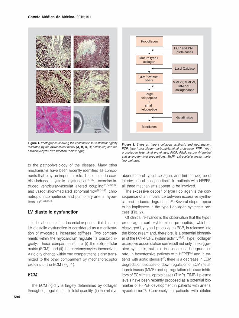
In the absence of endocardial or pericardial disease, LV diastolic dysfunction is considered as a manifesta-tion of myocardial increased stiffness. Two compart-ments within the myocardium regulate its diastolic ri-gidity. These compartments are (i) the extracellular matrix (ECM), and (ii) the cardiomyocytes themselves. A rigidity change within one compartment is also trans-mitted to the other comparment by mechanoreceptor proteins of the ECM (Fig. 1).
ECM
The ECM rigidity is largely determined by collagen through: (i) regulation of its total quantity, (ii) the relative
abundance of type I collagen, and (iii) the degree of intertwining of collagen itself. In patients with HFPEF, all three mechanisms appear to be involved.
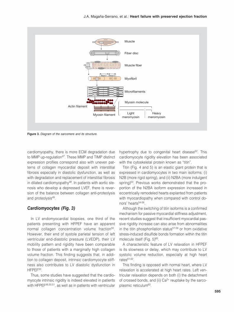
The excessive deposit of type I collagen is the con-sequence of an imbalance between excessive synthe-sis and reduced degradation41. Several steps appear to be implicated in the type I collagen synthesis pro-cess (Fig. 2).
Of clinical relevance is the observation that the type I procollagen carboxyl-terminal propeptide, which is cleavaged by type I procollagen PCP, is released into the bloodstream and, therefore, is a potential biomark-er of the PCP-PCPE system activity42,43. Type I collagen excessive accumulation can result not only in exagger-ated synthesis, but also in a decreased degradation rate. In hypertensive patients with HFPEF44 and in pa-tients with aortic stenosis45, there is a decrease in ECM degradation because of down-regulation of ECM metal-loproteinases (MMP) and up-regulation of tissue inhib-itors of ECM metalloproteinases (TIMP). TIMP-1 plasma levels have been recently proposed as a potential bio-marker of HFPEF development in patients with arterial hypertension46. Conversely, in patients with dilated
Figure 1. Photographs showing the contribution to ventricular rigidity mediated by the extracellular matrix (A, B, C, D; below left) and the cardiomyocytes own function (below right).
Procollagen
Mature type I collagen
Type I collagen fibers
Large telopeptide
+small
telopeptide
Matrikines
Gelatinases
MMP-1, MMP-9, MMP-13
collagenases
Lysyl Oxidase
PCP and PNP proteinases
Figure 2. Steps on type I collagen synthesis and degradation. PCP: type I procollagen carboxyl-terminal proteinase; PNP: type I procollagen N-terminal proteinase; PICP, PINP, carboxyl-terminal and amino-terminal propeptides; MMP: extracellular matrix meta-lloproteinase.
J.A. Magaña-Serrano, et al.: Heart failure with preserved ejection fraction
595
cardiomyopathy, there is more ECM degradation due to MMP up-regulation47. These MMP and TIMP distinct expression profiles correspond also with uneven pat-terns of collagen myocardial deposit with interstitial fibrosis especially in diastolic dysfunction, as well as with degradation and replacement of interstitial fibrosis in dilated cardiomyopathy48. In patients with aortic ste-nosis who develop a depressed LVEF, there is rever-sion of the balance between collagen anti-proteolysis and proteolysis49.
Cardiomyocytes (Fig. 3)
In LV endomyocardial biopsies, one third of the patients presenting with HFPEF have an apparent normal collagen concentration volume fraction50. However, their end of systole parietal tension of left ventricular end-diastolic pressure (LVEDP), their LV mobility pattern and rigidity have been comparable to those of patients with a marginally high collagen volume fraction. This finding suggests that, in addi-tion to collagen deposit, intrinsic cardiomyocyte stiff-ness also contributes to LV diastolic dysfunction in HFPEF50.
Thus, some studies have suggested that the cardio-myocyte intrinsic rigidity is indeed elevated in patients with HFPEF48,50,51, as well as in patients with ventricular
hypertrophy due to congenital heart disease52. This cardiomyocyte rigidity elevation has been associated with the cytoskeletal protein known as “titin”.
Titin (Fig. 4 and 5) is an elastic giant protein that is expressed in cardiomyocytes in two main isoforms; (i) N2B (more rigid spring), and (ii) N2BA (more indulgent spring)53. Previous works demonstrated that the pro-portion of the N2BA isoform expression increased in eccentrically remodeled hearts explanted from patients with myocardiopathy when compared with control do-nors’ hearts54-56.
Although the switching of titin isoforms is a confirmed mechanism for passive myocardial stiffness adjustment, recent studies suggest that insufficient myocardial pas-sive rigidity increase can also arise from abnormalities in the titin phosphorilation status57-59 or from oxidative stress-induced disulfide bonds formation within the titin molecule itself (Fig. 5)60.
A characteristic feature of LV relaxation in HFPEF is its slowness or delay, which may contribute to LV systolic volume reduction, especially at high heart rates61,62.
This finding is opposed with normal heart, where LV relaxation is accelerated at high heart rates. Left ven-tricular relaxation depends on both (i) the detachment of crossed bonds, and (ii) Ca2+ reuptake by the sarco-plasmic reticulum63.
Muscle
Fiber disc
Muscle �ber
Myo�bril
Micro�laments
Myosin molecule
Light meromyosin
Myosin �lament
Actin �lament
Heavy meromyosin
Band 1 Band 2
⎧⎨⎩
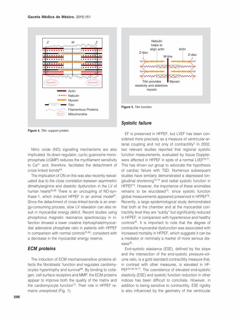
Figure 3. Diagram of the sarcomere and its structure.
Gaceta Médica de México. 2015;151
596
Nitric oxide (NO) signalling mechanisms are also implicated. Its down-regulator, cyclic guanosine mono-phosphate (cGMP) reduces the myofilament sensitivity to Ca2+ and, therefore, facilitates the detachment of cross-linked bonds64.
The implication of ON on this was also recently reeval-uated due to the close correlation between asymmetric dimethylarginine and diastolic dysfunction in the LV of human hearts65,66 There is an uncoupling of NO-syn-thase-1, which induced HFPEF in an animal model67. Since the detachment of cross-linked bonds is an ener-gy-consuming process, slow LV relaxation can also re-sult in myocardial energy deficit. Recent studies using phosphorus magnetic resonance spectroscopy in in-farction showed a lower creatine triphosphate/myocar-dial adenosine phosphate ratio in patients with HFPEF in comparison with normal controls35,68, consistent with a decrease in the myocardial energy reserve.
ECM proteins
The induction of ECM mechanosensitive proteins af-fects the fibroblasts’ function and regulates cardiomy-ocytes hypertrophy and survival69. By binding to colla-gen, cell-surface receptors and MMP, the ECM proteins appear to improve both the quality of the matrix and the cardiomyocyte function70. Their role in HFPEF re-mains unexplored (Fig. 1).
Systolic failure
EF is preserved in HFPEF, but LVEF has been con-sidered more precisely as a measure of ventricular-ar-terial coupling and not only of contractility3. In 2002, two relevant studies reported that regional systolic function measurements, evaluated by tissue Doppler, were affected in HFPEF in spite of a normal LVEF29,71. This has driven our group to advocate the hypothesis of cardiac failure with TSD. Numerous subsequent studies have similarly demonstrated a depressed lon-gitudinal shortening72,73 and radial systolic function in HFPEF74. However, the importance of these anomalies remains to be elucidated75, since systolic function global measurements appeared preserved in HFPEF76. Recently, a large epidemiological study demonstrated that both at the chamber and at the myocardial con-tractility level they are “subtly” but significantly reduced in HFPEF, in comparison with hypertensive and healthy controls30. It is important to note that the degree of contractile myocardial dysfunction was associated with increased mortality in HFPEF, which suggests it can be a mediator or nominally a marker of more serious dis-ease30.
End-systolic elastance (ESE), defined by the slope and the intersection of the end-systolic pressure-vol-ume ratio, is a gold standard contractility measure that, in contrast with other measures, is elevated in HF-PEF30,36,76,77. The coexistence of elevated end-systolic elasticity (ESE) and systolic function reduction in other indices has been difficult to conciliate. However, in addition to being sensitive to contractility, ESE rigidity is also influenced by the geometry of the ventricular
Actin
Z M Z
TitinFilamentous ProteinsMitochondria
MyosinNebulin
Figure 4. Titin: support protein.
Z-discZ-disc
Actin
Myosin
M-line
Nebulin helps to
align actin
Titin provideselasticity and stabilizes
myosin
Figure 5. Titin function.
J.A. Magaña-Serrano, et al.: Heart failure with preserved ejection fraction
597
chamber in the rest/effort cycle. ESE is elevated in HFPEF in spite of a contractility depression, when mea-sured across each pattern of the ventricular chamber geometry30. The same processes that promote ventric-ular diastolic stiffness in HFPEF are thought to increase ESE and contribute to contractility in infarction by lim-iting the systolic reserve as well. The systolic function is not as clearly deteriorated in HFPEF as it is in HFREF73, but recent studies have demonstrated that even mild basal contractility limitations in HFPFS can turn out to be more problematic with tension adjust-ment during exercise31-35, where incapacity to im-prove contractility can be associated with deteriorat-ed cardiac output reserve, thus making intolerance to exercise and decreased aerobic capacity symptoms more severe.
Defects in the ventricular-arterial coupling
Ventricular and vascular rigidity is known to increase with age, systemic arterial hypertension and diabetes,
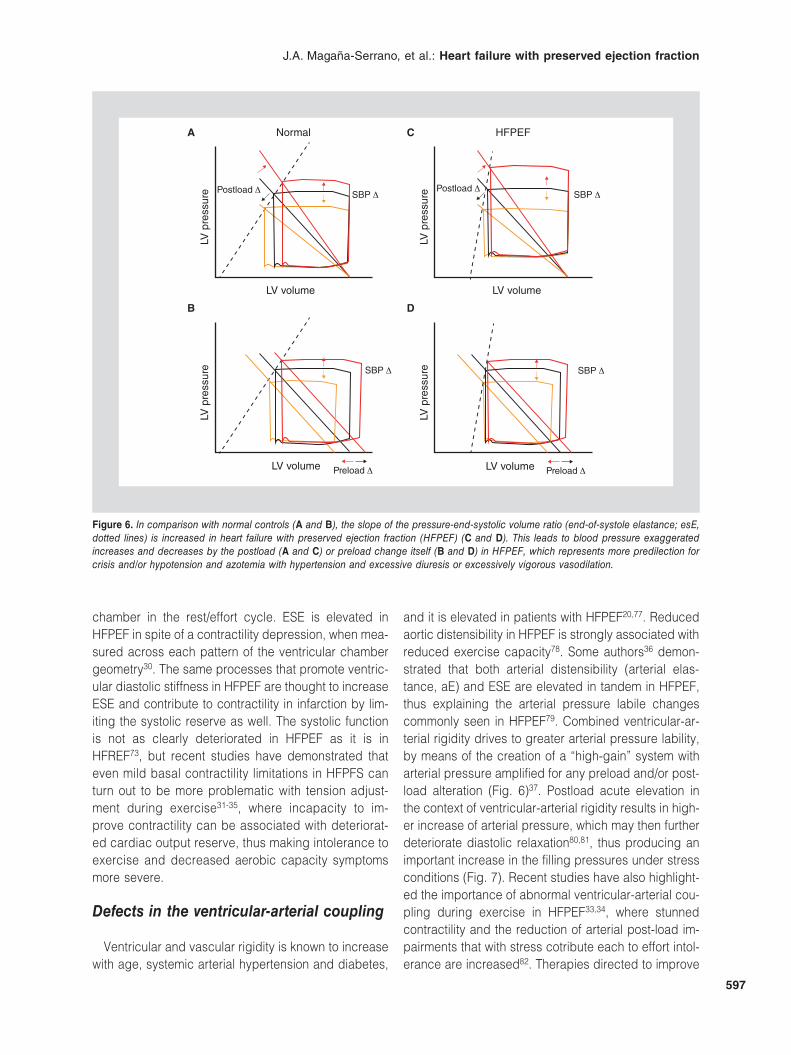
and it is elevated in patients with HFPEF20,77. Reduced aortic distensibility in HFPEF is strongly associated with reduced exercise capacity78. Some authors36 demon-strated that both arterial distensibility (arterial elas-tance, aE) and ESE are elevated in tandem in HFPEF, thus explaining the arterial pressure labile changes commonly seen in HFPEF79. Combined ventricular-ar-terial rigidity drives to greater arterial pressure lability, by means of the creation of a “high-gain” system with arterial pressure amplified for any preload and/or post-load alteration (Fig. 6)37. Postload acute elevation in the context of ventricular-arterial rigidity results in high-er increase of arterial pressure, which may then further deteriorate diastolic relaxation80,81, thus producing an important increase in the filling pressures under stress conditions (Fig. 7). Recent studies have also highlight-ed the importance of abnormal ventricular-arterial cou-pling during exercise in HFPEF33,34, where stunned contractility and the reduction of arterial post-load im-pairments that with stress cotribute each to effort intol-erance are increased82. Therapies directed to improve
Normal
SBP ΔPostload Δ
LV p
ress
ure
LV volume
A HFPEF
SBP ΔPostload Δ
LV p
ress
ure
LV volume
C
SBP Δ
Preload Δ
LV p
ress
ure
LV volume
B
LV p
ress
ure
D
SBP Δ
Preload Δ LV volume
Figure 6. In comparison with normal controls (A and B), the slope of the pressure-end-systolic volume ratio (end-of-systole elastance; esE, dotted lines) is increased in heart failure with preserved ejection fraction (HFPEF) (C and D). This leads to blood pressure exaggerated increases and decreases by the postload (A and C) or preload change itself (B and D) in HFPEF, which represents more predilection for crisis and/or hypotension and azotemia with hypertension and excessive diuresis or excessively vigorous vasodilation.

Gaceta Médica de México. 2015;151
598
the effort ventricular-arterial interaction in hypertensive elderly patients83 suggest its possible role in HFPEF.
Systemic vasorelaxation with exercise is atennuated in HFPEF31-33, which promotes alteration in blood-flow delivery to skeletal muscle. Vascular dysfunction in HFPEF may be due in part to endothelial dysfunction, as shown in a recent study, which demonstrated flow-mediated deteriorated vasodilation in HFPEF in comparison with healthy controls matched by age33. The dyspnea and fatigue symptoms in heart failure may be related to this ergoreflex pathological activa-tion, which is also related to NO bioavailability84.
Curiosly, the degree of flow-mediated vasodilation (an endothelial function marker) is related to the seri-ousness of effort intolerance symptoms during low in-tensity exercise in HFPEF33, with an emphasis on the complex between peripheral processes and the per-ception of symptoms in HF85. These data provide a further explanation of possible therapies targeting NO in HFPEF.
But vascular dysfunction is not limited to systemic circulation in HFPEF, since pulmonary hypertension is very frequently observed as well40. Among old-age patients with normal LVEF and high pulmonary artery pressure, HFPEF is usually the most common etiology86. Pulmonary pressures increase with aging and have been correlated with systemic vascular rigidity; both, common risk factors for HFPEF87. Pulmonary hyperten-sion in HFPEF appears to be due both to elevated telediastolic pressures of the heart’s LV and to high pulmonary vascular resistence, which can develop as a response to the former40. In the early stages of HFPEF,
pulmonary vasodilation with exercise is preserved, and effort pulmonary hypertension is passive and mainly secondary to high pressures of the left heart28. Pulmo-nary artery elevated pressures can predict an increase in HFPEF mortality40 and might represent a new thera-peutic target, although arterial pulmonary unbalanced vasodilation can lead to pathological elevations in the left pressures of the heart or even to overt pulmonary edema, and further studies are required to define the possible role of pulmonary vasodilators in HFPEF88.
Chronotropic response and cardiovascular reserve reduction
Most patients with HF do not complaint about symp-toms at rest, but rather with physical effort. A number of recent studies have emphasized on the importance of cardiovascular reserve function alterations at exer-cise stress in the physiopathology of HFPEF31-35,38. During physical exertion, cardiac output is augmented through comprehensive improvements in the venous re-turn, contractility, heart rate and peripheral vasodilation89.
Abnormalities in each one of these components of the abnormal reserve function in response to exercise have been identified in HFPEF and all of them may contribute to the pathophysiology in individual pa-tients (Fig. 8).
Normal diastolic reserve with exercise allows for the ventricle to fill up to a larger preload volume, in a short period of time, with no increase in filling pressures90. An old heart is indeed more labile than normal as a result of an increase in preload reserves to compensate
LV p
ress
ure
LV volume
A
LV p
ress
ure
Time
B
Figure 7. A: arterial ventricle rigidity combined in heart failure with preserved ejection fraction can lead to dramatic blood pressure elevations with postload increase (grey arrow). This leads to a new increase of the end-diastolic LV pressures (arrowhead), by alteration of the slope or position of the diastolic pressure-volume ratio, and/or (B) by prolongation of the LV pressure fall during isovolumetric relaxation (arrowhead).
J.A. Magaña-Serrano, et al.: Heart failure with preserved ejection fraction
599
the age-related reductions in the contractile and chro-notropic reserves91. In the same way the diastolic func-tion is altered in HFPEF, the diastolic function is also reduced, with patients showing effort-induced preload volume increases, in spite of marked elevations in the filling pressure28,92. This is probably related to an in-creased rigidity of the chamber27 and inadequate im-provement of early relaxation61,62 in spite of modulation by the pericardium and improvement of ventricular interaction, which may also contribute93.
The systolic reserve (SR) is also altered with exercise in HFPEF, causing non sufficient EFs, with stunned contractility and systolic-longitudinal shortening veloc-ities during exercise31-35. Exercise-induced stress can “unmask” mild deficits in the systolic function rest, and incapacity to reduce the telesystolic volume, combined with lower increase in end-diastolic volume, which largely limits the systolic volume responses during ex-ercise. The causes of SR and diastolic reserve (DR) remain unclear, but may be related to myocardial isch-emia (epicardial/microvascular coronary disease or
vascular distensibility alterations), to beta-adrenergic system deteriorated signalling94, to myocardial ener-getics34,68 or to abnormal management of calcium95.
The chronotropic reserve is depressed in HF-PEF31,33,34,38,96, even in comparison with controls, older of the same age and use of rate-deceleration-indepen-dent drugs. Similar to HFREF97, this is probably asso-ciated wit downwards deficits in beta-adrenergic stim-ulation, since the increase in plasma catecholamines with exercise is similar in HFPEF and in healthy controls31. Autonomic dysfunction can contribute to chronotropic incompetence, since baroreflex sensitivity31 is reduced and heart rate recovery is altered in HFPEF31,96.
Patients with HFPEF show exercise-induced reduc-tions in mean vascular arterial resistance and distensi-bility, together with endothelial function and dynamic ventricular-arterial coupling alterations31-33. Many of these anomalies are observed with normal aging and are symply more abnormally marked in HFPEF, consis-tent with the idea that HFPEF developes as an exag-gerated, progressive and pathological form of aging in
A
EDVAltered volume-
beat reserve
Vascular and
inotropic reserve
Preload reserve
C
Rest
Hea
rt r
ate
(BP
M)
50% Peak
p = 0.002 r = 0.7, p = 0.000150
00
50
D
0
Pea
k V
O2
(ml/m
in*K
g)
50Rate change (BPM)
100
30
25
20
15
10
5
p = 0.002
E
30
Per
iphe
ral a
rter
ial
tono
met
ry r
ate
60Time after release
90 120150180210240
8
6
4
2
0
B C
Exercise Exercise
VL
LV p
ress
ure
(mm
Hg)
160
120
80
40
0RestRest
LV p
ress
ure
(mm
Hg)
LV v
olum
e 160
120
80
40
0
VLVL
ESV
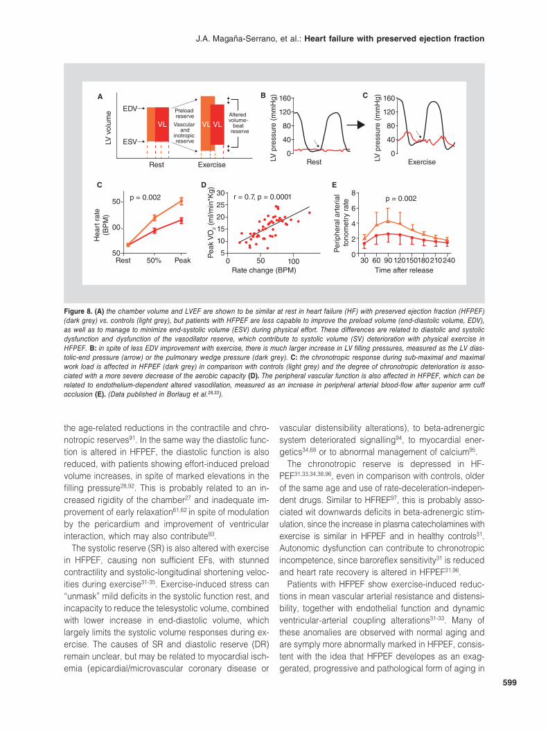
Figure 8. (A) the chamber volume and LVEF are shown to be similar at rest in heart failure (HF) with preserved ejection fraction (HFPEF) (dark grey) vs. controls (light grey), but patients with HFPEF are less capable to improve the preload volume (end-diastolic volume, EDV), as well as to manage to minimize end-systolic volume (ESV) during physical effort. These differences are related to diastolic and systolic dysfunction and dysfunction of the vasodilator reserve, which contribute to systolic volume (SV) deterioration with physical exercise in HFPEF. B: in spite of less EDV improvement with exercise, there is much larger increase in LV filling pressures, measured as the LV dias-tolic-end pressure (arrow) or the pulmonary wedge pressure (dark grey). C: the chronotropic response during sub-maximal and maximal work load is affected in HFPEF (dark grey) in comparison with controls (light grey) and the degree of chronotropic deterioration is asso-ciated with a more severe decrease of the aerobic capacity (D). The peripheral vascular function is also affected in HFPEF, which can be related to endothelium-dependent altered vasodilation, measured as an increase in peripheral arterial blood-flow after superior arm cuff occlusion (E). (Data published in Borlaug et al.28,33).
Gaceta Médica de México. 2015;151
600
hypertensive heart disease82. Patients with HFPEF are more prone to show higher number of slight individual anomalies in the ventricular and vascular reserve; re-cent evidence suggests that the acquisition of a suffi-cient number of individual abnormalities in the reserve promotes the transition of asymptomatic to symptom-atic diastolic function in hypertensive HFPEF33. This way, HFPEF can be conceived as a fundamental dis-order of the complex: cardiovascular reserve, of the diastolic, systolic, chronotropic and vascular function. Furter investigation is required to determine how these abnormalities can be efficaciously treated.
Diagnosis
In contrast with HFREF, the HFPEF diagnosis is more laborious, especially in patients who attend an outpa-tient clinic with effort dyspnea and multiple comorbid-ities, but without evident physical signs of fluid over-load. To avoid low specificity in the diagnosis of HFPEF, effort dyspnea and a normal LVEF, should be comple-mented with objective measurements of LV diastolic dysfunction, left ventricular hypertrophy, left atrial (LA) compliance and area, or plasma levels of natriuretic peptides (NP), such as BNP.
So far, several guidelines have been published for the diagnosis of HFPEF13,98-100. All of them require the simul-taneous and mandatory presence of signs and/or symp-toms of HF, “normal” LV systolic function tests and evi-dence of LV diastolic dysfunction, such as LV hypertrophy, LA compliance and size, atrial fibrillation or plasma BNP elevated levels. The first working group on myo-cardial function is the European Society of Cardiology98.
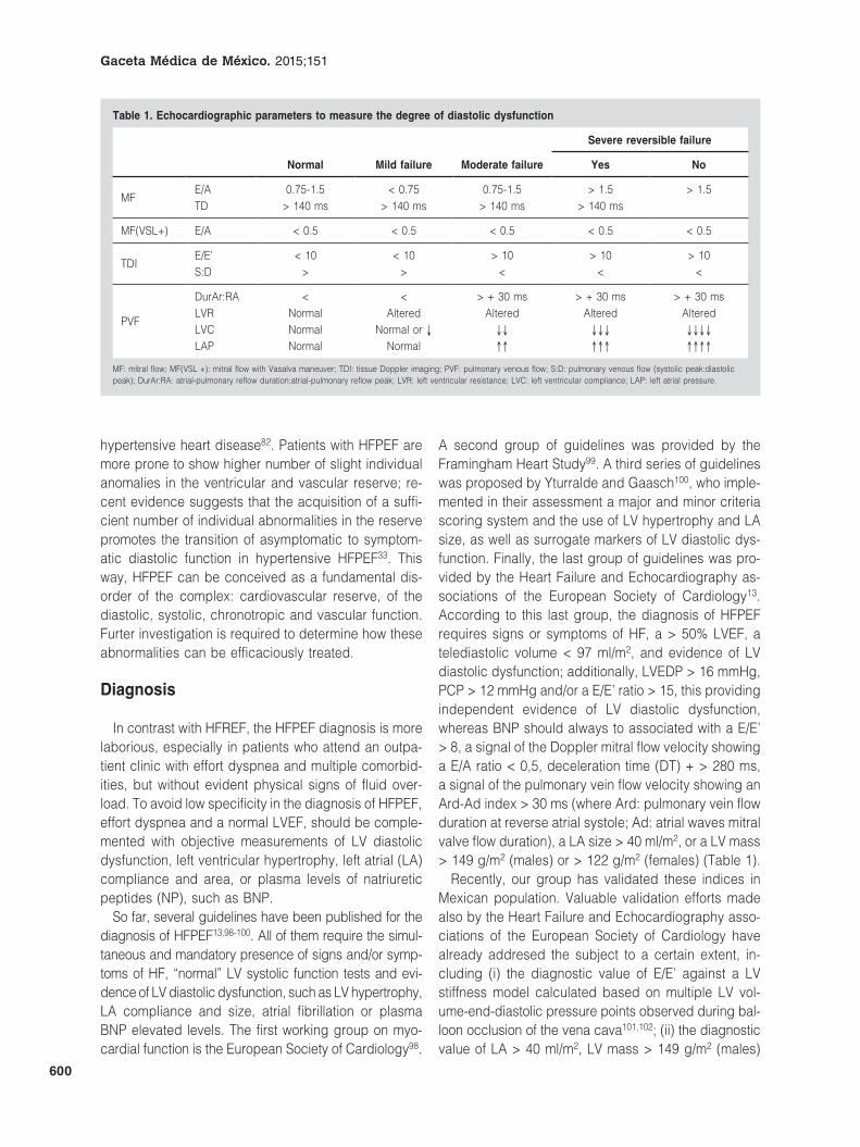
A second group of guidelines was provided by the Framingham Heart Study99. A third series of guidelines was proposed by Yturralde and Gaasch100, who imple-mented in their assessment a major and minor criteria scoring system and the use of LV hypertrophy and LA size, as well as surrogate markers of LV diastolic dys-function. Finally, the last group of guidelines was pro-vided by the Heart Failure and Echocardiography as-sociations of the European Society of Cardiology13. According to this last group, the diagnosis of HFPEF requires signs or symptoms of HF, a > 50% LVEF, a telediastolic volume < 97 ml/m2, and evidence of LV diastolic dysfunction; additionally, LVEDP > 16 mmHg, PCP > 12 mmHg and/or a E/E’ ratio > 15, this providing independent evidence of LV diastolic dysfunction, whereas BNP should always to associated with a E/E’ > 8, a signal of the Doppler mitral flow velocity showing a E/A ratio < 0,5, deceleration time (DT) + > 280 ms, a signal of the pulmonary vein flow velocity showing an Ard-Ad index > 30 ms (where Ard: pulmonary vein flow duration at reverse atrial systole; Ad: atrial waves mitral valve flow duration), a LA size > 40 ml/m2, or a LV mass > 149 g/m2 (males) or > 122 g/m2 (females) (Table 1).
Recently, our group has validated these indices in Mexican population. Valuable validation efforts made also by the Heart Failure and Echocardiography asso-ciations of the European Society of Cardiology have already addresed the subject to a certain extent, in-cluding (i) the diagnostic value of E/E’ against a LV stiffness model calculated based on multiple LV vol-ume-end-diastolic pressure points observed during bal-loon occlusion of the vena cava101,102; (ii) the diagnostic value of LA > 40 ml/m2, LV mass > 149 g/m2 (males)
Table 1. Echocardiographic parameters to measure the degree of diastolic dysfunction
Normal Mild failure Moderate failure
Severe reversible failure
Yes No
MFE/A 0.75-1.5 < 0.75 0.75-1.5 > 1.5 > 1.5TD > 140 ms > 140 ms > 140 ms > 140 ms
MF(VSL+) E/A < 0.5 < 0.5 < 0.5 < 0.5 < 0.5
TDIE/E’ < 10 < 10 > 10 > 10 > 10S:D > > < < <
PVF
DurAr:RA < < > + 30 ms > + 30 ms > + 30 msLVR Normal Altered Altered Altered AlteredLVC Normal Normal or ↓ ↓↓ ↓↓↓ ↓↓↓↓
LAP Normal Normal ↑↑ ↑↑↑ ↑↑↑↑
MF: mitral flow; MF(VSL +): mitral flow with Vasalva maneuver; TDI: tissue Doppler imaging; PVF: pulmonary venous flow; S:D: pulmonary venous flow (systolic peak:diastolic peak); DurAr:RA: atrial-pulmonary reflow duration:atrial-pulmonary reflow peak; LVR: left ventricular resistance; LVC: left ventricular compliance; LAP: left atrial pressure.
J.A. Magaña-Serrano, et al.: Heart failure with preserved ejection fraction
601
or > 122 g/m2 (females), Ard-Ad > 30 ms and E/A ratio < 0,5 DT + > 280 ms against E/E’103, and (iii) the diag-nostic value of NT-proBNP > 220 pg/ml against E/E’104.
In contrast with recent criticism on the validity of E/E’ as a measure of the LV filling pressures in patients with acute decompensated HFPEF105, a direct comparison of E/E’ against the flotation catheter derived from LV diastolic dysfunction models yielded a sensitivity of 83%, specificity of 92% and an area under the ROC curve of 0,907 for E/E’ > as a measure of the high-ri-gidity model in HFPEF patients102.
These results have suggested that an E/E’ value > 8 can be able to provide independent evidence of LV diastolic dysfunction, without further need of non-inva-sive tests106. Hence, the distinctive value of E/E’ can be explained as an indirect measurement of the LV filling pressures in HFREF and HFPEF107. A direct com-parison between the diagnostic values of E/E’ and pul-monary flow velocity showed that the latter is even poorly reliable for the diagnosis of LV diastolic dys-function103. In contrast, however, a LA size > 40 ml/m2 provides both high sensitivity and high specificity to detect E/E’ > 15.
Effort test: An underestimated risk marker?
HFREF is characterized by dilation of the chamber and low LVEF, easily detectable by echocardiography. In HFPEF, the size of the chamber and the LVEF are normal, and the main hemodynamical alteration is an elevation of filling pressures26.
When pressures are high and congestion is present at rest, HFPEF is easily diagnosed based on history and physical examination, x-rays, BNP levels and echocardiographic parameters13.
However, many patients with early-stage HFPEF have significant effort intolerance symptoms in the ab-sence of apparent volume overload. In some patients, an invasive evaluation can reveal pathological eleva-tion of the filling pressures that was not previously suspected108, and a recent study found that even among patients with normal echocardiographic tests, BNP levels and normal hemodynamics at rest, many can anyway develop pathological elevations in the fill-ing pressures that are characteristic of HFPEF during exercise-induced stress28. The HFPEF diagnosis could only be made by using hemodynamical evaluation with exercise, since in these patients it was also a strong predictor of HFPEF. Pulmonary artery pressures give a very good idea of the left heart filling pressures in the
early stages of HFPEF28. Therefore, assessment during effort is highly recommendable and necessary.
The E/E’ ratio is one of the cornerstones in the non-in-vasive assessment of diastolic function at rest13,14, and some groups have started using evaluations based on tissue Doppler imaging (TDI) during exercise, with the first studies showing reasonable correlations with inva-sive measurements109. However, E/E’ can be less ro-bust in the tachycardia and hyperventilation settings and in the fusion of early and late transmitral filling velocities. In patients not fulfilling the established cri-teria for positive HFPEF diagnosis13, but in whom there is reasonable strong clinical suspicion, invasive as-sessment should be seriously considered when avail-able measures on stress with exercise and at rest are normal28.
Conclusions
HFPEF is an important and growing public health problem, given that currently it accounts for half of all patients with HF. In spite of improvements in the un-derstanding of the disease, there are no treatments with entirely proven benefits. The advances on diag-nostic algorithms, imaging projection and invasive as-sessment will allow for more accurate and early diag-nosis, in order for treatments to be applied earlier in the progression of the disease, since the potential for benefit can be greater. Although important advances had been made in our understanding of the pathophys-iology, hemodynamics and cell mechanisms of diastol-ic failure, as well as non-diastolic mechanisms of the disease, further investigation is urgently needed to determine how to better direct these abnormalities in order to reduce the important burden of morbidity and mortality of this form of HF, which is reaching pandem-ic proportions.
References
1. Sanderson JE, Gibson DG, Brown DJ, et al. Left ventricular filling in hypertrophic cardiomyopathy. An angiographic study. Br Heart J. 1977;39:661-70.
2. Hanrath P, Mathey DG, Siegert R, et al. Left ventricular relaxation and filling pattern in different forms of left ventricular hypertrophy: an echo-cardiographic study. Am J Cardiol. 1980;45:15-23.
3. Hess OM, Grimm J, Krayenbuehl HP. Diastolic simple elastic and visco-elastic properties of the left ventricle in man. Circulation. 1979;59:1178-87.
4. Soufer R, Wohlgelernter D, Vita NA, et al. Intact systolic left ventricular function in clinical congestive heart failure. Am J Cardiol. 1985;55:1032-6.
5. Aronow WS, Kronzon I. Effect of enalapril on congestive heart failure treated with diuretics in elderly patients with prior myocardial infarc-tion and normal left ventricular ejection fraction. Am J Cardiol. 1993; 71:602-4.
6. Carson P, Johnson G, Fletcher R, et al. Mild systolic dysfunction in heart failure (left ventricular ejection fraction .35%): baseline characteristics, prognosis and response to therapy in the Vasodilator in Heart Failure Trials (V-HeFT). J Am Coll Cardiol. 1996;27:642-9.
Gaceta Médica de México. 2015;151
602
7. Aronow WS, Ahn C, Kronzon I. Effect of propranolol vs. no propranolol on total mortality plus nonfatal myocardial infarction in older patients with prior myocardial infarction, congestive heart failure, and left ventricular ejection fraction > or = 40% treated with diuretics plus angiotensin-con-verting enzyme inhibitors. Am J Cardiol. 1997; 80:207-9.
8. Petrie MC, Caruana L, Berry C, et al. “Diastolic heart failure” or heart failure caused by subtle left ventricular systolic dysfunction? Heart. 2002;87:29-31.
9. Heusch G. Diastolic heart failure: a misNOmer. Basic Res Cardiol. 2009;104:465-7.
10. De Keulenaer GW, Brutsaert DL. The heart failure spectrum: time for a phenotype-oriented approach. Circulation. 2009;119:3044-6.
11. Yusuf S, Pfeffer MA, Swedberg K, et al. Effects of candesartan in patients with chronic heart failure and preserved left-ventricular ejection fraction: the CHARM-Preserved Trial. Lancet. 2003;362:777-81.
12. Massie BM, Carson PE, McMurray JJ, et al. Irbesartan in patients with heart failure and preserved ejection fraction. N Engl J Med. 2008;359: 2456-67.
13. Paulus WJ, Tschope C, Sanderson JE, et al. How to diagnose diastolic heart failure: a consensus statement on the diagnosis of heart failure with normal left ventricular ejection fraction by the Heart Failure and Echocardiography Associations of the European Society of Cardiology. Eur Heart J. 2007;28:2539-50.
14. Nagueh SF, Appleton CP, Gillebert TC, et al. Recommendations for the evaluation of left ventricular diastolic function by echocardiography. J Am Soc Echocardiogr. 2009;22:107-33.
15. McMurray JJ, Carson PE, Komajda M, et al. Heart failure with preserved ejection fraction: clinical characteristics of 4133 patients enrolled in the I-PRESERVE trial. Eur J Heart Fail. 2008;10:149-56.
16. Sanderson JE. Heart failure with a normal ejection fraction. Heart. 2007; 93:155-8.
17. McMurray J, Pfeffer MA. New therapeutic options in congestive heart failure: part II. Circulation. 2002;105:2223-8.
18. Davies M, Hobbs F, Davis R, et al. Prevalence of left-ventricular systolic dysfunction and heart failure in the Echocardiographic Heart of England Screening study: a population based study. Lancet. 2001;358:439-44.
19. Petrie M, McMurray J. Changes in notions about heart failure. Lancet. 2001;358:432-4.
20. Owan TE, Hodge DO, Herges RM, et al. Trends in prevalence and outcome of heart failure with preserved ejection fraction. N Engl J Med. 2006;355:251-9.
21. Maeder MT, Kaye DM. Heart failure with normal left ventricular ejection fraction. J Am Coll Cardiol. 2009;53:905-18.
22. Paulus WJ, van Ballegoij JJ. Treatment of heart failure with normal ejec-tion fraction: an inconvenient truth! J Am Coll Cardiol. 2010; 55:526-37.
23. Nishimura RA, Tajik AJ. Evaluation of diastolic filling of left ventricle in health and disease: Doppler echocardiography is the clinician’s Rosetta Stone. J Am Coll Cardiol. 1997;30:8-18.
24. Mantero A, Gentile F, Gualtierotti C, et al. Left ventricular diastolic pa-rameters in 288 normal subjects from 20 to 80 years old. Eur Heart J. 1995; 16:94-105.
25. Belardinelli R, Georgiou D, Cianci G, et al. Exercise training improves left ventricular diastolic filling in patients with dilated cardiomyopathy. Clinical and prognostic implications. Circulation. 1995;91:2775-84.
26. Zile MR, Baicu CF, Gaasch WH. Diastolic heart failure—abnormalities in active relaxation and passive stiffness of the left ventricle. N Engl J Med. 2004;350:1953-9.
27. Westermann D, Kasner M, Steendijk P, et al. Role of left ventricular stiffness in heart failure with normal ejection fraction. Circulation. 2008;117:2051-60.
28. Borlaug BA, Nishimura RA, Sorajja P, et al. Exercise hemodynamics enhance diagnosis of early heart failure with preserved ejection fraction. Circ Heart Fail. 2010;117:2051-60.
29. Yu CM, Lin H, Yang H, et al. Progression of systolic abnormalities in patients with “isolated” diastolic heart failure and diastolic dysfunction. Circulation. 2002; 05:1195-201.
30. Borlaug BA, Lam CS, Roger VL, et al. Contractility and ventricular sys-tolic stiffening in hypertensive heart disease insights into the pathogen-esis of heart failure with preserved ejection fraction. J Am Coll Cardiol. 2009;54:410-8.
31. Borlaug BA, Melenovsky V, Russell SD, et al. Impaired chronotropic and vasodilator reserves limit exercise capacity in patients with heart failure and a preserved ejection fraction. Circulation. 2006;114:2138-47.
32. Ennezat PV, Lefetz Y, Marechaux S, et al. Left ventricular abnormal re-sponse during dynamic exercise in patients with heart failure and pre-served left ventricular ejection fraction at rest. J Card Fail. 2008;14:475-80.
33. Borlaug BA, Olson TP, Lam CS, et al. Global cardiovascular reserve dysfunction in heart failure with preserved ejection fraction. J Am Coll Cardiol. 2010;56:845-54.
34. Phan TT, Abozguia K, Nallur Shivu G, et al. Heart failure with preserved ejection fraction is characterized by dynamic impairment of active relax-ation and contraction of the left ventricle on exercise and associated with myocardial energy deficiency. J Am Coll Cardiol. 2009;54:402-9.
35. Tan YT, Wenzelburger F, Lee E, et al. The pathophysiology of heart failure with normal ejection fraction: exercise echocardiography reveals complex abnormalities of both systolic and diastolic ventricular function involving torsion, untwist, and longitudinal motion. J Am Coll Cardiol. 2009;54:36-46.
36. Kawaguchi M, Hay I, Fetics B, et al. Combined ventricular systolic and arterial stiffening in patients with heart failure and preserved ejection fraction: implications for systolic and diastolic reserve limitations. Circu-lation. 2003;107:714-20.
37. Borlaug BA, Kass DA. Ventricular-vascular interaction in heart failure. Heart Fail Clin. 2008;4:23-36.
38. Brubaker PH, Joo KC, Stewart KP, et al. Chronotropic incompetence and its contribution to exercise intolerance in older heart failure patients. J Cardiopulm Rehabil. 2006;26:86-9.
39. Kjaergaard J, Akkan D, Iversen KK, et al. Prognostic importance of pulmonary hypertension in patients with heart failure. Am J Cardiol. 2007;99:1146-50.
40. Lam CS, Roger VL, Rodeheffer RJ, et al. Pulmonary hypertension in heart failure with preserved ejection fraction: a community based study. J Am Coll Cardiol. 2009;53:1119-26.
41. Weber KT, Brilla CG, Janicki JS. Myocardial fibrosis: functional signifi-cance and regulatory factors. Cardiovasc Res. 1993;27:341-8.
42. Querejeta R, Varo N, López B, et al. Serum carboxy-terminal propeptide of procollagen type I is a marker of myocardial fibrosis in hypertensive heart disease. Circulation. 2000;101:1729-35.
43. López B, González A, Díez J. Circulating biomarkers of collagen metab-olism in cardiac diseases. Circulation. 2010;121:1645-54.
44. Ahmed SH, Clark LL, Pennington WR, et al. Matrix metalloproteinases/tissue inhibitors of metalloproteinases: relationship between changes in proteolytic determinants of matrix composition and structural, functional, and clinical manifestations of hypertensive heart disease. Circulation. 2006;113:2089-96.
45. Heymans S, Schroen B, Vermeersch P, et al. Increased cardiac expres-sion of tissue inhibitor of metalloproteinase-1 and tissue inhibitor of metal-loproteinase-2 is related to cardiac fibrosis and dysfunction in the chron-ic pressure-overloaded human heart. Circulation. 2005;112:1136-44.
46. González A, López B, Querejeta R, et al. Filling pressures and collagen metabolism in hypertensive patients with heart failure and normal ejec-tion fraction. Hypertension. 2010;55:1418-24.
47. Spinale FG, Coker ML, Heung LJ, et al. A matrix metalloproteinase in-duction/activation system exists in the human left ventricular myocardium and is up regulated in heart failure. Circulation. 2000;102:1944-9.
48. van Heerebeek L, Borbely A, Niessen HW, et al. Myocardial structure and function differ in systolic and diastolic heart failure. Circulation. 2006;113:1966-73.
49. Polyakova V, Hein S, Kostin S, et al. Matrix metalloproteinases and their tissue inhibitors in pressure-overloaded human myocardium during heart failure progression. J Am Coll Cardiol. 2004;44:1609-18.
50. Borbely A, van der Velden J, Papp Z, et al. Cardiomyocyte stiffness in diastolic heart failure. Circulation. 2005;111:774-81.
51. van Heerebeek L, Hamdani N, Handoko ML, et al. Diastolic stiffness of the failing diabetic heart: importance of fibrosis, advanced glycation end products, and myocyte resting tension. Circulation. 2008;117:43-51.
52. Chaturvedi RR, Herron T, Simmons R, et al. Passive stiffness of myocar-dium from congenital heart disease and implications for diastole. Circu-lation. 2010;121:979-88.
53. Bang ML, Centner T, Fornoff F, et al. The complete gene sequence of titin, expression of an unusual approximately 700-kDa titin isoform, and its interaction with obscurin identify a novel Z-line to I-band linking system. Circ Res. 2001;89:1065-72.
54. Neagoe C, Opitz CA, Makarenko I, et al. Gigantic variety: expression patterns of titin isoforms in striated muscles and consequences for myofibrillar passive stiffness. J Muscle Res Cell Motil. 2003;24:175-89.
55. Nagueh SF, Shah G, Wu Y, et al. Altered titin expression, myocardial stiffness, and left ventricular function in patients with dilated cardiomy-opathy. Circulation. 2004;110:155-62.
56. Makarenko I, Opitz CA, Leake MC, et al. Passive stiffness changes caused by upregulation of compliant titin isoforms in human dilated cardiomyopathy hearts. Circ Res. 2004;95:708-16.
57. Kruger M, Kotter S, Grutzner A, et al. Protein kinase G modulates human myocardial passive stiffness by phosphorylation of the titin springs. Circ Res. 2009;104:87-94.
58. Borbely A, Falcao-Pires I, van Heerebeek L, et al. Hypophosphorylation of the stiff N2B titin isoform raises cardiomyocyte resting tension in failing human myocardium. Circ Res. 2009;104:780-6.
59. Hidalgo C, Hudson B, Bogomolovas J, et al. PKC phosphorylation of titin’s PEVK element: a novel and conserved pathway for modulating myocardial stiffness. Circ Res. 2009;105:631-8.
60. Grutzner A, Garcia-Manyes S, Kotter S, et al. Modulation of titin-based stiffness by disulfide bonding in the cardiac titin N2-B unique sequence. Biophys J. 2009;97:825-34.
61. Wachter R, Schmidt-Schweda S, Westermann D, et al. Blunted frequen-cy dependent up regulation of cardiac output is related to impaired relaxation in diastolic heart failure. Eur Heart J. 2009;30:3027-36.
J.A. Magaña-Serrano, et al.: Heart failure with preserved ejection fraction
603
62. Sohn DW, Kim HK, Park JS, et al. Hemodynamic effects of tachycardia in patients with relaxation abnormality: abnormal stroke volume response as an overlooked mechanism of dyspnea associated with tachycardia in diastolic heart failure. J Am Soc Echocardiogr. 2007;20:171-6.
63. Ramirez-Correa GA, Murphy AM. Is phospholamban or troponin I the “prima donna” in beta-adrenergic induced lusitropy? Circ Res. 2007;101:326-7.
64. Paulus WJ, Vantrimpont PJ, Shah AM. Acute effects of nitric oxide on left ventricular relaxation and diastolic distensibility in humans: assess-ment by bicoronary sodium nitroprusside infusion. Circulation. 1994; 89:2070-8.
65. Wilson Tang WH, Tong W, Shrestha K, et al. Differential effects of argi-nine methylation on diastolic dysfunction and disease progression in patients with chronic systolic heart failure. Eur Heart J. 2008;29:2506-13.
66. Bronzwaer JG, Paulus WJ. Nitric oxide: the missing lusitrope in failing myocardium. Eur Heart J. 2008;29:2453-5.
67. Silberman GA, Fan TH, Liu H, et al. Uncoupled cardiac nitric oxide synthase mediates diastolic dysfunction. Circulation. 2010;121:519-28.
68. Smith CS, Bottomley PA, Schulman SP, et al. Altered creatine kinase adenosine triphosphate kinetics in failing hypertrophied human myocar-dium. Circulation. 2006;114:1151-8.
69. Schellings MW, Pinto YM, Heymans S. Matricellular proteins in the heart: possible role during stress and remodeling. Cardiovasc Res. 2004;64: 24-31.
70. Schroen B, Heymans S, Sharma U, et al. Thrombospondin-2 is essential for myocardial matrix integrity: increased expression identifies failure prone cardiac hypertrophy. Circ Res. 2004;95:515-22.
71. Yip G, Wang M, Zhang Y, et al. Left ventricular long axis function in diastolic heart failure is reduced in both diastole and systole: time for a redefinition? Heart. 2002;87:121-5.
72. Brucks S, Little WC, Chao T, et al. Contribution of left ventricular diastol-ic dysfunction to heart failure regardless of ejection fraction. Am J Cardiol. 2005;95:603-6.
73. Fukuta H, Little WC. Contribution of systolic and diastolic abnormalities to heart failure with a normal and a reduced ejection fraction. Prog Cardiovasc Dis. 2007;49:229-40.
74. Wang J, Khoury DS, Yue Y, et al. Preserved left ventricular twist and cir-cumferential deformation, but depressed longitudinal and radial deforma-tion in patients with diastolic heart failure. Eur Heart J. 2008;29:1283-9.
75. Aurigemma GP, Zile MR, Gaasch WH. Contractile behavior of the left ventricle in diastolic heart failure: with emphasis on regional systolic function. Circulation. 2006;113:296-304.
76. Baicu CF, Zile MR, Aurigemma GP, et al. Left ventricular systolic perfor-mance, function, and contractility in patients with diastolic heart failure. Circulation. 2005;111:2306-12.
77. Melenovsky V, Borlaug BA, Rosen B, et al. Cardiovascular features of heart failure with preserved ejection fraction vs. nonfailing hypertensive left ventricular hypertrophy in the urban Baltimore community: the role of atrial remodeling/dysfunction. J Am Coll Cardiol. 2007;49:198-207.
78. Hundley WG, Kitzman DW, Morgan TM, et al. Cardiac cycle-dependent changes in aortic area and distensibility are reduced in older patients with isolated diastolic heart failure and correlate with exercise intoler-ance. J Am Coll Cardiol. 2001;38:796-802.
79. Gandhi SK, Powers JC, Nomeir AM, et al. The pathogenesis of acute pulmonary edema associated with hypertension. N Engl J Med. 2001;344:17-22.
80. Gillebert TC, Leite-Moreira AF, De Hert SG. Load dependent diastolic dysfunction in heart failure. Heart Fail Rev. 2000;5:345-55.
81. Borlaug BA, Melenovsky V, Redfield MM, et al. Impact of arterial load and loading sequence on left ventricular tissue velocities in humans. J Am Coll Cardiol. 2007;50:1570-7.
82. Chantler PD, Lakatta EG, Najjar SS. Arterial-ventricular coupling: mech-anistic insights into cardiovascular performance at rest and during ex-ercise. J Appl Physiol. 2008;105:1342-51.
83. Chen CH, Nakayama M, Talbot M, et al. Verapamil acutely reduces ventricular-vascular stiffening and improve aerobic exercise perfor-mance in elderly individuals. J Am Coll Cardiol. 1999;33:1602-9.
84. Guazzi M, Samaja M, Arena R, et al. Long-term use of sildenafil in the ther-apeutic management of heart failure. J Am Coll Cardiol. 2007;50:2136-44.
85. Clark AL, Poole-Wilson PA, Coats AJ. Exercise limitation in chronic heart failure: central role of the periphery. J Am Coll Cardiol. 1996;28:1092-102.
86. Shapiro BP, McGoon MD, Redfield MM. Unexplained pulmonary hyper-tension in elderly patients. Chest. 2007;131:94-100.
87. Lam CS, Borlaug BA, Kane GC, et al. Age-associated increases in pulmonary artery systolic pressure in the general population. Circulation. 2009;119:2663-70.
88. Boilson BA, Schirger JA, Borlaug BA. Caveat medicus! Pulmonary hyper-tension in the elderly: a word of caution. Eur J Heart Fail. 2010;12:89-93.
89. Higginbotham MB, Morris KG, Williams RS, et al. Regulation of stroke volume during submaximal and maximal upright exercise in normal man. Circ Res. 1986;58:281-91.
90. Nonogi H, Hess OM, Ritter M, et al. Diastolic properties of the normal left ventricle during supine exercise. Br Heart J. 1988;60:30-8.
91. Rodeheffer RJ, Gerstenblith G, Becker LC, et al. Exercise cardiac output is maintained with advancing age in healthy human subjects: cardiac dilatation and increased stroke volume compensate for a diminished heart rate. Circulation. 1984;69:203-13.
92. Kitzman DW, Higginbotham MB, Cobb FR, et al. Exercise intolerance in patients with heart failure and preserved left ventricular systolic function: failure of the Frank-Starling mechanism. J Am Coll Cardiol. 1991;17:1065-72.
93. Dauterman K, Pak PH, Maughan WL, et al. Contribution of external forces to left ventricular diastolic pressure. Implications for the clinical use of the Starling law. Ann Intern Med. 1995;122:737-42.
94. Chattopadhyay S, Alamgir MF, Nikitin NP, et al. Lack of diastolic reserve in patients with heart failure and normal ejection fraction. Circ Heart Fail. 2010;3:35-43.
95. Liu C-P, Ting C-T, Lawrence W, et al. Diminished contractile response to increased heart rate in intact human left ventricular hypertrophy: systolic vs. diastolic determinants. Circulation. 1993; 88(part 1):1893-906.
96. Phan TT, Nallur Shivu G, Abozguia K, et al. Impaired heart rate recovery and chronotropic incompetence in patients with heart failure with pre-served ejection fraction. Circ Heart Fail. 2010;3:29-34.
97. Colucci WS, Ribeiro JP, Rocco MB, et al. Impaired chronotropic response to exercise in patients with congestive heart failure. Role of post-synaptic beta-receptor desensitization. Circulation. 1989;80:314-23.
98. How to diagnose diastolic heart failure. European Study Group on Dia-stolic Heart Failure. Eur Heart J. 1998;19:990-1003.
99. Vasan RS, Levy D. Defining diastolic heart failure: a call for standardized diagnostic criteria. Circulation. 2000;101:2118-21.
100. Yturralde RF, Gaasch WH. Diagnostic criteria for diastolic heart failure. Prog Cardiovasc Dis. 2005;47:314-9.
101. Lam CS. Heart failure with preserved ejection fraction: invasive solution to diagnostic confusion? J Am Coll Cardiol. 2010;55:1711-2.
102. Kasner M, Westermann D, Steendijk P, et al. Utility of Doppler echo-cardiography and tissue Doppler imaging in the estimation of diastolic function in heart failure with normal ejection fraction: a comparative Dop-pler-conductance catheterization study. Circulation. 2007;116:637-47.
103. Emery WT, Jadavji I, Choy JB, et al. Investigating the European Society of Cardiology Diastology Guidelines in a practical scenario. Eur J Echo-cardiogr. 2008;9:685-91.
104. Edelmann F, Schmidt AG, Gelbrich G, et al. Rationale and design of the aldosterone’ receptor blockade in diastolic heart failure’ trial: a dou-ble-blind, randomized, placebo-controlled, parallel group study to de-termine the effects of spironolactone on exercise capacity and diastolic function in patients with symptomatic diastolic heart failure (Aldo-DHF). Eur J Heart Fail. 2010;12:874-82.
105. Mullens W, Borowski AG, Curtin RJ, et al. Tissue Doppler imaging in the estimation of intracardiac filling pressure in decompensated patients with advanced systolic heart failure. Circulation. 2009;119:62-70.
106. Handoko ML, Paulus WJ. Polishing the diastolic dysfunction measure-ment stick. Eur J Echocardiogr. 2008;9:575-7.
107. Tschope C, Paulus WJ. Is echocardiographic evaluation of diastolic function useful in determining clinical care? Doppler echocardiography yields dubious estimates of left ventricular diastolic pressures. Circula-tion. 2009;120:810-20.
108. Penicka M, Bartunek J, Trakalova H, et al. Heart failure with preserved ejection fraction in outpatients with unexplained dyspnea: a pres-sure-volume loop analysis. J Am Coll Cardiol. 2010;55:1701-10.
109. Burgess MI, Jenkins C, Sharman JE, et al. Diastolic stress echocardi-ography: hemodynamic validation and clinical significance of estima-tion of ventricular filling pressure with exercise. J Am Coll Cardiol. 2006; 47:1891-1900.