Embed Size (px)
Citation preview
Hematopoiesis
Michael A. Rieger1 and Timm Schroeder2
1Georg-Speyer-Haus, Institute for Biomedical Research, Frankfurt (Main), Germany2Research Unit Stem Cell Dynamics, Helmholtz Zentrum Muenchen-German Research Center for EnvironmentalHealth, Neuherberg, Germany
Correspondence: [email protected]
SUMMARY
Enormous numbers of adult blood cells are constantly regenerated throughout life from he-matopoietic stem cells through a series of progenitor stages. Accessibility, robust functionalassays, well-established prospective isolation, and successful clinical application made he-matopoiesis the classical mammalian stem cell system. Most of the basic concepts of stem cellbiology have been defined in this system. At the same time, many long-standing disputes inhematopoiesis research illustrate our still limited understanding. Here we discuss the embry-onic development and lifelong maintenance of the hematopoietic system, its cellular compo-nents, and some of the hypotheses about the molecular mechanisms involved in controllinghematopoietic cell fates.
Outline
1 Introduction
2 Embryonic hematopoiesis
3 Adult hematopoiesis
4 Conclusion/outlook
References
Editors: Patrick P.L. Tam, W. James Nelson, and Janet Rossant
Additional Perspectives on Mammalian Development available at www.cshperspectives.org
Copyright # 2012 Cold Spring Harbor Laboratory Press; all rights reserved; doi: 10.1101/cshperspect.a008250
Cite this article as Cold Spring Harb Perspect Biol 2012;4:a008250
1
on March 3, 2019 - Published by Cold Spring Harbor Laboratory Press http://cshperspectives.cshlp.org/Downloaded from
1 INTRODUCTION
The blood system contains more than 10 different bloodcell types (lineages) with various functions: Leukocytesrepresent many specialized cell types involved in innateand acquired immunity. Erythrocytes provide O2 andCO2 transport, whereas megakaryocytes generate plateletsfor blood clotting and wound healing. All blood cell typesarise from hematopoietic stem cells (HSCs) that residemainly in the bone marrow (BM), a major site of adulthematopoiesis. Blood is one of the most regenerative andplastic tissues, and millions of “old” blood cells are replen-ished with new ones each second during life. In emergencysituations such as anemia or infections, blood cell countsrapidly increase. The cell number then declines back tonormal after recovery. The lifetimes of various matureblood cell types range from hours to years.
The hematopoietic system is a prime example of suc-cessful applied regenerative medicine. For more than 30years, stem cell transplantation has become a routine treat-ment for blood disorders and malignant diseases. After theeradication of the patient’s own hematopoietic system, thetransplanted donor hematopoietic stem and progenitorcells (HSPCs) provide lifelong reconstitution of the bloodsystem of the patient. The experimental evidence that HSCsnaturally migrate back and forth from the BM periodically,as well as the identification of agents that increase HSCmobilization (e.g., granulocyte colony-stimulating factor[G-CSF]), have opened new avenues for stem cell trans-plantation. However, although stem cell transplants worksuccessfully in the clinic, further improvement of the meth-od is needed to minimize engraftment failure and post-transplant infections. The ex vivo expansion of HSCswould be beneficial for grafts with limiting numbers ofHSCs (umbilical cord blood) and for gene therapy ap-proaches for monogenetic inherited blood disorders. How-ever, despite decades of research, the robust expansion (oreven maintenance) of HSCs ex vivo is not yet routinelyachieved.
HSCs are the most-studied adult stem cells. Five de-cades ago researchers passed the stage of providing purelydescriptive data to start quantitative HSC research (Till andMcCulloch 1961; Becker et al. 1963; Siminovitch et al.1963). Several properties of hematopoietic cells are favor-able for stem cell research. First, they are not tightly inter-connected in a tissue. Therefore, cells can be physicallyseparated without too much stress. The isolation from pe-ripheral blood is minimally invasive, and millions of cellscan easily be harvested. Many blood cell types are naturallycapable of extravasating into tightly packed tissues andsustaining enormous shear forces. Therefore, they tolerateisolation by flow cytometry well. This enabled the early
correlation of surface marker expression patterns withfunctional tests examining self-renewal capacity, clonoge-nicity, and lineage potential, leading to successful prospec-tive enrichment of distinct HSPC populations (Morrisonand Weissman 1994; Osawa et al. 1996; Kondo et al. 1997;Akashi et al. 2000; Kiel et al. 2005, Inlay et al. 2009). Last,HSPCs grow into colonies from single cells under appro-priate culture conditions, allowing assays at the clonal level.
Only functional tests in vivo can retrospectively deter-mine the true identity of HSCs by demonstrating theirunique abilities of long-term or even lifelong self-renewaland multilineage differentiation. Robust HSPC transplantsbetween congenic mouse strains, in which the donor cellsdiffer from the recipient cells by only a single surface mark-er, allow quantitative functional HSC readouts. Once in-jected intravenously, even single transplanted HSCs canfind their way to the appropriate location in the BM toinitiate lifelong blood regeneration (Osawa et al. 1996).Serial transplantation shows that HSC self-renewal canlast longer than the normal lifetime of the organism inthe mouse model (Morrison and Weissman 1994). TheHSC frequency within a mixture of undefined cells canbe quantified by transplanting limiting dilutions of cells(Szilvassy et al. 1990).
The murine hematopoietic system is by far the bestunderstood of all species. The generation of geneticallymodified mouse strains for gain- and loss-of-functionstudies and for lineage and cell tracing enables the studyof hematopoiesis in vivo. Conditional and inducible sys-tems allow the timed and tissue-specific manipulation ofgene expression under homeostatic conditions. For thisapproach, mouse strains expressing Cre recombinase onlyin specific hematopoietic cell types have proven extremelyvaluable (Kuhn et al. 1995; de Boer et al. 2003). Murinetransplantation models for murine and human HSPCshave been established for decades. The homing and engraft-ment of murine cells transplanted into congenic mousestrains is very efficient, although not absolute. Sophisticat-ed humanized mouse strains with an incompetent adaptiveimmune system have been developed to prevent xenograftrejection for reconstitution studies with human BM cells(McDermott et al. 2010). Existing species incompatibili-ties in receptor–ligand or adhesion molecule binding canbe partly compensated for by intrafemoral cell injection(Notta et al. 2011) or by transgenic recipient mice express-ing human growth factors (Rongvaux et al. 2011). Lesscommonly used but extremely valuable model organismsfor hematopoiesis research are Danio rerio (zebrafish) (Jingand Zon 2011), and increasingly also Drosophila mela-nogaster (fruit fly) (Martinez-Agosto et al. 2007). The ge-netic manipulation of these organisms is faster and cheaperthan in the murine model, and genetically modified
M.A. Rieger and T. Schroeder
2 Cite this article as Cold Spring Harb Perspect Biol 2012;4:a008250
on March 3, 2019 - Published by Cold Spring Harbor Laboratory Press http://cshperspectives.cshlp.org/Downloaded from
offspring can be generated within days and weeks. More-over, their transparent embryos, which develop extremelyfast and ex utero, allow relatively easy live imaging.
2 EMBRYONIC HEMATOPOIESIS
The analysis of how blood cells are first produced duringembryonic development is not only of great scientific in-terest, but it can also offer important insights into thecellular and molecular mechanisms that lead to the speci-fication of clinically important HSCs. This could lead totheir improved expansion or even de novo generation invitro from other cells like embryonic stem cells or inducedpluripotent stem cells by improved differentiation proto-cols or from any other cell type by direct reprogramming.
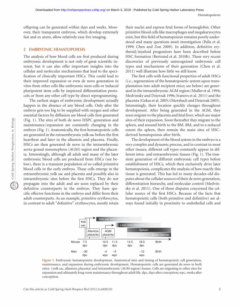
The earliest stages of embryonic development actuallyhappen in the absence of any blood cells. Only after theembryo gets too big to be supplied with oxygen and otheressential factors by diffusion are blood cells first generated(Fig. 1). The sites of both de novo HSPC generation andmaintenance/expansion are constantly changing in theembryo (Fig. 1). Anatomically, the first hematopoietic cellsare generated in the extraembryonic yolk sac before the firstheartbeat and later in the allantois and placenta. Finally,HSCs are then generated de novo in the intraembryonicaorta-gonad-mesonephros (AGM) region and the placen-ta. Interestingly, although all adult and many of the laterembryonic blood cells are produced from HSCs (see be-low), there is a transient population of so-called primitiveblood cells in the early embryo. These cells emerge in theextraembryonic yolk sac and placenta and possibly also inintraembryonic sites before the first HSCs. They do notpropagate into the adult and are soon replaced by theirdefinitive counterparts in the embryo. They have spe-cific effector functions in the embryo and differ from theiradult counterparts. As an example, primitive erythrocytes,in contrast to adult “definitive” erythrocytes, mostly retain
their nuclei and express fetal forms of hemoglobin. Otherprimitive blood cells like macrophages and megakaryocytesexist, but this field of hematopoiesis remains poorly under-stood and many questions await investigation (Palis et al.1999; Chen and Zon 2009). In addition, definitive ery-throid/myeloid progenitors have been described beforeHSC formation (Bertrand et al. 2010b). These very recentdiscoveries of previously unrecognized embryonic celltypes and mechanisms of their generation (Chen et al.2011) well illustrate how little we still know.
The first cells with functional properties of adult HSCs(i.e., regeneration of the hematopoietic system upon trans-plantation into adult recipient mice; see below) are gener-ated in the intraembryonic AGM region (Muller et al. 1994;Medvinsky and Dzierzak 1996; Ivanovs et al. 2011) and theplacenta (Gekas et al. 2005; Ottersbach and Dzierzak 2005).Interestingly, their location quickly changes throughoutdevelopment. After being generated in the AGM, theysoon migrate to the placenta and fetal liver, which are majorsites of their expansion. Soon thereafter they migrate to thespleen, and around birth to the BM. BM, and to a reducedextent the spleen, then remain the main sites of HSC-derived hematopoiesis after birth.
The development of the blood system in the embryo is avery complex and dynamic process, and in contrast to mostother tissues, different cell types constantly appear in dif-ferent intra- and extraembryonic tissues (Fig. 1). The tran-sient generation of different embryonic cell types beforeestablishment of HSCs, which then exclusively drive laterhematopoiesis, complicates the analysis of how exactly thistissue is generated. This has led to many decades-old dis-putes about the cellular sources of their de novo generation,differentiation hierarchy, and molecular control (Medvin-sky et al. 2011). One of those disputes concerned the cel-lular source of the first HSCs. Because of the facts thathematopoietic cells (both primitive and definitive) are al-ways found initially in proximity to endothelial cells and
Mouse 7.5
Yolksac
AGMplacenta
Allantoisplacenta
~30
dpc
dpc
10.5
~4
dpc
wpc
11.5
~5
dpc14.5dpc
wpc
18.5
~12
Birth
Bone marrow
Spleen
Liver and placenta
Yolk sac and placenta
Birth
dpc
wpcMan
Figure 1. Embryonic hematopoietic development. Anatomical sites and timing of hematopoietic cell generation,maintenance, and expansion during embryonic development. Hematopoietic cells are generated de novo in bothextra- (yolk sac, allantois, placenta) and intraembryonic (AGM region) tissues. Cells are migrating to other sites forexpansion and ultimately long-term maintenance throughout adult life. dpc, days after conception; wpc, weeks afterconception.
Hematopoiesis
Cite this article as Cold Spring Harb Perspect Biol 2012;4:a008250 3
on March 3, 2019 - Published by Cold Spring Harbor Laboratory Press http://cshperspectives.cshlp.org/Downloaded from
that these cell types share many molecular markers, it hadbeen hypothesized for decades that the first hematopoieticcells are actually generated from endothelium (reviewed byDieterlen-Lievre et al. 2006; Medvinsky et al. 2011). Thesehemogenic endothelial cells would be integrated into vesselwalls but would be able, under the influence of unknownmolecular signals, to generate hematopoietic cells. Howev-er, proving the existence of hemogenic endothelial cells wassurprisingly difficult. For decades, static analyses of cellseither before or after the endothelial-to-hematopoietictransition yielded data that could also be explained by thealternative conclusion that hematopoietic cells were gener-ated elsewhere and then migrated toward endothelial cells.Final proof of direct generation of hematopoietic cells fromhemogenic endothelium came from continuous single-cellobservations of endothelial-to-hematopoietic transitionsby time-lapse microscopy (Eilken et al. 2009). Subsequentstudies could then also detect this direct transition in vivoin the zebrafish embryo (Bertrand et al. 2010a; Kissa andHerbomel 2010). It is worth mentioning that these studiesdo not rule out the possible existence of other sources ofhemogenic cell types in the embryo. Importantly, compa-rable timing and location of hemogenic activity in humanembryos makes it likely that the same cells, molecules, andmechanisms are also involved in human embryonic hema-topoiesis (Ivanovs et al. 2011).
The proof of the existence of hemogenic endotheliumnow allows the improved analysis of the molecular mech-anisms involved in the generation of the first blood cells,including the therapeutically important HSCs. This mo-lecular mechanism is only partially understood. It involvescomplex signaling from different cell types surround-ing developing hemogenic endothelium at different sitesand times in the fast-developing and -changing embryo.Mesodermal cells expressing Flk1 and ETV2 were recentlyidentified as the common precursors for endothelial andhematopoietic cells (Lee et al. 2008; Kataoka et al. 2011a).The signals inducing hemogenic fate in these cells and theirprogeny are the subject of ongoing research. Moleculeslike membrane-bound Notch ligands or soluble hedgehog,bone morphogenetic protein, or vascular endothelialgrowth factor, but also shear stress and nitric oxide signal-ing, have been involved in the induction of the hemogenicprogram in endothelial cells—possibly even in mesodermalprecursors before they have reached endothelial identity(Medvinsky et al. 2011). Within the hemogenic endothelialcell, a number of transcription factors (TFs) seem to becrucial for the induction of the hemogenic program, withRunx1 and CBFb apparently being critical core factors(Chen et al. 2011).
Importantly, fetal HSCs differ from adult HSCs in theirregeneration behavior. In contrast to mostly quiescent
adult HSCs, they are actively cycling and regenerate hema-topoiesis faster and more robustly upon transplantation.The switch from this fetal to adult HSC behavior occurs in avery short time window a few weeks after birth of a mouse(Bowie et al. 2007). This sharp switch at a defined time isuseful for the identification of the involved molecularmechanisms (Kim et al. 2007; He et al. 2011), which couldbe of great interest for the induction of a clinically preferredfetal phenotype in HSCs.
3 ADULT HEMATOPOIESIS
3.1 The Hematopoietic Differentiation Hierarchy
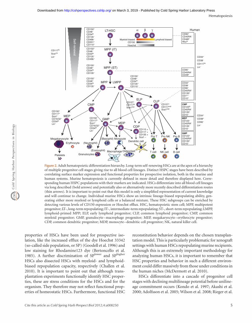
In adult mammals, HSCs are at the apex of a hierarchy ofnumerous progenitor cell stages with increasingly restrictedlineage potentials that give rise to all blood cell lineages(Fig. 2). Of all blood cells, only HSCs fulfill the criteriafor somatic stem cells, namely, long-term (and possiblylifelong) self-renewal and differentiation potential. In themurine system they robustly self-renew (produce moreHSCs) while also reconstituting all blood cell lineages for.24 weeks in congenic transplant recipients (Benvenisteet al. 2010), and are further able to repopulate secondaryrecipients. These functional assays have also revealed multi-potent progenitor (MPP) populations with intermediate-or short-term repopulating capacity (formerly called IT-and ST-HSCs) with a finite self-renewal potential (Osawaet al. 1996; Benveniste et al. 2010). The stepwise identifica-tion of multiple surface markers in the last 25 years enabledthe prospective isolation of defined stem and progenitorcell populations by flow cytometry-based cell sorting. Im-mature multipotent cells express CD117 (c-Kit) and stemcell antigen (Sca)-1 and are low in mature cell marker ex-pression (lineage markers) (Spangrude et al. 1988; Okadaet al. 1992; Uchida and Weissman 1992; Morrison andWeissman 1994). About one in 30 cells within this so-calledKSL (c-Kit+/Sca-1+/Lineage2) population (0.06% in BM)is an HSC (0.002% in BM), and there is no HSC activityoutside the KSL fraction. These findings laid the groundfor the identification of additional surface markers forimproved prospective HSC identification (Table 1). Cur-rently, the highest purity of at least 50% HSCs in the sortedfraction is obtained with CD150+/CD482/CD34low KSLcells (Kiel et al. 2005). Moreover, CD150 expression levelsmight indicate distinct subpopulations of HSCs with pre-disposed myeloid- or lymphoid-biased reconstitution pat-terns (see also Sec. 3.2.1) (Morita et al. 2010). CD49bexpression could be attributed to intermediate-term re-populating MPPs, a population that shares many function-al properties of myeloid-biased HSCs (Benveniste et al.2010). In addition to surface markers, distinct functional
M.A. Rieger and T. Schroeder
4 Cite this article as Cold Spring Harb Perspect Biol 2012;4:a008250
on March 3, 2019 - Published by Cold Spring Harbor Laboratory Press http://cshperspectives.cshlp.org/Downloaded from
properties of HSCs have been used for prospective iso-lation, like the increased efflux of the dye Hoechst 33342(so-called side population, or SP) (Goodell et al. 1996) andlow staining for Rhodamine123 dye (Bertoncello et al.1985). A further discrimination of SPlower and SPhigher
HSCs also dissected HSCs with myeloid- and lymphoid-biased repopulation capacity, respectively (Challen et al.2010). It is important to point out that although trans-plantation experiments functionally identify HSC proper-ties, these are stress conditions for the HSCs and for theorganism. They therefore may not reflect functional prop-erties of homeostatic HSCs. Furthermore, functional HSC
reconstitution behavior depends on the chosen transplan-tation model. This is particularly problematic for xenograftsettings with human HSCs repopulating murine recipients.Although this is an extremely important methodology foranalyzing human HSCs, it is important to remember thatHSC properties and behavior in such a different environ-ment could differ massively from those under conditions inthe human niches (McDermott et al. 2010).
HSCs differentiate into a cascade of progenitor cellstages with declining multilineage potential before uniline-age commitment occurs (Kondo et al. 1997; Akashi et al.2000; Adolfsson et al. 2005; Wilson et al. 2008; Rieger et al.
CD150+
CD34lo
CD135–
CD49b– Myeloid biasedCD150CD201+
CD110+
CD48–
CD34+
CD90–
CD45RA+
CD135+
CD7+/–
CD10+
CD38–
CD115 +
CD135 +Sca1 –
CD117 +
CD127+
Sca1midCD117mid
CD127–
Sca1midCD117hi CMP
CD45RA–
CD135+
CD10–
CD123lo
MEP
CD45RA–
CD135–
CD10–
CD123–CD38+
CD90–
Lin–
CD34+
GMP
CD45RA+
CD135+
CD10–
CD123+
CD90+
CD49f+
Rholo
CD45RA–
CD34+
CD117hi
Lin–
CD38–
CD90–
CD49f–CD45RA–
CD150–
CD34hi
CD135+
CD110–
CD48+
CD150+
CD34lo
CD135–
CD49b+
CD48–CD117hi
Lin–
Sca1hi
CD150–
CD34hi
CD135–
CD48–
CD150–
CD34hi
CD135–
CD48+
CD150+
Sca1–
CD16/32–
CD34lo
Lin–
CD117+
CD150–
Sca1–
CD16/32+
CD34hi
Lin–
CD117+
Hoechst
Lymphoid biased
MPP (IT)
LT-HSC Human
Self-renew
al
MPP (ST)
MPP LMPP
ELP
CMP
GMPMEP
Megakaryocyte
Erythrocyte
Platelets GranulocytesMacrophage Osteoclast T cell NK cell B cell
(Eosinophil)
(Neutrophil)
(Basophil) MonocyteDendritic cell
Pro T Pro NK Pro B
MDPCDP
CLP
α β γ
Figure 2. Adult hematopoietic differentiation hierarchy. Long-term self-renewing HSCs are at the apex of a hierarchyof multiple progenitor cell stages giving rise to all blood cell lineages. Distinct HSPC stages have been described bycorrelating surface marker expression and functional properties for prospective isolation, both in the murine andhuman systems. Murine hematopoiesis is currently defined in more detail and therefore displayed here. Corre-sponding human HSPC populations with their markers are indicated. HSCs differentiate into all blood cell lineagesvia long described (bold arrows) and potentially also or alternatively more recently described differentiation routes(thin arrows). It is important to point out that this model is only a simplified representation of current knowledgeand will continue to change. Individual murine HSCs show an intrinsic lineage-biased repopulating ability, gen-erating either more myeloid or lymphoid cells or a balanced mixture. These HSC subgroups can be enriched bydetecting various levels of CD150 expression or Hoechst efflux. HSC, hematopoietic stem cell; MPP, multipotentprogenitor; LT-, long-term repopulating; IT-, intermediate-term repopulating; ST-, short-term repopulating; LMPP,lymphoid-primed MPP; ELP, early lymphoid progenitor; CLP, common lymphoid progenitor; CMP, commonmyeloid progenitor; GMP, granulocyte–macrophage progenitor; MEP, megakaryocyte–erythrocyte progenitor;CDP, common dendritic progenitor; MDP, monocyte–dendritic cell progenitor; NK, natural killer cell.
Hematopoiesis
Cite this article as Cold Spring Harb Perspect Biol 2012;4:a008250 5
on March 3, 2019 - Published by Cold Spring Harbor Laboratory Press http://cshperspectives.cshlp.org/Downloaded from
2009b). Current models of the hematopoietic hierarchydescribe a successive loss of individual lineage potentials(Adolfsson et al. 2005; Arinobu et al. 2007; Mansson et al.2007; Pronk et al. 2007). MPPs first lose their erythroid/megakaryocytic potential and develop into lymphoid-primed MPPs (LMPPs) and early lymphoid progenitors(ELPs) (Adolfsson et al. 2005). Then they lose their myeloidpotential and become common lymphoid progenitors(CLPs) (Kondo et al. 1997; Inlay et al. 2009; Schlenneret al. 2010), and finally differentiate into lymphoid cells(Fig. 2). However, the successive restriction to solely lym-phoid fate should not imply a picture of default lymphoidcell fate. The opposite might be the case, namely, that activerepression of the myeloid cell fate is required to inducelymphoid development. Interestingly, B- and T-cell pro-genitors retain, at least in vitro, some myeloid potential(Kawamoto et al. 2010). In contrast, lineage-tracing exper-iments revealed only a minor contribution of myeloid cellsfrom interleukin-7 (IL-7) receptor–positive CLPs in mu-rine homeostatic hematopoiesis (Schlenner et al. 2010). Itis important to point out that this model of the hemato-poietic differentiation hierarchy clearly only reflects currentknowledge and will continue to change over time. In par-ticular, the early progenitor stages are still ill defined, andnumerous novel maturation stages and differentiationpathways will certainly be described in coming years.
According to current data, the human hematopoieticdifferentiation hierarchy closely resembles the murine one(Majeti et al. 2007; Doulatov et al. 2010). However, mostlyas a result of more difficult experimentation, and despitetheir clinical importance, purification methods for humanHSPCs are far less advanced than for murine HSPCs (Table1). Human HSCs express the surface molecule CD34, incontrast to their murine counterparts (Osawa et al. 1996;Dick 2008). However, although many reports describeCD34+/CD382/Lineage2 cells as human HSCs, it is veryimportant to point out that this population comprises,1% functional HSCs (as determined by xenograft trans-plantation assays). Only recently have improved methodsfor the prospective enrichment of human HSCs beendescribed that now allow enrichment to purities of ~15%(Notta et al. 2011) (Table 1). Prospective isolation ofhuman hematopoietic progenitors has also recently beenimproved, allowing the functional and molecular charac-terization of these important cell types with much im-proved resolution (Manz et al. 2002; Majeti et al. 2007;Doulatov et al. 2010). However, the percentage of pro-spectively isolated human HSCs may be underestimatedby xenotransplantations because the murine environmentis less permissive to human HSCs, and advanced human-ized mouse models have improved engraftment efficiency(Rongvaux et al. 2011).
Table 1. Prospective HSC isolation
HSPC populationTransplanted
cells Assay/modelHSCs(%) References
MurineThy-1low/Sca-1+/Lin2 30 Competitive 1.5–3 Spangrude et al. 1988KSL Limiting
dilutionsCompetitive 3–7 Okada et al. 1992; Bryder et al.
2006Thy-1low KSL 5–10 Competitive 10–20 Morrison and Weissman 1994CD342 KSL 1 Competitive 21 Osawa et al. 1996SP Rholow/CD45mid/Lin2 1 Sublethal W-41 42/33 Uchida et al. 2003; Dykstra et al.
2006SP CD201+ 10 Competitive .10 Balazs et al. 2006CD150+/CD482/CD412 KSL 1 Competitive 47 Kiel et al. 2005CD150+/CD482/CD49blow KSL 1 Sublethal W-41
competitive29 Benveniste et al. 2010
CD150+/CD482/CD201+/CD45+ 1 Sublethal W-41 43 Kent et al. 2009
HumanCD34+/CD382/Lin2 Limiting
dilutionsNOD/SCID ,1 Bhatia et al. 1997
CD34+/CD382/CD90+/CD45RA2/Lin2 Limitingdilutions
Sublethal NSG 5 Majeti et al. 2007; Notta et al.2011
Rholow/CD49f+/CD34+/CD382/CD90+/CD45RA2/Lin2
1 Sublethal NSG 15 Notta et al. 2011
HSPC, hematopoietic stem or progenitor cell; KSL, CD117+, Sca-1+, and Lin2 cells; SP, side population; Rho, Rhodamine123; W-41, mouse line with homo-
zygous KitW-41J/KitW-41J mutation; NOD/SCID, nonobese diabetic/severe combined immune deficiency mouse line; NSG, NOD/SCID/Il2 g-chain knockout
mouse line; competitive, transplantation in lethally irradiated recipients with “competitor-recipient” HSPCs.
M.A. Rieger and T. Schroeder
6 Cite this article as Cold Spring Harb Perspect Biol 2012;4:a008250
on March 3, 2019 - Published by Cold Spring Harbor Laboratory Press http://cshperspectives.cshlp.org/Downloaded from
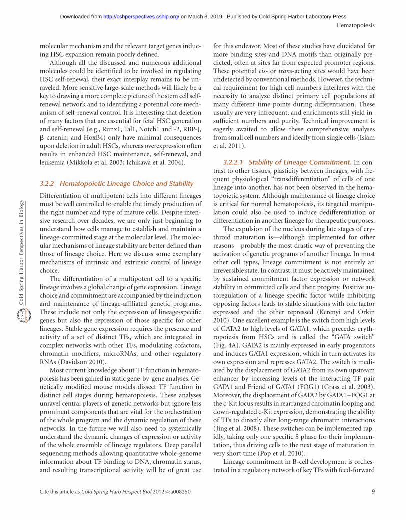
3.2 Molecular Control of HematopoieticFate Choices
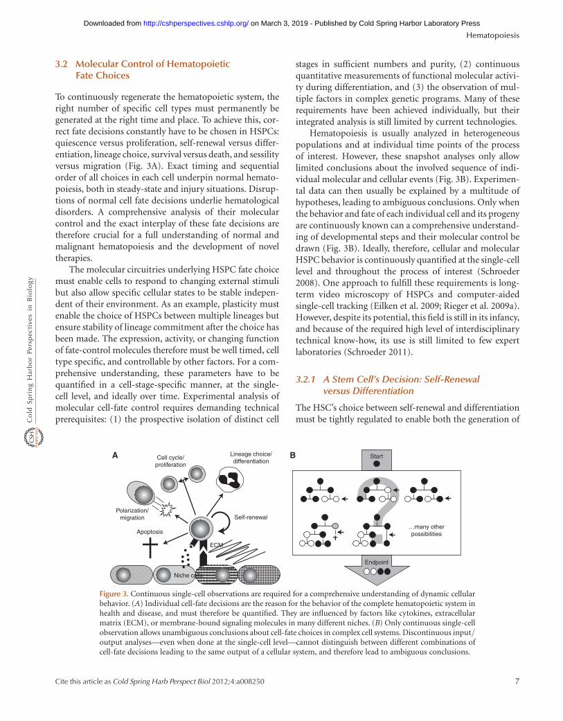
To continuously regenerate the hematopoietic system, theright number of specific cell types must permanently begenerated at the right time and place. To achieve this, cor-rect fate decisions constantly have to be chosen in HSPCs:quiescence versus proliferation, self-renewal versus differ-entiation, lineage choice, survival versus death, and sessilityversus migration (Fig. 3A). Exact timing and sequentialorder of all choices in each cell underpin normal hemato-poiesis, both in steady-state and injury situations. Disrup-tions of normal cell fate decisions underlie hematologicaldisorders. A comprehensive analysis of their molecularcontrol and the exact interplay of these fate decisions aretherefore crucial for a full understanding of normal andmalignant hematopoiesis and the development of noveltherapies.
The molecular circuitries underlying HSPC fate choicemust enable cells to respond to changing external stimulibut also allow specific cellular states to be stable indepen-dent of their environment. As an example, plasticity mustenable the choice of HSPCs between multiple lineages butensure stability of lineage commitment after the choice hasbeen made. The expression, activity, or changing functionof fate-control molecules therefore must be well timed, celltype specific, and controllable by other factors. For a com-prehensive understanding, these parameters have to bequantified in a cell-stage-specific manner, at the single-cell level, and ideally over time. Experimental analysis ofmolecular cell-fate control requires demanding technicalprerequisites: (1) the prospective isolation of distinct cell
stages in sufficient numbers and purity, (2) continuousquantitative measurements of functional molecular activi-ty during differentiation, and (3) the observation of mul-tiple factors in complex genetic programs. Many of theserequirements have been achieved individually, but theirintegrated analysis is still limited by current technologies.
Hematopoiesis is usually analyzed in heterogeneouspopulations and at individual time points of the processof interest. However, these snapshot analyses only allowlimited conclusions about the involved sequence of indi-vidual molecular and cellular events (Fig. 3B). Experimen-tal data can then usually be explained by a multitude ofhypotheses, leading to ambiguous conclusions. Only whenthe behavior and fate of each individual cell and its progenyare continuously known can a comprehensive understand-ing of developmental steps and their molecular control bedrawn (Fig. 3B). Ideally, therefore, cellular and molecularHSPC behavior is continuously quantified at the single-celllevel and throughout the process of interest (Schroeder2008). One approach to fulfill these requirements is long-term video microscopy of HSPCs and computer-aidedsingle-cell tracking (Eilken et al. 2009; Rieger et al. 2009a).However, despite its potential, this field is still in its infancy,and because of the required high level of interdisciplinarytechnical know-how, its use is still limited to few expertlaboratories (Schroeder 2011).
3.2.1 A Stem Cell’s Decision: Self-Renewalversus Differentiation
The HSC’s choice between self-renewal and differentiationmust be tightly regulated to enable both the generation of
Niche cells
ECM
Self-renewal
Endpoint
…many otherpossibilities
Start
Apoptosis
Polarization/migration
Cell cycle/proliferation
A BLineage choice/differentiation
Figure 3. Continuous single-cell observations are required for a comprehensive understanding of dynamic cellularbehavior. (A) Individual cell-fate decisions are the reason for the behavior of the complete hematopoietic system inhealth and disease, and must therefore be quantified. They are influenced by factors like cytokines, extracellularmatrix (ECM), or membrane-bound signaling molecules in many different niches. (B) Only continuous single-cellobservation allows unambiguous conclusions about cell-fate choices in complex cell systems. Discontinuous input/output analyses—even when done at the single-cell level—cannot distinguish between different combinations ofcell-fate decisions leading to the same output of a cellular system, and therefore lead to ambiguous conclusions.
Hematopoiesis
Cite this article as Cold Spring Harb Perspect Biol 2012;4:a008250 7
on March 3, 2019 - Published by Cold Spring Harbor Laboratory Press http://cshperspectives.cshlp.org/Downloaded from
differentiated cells and the accurate maintenance of theright HSC number. The BM provides the environmentfor sustained HSC function, and HSCs rapidly lose theirself-renewal capacity once isolated from their in vivoniches. Extrinsic signals from membrane-bound, soluble,or extracellular matrix-associated ligands from the nicheare necessary for appropriate HSC behavior. Two majorHSC niches are currently proposed to exist in BM: theendosteal osteoblastic niche (Calvi et al. 2003; Zhanget al. 2003; Lo Celso et al. 2009; Xie et al. 2009) and theperivascular endothelial niche (Kiel et al. 2005; Ding et al.2012). It is unclear if both niches, although spatially sepa-rated, fulfill a similar function; if both niches provide dis-tinct properties, for example, for dormant versus cyclingHSCs; or if the niche comprises both osteoblasts and en-dothelial cells working synergistically on HSC function.Also, BM comprises a heterogeneous mixture of variouscell types including blood cells, mesenchymal cells, osteo-blasts, osteoclasts, endothelial cells, reticular cells, fat cells,and many other less-defined types, which will also influ-ence hematopoietic fates. A multitude of signaling path-ways have been shown to be activated in HSCs by the niche(e.g., the cytokine receptors c-Kit and Mpl, Wnt, Notch,Sonic hedgehog, and integrin signaling). However, theirprecise involvement in HSC maintenance remains sur-prisingly controversial. For most of them, contradictoryconclusions have been drawn in separate studies. Our in-complete understanding of the molecular control of HSCself-renewal is well illustrated by the fact that therapeuti-cally relevant robust maintenance or even expansion ofthese clinically important cells ex vivo remains elusivewithout genetic manipulation. Nevertheless, several recentstudies with murine and human HSCs describe very prom-ising approaches for the ex vivo expansion of HSCs withoutgenetic manipulation (Zhang et al. 2008; Boitano et al.2010). A comprehensive discussion of all potential molec-ular HSC self-renewal modulators goes beyond the scope ofthis chapter, and can be found elsewhere (Ehninger andTrumpp 2011; Mercier et al. 2011).
At the single-cell level, even HSCs with retrospective-ly proven functionality (.1% long-term contributionto mature myeloid and lymphoid cells in peripheralblood) are heterogeneous in their reconstitution efficacyand their lineage contribution pattern. They range from1% to almost 100% total contribution, with varying ratiosof myeloid and lymphoid lineages (Muller-Sieburg et al.2002, 2004; Sieburg et al. 2006; Dykstra et al. 2007; Kentet al. 2009). Importantly, these patterns are conservedthrough serial transplantations, indicating the existenceof stable inheritable stem cell intrinsic programs (Muller-Sieburg et al. 2002, 2004; Sieburg et al. 2006; Dykstraet al. 2007; Kent et al. 2009). Therefore, not only are self-
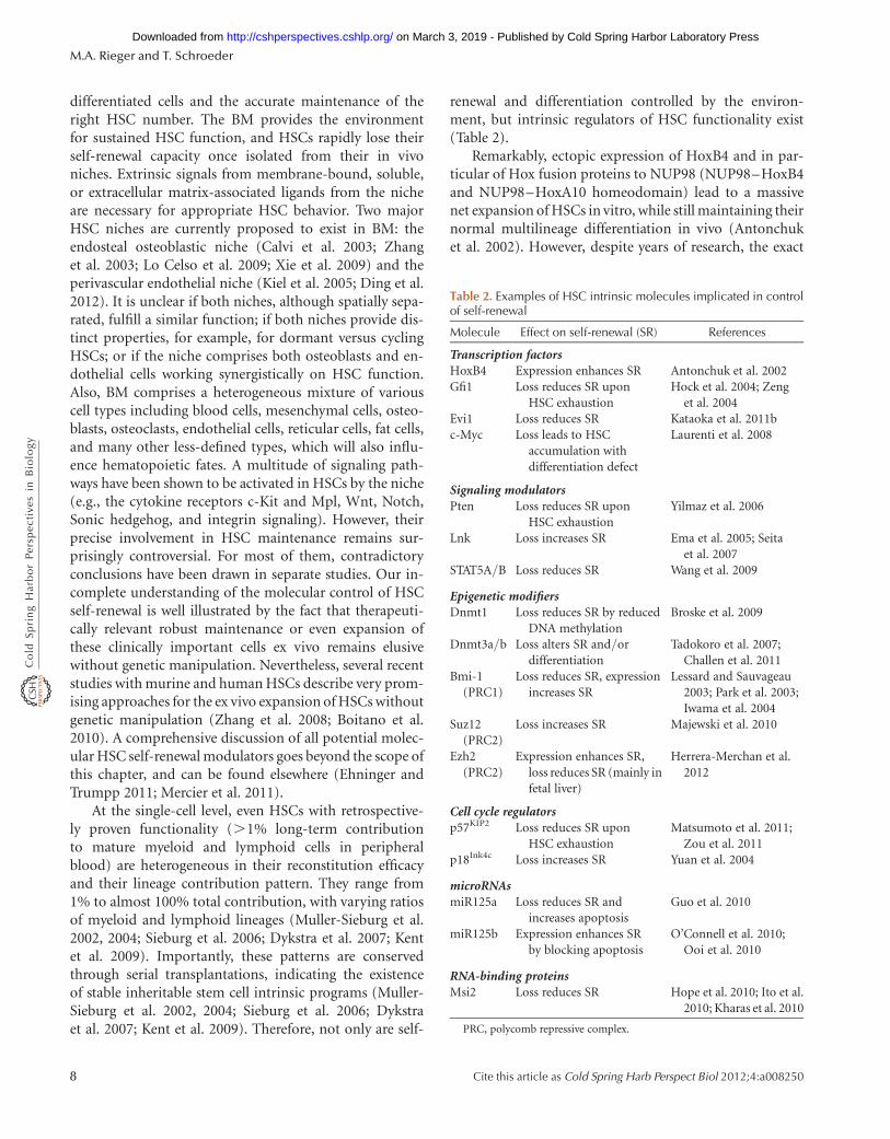
renewal and differentiation controlled by the environ-ment, but intrinsic regulators of HSC functionality exist(Table 2).
Remarkably, ectopic expression of HoxB4 and in par-ticular of Hox fusion proteins to NUP98 (NUP98–HoxB4and NUP98–HoxA10 homeodomain) lead to a massivenet expansion of HSCs in vitro, while still maintaining theirnormal multilineage differentiation in vivo (Antonchuket al. 2002). However, despite years of research, the exact
Table 2. Examples of HSC intrinsic molecules implicated in controlof self-renewal
Molecule Effect on self-renewal (SR) References
Transcription factorsHoxB4 Expression enhances SR Antonchuk et al. 2002Gfi1 Loss reduces SR upon
HSC exhaustionHock et al. 2004; Zeng
et al. 2004Evi1 Loss reduces SR Kataoka et al. 2011bc-Myc Loss leads to HSC
accumulation withdifferentiation defect
Laurenti et al. 2008
Signaling modulatorsPten Loss reduces SR upon
HSC exhaustionYilmaz et al. 2006
Lnk Loss increases SR Ema et al. 2005; Seitaet al. 2007
STAT5A/B Loss reduces SR Wang et al. 2009
Epigenetic modifiersDnmt1 Loss reduces SR by reduced
DNA methylationBroske et al. 2009
Dnmt3a/b Loss alters SR and/ordifferentiation
Tadokoro et al. 2007;Challen et al. 2011
Bmi-1(PRC1)
Loss reduces SR, expressionincreases SR
Lessard and Sauvageau2003; Park et al. 2003;Iwama et al. 2004
Suz12(PRC2)
Loss increases SR Majewski et al. 2010
Ezh2(PRC2)
Expression enhances SR,loss reduces SR (mainly infetal liver)
Herrera-Merchan et al.2012
Cell cycle regulatorsp57KIP2 Loss reduces SR upon
HSC exhaustionMatsumoto et al. 2011;
Zou et al. 2011p18Ink4c Loss increases SR Yuan et al. 2004
microRNAsmiR125a Loss reduces SR and
increases apoptosisGuo et al. 2010
miR125b Expression enhances SRby blocking apoptosis
O’Connell et al. 2010;Ooi et al. 2010
RNA-binding proteinsMsi2 Loss reduces SR Hope et al. 2010; Ito et al.
2010; Kharas et al. 2010
PRC, polycomb repressive complex.
M.A. Rieger and T. Schroeder
8 Cite this article as Cold Spring Harb Perspect Biol 2012;4:a008250
on March 3, 2019 - Published by Cold Spring Harbor Laboratory Press http://cshperspectives.cshlp.org/Downloaded from
molecular mechanism and the relevant target genes induc-ing HSC expansion remain poorly defined.
Although all the discussed and numerous additionalmolecules could be identified to be involved in regulatingHSC self-renewal, their exact interplay remains to be un-raveled. More sensitive large-scale methods will likely be akey to drawing a more complete picture of the stem cell self-renewal network and to identifying a potential core mech-anism of self-renewal control. It is interesting that deletionof many factors that are essential for fetal HSC generationand self-renewal (e.g., Runx1, Tal1, Notch1 and -2, RBP-J,b-catenin, and HoxB4) only have minimal consequencesupon deletion in adult HSCs, whereas overexpression oftenresults in enhanced HSC maintenance, self-renewal, andleukemia (Mikkola et al. 2003; Ichikawa et al. 2004).
3.2.2 Hematopoietic Lineage Choice and Stability
Differentiation of multipotent cells into different lineagesmust be well controlled to enable the timely production ofthe right number and type of mature cells. Despite inten-sive research over decades, we are only just beginning tounderstand how cells manage to establish and maintain alineage-committed stage at the molecular level. The molec-ular mechanisms of lineage stability are better defined thanthose of lineage choice. Here we discuss some exemplarymechanisms of intrinsic and extrinsic control of lineagechoice.
The differentiation of a multipotent cell to a specificlineage involves a global change of gene expression. Lineagechoice and commitment are accompanied by the inductionand maintenance of lineage-affiliated genetic programs.These include not only the expression of lineage-specificgenes but also the repression of those specific for otherlineages. Stable gene expression requires the presence andactivity of a set of distinct TFs, which are integrated incomplex networks with other TFs, modulating cofactors,chromatin modifiers, microRNAs, and other regulatoryRNAs (Davidson 2010).
Most current knowledge about TF function in hemato-poiesis has been gained in static gene-by-gene analyses. Ge-netically modified mouse models dissect TF function indistinct cell stages during hematopoiesis. These analysesunravel central players of genetic networks but ignore lessprominent components that are vital for the orchestrationof the whole program and the dynamic regulation of thesenetworks. In the future we will also need to systemicallyunderstand the dynamic changes of expression or activityof the whole ensemble of lineage regulators. Deep parallelsequencing methods allowing quantitative whole-genomeinformation about TF binding to DNA, chromatin status,and resulting transcriptional activity will be of great use
for this endeavor. Most of these studies have elucidated farmore binding sites and DNA motifs than originally pre-dicted, often at sites far from expected promoter regions.These potential cis- or trans-acting sites would have beenundetected by conventional methods. However, the techni-cal requirement for high cell numbers interferes with thenecessity to analyze distinct primary cell populations atmany different time points during differentiation. Theseusually are very infrequent, and enrichments still yield in-sufficient numbers and purity. Technical improvement iseagerly awaited to allow these comprehensive analysesfrom small cell numbers and ideally from single cells (Islamet al. 2011).
3.2.2.1 Stability of Lineage Commitment. In con-trast to other tissues, plasticity between lineages, with fre-quent physiological “transdifferentiation” of cells of onelineage into another, has not been observed in the hema-topoietic system. Although maintenance of lineage choiceis critical for normal hematopoiesis, its targeted manipu-lation could also be used to induce dedifferentiation ordifferentiation in another lineage for therapeutic purposes.
The expulsion of the nucleus during late stages of ery-throid maturation is—although implemented for otherreasons—probably the most drastic way of preventing theactivation of genetic programs of another lineage. In mostother cell types, lineage commitment is not entirely anirreversible state. In contrast, it must be actively maintainedby sustained commitment factor expression or networkstability in committed cells and their progeny. Positive au-toregulation of a lineage-specific factor while inhibitingopposing factors leads to stable situations with one factorexpressed and the other repressed (Kerenyi and Orkin2010). One excellent example is the switch from high levelsof GATA2 to high levels of GATA1, which precedes eryth-ropoiesis from HSCs and is called the “GATA switch”(Fig. 4A). GATA2 is mainly expressed in early progenitorsand induces GATA1 expression, which in turn activates itsown expression and represses GATA2. The switch is medi-ated by the displacement of GATA2 from its own upstreamenhancer by increasing levels of the interacting TF pairGATA1 and Friend of GATA1 (FOG1) (Grass et al. 2003).Moreover, the displacement of GATA2 by GATA1–FOG1 atthe c-Kit locus results in rearranged chromatin looping anddown-regulated c-Kit expression, demonstrating the abilityof TFs to directly alter long-range chromatin interactions(Jing et al. 2008). These switches can be implemented rap-idly, taking only one specific S phase for their implemen-tation, thus driving cells to the next stage of maturation invery short time (Pop et al. 2010).
Lineage commitment in B-cell development is orches-trated in a regulatory network of key TFs with feed-forward
Hematopoiesis
Cite this article as Cold Spring Harb Perspect Biol 2012;4:a008250 9
on March 3, 2019 - Published by Cold Spring Harbor Laboratory Press http://cshperspectives.cshlp.org/Downloaded from
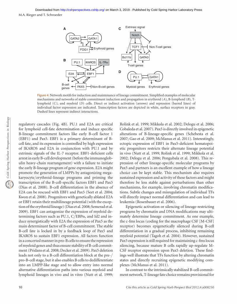
regulatory cascades (Fig. 4B). PU.1 and E2A are criticalfor lymphoid cell-fate determination and induce specificB-lineage commitment factors like early B-cell factor 1(EBF1) and Pax5. EBF1 is a primary determinant of B-cell fate, and its expression is controlled by high expressionof IKAROS and E2A in conjunction with PU.1 and byextrinsic signals of the IL-7 receptor. EBF1-deficient cellsarrest in early B-cell development (before the immunoglob-ulin heavy-chain rearrangement) with a failure to initiatethe early B-lineage program of gene expression. E2A mightpromote the generation of LMPPs by antagonizing mega-karyocyte/erythroid-lineage programs and priming thetranscription of the B-cell-specific factors EBF1 and Pax5(Dias et al. 2008). B-cell differentiation in the absence ofE2A can be rescued with EBF1 and Pax5 (Seet et al. 2004;Kwon et al. 2008). Progenitors with genetically ablated E2Aor EBF1 retain their multilineage potential (with the excep-tion of the erythroid lineage) (Dias et al. 2008; Semerad et al.2009). EBF1 can antagonize the expression of myeloid de-termining factors such as PU.1, C/EBPa, and Id2 and in-duce synergistically with E2A the expression of Pax5 as themain determinant factor of B-cell commitment. The stableB-cell fate is locked in by a feedback loop of Pax5 andIKAROS to sustain EBF1 expression. All factors functionin aconcerted manner in pro-B cells to ensure the repressionof myeloid genes and thus ensure stability of B-cell commit-ment (Pridans et al. 2008; Decker et al. 2009). Pax5 deletionleads not only to a B-cell differentiation block at the pre-/pro-B-cell stage, but it also enables B cells to dedifferentiateinto an LMPP-like stage and to allow entry into normalalternative differentiation paths into various myeloid andlymphoid lineages in vivo and in vitro (Nutt et al. 1999;
Rolink et al. 1999; Mikkola et al. 2002; Delogu et al. 2006;Cobaleda et al. 2007). Pax5 is directly involved in epigeneticalterations of B-lineage-specific genes (Schebesta et al.2007; Gao et al. 2009; McManus et al. 2011). Interestingly,ectopic expression of EBF1 in Pax5-deficient hematopoi-etic progenitors restricts their alternate lineage potentialin vivo (Nutt et al. 1999; Rolink et al. 1999; Mikkola et al.2002; Delogu et al. 2006; Pongubala et al. 2008). This re-pression of other lineage-specific molecular programs byPax5 and partners is an excellent example of how a lineagechoice can be kept stable. This mechanism also requiressustained expression and activity of these factors and mighttherefore be less stable against perturbations than othermechanisms, for example, involving chromatin modifica-tions. Subtle changes and misregulation of individual TFswill directly impact normal differentiation and can lead toleukemia (Rosenbauer et al. 2006).
Epigenetic activation or silencing of lineage-restrictingprograms by chromatin and DNA modifications may ulti-mately determine lineage commitment. As one example,the c-fms locus (coding for the macrophage CSF [M-CSF]receptor) becomes epigenetically silenced during B-celldifferentiation in a gradual process, inhibiting remainingmyeloid potential (Tagoh et al. 2004). However, sustainedPax5 expression is still required for maintaining c-fms locussilencing, because mature B cells rapidly up-regulate M-CSF receptor expression upon Pax5 deletion. These find-ings well illustrate that TFs function by altering chromatinstates and directly recruiting epigenetic modifying com-plexes (McManus et al. 2011).
In contrast to the intrinsically stabilized B-cell commit-ment network, T-lineage fate choice remains provisional for
GATA2
A
B
C
DPU.1 GATA1
GATA1
IKAROSPU.1
EBF1
PAX5
E2A
IL-7Rα
GATA3Bcl11b
TCF1
Notch
Erythroid genes
T-cell genes
Non-T-cell genes
Extrinsic signal
Non-B-cell genes Myeloid genes Erythroid genesB-cellgenes
FOG1
Figure 4. Network motifs for induction and maintenance of lineage commitment. Simplified examples of molecularmechanisms and networks of stable commitment induction and propagation in erythroid (A), B-lymphoid (B), T-lymphoid (C), and myeloid (D) cells. Direct or indirect activation (arrows) and repression (barred lines) ofindividual factor expression are indicated. Transcription factors are depicted in white, surface receptors in gray.Dashed lines represent indirect interactions.
M.A. Rieger and T. Schroeder
10 Cite this article as Cold Spring Harb Perspect Biol 2012;4:a008250
on March 3, 2019 - Published by Cold Spring Harbor Laboratory Press http://cshperspectives.cshlp.org/Downloaded from
an extended period of differentiation with high dependencyon extrinsic stimulation from the thymic environment (Fig.4C). T-cell fate initiation and determination requires per-sistent extrinsic Notch receptor signaling, which is stimu-lated by Delta-like 1 and 4 expressing cortical endothelialcells in the thymus (Radtke et al. 1999). Notch signalingturns LMPPs into early T-cell progenitors by inducing apro-T-cell developmental program with direct and indirectactivation of GATA3, Tcf7, and other T-cell-restrictinggenes, which, once initiated, positively autoregulate theirown expression. The activation of a T-lineage program byNotch signaling is, however, not sufficient for T-cell fatedetermination alone. Additionally, Notch suppresses B-cell-lineage-determining EBF1 and E2A expression and di-minishes remaining myeloid potential by down-regulationof PU.1 and C/EBPa (Ordentlich et al. 1998; Smith et al.2005; Laiosa et al. 2006b). Id2 expression is blocked byNotch signaling to circumvent NK cell development (Ikawaet al. 2001). Conversely, the TF leukemia/lymphoma-relat-ed factor (LRF) inhibits Notch signaling in the BM, thuspreventing T-cell development. Consequently, the condi-tional deletion of LRF in HSCs results in the generation ofT-lineage progeny in the BM (Maeda et al. 2007).
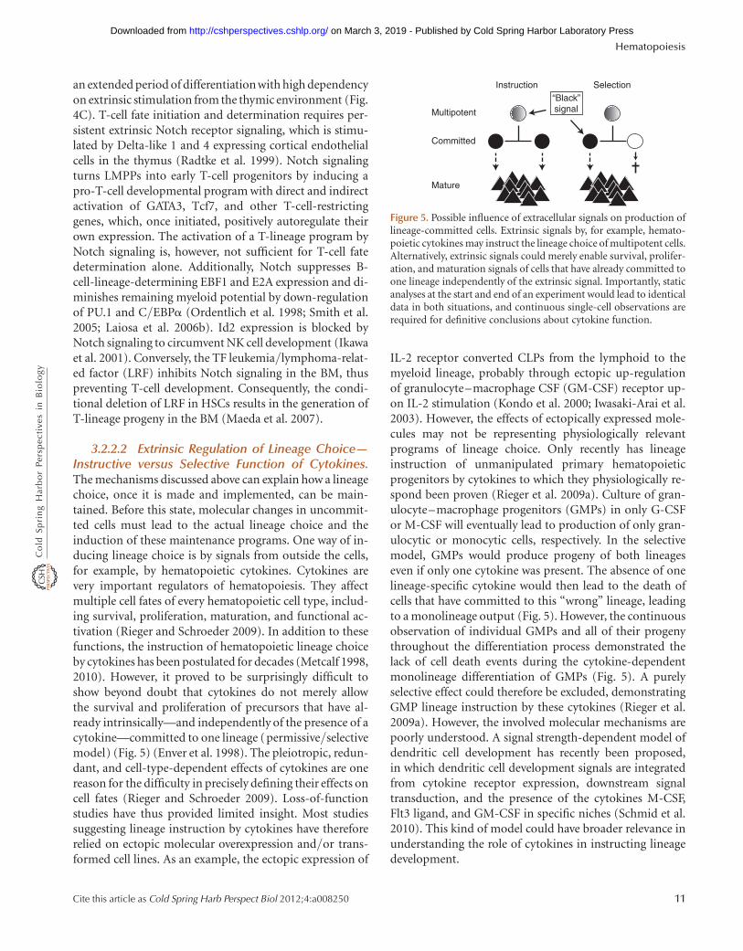
3.2.2.2 Extrinsic Regulation of Lineage Choice—Instructive versus Selective Function of Cytokines.The mechanisms discussed above can explain how a lineagechoice, once it is made and implemented, can be main-tained. Before this state, molecular changes in uncommit-ted cells must lead to the actual lineage choice and theinduction of these maintenance programs. One way of in-ducing lineage choice is by signals from outside the cells,for example, by hematopoietic cytokines. Cytokines arevery important regulators of hematopoiesis. They affectmultiple cell fates of every hematopoietic cell type, includ-ing survival, proliferation, maturation, and functional ac-tivation (Rieger and Schroeder 2009). In addition to thesefunctions, the instruction of hematopoietic lineage choiceby cytokines has been postulated for decades (Metcalf 1998,2010). However, it proved to be surprisingly difficult toshow beyond doubt that cytokines do not merely allowthe survival and proliferation of precursors that have al-ready intrinsically—and independently of the presence of acytokine—committed to one lineage (permissive/selectivemodel) (Fig. 5) (Enver et al. 1998). The pleiotropic, redun-dant, and cell-type-dependent effects of cytokines are onereason for the difficulty in precisely defining their effects oncell fates (Rieger and Schroeder 2009). Loss-of-functionstudies have thus provided limited insight. Most studiessuggesting lineage instruction by cytokines have thereforerelied on ectopic molecular overexpression and/or trans-formed cell lines. As an example, the ectopic expression of
IL-2 receptor converted CLPs from the lymphoid to themyeloid lineage, probably through ectopic up-regulationof granulocyte–macrophage CSF (GM-CSF) receptor up-on IL-2 stimulation (Kondo et al. 2000; Iwasaki-Arai et al.2003). However, the effects of ectopically expressed mole-cules may not be representing physiologically relevantprograms of lineage choice. Only recently has lineageinstruction of unmanipulated primary hematopoieticprogenitors by cytokines to which they physiologically re-spond been proven (Rieger et al. 2009a). Culture of gran-ulocyte–macrophage progenitors (GMPs) in only G-CSFor M-CSF will eventually lead to production of only gran-ulocytic or monocytic cells, respectively. In the selectivemodel, GMPs would produce progeny of both lineageseven if only one cytokine was present. The absence of onelineage-specific cytokine would then lead to the death ofcells that have committed to this “wrong” lineage, leadingto a monolineage output (Fig. 5). However, the continuousobservation of individual GMPs and all of their progenythroughout the differentiation process demonstrated thelack of cell death events during the cytokine-dependentmonolineage differentiation of GMPs (Fig. 5). A purelyselective effect could therefore be excluded, demonstratingGMP lineage instruction by these cytokines (Rieger et al.2009a). However, the involved molecular mechanisms arepoorly understood. A signal strength-dependent model ofdendritic cell development has recently been proposed,in which dendritic cell development signals are integratedfrom cytokine receptor expression, downstream signaltransduction, and the presence of the cytokines M-CSF,Flt3 ligand, and GM-CSF in specific niches (Schmid et al.2010). This kind of model could have broader relevance inunderstanding the role of cytokines in instructing lineagedevelopment.
SelectionInstruction
Multipotent
Committed
Mature
“Black”signal
Figure 5. Possible influence of extracellular signals on production oflineage-committed cells. Extrinsic signals by, for example, hemato-poietic cytokines may instruct the lineage choice of multipotent cells.Alternatively, extrinsic signals could merely enable survival, prolifer-ation, and maturation signals of cells that have already committed toone lineage independently of the extrinsic signal. Importantly, staticanalyses at the start and end of an experiment would lead to identicaldata in both situations, and continuous single-cell observations arerequired for definitive conclusions about cytokine function.
Hematopoiesis
Cite this article as Cold Spring Harb Perspect Biol 2012;4:a008250 11
on March 3, 2019 - Published by Cold Spring Harbor Laboratory Press http://cshperspectives.cshlp.org/Downloaded from
Despite this accumulating evidence for lineage instruc-tion by cell-extrinsic signals in some of the more restrictedprogenitors, their impact on earlier branching points of thehematopoietic hierarchy still remains poorly understood.Cytokines contribute in a major way to the survival andproliferation of different hematopoietic cell types, and notall lineage-affiliated cytokines have a lineage-instructiveability. Even if specific lineages can be instructed by cyto-kines, this does not preclude the existence of additionalcell-intrinsic lineage choice mechanisms that could leadto lineage choice in the absence of cell-extrinsic instructivesignals. The capacity of individual extrinsic signals to in-struct lineage choice, as well as its integration with othercytokine signals and with other intracellular molecularstates like TF expression, must be further analyzed foreach individual cytokine, in specific cell types, under chem-ically defined conditions, and ideally continuously at thesingle-cell level.
3.2.2.3 Intrinsic Regulation of Lineage Choice byTFs—Decision Makers or Only Executors? It is clearthat individual TFs can instruct lineage choice and areeven able to reprogram committed cells, leading to cross-ing of lineage borders. For example, ectopic expressionof GATA1 in multipotent cells or cells committed to otherthan the megakaryocyte–erythrocyte lineages (e.g., CLPs)enforces the development into erythroid and megakar-yocytic cells (Heyworth et al. 2002). C/EBPa and PU.1are essential for the generation of granulocyte–macro-phage progenitors. C/EBPa expression in committed lym-phoid cells (B and T cells) instructs the development ofmacrophages (Xie et al. 2004; Laiosa et al. 2006a,b). Fur-thermore, committed T cells transdifferentiate into mye-loid dendritic cells upon ectopic PU.1 expression (Laiosaet al. 2006b). TFs also actively repress lineage programs. Tothis end, the myeloid potential as well as PU.1- or C/EBPa-dependent myeloid reprogramming of thymic precursorscan be blocked by active Notch signaling (Franco et al.2006; Laiosa et al. 2006b; Rothenberg 2007), suggestingan instructive extracellular non-cytokine-mediated induc-tion of T-cell identity. Findings like these clearly demon-strate the ability of many lineage-specific TFs to drive cellsinto one lineage. Upon their up-regulation or activation,they will start the transcription of myriads of direct andindirect target genes and chromatin modifications, whichlead to the changing phenotypes of lineage-committingand maturing cells.
However, the ability of TFs to drive a cell into onelineage does not yet explain how the initial decision forthis lineage was made—it only explains how this decisionis then executed. What led to the up-regulation or activa-tion of, for example, the executing TF remains unclear. One
possibility is the instruction of lineage choice by cell-ex-trinsic signals. In this case, TFs are most likely the finalexecutors, implementing the lineage decision, but are notinvolved in the actual decision making. Another intriguingpossible mechanism is the stochastic output of fluctuatingcell-intrinsic networks of TFs. In this case, TFs would notonly execute a previously made decision, they would also bethe relevant components of the molecular machinery gen-erating the lineage choice. This hypothesis stems from theinitially surprising findings that multiple “lineage-specific”TFs are coexpressed in multipotent cells before lineagecommitment (“lineage priming”; see, e.g., Miyamotoet al. 2002). These factors are expressed only exclusivelyin different mature lineages, and they can drive cells intothis lineage upon overexpression. Initially, therefore, it wasassumed that multipotent cells have the potential to differ-entiate into multiple lineages because they lack the expres-sion of these lineage-specific TFs. However, with theavailability of improved prospective purification of multi-potent cells and more sensitive molecular methods for thedetection of TF expression, it then became clear that theopposite could be true. Multipotency might in fact be char-acterized not by the absence of lineage-specific propertiesbut by the presence of properties of multiple lineages, inparticular the coexpression of multiple “lineage-specific”TFs. The additional finding that these TFs often inhibiteach other by binding mutually or to cofactors of opposingTFs led to the idea that these factors may neutralize eachother in uncommitted cells, thus retaining multipotency.Random fluctuations in their expression could then lead toone TF gaining dominance; positive autoregulation andrepression of other TFs would lead to a lineage-committedstate in which only one lineage-specific TF is expressed(Enver et al. 1998; Cantor and Orkin 2001; Graf and Enver2009). The production of all lineages would be ensured bythe overall wiring of the TF network leading to stable sto-chastic lineage output. Although not predictable for anindividual cell, different cells within a population wouldfluctuate into decisions for different lineages with specificprobabilities. Adaptation of the blood system to stress orinjury would then be regulated by the selection of existinglineage-committed progenitors for survival and prolifera-tion (Suda et al. 1984).
Studies on transcriptional pathways that control binarylineage choices in hematopoiesis revealed some similaritiesin the gene regulatory network circuits of different lineages.Pairs of TFs that mutually antagonize each other’s activityand expression are often involved. One example is the an-tagonistic interplay between the lineage-determining fac-tors PU.1 and GATA1 as a molecular mechanism of lineagechoice between myeloid and megakaryocytic–erythroidfate, respectively (Fig. 4D). PU.1 and GATA1 physically
M.A. Rieger and T. Schroeder
12 Cite this article as Cold Spring Harb Perspect Biol 2012;4:a008250
on March 3, 2019 - Published by Cold Spring Harbor Laboratory Press http://cshperspectives.cshlp.org/Downloaded from
bind each other and cross-antagonize their activity (Zhanget al. 1999, 2000; Stopka et al. 2005; Liew et al. 2006). PU.1directly inhibits GATA1 DNA-binding capacity, whileGATA1 inhibits the transactivation potential of PU.1.Both PU.1 and GATA1 are autoregulatory for their ownexpression (Nishimura et al. 2000; Okuno et al. 2005; Laio-sa et al. 2006a), thereby providing stability to their levels,once expressed. The genetic ablation of these factors un-derpins their central role in implementing lineage choice.Loss of GATA1 demonstrates its absolute necessity formegakaryocyte/erythrocyte development, whereas PU.1deficiency leads to a lack of granulocytes, macrophages,and B cells. Furthermore, both factors have instructive lin-eage commitment ability by implementing lineage-affiliat-ed gene programs (Iwasaki et al. 2003, 2006). However,recent studies suggest a more complicated dynamic imple-mentation. In fish hematopoiesis, the interplay of PU.1 andGATA1 differs in various cell stages during hematopoiesisand is influenced by other factors, such as the transcriptionintermediate factor 1g (tif1g), a RING domain E3 ubiqui-tin ligase (Monteiro et al. 2011). Moreover, PU.1 showedpositive autoregulation in all analyzed cell stages, butGATA1 only in some of them. C/EBPa and FOG1 haverecently been implicated in the lineage choice between my-eloid and megakaryocyte/erythrocyte lineages by exhibit-ing transcriptional cross-regulation, potentially beingbetter candidates for lineage choice making than PU.1and GATA1 (Mancini et al. 2011).
Again, all of these models suffer from the fact that theyare based on expression analyses with very low resolution.Precise timing of changing PU.1 and GATA1 protein levelsbefore and during lineage choice in the individual cellwould be critical for this mechanism of lineage choice.However, current expression analyses measure only popu-lation averages and/or RNA levels and/or single timepoints or maturation stages. This static data allows manyalternative explanations and is therefore not sufficientto prove the mechanism of lineage choice. Ideally, noveltechnology will enable the simultaneous quantification ofprotein levels of multiple TFs, at the single-cell level, con-tinuously with high temporal resolution, and throughoutlineage choice of living HSCs.
4 CONCLUSION/OUTLOOK
In conclusion, hematopoiesis is an excellent model forstudying the molecular mechanisms of cell-fate control.Being the classical mammalian stem cell model, withhigh clinical relevance, robust quantitative functional as-says, and well-established culture and prospective isolationtechniques, it allows us to address the underlying mecha-nisms of cell-fate control. Most of the basic concepts in
stem cell research have been defined in the hematopoieticsystem, and many novel technical approaches are first de-veloped or applied in hematopoiesis research. Because ofthe routine use of hematopoietic cells for clinical therapy,the chances for a quick transfer of novel basic insights topatient benefit in the clinic are higher than for most othertissues. Nevertheless, despite decades of successful research,many questions posed very long ago are still awaiting ananswer, and long-standing disputes illustrate the need forever improving technological approaches. These continueto be exciting times in hematopoiesis research.
ACKNOWLEDGMENTS
M.A.R. is thankful for the support of the LOEWE Centerfor Cell and Gene Therapy Frankfurt (HMWK III L 4- 518/17.004 [2010]) and institutional funds of the Georg-Speyer-Haus. The Georg-Speyer-Haus is funded jointlyby the German Federal Ministry of Health (BMG) andthe Ministry of Higher Education, Research and the Artsof the state of Hessen (HMWK).
REFERENCES
Adolfsson J, Mansson R, Buza-Vidas N, Hultquist A, Liuba K, Jensen CT,Bryder D, Yang L, Borge OJ, Thoren LA, et al. 2005. Identification ofFlt3+ lympho-myeloid stem cells lacking erythro-megakaryocytic po-tential: A revised road map for adult blood lineage commitment. Cell121: 295–306.
Akashi K, Traver D, Miyamoto T, Weissman IL. 2000. A clonogenic com-mon myeloid progenitor that gives rise to all myeloid lineages. Nature404: 193–197.
Antonchuk J, Sauvageau G, Humphries RK. 2002. HOXB4-induced ex-pansion of adult hematopoietic stem cells ex vivo. Cell 109: 39–45.
Arinobu Y, Mizuno S, Chong Y, Shigematsu H, Iino T, Iwasaki H, Graf T,Mayfield R, Chan S, Kastner P, et al. 2007. Reciprocal activation ofGATA-1 and PU.1 marks initial specification of hematopoietic stemcells into myeloerythroid and myelolymphoid lineages. Cell Stem Cell1: 416–427.
Balazs AB, Fabian AJ, Esmon CT, Mulligan RC. 2006. Endothelial proteinC receptor (CD201) explicitly identifies hematopoietic stem cells inmurine bone marrow. Blood 107: 2317–2321.
Becker AJ, McCulloch EA, Till JE. 1963. Cytological demonstration of theclonal nature of spleen colonies derived from transplanted mousemarrow cells. Nature 197: 452–454.
Benveniste P, Frelin C, Janmohamed S, Barbara M, Herrington R, HyamD, Iscove NN. 2010. Intermediate-term hematopoietic stem cells withextended but time-limited reconstitution potential. Cell Stem Cell 6:48–58.
Bertoncello I, Hodgson GS, Bradley TR. 1985. Multiparameter analysis oftransplantable hemopoietic stem cells: I. The separation and enrich-ment of stem cells homing to marrow and spleen on the basis ofrhodamine-123 fluorescence. Exp Hematol 13: 999–1006.
Bertrand JY, Chi NC, Santoso B, Teng S, Stainier DY, Traver D. 2010a.Haematopoietic stem cells derive directly from aortic endotheliumduring development. Nature 464: 108–111.
Bertrand JY, Cisson JL, Stachura DL, Traver D. 2010b. Notch signalingdistinguishes 2 waves of definitive hematopoiesis in the zebrafish em-bryo. Blood 115: 2777–2783.
Hematopoiesis
Cite this article as Cold Spring Harb Perspect Biol 2012;4:a008250 13
on March 3, 2019 - Published by Cold Spring Harbor Laboratory Press http://cshperspectives.cshlp.org/Downloaded from
Bhatia M, Wang JC, Kapp U, Bonnet D, Dick JE. 1997. Purification ofprimitive human hematopoietic cells capable of repopulating im-mune-deficient mice. Proc Natl Acad Sci 94: 5320–5325.
Boitano AE, Wang J, Romeo R, Bouchez LC, Parker AE, Sutton SE,Walker JR, Flaveny CA, Perdew GH, Denison MS, et al. 2010. Arylhydrocarbon receptor antagonists promote the expansion of humanhematopoietic stem cells. Science 329: 1345–1348.
Bowie MB, Kent DG, Dykstra B, McKnight KD, McCaffrey L, HoodlessPA, Eaves CJ. 2007. Identification of a new intrinsically timed devel-opmental checkpoint that reprograms key hematopoietic stem cellproperties. Proc Natl Acad Sci 104: 5878–5882.
Broske AM, Vockentanz L, Kharazi S, Huska MR, Mancini E, Scheller M,Kuhl C, Enns A, Prinz M, Jaenisch R, et al. 2009. DNA methylationprotects hematopoietic stem cell multipotency from myeloerythroidrestriction. Nat Genet 41: 1207–1215.
Bryder D, Rossi DJ, Weissman IL. 2006. Hematopoietic stem cells: Theparadigmatic tissue-specific stem cell. Am J Pathol 169: 338–346.
Calvi LM, Adams GB, Weibrecht KW, Weber JM, Olson DP, Knight MC,Martin RP, Schipani E, Divieti P, Bringhurst FR, et al. 2003. Osteo-blastic cells regulate the haematopoietic stem cell niche. Nature 425:841–846.
Cantor AB, Orkin SH. 2001. Hematopoietic development: A balancingact. Curr Opin Genet Dev 11: 513–519.
Challen GA, Boles NC, Chambers SM, Goodell MA. 2010. Distinct he-matopoietic stem cell subtypes are differentially regulated by TGF-b1.Cell Stem Cell 6: 265–278.
Challen GA, Sun D, Jeong M, Luo M, Jelinek J, Berg JS, Bock C, Vasan-thakumar A, Gu H, Xi Y, et al. 2011. Dnmt3a is essential for hemato-poietic stem cell differentiation. Nat Genet 44: 23–31.
Chang JT, Palanivel VR, Kinjyo I, Schambach F, Intlekofer AM, BanerjeeA, Longworth SA, Vinup KE, Mrass P, Oliaro J, et al. 2007. AsymmetricT lymphocyte division in the initiation of adaptive immune responses.Science 315: 1687–1691.
Chen AT, Zon LI. 2009. Zebrafish blood stem cells. J Cell Biochem 108:35–42.
Chen MJ, Li Y, De Obaldia ME, Yang Q, Yzaguirre AD, Yamada-InagawaT, Vink CS, Bhandoola A, Dzierzak E, Speck NA. 2011. Erythroid/myeloid progenitors and hematopoietic stem cells originate from dis-tinct populations of endothelial cells. Cell Stem Cell 9: 541–552.
Cobaleda C, Jochum W, Busslinger M. 2007. Conversion of mature B cellsinto T cells by dedifferentiation to uncommitted progenitors. Nature449: 473–477.
Dahl R, Walsh JC, Lancki D, Laslo P, Iyer SR, Singh H, Simon MC. 2003.Regulation of macrophage and neutrophil cell fates by the PU.1:C/EBPa ratio and granulocyte colony-stimulating factor. Nat Immunol4: 1029–1036.
Davidson EH. 2010. Emerging properties of animal gene regulatory net-works. Nature 468: 911–920.
de Boer J, Williams A, Skavdis G, Harker N, Coles M, Tolaini M, NortonT, Williams K, Roderick K, Potocnik AJ, et al. 2003. Transgenic micewith hematopoietic and lymphoid specific expression of Cre. Eur JImmunol 33: 314–325.
Decker T, Pasca di Magliano M, McManus S, Sun Q, Bonifer C, Tagoh H,Busslinger M. 2009. Stepwise activation of enhancer and promoterregions of the B cell commitment gene Pax5 in early lymphopoiesis.Immunity 30: 508–520.
Delogu A, Schebesta A, Sun Q, Aschenbrenner K, Perlot T, Busslinger M.2006. Gene repression by Pax5 in B cells is essential for blood cellhomeostasis and is reversed in plasma cells. Immunity 24: 269–281.
Dias S, Mansson R, Gurbuxani S, Sigvardsson M, Kee BL. 2008. E2Aproteins promote development of lymphoid-primed multipotent pro-genitors. Immunity 29: 217–227.
Dick JE. 2008. Stem cell concepts renew cancer research. Blood 112:4793–4807.
Dieterlen-Lievre F, Pouget C, Bollerot K, Jaffredo T. 2006. Are intra-aortichemopoietic cells derived from endothelial cells during ontogeny?Trends Cardiovasc Med 16: 128–139.
Ding L, Saunders TL, Enikolopov G, Morrison SJ. 2012. Endothelial andperivascular cells maintain haematopoietic stem cells. Nature 481:457–462.
Doulatov S, Notta F, Eppert K, Nguyen LT, Ohashi PS, Dick JE. 2010.Revised map of the human progenitor hierarchy shows the origin ofmacrophages and dendritic cells in early lymphoid development. NatImmunol 11: 585–593.
Dykstra B, Ramunas J, Kent D, McCaffrey L, Szumsky E, Kelly L, Farn K,Blaylock A, Eaves C, Jervis E. 2006. High-resolution video monitoringof hematopoietic stem cells cultured in single-cell arrays identifies newfeatures of self-renewal. Proc Natl Acad Sci 103: 8185–8190.
Dykstra B, Kent D, Bowie M, McCaffrey L, Hamilton M, Lyons K, Lee SJ,Brinkman R, Eaves C. 2007. Long-term propagation of distinct hema-topoietic differentiation programs in vivo. Cell Stem Cell 1: 218–229.
Ehninger A, Trumpp A. 2011. The bone marrow stem cell niche grows up:Mesenchymal stem cells and macrophages move in. J Exp Med 208:421–428.
Eilken HM, Nishikawa S, Schroeder T. 2009. Continuous single-cell im-aging of blood generation from haemogenic endothelium. Nature 457:896–900.
Ema H, Sudo K, Seita J, Matsubara A, Morita Y, Osawa M, Takatsu K,Takaki S, Nakauchi H. 2005. Quantification of self-renewal capacity insingle hematopoietic stem cells from normal and Lnk-deficient mice.Dev Cell 8: 907–914.
Enver T, Heyworth CM, Dexter TM. 1998. Do stem cells play dice? Blood92: 348–351; discussion 352.
Franco CB, Scripture-Adams DD, Proekt I, Taghon T, Weiss AH, Yui MA,Adams SL, Diamond RA, Rothenberg EV. 2006. Notch/Delta signalingconstrains reengineering of pro-T cells by PU.1. Proc Natl Acad Sci 103:11993–11998.
Gao H, Lukin K, Ramirez J, Fields S, Lopez D, Hagman J. 2009. Opposingeffects of SWI/SNF and Mi-2/NuRD chromatin remodeling complex-es on epigenetic reprogramming by EBF and Pax5. Proc Natl Acad Sci106: 11258–11263.
Gekas C, Dieterlen-Lievre F, Orkin SH, Mikkola HK. 2005. The placentais a niche for hematopoietic stem cells. Dev Cell 8: 365–375.
Goodell MA, Brose K, Paradis G, Conner AS, Mulligan RC. 1996. Isola-tion and functional properties of murine hematopoietic stem cells thatare replicating in vivo. J Exp Med 183: 1797–1806.
Graf T, Enver T. 2009. Forcing cells to change lineages. Nature 462:587–594.
Grass JA, Boyer ME, Pal S, Wu J, Weiss MJ, Bresnick EH. 2003. GATA-1-dependent transcriptional repression of GATA-2 via disruption ofpositive autoregulation and domain-wide chromatin remodeling.Proc Natl Acad Sci 100: 8811–8816.
Guo S, Lu J, Schlanger R, Zhang H, Wang JY, Fox MC, Purton LE, FlemingHH, Cobb B, Merkenschlager M, et al. 2010. MicroRNA miR-125acontrols hematopoietic stem cell number. Proc Natl Acad Sci 107:14229–14234.
He S, Kim I, Lim MS, Morrison SJ. 2011. Sox17 expression confers self-renewal potential and fetal stem cell characteristics upon adult hema-topoietic progenitors. Genes Dev 25: 1613–1627.
Herrera-Merchan A, Arranz L, Ligos JM, de Molina A, Dominguez O,Gonzalez S. 2012. Ectopic expression of the histone methyltransferaseEzh2 in haematopoietic stem cells causes myeloproliferative disease.Nat Commun 3: 623.
Heyworth C, Pearson S, May G, Enver T. 2002. Transcription factor-mediated lineage switching reveals plasticity in primary committedprogenitor cells. EMBO J 21: 3770–3781.
Hock H, Hamblen MJ, Rooke HM, Schindler JW, Saleque S, Fujiwara Y,Orkin SH. 2004. Gfi-1 restricts proliferation and preserves functionalintegrity of haematopoietic stem cells. Nature 431: 1002–1007.
Hope KJ, Cellot S, Ting SB, MacRae T, Mayotte N, Iscove NN, SauvageauG. 2010. An RNAi screen identifies Msi2 and Prox1 as having oppositeroles in the regulation of hematopoietic stem cell activity. Cell StemCell 7: 101–113.
M.A. Rieger and T. Schroeder
14 Cite this article as Cold Spring Harb Perspect Biol 2012;4:a008250
on March 3, 2019 - Published by Cold Spring Harbor Laboratory Press http://cshperspectives.cshlp.org/Downloaded from
Ichikawa M, Asai T, Saito T, Seo S, Yamazaki I, Yamagata T, Mitani K,Chiba S, Ogawa S, Kurokawa M, et al. 2004. AML-1 is required formegakaryocytic maturation and lymphocytic differentiation, but notfor maintenance of hematopoietic stem cells in adult hematopoiesis.Nat Med 10: 299–304.
Ikawa T, Fujimoto S, Kawamoto H, Katsura Y, Yokota Y. 2001. Commit-ment to natural killer cells requires the helix-loop-helix inhibitor Id2.Proc Natl Acad Sci 98: 5164–5169.
Inlay MA, Bhattacharya D, Sahoo D, Serwold T, Seita J, Karsunky H,Plevritis SK, Dill DL, Weissman IL. 2009. Ly6d marks the earliest stageof B-cell specification and identifies the branchpoint between B-celland T-cell development. Genes Dev 23: 2376–2381.
Islam S, Kjallquist U, Moliner A, Zajac P, Fan JB, Lonnerberg P, Linnars-son S. 2011. Characterization of the single-cell transcriptional land-scape by highly multiplex RNA-seq. Genome Res 21: 1160–1167.
Ito T, Kwon HY, Zimdahl B, Congdon KL, Blum J, Lento WE, Zhao C,Lagoo A, Gerrard G, Foroni L, et al. 2010. Regulation of myeloidleukaemia by the cell-fate determinant Musashi. Nature 466: 765–768.
Ivanovs A, Rybtsov S, Welch L, Anderson RA, Turner ML, Medvinsky A.2011. Highly potent human hematopoietic stem cells first emerge inthe intraembryonic aorta-gonad-mesonephros region. J Exp Med 208:2417–2427.
Iwama A, Oguro H, Negishi M, Kato Y, Morita Y, Tsukui H, Ema H,Kamijo T, Katoh-Fukui Y, Koseki H, et al. 2004. Enhanced self-renewalof hematopoietic stem cells mediated by the polycomb gene productBmi-1. Immunity 21: 843–851.
Iwasaki H, Mizuno S, Wells RA, Cantor AB, Watanabe S, Akashi K. 2003.GATA-1 converts lymphoid and myelomonocytic progenitors into themegakaryocyte/erythrocyte lineages. Immunity 19: 451–462.
Iwasaki H, Mizuno S, Arinobu Y, Ozawa H, Mori Y, Shigematsu H,Takatsu K, Tenen DG, Akashi K. 2006. The order of expression oftranscription factors directs hierarchical specification of hematopoi-etic lineages. Genes Dev 20: 3010–3021.
Iwasaki-Arai J, Iwasaki H, Miyamoto T, Watanabe S, Akashi K. 2003.Enforced granulocyte/macrophage colony-stimulating factor signalsdo not support lymphopoiesis, but instruct lymphoid to myelomo-nocytic lineage conversion. J Exp Med 197: 1311–1322.
Jing L, Zon LI. 2011. Zebrafish as a model for normal and malignanthematopoiesis. Dis Model Mech 4: 433–438.
Jing H, Vakoc CR, Ying L, Mandat S, Wang H, Zheng X, Blobel GA. 2008.Exchange of GATA factors mediates transitions in looped chromatinorganization at a developmentally regulated gene locus. Mol Cell 29:232–242.
Kataoka H, Hayashi M, Nakagawa R, Tanaka Y, Izumi N, Nishikawa S,Jakt ML, Tarui H, Nishikawa S. 2011a. Etv2/ER71 induces vascularmesoderm from Flk1+PDGFRa+ primitive mesoderm. Blood 118:6975–6986.
Kataoka K, Sato T, Yoshimi A, Goyama S, Tsuruta T, Kobayashi H, Shi-mabe M, Arai S, Nakagawa M, Imai Y, et al. 2011b. Evi1 is essential forhematopoietic stem cell self-renewal, and its expression marks hema-topoietic cells with long-term multilineage repopulating activity. J ExpMed 208: 2403–2416.
Kawamoto H, Ikawa T, Masuda K, Wada H, Katsura Y. 2010. A map forlineage restriction of progenitors during hematopoiesis: The essenceof the myeloid-based model. Immunol Rev 238: 23–36.
Kent DG, Copley MR, Benz C, Wohrer S, Dykstra BJ, Ma E, Cheyne J,Zhao Y, Bowie MB, Zhao Y, et al. 2009. Prospective isolation andmolecular characterization of hematopoietic stem cells with durableself-renewal potential. Blood 113: 6342–6350.
Kerenyi MA, Orkin SH. 2010. Networking erythropoiesis. J Exp Med 207:2537–2541.
Kharas MG, Lengner CJ, Al-Shahrour F, Bullinger L, Ball B, Zaidi S,Morgan K, Tam W, Paktinat M, Okabe R, et al. 2010. Musashi-2regulates normal hematopoiesis and promotes aggressive myeloid leu-kemia. Nat Med 16: 903–908.
Kiel MJ, Yilmaz OH, Iwashita T, Yilmaz OH, Terhorst C, Morrison SJ.2005. SLAM family receptors distinguish hematopoietic stem and
progenitor cells and reveal endothelial niches for stem cells. Cell 121:1109–1121.
Kim I, Saunders TL, Morrison SJ. 2007. Sox17 dependence distinguishesthe transcriptional regulation of fetal from adult hematopoietic stemcells. Cell 130: 470–483.
Kissa K, Herbomel P. 2010. Blood stem cells emerge from aortic endo-thelium by a novel type of cell transition. Nature 464: 112–115.
Kondo M, Weissman IL, Akashi K. 1997. Identification of clonogeniccommon lymphoid progenitors in mouse bone marrow. Cell 91:661–672.
Kondo M, Scherer DC, Miyamoto T, King AG, Akashi K, Sugamura K,Weissman IL. 2000. Cell-fate conversion of lymphoid-committed pro-genitors by instructive actions of cytokines. Nature 407: 383–386.
Kuhn R, Schwenk F, Aguet M, Rajewsky K. 1995. Inducible gene targetingin mice. Science 269: 1427–1429.
Kwon K, Hutter C, Sun Q, Bilic I, Cobaleda C, Malin S, Busslinger M.2008. Instructive role of the transcription factor E2A in early B lym-phopoiesis and germinal center B cell development. Immunity 28:751–762.
Laiosa CV, Stadtfeld M, Graf T. 2006a. Determinants of lymphoid-mye-loid lineage diversification. Annu Rev Immunol 24: 705–738.
Laiosa CV, Stadtfeld M, Xie H, de Andres-Aguayo L, Graf T. 2006b.Reprogramming of committed T cell progenitors to macrophagesand dendritic cells by C/EBPa and PU.1 transcription factors. Immu-nity 25: 731–744.
Laurenti E, Varnum-Finney B, Wilson A, Ferrero I, Blanco-Bose WE,Ehninger A, Knoepfler PS, Cheng PF, MacDonald HR, EisenmanRN, et al. 2008. Hematopoietic stem cell function and survival dependon c-Myc and N-Myc activity. Cell Stem Cell 3: 611–624.
Lee D, Park C, Lee H, Lugus JJ, Kim SH, Arentson E, Chung YS, Gomez G,Kyba M, Lin S, et al. 2008. ER71 acts downstream of BMP, Notch, andWnt signaling in blood and vessel progenitor specification. Cell StemCell 2: 497 – 507.
Lessard J, Sauvageau G. 2003. Bmi-1 determines the proliferative capacityof normal and leukaemic stem cells. Nature 423: 255–260.
Liew CW, Rand KD, Simpson RJ, Yung WW, Mansfield RE, Crossley M,Proetorius-Ibba M, Nerlov C, Poulsen FM, Mackay JP. 2006.Molecular analysis of the interaction between the hematopoietic mas-ter transcription factors GATA-1 and PU.1. J Biol Chem 281:28296–28306.
Lo Celso C, Fleming HE, Wu JW, Zhao CX, Miake-Lye S, Fujisaki J, CoteD, Rowe DW, Lin CP, Scadden DT. 2009. Live-animal tracking ofindividual haematopoietic stem/progenitor cells in their niche. Na-ture 457: 92–96.
Maeda T, Merghoub T, Hobbs RM, Dong L, Maeda M, Zakrzewski J, vanden Brink MR, Zelent A, Shigematsu H, Akashi K, et al. 2007. Regu-lation of B versus T lymphoid lineage fate decision by the proto-oncogene LRF. Science 316: 860–866.
Majeti R, Park CY, Weissman IL. 2007. Identification of a hierarchy ofmultipotent hematopoietic progenitors in human cord blood. CellStem Cell 1: 635–645.
Majewski IJ, Ritchie ME, Phipson B, Corbin J, Pakusch M, Ebert A,Busslinger M, Koseki H, Hu Y, Smyth GK, et al. 2010. Opposing rolesof polycomb repressive complexes in hematopoietic stem and progen-itor cells. Blood 116: 731–739.
Mancini E, Sanjuan-Pla A, Luciani L, Moore S, Grover A, Zay A, Ras-mussen KD, Luc S, Bilbao D, O’Carroll D, et al. 2011. FOG-1 andGATA-1 act sequentially to specify definitive megakaryocytic and ery-throid progenitors. EMBO J 31: 351–365.
Mansson R, Hultquist A, Luc S, Yang L, Anderson K, Kharazi S, Al-Hashmi S, Liuba K, Thoren L, Adolfsson J, et al. 2007. Molecularevidence for hierarchical transcriptional lineage priming in fetal andadult stem cells and multipotent progenitors. Immunity 26: 407–419.
Manz MG, Miyamoto T, Akashi K, Weissman IL. 2002. Prospective iso-lation of human clonogenic common myeloid progenitors. Proc NatlAcad Sci 99: 11872–11877.
Hematopoiesis
Cite this article as Cold Spring Harb Perspect Biol 2012;4:a008250 15
on March 3, 2019 - Published by Cold Spring Harbor Laboratory Press http://cshperspectives.cshlp.org/Downloaded from
Martinez-Agosto JA, Mikkola HK, Hartenstein V, Banerjee U. 2007. Thehematopoietic stem cell and its niche: A comparative view. Genes Dev21: 3044–3060.
Matsumoto A, Takeishi S, Kanie T, Susaki E, Onoyama I, Tateishi Y,Nakayama K, Nakayama KI. 2011. p57 is required for quiescenceand maintenance of adult hematopoietic stem cells. Cell Stem Cell 9:262–271.
McDermott SP, Eppert K, Lechman ER, Doedens M, Dick JE. 2010.Comparison of human cord blood engraftment between immuno-compromised mouse strains. Blood 116: 193–200.
McManus S, Ebert A, Salvagiotto G, Medvedovic J, Sun Q, Tamir I, JaritzM, Tagoh H, Busslinger M. 2011. The transcription factor Pax5 regu-lates its target genes by recruiting chromatin-modifying proteins incommitted B cells. EMBO J 30: 2388–2404.
Medvinsky A, Dzierzak E. 1996. Definitive hematopoiesis is autono-mously initiated by the AGM region. Cell 86: 897–906.
Medvinsky A, Rybtsov S, Taoudi S. 2011. Embryonic origin of the adulthematopoietic system: Advances and questions. Development 138:1017–1031.
Mercier FE, Ragu C, Scadden DT. 2011. The bone marrow at the cross-roads of blood and immunity. Nat Rev Immunol 12: 49–60.
Metcalf D. 1998. Lineage commitment and maturation in hematopoieticcells: The case for extrinsic regulation. Blood 92: 345–347; discussion352.
Metcalf D. 2010. The colony-stimulating factors and cancer. Nat RevCancer 10: 425–434.
Mikkola I, Heavey B, Horcher M, Busslinger M. 2002. Reversion of B cellcommitment upon loss of Pax5 expression. Science 297: 110–113.
Mikkola HK, Klintman J, Yang H, Hock H, Schlaeger TM, Fujiwara Y,Orkin SH. 2003. Haematopoietic stem cells retain long-term repopu-lating activity and multipotency in the absence of stem-cell leukaemiaSCL/tal-1 gene. Nature 421: 547–551.
Miyamoto T, Iwasaki H, Reizis B, Ye M, Graf T, Weissman IL, Akashi K.2002. Myeloid or lymphoid promiscuity as a critical step in hemato-poietic lineage commitment. Dev Cell 3: 137–147.
Monteiro R, Pouget C, Patient R. 2011. The gata1/pu.1 lineage fateparadigm varies between blood populations and is modulated bytif1g. EMBO J 30: 1093–1103.
Morita Y, Ema H, Nakauchi H. 2010. Heterogeneity and hierarchy withinthe most primitive hematopoietic stem cell compartment. J Exp Med207: 1173–1182.
Morrison SJ, Weissman IL. 1994. The long-term repopulating subset ofhematopoietic stem cells is deterministic and isolatable by phenotype.Immunity 1: 661–673.
Muller AM, Medvinsky A, Strouboulis J, Grosveld F, Dzierzak E. 1994.Development of hematopoietic stem cell activity in the mouse em-bryo. Immunity 1: 291–301.
Muller-Sieburg CE, Cho RH, Thoman M, Adkins B, Sieburg HB. 2002.Deterministic regulation of hematopoietic stem cell self-renewal anddifferentiation. Blood 100: 1302–1309.
Muller-Sieburg CE, Cho RH, Karlsson L, Huang JF, Sieburg HB. 2004.Myeloid-biased hematopoietic stem cells have extensive self-renewalcapacity but generate diminished lymphoid progeny with impairedIL-7 responsiveness. Blood 103: 4111–4118.
Nishimura S, Takahashi S, Kuroha T, Suwabe N, Nagasawa T, Trainor C,Yamamoto M. 2000. A GATA box in the GATA-1 gene hematopoieticenhancer is a critical element in the network of GATA factors and sitesthat regulate this gene. Mol Cell Biol 20: 713–723.
Notta F, Doulatov S, Laurenti E, Poeppl A, Jurisica I, Dick JE. 2011.Isolation of single human hematopoietic stem cells capable of long-term multilineage engraftment. Science 333: 218–221.
Nutt SL, Heavey B, Rolink AG, Busslinger M. 1999. Commitment to theB-lymphoid lineage depends on the transcription factor Pax5. Nature401: 556–562.
O’Connell RM, Chaudhuri AA, Rao DS, Gibson WS, Balazs AB, Balti-more D. 2010. MicroRNAs enriched in hematopoietic stem cells
differentially regulate long-term hematopoietic output. Proc NatlAcad Sci 107: 14235–14240.
Okada S, Nakauchi H, Nagayoshi K, Nishikawa S, Miura Y, Suda T. 1992.In vivo and in vitro stem cell function of c-kit– and Sca-1–positivemurine hematopoietic cells. Blood 80: 3044–3050.
Okuno Y, Huang G, Rosenbauer F, Evans EK, Radomska HS, Iwasaki H,Akashi K, Moreau-Gachelin F, Li Y, Zhang P, et al. 2005. Potentialautoregulation of transcription factor PU.1 by an upstream regulatoryelement. Mol Cell Biol 25: 2832–2845.
Ooi AG, Sahoo D, Adorno M, Wang Y, Weissman IL, Park CY. 2010.MicroRNA-125b expands hematopoietic stem cells and enriches forthe lymphoid-balanced and lymphoid-biased subsets. Proc Natl AcadSci 107: 21505–21510.
Ordentlich P, Lin A, Shen CP, Blaumueller C, Matsuno K, Artavanis-Tsakonas S, Kadesch T. 1998. Notch inhibition of E47 supports theexistence of a novel signaling pathway. Mol Cell Biol 18: 2230–2239.
Osawa M, Hanada K, Hamada H, Nakauchi H. 1996. Long-term lym-phohematopoietic reconstitution by a single CD34-low/negative he-matopoietic stem cell. Science 273: 242–245.
Ottersbach K, Dzierzak E. 2005. The murine placenta contains hemato-poietic stem cells within the vascular labyrinth region. Dev Cell 8:377–387.
Palis J, Robertson S, Kennedy M, Wall C, Keller G. 1999. Development oferythroid and myeloid progenitors in the yolk sac and embryo properof the mouse. Development 126: 5073–5084.
Park IK, Qian D, Kiel M, Becker MW, Pihalja M, Weissman IL, MorrisonSJ, Clarke MF. 2003. Bmi-1 is required for maintenance of adult self-renewing haematopoietic stem cells. Nature 423: 302–305.
Pongubala JM, Northrup DL, Lancki DW, Medina KL, Treiber T, Berto-lino E, Thomas M, Grosschedl R, Allman D, Singh H. 2008. Transcrip-tion factor EBF restricts alternative lineage options and promotesB cell fate commitment independently of Pax5. Nat Immunol 9:203–215.
Pop R, Shearstone JR, Shen Q, Liu Y, Hallstrom K, Koulnis M, Gribnau J,Socolovsky M. 2010. A key commitment step in erythropoiesis issynchronized with the cell cycle clock through mutual inhibition be-tween PU.1 and S-phase progression. PLoS Biol 8: e1000484.
Pridans C, Holmes ML, Polli M, Wettenhall JM, Dakic A, Corcoran LM,Smyth GK, Nutt SL. 2008. Identification of Pax5 target genes in early Bcell differentiation. J Immunol 180: 1719–1728.
Pronk CJ, Rossi DJ, Mansson R, Attema JL, Norddahl GL, Chan CK,Sigvardsson M, Weissman IL, Bryder D. 2007. Elucidation of the phe-notypic, functional, and molecular topography of a myeloerythroidprogenitor cell hierarchy. Cell Stem Cell 1: 428–442.
Radtke F, Wilson A, Stark G, Bauer M, van Meerwijk J, MacDonald HR,Aguet M. 1999. Deficient T cell fate specification in mice with aninduced inactivation of Notch1. Immunity 10: 547–558.
Rieger MA, Schroeder T. 2009. Analyzing cell fate control by cytokinesthrough continuous single cell biochemistry. J Cell Biochem 108:343–352.
Rieger MA, Hoppe PS, Smejkal BM, Eitelhuber AC, Schroeder T. 2009a.Hematopoietic cytokines can instruct lineage choice. Science 325:217–218.
Rieger MA, Smejkal BM, Schroeder T. 2009b. Improved prospectiveidentification of megakaryocyte-erythrocyte progenitor cells. Br JHaematol 144: 448–451.
Rolink AG, Nutt SL, Melchers F, Busslinger M. 1999. Long-term in vivoreconstitution of T-cell development by Pax5-deficient B-cell progen-itors. Nature 401: 603–606.
Rongvaux A, Willinger T, Takizawa H, Rathinam C, Auerbach W, MurphyAJ, Valenzuela DM, Yancopoulos GD, Eynon EE, Stevens S, et al. 2011.Human thrombopoietin knockin mice efficiently support human he-matopoiesis in vivo. Proc Natl Acad Sci 108: 2378–2383.
Rosenbauer F, Owens BM, Yu L, Tumang JR, Steidl U, Kutok JL, ClaytonLK, Wagner K, Scheller M, Iwasaki H, et al. 2006. Lymphoid cellgrowth and transformation are suppressed by a key regulatory elementof the gene encoding PU.1. Nat Genet 38: 27–37.
M.A. Rieger and T. Schroeder
16 Cite this article as Cold Spring Harb Perspect Biol 2012;4:a008250
on March 3, 2019 - Published by Cold Spring Harbor Laboratory Press http://cshperspectives.cshlp.org/Downloaded from
Rothenberg EV. 2007. Negotiation of the T lineage fate decision by tran-scription-factor interplay and microenvironmental signals. Immunity26: 690–702.
Schebesta A, McManus S, Salvagiotto G, Delogu A, Busslinger GA, Bus-slinger M. 2007. Transcription factor Pax5 activates the chromatin ofkey genes involved in B cell signaling, adhesion, migration, and im-mune function. Immunity 27: 49–63.
Schlenner SM, Madan V, Busch K, Tietz A, Laufle C, Costa C, Blum C,Fehling HJ, Rodewald HR. 2010. Fate mapping reveals separate originsof T cells and myeloid lineages in the thymus. Immunity 32: 426–436.
Schmid MA, Kingston D, Boddupalli S, Manz MG. 2010. Instructivecytokine signals in dendritic cell lineage commitment. Immunol Rev234: 32–44.
Schroeder T. 2008. Imaging stem cell driven mammalian regeneration.Nature 453: 345–351.
Schroeder T. 2011. Long-term single-cell imaging of mammalian stemcells. Nat Methods 8: S30–S35.
Seet CS, Brumbaugh RL, Kee BL. 2004. Early B cell factor promotes Blymphopoiesis with reduced interleukin 7 responsiveness in the ab-sence of E2A. J Exp Med 199: 1689–1700.
Seita J, Ema H, Ooehara J, Yamazaki S, Tadokoro Y, Yamasaki A, Eto K,Takaki S, Takatsu K, Nakauchi H. 2007. Lnk negatively regulates self-renewal of hematopoietic stem cells by modifying thrombopoietin-mediated signal transduction. Proc Natl Acad Sci 104: 2349–2354.
Semerad CL, Mercer EM, Inlay MA, Weissman IL, Murre C. 2009. E2Aproteins maintain the hematopoietic stem cell pool and promote thematuration of myelolymphoid and myeloerythroid progenitors. ProcNatl Acad Sci 106: 1930–1935.
Sieburg HB, Cho RH, Dykstra B, Uchida N, Eaves CJ, Muller-Sieburg CE.2006. The hematopoietic stem compartment consists of a limitednumber of discrete stem cell subsets. Blood 107: 2311–2316.
Siminovitch L, McCulloch EA, Till JE. 1963. The distribution of colony-forming cells among spleen colonies. J Cell Physiol 62: 327–336.
Smith EM, Akerblad P, Kadesch T, Axelson H, Sigvardsson M. 2005.Inhibition of EBF function by active Notch signaling reveals a novelregulatory pathway in early B-cell development. Blood 106: 1995–2001.
Spangrude GJ, Heimfeld S, Weissman IL. 1988. Purification and charac-terization of mouse hematopoietic stem cells. Science 241: 58–62.
Stopka T, Amanatullah DF, Papetti M, Skoultchi AI. 2005. PU.1 inhibitsthe erythroid program by binding to GATA-1 on DNA and creating arepressive chromatin structure. EMBO J 24: 3712–3723.
Suda T, Suda J, Ogawa M. 1984. Disparate differentiation in mouse he-mopoietic colonies derived from paired progenitors. Proc Natl AcadSci 81: 2520–2524.
Szilvassy SJ, Humphries RK, Lansdorp PM, Eaves AC, Eaves CJ. 1990.Quantitative assay for totipotent reconstituting hematopoietic stemcells by a competitive repopulation strategy. Proc Natl Acad Sci 87:8736–8740.
Tadokoro Y, Ema H, Okano M, Li E, Nakauchi H. 2007. De novo DNAmethyltransferase is essential for self-renewal, but not for differentia-tion, in hematopoietic stem cells. J Exp Med 204: 715–722.
Tagoh H, Schebesta A, Lefevre P, Wilson N, Hume D, Busslinger M,Bonifer C. 2004. Epigenetic silencing of the c-fms locus during B-
lymphopoiesis occurs in discrete steps and is reversible. EMBO J 23:4275–4285.
Till JE, McCulloch EA. 1961. A direct measurement of the radiationsensitivity of normal mouse bone marrow cells. Radiat Res 14: 213–222.
Uchida N, Weissman IL. 1992. Searching for hematopoietic stem cells:Evidence that Thy-1.1lo Lin – Sca-1+ cells are the only stem cells inC57BL/Ka-Thy-1.1 bone marrow. J Exp Med 175: 175–184.
Uchida N, Dykstra B, Lyons KJ, Leung FY, Eaves CJ. 2003. Different invivo repopulating activities of purified hematopoietic stem cells beforeand after being stimulated to divide in vitro with the same kinetics. ExpHematol 31: 1338–1347.
Wang Z, Li G, Tse W, Bunting KD. 2009. Conditional deletion of STAT5 inadult mouse hematopoietic stem cells causes loss of quiescence andpermits efficient nonablative stem cell replacement. Blood 113:4856–4865.
Wilson A, Laurenti E, Oser G, van der Wath RC, Blanco-Bose W, JaworskiM, Offner S, Dunant CF, Eshkind L, Bockamp E, et al. 2008. Hema-topoietic stem cells reversibly switch from dormancy to self-renewalduring homeostasis and repair. Cell 135: 1118–1129.
Xie H, Ye M, Feng R, Graf T. 2004. Stepwise reprogramming of B cells intomacrophages. Cell 117: 663–676.
Xie Y, Yin T, Wiegraebe W, He XC, Miller D, Stark D, Perko K, AlexanderR, Schwartz J, Grindley JC, et al. 2009. Detection of functional hae-matopoietic stem cell niche using real-time imaging. Nature 457:97–101.
Yilmaz OH, Valdez R, Theisen BK, Guo W, Ferguson DO, Wu H, Mor-rison SJ. 2006. Pten dependence distinguishes haematopoietic stemcells from leukaemia-initiating cells. Nature 441: 475–482.
Yuan Y, Shen H, Franklin DS, Scadden DT, Cheng T. 2004. In vivo self-renewing divisions of haematopoietic stem cells are increased in theabsence of the early G1-phase inhibitor, p18INK4C. Nat Cell Biol 6:436–442.
Zeng H, Yucel R, Kosan C, Klein-Hitpass L, Moroy T. 2004. Transcriptionfactor Gfi1 regulates self-renewal and engraftment of hematopoieticstem cells. EMBO J 23: 4116–4125.
Zhang P, Behre G, Pan J, Iwama A, Wara-Aswapati N, Radomska HS,Auron PE, Tenen DG, Sun Z. 1999. Negative cross-talk between he-matopoietic regulators: GATA proteins repress PU.1. Proc Natl Acad Sci96: 8705–8710.
Zhang P, Zhang X, Iwama A, Yu C, Smith KA, Mueller BU, Narravula S,Torbett BE, Orkin SH, Tenen DG. 2000. PU.1 inhibits GATA-1 func-tion and erythroid differentiation by blocking GATA-1 DNA binding.Blood 96: 2641–2648.
Zhang J, Niu C, Ye L, Huang H, He X, Tong WG, Ross J, Haug J, JohnsonT, Feng JQ, et al. 2003. Identification of the haematopoietic stem cellniche and control of the niche size. Nature 425: 836–841.
Zhang CC, Kaba M, Iizuka S, Huynh H, Lodish HF. 2008. Angiopoietin-like 5 and IGFBP2 stimulate ex vivo expansion of human cord bloodhematopoietic stem cells as assayed by NOD/SCID transplantation.Blood 111: 3415–3423.
Zou P, Yoshihara H, Hosokawa K, Tai I, Shinmyozu K, Tsukahara F, MaruY, Nakayama K, Nakayama KI, Suda T. 2011. p57Kip2 and p27Kip1
cooperate to maintain hematopoietic stem cell quiescence throughinteractions with Hsc70. Cell Stem Cell 9: 247–261.
Hematopoiesis
Cite this article as Cold Spring Harb Perspect Biol 2012;4:a008250 17
on March 3, 2019 - Published by Cold Spring Harbor Laboratory Press http://cshperspectives.cshlp.org/Downloaded from
2012; doi: 10.1101/cshperspect.a008250Cold Spring Harb Perspect Biol Michael A. Rieger and Timm Schroeder Hematopoiesis
Subject Collection Mammalian Development
Mouse EmbryoThe Dynamics of Morphogenesis in the Early
HadjantonakisJaime A. Rivera-Pérez and Anna-Katerina Development
Neural Progenitors during Mammalian Cortical Cell Division Modes and Cleavage Planes of
Fumio Matsuzaki and Atsunori ShitamukaimicroRNAs as Developmental Regulators
Kathryn N. Ivey and Deepak SrivastavaBlood and Lymphatic Vessel Formation
Victoria L. Bautch and Kathleen M. CaronDevelopment of the Endochondral Skeleton
Fanxin Long and David M. Ornitz DevelopmentTranscriptional Networks in Liver and Intestinal
Karyn L. Sheaffer and Klaus H. KaestnerAdipogenesis
Kelesha Sarjeant and Jacqueline M. StephensPluripotency in the Embryo and in Culture
Jennifer Nichols and Austin SmithMolecular Mechanisms of Inner Ear Development
Doris K. Wu and Matthew W. Kelley Development and RegenerationSignaling and Transcriptional Networks in Heart
Benoit G. BruneauPolarity in Mammalian Epithelial Morphogenesis
Julie Roignot, Xiao Peng and Keith Mostov Cell DifferentiationSignals and Switches in Mammalian Neural Crest
Shachi Bhatt, Raul Diaz and Paul A. TrainorEye Development and Retinogenesis
Whitney Heavner and Larysa PevnyHematopoiesis
Michael A. Rieger and Timm SchroederPrimordial Germ Cells in Mice
Mitinori Saitou and Masashi YamajiEmbryonic AxesEstablishing Blastocyst Cell Lineages and Intercellular Interactions, Position, and Polarity in
P.L. TamRobert O. Stephenson, Janet Rossant and Patrick
http://cshperspectives.cshlp.org/cgi/collection/ For additional articles in this collection, see
Copyright © 2012 Cold Spring Harbor Laboratory Press; all rights reserved
on March 3, 2019 - Published by Cold Spring Harbor Laboratory Press http://cshperspectives.cshlp.org/Downloaded from