Embed Size (px)
Citation preview
JOURNAL OF VIROLOGY, Dec. 2009, p. 12714–12724 Vol. 83, No. 240022-538X/09/$12.00 doi:10.1128/JVI.00717-09Copyright © 2009, American Society for Microbiology. All Rights Reserved.
Heparan Sulfate Proteoglycans Are Required for Cellular Binding ofthe Hepatitis E Virus ORF2 Capsid Protein and for Viral Infection�†
Manjula Kalia,* Vivek Chandra, Sheikh Abdul Rahman, Deepak Sehgal,# and Shahid JameelInternational Centre for Genetic Engineering & Biotechnology, New Delhi, India
Received 7 April 2009/Accepted 30 September 2009
The hepatitis E virus (HEV), a nonenveloped RNA virus, is the causative agent of hepatitis E. The mode bywhich HEV attaches to and enters into target cells for productive infection remains unidentified. Open readingframe 2 (ORF2) of HEV encodes its major capsid protein, pORF2, which is likely to have the determinants forvirus attachment and entry. Using an �56-kDa recombinant pORF2 that can self-assemble as virus-likeparticles, we demonstrated that cell surface heparan sulfate proteoglycans (HSPGs), specifically syndecans,play a crucial role in the binding of pORF2 to Huh-7 liver cells. Removal of cell surface heparan sulfate byenzymatic (heparinase) or chemical (sodium chlorate) treatment of cells or competition with heparin, heparansulfate, and their oversulfated derivatives caused a marked reduction in pORF2 binding to the cells. Synde-can-1 is the most abundant proteoglycan present on these cells and, hence, plays a key role in pORF2 binding.Specificity is likely to be dictated by well-defined sulfation patterns on syndecans. We show that pORF2 bindssyndecans predominantly via 6-O sulfation, indicating that binding is not entirely due to random electrostaticinteractions. Using an in vitro infection system, we also showed a marked reduction in HEV infection ofheparinase-treated cells. Our results indicate that, analogous to some enveloped viruses, a nonenveloped viruslike HEV may have also evolved to use HSPGs as cellular attachment receptors.
Hepatitis E virus (HEV), the causative agent of hepatitis E,is responsible for sporadic infections as well as large outbreaksof waterborne acute hepatitis (9). It is a nonenveloped andsingle- and positive-stranded RNA virus of about 27 to 34 nm(30). The virus has been classified as the sole member of thegenus Hepevirus, family Hepeviridae (15). The viral genomeconsists of short 5� and 3� untranslated regions and three openreading frames (ORFs), called ORF1, ORF2, and ORF3 (62).ORF1 encodes the nonstructural proteins that are involved invirus replication and viral protein processing (1, 56), ORF2encodes the viral capsid protein, and ORF3, which overlaps the5� end of ORF2 (62), encodes a small protein shown to regu-late the cellular environment (8, 29, 44). The AUG start codonof ORF3 was predicted to overlap with the UGA stop codon ofORF1; however, recent studies have shown that the third in-frame AUG in the junction region is the authentic initiationsite of ORF3 and is critical for virus infection (19, 26). Thus,ORF2 and ORF3 are proposed to be translated from a singlebicistronic mRNA and overlap each other, but neither overlapsORF1.
Until recently due to the lack of a suitable cell culture systemor small animal models for the propagation of HEV, studiesconcerning the properties of individual gene products and theirrole(s) in replication were restricted to subgenomic or replicon
expression strategies. Viral genomic RNA is infectious forsome cultured cells and nonhuman primates, and transfectionwith capped recombinant genomes can generate infectious viri-ons (16, 47). It is now well established that HEV causes azoonotic infection, and pigs and chickens are reservoirs forHEV (22, 42, 43). Hence, it is now possible to study HEVpathogenesis by using these animals as disease models. A re-cent study has examined the role of the hypervariable region ofORF1 in replication and pathogenesis of HEV in chicken andpigs by using species-specific infectious clones (50).
The ORF2 of HEV encodes its major capsid protein(pORF2) of 660 amino acids. This protein has been expressedusing various in vitro systems and is also the basis for a recom-binant subunit vaccine against hepatitis E. In animal cells,pORF2 is expressed in a 74-kDa form and an 88-kDa glycosyl-ated form (gpORF2). Three N-linked glycosylation sites onpORF2 have been mapped to asparagine (Asn) residues atpositions 137, 310, and 562 (70). A study using in vitro tran-scripts from an infectious cDNA clone mutated to eliminatepotential glycosylation sites showed that none of the mutationsaffected genome replication or capsid protein synthesis in cellculture. However, these mutants were noninfectious, indicat-ing that glycosylation of the capsid protein is biologically rel-evant (20).
In insect cells, the recombinant ORF2 protein of 72 kDa isproteolytically processed into 63-, 56-, and 53-kDa proteinscontaining a common N terminus starting at amino acid (aa)112 but with different C termini. The 56-kDa truncated protein(aa 112 to 607) can self-assemble to form virus-like particles(VLPs) (34, 55), which, except for their smaller size, are mor-phologically similar to the native virus particle and possessdominant antigenic epitopes (34).
The antigenic composition of HEV proteins has been exam-ined with synthetic peptides and recombinant proteins of dif-
* Corresponding author. Mailing address: Virology ResearchGroup, International Centre for Genetic Engineering and Biotechnol-ogy (ICGEB), Aruna Asaf Ali Marg, New Delhi 110 067, India. Phone:91-11-26742357, ext. 251. Fax: 91-11-26742316. E-mail: [email protected].
# Present address: Biopharmaceuticals, R&D, Panacea BiotechLtd., Mohan Cooperative Industrial Estate, New Delhi, India.
† Supplemental material for this article may be found at http://jvi.asm.org/.
� Published ahead of print on 7 October 2009.
12714
on March 28, 2018 by guest
http://jvi.asm.org/
Dow
nloaded from
ferent sizes expressed in Escherichia coli (54). Expression of atruncated pORF2 (aa 394 to 660) results in the presentation ofconformational epitopes. The shortest peptide reactive withneutralizing monoclonal antibodies (MAbs) was found to spanaa 458 to 607 and is named the “neutralization peptide.” Thisregion appears to be critical, as removal of only a few aminoacids from either end abrogates its recognition by neutralizingantibodies (71). Despite a number of studies on the antigenicdomains of the ORF2 protein, the cellular receptor for HEV,its mode of infection, and viral determinants of attachment andinfection remain unidentified.
Recently, two groups have reported the crystal structure ofHEV-like particle (HEV-LP) (21, 69). Both studies show thatthe HEV-LP has three structural elements: an inner-shell do-main that serves as an internal scaffold and forms a jelly roll-like �-sandwich, a middle domain that has a twisted antipar-allel �-barrel structure, and the protruding domain thatencompasses aa 456 to 606 and forms spikes. The protrudingdomain is dimeric and, being highly exposed, plays a major rolein antigenicity determination and virus neutralization (21). An-other group has reported the crystal structure of the HEVantigenic domain spanning aa 455 to 602 of the ORF2 protein(33). Dimerization of this domain is a prerequisite for virus-host interaction and binding of neutralizing antibodies. Thesestructural studies are relevant to understanding the molecularmechanisms of HEV assembly and entry.
A number of viruses utilize cell surface heparan sulfate (HS)proteoglycans (PGs; HSPGs) as attachment factors. These in-clude the herpes simplex virus (HSV) (58, 67), vaccinia virus(11), Sindbis virus (6), respiratory syncytial virus (32), norovi-ruses (63), adeno-associated virus (13), flaviviruses such asdengue virus (10), hepatitis C virus (3), and tick-borne enceph-alitis virus (39), and retroviruses such as human immunodefi-ciency virus type 1 (HIV-1) (45) and the human T-cell leuke-mia virus (48). The HSPGs also interact with numerousbiological effector molecules, such as growth factors and theirreceptors, extracellular matrix proteins, and cell-cell adhesionmolecules, and are known to provide docking sites for thebinding of various enveloped viruses and other microorganismsto eukaryotic cells (5). The HSPGs are present almost ubiqui-tously on cell surfaces but are extensively heterogeneous withrespect to their compositions and quantities among differentspecies, cell types, tissues, and developmental stages. HS isfound consistently on members of two major families of mem-brane-bound PGs: the transmembrane syndecans and glyco-sylphosphatidylinositol (GPI)-anchored glypicans. The PGcore protein is often expressed on mammalian cells in a tissue-specific manner. The HS is a repeating, highly negativelycharged, linear polymer of 30 to 400 variously sulfated uronicacid and glucosamine residues (17). During and after assembly,individual saccharide units are subjected to variable enzymaticmodifications, which ultimately generate the HS fine structure.These variations include modifications in the length, degree ofsulfation, and positions of the sulfate groups in the disaccha-ride repeats (35). In many cases, the binding of a particularligand to HS depends on its specific modification pattern (37).It has been proposed that the tissue tropism of some virusesmay be determined by the HS fine structure (51, 60).
In the present study, we demonstrated that the capsid ORF2protein of HEV interacts with highly sulfated HSPGs on the
surface of Huh-7 liver cells. We have characterized the HSPGprofile of these cells and observed that Syndecan-1 is the mostabundantly expressed PG. We showed the specificity of bindingto be dictated mainly by the 6-O sulfation of syndecans. Usinga replicon-based in vitro infection system, we also showed thatthe HSPGs are required for HEV infection of liver cells.
MATERIALS AND METHODS
Reagents and cell lines. Huh-7 and S10-3 (a subclone of human hepatoma cellline Huh-7; a kind gift from Suzanne Emerson, NIAID, NIH) cells were grownas a monolayer in Dulbecco’s modified Eagle’s medium (DMEM) supplementedwith 10% fetal bovine serum. Heparin-agarose beads, heparin sodium salt, HSsodium salt (from bovine kidney), dextran sulfate, chondroitin sulfate A sodiumsalt (from bovine trachea), chondroitin ABC lyase, heparinase I, sodium chlo-rate, and SYBR green I were obtained from Sigma-Aldrich Chemicals (St. Louis,MO). A panel of differentially derived heparins, which included oversulfated(OS) heparin, OS heparan sulfate, 2-O-desulfated heparin, 6-O-desulfated hep-arin, and fully de-O-sulfated heparin, was obtained from Neoparin Inc. (Ala-meda, CA). The anti-HS antibody F58-10E4 and anti-del-HS antibody F69-3G10were obtained from the Seikagaku Biobusiness Corporation, Japan (marketed byCape Cod Inc., East Falmouth, MA). The anti-Syndecan-1 and anti-Syndecan-4antibodies were obtained from Zymed Laboratories (Invitrogen Corporation,CA). The anti-Syndecan-2 and anti-Syndecan-3 antibodies were purchased fromSanta Cruz Biotechnology, Inc., CA. The small interfering RNA (siRNA) re-agents against Syndecan-1 and Syndecan-4 were obtained from Dharmacon Inc.(Thermo Scientific). The anti-ORF2 antibody has been described earlier (27).Alexa 488/594-coupled anti-rabbit and anti-mouse antibodies and ProLong Goldantifade reagent with DAPI (4�,6-diamidino-2-phenylindole) were obtained fromInvitrogen Corporation, NY. Alexa 647-conjugated anti-CD59 and phosphati-dylinositol phospholipase C (PIPLC) were a kind gift from Satyajit Mayor(NCBS, Bangalore, India). The T7 Riboprobe in vitro transcription system andthe Ribo m7G cap analog were obtained from Promega Pte. Ltd. (Singapore).
Generation of ORF2 proteins. The 56-kDa ORF2 protein was purified follow-ing expression through a baculovirus system by using the insect cell line Tn5(Invitrogen, Carlsbad, CA) as described earlier (57). For the generation of the aa458 to 607 ORF2 polypeptide, the region was PCR amplified and cloned into theprokaryotic expression vector pRSET. After transformation into E. coliBL21(DE3) and induction with IPTG (isopropyl-�-D-thiogalactopyranoside), theprotein was purified by Ni-nitrilotriacetic acid affinity chromatography on His-Bind resin (Qiagen, Germany) according to the manufacturer’s protocol.
Sedimentation analysis of pORF2 by sucrose density gradients. The purifiedpORF2 was tested for its ability to be pelleted in a 20% sucrose cushion. TheORF2 protein (40 �l of 1 mg/ml) was loaded on top of a 4.3-ml 20% sucrosecushion and subjected to ultracentrifugation at 35,000 rpm (164,000 � g) for 16 hat 4°C in a Beckman SW60 Ti rotor. The pellet was suspended in phosphate-buffered saline (PBS) and was analyzed by sodium dodecyl sulfate-polyacryl-amide gel electrophoresis (SDS-PAGE) and Western blot analysis for ORF2. Todetermine the buoyant density of the purified pORF2, an isopycnic centrifuga-tion in a sucrose density gradient (20%-60%) was performed. Briefly, a sucrosegradient was prepared in Beckman ultracentrifuge SW60 Ti tubes by layering 2.2ml of 20% sucrose on top of 2 ml 60% sucrose. The ORF2 protein (1 mg/ml) waslayered on top in a volume of 40 �l. Centrifugation was carried out as describedabove, and six fractions of 0.7 ml each were collected from the top. The 20%-60% interface was collected in fraction 4. The fractions were run on SDS-PAGEand Western blotted with anti-pORF2 antibodies.
pORF2-heparin interaction studies. HEV pORF2 (40 �g) or bovine serumalbumin (BSA) (40 �g) was allowed to bind to 100 �l of heparin-agarose beadsfor 2 h at 4°C. The beads were washed thrice with PBS, and the bound proteinwas eluted by direct addition of 2� Laemmli SDS-PAGE buffer. The elutedfractions were analyzed by SDS-PAGE, Coomassie staining, and Western blot-ting for ORF2. In a parallel experiment, after binding to heparin-agarose beads,the protein was eluted with a stepwise 0.1-to-0.6 M NaCl gradient and analyzedby SDS-PAGE and Western blotting for pORF2.
Analysis of ORF2 protein binding to cells by flow cytometry. Cells weredetached from culture dishes with PBS containing 5 mM EDTA for 10 min at37°C, pelleted at 400 � g for 5 min and resuspended in cold PBS supplementedwith 1% BSA (wash buffer). The cells were incubated with pORF2 (20 �g/ml) for30 min on ice and washed twice with wash buffer followed by fixation with 2%paraformaldehyde. Fixed and washed cells were stained with anti-pORF2 anti-body (1/1,000 dilution) and Alexa 488 anti-rabbit secondary antibody (1/500dilution). For enzyme pretreatment, Huh-7 cells were removed from culture
VOL. 83, 2009 HEV ORF2 PROTEIN BINDS HSPGs 12715
on March 28, 2018 by guest
http://jvi.asm.org/
Dow
nloaded from
plates as described above and incubated with heparinase I (5 U/ml), for 1 h at37°C. The pORF2 binding and staining were then done as described above. Fora control, cells were also stained with the primary and secondary antibodieswithout prior pORF2 binding. Acquisition was done on a FACSCalibur flowcytometer (Becton Dickinson), and data were analyzed using the Flow-Jo soft-ware (Tree Star, Inc.).
Detection and quantitation of pORF2 binding by immunofluorescence. Forimaging studies, cells were grown on coverslips, and purified recombinantpORF2 was added at different concentrations ranging from 20 to 100 �g/ml.Binding was carried out on ice for 30 min, following which cells were washedextensively with chilled PBS and fixed with 2% formaldehyde. Staining was doneusing anti-pORF2 polyclonal antibody (1/1,000 dilution) and Alexa 488/594-coupled anti-rabbit secondary antibody (1/500 dilution). Coverslips weremounted in ProLong antifade containing DAPI. Imaging was done with 20�/40�/60� objectives on a Nikon A-1R confocal microscope. For the effects ofheparinase I or chondroitin ABC lyase, cells were treated with the enzyme at37°C for 1 h, following which pORF2 binding studies were performed. Forstudies with various glycosaminoglycans (GAGs), pORF2 was preincubated withdefined GAGs at various concentrations for 20 min at room temperature. Theprotein-GAG complexes were then added to cells, and cellular binding wasassessed. For quantitative measurement studies, images were acquired at 40� onthe Nikon A1-R microscope. In each experiment, fluorescence was calculatedfrom 10 to 12 fields (100 to 120 cells) from duplicate slides for each inhibitorconcentration. Total fluorescence of cells was determined by marking out a celloutline from the digital interference contrast image, and the fluorescence inten-sity value per cell was obtained using Metamorph software (Universal ImagingCorporation). Integrated values of cell fluorescence were corrected for back-ground autofluorescence. The weighted mean value and the uncertainty in themean were determined on a per-field basis, considering each field (consistingof 5 to 20 cells) to be an independent event. For a control, background bindingof BSA (instead of pORF2) to cells was determined. Intensity measurements ofpORF2 binding in the presence of inhibitors were normalized, consideringpORF2 binding in the absence of inhibitors as 100%.
Inhibition of cellular GAG sulfation by sodium chlorate. To reduce the extentof sulfation on HSPGs, Huh-7 cells were cultured for 48 to 96 h in DMEM(Invitrogen) containing 10% fetal bovine serum (FBS) in the presence of sodiumchlorate at concentrations ranging from 25 mM to 75 mM. Sodium chlorate is apotent inhibitor of all cellular sulfation reactions but has no effect on proteinsynthesis or other posttranslational modifications.
In vitro HEV replicon transfection of cells. Plasmid pSK-E2 was linearized ata unique BglII site located immediately downstream of the HEV poly(A) tract,and capped transcripts were synthesized as previously described (8, 16). Tran-scription mixtures were cooled on ice and mixed with a liposome mixture con-sisting of 25 �l of DMRIE-C (1,2-dimyristyloxypropyl-3-dimethyl-hydroxy ethylammonium bromide and cholesterol; Invitrogen, Carlsbad, CA) in 1 ml of Opti-MEM. This was added to a T25 flask containing washed S10-3 cells at 40%confluence. The flasks were incubated at 34.5°C for 6 h, an additional 1 ml ofOpti-MEM was added, and incubation was continued overnight. Cells were thentrypsinized, split into two T25 flasks, and incubated in DMEM containing 10%FBS. On day 8 posttransfection, cells were again trypsinized and centrifuged. Thesupernatant was aspirated, and the cell pellet was stored at �80°C. Frozen pelletswere extracted at room temperature by adding 0.9 ml of water per T25 pellet andvortexing vigorously until the pellet dispersed and the solution became cloudy.The sample was vortexed once or twice more in the next 10 min, 0.1 ml of10-times-concentrated PBS was added, and debris was removed by centrifugationat 15,700 � g for 2 min. The supernatant was removed, placed on ice, and takenas a source of virions.
In vitro infectivity assay and real-time RT-PCR. The S10-3 cells at 60%confluence in 12-well plates were incubated with different amounts of heparinaseI as indicated or with PBS as a control at 37°C in a 5% CO2 atmosphere. Aftera 1-h incubation, cells were washed twice with PBS, 0.2 ml of virion mix wasadded, and the mixture was incubated at 37°C in a 5% CO2 atmosphere for 2 h,after which the liquid was replaced with 1 ml of growth medium containingantibiotics. Two days postinfection, the cells were trypsinized, split into 2 wells ofthe 12-well plate, and incubated in DMEM containing 10% FBS. Five dayspostinfection, the cells were washed twice with PBS, and total RNA was isolatedusing the Trizol reagent (Invitrogen, Carlsbad, CA). Two micrograms of RNA ina 25-�l reaction mixture was used for cDNA synthesis with reverse transcriptase(RT; Promega, Madison, WI) according to the supplier’s protocol by usingprimer EU4 (5�-GCCTGCGCGCCGGTCGCAACA-3�) for ORF2 and oligo(dT)for the histone H4 control. Of this, 2 �l of the cDNA mixture was used as atemplate for PCR amplification of the target gene, ORF2. The PCRs wereperformed in a 50-�l volume containing 1� reaction buffer, 200 �M (each)
deoxynucleoside triphosphates, 10 pmol of each primer, and 1.25 U of Taq DNApolymerase (Real Biotech Corporation, Taipei, Taiwan) for 25 cycles of 94°C for30 s, 55°C for 30 s, 72°C for 30 s, and a final extension at 72°C for 5 min. Theprimers used for PCR were EU2 (5�-TGGAGAATGCTCAGCAGGATAA-3�)and EU3 (5�-TAAGTGGACTGGTCGTACTCGGC-3�). From this, 2 �l of theamplified product was used as a template and reamplified for another 20 cycles.As a control for RNA loading, histone H4 RT-PCR amplification was performedas described earlier (44). For real-time PCR amplification, SYBR green I wasused to monitor the amplification. The real-time PCRs were performed using the2 �l of the amplified product in a 25-�l volume containing 0.5� SYBR green, 1�reaction buffer, 200 �M (each) deoxynucleoside triphosphates, 5 pmol of eachprimer, 5% dimethyl sulfoxide, and 1 U of Taq DNA polymerase (Real BiotechCorporation, Taipei, Taiwan) for 40 cycles of 95°C for 30 s, 55°C for 30 s, 72°Cfor 30 s, and a final extension at 72°C for 5 min. The real-time fluorescence signalgenerated was analyzed using the iCycler software. The threshold fluorescencelevel was automatically set by the software, and the threshold cycle (CT) wasdetermined for each sample. For all assays, a melting curve from 60°C to 100°Cwas recorded. A negative control without DNA template and positive controlwith pSK-E2 plasmid were run with every assay to assess the overall specificity.
RESULTS
Characterization of the ORF2 56-kDa VLP generated inbaculovirus. The 56-kDa HEV ORF2 protein was checked forits capacity to form VLPs. The ORF2 protein could be pelletedthrough a 20% sucrose cushion (see Fig. S1A, lane 8, in thesupplemental material), indicating the formation of a VLP.The protein was also subjected to sucrose equilibrium gradientcentrifugation localized to fractions at or near the 20%-60%sucrose interface (see Fig. S1A, lanes 1 to 6, in the supplemen-tal material), confirming the assembly of VLPs. The densitiesof the fractions demonstrating immunoreactivity for pORF2were measured to be 1.12 to 1.22 g/cm3. These studies estab-lished that the 56-kDa pORF2 used later in these studiesassembled as VLPs.
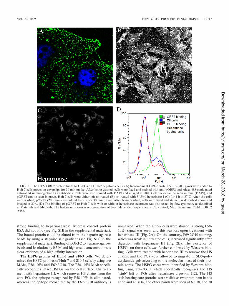
The HEV ORF2 protein binds to HSPGs on Huh-7 cells.The 56-kDa HEV ORF2 VLPs were found to attach as punc-tate structures all over and just outside the cellular membraneof Huh-7 cells (Fig. 1A). The punctate staining pattern forpORF2 suggested binding to some ubiquitous molecules oncells, such as HSPGs, which could also be shed into the me-dium. To check if pORF2 bound to Huh-7 cells throughHSPGs, cells were treated with heparinase I, an enzyme whichdegrades heparin and highly sulfated domains in heparin sul-fate (36). This reduced the binding of pORF2 to nearly controllevels in cells (Fig. 1B and C). This result is consistent withHSPGs being attachment receptors for pORF2 on Huh-7 cells.This was also evident from a flow cytometric analysis ofpORF2 binding to control and heparinase-treated Huh-7 cells(Fig. 1D). We also tested an aa 458 to 607 ORF2 polypeptideexpressed in bacteria for its binding to Huh-7 cells. This ORF2polypeptide was reported to be recognized by anti-HEV neu-tralizing antibodies, and the critical receptor binding domainof HEV was proposed to lie within this region (71). We ob-served no binding of this polypeptide to Huh-7 cells (data notshown), suggesting that binding of pORF2 to HSPGs is eithermediated by determinants that are outside the aa 458 to 607region or by positively charged residues that are brought to-gether in the VLP structure to create an HSPG binding motif.
Since the pORF2 VLP showed HSPG binding on cells, theheparin-binding activity of pORF2 was directly examined byincubating the protein with heparin-agarose beads (see Fig.S1B and C in the supplemental material). pORF2 showed
12716 KALIA ET AL. J. VIROL.
on March 28, 2018 by guest
http://jvi.asm.org/
Dow
nloaded from
strong binding to heparin-agarose, whereas control proteinBSA did not bind (see Fig. S1B in the supplemental material).The bound protein could be eluted from the heparin-agarosebeads by using a stepwise salt gradient (see Fig. S1C in thesupplemental material). Binding of pORF2 to heparin-agarosebeads and its elution by 0.3 M and higher salt concentrations isclear evidence of a high-affinity interaction.
The HSPG profiles of Huh-7 and S10-3 cells. We deter-mined the HSPG profiles of Huh-7 and S10-3 cells by using twoMAbs, F58-10E4 and F69-3G10. The F58-10E4 MAb specifi-cally recognizes intact HSPGs on the cell surface. On treat-ment with heparinase III, which removes HS chains from thecore PG, the epitope recognized by F58-10E4 is eliminated,whereas the epitope recognized by the F69-3G10 antibody is
unmasked. When the Huh-7 cells were stained, a strong F58-10E4 signal was seen, and this was lost upon treatment withheparinase III (Fig. 2A). On the contrary, F69-3G10 staining,which was weak in untreated cells, increased significantly afterdigestion with heparinase III (Fig. 2B). The existence ofHSPGs on these cells was further confirmed by Western blot-ting. Cells were treated with heparinase III to remove the HSchains, and the PGs were allowed to migrate in SDS-poly-acrylamide gels according to the molecular mass of their pro-tein cores. The HSPG cores were identified by Western blot-ting using F69-3G10, which specifically recognizes the HS“stub” left on PGs after heparinase digestion (12). The HSstub-bearing core proteins were visible as two prominent bandsat 85 and 48 kDa, and other bands were seen at 60, 38, and 30
FIG. 1. The HEV ORF2 protein binds to HSPGs on Huh-7 hepatoma cells. (A) Recombinant ORF2 protein VLPs (20 �g/ml) were added toHuh-7 cells grown on coverslips for 30 min on ice. After being washed, cells were fixed and stained with anti-pORF2 and Alexa 488-conjugatedanti-rabbit immunoglobulin G antibodies. Cells were also stained with DAPI and imaged at 60�. Cell nuclei can be seen in blue (DAPI), andpORF2 can be seen in green. Huh-7 cells were either left untreated (B) or treated with 5 U/ml heparinase I (C) for 1 h at 37°C. After the cellswere washed, pORF2 (20 �g/ml) was added to cells for 30 min on ice. After being washed, cells were fixed and stained as described above andimaged at 20�. (D) The binding of pORF2 to Huh-7 cells with or without heparinase treatment was also tested by flow cytometry as describedin Materials and Methods. The histogram shown is representative of two independent experiments. Ctl, control; Max, maximum; FL1-H, ORF2A488.
VOL. 83, 2009 HEV ORF2 PROTEIN BINDS HSPGs 12717
on March 28, 2018 by guest
http://jvi.asm.org/
Dow
nloaded from
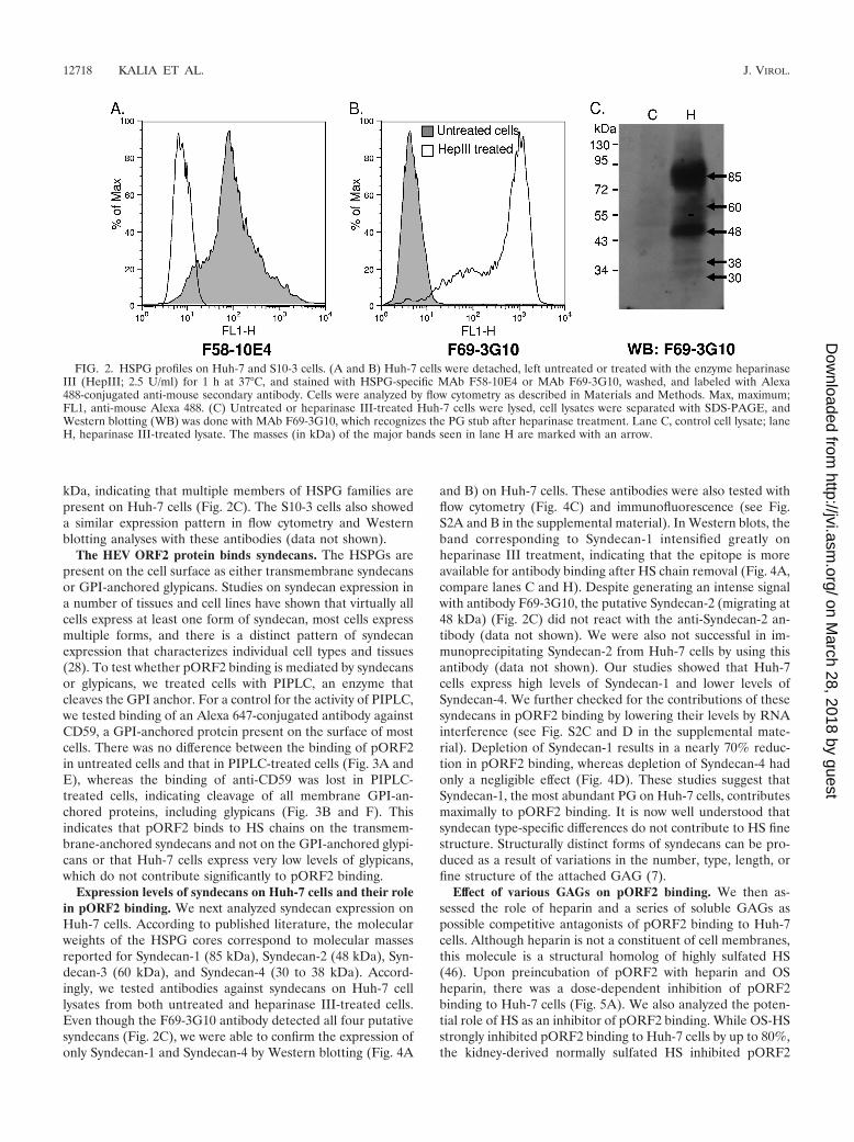
kDa, indicating that multiple members of HSPG families arepresent on Huh-7 cells (Fig. 2C). The S10-3 cells also showeda similar expression pattern in flow cytometry and Westernblotting analyses with these antibodies (data not shown).
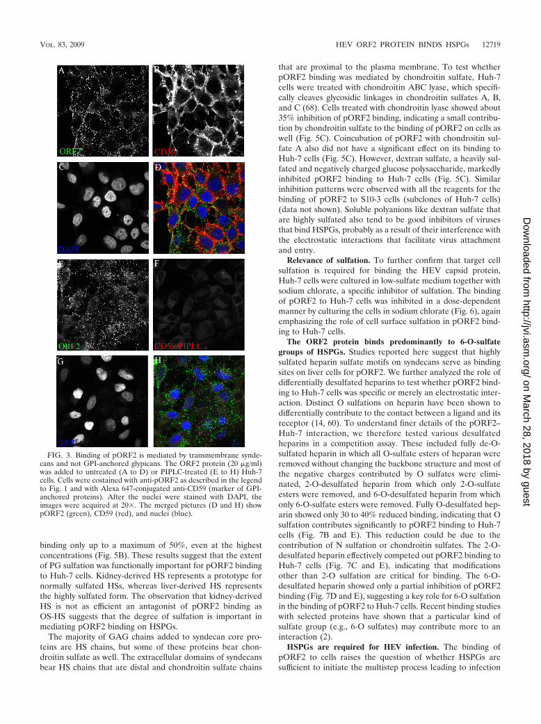
The HEV ORF2 protein binds syndecans. The HSPGs arepresent on the cell surface as either transmembrane syndecansor GPI-anchored glypicans. Studies on syndecan expression ina number of tissues and cell lines have shown that virtually allcells express at least one form of syndecan, most cells expressmultiple forms, and there is a distinct pattern of syndecanexpression that characterizes individual cell types and tissues(28). To test whether pORF2 binding is mediated by syndecansor glypicans, we treated cells with PIPLC, an enzyme thatcleaves the GPI anchor. For a control for the activity of PIPLC,we tested binding of an Alexa 647-conjugated antibody againstCD59, a GPI-anchored protein present on the surface of mostcells. There was no difference between the binding of pORF2in untreated cells and that in PIPLC-treated cells (Fig. 3A andE), whereas the binding of anti-CD59 was lost in PIPLC-treated cells, indicating cleavage of all membrane GPI-an-chored proteins, including glypicans (Fig. 3B and F). Thisindicates that pORF2 binds to HS chains on the transmem-brane-anchored syndecans and not on the GPI-anchored glypi-cans or that Huh-7 cells express very low levels of glypicans,which do not contribute significantly to pORF2 binding.
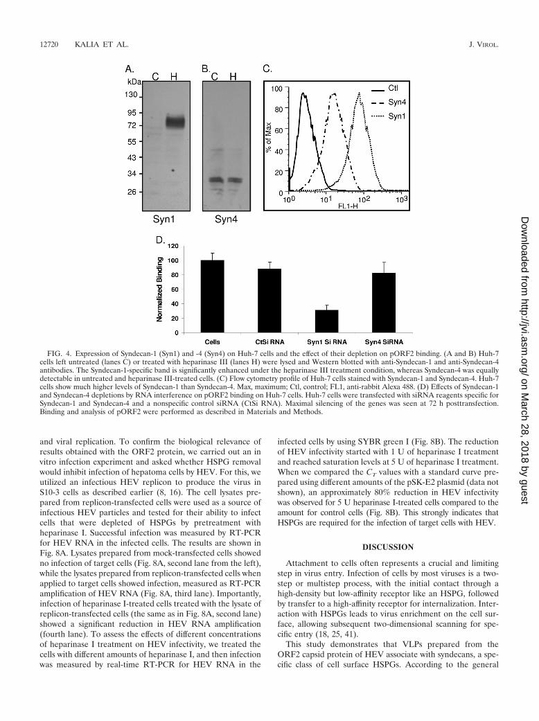
Expression levels of syndecans on Huh-7 cells and their rolein pORF2 binding. We next analyzed syndecan expression onHuh-7 cells. According to published literature, the molecularweights of the HSPG cores correspond to molecular massesreported for Syndecan-1 (85 kDa), Syndecan-2 (48 kDa), Syn-decan-3 (60 kDa), and Syndecan-4 (30 to 38 kDa). Accord-ingly, we tested antibodies against syndecans on Huh-7 celllysates from both untreated and heparinase III-treated cells.Even though the F69-3G10 antibody detected all four putativesyndecans (Fig. 2C), we were able to confirm the expression ofonly Syndecan-1 and Syndecan-4 by Western blotting (Fig. 4A
and B) on Huh-7 cells. These antibodies were also tested withflow cytometry (Fig. 4C) and immunofluorescence (see Fig.S2A and B in the supplemental material). In Western blots, theband corresponding to Syndecan-1 intensified greatly onheparinase III treatment, indicating that the epitope is moreavailable for antibody binding after HS chain removal (Fig. 4A,compare lanes C and H). Despite generating an intense signalwith antibody F69-3G10, the putative Syndecan-2 (migrating at48 kDa) (Fig. 2C) did not react with the anti-Syndecan-2 an-tibody (data not shown). We were also not successful in im-munoprecipitating Syndecan-2 from Huh-7 cells by using thisantibody (data not shown). Our studies showed that Huh-7cells express high levels of Syndecan-1 and lower levels ofSyndecan-4. We further checked for the contributions of thesesyndecans in pORF2 binding by lowering their levels by RNAinterference (see Fig. S2C and D in the supplemental mate-rial). Depletion of Syndecan-1 results in a nearly 70% reduc-tion in pORF2 binding, whereas depletion of Syndecan-4 hadonly a negligible effect (Fig. 4D). These studies suggest thatSyndecan-1, the most abundant PG on Huh-7 cells, contributesmaximally to pORF2 binding. It is now well understood thatsyndecan type-specific differences do not contribute to HS finestructure. Structurally distinct forms of syndecans can be pro-duced as a result of variations in the number, type, length, orfine structure of the attached GAG (7).
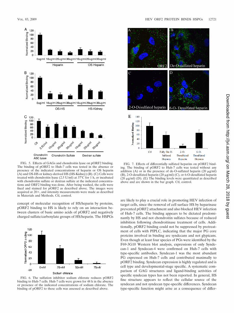
Effect of various GAGs on pORF2 binding. We then as-sessed the role of heparin and a series of soluble GAGs aspossible competitive antagonists of pORF2 binding to Huh-7cells. Although heparin is not a constituent of cell membranes,this molecule is a structural homolog of highly sulfated HS(46). Upon preincubation of pORF2 with heparin and OSheparin, there was a dose-dependent inhibition of pORF2binding to Huh-7 cells (Fig. 5A). We also analyzed the poten-tial role of HS as an inhibitor of pORF2 binding. While OS-HSstrongly inhibited pORF2 binding to Huh-7 cells by up to 80%,the kidney-derived normally sulfated HS inhibited pORF2
FIG. 2. HSPG profiles on Huh-7 and S10-3 cells. (A and B) Huh-7 cells were detached, left untreated or treated with the enzyme heparinaseIII (HepIII; 2.5 U/ml) for 1 h at 37°C, and stained with HSPG-specific MAb F58-10E4 or MAb F69-3G10, washed, and labeled with Alexa488-conjugated anti-mouse secondary antibody. Cells were analyzed by flow cytometry as described in Materials and Methods. Max, maximum;FL1, anti-mouse Alexa 488. (C) Untreated or heparinase III-treated Huh-7 cells were lysed, cell lysates were separated with SDS-PAGE, andWestern blotting (WB) was done with MAb F69-3G10, which recognizes the PG stub after heparinase treatment. Lane C, control cell lysate; laneH, heparinase III-treated lysate. The masses (in kDa) of the major bands seen in lane H are marked with an arrow.
12718 KALIA ET AL. J. VIROL.
on March 28, 2018 by guest
http://jvi.asm.org/
Dow
nloaded from
binding only up to a maximum of 50%, even at the highestconcentrations (Fig. 5B). These results suggest that the extentof PG sulfation was functionally important for pORF2 bindingto Huh-7 cells. Kidney-derived HS represents a prototype fornormally sulfated HSs, whereas liver-derived HS representsthe highly sulfated form. The observation that kidney-derivedHS is not as efficient an antagonist of pORF2 binding asOS-HS suggests that the degree of sulfation is important inmediating pORF2 binding on HSPGs.
The majority of GAG chains added to syndecan core pro-teins are HS chains, but some of these proteins bear chon-droitin sulfate as well. The extracellular domains of syndecansbear HS chains that are distal and chondroitin sulfate chains
that are proximal to the plasma membrane. To test whetherpORF2 binding was mediated by chondroitin sulfate, Huh-7cells were treated with chondroitin ABC lyase, which specifi-cally cleaves glycosidic linkages in chondroitin sulfates A, B,and C (68). Cells treated with chondroitin lyase showed about35% inhibition of pORF2 binding, indicating a small contribu-tion by chondroitin sulfate to the binding of pORF2 on cells aswell (Fig. 5C). Coincubation of pORF2 with chondroitin sul-fate A also did not have a significant effect on its binding toHuh-7 cells (Fig. 5C). However, dextran sulfate, a heavily sul-fated and negatively charged glucose polysaccharide, markedlyinhibited pORF2 binding to Huh-7 cells (Fig. 5C). Similarinhibition patterns were observed with all the reagents for thebinding of pORF2 to S10-3 cells (subclones of Huh-7 cells)(data not shown). Soluble polyanions like dextran sulfate thatare highly sulfated also tend to be good inhibitors of virusesthat bind HSPGs, probably as a result of their interference withthe electrostatic interactions that facilitate virus attachmentand entry.
Relevance of sulfation. To further confirm that target cellsulfation is required for binding the HEV capsid protein,Huh-7 cells were cultured in low-sulfate medium together withsodium chlorate, a specific inhibitor of sulfation. The bindingof pORF2 to Huh-7 cells was inhibited in a dose-dependentmanner by culturing the cells in sodium chlorate (Fig. 6), againemphasizing the role of cell surface sulfation in pORF2 bind-ing to Huh-7 cells.
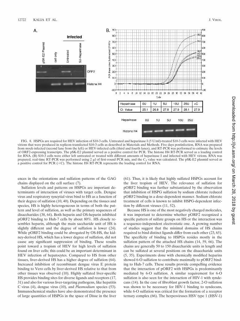
The ORF2 protein binds predominantly to 6-O-sulfategroups of HSPGs. Studies reported here suggest that highlysulfated heparin sulfate motifs on syndecans serve as bindingsites on liver cells for pORF2. We further analyzed the role ofdifferentially desulfated heparins to test whether pORF2 bind-ing to Huh-7 cells was specific or merely an electrostatic inter-action. Distinct O sulfations on heparin have been shown todifferentially contribute to the contact between a ligand and itsreceptor (14, 60). To understand finer details of the pORF2–Huh-7 interaction, we therefore tested various desulfatedheparins in a competition assay. These included fully de-O-sulfated heparin in which all O-sulfate esters of heparan wereremoved without changing the backbone structure and most ofthe negative charges contributed by O sulfates were elimi-nated, 2-O-desulfated heparin from which only 2-O-sulfateesters were removed, and 6-O-desulfated heparin from whichonly 6-O-sulfate esters were removed. Fully O-desulfated hep-arin showed only 30 to 40% reduced binding, indicating that Osulfation contributes significantly to pORF2 binding to Huh-7cells (Fig. 7B and E). This reduction could be due to thecontribution of N sulfation or chondroitin sulfates. The 2-O-desulfated heparin effectively competed out pORF2 binding toHuh-7 cells (Fig. 7C and E), indicating that modificationsother than 2-O sulfation are critical for binding. The 6-O-desulfated heparin showed only a partial inhibition of pORF2binding (Fig. 7D and E), suggesting a key role for 6-O sulfationin the binding of pORF2 to Huh-7 cells. Recent binding studieswith selected proteins have shown that a particular kind ofsulfate group (e.g., 6-O sulfates) may contribute more to aninteraction (2).
HSPGs are required for HEV infection. The binding ofpORF2 to cells raises the question of whether HSPGs aresufficient to initiate the multistep process leading to infection
FIG. 3. Binding of pORF2 is mediated by transmembrane synde-cans and not GPI-anchored glypicans. The ORF2 protein (20 �g/ml)was added to untreated (A to D) or PIPLC-treated (E to H) Huh-7cells. Cells were costained with anti-pORF2 as described in the legendto Fig. 1 and with Alexa 647-conjugated anti-CD59 (marker of GPI-anchored proteins). After the nuclei were stained with DAPI, theimages were acquired at 20�. The merged pictures (D and H) showpORF2 (green), CD59 (red), and nuclei (blue).
VOL. 83, 2009 HEV ORF2 PROTEIN BINDS HSPGs 12719
on March 28, 2018 by guest
http://jvi.asm.org/
Dow
nloaded from
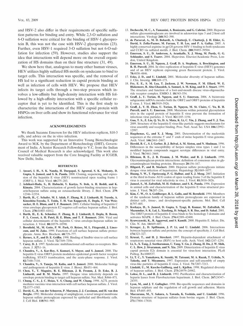
and viral replication. To confirm the biological relevance ofresults obtained with the ORF2 protein, we carried out an invitro infection experiment and asked whether HSPG removalwould inhibit infection of hepatoma cells by HEV. For this, weutilized an infectious HEV replicon to produce the virus inS10-3 cells as described earlier (8, 16). The cell lysates pre-pared from replicon-transfected cells were used as a source ofinfectious HEV particles and tested for their ability to infectcells that were depleted of HSPGs by pretreatment withheparinase I. Successful infection was measured by RT-PCRfor HEV RNA in the infected cells. The results are shown inFig. 8A. Lysates prepared from mock-transfected cells showedno infection of target cells (Fig. 8A, second lane from the left),while the lysates prepared from replicon-transfected cells whenapplied to target cells showed infection, measured as RT-PCRamplification of HEV RNA (Fig. 8A, third lane). Importantly,infection of heparinase I-treated cells treated with the lysate ofreplicon-transfected cells (the same as in Fig. 8A, second lane)showed a significant reduction in HEV RNA amplification(fourth lane). To assess the effects of different concentrationsof heparinase I treatment on HEV infectivity, we treated thecells with different amounts of heparinase I, and then infectionwas measured by real-time RT-PCR for HEV RNA in the
infected cells by using SYBR green I (Fig. 8B). The reductionof HEV infectivity started with 1 U of heparinase I treatmentand reached saturation levels at 5 U of heparinase I treatment.When we compared the CT values with a standard curve pre-pared using different amounts of the pSK-E2 plasmid (data notshown), an approximately 80% reduction in HEV infectivitywas observed for 5 U heparinase I-treated cells compared to theamount for control cells (Fig. 8B). This strongly indicates thatHSPGs are required for the infection of target cells with HEV.
DISCUSSION
Attachment to cells often represents a crucial and limitingstep in virus entry. Infection of cells by most viruses is a two-step or multistep process, with the initial contact through ahigh-density but low-affinity receptor like an HSPG, followedby transfer to a high-affinity receptor for internalization. Inter-action with HSPGs leads to virus enrichment on the cell sur-face, allowing subsequent two-dimensional scanning for spe-cific entry (18, 25, 41).
This study demonstrates that VLPs prepared from theORF2 capsid protein of HEV associate with syndecans, a spe-cific class of cell surface HSPGs. According to the general
FIG. 4. Expression of Syndecan-1 (Syn1) and -4 (Syn4) on Huh-7 cells and the effect of their depletion on pORF2 binding. (A and B) Huh-7cells left untreated (lanes C) or treated with heparinase III (lanes H) were lysed and Western blotted with anti-Syndecan-1 and anti-Syndecan-4antibodies. The Syndecan-1-specific band is significantly enhanced under the heparinase III treatment condition, whereas Syndecan-4 was equallydetectable in untreated and heparinase III-treated cells. (C) Flow cytometry profile of Huh-7 cells stained with Syndecan-1 and Syndecan-4. Huh-7cells show much higher levels of Syndecan-1 than Syndecan-4. Max, maximum; Ctl, control; FL1, anti-rabbit Alexa 488. (D) Effects of Syndecan-1and Syndecan-4 depletions by RNA interference on pORF2 binding on Huh-7 cells. Huh-7 cells were transfected with siRNA reagents specific forSyndecan-1 and Syndecan-4 and a nonspecific control siRNA (CtSi RNA). Maximal silencing of the genes was seen at 72 h posttransfection.Binding and analysis of pORF2 were performed as described in Materials and Methods.
12720 KALIA ET AL. J. VIROL.
on March 28, 2018 by guest
http://jvi.asm.org/
Dow
nloaded from
concept of molecular recognition of HS/heparin by proteins,pORF2 binding to HS is likely to rely on an interaction be-tween clusters of basic amino acids of pORF2 and negativelycharged sulfate/carboxylate groups of HS/heparin. The HSPGs
are likely to play a crucial role in promoting HEV infection oftarget cells, since the removal of cell surface HS by heparinaseprevented pORF2 attachment and also blocked HEV infectionof Huh-7 cells. The binding appears to be dictated predomi-nantly by HS and not chondroitin sulfates because of reducedinhibition following chondroitinase treatment of cells. Addi-tionally, pORF2 binding could not be suppressed by pretreat-ment of cells with PIPLC, indicating that the major PG coreproteins involved in binding are syndecans and not glypicans.Even though at least four species of PGs were identified by theF69-3G10 Western blot analysis, expressions of only Synde-can-1 and Syndecan-4 were confirmed on Huh-7 cells withtype-specific antibodies. Syndecan-1 was the most abundantPG expressed on Huh-7 cells and contributed maximally topORF2 binding. Syndecan expression is highly regulated and iscell type and developmental-stage specific. A systematic com-parison of GAG structures and ligand-binding activities ofspecific syndecan types has not been reported. In general, HSfine structure appears to reflect the cellular source of thesyndecan and not syndecan type-specific differences. Syndecantype-specific function might arise as a consequence of differ-
FIG. 5. Effects of GAGs and chondroitin lyase on pORF2 binding.The binding of pORF2 to Huh-7 cells was tested in the absence orpresence of the indicated concentrations of heparin or OS heparin(A) and OS-HS or kidney-derived HS (HS-Kidney) (B). (C) Cells weretreated with chondroitin lyase (2.5 U/ml) at 37°C for 1 h, or incubatedwith chondroitin sulfate or dextran sulfate at the indicated concentra-tions and ORF2 binding was done. After being washed, the cells werefixed and stained for pORF2 as described above. The images wereacquired at 20�, and intensity measurements were made as describedin Materials and Methods. Ctl, control.
FIG. 6. The sulfation inhibitor sodium chlorate reduces pORF2binding to Huh-7 cells. Huh-7 cells were grown for 48 h in the absenceor presence of the indicated concentrations of sodium chlorate. Thebinding of pORF2 to these cells was assessed as described above.
FIG. 7. Effects of differentially sulfated heparins on pORF2 bind-ing. The binding of pORF2 to Huh-7 cells was tested without anyaddition (A) or in the presence of de-O-sulfated heparin (20 �g/ml)(B), 2-O-desulfated heparin (20 �g/ml) (C), or 6-O-desulfated heparin(20 �g/ml) (D). (E) The binding levels were quantitated as describedabove and are shown in the bar graph. Ctl, control.
VOL. 83, 2009 HEV ORF2 PROTEIN BINDS HSPGs 12721
on March 28, 2018 by guest
http://jvi.asm.org/
Dow
nloaded from
ences in the orientations and sulfation patterns of the GAGchains displayed on the cell surface (7).
Sulfation levels and patterns on HSPGs are important de-terminants of interaction of viruses with target cells. Denguevirus and respiratory syncytial virus bind to HS as a function oftheir degree of sulfation (10, 40). Depending on the tissues andspecies, HS is highly heterogeneous in terms of both the pat-tern and level of sulfation, as well as the primary sequences ofdisaccharides (38, 64). Both heparin and OS-heparin inhibitedpORF2 binding to Huh-7 cells by about 80%. HS closely re-sembles heparin, although the main saccharide unit of HS isslightly different and the degree of sulfation is lower (24).While pORF2 binding could be abrogated by OS-HS, the kid-ney-derived HS, which has a lower degree of sulfation, did notcause any significant suppression of binding. These resultspoint toward a tropism of HEV for high levels of sulfationfound on liver cells; this could be an important determinant inHEV infection of hepatocytes. Compared to HS from othertissues, liver-derived HS has a higher degree of sulfation (64).Increased inhibition of recombinant dengue virus E proteinbinding to Vero cells by liver-derived HS relative to that fromother tissues was observed (10). Highly sulfated liver-specificHS provides binding sites for diverse ligands and receptors (17,31) and also for various liver-targeting pathogens, like hepatitisC virus (4), dengue virus (10), and Plasmodium species (53).Immunochemical studies have also demonstrated the presenceof large quantities of HSPGs in the space of Disse in the liver
(61). Thus, it is likely that highly sulfated HSPGs account forthe liver tropism of HEV. The relevance of sulfation forpORF2 binding was further substantiated by the observationthat inhibition of HSPG sulfation by sodium chlorate reducedpORF2 binding in a dose-dependent manner. Sodium chloratetreatment of cells is known to inhibit HSPG-dependent infec-tion by different viruses (11, 52).
Because HS is one of the most negatively charged molecules,it was important to determine whether pORF2 recognized aspecific pattern of sulfate groups on HS or the interaction wasa sequence-independent electrostatic one. A growing numberof studies suggest that the minimal domains of HS chainsrequired to bind distinct ligands differ from each other (23, 65).The specificity of binding to HSPGs resides mostly in thesulfation pattern of the attached HS chains (14, 59, 66). Thechains are generally 50 to 150 disaccharide units in length andcan be sulfated at several positions on the disaccharide units(5, 35). Experiments done with chemically modified heparinsshowed 6-O sulfation to contribute maximally to pORF2 bind-ing to Huh-7 cells. These results provide compelling evidencethat the interaction of pORF2 with HSPGs is predominantlymediated by 6-O sulfation. A similar requirement for 6-Osulfation is also seen for the interaction of HIV-1 with synde-cans (14). In the case of fibroblast growth factor, 2-O sulfationwas shown to be necessary for HIV-1 binding to syndecans,while 6-O sulfation was critical for the formation of a receptorternary complex (66). The herpesviruses HSV type 1 (HSV-1)
FIG. 8. HSPGs are required for HEV infection of S10-3 cells. Untreated and heparinase I (5 U/ml)-treated S10-3 cells were infected with HEVvirions that were produced in replicon-transfected S10-3 cells as described in Materials and Methods. Five days postinfection, RNA was preparedfrom mock-infected (second lane from the left) or HEV-infected cells (third and fourth lanes), and RT-PCR was performed to estimate the levelsof ORF2-expressing transcripts. The pSK-E2 plasmid served as a positive control for PCR. The histone H4 RT-PCR served as a loading controlfor RNA. (B) S10-3 cells were either left untreated or treated with different amounts of heparinase I and infected with HEV virions. RNA wasprepared, real-time RT-PCR was performed using 2 �l of first-round PCR mix, and the CT value was calculated. The pSK-E2 plasmid served asa positive control for PCR (�C). The histone H4 RT-PCR represents the loading control for RNA.
12722 KALIA ET AL. J. VIROL.
on March 28, 2018 by guest
http://jvi.asm.org/
Dow
nloaded from
and HSV-2 also differ in their requirements of specific sulfa-tion patterns for binding and entry. While 2,3-O sulfation and6-O sulfation were critical for the binding of HSV-1 glycopro-tein B, this was not the case with HSV-2 glycoproteins (23).Further, even HSV-1 required 3-O sulfation but not 6-O-sul-fation for infection (59). However, studies also support theidea that interactions will depend more on the overall organi-zation of HS domains than on their fine structure (31, 49).
We show here that, analogous to many other human viruses,HEV utilizes highly sulfated HS motifs on syndecans to bind totarget cells. This interaction was specific, and the removal ofHS led to a significant reduction in capsid protein binding aswell as infection of cells with HEV. We propose that HEVinfects its target cells through a two-step process which in-volves a low-affinity but high-density interaction with HS fol-lowed by a high-affinity interaction with a specific cellular re-ceptor that is yet to be identified. This is the first study tocharacterize the interactions of the HEV capsid protein withHSPGs on liver cells and show its functional relevance for viralinfection.
ACKNOWLEDGMENTS
We thank Suzanne Emerson for the HEV infectious replicon, S10-3cells, and advice on the in vitro infection.
This work was supported by an Innovative Young BiotechnologistAward to M.K. by the Department of Biotechnology (DBT), Govern-ment of India. A Senior Research Fellowship to V.C. from the IndianCouncil of Medical Research is also acknowledged. This work alsoreceived valuable support from the Core Imaging Facility at ICGEB,New Delhi, India.
REFERENCES
1. Ansari, I. H., S. K. Nanda, H. Durgapal, S. Agrawal, S. K. Mohanty, D.Gupta, S. Jameel, and S. K. Panda. 2000. Cloning, sequencing, and expres-sion of the hepatitis E virus (HEV) nonstructural open reading frame 1(ORF1). J. Med. Virol. 60:275–283.
2. Ashikari-Hada, S., H. Habuchi, Y. Kariya, N. Itoh, A. H. Reddi, and K.Kimata. 2004. Characterization of growth factor-binding structures in hep-arin/heparan sulfate using an octasaccharide library. J. Biol. Chem. 279:12346–12354.
3. Barth, H., C. Schafer, M. I. Adah, F. Zhang, R. J. Linhardt, H. Toyoda, A.Kinoshita-Toyoda, T. Toida, T. H. Van Kuppevelt, E. Depla, F. Von Weiz-sacker, H. E. Blum, and T. F. Baumert. 2003. Cellular binding of hepatitis Cvirus envelope glycoprotein E2 requires cell surface heparan sulfate. J. Biol.Chem. 278:41003–41012.
4. Barth, H., E. K. Schnober, F. Zhang, R. J. Linhardt, E. Depla, B. Boson,F. L. Cosset, A. H. Patel, H. E. Blum, and T. F. Baumert. 2006. Viral andcellular determinants of the hepatitis C virus envelope-heparan sulfate in-teraction. J. Virol. 80:10579–10590.
5. Bernfield, M., M. Gotte, P. W. Park, O. Reizes, M. L. Fitzgerald, J. Lince-cum, and M. Zako. 1999. Functions of cell surface heparan sulfate proteo-glycans. Annu. Rev. Biochem. 68:729–777.
6. Byrnes, A. P., and D. E. Griffin. 1998. Binding of Sindbis virus to cell surfaceheparan sulfate. J. Virol. 72:7349–7356.
7. Carey, D. J. 1997. Syndecans: multifunctional cell-surface co-receptors. Bio-chem. J. 327:1–16.
8. Chandra, V., A. Kar-Roy, S. Kumari, S. Mayor, and S. Jameel. 2008. Thehepatitis E virus ORF3 protein modulates epidermal growth factor receptortrafficking, STAT3 translocation, and the acute-phase response. J. Virol.82:7100–7110.
9. Chandra, V., S. Taneja, M. Kalia, and S. Jameel. 2008. Molecular biologyand pathogenesis of hepatitis E virus. J. Biosci. 33:451–464.
10. Chen, Y., T. Maguire, R. E. Hileman, J. R. Fromm, J. D. Esko, R. J.Linhardt, and R. M. Marks. 1997. Dengue virus infectivity depends onenvelope protein binding to target cell heparan sulfate. Nat. Med. 3:866–871.
11. Chung, C. S., J. C. Hsiao, Y. S. Chang, and W. Chang. 1998. A27L proteinmediates vaccinia virus interaction with cell surface heparan sulfate. J. Virol.72:1577–1585.
12. David, G., B. van der Schueren, P. Marynen, J. J. Cassiman, and H. van denBerghe. 1992. Molecular cloning of amphiglycan, a novel integral membraneheparan sulfate proteoglycan expressed by epithelial and fibroblastic cells.J. Cell Biol. 118:961–969.
13. Dechecchi, M. C., A. Tamanini, A. Bonizzato, and G. Cabrini. 2000. Heparansulfate glycosaminoglycans are involved in adenovirus type 5 and 2-host cellinteractions. Virology 268:382–390.
14. de Parseval, A., M. D. Bobardt, A. Chatterji, U. Chatterji, J. H. Elder, G.David, S. Zolla-Pazner, M. Farzan, T. H. Lee, and P. A. Gallay. 2005. Ahighly conserved arginine in gp120 governs HIV-1 binding to both syndecansand CCR5 via sulfated motifs. J. Biol. Chem. 280:39493–39504.
15. Emerson, S. U., D. Anderson, A. Arankalle, X. J. Meng, M. Purdy, G. G.Schlauder, and S. Tsarev. 2004. Hepevirus. Elsevier/Academic Press, Lon-don, United Kingdom.
16. Emerson, S. U., H. Nguyen, J. Graff, D. A. Stephany, A. Brockington, andR. H. Purcell. 2004. In vitro replication of hepatitis E virus (HEV) genomesand of an HEV replicon expressing green fluorescent protein. J. Virol.78:4838–4846.
17. Esko, J. D., and U. Lindahl. 2001. Molecular diversity of heparan sulfate.J. Clin. Investig. 108:169–173.
18. Fry, E. E., S. M. Lea, T. Jackson, J. W. Newman, F. M. Ellard, W. E.Blakemore, R. Abu-Ghazaleh, A. Samuel, A. M. King, and D. I. Stuart. 1999.The structure and function of a foot-and-mouth disease virus-oligosaccha-ride receptor complex. EMBO J. 18:543–554.
19. Graff, J., U. Torian, H. Nguyen, and S. U. Emerson. 2006. A bicistronicsubgenomic mRNA encodes both the ORF2 and ORF3 proteins of hepatitisE virus. J. Virol. 80:5919–5926.
20. Graff, J., Y. H. Zhou, U. Torian, H. Nguyen, M. St. Claire, C. Yu, R. H.Purcell, and S. U. Emerson. 2008. Mutations within potential glycosylationsites in the capsid protein of hepatitis E virus prevent the formation ofinfectious virus particles. J. Virol. 82:1185–1194.
21. Guu, T. S., Z. Liu, Q. Ye, D. A. Mata, K. Li, C. Yin, J. Zhang, and Y. J. Tao.2009. Structure of the hepatitis E virus-like particle suggests mechanisms forvirus assembly and receptor binding. Proc. Natl. Acad. Sci. USA 106:12992–12997.
22. Haqshenas, G., and X. J. Meng. 2001. Determination of the nucleotidesequences at the extreme 5� and 3� ends of swine hepatitis E virus genome.Arch. Virol. 146:2461–2467.
23. Herold, B. C., S. I. Gerber, B. J. Belval, A. M. Siston, and N. Shulman. 1996.Differences in the susceptibility of herpes simplex virus types 1 and 2 tomodified heparin compounds suggest serotype differences in viral entry.J. Virol. 70:3461–3469.
24. Hileman, R. E., J. R. Fromm, J. M. Weiler, and R. J. Linhardt. 1998.Glycosaminoglycan-protein interactions: definition of consensus sites in gly-cosaminoglycan binding proteins. Bioessays 20:156–167.
25. Hilgard, P., and R. Stockert. 2000. Heparan sulfate proteoglycans initiatedengue virus infection of hepatocytes. Hepatology 32:1069–1077.
26. Huang, Y. W., T. Opriessnig, P. G. Halbur, and X. J. Meng. 2007. Initiationat the third in-frame AUG codon of open reading frame 3 of the hepatitis Evirus is essential for viral infectivity in vivo. J. Virol. 81:3018–3026.
27. Jameel, S., M. Zafrullah, M. H. Ozdener, and S. K. Panda. 1996. Expressionin animal cells and characterization of the hepatitis E virus structural pro-teins. J. Virol. 70:207–216.
28. Kim, C. W., O. A. Goldberger, R. L. Gallo, and M. Bernfield. 1994. Membersof the syndecan family of heparan sulfate proteoglycans are expressed indistinct cell-, tissue-, and development-specific patterns. Mol. Biol. Cell5:797–805.
29. Korkaya, H., S. Jameel, D. Gupta, S. Tyagi, R. Kumar, M. Zafrullah, M.Mazumdar, S. K. Lal, L. Xiaofang, D. Sehgal, S. R. Das, and D. Sahal. 2001.The ORF3 protein of hepatitis E virus binds to Src homology 3 domains andactivates MAPK. J. Biol. Chem. 276:42389–42400.
30. Krawczynski, K., R. Aggarwal, and S. Kamili. 2000. Hepatitis E. Infect. Dis.Clin. N. Am. 14:669–687.
31. Kreuger, J., D. Spillmann, J. P. Li, and U. Lindahl. 2006. Interactionsbetween heparan sulfate and proteins: the concept of specificity. J. Cell Biol.174:323–327.
32. Krusat, T., and H. J. Streckert. 1997. Heparin-dependent attachment ofrespiratory syncytial virus (RSV) to host cells. Arch. Virol. 142:1247–1254.
33. Li, S., X. Tang, J. Seetharaman, C. Yang, Y. Gu, J. Zhang, H. Du, J. W. Shih,C. L. Hew, J. Sivaraman, and N. Xia. 2009. Dimerization of hepatitis E viruscapsid protein E2s domain is essential for virus-host interaction. PLoSPathog. 5:e1000537.
34. Li, T. C., Y. Yamakawa, K. Suzuki, M. Tatsumi, M. A. Razak, T. Uchida, N.Takeda, and T. Miyamura. 1997. Expression and self-assembly of emptyvirus-like particles of hepatitis E virus. J. Virol. 71:7207–7213.
35. Lindahl, U., M. Kusche-Gullberg, and L. Kjellen. 1998. Regulated diversityof heparan sulfate. J. Biol. Chem. 273:24979–24982.
36. Lohse, D. L., and R. J. Linhardt. 1992. Purification and characterization ofheparin lyases from Flavobacterium heparinum. J. Biol. Chem. 267:24347–24355.
37. Lyon, M., and J. T. Gallagher. 1998. Bio-specific sequences and domains inheparan sulphate and the regulation of cell growth and adhesion. MatrixBiol. 17:485–493.
38. Maccarana, M., Y. Sakura, A. Tawada, K. Yoshida, and U. Lindahl. 1996.Domain structure of heparan sulfates from bovine organs. J. Biol. Chem.271:17804–17810.
VOL. 83, 2009 HEV ORF2 PROTEIN BINDS HSPGs 12723
on March 28, 2018 by guest
http://jvi.asm.org/
Dow
nloaded from
39. Mandl, C. W., H. Kroschewski, S. L. Allison, R. Kofler, H. Holzmann, T.Meixner, and F. X. Heinz. 2001. Adaptation of tick-borne encephalitis virusto BHK-21 cells results in the formation of multiple heparan sulfate bindingsites in the envelope protein and attenuation in vivo. J. Virol. 75:5627–5637.
40. Martínez, I., and J. A. Melero. 2000. Binding of human respiratory syncytialvirus to cells: implication of sulfated cell surface proteoglycans. J. Gen. Virol.81:2715–2722.
41. McClain, D. S., and A. O. Fuller. 1994. Cell-specific kinetics and efficiency ofherpes simplex virus type 1 entry are determined by two distinct phases ofattachment. Virology 198:690–702.
42. Meng, X. J. 2009. Hepatitis E virus: animal reservoirs and zoonotic risk. Vet.Microbiol. [Epub ahead of print.] doi:10.1016/j.vetmic.2009.03.017.
43. Meng, X. J., R. H. Purcell, P. G. Halbur, J. R. Lehman, D. M. Webb, T. S.Tsareva, J. S. Haynes, B. J. Thacker, and S. U. Emerson. 1997. A novel virusin swine is closely related to the human hepatitis E virus. Proc. Natl. Acad.Sci. USA 94:9860–9865.
44. Moin, S. M., M. Panteva, and S. Jameel. 2007. The hepatitis E virus Orf3protein protects cells from mitochondrial depolarization and death. J. Biol.Chem. 282:21124–21133.
45. Mondor, I., S. Ugolini, and Q. J. Sattentau. 1998. Human immunodeficiencyvirus type 1 attachment to HeLa CD4 cells is CD4 independent and gp120dependent and requires cell surface heparans. J. Virol. 72:3623–3634.
46. Nader, H. B., C. P. Dietrich, V. Buonassisi, and P. Colburn. 1987. Heparinsequences in the heparan sulfate chains of an endothelial cell proteoglycan.Proc. Natl. Acad. Sci. USA 84:3565–3569.
47. Panda, S. K., I. H. Ansari, H. Durgapal, S. Agrawal, and S. Jameel. 2000.The in vitro-synthesized RNA from a cDNA clone of hepatitis E virus isinfectious. J. Virol. 74:2430–2437.
48. Pinon, J. D., P. J. Klasse, S. R. Jassal, S. Welson, J. Weber, D. W. Brighty,and Q. J. Sattentau. 2003. Human T-cell leukemia virus type 1 envelopeglycoprotein gp46 interacts with cell surface heparan sulfate proteoglycans.J. Virol. 77:9922–9930.
49. Powell, A. K., E. A. Yates, D. G. Fernig, and J. E. Turnbull. 2004. Interac-tions of heparin/heparan sulfate with proteins: appraisal of structural factorsand experimental approaches. Glycobiology 14:17R–30R.
50. Pudupakam, R. S., Y. W. Huang, T. Opriessnig, P. G. Halbur, F. W. Pierson,and X. J. Meng. 2009. Deletions of the hypervariable region (HVR) in openreading frame 1 of hepatitis E virus do not abolish virus infectivity: evidencefor attenuation of HVR deletion mutants in vivo. J. Virol. 83:384–395.
51. Putnak, J. R., N. Kanesa-Thasan, and B. L. Innis. 1997. A putative cellularreceptor for dengue viruses. Nat. Med. 3:828–829.
52. Qiu, J., A. Handa, M. Kirby, and K. E. Brown. 2000. The interaction ofheparin sulfate and adeno-associated virus 2. Virology 269:137–147.
53. Rathore, D., T. F. McCutchan, D. N. Garboczi, T. Toida, M. J. Hernaiz, L. A.LeBrun, S. C. Lang, and R. J. Linhardt. 2001. Direct measurement of theinteractions of glycosaminoglycans and a heparin decasaccharide with themalaria circumsporozoite protein. Biochemistry 40:11518–11524.
54. Riddell, M. A., F. Li, and D. A. Anderson. 2000. Identification of immuno-dominant and conformational epitopes in the capsid protein of hepatitis Evirus by using monoclonal antibodies. J. Virol. 74:8011–8017.
55. Robinson, R. A., W. H. Burgess, S. U. Emerson, R. S. Leibowitz, S. A.Sosnovtseva, S. Tsarev, and R. H. Purcell. 1998. Structural characterizationof recombinant hepatitis E virus ORF2 proteins in baculovirus-infectedinsect cells. Protein Expr. Purif. 12:75–84.
56. Ropp, S. L., A. W. Tam, B. Beames, M. Purdy, and T. K. Frey. 2000.Expression of the hepatitis E virus ORF1. Arch. Virol. 145:1321–1337.
57. Sehgal, D., P. S. Malik, and S. Jameel. 2003. Purification and diagnosticutility of a recombinant hepatitis E virus capsid protein expressed in insectlarvae. Protein Expr. Purif. 27:27–34.
58. Shieh, M. T., D. WuDunn, R. I. Montgomery, J. D. Esko, and P. G. Spear.1992. Cell surface receptors for herpes simplex virus are heparan sulfateproteoglycans. J. Cell Biol. 116:1273–1281.
59. Shukla, D., J. Liu, P. Blaiklock, N. W. Shworak, X. Bai, J. D. Esko, G. H.Cohen, R. J. Eisenberg, R. D. Rosenberg, and P. G. Spear. 1999. A novelrole for 3-O-sulfated heparan sulfate in herpes simplex virus 1 entry. Cell99:13–22.
60. Shukla, D., and P. G. Spear. 2001. Herpesviruses and heparan sulfate: anintimate relationship in aid of viral entry. J. Clin. Investig. 108:503–510.
61. Stow, J. L., L. Kjellen, E. Unger, M. Hook, and M. G. Farquhar. 1985.Heparan sulfate proteoglycans are concentrated on the sinusoidal plasmalem-mal domain and in intracellular organelles of hepatocytes. J. Cell Biol.100:975–980.
62. Tam, A. W., M. M. Smith, M. E. Guerra, C. C. Huang, D. W. Bradley, K. E.Fry, and G. R. Reyes. 1991. Hepatitis E virus (HEV): molecular cloning andsequencing of the full-length viral genome. Virology 185:120–131.
63. Tamura, M., K. Natori, M. Kobayashi, T. Miyamura, and N. Takeda. 2004.Genogroup II noroviruses efficiently bind to heparan sulfate proteoglycanassociated with the cellular membrane. J. Virol. 78:3817–3826.
64. Toida, T., H. Yoshida, H. Toyoda, I. Koshiishi, T. Imanari, R. E. Hileman,J. R. Fromm, and R. J. Linhardt. 1997. Structural differences and thepresence of unsubstituted amino groups in heparan sulphates from differenttissues and species. Biochem. J. 322:499–506.
65. Trybala, E., J. A. Liljeqvist, B. Svennerholm, and T. Bergstrom. 2000. Her-pes simplex virus types 1 and 2 differ in their interaction with heparan sulfate.J. Virol. 74:9106–9114.
66. Wang, S., X. Ai, S. D. Freeman, M. E. Pownall, Q. Lu, D. S. Kessler, andC. P. Emerson, Jr. 2004. QSulf1, a heparan sulfate 6-O-endosulfatase, in-hibits fibroblast growth factor signaling in mesoderm induction and angio-genesis. Proc. Natl. Acad. Sci. USA 101:4833–4838.
67. WuDunn, D., and P. G. Spear. 1989. Initial interaction of herpes simplexvirus with cells is binding to heparan sulfate. J. Virol. 63:52–58.
68. Yamagata, T., H. Saito, O. Habuchi, and S. Suzuki. 1968. Purification andproperties of bacterial chondroitinases and chondrosulfatases. J. Biol. Chem.243:1523–1535.
69. Yamashita, T., Y. Mori, N. Miyazaki, R. H. Cheng, M. Yoshimura, H. Unno,R. Shima, K. Moriishi, T. Tsukihara, T. C. Li, N. Takeda, T. Miyamura, andY. Matsuura. 2009. Biological and immunological characteristics of hepatitisE virus-like particles based on the crystal structure. Proc. Natl. Acad. Sci.USA 106:12986–12991.
70. Zafrullah, M., M. H. Ozdener, R. Kumar, S. K. Panda, and S. Jameel. 1999.Mutational analysis of glycosylation, membrane translocation, and cell sur-face expression of the hepatitis E virus ORF2 protein. J. Virol. 73:4074–4082.
71. Zhou, Y. H., R. H. Purcell, and S. U. Emerson. 2004. An ELISA for putativeneutralizing antibodies to hepatitis E virus detects antibodies to genotypes 1,2, 3, and 4. Vaccine 22:2578–2585.
12724 KALIA ET AL. J. VIROL.
on March 28, 2018 by guest
http://jvi.asm.org/
Dow
nloaded from