Embed Size (px)
Citation preview
BMC Developmental Biology (2001) 1:2 http://www.biomedcentral.com/1471-213X/1/2
BMC Developmental Biology (2001) 1:2Research articleHigh copy arrays containing a sequence upstream of mec-3 alter cell migration and axonal morphology in C. elegansNicole Toms, Jennifer Cooper, Brandi Patchen and Eric Aamodt*
Address: Louisiana State University Health Sciences Center-Shreveport, Department of Biochemistry and Molecular Biology, Shreveport, USA
E-mail: Nicole Toms - [email protected]; Jennifer Cooper - [email protected]; Brandi Patchen - [email protected];
Eric Aamodt* - [email protected]*Corresponding author
AbstractBackground: The Caenorhabditis elegans gene mec-3 encodes a LIM-homeodomain protein that isa master regulator of touch receptor neuron genes. Two of the touch neurons, the ALM neurons,are generated in the anterior of the animal and then migrate to near the middle of the animal. Inanimals transformed with a sequence upstream of mec-3, the ALM touch receptor neurons failedto migrate to their normal positions and sometimes migrated in the wrong direction, and the PLMtouch receptor neurons showed axonal defects. Here we characterize this effect and identify thesequence causing the cell migration and axonal defects.
Results: The ALM migration defect did not result from RNA interference (RNAi), nonspecificeffects of carrying a transgenic array, expression of GFP, or the marker gene used to make thetransformants. Instead, the ALM migration defect resulted from transgenic arrays containing manycopies of a specific 104 bp DNA sequence. Transgenic arrays containing this sequence did not affectall cell migrations.
Conclusions: The mec-3 upstream sequence appeared to be sequestering (titrating out) a specificDNA-binding factor that is required for the ALMs to migrate correctly. Because titration of thisfactor could reverse the direction of ALM migrations, it may be part of a program that specifiesboth the direction and extent of ALM migrations. mec-3 is a master regulator of touch receptorneuron genes, so the factor or factors that bind this sequence may also be involved in specifyingthe fate of touch receptor neurons.
BackgroundCell migration is one of the most important and complex
cellular behaviors. It is essential for animal develop-
ment, immune system function, and wound repair. De-
fects in cell migration can lead to human diseases such as
birth defects, and failure to control cell migration is an
important step in tumor metastasis.
We currently believe that migrating cells extend and re-
tract actin rich protrusions, lamellipodia and filopodia,
into their environment. Protrusions that adhere strongly
enough are stabilized and fail to retract. In this way, cells
(or cell processes) can follow adhesive guidance cues.
While this model has been around for some time [1], we
are only now beginning to understand the molecular sig-
nals that cause cells to initiate movement, how cells
Published: 31 January 2001
BMC Developmental Biology 2001, 1:2
This article is available from: http://www.biomedcentral.com/1471-213X/1/2
(c) 2001 Toms et al, licensee BioMed Central Ltd.
Received: 22 November 2000Accepted: 31 January 2001
BMC Developmental Biology (2001) 1:2 http://www.biomedcentral.com/1471-213X/1/2
move, the signals that guide the cell migrations, and the
signals that stop cells at their appropriate positions.
Recently, progress in understanding cell migration hascome from studies of Caenorhabditis elegans and Dro-
sophila [2]. Most cell migration genes identified in these
simple invertebrates are conserved in vertebrates, which
confirms the efficacy of these genetically tractable sys-
tems for studying cell migration.
C. elegans is a particularly attractive system for the study
of cell migration. These animals are transparent and an-
atomically simple, so cell migrations can be followed in
the living animal at all stages of development by fluores-
cence microscopy of GFP fusion proteins or by Nomarski
microscopy. Both the cell lineage and the cell migrations
are invariant from animal to animal, so migration defects
can be easily identified.
Several genetic screens performed with C. elegans have
identified mutations that interfere with cell migrations
(for reviews see [2, 3]). Some of these mutations affect all
cell migrations, whereas others only affect the migra-
tions of a limited subset of cells. Not surprisingly, many
of these mutants also show defects in axon extension,
bundling and pathfinding. The genes identified by these
mutations encode extracellular proteins, cell surface re-
ceptors, fibroblast growth factor-like proteins and their
receptors, adhesion molecules, small GTPases, non-muscle myosins and transcription factors.
In C. elegans, three genes, unc-6, unc-5 and unc-40,
guide cells and processes along the dorsal-ventral axis
[4]. All of these genes are conserved across broad groups
of animals from C. elegans to man. UNC-6 protein, a
laminin-like protein that is located in the ventral region
of the animal, is a homolog of the vertebrate protein ne-
trin. UNC-5 and UNC-40 are cell surface receptors that
interact with UNC-6. UNC-5 promotes dorsal migra-
tions, whereas UNC-40 promotes ventral migrations,
both in response to UNC-6 signals. Based on studies in
other species, the difference in how UNC-5 and UNC-40
guide cells and axons lies in their intracellular domains
[5, 6].
Mutations in unc-129, a member of the transforming
growth factor β (TGFβ) superfamily, were identified as
genetic suppressors of ectopic UNC-6 signaling [7]. unc-
129 is expressed dorsally and loss of UNC-129 function
disrupts dorsal axon migrations. In Drosophila, TGFβfamily members are also involved in controlling dorsal-
ventral migrations [8]. It appears, therefore, that the
UNC-6/netrin and TGFβ guidance systems act redun-
dantly and are conserved across species.
The guidance of cells and axons along the anterior-pos-
terior axis of C. elegans is not as well understood. Two
genes involved in anterior-posterior cell migration are
vab-8 and mig-13. VAB-8 is a kinesin-related proteinthat acts cell autonomously and is involved in posterior
cell migrations [9, 10]. Therefore, VAB-8 is probably in-
volved in the cellular response to guidance cues. MIG-13
is a novel transmembrane protein that acts non-cell au-
tonomously and is involved in anterior cell migrations
[11, 12]. The dose of MIG-13 appears to affect the extent
of anterior cell migrations [12]. MIG-13 may, therefore,
signal to cells their direction and extent of migration.
Chalfie and colleagues have identified a transcriptional
cascade that leads to the activation of touch neuron-spe-
cific genes. UNC-86 is a POU homeodomain transcrip-
tion factor needed to activate the mec-3 gene [13,14,15].
mec-3 in turn encodes a LIM homeodomain protein that
is expressed in the six touch receptor neurons, two FLP
neurons and two PVD neurons. MEC-3 and UNC-86 pro-
teins form a heterodimer that binds to and activates the
mec-3 promoter and the promoters of touch receptor-
specific genes such as mec-7 and mec-4 [13,14,15]. In this
way, MEC-3 activates its own transcription, which prob-
ably prevents the dedifferentiation of the touch neurons.
Later in development, mec-17 also contributes to the
maintenance of mec-3 expression [16].
We show here that a sequence upstream of mec-3, whentransformed into C. elegans in high copy arrays, altered
the extent and direction of ALM touch receptor neuron
migrations. This sequence also disrupted extension of
the PLM touch receptor axon. These defects did not re-
sult from RNA interference (RNAi), the heavy genetic
load of carrying a transgenic array, the expression of
GFP, or the rol-6 marker gene used to make the trans-
genic arrays. The ALM migration defects were due to a
specific DNA sequence and only occurred when there
were many copies of that sequence in the array. This se-
quence did not affect all cell migrations, the ALM/BDU
cell division or the positions of the BDU cells. We con-
clude, therefore, that the sequence is sequestering a fac-
tor that helps control ALM migrations and PLM axon
outgrowth. We also suggest that this factor may be differ-
entially segregated into touch receptor neurons and that
it may help specify the touch receptor neuron cell fate.
ResultsThe ALM touch receptor neurons are the lineal sisters of
the BDU neurons [17]. The ALMs are generated anterior
to the BDUs but then migrate posteriorly to near the
middle of the animal, while the BDUs migrate anteriorly
a short distance (Figure 1A) [12]. While looking for the
DNA element that mediates the PAG-3 dependent sup-pression of mec-3 in the BDU neurons [18, 19], we iden-

BMC Developmental Biology (2001) 1:2 http://www.biomedcentral.com/1471-213X/1/2
tified extrachromosomal arrays that alter ALM touch
neuron migration. In animals containing these arrays,
the ALM touch neurons are found anterior to their nor-
mal positions. Some ALMs even migrated anteriorly
rather than posteriorly with final positions anterior to
where they originated.
To measure the ALM migration defect in strains contain-
ing these arrays, we conceptually divided the region of
the animal from the rear bulb of the pharynx to the mid-
dle of the animal into 10 sections, identified as positions
1-10 (Figure 1B), and visually scored ALM positions
along this scale. The normal positions of ALMs would be
between 8 and 10 on this scale.
ALM migration and PLM axonal morphology defectsFigure 2 shows fluorescence micrographs and bar graphs
of the positions of the ALMs from strain TU2562, which
has an integrated mec-3gfp (equivalent to pJC8 in Fig-
ure 4), and strain EA485, which contains a high copy ex-
trachromosomal array made from plasmid pJC4 (seeFigure 4 and Materials and Methods). In strain TU2562,
the ALMs migrated close to their normal positions; the
average position was 8.6 (n=190) on the scale described
above. In EA485, the ALMs were usually anterior to their
normal positions; the average position was 4.2 (n=175)
on this scale.
Strains that showed the ALM migration defects also
showed defects in another pair of touch neurons, the
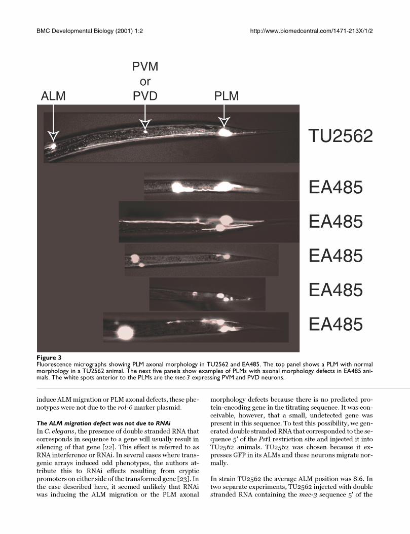
PLMs. Figure 3 shows an example of a normal PLM in
strain TU2562 and several examples of PLMs with axon-
al defects in strain EA485. All of the 189 PLMs scored in
TU2562 had normal morphology, whereas 64% of the
183 PLMs scored in EA485 had defects in axonal mor-
phology. In EA485, the PLM axons were often short,
misdirected and had extra branches.
Axonal defects in the ALMs were less common than in
the PLMs. In EA485, 77.4% of 199 ALMs scored had nor-
mal axonal morphology, whereas in TU2562, all of the
117 ALMs scored had normal morphology. The majority
of ALMs with abnormal axonal morphology were in po-
sitions 1-3 with a few as posterior as position 5. All ALMs
located posterior to position 5 had normal axonal mor-
phology.
The ALM migration defect was due to a specific DNA se-quenceTo determine whether the ALM migration defect was due
to the presence of a specific DNA sequence in the extra-chromosomal arrays, we generated mec-3gfp deletion
constructs containing various portions of the mec-3 up-
stream sequences. Plasmids pJC8, pJC3, pJC4, pJC1,
pJC18 and pJC19 were made as described in Materials
and Methods and plasmid pPD118.17 was a gift from A.
Fire. We transformed these plasmids into wild type ani-
mals as described in Materials and Methods and
screened for transgenic lines. All of the resulting strains
expressed mec-3gfp in the ten cells where mec-3 is nor-
Figure 1A. Origin and migration of the BDU and ALM neurons. The ALM touch receptor neurons and the BDU interneurons are linealsisters (the AB.arpppapp and AB.arppaapp cells divide to give ALMR/BDUR and ALML/BDUL, respectively). The ALMs are gen-erated anterior to the BDUs but migrate posteriorly, while the BDUs migrate anteriorly so that each cell stops near the posi-tions shown. B. Position and axonal morphology of the ALM, AVM and PLM touch receptor neurons. The scale shown at thebottom was used to measure ALM, BDU and AVM positions in the experiments shown below. The ALMs are located aroundposition 9, the AVMs are located between positions 6 and 7 and the BDUs are located between positions 3 and 4 in wild typeanimals.
BMC Developmental Biology (2001) 1:2 http://www.biomedcentral.com/1471-213X/1/2
mally expressed. We measured the ALM positions in 100
animals from each strain.
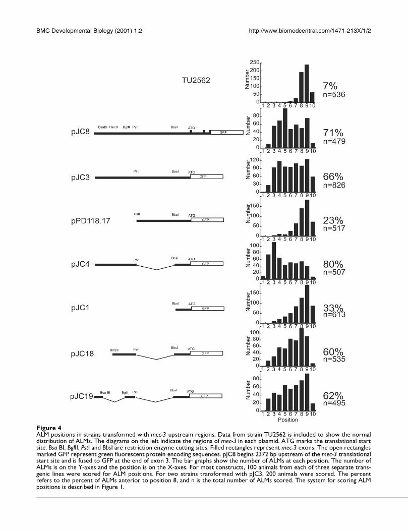
Figure 4 shows the region of mec-3gfp included in each
of the plasmids, the combined distribution of ALM posi-tions from three strains transformed with each con-
struct, the percent of ALMs that showed migration
defects (those located anterior to position 8) and the to-
tal number of ALMs scored. Strains transformed with
pJC8, pJC3, and pJC4 showed strong ALM migration de-
fects but strains transformed with plasmids pPD118.17
and pJC1 showed few ALM migration defects. The region
from PstI to BbsI appeared to suppress the migration de-
fect (compare pJC3 to pJC4). The 495 bp region from the
5' end of the mec-3 clone to the PstI site appeared to most
strongly affect ALM migrations (compare pJC3 and
pJC4 to pPD118.17 and pJC1). Further deletions of this
region (pJC18 and pJC19) resulted in less severe migra-
tion defects. These data and results described below are
consistent with the hypothesis that this sequence con-
tains multiple binding sites that titrate out a cell migra-
tion factor or factors. Below we refer to this sequence as
the titrating sequence.
The migration defect was not due to nonspecific effects of carrying an array, expression of GFP, or the rol-6 transfor-mation marker geneThe above data also show that the ALM migration and
PLM axonal morphology defects are not due to nonspe-cific effects of carrying an extrachromosomal array.
Some arrays such as those made with plasmids
pPD118.17 and pJC1 had little effect on ALM migration
and PLM axonal morphology.
There have been reports that expression of GFP in neu-
rons can have toxic effects [20, 21]. The above results,
however, show that neither the migration defects nor the
PLM axonal morphology defects resulted from expres-
sion of GFP. Some arrays, such as the integrated array in
strain TU2562 and the extrachromosomal arrays con-
taining plasmids pPD118.17 and pJC1, express GFP yet
had few ALM migration or PLM axonal morphology de-
fects. The lines transformed with pJC1 had especially
bright GFP fluorescence yet had few ALM or PLM de-
fects.
All of the lines shown in Figure 4 were made by co-injec-
tion of the plasmid of interest and the plasmid pRF4,
which contains the rol-6(su1006) marker gene. Because
some of these arrays, and others described below, did not
Figure 2ALM positions in lines TU2562 and EA485. Micrographs on the left show examples of the positions of the FLPs, AVM andALMs in TU2562 and EA485. The animals are positioned with anterior to the left. White areas are cells expressing green fluo-rescent protein. Bar graphs on the right show the number of ALMs at each position in 100 animals from each strain. The ALMpositions in TU2562 were similar to those in wild type animals whereas the ALMs in EA485 were located significantly anteriorto their normal positions. The system for scoring ALM positions is described in Figure 1.
BMC Developmental Biology (2001) 1:2 http://www.biomedcentral.com/1471-213X/1/2
induce ALM migration or PLM axonal defects, these phe-
notypes were not due to the rol-6 marker plasmid.
The ALM migration defect was not due to RNAiIn C. elegans, the presence of double stranded RNA that
corresponds in sequence to a gene will usually result in
silencing of that gene [22]. This effect is referred to as
RNA interference or RNAi. In several cases where trans-
genic arrays induced odd phenotypes, the authors at-
tribute this to RNAi effects resulting from cryptic
promoters on either side of the transformed gene [23]. In
the case described here, it seemed unlikely that RNAiwas inducing the ALM migration or the PLM axonal
morphology defects because there is no predicted pro-
tein-encoding gene in the titrating sequence. It was con-
ceivable, however, that a small, undetected gene was
present in this sequence. To test this possibility, we gen-
erated double stranded RNA that corresponded to the se-
quence 5' of the PstI restriction site and injected it into
TU2562 animals. TU2562 was chosen because it ex-
presses GFP in its ALMs and these neurons migrate nor-
mally.
In strain TU2562 the average ALM position was 8.6. In
two separate experiments, TU2562 injected with doublestranded RNA containing the mec-3 sequence 5' of the
Figure 3Fluorescence micrographs showing PLM axonal morphology in TU2562 and EA485. The top panel shows a PLM with normalmorphology in a TU2562 animal. The next five panels show examples of PLMs with axonal morphology defects in EA485 ani-mals. The white spots anterior to the PLMs are the mec-3 expressing PVM and PVD neurons.
BMC Developmental Biology (2001) 1:2 http://www.biomedcentral.com/1471-213X/1/2
Figure 4ALM positions in strains transformed with mec-3 upstream regions. Data from strain TU2562 is included to show the normaldistribution of ALMs. The diagrams on the left indicate the regions of mec-3 in each plasmid. ATG marks the translational startsite. Bsa BI, BglII, PstI and BbsI are restriction enzyme cutting sites. Filled rectangles represent mec-3 exons. The open rectanglesmarked GFP represent green fluorescent protein encoding sequences. pJC8 begins 2372 bp upstream of the mec-3 translationalstart site and is fused to GFP at the end of exon 3. The bar graphs show the number of ALMs at each position. The number ofALMs is on the Y-axes and the position is on the X-axes. For most constructs, 100 animals from each of three separate trans-genic lines were scored for ALM positions. For two strains transformed with pJC3, 200 animals were scored. The percentrefers to the percent of ALMs anterior to position 8, and n is the total number of ALMs scored. The system for scoring ALMpositions is described in Figure 1.
BMC Developmental Biology (2001) 1:2 http://www.biomedcentral.com/1471-213X/1/2
PstI restriction site also had average ALM positions of
8.6 (n=190 and n=167). These RNA injected animals also
had normal PLM axonal morphology. Therefore, the
ALM migration and the PLM axonal morphology defectswere not due to RNAi.
Many copies of the titrating sequence were required to in-duce the ALM migration defectTo determine whether the migration defect resulted
from having many copies of the titrating sequence, we
made transformants with different mixtures of plasmid
and genomic DNA. Kelly et al. [24] have shown that
blunt end cut plasmids injected with blunt end cut ge-
nomic DNA will form complex arrays that contain ge-
nomic DNA intermixed with a few copies of the plasmid.
To generate arrays with different numbers of copies of
pJC4, we co-injected pJC4 plasmid DNA with pRF4 and
genomic DNA as described in Materials and Methods.
We measured the positions of the ALMs in 100 animals
from one transgenic strain made with each DNA mix-
ture.
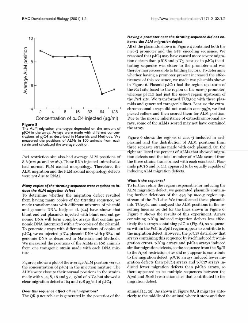
Figure 5 shows a plot of the average ALM position versus
the concentration of pJC4 in the injection mixture. The
ALMs were close to their normal positions in the strains
made with 2, 4, 8, 16 and 32 µg/ml of pJC4 but showed a
clear migration defect at 64 and 128 µg/ml of pJC4.
Having a promoter near the titrating sequence did not en-hance the ALM migration defectAll of the plasmids shown in Figure 4 contained both the
mec-3 promoter and the GFP encoding sequence. Wereasoned that pJC4 may have caused more severe migra-
tion defects than pJC8 and pJC3 because in pJC4 the ti-
trating sequence was closer to the promoter and was
thereby more accessible to binding factors. To determine
whether having a promoter present increased the effec-
tiveness of this sequence, we made two plasmids shown
in Figure 6. Plasmid pJC11 had the region upstream of
the PstI site fused to the region of the mec-3 promoter,
whereas pJC10 had just the mec-3 region upstream of
the PstI site. We transformed TU2562 with these plas-
mids and generated transgenic lines. Because the extra-
chromosomal arrays did not contain mec-3gfp, we first
picked rollers and then scored them for ALM position.
Due to the mosaic inheritance of extrachromosomal ar-
rays, some of the ALMs scored may not have contained
the array.
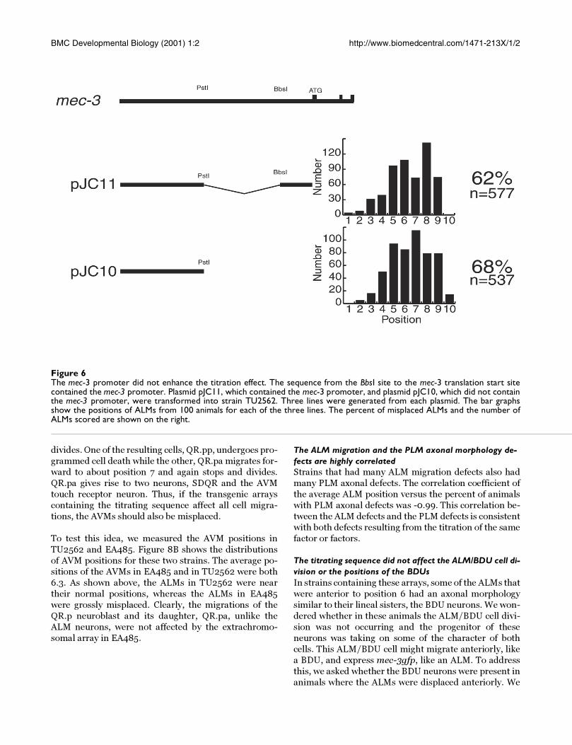
Figure 6 shows the regions of mec-3 included in each
plasmid and the distribution of ALM positions from
three separate strains made with each plasmid. On the
right are listed the percent of ALMs that showed migra-
tion defects and the total number of ALMs scored from
the three strains transformed with each construct. Plas-
mids pJC10 and pJC11 appeared to be equally capable of
inducing ALM migration defects.
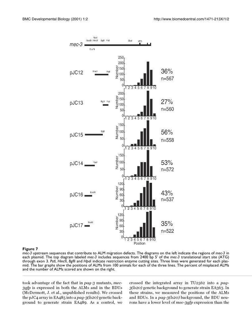
What is the sequence?To further refine the region responsible for inducing the
ALM migration defect, we generated plasmids contain-
ing further deletions of the 495 bp mec-3 region up-
stream of the PstI site. We transformed these plasmids
into TU2562 and analyzed the ALM positions in the re-
sulting lines as we did for the lines shown in Figure 6.
Figure 7 shows the results of this experiment. Arrays
containing pJC15 induced migration defects less effec-
tively than arrays containing pJC10 (Fig. 6), so sequenc-
es within the PstI to BglII region appear to contribute to
the migration defect. However, the pJC13 data show that
arrays containing this sequence by itself induced few mi-
gration errors. pJC15 arrays and pJC14 arrays induced
similar migration defects, so the sequence from the BglII
to the HpaI restriction sites did not appear to contribute
to the migration defect. pJC16 arrays induced fewer mi-
gration defects than pJC14 arrays and pJC17 arrays in-
duced fewer migration defects than pJC16 arrays, so
there appeared to be multiple sequences between the
HpaI and BsaBI restriction sites that contributed to the
migration defect.
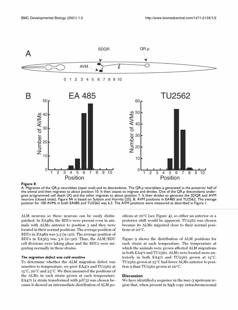
Does this sequence affect all cell migrations?The QR.p neuroblast is generated in the posterior of the
animal [12, 25]. As shown in Figure 8A, it migrates ante-riorly to the middle of the animal where it stops and then
Figure 5The ALM migration phenotype depended on the amount ofpJC4 in the array. Arrays were made with different concen-trations of pJC4 as described in Materials and Methods. Wemeasured the positions of ALMs in 100 animals from eachstrain and calculated the average position.
BMC Developmental Biology (2001) 1:2 http://www.biomedcentral.com/1471-213X/1/2
divides. One of the resulting cells, QR.pp, undergoes pro-
grammed cell death while the other, QR.pa migrates for-
ward to about position 7 and again stops and divides.
QR.pa gives rise to two neurons, SDQR and the AVM
touch receptor neuron. Thus, if the transgenic arrays
containing the titrating sequence affect all cell migra-
tions, the AVMs should also be misplaced.
To test this idea, we measured the AVM positions in
TU2562 and EA485. Figure 8B shows the distributions
of AVM positions for these two strains. The average po-
sitions of the AVMs in EA485 and in TU2562 were both
6.3. As shown above, the ALMs in TU2562 were near
their normal positions, whereas the ALMs in EA485
were grossly misplaced. Clearly, the migrations of the
QR.p neuroblast and its daughter, QR.pa, unlike the
ALM neurons, were not affected by the extrachromo-
somal array in EA485.
The ALM migration and the PLM axonal morphology de-fects are highly correlatedStrains that had many ALM migration defects also had
many PLM axonal defects. The correlation coefficient of
the average ALM position versus the percent of animals
with PLM axonal defects was -0.99. This correlation be-
tween the ALM defects and the PLM defects is consistent
with both defects resulting from the titration of the same
factor or factors.
The titrating sequence did not affect the ALM/BDU cell di-vision or the positions of the BDUsIn strains containing these arrays, some of the ALMs that
were anterior to position 6 had an axonal morphology
similar to their lineal sisters, the BDU neurons. We won-
dered whether in these animals the ALM/BDU cell divi-
sion was not occurring and the progenitor of these
neurons was taking on some of the character of both
cells. This ALM/BDU cell might migrate anteriorly, like
a BDU, and express mec-3gfp, like an ALM. To address
this, we asked whether the BDU neurons were present in
animals where the ALMs were displaced anteriorly. We
Figure 6The mec-3 promoter did not enhance the titration effect. The sequence from the BbsI site to the mec-3 translation start sitecontained the mec-3 promoter. Plasmid pJC11, which contained the mec-3 promoter, and plasmid pJC10, which did not containthe mec-3 promoter, were transformed into strain TU2562. Three lines were generated from each plasmid. The bar graphsshow the positions of ALMs from 100 animals for each of the three lines. The percent of misplaced ALMs and the number ofALMs scored are shown on the right.
BMC Developmental Biology (2001) 1:2 http://www.biomedcentral.com/1471-213X/1/2
took advantage of the fact that in pag-3 mutants, mec-
3gfp is expressed in both the ALMs and in the BDUs
(McDermott, J. et al., unpublished results). We crossed
the pJC4 array in EA485 into a pag-3(ls20) genetic back-
ground to generate strain EA489. As a control, we
crossed the integrated array in TU2562 into a pag-
3(ls20) genetic background to generate strain EA363. In
these strains, we measured the positions of the ALMs
and BDUs. In a pag-3(ls20) background, the BDU neu-
rons have a lower level of mec-3gfp expression than the
Figure 7mec-3 upstream sequences that contribute to ALM migration defects. The diagrams on the left indicate the regions of mec-3 ineach plasmid. The top diagram labeled mec-3 includes sequences from 2400 bp 5' of the mec-3 translational start site (ATG)through exon 3. PstI, HincII, BglII and HpaI indicate restriction enzyme cutting sites. Three lines were generated for each plas-mid. The bar graphs show the positions of ALMs from 100 animals for each of the three lines. The percent of misplaced ALMsand the number of ALMs scored are shown on the right.
BMC Developmental Biology (2001) 1:2 http://www.biomedcentral.com/1471-213X/1/2
ALM neurons so these neurons can be easily distin-
guished. In EA489, the BDUs were present even in ani-
mals with ALMs anterior to position 3 and they were
located in their normal positions. The average position of
BDUs in EA489 was 3.3 (n=50). The average position of
BDUs in EA363 was 3.6 (n=50). Thus, the ALM/BDU
cell divisions were taking place and the BDUs were mi-
grating normally in these strains.
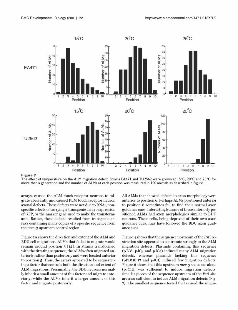
The migration defect was cold sensitiveTo determine whether the ALM migration defect was
sensitive to temperature, we grew EA471 and TU2562 at
15°C, 20°C and 25°C. We then measured the positions ofthe ALMs in each strain grown at each temperature.
EA471 (a strain transformed with pJC3) was chosen be-
cause it showed an intermediate distribution of ALM po-
sitions at 20°C (see Figure 4), so either an anterior or a
posterior shift would be apparent. TU2562 was chosen
because its ALMs migrated close to their normal posi-
tions at 20°C.
Figure 9 shows the distribution of ALM positions for
each strain at each temperature. The temperature at
which the animals were grown affected ALM migrations
in both EA471 and TU2562. ALMs were located more an-
teriorly in both EA471 and TU2562 grown at 15°C.
TU2562 grown at 25°C had fewer ALMs anterior to posi-
tion 9 than TU2562 grown at 20°C.
DiscussionWe have identified a sequence in the mec-3 upstream re-
gion that, when present in high copy extrachromosomal
Figure 8A. Migration of the QR.p neuroblast (open oval) and its descendants. The QR.p neuroblast is generated in the posterior half ofthe animal and then migrates to about position 10. It then ceases to migrate and divides. One of the QR.p descendants under-goes programmed cell death (X) and the other migrates to about position 7. It then divides to generate the SDQR and AVMneurons (closed ovals). Figure 9A is based on Sulston and Horvitz [25]. B. AVM positions in EA485 and TU2562. The averageposition for 100 AVMs in both EA485 and TU2562 was 6.3. The AVM positions were measured as described in Figure 1.
BMC Developmental Biology (2001) 1:2 http://www.biomedcentral.com/1471-213X/1/2
arrays, caused the ALM touch receptor neurons to mi-
grate aberrantly and caused PLM touch receptor neuron
axonal defects. These defects were not due to RNAi, non-specific effects of carrying a transgenic array, expression
of GFP, or the marker gene used to make the transform-
ants. Rather, these defects resulted from transgenic ar-
rays containing many copies of a specific sequence from
the mec-3 upstream control region.
Figure 1A shows the direction and extent of the ALM and
BDU cell migrations. ALMs that failed to migrate would
remain around position 5 [12]. In strains transformed
with the titrating sequence, the ALMs often migrated an-
teriorly rather than posteriorly and were located anterior
to position 5. Thus, the arrays appeared to be sequester-
ing a factor that controls both the direction and extent of
ALM migrations. Presumably, the BDU neurons normal-
ly inherit a small amount of this factor and migrate ante-
riorly, while the ALMs inherit a larger amount of this
factor and migrate posteriorly.
All ALMs that showed defects in axon morphology were
anterior to position 6. Perhaps ALMs positioned anterior
to position 6 sometimes fail to find their normal axonguidance cues. Interestingly, some of these anteriorly po-
sitioned ALMs had axon morphologies similar to BDU
neurons. These cells, being deprived of their own axon
guidance cues, may have followed the BDU axon guid-
ance cues.
Figure 4 shows that the sequence upstream of the PstI re-
striction site appeared to contribute strongly to the ALM
migration defects. Plasmids containing this sequence
(pJC8, pJC3 and pJC4) induced many ALM migration
defects, whereas plasmids lacking this sequence
(pPD118.17 and pJC1) induced few migration defects.
Figure 6 shows that this upstream mec-3 sequence alone
(pJC10) was sufficient to induce migration defects.
Smaller pieces of the sequence upstream of the PstI site
are also sufficient to induce ALM migration defects (Fig.
7). The smallest sequence tested that caused the migra-
Figure 9The effect of temperature on the ALM migration defect. Strains EA471 and TU2562 were grown at 15°C, 20°C and 25°C formore than a generation and the number of ALMs at each position was measured in 100 animals as described in Figure 1.
BMC Developmental Biology (2001) 1:2 http://www.biomedcentral.com/1471-213X/1/2
tion defects (pJC16, Fig. 7) contained only 157 bp up-
stream of the EcoRI restriction site.
These conclusions assume that the arrays generated foreach construct have roughly the same number of copies
of the titrating sequence. We controlled for strain-to-
strain variability in the array size by discarding strains
with low transmission frequencies (see Materials and
Methods) and by combining the data from at least three
independently generated strains transformed with each
construct. Because the strain-to-strain variability was
small, this assumption is probably valid.
Other sequences around the titrating sequence appeared
to facilitate the binding of the factor or factors involved
in controlling ALM cell migrations. For instance, plas-
mid pJC10, which contained more surrounding se-
quence, was more effective than pJC15. These
surrounding sequences may contain additional binding
sites for this factor or the migration factor may interact
with other proteins that bind the mec-3 upstream se-
quences. Thus, the binding of one factor might coopera-
tively facilitate the binding of other factors. Li et al. [26]
identified a sequence upstream of her-1 that phenocop-
ied an sdc mutation when transformed into C. elegans in
high copy arrays. They too found that several regions of
the her-1 upstream sequence contributed to the effect.
The results shown in Figure 7 suggest that a 104 bp se-quence, 5'AAATCTCAAT-10-CTAAAAACAT-20-TTTTA-
GAACT-30-TTTTCAAAGA-40-TATGTTGAAT-50-TCTG
GCAACT-60-TTAGGTAGTT-70-TTAAATTTTC-80-CAC
GAAAT TT-90-CAAATC AAAT-1 00- GTTA3', contribut-
ed strongly to the ALM migration defects. The region
from nucleotide 39-96 is a degenerate palindrome. With-
in this palindrome there are two more levels of nested
palindromes. For instance, the 69-96 region is palindro-
mic, and within the 69-96 region, the 84-96 region is pal-
indromic.
Some of this interesting sequence structure may result
from the juxtaposition of transcription factor binding
sites. For instance, the sequence from 72-92 would be an
excellent binding site in both its forward and reverse ori-
entations for the Drosophila STAT transcription factor
D-STAT [27]. There are two proteins with homology to
the STAT family of transcription factors in C. elegans.
This sequence is followed by the repeated sequence
caaatcaaat, which would provide two good binding sites
for the mammalian brain POU transcription factor Brn-
2 [28]. Brn-2 is related to UNC-86, which is required to
initiate and sustain mec-3 expression.
The titrating sequence is located between two genes thatare positioned head-to-head, mec-3 and F01D4.1, which
encodes a UDP-glucoronosyltransferase. The titrating
element could be regulating either, neither, or both of
these genes. Because this element appeared to sequester
a factor involved in ALM migration and PLM axon out-growth, it is probably a mec-3 control element.
mec-3gfp fusion genes that lack this element appear to
be correctly expressed. What aspect, then, of mec-3 ex-
pression is affected by this control element? mec-3 is ac-
tivated by UNC-86 protein [13,14,15]. Once MEC-3
protein is made, UNC-86 and MEC-3 form a heterodim-
er that more effectively activates mec-3. Perhaps the ti-
trating element is involved in the initial mec-3 activation.
This role would not be apparent in mec-3gfp fusion
genes because they would be activated by the endog-
enous UNC-86/MEC-3 heterodimer. Alternatively, this
element might help sustain mec-3 expression. Finally,
this element might modulate mec-3 expression in a way
that is not evident by looking at the MEC-3GFP fluores-
cence.
Figure 5 demonstrates that the ALM migration defect
arises due to having many copies of the titrating se-
quence. Arrays made with few copies of pJC4 did not in-
duce ALM migration defects, whereas arrays containing
many copies of pJC4 did, thus suggesting that the arrays
that altered ALM cell migrations acted by titrating a con-
trol factor.
It is somewhat surprising that high copy arrays could ti-
trate this factor to low enough levels to induce pheno-
types. Transgenic arrays in C. elegans contain about 50-
100 copies of the plasmids used to make the array [29].
Assuming that each copy of the titrating sequence bound
only one molecule, then shifting the concentration of this
DNA binding factor by as few as 100 molecules caused
the ALM migration defects. Because this factor is titrata-
ble, it may be present at low concentrations and bind
with high affinity to the mec-3 upstream sequence.
Perhaps this factor is present at low concentration be-
cause it is part of a mechanism for sensitive regulation of
a cell state. The relative level of a factor present at low
concentration should be easier to change than that of a
factor present at high concentration. These results sug-
gest that ALM migrations are carefully regulated by the
concentration of a specific DNA binding protein that is
present in the cell in only a few copies.
The need for an all-or-nothing response could also ex-
plain why there appear to be multiple sequences that
contribute to the titration. One mechanism for steepen-
ing the response of an effector molecule is to require that
more than one molecule bind to the target to induce a re-sponse. Perhaps the migration factor binds to several

BMC Developmental Biology (2001) 1:2 http://www.biomedcentral.com/1471-213X/1/2
sites in both mec-3 and in the cell migration genes, and
multiple sites must be bound to affect expression.
The factor sequestered by the titrating sequence does notaffect all cell migrations. The AVM touch receptor neu-
rons in EA485, a strain which showed a strong ALM mi-
gration defect, were located in the same position as the
AVMs in TU2562, a strain which showed few ALM mi-
gration defects (Fig. 8). The AVM's lineal mother and
grandmother both migrate anteriorly and if either of
these migrations were defective, the AVMs would be mis-
positioned. We do not know whether other cell migra-
tions were affected by the titrating sequence, but no
other phenotypes were observed in animals with severe
ALM migration defects.
The incidence of PLM axonal defects correlated well with
the ALM migration defects. We believe, therefore, that
the PLM defects were induced by titration of the same
factor or factors that control ALM migrations. In both
cases, the cells failed to correctly interpret anterior-pos-
terior spatial information. However, the ALMs migrated
to more anterior positions, whereas the PLMs axons of-
ten terminated at more posterior positions.
Temperature had a strong effect on the ALM migration
defects (Fig. 9). In cells grown at lower temperatures, the
titrated factor may be less active, it may be produced at
low concentrations, it may bind to arrays more tightly, orit may interact with its ligand less effectively. The cold
sensitivity of the ALM migration defect may be useful for
identifying genetic suppressors of the migration defect.
The factor titrated by this sequence may also be involved
in differentiating ALMs and BDUs. It may be present in
AB.arpppapp and AB.arppaapp, the progenitors of
ALMR/BDUR and ALML/BDUL, respectively. When
these cells divide to generate ALMs and BDUs, the factor
may differentially segregate into the ALMs. A high con-
centration of this factor may direct the ALMs to migrate
posteriorly while a low level of this factor may direct the
BDUs to migrate anteriorly. Furthermore, this factor
may help activate mec-3 expression and thereby cause
the ALMs to differentiate into touch receptor neurons.
As was true in sea urchin embryos [30, 31], titration of
transcription factors with high copy transgenes may be
useful for characterizing control elements in C. elegans.
The ALM migration defect should be useful for identify-
ing optimal conditions for the titration of DNA binding
protein with high copy arrays. We do not know, at this
point, how often transcription factors will be present at
low enough concentrations and bind DNA with enough
affinity to apply this approach. Because others have alsoreported competitive titration by transgenic sequences
in C. elegans [26, 32], we expect that it will not be un-
common. Certainly, some of the artifacts associated with
transformation of C. elegans with high copy arrays may
result from titration of DNA binding factors [23]. Trans-formation of C. elegans with low copy arrays made with
complex carrier DNA prevented the titration effects, so
the use of low copy arrays may be prudent where titra-
tion effects are not desired.
Kelly et al. [24] found that inclusion of complex carrier
DNA in C. elegans transformation experiments im-
proves the function of certain co-injected reporter con-
structs. These effects include both increased expression
and improved uniformity of transgene expression. Fig-
ure 5 shows that including complex carrier DNA may
also prevent transformation artifacts resulting from ti-
tration of transcription factors.
ConclusionsThe experiments described here show that transforma-
tion of C. elegans with many copies of a specific sequence
located upstream of mec-3 induced cell migration and
axonal guidance defects. This mec-3 upstream sequence
appeared to be sequestering a factor involved in control-
ling ALM migration and PLM axonal outgrowth. This
factor may also regulate mec-3 and thereby control touch
receptor neuron fate. Titration of transcription factors
with high copy arrays may become widely applicable in
C. elegans once conditions that optimize this effect arefound. The ALM defect described here may be useful for
finding these conditions.
Materials and MethodsWorm culture and strain methodsC. elegans were maintained as described by Brenner
[33]. Animals were grown at 20°C unless otherwise stat-
ed. The wild type strain was N2 var. Bristol. Strain
TU2562, mec-3gfp(uIs22), was a gift from M. Chalfie.
Plasmid constructsStandard molecular biology methods were used in mak-
ing the following plasmids [34]. To make pJC1, we re-
moved a PstI-BbsI fragment from pPD118.17, a gift from
A. Fire. To make pJC3, we subcloned the NsiI-PstI frag-
ment (1246 bp) of pTU23 [35] into the PstI site of
pPD118.17. We made pJC4 by deleting the PstI-BbsI
fragment (1329 bp) from pJC3.
We made pJC8 from pBS1 and pJC3. To make pBS1, the
oligonucleotides 5' ACCTCCCAAACTATAGATTGGGTG3'
and 5'CGGCCAGAGTCGACTCACATATTG3' were used to
amplify a 1370 bp fragment from pTU23. This intro-
duced a SalI site near the 3' end of the third exon of mec-
3. We digested this fragment with HindIII and SalI, andligated it into the HindIII and SalI sites of pPD95.67, a
BMC Developmental Biology (2001) 1:2 http://www.biomedcentral.com/1471-213X/1/2
gift from A. Fire. We removed the KpnI-KpnI fragment
from pBS1 to make pBS1∆K. We digested pBS1∆K and
pJC3 with both BbsI and XhoI to remove a 1657 bp piece
and a 908 bp piece, respectively. The remaining frag-ments were religated to make pJC8.
The only mec-3 sequence in plasmid pJC10 was the 495
bp sequence 5' to the PstI site. We constructed pJC10 by
deleting the PstI-NheI fragment (2861 bp) from pJC3. To
make pJC11, we removed the NotI-NheI fragment (944
bp) from pJC4. Plasmid pJC12 was a HincII-HincII dele-
tion (443 bp) of pJC10. To make pJC13, we removed the
HincII-BglII fragment (602 bp) from pJC10. pJC14 was
a HpaI-BspEI deletion (309 bp) of pJC10, and pJC15 was
a BglII-BspEI deletion (150 bp) of pJC10. To make
pJC16, we digested pJC10 with BspEI and EcoRI to re-
move a 365 bp fragment. pJC17 was a BspEI-BsaBI dele-
tion (412 bp) of pJC10. pJC18 was a HincII-HincII
deletion (443 bp) of pJC4, and pJC19 was a BsaBI-BglII
deletion (262 bp) of pJC4. All plasmids were prepared
with the Wizard midiprep kit (Promega).
Transformation of C. elegansGerm-line transformation was done as described by Mel-
lo et al. [29]. All constructs were co-injected with plas-
mid pRF4 containing the semidominant rol-6(su1006)
allele, which causes a roller phenotype. Plasmids pJC1,
pJC3, pJC4, and pJC8 were injected into N2 wild type
animals at 100 µg/ml. Plasmids pJC10-pJC15 were in-jected into TU2562 at 100 µg/ml. Plasmid pRF4 was co-
injected with all plasmids at 100 µg/ml.
To test whether the number of copies in the arrays had an
effect on ALM migration, StuI cut pJC4 was co-injected
with SmaI cut pRF4 and PvuII cut genomic DNA at the
following concentrations (µg/ml): 2, 2, 100; 4, 4, 92; 8,
8, 84; 16, 16, 68; 32, 32, 36; 64, 32, 18; and 128, 32, 9, re-
spectively. Larger arrays are transmitted to the next gen-
eration more effectively than smaller arrays [36]. All of
the strains presented here had transmission frequencies
greater than 16%.
MicroscopyMicroscopy was done on an Olympus IMT-2 inverted mi-
croscope and on an Olympus AX70 microscope. A Power
Mac G3 7100/80 with a frame grabber and IPlab soft-
ware was used to collect and process images from the
AX70. Both microscopes were equipped with fluores-
cence and Nomarski optics. Animals were immobilized
with approximately 30 mM sodium azide and viewed at
100 X or 400 X.
RNAiTo make double stranded RNA for RNAi, we placed thePstI-RsaI fragment from pJC3 into pBluescript II KS+.
RNA was made from the T3 and T7 promoters in pBlue-
script with a Maxiscript kit (Ambion). The dsRNA was
injected at a concentration of 25 µg/ml.
Computer analysisSequence homologies were identified with the Blast algo-
rithm [37, 38] at the National Center for Biotechnology
Information (http://www.ncbi.nlm.nih.gov/). To identi-
fy long-range palindromic structures, an RNA secondary
structure algorithm in the program DNASIS version 2.0
was used. Transcription factor binding sites were identi-
fied with MatInspector at http://genomatix.gsf.de/cgi-
bin/matinspector/matinspector.pl [39].
AcknowledgementsThis work was supported by grant RR10296 from the NIH. We thank Dr. M. Chalfie for strain TU2562 and Dr. A. Fire for plasmid pPD118.17. We thank Dr. Peter Good, Dr. Kelly Tatchell, Dr. Stephanie Aamodt and Dr. Joan McDermott for comments on an earlier draft of this manuscript. We are especially grateful to Dr. Kelly Tatchell for use of his Olympus AX70 microscope.
References1. Cajal R: Degeneration and Regeneration of the Nervous System. London:
Oxford University Press; 1928, 2. Montell DJ: The genetics of cell migration in Drosophila mel-
anogaster and Caenorhabditis elegans development. Develop-ment 1999, 126:3035-3046
3. Blelloch R, Newman C, Kimble J: Control of cell migration duringCaenorhabditis elegans development. Curr Opin Cell Biol 1999,11:608-613
4. Hedgecock EM, Culotti JG, Hall DH: The unc-5, unc-6 and unc-40genes guide circumferential migrations of pioneer axons andmesodermal cells on the epidermis in C. elegans. Neuron 1990,2:61-85
5. Hong K, Hinck L, Nishiyama M, Poo MM, Tessier-Lavigne M, Stein E:A ligand-gated association between cytoplasmic domains ofUNC5 and DCC family receptors converts netrin-inducedgrowth cone attraction to repulsion. Cell 1999, 97:927-941
6. Bashaw GJ, Goodman CS: Chimeric axon guidance receptors:the cytoplasmic domains of slit and netrin receptors specifyattraction versus repulsion. Cell 1999, 97:917-926
7. Colavita A, Krishna S, Zheng H, Padgett RW, Culotti JG: Pioneeraxon guidance by UNC-129, a C. elegans TGF-beta. Science1998, 281:706-709
8. Vincent S, Ruberte E, Grieder NC, Chen CK, Haerry T, Schuh R, Af-folter M: DPP controls tracheal cell migration along the dor-soventral body axis of the Drosophila embryo. Development1997, 124:2741-2750
9. Wightman B, Clark SG, Taskar AM, Forrester WC, Maricq AV, Barg-mann CI, Garriga G: The C. elegans gene vab-8 guides posteri-orly directed axon outgrowth and cell migration. Development1996, 122:671-682
10. Wolf FW, Hung MS, Wightman B, Way J, Garriga G: vab-8 is a keyregulator of posteriorly directed migrations in C. elegansand encodes a novel protein with kinesin motor similarity.Neuron 1998, 20:655-666
11. Harris J, Honigberg L, Robinson N, Kenyon C: Neuronal cell mi-gration in C. elegans : regulation of Hox gene expression andcell position. Development 1996, 122:3117-3131
12. Sym M, Robinson N, Kenyon C: MIG-13 positions migrating cellsalong the anteroposterior body axis of C. elegans. Cell 1999,98:25-36
13. Lichtsteiner S, Tjian R: Synergistic activation of transcription byUNC-86 and MEC-3 in Caenorhabditis elegans embryo ex-tracts. EMBO J. 1995, 14:3937-3945
14. Xue D, Finney M, Ruvkun G, Chalfie M: Regulation of the mec-3gene by the C. elegans homeoproteins UNC-86 and MEC-3.EMBO J. 1992, 11:4969-4979
BMC Developmental Biology (2001) 1:2 http://www.biomedcentral.com/1471-213X/1/2
15. Xue D, Tu Y, Chalfie M: Cooperative interactions between theCaenorhabditis elegans homeoproteins UNC-86 and MEC-3.Science 1993, 261:1324-1328
16. Chalfie M, Au M: Genetic control of differentiation of theCaenorhabditis elegans touch receptor neurons. Science 1989,243:1027-1033
17. Sulston JE, Schierenberg E, White JG, Thomson JN: The embryoniccell lineage of the nematode Caenorhabditis elegans. Dev. Biol.1983, 100:64-119
18. Jia Y, Xie G, Aamodt EJ: pag-3, a C. elegans gene involved intouch neuron gene expression and coordinated movement.Genetics 1996, 142:141-147
19. Jia Y, Xie G, McDermott JB, Aamodt E: The C. elegans gene pag-3 is homologous to the zinc finger proto-oncogene Gfi1. De-velopment 1997, 124:2063-2073
20. Martinez-Serrano A, Villa A, Navarro B, Rubio FJ, Bueno C: Humanneural progenitor cells: better blue than green? Nat Med 2000,6:483-484
21. Goldman S, Roy N: Reply to "Human neural progenitor cells:better blue than green?". Nat Med 2000, 6:483-484
22. Fire A, Xu S, Montgomery MK, Kostas SA, Driver SE, Mello CC: Po-tent and specific genetic interference by double-strandedRNA in Caenorhabditis elegans. Nature 1998, 391:806-811
23. McGhee JD, Krause MW: Transcription factors and transcrip-tional regulation. In: C. ELEGANS II Edited by Riddle DL, Blumenthal T,Meyer BJ, Priess JR. pp. 147-184. Cold Spring Harbor: Cold Spring HarborLaboratory Press; 1997, 147-184
24. Kelly WG, Xu S, Montgomery MK, Fire A: Distinct requirementsfor somatic and germline expression of a generally ex-pressed Caenorhabditis elegans gene. Genetics 1997, 146:227-238
25. Sulston JE, Horvitz HR: Post-embryonic cell lineages of thenematode Caenorhabditis elegans. Dev. Biol. 1977, 56:110-156
26. Li W, Streit A, Robertson B, Wood WB: Evidence for multiplepromoter elements orchestrating male-specific regulationof the her-1 gene in Caenorhabditis elegans. Genetics 1999,152:237-248
27. Yan R, Small S, Desplan C, Dearolf CR, Darnell JE Jr: Identificationof a Stat gene that functions in Drosophila development. Cell1996, 84:421-430
28. Li P, He X, Gerrero MR, Mok M, Aggarwal A, Rosenfeld MG: Spacingand orientation of bipartite DNA-binding motifs as potentialfunctional determinants for POU domain factors. Genes Dev1993, 7:2483-2496
29. Mello CC, Kramer JM, Stinchcomb D, Ambros V: Efficient genetransfer in C. elegans : extrachromosomal maintenance andintegration of transforming sequences. EMBO J. 1991, 10:3959-3970
30. Livant DL, Hough-Evans BR, Moore JG, Britten RJ, Davidson EH: Dif-ferential stability of expression of similarly specified endog-enous and exogenous genes in the sea urchin embryo.Development 1991, 113:385-398
31. Franks RR, Anderson R, Moore JG, Hough-Evans BR, Britten RJ, Dav-idson EH: Competitive titration in living sea urchin embryosof regulatory factors required for expression of the CyIIIa ac-tin gene. Development 1990, 110:31-40
32. Carmi I, Kopczynski JB, Meyer BJ: The nuclear hormone receptorSEX-1 is an X-chromosome signal that determines nema-tode sex. Nature 1998, 396:168-173
33. Brenner S: The genetics of Caenorhabditis elegans. Genetics1974, 77:71-94
34. Sambrook J, Fritsch EF, Maniatis T: Molecular Cloning: A LaboratoryManual Second Edition. Cold Spring Harbor, New York: Cold Spring HarborLaboratory Press; 1989,
35. Way JC, Chalfie M: mec-3, a homeobox-containing gene thatspecifies differentiation of the touch receptor neurons in C.elegans. Cell 1988, 54:5-16
36. Mello C, Fire A: DNA transformation. Methods Cell Biol 1995,48:451-482
37. Altschul SF, Gish W, Miller W, Myers EW, Lipman DJ: Basic localalignment search tool. J. Mol. Biol. 1990, 215:403-410
38. Altschul SF, Madden TL, Schäffer AA, Zhang J, Zhang Z, Miller W, Lip-man DJ: Gapped BLAST and PSI-BLAST: a new generation ofprotein database search programs. Nucleic Acids Res. 1997,25:3389-3402
39. Quandt K, Frech K, Karas H, Wingender E, Werner T: MatInd andMatInspector - New fast and versatile tools for detection ofconsensus matches in nucleotide sequence data. Nucleic AcidsResearch 1995, 23:4878-4884
?How do youkeep informed of thelatest innovationsin RNA research
Sign up for RNA Flashnotes. Once every threeweeks Ambion, The RNA Company, will emailinformation to you concerning the hottest technologies,the newest products, and the most influential RNAbased research to enable you to make informeddecisions about your RNA research methodologies.
Get your free
subscription today.
E-mail your
complete postal
mailing address to
THE RNA COMPANY™