Embed Size (px)
Citation preview
Vol. 55, No. 12
High-Frequency Transformation, by Electroporation, ofLactococcus lactis subsp. cremoris Grown with Glycine in
Osmotically Stabilized MediaHELGE HOLO't* AND INGOLF F. NES2
Norwegian Dairies Association, Oslo,' and Laboratory of Microbial Gene Technology, N-1432 As-NLH,2 Norway
Received 23 May 1989/Accepted 12 September 1989
An efficient method for genetic transformation of lactococci by electroporation is presented. Highlycompetent lactococci for electrotransformation were obtained by growing cells in media containing highconcentrations of glycine and 0.5 M sucrose as the osmotic stabilizers. These cells could be stored at -85°Cwithout loss of competence. With Lactococcus lactis subsp. cremoris BC101, a transformation frequency of 5.7x 10⁷ transformants per ,ug of pIL253 DNA was obtained, which represents 5% of the surviving cells. All thelactococcal strains tested could be transformed by the present method.
Strains of Lactococcus lactis subsp. cremoris are impor-tant starter organisms in the production of cheese. Strainimprovement by modern gene technology as well as geneticstudies of these organisms has been hampered by the lack ofefficient genetic transformation systems. For the closelyrelated L. lactis subsp. lactis, successful transformationprocedures have been developed. The highest transforma-tion frequency reported was 5 x 10⁶ erythromycin-resistanttransformants per ,ug of pIL204 DNA (16). Polyethyleneglycol-mediated protoplast transformation was used in thiswork. However, this method is not effective for all strainsand has been shown to work for only a few L. lactis subsp.cremoris strains with transformation frequencies of about103/,ug of DNA or less (15, 17).Transformation by electroporation is a recent method that
has been successfully employed with a number of bacterialspecies (8; for a review, see reference 2). This technique isless tedious and time consuming than protoplast transforma-tion and has proved useful in species previously regarded asuntransformable. Harlander (6) was the first to transform L.lactis subsp. lactis by electroporation. Extended work byMcIntyre and Harlander (10) did not improve the transfor-mation frequency significantly.
Powell et al. (13) were able to transform several strains ofL. lactis subsp. lactis and L. lactis subsp. cremoris byelectroporation, but the transformation frequencies obtainedwere generally very low, and not all of the strains testedcould be transformed by their procedure. However, a hightransformation frequency was obtained with L. lactis subsp.lactis LM0230 treated with lysozyme before electroporation,indicating that the cell wall is a physical barrier to theentering DNA. This notion was supported by the work ofvan der Lelie et al. (17), who incorporated 40 mM DL-threonine in the growth medium to obtain cells with weak-ened cell walls. These cells could be transformed by elec-troporation, but transformation frequencies were still quitelow. Transformants were not obtained when threonine wasomitted from the growth medium.Our initial attempts to use these two procedures (13, 17) in
transforming L. lactis subsp. cremoris BC101 were unsuc-
* Corresponding author.t Present address: Laboratory of Microbial Gene Technology,
P. 0. Box 51, N-1432 As-NLH, Norway.
cessful. The published data suggested that this could resultfrom inadequate weakening of the cell wall; the data ofPowell et al. (13) indicate that most of the cells able to takeup DNA are osmotically fragile. Such cells cannot be ob-tained by using cell wall-weakening agents in hypotonicgrowth media. In the work presented here, the cells weregrown in osmotically stabilized media containing high con-centrations of glycine as an inhibitor of cell wall formation.These cells were transformed by electroporation at highfrequencies.
MATERIALS AND METHODSBacterial strains and plasmids. L. lactis subsp. cremoris
BC101 and L. lactis subsp. lactis LM2336 (both plasmid-free, Lac-) were kindly provided by T. Langsrud, Agricul-tural University of Norway, As, Norway. L. lactis subsp.cremoris GS was isolated from a commercial starter culture(C. Hansen, Copenhagen, Denmark). L. lactis subsp. lactisIL1837(pIL253) was kindly provided by A. Chopin, InstitutNational de la Recherche Agronomique, Jouy-en-Josas,France. Other lactococcal strains used in this study werefrom The National Collection of Food Bacteria (Reading,United Kingdom). All lactococcal strains were grown at30°C in M17 supplemented with 0.5% glucose (GM17) unlessotherwise stated. E. coli JM109(pSA3) was kindly providedby J. J. Ferretti, University of Oklahoma Health ScienceCenter, Oklahoma City.
Transformation protocol. To obtain competent cells, thecultures were grown to an optical density at 600 nm of 0.5 to0.8 and then diluted 100-fold in SGM17 (GM17 containing 0.5M sucrose) supplemented with glycine as indicated in thetext. After growth at 30°C to an optical density at 600 nm of0.2 to 0.7, the cells were harvested by centrifugation at 4°Cat 5,000 x g. Following two washes in ice-cold 0.5 M sucrosecontaining 10% glycerol, the cells were suspended in 1/100culture volume of washing solution and then stored inaliquots at -85°C until use.The cell suspensions were thawed on ice. Portions (40 ,ul)
were mixed with 1 ,ul of DNA dissolved in 10 mM Trishydrochloride-1 mM EDTA (pH 7.5) and then transferred toan ice-cooled electroporation cuvette (2-mm electrode gap)and exposed to a single electrical pulse. The pulse wasdelivered by a Gene-Pulser (Bio-Rad Laboratories, Rich-mond, Calif.) set at 25 ,uF and normally at 2.0 kV. The
3119
APPLIED AND ENVIRONMENTAL MICROBIOLOGY, Dec. 1989, p. 3119-31230099-2240/89/123119-05$02.00/0Copyright C 1989, American Society for Microbiology
on April 19, 2018 by guest
http://aem.asm
.org/D
ownloaded from

3120 HOLO AND NES
cuvette was connected in parallel to a 200-fl resistor (pulsecontroller; Bio-Rad), resulting in time constants of 4.5 to 5ms. Immediately following the discharge, the suspensionswere mixed with 0.96 ml of ice-cold SGM17 containing 20mM MgCl2 and 2 mM CaCl2 (SGM17MC) and left on ice forabout 5 min. Appropriate dilutions were then made inSGM17MC, and the cells were incubated at 30°C for 2 hbefore 100-,u portions were spread on selective streptococ-cal regeneration medium (SR) plates (12) containing (perliter) 10 g of tryptone, 5 g of yeast extract, 200 g of sucrose,10 g of glucose, 25 g of gelatin, 15 g of agar, 2.5 mM MgCl2,and 2.5 mM CaC12 (pH 6.8). The plates contained 1 p.g oferythromycin per ml for the selection of erythromycin-resistant transformants. Transformants were enumeratedafter 2 days of incubation at 30°C.DNA isolation and analysis. Erythromycin-resistant trans-
formants were grown in GM17 containing 10 ,ug of erythro-mycin per ml. Plasmids were isolated by the alkaline lysisprocedure described by Klaenhammer (7) and analyzed byagarose gel electrophoresis as described by Maniatis et al.(9). The plasmids were transferred from agarose gels toHybond-N (Amersham International, Amersham, UnitedKingdom) nylon filters by vacuum transfer and then sub-jected to Southern analysis according to Maniatis et al. (9).The plasmids used as probes were purified by CsCl equilib-rium centrifugation before being 32P-labeled by nick transla-tion. DNA concentrations were measured fluorometrically,by using a TKO Mini Fluorometer (Hoefer Scientific Instru-ments, San Francisco, Calif.); the Hoechst fluorochromedye 33258 (Polysciences Inc., Warrington, Pa.), which bindsspecific for DNA, was also used.
RESULTS
Optimization of transformation of L. lactis subsp. cremorisBC101 by electroporation. By modifying the procedure ofDower et al. (4) for transformation of Escherichia coli, wewere able to transform strain BC101 by electroporation. Thecells were transformed with pIL253, a cloning vector de-rived from pAM31 that has been constructed for cloning ingram-positive organisms (3). To increase transformationfrequency, glycine was incorporated in the growth mediumof strain BC101 to weaken its cell wall. The effect of glycineon growth is shown in Fig. 1. The presence of sucrose itselfwill inhibit growth of strain BC101. In the absence ofsucrose, no growth was observed with more than 2% glycinein the medium, whereas 0.5 M sucrose-containing mediumsupported growth in up to 3% glycine. Consequently, thecells without sucrose were relatively more sensitive togrowth inhibition by glycine than were cells grown in thepresence of an osmotic stabilizer (Fig. 1). This suggestedthat growth inhibition caused by glycine was a result of itseffect on cell wall formation. Cells from stationary-phasecultures were less sensitive to glycine and could grow with4% glycine in SGM17. Microscopic examination revealedswollen cells, spheroplasts, and some ghosts from culturesgrown at the highest glycine concentrations in SGM17. Thiswas not observed in cultures grown without sucrose.
Figure 1 also shows the effect of glycine on the transform-ability of strain BC101. Adding glycine to the growth me-dium resulted in an increase in transformability. The highesttransformation frequencies were obtained with cells grownat the highest glycine concentrations. For cells grown inSGM17, transformation frequency increased exponentiallywith respect to glycine concentration in the range of 0.5 to2%. The effect of glycine was much less pronounced without
8
6
co.I-
E0,, 4C
0)0-i
2
1.2
Ec00.8 g(04
0.4
0 1 2 3% Glycine
FIG. 1. Effect of glycine on growth and plasmid transformationof L. lactis subsp. cremoris BC101 with and without 0.5 M sucrosein the growth medium. The cells were grown overnight. Symbols: Oand A, growth in the presence and absence of sucrose, respectively;* and A, transformation in the presence and absence of sucrose,respectively.
sucrose. Thus, sucrose caused an increase in transformabil-ity even at glycine concentrations that the cells could toler-ate without osmotic protection.
Figure 2 shows the effect of electrical field strength on the
3
o 2
UT
c
;E0
CU)
0
12
0
8
LLU
4
00 4 8 12
Field Strength (kV cm-i)
FIG. 2. Effect of electrical field strength on transformation effi-ciency (0) and survival (0) of L. lactis subsp. cremoris BC101. Thecells were grown with 3% glycine in SGM17.
APPL. ENVIRON. MICROBIOL.
on April 19, 2018 by guest
http://aem.asm
.org/D
ownloaded from
TRANSFORMATION OF L. LACTIS SUBSP. CREMORIS
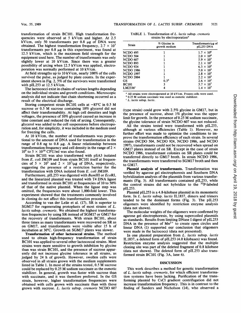
transformation of strain BC101. High transformation fre-quencies were observed at 5 kV/cm and higher. At 2.5kV/cm, only 50 transformants per 0.8 ,ug of DNA were
obtained. The highest transformation frequency, 2.7 x 10⁷transformants per 0.8 ,ug in this experiment, was found at12.5 kV/cm, which is the maximum field strength for theequipment used here. The number of transformants was onlyslightly lower at 10 kV/cm. Since there was a greaterpossibility of arcing when 12.5 kV/cm was applied, electro-poration was normally performed at 10 kV/cm.
At field strengths up to 10 kV/cm, nearly 100% of the cellssurvived the pulse, as judged by plate counts. In the exper-
iment shown in Fig. 2, 5% of the survivors were transformedwith pIL253 at 12.5 kV/cm.The lactococci exist in chains of various lengths depending
on the individual strains and growth conditions. Microscopicanalysis did not indicate that chain shortening occurred as a
result of the electrical discharge.Storing competent strain BC101 cells at -85°C in 0.5 M
sucrose or 0.5 M sucrose containing 10% glycerol did notaffect their transformability. At high cell densities and highvoltages, the presence of 10% glycerol caused an increase intime constant and reduced the risk of arcing. Consequently,glycerol was added to the cell suspensions before electropo-ration and, for simplicity, it was included in the medium usedfor freezing the cells.At 10 kV/cm, the number of transformants was propor-
tional to the amount of DNA used for transformation in therange of 0.8 ng to 0.8 ,ug. A linear relationship betweentransformation frequency and cell density in the range of 2 x
109 to 5 x 10¹⁰ CFU/ml was also found.Strain BC101 was also transformed with pSA3 isolated
from E. coli JM109 and from strain BC101 itself at frequen-cies of 5 x 10³ and 2 x 10⁷/,ug of DNA, respectively,suggesting the presence of a restriction barrier for thetransformation with DNA isolated from E. coli JM109.
Furthermore, pIL253 was digested with BamHI or EcoRI,and the linearized plasmid was treated with T4 DNA ligaseand transformed in strain BC101 at frequencies of 10 to 50%of that of the native plasmid. When the ligase step wasomitted, the frequencies were about 1,000-fold lower. Thisexperiment showed that the treatments commonly involvedin cloning do not affect this transformation procedure.According to van der Lelie et al. (17), SR is superior to
SGM17 for regenerating protoplasts of most strains of L.lactis subsp. cremoris. We obtained the highest transforma-tion frequencies by using SR instead of SGM17 or GM17 forthe recovery of transformants. With strain BC101, aboutthree times as many transformants were recovered on SR ason GM17, and transformants were visible after 15 h ofincubation at 30°C. Growth on SGM17 plates was slower.
Transformation of other lactococcal strains. The methodused to obtain high-frequency transformation of strainBC101 was applied to several other lactococcal strains. Moststrains were more sensitive to growth inhibition by glycinethan was strain BC101, and the presence of sucrose appar-ently did not increase glycine tolerance in all strains, as
judged by 24 h of growth. However, swollen cells wereobserved in all strains grown with the medium supplementslisted in Table 1. In most of the strains tested, 0.5 M sucrosecould be replaced by 0.25 M sodium succinate as the osmoticstabilizer. In general, growth was faster with sucrose thanwith succinate, and it was therefore preferred. In the GSstrain, however, higher transformation frequencies wereobtained with cells grown with succinate than with thosegrown with sucrose. L. lactis subsp. cremoris NCDO 607
TABLE 1. Transformation of L. lactis subsp. cremorisstrains by electroporationa
Strain % Glycine in Transformants/4Lg ofgrowth medium pIL253 DNA
NCDO 495 1 1.7 x 10⁶NCDO 504 1 2.0 x10⁴NCDO 607 1 5.9 xlONCDO 893 1 2.1 x10⁵NCDO 924 1 6.3 x 10³NCDO 1986 1 5.0x 10³NCDO 1997 1 2.8x 10⁵NCDO2W4 1 2.2x 10³GS 0.3b 2.6x 10⁵BC101 3 5.7 x 10⁷LM2336C 4 1.4x 10⁷
a All strains were electroporated at 10 kV/cm. Frozen cells were used.b 0.25 M sodium succinate was used as osmotic stabilizer.c L. lactis subsp. lactis.
(type strain) could grow with 2.5% glycine in GM17, but inthe presence of sucrose, about 1% glycine was the upperlimit for growth. In the presence of 0.25 M sodium succinate,the glycine tolerance of strain NCDO 607 was not reduced.
All the strains tested were transformed with pIL253,although at various efficiencies (Table 1). However, nofurther effort was made to optimize the conditions to im-prove the transformation efficiency of each strain. In severalstrains (NCDO 504, NCDO 924, NCDO 1986, and NCDO1997), transformants could not be recovered when spread onGM17 plates instead of on SR. Except in the case of strainNCDO 1986, transformant colonies on SR plates could betransferred directly to GM17 broth. In strain NCDO 1986,the transformants were transferred to SGM17 broth and thento GM17.
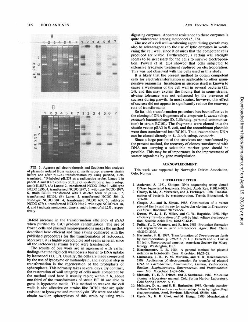
Plasmid analysis of transformants. Transformation wasverified by agarose gel electrophoresis and Southern DNAhybridization analysis of the plasmids from various transfor-mants (Fig. 3). All transformants contained pIL253, whilethe control strains did not hybridize to the 32P-labeledpIL253.
Plasmid pIL253 is a 4.8-kilobase plasmid in its monomericform. In L. lactis subsp. lactis IL1837, dimers and trimerstended to be the dominant forms (Fig. 3). The pIL253oligomers were identified by restriction enzyme analysis(data not shown).The molecular weights of the oligomers were confirmed by
agarose gel electrophoresis, by using supercoiled plasmidsas standards. Results from limiting DNase I digest of pIL253DNA in the presence of Mn2+ to create open circles andlinear DNA (1) supported our conclusion that oligomerswere made in the lactococci (data not presented).
In one plasmid preparation from L. lactis subsp. lactisIL1837, a deleted form of pIL253 (4.0 kilobases) was found.Restriction enzyme analysis suggested that the multiplecloning site was part of the deleted fragment of 0.8 kilobase(data not shown). The deleted form of pIL253 also trans-formed strain BC101 (Fig. 3A, lane 6).
DISCUSSION
This work describes a method for genetic transformationof L. lactis subsp. cremoris, for which efficient transforma-tion systems have been lacking. Purification of the trans-forming plasmid by CsCl gradient centrifugation did notincrease transformation frequency. This is in contrast to thefinding of Sanders and Nicholson (14), who observed a
VOL. 55, 1989 3121
on April 19, 2018 by guest
http://aem.asm
.org/D
ownloaded from
APPL. ENVIRON. MICROBIOL.
t
CO dIto
:0 ;00 fft iV: -;- ::D
ji ,z, .s,F, .:CE .'V:'
*Mee,, 00
'_" . t" ff 0 '
id i,
w ''' _=" 'S : 0 0.0t' *.
'VeV; 0 00d'<'S00 ti."'''?
'St
i: '.ffE'S ,4i 0i't 0 0 ; 0 f -.\ i ;;t ' ........ a.'5 ^LS ts Wf W; 't ': s;0;'im<>'os'}
!-13 4 5 6 7
y,,: 00WA0*S:0I
FIG. 3. Agarose gel electrophoresis and Southern blot analysesof plasmids isolated from various L. lactis subsp. cremoris strains
before and after pIL253 transformation by using purified, nick-
translated, 32P-labeled pIL253 as a radioactive probe. Lanes 1 in
panels A and B are controls of pIL253 isolated from L. lactis subsp.lactis IL1837. (A) Lanes: 2, transformed NCDO 1986; 3, wild-typeNCDO 1896; 4, transformed NCDO 1997; 5, wild-type NCDO 1997;
6, strain BC101 transformed with a deleted form of pIL263; 7,
transformed BC101. (B) Lanes: 2, transformed NCDO 504; 3,
wild-type NCDO 504; 4, transformed NCDO 607; 5, wild-type
NCDO 607; 6, transformed NCDO 924; 7, wild-type NCDO 924. m,
d, and t indicate monomers, dimers, and trimers of pIL253, respec-
tively.
10-fold increase in the transformation efficiency of pSA3when purified by CsCl gradient centrifugation. The use of
frozen cells and plasmid minipreparations makes the method
described here efficient and time saving compared with the
published procedures for the transformation of lactococci.
Moreover, it is highly reproducible and seems general, since
all the lactococcal strains tested were transformed.
The results of our work are in agreement with earlier
findings that the rigid cell wall poses a barrier to DNA uptakeby lactococci (13, 17). Usually, the cells are made competent
by the use of lysozyme or mutanolysin, and a crucial step in
transformation is the regeneration of the protoplasts or
spheroplasts. This normally takes several days. By contrast,
the restoration of wall integrity of cells made competent bythe method used here is usually rapid; within 2 h, about
one-third of the transformants of strain BC101 are able to
grow in hypotonic media. This method to weaken the cell
walls is also effective on strains like BC101 that are quiteresistant to lysozyme and mutanolysin. We were not able to
obtain swollen spheroplasts of this strain by using wall-
digesting enzymes. Apparent resistance to these enzymes isquite widespread among lactococci (5, 18).-T4e uae of a cell wall-weakening agent during growth may
also be advantageous to the use of lytic enzymes in weak-ening the cell wall, since it ensures that the competent cellsproduced are viable. Furthermore, a certain wall strengthseems to be necessary for the cells to survive electropora-tion. Powell et al. (13) showed that cells subjected toextensive lysozyme treatment ruptured on electroporation.This was not observed with the cells used in this study.
It is likely that the present method to obtain competentcells for electrotransformation is applicable to other gram-positive organisms. Incubation in sucrose itself is known tocause a weakening of the cell wall in several bacteria (11,14), and this may explain the finding that in some strains,glycine tolerance was not enhanced by the presence ofsucrose during growth. In most strains, however, this effectof sucrose did not appear to significantly reduce the recoveryrate of transformants.So far, this transformation procedure has been effective in
the cloning ofDNA fragments of a temperate L. lactis subsp.cremoris bacteriophage (D. Lillehaug, personal communica-tion) in strain BC101. The fragments were cloned into theshuttle vector pSA3 in E. coli, and the recombinant plasmidswere then transformed into BC101. Thus, recombinant DNAcan be cloned directly in L. lactis subsp. cremoris.
Since a large portion of the survivors are transformed bythe present method, the recovery of clones transformed withDNA not carrying a selectable marker gene should bepossible. This may be of importance in the improvement ofstarter organisms by gene manipulation.
ACKNOWLEDGMENT
This work was supported by Norwegian Dairies Association,Oslo, Norway.
LITERATURE CITED1. Anderson, S. 1981. Shotgun DNA sequencing using cloned
DNase I-generated fragments. Nucleic Acids Res. 9:3015-3027.2. Chassy, B. M., A. Mercenier, and J. Flickinger. 1988. Transfor-
mation of bacteria by electroporation. Trends Biotechnol. 6:303-309.
3. Chopin, A., and D. Simon. 1988. Construction of a vectorplasmid family and its use for molecular cloning in Streptococ-cus lactis. Biochimie 70:559-566.
4. Dower, W. J., J. F. Miller, and C. W. Ragsdale. 1988. Highefficiency transformation of E. coli by high voltage electropora-tion. Nucleic Acids Res. 16:6127-6145.
5. Fujita, Y., T. Okamoto, and R. Irie. 1983. Protoplast formationand regeneration in lactic streptococci. Agric. Biol. Chem.47:2103-2105.
6. Harlander, S. K. 1987. Transformation of Streptococcus lactisby electroporation, p. 229-233. In J. J. Ferretti and R. CurtissIII (ed.), Streptococcal genetics. American Society for Micro-biology, Washington, D.C.
7. Klaenhammer, T. R. 1984. A general method for plasmidisolation in lactobacilli. Curr. Microbiol. 10:23-28.
8. Luchansky, J. B., P. M. Muriana, and T. R. Klaenhammer.1988. Application of electroporation for transfer of plasmidDNA to Lactobacillus, Leuconostoc, Listeria, Pediococcus,Bacillus, Staphylococcus, Enterococcus, and Propionibacte-rium. Mol. Microbiol. 2:637-646.
9. Maniatis, T., E. F. Fritsch, and J. Sambrook. 1982. Molecularcloning: a laboratory manual. Cold Spring Harbor Laboratory,Cold Spring Harbor, N.Y.
10. McIntyre, D. A., and S. K. Harlander. 1989. Genetic transfor-mation of intact Lactococcus lactis subsp. lactis by high-voltageelectroporation. Appl. Environ. Microbiol. 55:604-610.
11. Ogata, S., K. H. Choi, and M. Hongo. 1980. Morphological
3122 HOLO AND NES
on April 19, 2018 by guest
http://aem.asm
.org/D
ownloaded from
TRANSFORMATION OF L. LACTIS SUBSP. CREMORIS
changes during conversion of Clostridium saccharoperbutylac-etonicum to protoplasts by sucrose-induced autolysis. Micro-biol. Immunol. 24:393-400.
12. Okamoto, T., Y. Fujita, and R. Irie. 1983. Protoplast formationand regeneration of Streptococcus lactis cells. Agric. Biol.Chem. 47:259-263.
13. Powell, I. B., M. G. Achen, A. J. Hillier, and B. E. Davidson.1988. A simple and rapid method for genetic transformation oflactic streptococci by electroporation. Appl. Environ. Micro-biol. 54:655-660.
14. Sanders, M. E., and M. A. Nicholson. 1987. A method forgenetic transformation of nonprotoplasted Streptococcus lactis.Appl. Environ. Microbiol. 53:1730-1736.
15. Simon, D., A. Rouault, and M. C. Chopin. 1985. Protoplasttransformation of group N streptococci with cryptic plasmids.FEMS Microbiol. Lett. 26:239-241.
16. Simon, D., A. Rouault, and M. C. Chopin. 1986. High-efficiencytransformation of Streptococcus lactis protoplasts by plasmidDNA. Appl. Environ. Microbiol. 52:394-395.
17. van der Lelie, D., J. M. B. M. van der Vossen, and G. Venema.1988. Effect of plasmid incompatibility on DNA transfer toStreptococcus cremoris. Appl. Environ. Microbiol. 54:865-871.
18. Woskow, S. A., and J. K. Kondo. 1987. Effect of proteolyticenzymes on transfection and transformation of Streptococcuslactis protoplasts. Appl. Environ. Microbiol. 53:2583-2587.
VOL. 55, 1989 3123
on April 19, 2018 by guest
http://aem.asm
.org/D
ownloaded from