Embed Size (px)
Citation preview
R
Hhr
Za
Nb
h
�
�
�
�
a
ARR2AA
KHCHTT
C
0h
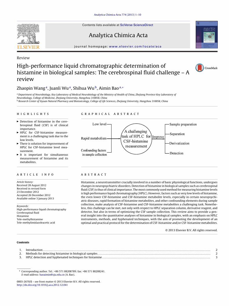
Analytica Chimica Acta 774 (2013) 1– 10
Contents lists available at SciVerse ScienceDirect
Analytica Chimica Acta
j ourna l ho me page: www.elsev ier .com/ locate /aca
eview
igh-performance liquid chromatographic determination ofistamine in biological samples: The cerebrospinal fluid challenge – Aeview
haopin Wanga, Juanli Wua, Shihua Wub, Aimin Baoa,∗
Department of Neurobiology, Key Laboratory of Medical Neurobiology of the Ministry of Health of China, Zhejiang Province Key Laboratory ofeurobiology, College of Medicine, Zhejiang University, Hangzhou 310058, ChinaResearch Center of Siyuan Natural Pharmacy and Biotoxicology, College of Life Sciences, Zhejiang University, Hangzhou 310058, China
i g h l i g h t s
Detection of histamine in the cere-brospinal fluid (CSF) is of clinicalimportance.HPLC for CSF-histamine measure-ment is a challenging task due to thelow levels.There is solution for improvement ofHPLC for CSF-histamine level mea-surement.It is important for simultaneousmeasurement of histamine and itsmetabolites.
g r a p h i c a l a b s t r a c t
r t i c l e i n f o
rticle history:eceived 28 August 2012eceived in revised form3 December 2012ccepted 26 December 2012vailable online 3 January 2013
eywords:
a b s t r a c t
Histamine, a neurotransmitter crucially involved in a number of basic physiological functions, undergoeschanges in neuropsychiatric disorders. Detection of histamine in biological samples such as cerebrospinalfluid (CSF) is thus of clinical importance. The most commonly used method for measuring histamine levelsis high performance liquid chromatography (HPLC). However, factors such as very low levels of histamine,the even lower CSF-histamine and CSF-histamine metabolite levels, especially in certain neuropsychi-atric diseases, rapid formation of histamine metabolites, and other confounding elements during samplecollection, make analysis of CSF-histamine and CSF-histamine metabolites a challenging task. Nonethe-
igh-performance liquid chromatographyerebrospinal fluidistamineele-methylhistamineele-methylimidazoleacetic acid
less, this challenge can be met, not only with respect to HPLC separation column, derivative reagent, anddetector, but also in terms of optimizing the CSF sample collection. This review aims to provide a gen-eral insight into the quantitative analyses of histamine in biological samples, with an emphasis on HPLCinstruments, methods, and hyphenated techniques, with the aim of promoting the development of anoptimal and practical protocol for the determination of CSF-histamine and/or CSF-histamine metabolites.
© 2013 Elsevier B.V. All rights reserved.
ontents
1. Introduction . . . . . . . . . . . . . . . . . . . . . . . . . . . . . . . . . . . . . . . . . . . . . . . . . . . . . . . . . . . . . . . .
2. Methods for detecting histamine in biological samples . . . . . . . . . . . . . . . . . . .
3. HPLC detection and hyphenated techniques for histamine . . . . . . . . . . . . . . .
∗ Corresponding author. Tel.: +86 571 88208789; fax: +86 571 88208241.E-mail address: [email protected] (A. Bao).
003-2670/$ – see front matter © 2013 Elsevier B.V. All rights reserved.ttp://dx.doi.org/10.1016/j.aca.2012.12.041
. . . . . . . . . . . . . . . . . . . . . . . . . . . . . . . . . . . . . . . . . . . . . . . . . . . . . . . . . . . . . . . . . . . . . . . . . . 2
. . . . . . . . . . . . . . . . . . . . . . . . . . . . . . . . . . . . . . . . . . . . . . . . . . . . . . . . . . . . . . . . . . . . . . . . . . 2. . . . . . . . . . . . . . . . . . . . . . . . . . . . . . . . . . . . . . . . . . . . . . . . . . . . . . . . . . . . . . . . . . . . . . . . . . 3
2 Z. Wang et al. / Analytica Chimica Acta 774 (2013) 1– 10
3.1. Sample preparation . . . . . . . . . . . . . . . . . . . . . . . . . . . . . . . . . . . . . . . . . . . . . . . . . . . . . . . . . . . . . . . . . . . . . . . . . . . . . . . . . . . . . . . . . . . . . . . . . . . . . . . . . . . . . . . . . . . . . . . . . . 33.1.1. Extraction . . . . . . . . . . . . . . . . . . . . . . . . . . . . . . . . . . . . . . . . . . . . . . . . . . . . . . . . . . . . . . . . . . . . . . . . . . . . . . . . . . . . . . . . . . . . . . . . . . . . . . . . . . . . . . . . . . . . . . . . . . . 33.1.2. Purification . . . . . . . . . . . . . . . . . . . . . . . . . . . . . . . . . . . . . . . . . . . . . . . . . . . . . . . . . . . . . . . . . . . . . . . . . . . . . . . . . . . . . . . . . . . . . . . . . . . . . . . . . . . . . . . . . . . . . . . . . 33.1.3. Derivatization . . . . . . . . . . . . . . . . . . . . . . . . . . . . . . . . . . . . . . . . . . . . . . . . . . . . . . . . . . . . . . . . . . . . . . . . . . . . . . . . . . . . . . . . . . . . . . . . . . . . . . . . . . . . . . . . . . . . . . . 4
3.2. Separation . . . . . . . . . . . . . . . . . . . . . . . . . . . . . . . . . . . . . . . . . . . . . . . . . . . . . . . . . . . . . . . . . . . . . . . . . . . . . . . . . . . . . . . . . . . . . . . . . . . . . . . . . . . . . . . . . . . . . . . . . . . . . . . . . . . . 43.3. Detection . . . . . . . . . . . . . . . . . . . . . . . . . . . . . . . . . . . . . . . . . . . . . . . . . . . . . . . . . . . . . . . . . . . . . . . . . . . . . . . . . . . . . . . . . . . . . . . . . . . . . . . . . . . . . . . . . . . . . . . . . . . . . . . . . . . . . 7
4. Progress in the determination of CSF-histamine levels . . . . . . . . . . . . . . . . . . . . . . . . . . . . . . . . . . . . . . . . . . . . . . . . . . . . . . . . . . . . . . . . . . . . . . . . . . . . . . . . . . . . . . . . . . . . . 75. The determination of histamine metabolite levels in CSF . . . . . . . . . . . . . . . . . . . . . . . . . . . . . . . . . . . . . . . . . . . . . . . . . . . . . . . . . . . . . . . . . . . . . . . . . . . . . . . . . . . . . . . . . . . 96. Conclusion. . . . . . . . . . . . . . . . . . . . . . . . . . . . . . . . . . . . . . . . . . . . . . . . . . . . . . . . . . . . . . . . . . . . . . . . . . . . . . . . . . . . . . . . . . . . . . . . . . . . . . . . . . . . . . . . . . . . . . . . . . . . . . . . . . . . . . . . . . . . 9
Acknowledgements . . . . . . . . . . . . . . . . . . . . . . . . . . . . . . . . . . . . . . . . . . . . . . . . . . . . . . . . . . . . . . . . . . . . . . . . . . . . . . . . . . . . . . . . . . . . . . . . . . . . . . . . . . . . . . . . . . . . . . . . . . . . . . . . . . 9 . . . . . .
1
mptlohtnhcoo
cbttarth
References . . . . . . . . . . . . . . . . . . . . . . . . . . . . . . . . . . . . . . . . . . . . . . . . . . . . . . . . . . . .
Zhaopin Wang is a M.S. candidate in ZhejiangUniversity School of Medicine (China). He is cur-rently working on the neurobiological basis, espe-cially on the interaction among the neurotrans-mitters/neuromodulators in depression under thesupervision of Prof. Aimin Bao.
Juanli Wu is a Ph.D. candidate in Zhejiang UniversitySchool of Medicine (China). She is currently studyingthe role of histamine in mood disorders and is focusingon the development of optimal HPLC protocol for CSF-and plasma histamine analysis under the supervisionof Prof. Aimin Bao.
. Introduction
Histaminergic neurons are exclusively located in the tubero-amillary nucleus of the posterior hypothalamus, from where they
roject to practically all brain regions. Histamine is involved inhe regulation of important neurophysiological functions, such asocomotor activity, sleep–wake cycle, attention, cognition, mem-ry, and stress responses [1–3]. Changes in brain histamine levelsave been observed in many neurological disorders, including mul-iple sclerosis [4], Alzheimer’s disease [5], febrile convulsions [6],arcolepsy [7,8], and hypersomnia [9,10]. The concentrations ofistamine in cerebrospinal fluid (CSF) may be regarded as an indi-ator for central histaminergic activity [5]. This makes the detectionf CSF-histamine levels of potentially great significance for physi-logical research as well as for the clinic.
High performance liquid chromatography (HPLC) is the mostommonly used method for measuring histamine levels. However,ecause of the low levels of histamine, the rapid formation of his-amine metabolites [5,11,12], and the many confounding factors
hat arise during sample collection, the analysis of CSF-histaminend CSF-histamine metabolite levels remains a challenging task foresearchers. The solution for this problem is to improve the sensi-ivity and specificity of assays such as HPLC. Similar solutions willold for histamine determination in other biological samples. The. . . . . . . . . . . . . . . . . . . . . . . . . . . . . . . . . . . . . . . . . . . . . . . . . . . . . . . . . . . . . . . . . . . . . . . . . 9
Shihua Wu received his Ph.D. of Organic Chem-istry (2004) in Zhejiang University School of Science(China). He was appointed as a lecturer of Collegeof Life Sciences Zhejiang University in 2004 and wasappointed as an associate professor from 2006. Hiswork mainly focuses on the extraction, separation,and identification of the structures of biotoxins in theantitumor natural products, as well as studying theirworking mechanism and application. The main toolsused are high-speed countercurrent chromatographyand related chromatographies.
Aimin Bao received her Ph.D. of Neurobiology (2003)in School of Life Science, University of Science andTechnology of China. She worked as a Post-Docresearcher at the Netherlands Institute for Neu-roscience, Amsterdam, the Netherlands, and wasappointed as professor of Neurobiology and PI in Zhe-jiang University School of Medicine from 2007 tillnow. Her work focuses on the neurobiological basisof signs and symptoms of neuropsychiatric disordersand the biological rhythm of hormones and behav-iors. The main approaches used include hormones andneurotransmitters assays.
current review therefore aims at providing a general insight intothe quantitative analyses of histamine in biological samples, par-ticularly in terms of HPLC instruments, methods, and hyphenatedtechniques, with the purpose of promoting the development of anoptimal protocol for the determination of CSF-histamine and/orhistamine metabolites.
2. Methods for detecting histamine in biological samples
The main methods for determining the levels of histaminein biological samples such as blood, urine and brain tis-sue, are radioenzymatic assay (REA), radioimmunoassay (RIA),enzymeimmunoassay (EIA), gas chromatography (GC), capillaryelectrophoresis (CE), capillary electrochromatography (CEC), HPLC[13,14] and ultra performance liquid chromatography (UPLC).
Since REA, RIA and EIA are based upon the specific reactionsbetween antigen and antibody, the key step for developing anoptimal protocol for measuring histamine concentrations withthese techniques is the production of a potent and specific mono-
clonal or polyclonal antibody against histamine. Because histamineis a small molecule and a hapten with antigenic (but withoutimmunogenic) potency [13], it is relatively difficult to produce ahistamine-antibody with both high specificity and high sensitivity.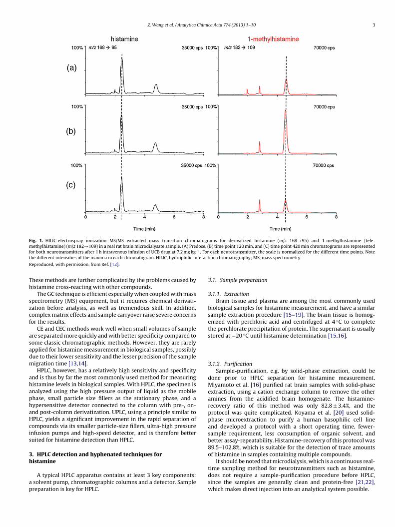
Z. Wang et al. / Analytica Chimica Acta 774 (2013) 1– 10 3
Fig. 1. HILIC-electrospray ionization MS/MS extracted mass transition chromatograms for derivatized histamine (m/z 168→95) and 1-methylhistamine (tele-methylhistamine) (m/z 182→109) in a real rat brain microdialysate sample. (A) Predose, (B) time point 120 min, and (C) time point 420 min chromatograms are representedf −1. Fort teract
R
Th
szcf
asadm
ahaphaHcis
3h
ap
or both neurotransmitters after 1 h intravenous infusion of UCB drug at 7.2 mg kghe different intensities of the maxima in each chromatogram. HILIC, hydrophilic in
eproduced, with permission, from Ref. [12].
hese methods are further complicated by the problems caused byistamine cross-reacting with other compounds.
The GC technique is efficient especially when coupled with masspectrometry (MS) equipment, but it requires chemical derivati-ation before analysis, as well as tremendous skill. In addition,omplex matrix effects and sample carryover raise severe concernsor the results.
CE and CEC methods work well when small volumes of samplere separated more quickly and with better specificity compared toome classic chromatographic methods. However, they are rarelypplied for histamine measurement in biological samples, possiblyue to their lower sensitivity and the lesser precision of the sampleigration time [13,14].HPLC, however, has a relatively high sensitivity and specificity
nd is thus by far the most commonly used method for measuringistamine levels in biological samples. With HPLC, the specimen isnalyzed using the high pressure output of liquid as the mobilehase, small particle size fillers as the stationary phase, and aypersensitive detector connected to the column with pre-, on-nd post-column derivatization. UPLC, using a principle similar toPLC, yields a significant improvement in the rapid separation ofompounds via its smaller particle-size fillers, ultra-high pressurenfusion pumps and high-speed detector, and is therefore betteruited for histamine detection than HPLC.
. HPLC detection and hyphenated techniques foristamine
A typical HPLC apparatus contains at least 3 key components: solvent pump, chromatographic columns and a detector. Samplereparation is key for HPLC.
each neurotransmitter, the scale is normalized for the different time points. Noteion chromatography; MS, mass spectrometry.
3.1. Sample preparation
3.1.1. ExtractionBrain tissue and plasma are among the most commonly used
biological samples for histamine measurement, and have a similarsample extraction procedure [15–19]. The brain tissue is homog-enized with perchloric acid and centrifuged at 4 ◦C to completethe perchlorate precipitation of protein. The supernatant is usuallystored at −20 ◦C until histamine determination [15,16].
3.1.2. PurificationSample-purification, e.g. by solid-phase extraction, could be
done prior to HPLC separation for histamine measurement.Miyamoto et al. [16] purified rat brain samples with solid-phaseextraction, using a cation exchange column to remove the otheramines from the acidified brain homogenate. The histamine-recovery ratio of this method was only 82.8 ± 3.4%, and theprotocol was quite complicated. Koyama et al. [20] used solid-phase microextraction to purify a human basophilic cell lineand developed a protocol with a short operating time, fewer-sample requirement, less consumption of organic solvent, andbetter assay-repeatability. Histamine-recovery of this protocol was89.5–102.8%, which is suitable for the detection of trace amountsof histamine in samples containing multiple compounds.
It should be noted that microdialysis, which is a continuous real-
time sampling method for neurotransmitters such as histamine,does not require a sample-purification procedure before HPLC,since the samples are generally clean and protein-free [21,22],which makes direct injection into an analytical system possible.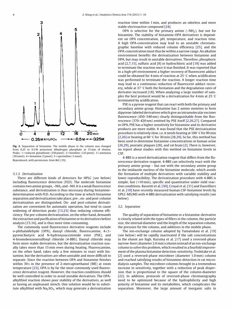
4 Z. Wang et al. / Analytica Chimi
Fig. 2. Separation of histamine. The mobile phase in the column was changedfrom 0.25 to 0.5 M potassium dihydrogen phosphate at 15 min of elution.Peaks: 1 = reduced glutathione (250 pmol); 2 = histidine (125 pmol); 3 = ammonia(50 nmol); 4 = histamine (5 pmol); 5 = spermidine (1 nmol).
R
3
icsdsdzwcta
op4faotswtcbaat
[22]. In addition, protocols of reversed-phase chromatography
eproduced, with permission, from Ref. [19].
.1.3. DerivatizationThere are different kinds of detectors for HPLC (see below)
ncluding fluorescence detection (FLD). The molecule histamineontains two amine groups, –NH2 and –NH. It is a weak fluorescenceubstance, and derivatization is thus necessary during histamine-etermination with FLD. According to the time at which histamineeparation and derivatization take place, pre-, on- and post-columnerivatization are distinguished. On- and post-column derivati-ation are convenient for automatic operation, but tend to causeidening of detection peaks [13,23] thus reducing column effi-
iency. The pre-column derivatization, on the other hand, demandshe extraction and purification of histamine or its derivatives beforenalysis [15,16], and is thus more time-consuming.
The commonly used fluorescence derivative reagents include-phthalaldehyde (OPA), dansyl chloride, fluorescamine, 4-(1-yrene)butyric acid N-hydroxysuccinimide ester (PSE), and-bromobenzenesulfonyl chloride (4-BBS). Dansyl chloride mayorm more stable derivatives, but the derivatization reaction usu-lly takes more than 15 min even during heating. Fluorescamine,n the other hand, takes only a few minutes to react with his-amine, but the derivatives are often unstable and more difficult toeparate. Since the reaction between OPA and histamine finishesithin 30 s in the presence of 2-mercaptoethanol (ME) at room
emperature [23], OPA is by far the most commonly used fluores-ence derivative reagent. However, the reaction conditions shoulde well controlled in order to avoid instable derivatives. The OPA-
lkylthiol reaction shows poor stability of the derivatives, as wells having an unpleasant stench. One solution would be to substi-ute alkylthiol with Na2SO3, which may generate a derivatizationca Acta 774 (2013) 1– 10
reaction time within 1 min, and produces an odorless and morestable electroactive compound [24].
OPA is selective for the primary amine (–NH2), but not forhistamine. The stability of histamine-OPA derivatives is depend-ent on OPA concentration, pH, temperature, and reaction time.A high OPA-concentration may lead to an unstable chromato-graphic baseline with reduced column efficiency [25], and theOPA-concentration must thus be within a narrow range. An alkalineenvironment benefits the derivatization between histamine andOPA, but may result in unstable derivatives. Therefore, phosphoricacid [2,7,15], sulfuric acid [8] or hydrochloric acid [18] was addedto terminate the reaction once it was finished. It was reported thatin a high-pH environment a higher recovery of fluorescent adductcould be obtained for 4 min of reaction at 25 ◦C when acidificationwas performed to terminate the reaction. A longer reaction timemay lead to a continuous reduction of fluorescent adduct recov-ery, while at 37 ◦C both the formation and the degradation rates ofderivates increased [18]. When analyzing a large number of sam-ples the best protocol would be a derivatization for 4 min at 25 ◦Cterminated by acidification.
PSE is a pyrene reagent that can react with both the primary andsecondary amine group. Histamine has 2 amino moieties to formdipyrene-labeled derivatives which give an intramolecular excimerfluorescence (450–540 nm) clearly distinguishable from the fluo-rescence (370–420 nm) emitted by PSE itself [2,26,27]. Comparedto OPA, PSE has a higher sensitivity for histamine and its derivativeproducts are more stable. It was found that the PSE derivatizationprocedure is relatively slow, i.e. it needs heating at 100 ◦C for 90 min[2,26] or heating at 60 ◦C for 30 min [28,29]. So far, this method hasbeen used to determine histamine-concentrations in human urine[26,29], psoriatic plaques [28], and rat brain [2]. There is, however,no report about studies with this method on histamine levels inCSF.
4-BBS is a novel derivatization reagent that differs from the flu-orescence derivative reagent. 4-BBS can selectively react with theprimary amine group – but not with the secondary amine groupin the imidazole nucleus of the histamine molecule, which avoidsthe formation of multiple derivatives with variable stability andlower reproducibility. The derivatization procedure with 4-BBS issimple, fast (<10 min), specific and quantitative under mild reac-tion conditions. Bassetti et al. [30], Croyal et al. [31] and Dauvillierset al. [10] have recently measured human CSF-histamine levels byUPLC-MS/MS with 4-BBS derivatization with satisfying results (seebelow).
3.2. Separation
The quality of separation of histamine or a histamine-derivativeis closely related with the types of fillers in the column, the particlesize, the internal diameter and the length of the separation column,the pressure for the column, and additives in the mobile phase.
The ion-exchange column adopted by Yamatodani et al. [19](see below) will be rapidly inactivated if the salt concentrationsin the eluent are high. Kuruma et al. [17] used a reversed-phasenarrow-bore (diameter 2.0 mm) column instead of an ion-exchangecolumn to solve this problem, which resulted in a fourfold improve-ment of the plasma histamine detection-sensitivity. Yoshitake et al.[2] used a reversed-phase microbore (diameter 1.0 mm) columnand reached satisfying results of histamine-detection in rat micro-dialysis samples. The microbore columns brought in a tremendousincrease in sensitivity, together with a reduction of peak disper-sion that is proportional to the square of the column-diameter
need to be optimized because of the hydrophilicity and highpolarity of histamine and its metabolites, which complicates theseparation. Moreover, the large amount of inorganic salts in
Z. Wang et al. / Analytica Chimica Acta 774 (2013) 1– 10 5
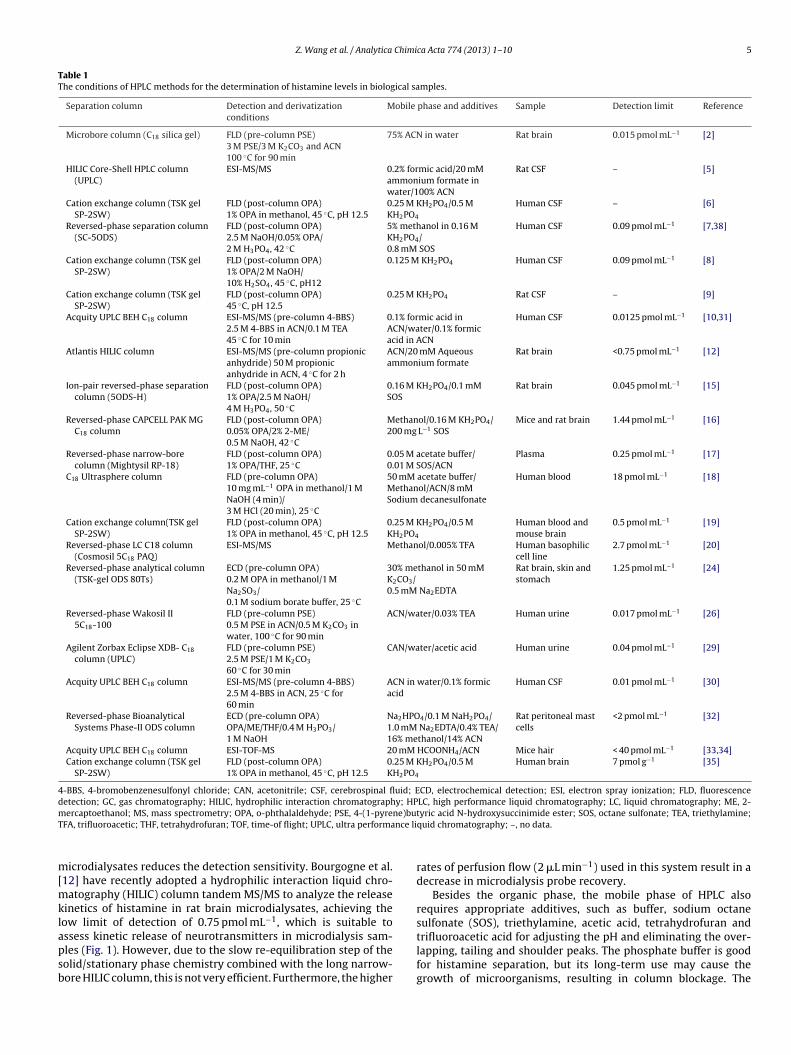
Table 1The conditions of HPLC methods for the determination of histamine levels in biological samples.
Separation column Detection and derivatizationconditions
Mobile phase and additives Sample Detection limit Reference
Microbore column (C18 silica gel) FLD (pre-column PSE)3 M PSE/3 M K2CO3 and ACN100 ◦C for 90 min
75% ACN in water Rat brain 0.015 pmol mL−1 [2]
HILIC Core-Shell HPLC column(UPLC)
ESI-MS/MS 0.2% formic acid/20 mMammonium formate inwater/100% ACN
Rat CSF – [5]
Cation exchange column (TSK gelSP-2SW)
FLD (post-column OPA)1% OPA in methanol, 45 ◦C, pH 12.5
0.25 M KH2PO4/0.5 MKH2PO4
Human CSF – [6]
Reversed-phase separation column(SC-5ODS)
FLD (post-column OPA)2.5 M NaOH/0.05% OPA/2 M H3PO4, 42 ◦C
5% methanol in 0.16 MKH2PO4/0.8 mM SOS
Human CSF 0.09 pmol mL−1 [7,38]
Cation exchange column (TSK gelSP-2SW)
FLD (post-column OPA)1% OPA/2 M NaOH/10% H2SO4, 45 ◦C, pH12
0.125 M KH2PO4 Human CSF 0.09 pmol mL−1 [8]
Cation exchange column (TSK gelSP-2SW)
FLD (post-column OPA)45 ◦C, pH 12.5
0.25 M KH2PO4 Rat CSF – [9]
Acquity UPLC BEH C18 column ESI-MS/MS (pre-column 4-BBS)2.5 M 4-BBS in ACN/0.1 M TEA45 ◦C for 10 min
0.1% formic acid inACN/water/0.1% formicacid in ACN
Human CSF 0.0125 pmol mL−1 [10,31]
Atlantis HILIC column ESI-MS/MS (pre-column propionicanhydride) 50 M propionicanhydride in ACN, 4 ◦C for 2 h
ACN/20 mM Aqueousammonium formate
Rat brain <0.75 pmol mL−1 [12]
Ion-pair reversed-phase separationcolumn (5ODS-H)
FLD (post-column OPA)1% OPA/2.5 M NaOH/4 M H3PO4, 50 ◦C
0.16 M KH2PO4/0.1 mMSOS
Rat brain 0.045 pmol mL−1 [15]
Reversed-phase CAPCELL PAK MGC18 column
FLD (post-column OPA)0.05% OPA/2% 2-ME/0.5 M NaOH, 42 ◦C
Methanol/0.16 M KH2PO4/200 mg L−1 SOS
Mice and rat brain 1.44 pmol mL−1 [16]
Reversed-phase narrow-borecolumn (Mightysil RP-18)
FLD (post-column OPA)1% OPA/THF, 25 ◦C
0.05 M acetate buffer/0.01 M SOS/ACN
Plasma 0.25 pmol mL−1 [17]
C18 Ultrasphere column FLD (pre-column OPA)10 mg mL−1 OPA in methanol/1 MNaOH (4 min)/3 M HCl (20 min), 25 ◦C
50 mM acetate buffer/Methanol/ACN/8 mMSodium decanesulfonate
Human blood 18 pmol mL−1 [18]
Cation exchange column(TSK gelSP-2SW)
FLD (post-column OPA)1% OPA in methanol, 45 ◦C, pH 12.5
0.25 M KH2PO4/0.5 MKH2PO4
Human blood andmouse brain
0.5 pmol mL−1 [19]
Reversed-phase LC C18 column(Cosmosil 5C18 PAQ)
ESI-MS/MS Methanol/0.005% TFA Human basophiliccell line
2.7 pmol mL−1 [20]
Reversed-phase analytical column(TSK-gel ODS 80Ts)
ECD (pre-column OPA)0.2 M OPA in methanol/1 MNa2SO3/0.1 M sodium borate buffer, 25 ◦C
30% methanol in 50 mMK2CO3/0.5 mM Na2EDTA
Rat brain, skin andstomach
1.25 pmol mL−1 [24]
Reversed-phase Wakosil II5C18-100
FLD (pre-column PSE)0.5 M PSE in ACN/0.5 M K2CO3 inwater, 100 ◦C for 90 min
ACN/water/0.03% TEA Human urine 0.017 pmol mL−1 [26]
Agilent Zorbax Eclipse XDB- C18
column (UPLC)FLD (pre-column PSE)2.5 M PSE/1 M K2CO3
60 ◦C for 30 min
CAN/water/acetic acid Human urine 0.04 pmol mL−1 [29]
Acquity UPLC BEH C18 column ESI-MS/MS (pre-column 4-BBS)2.5 M 4-BBS in ACN, 25 ◦C for60 min
ACN in water/0.1% formicacid
Human CSF 0.01 pmol mL−1 [30]
Reversed-phase BioanalyticalSystems Phase-II ODS column
ECD (pre-column OPA)OPA/ME/THF/0.4 M H3PO3/1 M NaOH
Na2HPO4/0.1 M NaH2PO4/1.0 mM Na2EDTA/0.4% TEA/16% methanol/14% ACN
Rat peritoneal mastcells
<2 pmol mL−1 [32]
Acquity UPLC BEH C18 column ESI-TOF-MS 20 mM HCOONH4/ACN Mice hair < 40 pmol mL−1 [33,34]Cation exchange column (TSK gel
SP-2SW)FLD (post-column OPA)1% OPA in methanol, 45 ◦C, pH 12.5
0.25 M KH2PO4/0.5 MKH2PO4
Human brain 7 pmol g−1 [35]
4-BBS, 4-bromobenzenesulfonyl chloride; CAN, acetonitrile; CSF, cerebrospinal fluid; ECD, electrochemical detection; ESI, electron spray ionization; FLD, fluorescencedetection; GC, gas chromatography; HILIC, hydrophilic interaction chromatography; HPLC, high performance liquid chromatography; LC, liquid chromatography; ME, 2-m ne)buT nce li
m[mklapsb
ercaptoethanol; MS, mass spectrometry; OPA, o-phthalaldehyde; PSE, 4-(1-pyreFA, trifluoroacetic; THF, tetrahydrofuran; TOF, time-of flight; UPLC, ultra performa
icrodialysates reduces the detection sensitivity. Bourgogne et al.12] have recently adopted a hydrophilic interaction liquid chro-
atography (HILIC) column tandem MS/MS to analyze the releaseinetics of histamine in rat brain microdialysates, achieving theow limit of detection of 0.75 pmol mL−1, which is suitable to
ssess kinetic release of neurotransmitters in microdialysis sam-les (Fig. 1). However, due to the slow re-equilibration step of theolid/stationary phase chemistry combined with the long narrow-ore HILIC column, this is not very efficient. Furthermore, the highertyric acid N-hydroxysuccinimide ester; SOS, octane sulfonate; TEA, triethylamine;quid chromatography; –, no data.
rates of perfusion flow (2 �L min−1) used in this system result in adecrease in microdialysis probe recovery.
Besides the organic phase, the mobile phase of HPLC alsorequires appropriate additives, such as buffer, sodium octanesulfonate (SOS), triethylamine, acetic acid, tetrahydrofuran and
trifluoroacetic acid for adjusting the pH and eliminating the over-lapping, tailing and shoulder peaks. The phosphate buffer is goodfor histamine separation, but its long-term use may cause thegrowth of microorganisms, resulting in column blockage. The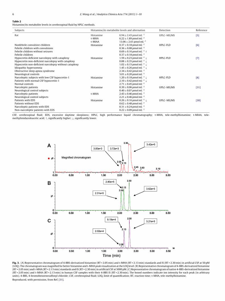
6 Z. Wang et al. / Analytica Chimica Acta 774 (2013) 1– 10
Table 2Histamine/its metabolite levels in cerebrospinal fluid by HPLC methods.
Subjects Histamine/its metabolite levels and alternation Detection Reference
Rat Histamine 6.94 ± 2.43 pmol mL−1 UPLC–MS/MS [5]t-MHA 6.22 ± 1.89 pmol mL−1
t-MIAA 13.06 ± 2.61 pmol mL−1
Nonfebrile convulsive children Histamine 0.37 ± 0.18 pmol mL−1 HPLC-FLD [6]Febrile children with convulsions 0.36 ± 0.08 pmol mL−1
Febrile children without seizures 0.69 ± 0.16 pmol mL−1 ↑Febrile children 0.37 ± 0.18 pmol mL−1
Hypocretin deficient narcolepsy with cataplexy Histamine 1.59 ± 0.23 pmol mL−1 ↓ HPLC-FLD [7]Hypocretin non-deficient narcolepsy with cataplexy 0.88 ± 0.35 pmol mL−1 ↓Hypocretin non-deficient narcolepsy without cataplexy 1.02 ± 0.15 pmol mL−1 ↓Idiopathic hypersomnia 1.45 ± 0.26 pmol mL−1 ↓Obstructive sleep apnea syndrome 2.34 ± 0.42 pmol mL−1
Neurological controls 3.01 ± 0.20 pmol mL−1
Narcoleptic subjects with low CSF hypocretin-1 Histamine 1.20 ± 0.18 pmol mL−1 ↓ HPLC-FLD [8]Patients with normal CSF hypocretin-1 2.10 ± 0.42 pmol mL−1 ↓Normal controls 2.71 ± 0.45 pmol mL−1
Narcoleptic patients Histamine 0.39 ± 0.06 pmol mL−1 UPLC–MS/MS [31]Neurological control subjects 0.40 ± 0.07 pmol mL−1
Narcoleptic patients t-MHA 2.43 ± 0.46 pmol mL−1
Neurological control subjects 2.21 ± 0.46 pmol mL−1
Patients with EDS Histamine 0.26 ± 0.16 pmol mL−1 ↓ UPLC–MS/MS [30]Patients without EDS 0.62 ± 0.48 pmol mL−1
Narcoleptic patients with EDS 0.31 ± 0.24 pmol mL−1
Non-narcoleptic patients with EDS 0.22 ± 0.09 pmol mL−1
CSF, cerebrospinal fluid; EDS, excessive daytime sleepiness; HPLC, high performance liquid chromatography; t-MHA, tele-methylhistamine; t-MIAA, tele-methylimidazoleacetic acid; ↑, significantly higher; ↓, significantly lower.
Fig. 3. (A) Representative chromatogram of 4-BBS-derivatized histamine (RT = 2.05 min) and t-MHA (RT = 2.13 min) standards and IS (RT = 2.30 min) in artificial CSF at 50 pM(LOQ). The chromatogram was magnified for better histamine and t-MHA peak visualization at the LOQ level. (B) Representative chromatogram of 4-BBS-derivatized histamine(RT = 2.05 min) and t-MHA (RT = 2.13 min) standards and IS (RT = 2.30 min) in artificial CSF at 5000 pM. (C) Representative chromatogram of native 4-BBS-derivatized histamine(RT = 2.05 min) and t-MHA (RT = 2.13 min) in human CSF samples with their 4-BBS IS (RT = 2.30 min). The boxed numbers indicate ion intensity for each peak (in arbitraryunits). 4-BBS, 4-bromobenzenesulfonyl chloride; CSF, cerebrospinal fluid; LOQ, limit of quantification; RT, reaction time; t-MHA, tele-methylhistamine.
Reproduced, with permission, from Ref. [31].
Chimica Acta 774 (2013) 1– 10 7
aatrawtaTdarms
3
mtotasd
lvHg2sempttfas(dmrsw
4
ciachetaRstfdr
Fig. 4. Representative chromatographs of basal levels of HA, t-mHA, and t-MIAAin rat cerebrospinal fluid at concentrations of 2.47 ng mL−1, 1.61 ng mL−1, and
−1
Z. Wang et al. / Analytica
cetate buffer is thus more suitable for long-term continuous uselthough it shows a slightly inferior effect for histamine separa-ion compared to the phosphate buffer system. SOS is an ion-paireagent which carries the opposite electric charges of histaminend of (R)�-methylhistamine [(R)�-MH], and it may thus combineith the latter to form a hydrophobic ion-pair, which could increase
he allocation of retention time for the nonpolar stationary phasend achieve the ideal separation of the various components [7,15].he nonpolar solvent triethylamine helps to stabilize the histamineerivative products when added to the OPA/ME reagent [17,32]nd thereby prevents the overlap of the histamine-peak witheagent peaks. Trifluoroacetic acid, acting as an ion-pair reagent,ay help to control the mobile phase pH and to improve the peak
hape.
.3. Detection
The detector transforms the component outflow from the chro-atographic column into detection signals, and thus determines
he sensitivity and precision of the system. There are different kindsf detectors including ultraviolet–visible detection (UV), FLD, elec-rochemical detection (ECD), chemiluminescence detection (CLD),nd MS [14], each of which requires its own specific detectingystem. Detector-selection should thus be the first priority wheneveloping an HPLC assay.
The sensitivity of histamine-determination by HPLC-UV is quiteow because histamine lacks strong absorption in the ultraviolet-isible light region [13,14]. A similar low sensitivity holds forPLC-CLD. HPLC-ECD has been used in one study [32] with aood sensitivity – the detection limit for histamine was less than
pmol mL−1 in rat peritoneal mast cells. It should be noted that theample-preparation prior to HPLC-ECD, which includes extraction,lution and purification, is rather complicated. Together with theany confounding factors, such as oxygen dissolved in the mobile
hase and changes in temperature that affect the ECD detection,he practical application of HPLC-ECD for histamine determina-ion is limited. HPLC-FLD, on the other hand, shows advantagesor trace analysis and for multiple compound sample analysis,lthough it needs histamine derivatization to improve the sen-itivity of FLD, since histamine is a weak-fluorescence materialsee above). Recently, HPLC-MS has been used in histamine-etermination in biological samples, including human CSF andice hair [5,20,30,31,33,34], with high specificity, since it not only
ecords the retention time of the compounds but also depicts theirtructure, thus solving the problem of distinguishing two aminesith similar physicochemical properties.
. Progress in the determination of CSF-histamine levels
HPLC-FLD with OPA-derivatization has been by far the mostommonly used method for measuring histamine levels in biolog-cal samples. Yamatodani et al. [19] developed a highly sensitivend specific protocol with post-column OPA-derivatization usingation exchange in the HPLC-FLD system for the determination ofistamine in mouse brain tissue and human plasma. The stage-lution and optimization of the post-column derivatization avoidedhe complex pre-column sample handling and/or derivatization. Inddition, there was a good correlation between this method andEA. As shown in Fig. 2, representative interfering substances inamples were well separated from histamine. This protocol was
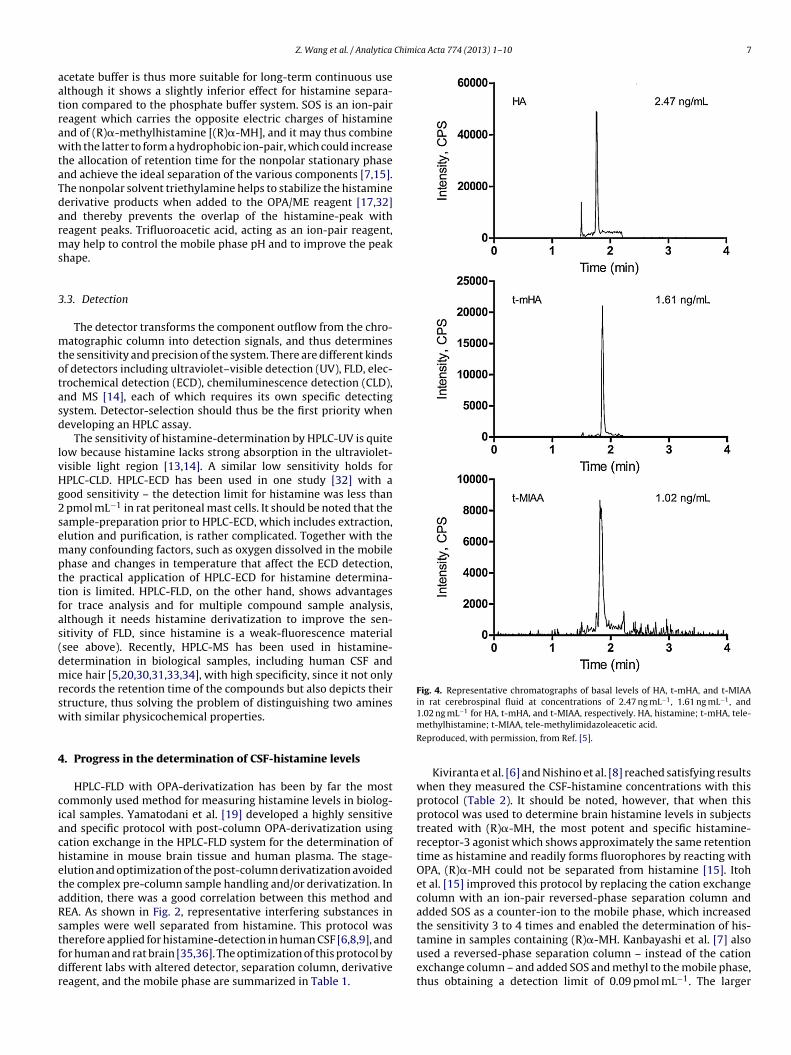
herefore applied for histamine-detection in human CSF [6,8,9], andor human and rat brain [35,36]. The optimization of this protocol byifferent labs with altered detector, separation column, derivativeeagent, and the mobile phase are summarized in Table 1.1.02 ng mL for HA, t-mHA, and t-MIAA, respectively. HA, histamine; t-mHA, tele-methylhistamine; t-MIAA, tele-methylimidazoleacetic acid.
Reproduced, with permission, from Ref. [5].
Kiviranta et al. [6] and Nishino et al. [8] reached satisfying resultswhen they measured the CSF-histamine concentrations with thisprotocol (Table 2). It should be noted, however, that when thisprotocol was used to determine brain histamine levels in subjectstreated with (R)�-MH, the most potent and specific histamine-receptor-3 agonist which shows approximately the same retentiontime as histamine and readily forms fluorophores by reacting withOPA, (R)�-MH could not be separated from histamine [15]. Itohet al. [15] improved this protocol by replacing the cation exchangecolumn with an ion-pair reversed-phase separation column andadded SOS as a counter-ion to the mobile phase, which increasedthe sensitivity 3 to 4 times and enabled the determination of his-
tamine in samples containing (R)�-MH. Kanbayashi et al. [7] alsoused a reversed-phase separation column – instead of the cationexchange column – and added SOS and methyl to the mobile phase,thus obtaining a detection limit of 0.09 pmol mL−1. The larger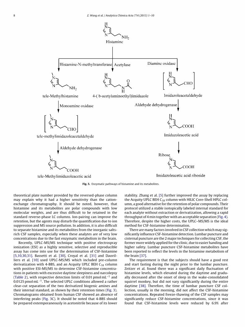
8 Z. Wang et al. / Analytica Chimica Acta 774 (2013) 1– 10
of his
tmehmsrstrc
ia[ldwt(0ctCib
Fig. 5. Enzymatic pathways
heoretical plate number provided by the reversed-phase columnay explain why it had a higher sensitivity than the cation-
xchange chromatography. It should be noted, however, thatistamine and its metabolites are polar compounds with lowolecular weights, and are thus difficult to be retained in the
tandard reverse-phase LC columns. Ion-pairing can improve theetention, but the agents may disturb the quantification due to ionuppression and MS source pollution. In addition, it is also difficulto separate histamine and its metabolites from the inorganic salts-ich CSF samples, especially when these analytes are of very lowoncentrations due to the fast enzymatic metabolism in the brain.
Recently, UPLC-MS/MS technique with positive electrosprayonization (ESI) as a highly sensitive, selective and reproduciblessay has come into use for the determination of CSF-histamine5,10,30,31]. Bassetti et al. [30], Croyal et al. [31] and Dauvil-iers et al. [10] used UPLC-MS/MS which included pre-columnerivatization with 4-BBS, and an Acquity UPLC BEH C18 columnith positive ESI-MS/MS to determine CSF-histamine concentra-
ions in patients with excessive daytime sleepiness and narcolepsyTable 2), with respective detection limits of 0.01 pmol mL−1 and.0125 pmol mL−1. The selected UPLC conditions allowed a ratherlear-cut separation of the two derivatized biogenic amines and
heir internal standard, as shown by their retention times (Fig. 3).hromatograms obtained from human CSF showed an absence ofnterfering peaks (Fig. 3C). It should be noted that 4-BBS shoulde prepared extemporaneously in acetonitrile because of its lower
tamine and its metabolites.
stability. Zhang et al. [5] further improved the assay by replacingthe Acquity UPLC BEH C18 column with HILIC Core-Shell HPLC col-umn, a good alternative for the retention of polar compounds. Theirprotocol utilized a stable isotopically labeled internal standard foreach analyte without extraction or derivatization, allowing a rapidthroughput of 4 min together with an acceptable separation (Fig. 4).Therefore, despite the higher costs, the UPLC–MS/MS is the idealmethod for CSF-histamine determination.
There are many factors involved in CSF collection which may sig-nificantly influence CSF-histamine detection. Lumbar puncture andcisternal puncture are the 2 major techniques for collecting CSF, theformer more widely applied for the clinic, due to easier handing andhigher safety. Lumbar puncture CSF-histamine metabolites havebeen reported to reflect the levels in the histamine metabolism ofthe brain [37].
The requirement is that the subjects should have a good restand start fasting during the night prior to the lumbar puncture.Zeitzer et al. found there was a significant daily fluctuation ofhistamine levels, which elevated during the daytime and gradu-ally decreased after the onset of sleep in the wake-consolidatedsquirrel monkey, but did not vary significantly during the entiredaytime [38]. Therefore, the time of lumbar puncture CSF col-
lection, usually in the morning, did not affect the CSF-histamineconcentrations. Repeated freeze-thawing of the CSF samples maysignificantly reduce CSF-histamine concentrations, since it wasfound that CSF-histamine levels were reduced by 6.9% after
Chimi
tTkPttlrcisto(Iao
5
n(omdbpip
alfiCic[mi
lFmimai
6
mpHfdCcuotbdfi
[
[
[
[
[[[
[[
[
[
[
[
[
[[
[
[
[
[
[
[
[[
[
[
Z. Wang et al. / Analytica
haw-freezing once, and by 44.4% after thaw-freezing twice [8].herefore, CSF samples should be aliquoted after collection andept at low temperature (−80 ◦C or −70 ◦C) until measurement.lease note there are no reports about the effect of storageemperature on the CSF-histamine concentrations. Moreover, con-amination of proteins or plasma may also affect CSF-histamineevels, and 60% perchloric acid is thus routinely applied to getid of proteins in the CSF [8,19]. CSF-histamine concentrations areonsiderably elevated upon contamination with blood [7–9] andt is therefore proposed that samples with visual (trace) bloodhould be excluded before CSF-histamine determination. In addi-ion, it is recommended that CSF-histamine concentrations shouldnly be determined when the samples have normal leukocyte0–5 × 10⁶ L−1) [6,39] and erythrocyte counts (0–5 × 10⁸ L−1) [39].n summary, strict sampling criteria related to exclusion of samplesnd sample handling are crucial for standardization of the protocolf CSF-histamine measurement.
. The determination of histamine metabolite levels in CSF
Histamine is metabolized via 3 enzymatic pathways (Fig. 5),amely (i) histamine is metabolized into tele-methylhistaminet-MHA) by histamine-N-methyltransferase and subsequentlyxidized into tele-methylimidazoleacetic acid (t-MIAA) byonoamine oxidase; (ii) histamine is deaminated by diamine oxi-
ase to form imidazoleacetic acid; and (iii) histamine is acetylatedy acetylase to form 4-(�-acetylaminoethyl)imidazole – but thisathway is found exclusively in enterobacteria and has not been
dentified in mammalians [13,14]. The first pathway is the majorathway, perhaps the only one in the mammalian brain [1,40].
Due to the fast enzymatic metabolism of histamine [5,11,12]nd the short half-life of brain histamine [41], together with theow levels of histamine in CSF and brain tissues, it is indeed dif-cult to estimate histamine activity in the brain on the basis ofSF-histamine measurements. However, since many reports have
ndicated that histamine metabolites (t-MHA and t-MIAA) in CSFan also be used as indicators of neuronal histaminergic activity5,11,40–42], a simultaneous measurement of histamine and its
etabolites [5,10,31] or merely of its metabolites [11,12,37,41,42]s therefore a serious option.
The methods for histamine-metabolite-determination in bio-ogical samples are the same as for histamine measurement.or research work, the major methods for measuring histamineetabolites in CSF are usually GC–MS or UPLC–MS/MS. The increas-
ng possibilities for funding and the advances in technology haveade it possible for UPLC–MS/MS to be gradually applied more
nd more to CSF-histamine-metabolite determination because ofts high sensitivity, specificity and accuracy.
. Conclusion
HPLC-FLD is a readily available method for histamine measure-ent in biological samples, and thus of importance for studying the
hysiological and pathological processes that involve histamine.owever, due to the low levels of histamine in CSF, the rapid
ormation of its metabolites and the many confounding factorsuring sample collection, it is still a challenging task to analyzeSF-histamine and CSF-histamine metabolites. Nonetheless, thishallenge can be met, not only for HPLC including separation col-mn, derivative reagent, and detector, but also in terms of theptimization of CSF sample collection. The development of the MS
echnique and the application of UPLC may lead to UPLC–MS/MSecoming the mainstream technique for CSF-histamine and theetection of its metabolites, due to its excellent sensitivity, speci-city and accuracy.[
[
ca Acta 774 (2013) 1– 10 9
Acknowledgements
The authors want to thank Mrs. Wilma Verweij for her secretar-ial assistance, Professor Dick Swaab for comments and correctionsof the paper. This review was supported by Nature Science Foun-dation of China (30970928 and 31271130), Science TechnologyProgram of Zhejiang Province (2009C34020), Zhejiang ProvincialNatural Science Foundation of China (LY12H09007).
References
[1] R.E. Brown, D.R. Stevens, H.L. Haas, Prog. Neurobiol. 63 (2001) 637–672.[2] T. Yoshitake, M. Yamaguchi, H. Nohta, F. Ichinose, H. Yoshida, S. Yoshitake, K.
Fuxe, J. Kehr, J. Neurosci. Methods 127 (2003) 11–17.[3] C. Ito, Biomed. Pharmacother. 54 (2000) 263–267.[4] F. Jadidi-Niaragh, A. Mirshafiey, Neuropharmacology 59 (2010) 180–189.[5] Y. Zhang, F.D. Tingley III, E. Tseng, M. Tella, X. Yang, E. Groeber, J. Liu, W. Li, C.J.
Schmidt, R. Steenwyk, J. Chromatogr. B Analyt. Technol. Biomed. Life Sci. 879(2011) 2023–2033.
[6] T. Kiviranta, L. Tuomisto, E.M. Airaksinen, Epilepsia 36 (1995) 276–280.[7] T. Kanbayashi, T. Kodama, H. Kondo, S. Satoh, Y. Inoue, S. Chiba, T. Shimizu, S.
Nishino, Sleep 32 (2009) 181–187.[8] S. Nishino, E. Sakurai, S. Nevsimalova, Y. Yoshida, T. Watanabe, K. Yanai, E.
Mignot, Sleep 32 (2009) 175–180.[9] A. Soya, Y.H. Song, T. Kodama, Y. Honda, N. Fujiki, S. Nishino, Neurosci. Lett. 430
(2008) 224–229.10] Y. Dauvilliers, N. Delallee, I. Jaussent, S. Scholz, S. Bayard, M. Croyal, J.C.
Schwartz, P. Robert, Sleep 35 (2012) 1359–1366.11] G.D. Prell, J.P. Green, C.A. Kaufmann, J.K. Khandelwal, A.M. Morrishow, D.G.
Kirch, M. Linnoila, R.J. Wyatt, Schizophr. Res. 14 (1995) 93–104.12] E. Bourgogne, F.X. Mathy, D. Boucaut, H. Boekens, O. Laprevote, Anal. Bioanal.
Chem. 402 (2012) 449–459.13] S. Oguri, Y. Yoneya, J. Chromatogr. B Analyt. Technol. Biomed. Life Sci. 781
(2002) 165–179.14] T. Toyo’oka, Biomed. Chromatogr. 22 (2008) 919–930.15] Y. Itoh, R. Oishi, N. Adachi, K. Saeki, J. Neurochem. 58 (1992) 884–889.16] Y. Miyamoto, R. Yoshimoto, M. Yumoto, A. Ishihara, K. Takahashi, H. Kotani, A.
Kanatani, S. Tokita, Anal. Biochem. 334 (2004) 89–96.17] K. Kuruma, T. Sakano, Anal. Sci. 15 (1999) 489–492.18] M. Previati, A. Raspadori, L. Bertolaso, A. Parmeggiani, D. Bindini, C. Vitali, I.
Lanzoni, E. Corbacella, M. Saviano, F. Fagioli, G. Blo, S. Capitani, J. Chromatogr.B Analyt. Technol. Biomed. Life Sci. 780 (2002) 331–339.
19] A. Yamatodani, H. Fukuda, H. Wada, T. Iwaeda, T. Watanabe, J. Chromatogr. 344(1985) 115–123.
20] J. Koyama, A. Takeuchi, C. Tode, M. Shimizu, I. Morita, M. Nobukawa, N.Kobayashi, J. Chromatogr. B Analyt. Technol. Biomed. Life Sci. 877 (2009)207–212.
21] C.S. Chaurasia, M. Muller, E.D. Bashaw, E. Benfeldt, J. Bolinder, R. Bullock,P.M. Bungay, E.C. DeLange, H. Derendorf, W.F. Elmquist, M. Hammarlund-Udenaes, C. Joukhadar, D.L. Kellogg Jr., C.E. Lunte, C.H. Nordstrom, H. Rollema,R.J. Sawchuk, B.W. Cheung, V.P. Shah, L. Stahle, U. Ungerstedt, D.F. Welty, H.Yeo, Pharm. Res. 24 (2007) 1014–1025.
22] M.I. Davies, J.D. Cooper, S.S. Desmond, C.E. Lunte, S.M. Lunte, Adv. Drug Deliv.Rev. 45 (2000) 169–188.
23] J.F. Peng, K.T. Fang, D.H. Xie, B. Ding, J.Y. Yin, X.M. Cui, Y. Zhang, J.F. Liu, J.Chromatogr. A 1209 (2008) 70–75.
24] M. Maldonado, K. Maeyama, Anal. Biochem. 432 (2012) 1–7.25] N.H. Kim, Y. Park, E.S. Jeong, C.S. Kim, M.K. Jeoung, K.S. Kim, S.H. Hong, J.K. Son,
J.T. Hong, I.Y. Park, D.C. Moon, Arch. Pharm. Res. 30 (2007) 1350–1357.26] T. Yoshitake, F. Ichinose, H. Yoshida, K. Todoroki, J. Kehr, O. Inoue, H. Nohta, M.
Yamaguchi, Biomed. Chromatogr. 17 (2003) 509–516.27] H. Yoshida, F. Ichinose, T. Yoshitake, Y. Nakano, K. Todoroki, H. Nohta, M. Yam-
aguchi, Anal. Sci. 20 (2004) 557–559.28] E. Guihen, W.L. Ho, A.M. Hogan, M.L. O’Connell, M.J. Leahy, B. Ramsay, W.T.
O’Connor, J. Chromatogr. B Analyt. Technol. Biomed. Life Sci. 880 (2012)119–124.
29] A.M. Hogan, C. Crean, U.M. Barrett, E. Guihen, J.D. Glennon, J. Sep. Sci. 35 (2012)1087–1093.
30] C.L. Bassetti, C.R. Baumann, Y. Dauvilliers, M. Croyal, P. Robert, J.C. Schwartz, J.Sleep Res. 19 (2010) 620–623.
31] M. Croyal, Y. Dauvilliers, O. Labeeuw, M. Capet, J.C. Schwartz, P. Robert, Anal.Biochem. 409 (2011) 28–36.
32] T.B. Jensen, P.D. Marley, J. Chromatogr. B Biomed. Appl. 670 (1995) 199–207.33] H. Kawanishi, T. Toyo’oka, K. Ito, M. Maeda, T. Hamada, T. Fukushima, M. Kato,
S. Inagaki, Clin. Chim. Acta 378 (2007) 122–127.34] H. Kawanishi, T. Toyo’oka, K. Ito, M. Maeda, T. Hamada, T. Fukushima, M. Kato,
S. Inagaki, J. Chromatogr. A 1132 (2006) 148–156.35] J.O. Rinne, O.V. Anichtchik, K.S. Eriksson, J. Kaslin, L. Tuomisto, H. Kalimo, M.
Roytta, P. Panula, J. Neurochem. 81 (2002) 954–960.36] A. Xu, E. Sakurai, A. Kuramasu, J. Zhang, J. Li, N. Okamura, D. Zhang, T. Yoshikawa,
T. Watanabe, K. Yanai, J. Pharmacol. Sci. 114 (2010) 444–453.37] G.D. Prell, J.K. Khandelwal, R.S. Burns, J.P. Green, J. Neurochem. 50 (1988)
1194–1199.

1 Chimi
[
[
0 Z. Wang et al. / Analytica
38] J.M. Zeitzer, T. Kodama, C.L. Buckmaster, Y. Honda, D.M. Lyons, S. Nishino, E.Mignot, J. Sleep Res. 21 (2012) 189–194.
39] K. Blennow, A. Wallin, C.G. Gottfries, J.E. Mansson, L. Sven-nerholm, J. Neural Transm. Park. Dis. Dement. Sect. 5 (1993)5–15.
[
[[
ca Acta 774 (2013) 1– 10
40] J.K. Khandelwal, L.B. Hough, J.P. Green, Klin. Wochenschr. 60 (1982)914–918.
41] G.D. Prell, J.P. Green, Agents Actions (1994) C5–C8, No. 41 Spec.42] G.D. Prell, J.K. Khandelwal, P.A. LeWitt, J.P. Green, Agents Actions 26 (1989)
267–272.