Embed Size (px)
Citation preview
ARTICLE OPEN
Highly flexible, wearable, and disposable cardiac biosensorsfor remote and ambulatory monitoringStephen P. Lee1, Grace Ha2, Don E. Wright1, Yinji Ma3,4, Ellora Sen-Gupta1, Natalie R. Haubrich2, Paul C. Branche1, Weihua Li1,Gilbert L. Huppert1, Matthew Johnson2, Hakan B. Mutlu1, Kan Li4, Nirav Sheth1, John A. Wright Jr.1, Yonggang Huang4,5,Moussa Mansour2, John A. Rogers5 and Roozbeh Ghaffari1,5
Contemporary cardiac and heart rate monitoring devices capture physiological signals using optical and electrode-based sensors.However, these devices generally lack the form factor and mechanical flexibility necessary for use in ambulatory and homeenvironments. Here, we report an ultrathin (~1mm average thickness) and highly flexible wearable cardiac sensor (WiSP) designedto be minimal in cost (disposable), light weight (1.2 g), water resistant, and capable of wireless energy harvesting. Theoreticalanalyses of system-level bending mechanics show the advantages of WiSP’s flexible electronics, soft encapsulation layers andbioadhesives, enabling intimate skin coupling. A clinical feasibility study conducted in atrial fibrillation patients demonstrates thatthe WiSP device effectively measures cardiac signals matching the Holter monitor, and is more comfortable. WiSP’s physicalattributes and performance results demonstrate its utility for monitoring cardiac signals during daily activity, exertion and sleep,with implications for home-based care.
npj Digital Medicine (2018) 1:2 ; doi:10.1038/s41746-017-0009-x
INTRODUCTIONWearable biosensing systems have become ubiquitous, providingeffective routes for quantifying important physiological metrics inboth medical and consumer applications.1–5 Multi-lead Holtermonitoring devices and event monitors, for example, representthe clinical standard of care for detecting and diagnosing cardiacrhythm6 and rate disorders based on continuous electrocardio-gram (ECG) waveforms and rate-related information.7–10 Althoughthese devices have been widely adopted, they are susceptible topoor patient compliance due in part to their bulky form factor andwired connections to leads.11,12
Advances in electronics miniaturization and semiconductorperformance have significantly reduced the areal footprint andoverall size of wearable sensors, creating new market opportu-nities for consumer and medical health monitoring.13 Single-leadcardiac devices14,15 have enabled continuous ECG monitoringwith significantly smaller footprints compared to Holter monitors,but these patch-based devices contain packaged electroniccomponents and are mechanically rigid,12,16 thus limiting intimateskin coupling for repeated, long-term daily use. Increasing interestin quantifying cardiac metrics at home has led to the integrationof low profile and high performance heart rate sensors in appareland wrist bands, but these devices usually rely on photoplethys-mography (PPG) and tend to be limited in signal accuracycompared to the clinical standards of care.17 Poor accuracy iscaused by the physical limitations of PPG, magnified by inherentchallenges associated with the peripheral wrist location of the
sensor and noisy interface with the skin.18 Despite theselimitations, the convenience of wrist-worn devices have ledresearchers to estimate ECG parameters from PPG signals,19
demonstrating that there is a continued desire to measure cardiacrhythm metrics with improved patient comfort.Recent advances in flexible epidermal electronics technologies
have begun to address many of the mechanical, wearability andcomfort limitations attributed to existing classes of wearable andapparel-based cardiac and heart rate monitoring devices.20–34
These novel epidermal devices contain soft, conformal sensorsand associated circuits embedded in ultrathin encapsulatinglayers that achieve intimate skin coupling.25,34 Here, we presenta highly flexible epidermal design and clinical implementation of anovel ECG and heart rate logging wearable sensor, henceforthreferred to as “WiSP”, which is low cost, light-weight (1.2 g), andcapable of energy harvesting. The WiSP device is comparable insize to a standard adhesive bandage (58 mm× 25mm× 1mm)and streams physiological data to commercial smartphones viastandard near-field-communication (NFC) for use in both ambu-latory and home-based settings.
RESULTSWiSP device design and characterizationThe WiSP epidermal sensor design in Fig. 1a, b highlights the keystructures, as well as the multi-layered conformal mechanics thatlead to tight skin coupling and robust cardiac signals. An elastic
Received: 19 April 2017 Revised: 15 June 2017 Accepted: 7 July 2017
1MC10 Inc, Lexington, MA 02421, USA; 2Massachusetts General Hospital, Boston, MA 02114, USA; 3AML, Department of Engineering Mechanics, Center for Mechanics andMaterials, Tsinghua University, Beijing 100084, China; 4Department of Civil and Environmental Engineering, Mechanical Engineering, and Materials Science and Engineering,Northwestern University, Evanston, IL 60208, USA and 5Center for Bio-Integrated Electronics, Departments of Materials Science and Engineering, Biomedical Engineering,Chemistry, Mechanical Engineering, Electrical Engineering and Computer Science, and Neurological Surgery, Simpson Querrey Institute for Nano/Biotechnology, McCormickSchool of Engineering, Northwestern University, Evanston, IL 60208, USACorrespondence: Moussa Mansour ([email protected]) or John A. Rogers ([email protected]) or Roozbeh Ghaffari ([email protected])Stephen P. Lee and Grace Ha contributed equally to this work.
www.nature.com/npjdigitalmed
Published in partnership with the Scripps Translational Science Institute
layer of polyurethane serves as an encapsulating layer. Measure-ments of water ingress rate under full immersion conditions showno water permeability over several hours, with no changes in WiSPdevice performance. The electronics are electrically and mechani-cally coupled to a flexible substrate layer that includes theelectroless nickel immersion gold electrodes. The user-facing sideof the device is covered with a biocompatible, medical grade skinadhesive and selectively coated conductive hydrogels for directskin contact. A significant fraction of the skin-side surface area isdedicated to the two electrodes. They are spaced apartsufficiently35 to allow for capture of the full PQRST waveforms(Fig. 1c), yet mechanically flexible (Fig. 1d, e).We applied ABAQUS commercial software36 to study the
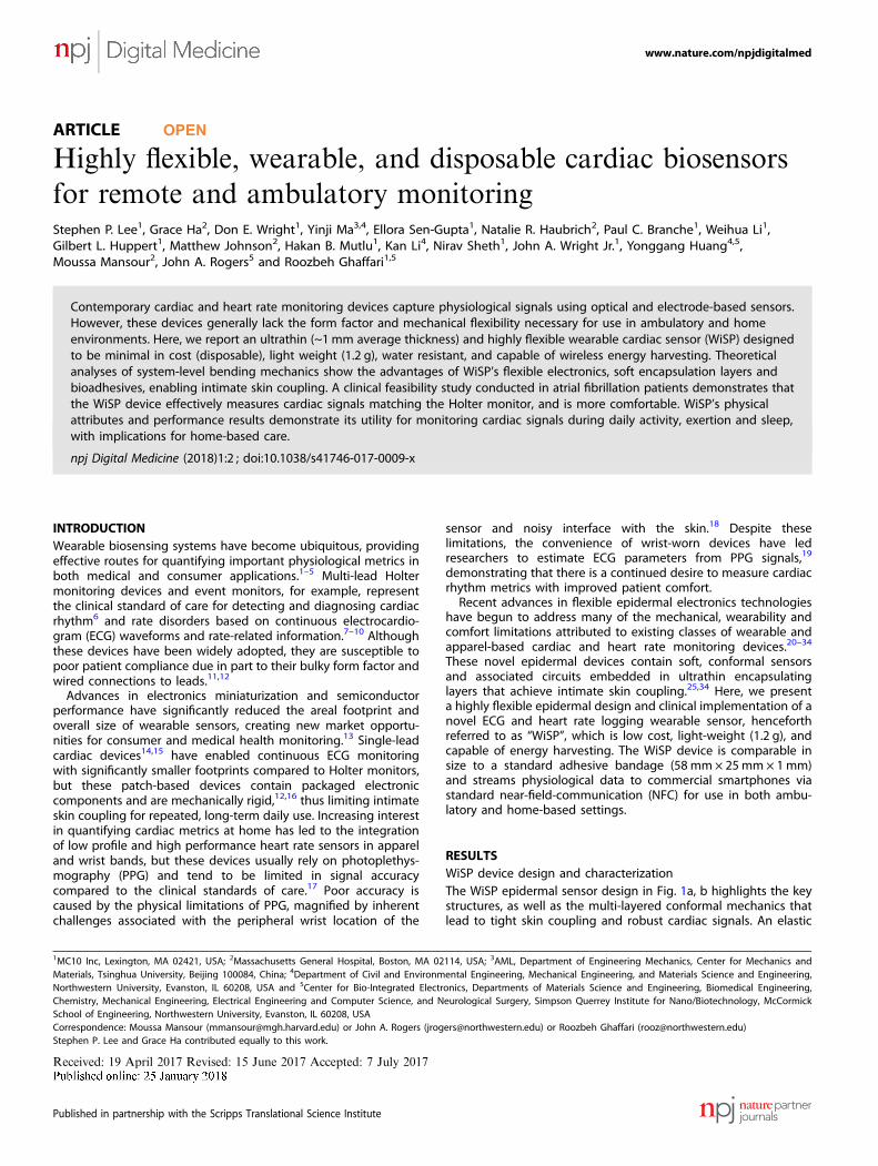
bending response of a simplified WiSP device during wear onthe skin (Fig. 2a, b). The simplified WiSP device, consisting of apolyimide (PI) layer with elastic modulus ~2.5 GPa and Poisson’sratio of 0.34, was mounted on a skin substrate (elastic modulus~130 kPa and Poisson’s ratio 0.5; ref. 37) through a 0.05mm-thickadhesive layer (elastic modulus ~17 kPa and Poisson’s ratio 0.5;ref. 38). Supplementary Fig. S1 shows that the interfacial stresses(particularly normal stresses) on the skin increase as the PIthickness increases under a fixed applied curvature of 0.005 mm−1
(defined by the curvature of the skin under the device, κ = α/L, asshown in Fig. 2a, b). These results indicate that thin form factors(e.g., ~0.2 mm PI thickness) for WiSP are well matched to themechanical properties and curvature of the body.With an average PI thickness of 0.2 mm for WiSP in the model,
Fig. 2c shows the interfacial stresses between the adhesive layerand skin under different applied curvatures (0.002, 0.005, and 0.01mm−1). The interfacial stresses on skin are largest near theperimeter of the WiSP device, which are within the threshold fornormal skin perception34 (20 kPa) for applied curvatures of 0.002and 0.005mm−1. In response to aggressive applied curvatures(~0.01 mm−1), the interfacial stresses on skin only exceed 20 kPa ata small local area (Fig. 2c), and the maximum principal strains on
the top surface of the PI layer (where the electronics are located)are smaller than 0.2% (Supplementary Fig. S2). As a result, theflexible design of WiSP remains functional and comfortable duringbending, and unlikely to experience damage in the activeelectronics layers during wear.To ensure reliable heart rate computations and ECG capture, the
WiSP onboard hardware conditions biopotentials with an instru-mentation amplifier configured to amplify time varying signalsand attenuate common-mode signals. A second-order high-passfilter and an amplified second-order low-pass active filter thencondition the signal. A right-leg drive circuit servos the common-mode voltage at the electrodes and improves common-moderejection. Ancillary active and passive components provide leads-off detection. To further condition cardiac signals, the microcon-troller with integrated ADC oversamples the signal and feeds itinto a 25-tap IIR digital filter. A state-machine computes heart rate,logs data, streams live ECG, controls LEDs and manages NFC, dataencryption and writing to memory. A learning algorithm measuresthe ECG baseline level, identifies aspects of the ECG morphology,detects R-wave signatures, and computes heart rate based on thesubject’s personal ECG signature.The WiSP microcontroller also optimizes power consumption by
coordinating power harvesting from NFC, system activation, wake,sleep, and power down. Empirically tested switching times controlthe surge current from the load at system activation and ensurereliable startup of the WiSP device from either the battery or NFCpower harvester. Once the system is initialized, active componentsthat are not being used at any given time enter a low-power stateand are initiated again only when needed. To remove the need forbulky physical buttons, the WiSP device relies on wirelessactivation using an NFC-enabled smartphone. By positioning thesmartphone in close proximity to the WiSP device (~3 cm), theNFC field is detected by an NFC integrated circuit and a dedicatedcircuit that activates the system. The microcontroller thenconfirms whether activation is valid, and, in turn, continues to
Fig. 1 Schematic illustrations and images of soft flexible cardiac sensor in a thin elastic enclosure and data transfer to smartphone app. aExploded image of WiSP device showing the multiple polymeric, electronic, adhesive and hydrogel layers. Image was created by co-authorDon E. Wright (permission granted). b Illustration of assembled WiSP device consisting of five distinct layers (180 µm thick along the edges,1.2 g) attached to the torso (in lead I or lead II orientation). The human silhouette is based on an icon created by Freepik (permission granted).c Data are wirelessly transmitted to smartphone (via NFC) for visualization of logged heart rate data and/or real-time ECG waveforms, andsubsequently transmitted from smartphone to a cloud server (via WiFi or cellular connectivity). d, eWiSP device in mechanical twist and benddeformations
Biosensors for remote and ambulatory monitoringSP Lee et al.
2
npj Digital Medicine (2018) 2 Published in partnership with the Scripps Translational Science Institute
1234567890
power-up. A similar set of actions can wirelessly shut down theWiSP.A custom-designed primary cell battery (450 µm thick; 4 mAh
capacity) supplies power to the electronics and memory modulefor logging heart rate. The logged data may be retrieved with orwithout battery power. Likewise, the full ECG waveform may bestreamed to the smartphone solely using harvested NFC power.The system uses end-to-end encryption from the patch to thesmartphone, and from the smartphone to a cloud server. This NFCfunctionality is interoperable with most smartphones (ISO/IEC14443 Part 2 and 3 compliant) and takes advantage of the custom-designed flexible antenna that wraps around the circumference ofthe WiSP device. The antenna and system design ensure reliableoperation despite variation among different smartphones’ NFCbroadcast power. The flexible antenna was designed to operateefficiently on both soft tissue and in free air to ensure usability andreliability across use cases. Tests verify that WiSP works with awide variety of commercially available smartphones, allowingconsumers and/or patients to use WiSP with their own smart-phones at the home (Supplementary Table S1).
Heart rate and ECG measurementsThe WiSP ECG waveforms were captured from healthy subjectsand compared to measurements taken concurrently with a clinicalstandard of care product (Fig. 3a; GE Dash 3000 Patient Monitor).The WiSP device is capable of capturing full, differential ECGsignals across lead I, II, or III anatomical positions. Figure 3a showsrepresentative WiSP ECG waveforms using two distinct filtersettings tuned for ECG and heart rate signal capture, respectively.The ECG filter allows for visualization of P and T-waves (middletrace of Fig. 3a), whereas the heart rate filter (bottom trace ofFig. 3a) operates with a narrow pass-band, thereby eliminating Pand T-wave morphology as well as most motion and muscleactivation artifacts. By applying this heart rate filter, we could testR-wave detection algorithms and benchmark against existingheart rate sensors.A field study (Fig. 3b) conducted on healthy subjects provides
base-level validation for the WiSP hardware, adhesive, and wearlocations. In this validation test, we attached the WiSP and acommercially available Polar H7 heart rate monitor to the chest ofeach healthy volunteer (>4 h) and recorded heart rate data duringroutine daily activities. The Polar H7 is a chest-strap electrode-
Fig. 2 Summary of computational studies detailing the effects of normal and shear mechanical stresses from WiSP device on human skin. aSchematic drawing illustrating the cross-sectional design of simplified WiSP device attached to human skin with key lateral and transversedimensions (ztot= 0.25 mm and Lo= 55mm) and material layers (adhesive layer and PI layer) and their associated Young’s modulus, E. b Sideprofile view of WiSP device attached to skin surface under bending deformation with angle α. The applied curvature (κ= α/Lo) is defined bythe curvature of the skin under the device. c Finite element (FE) simulation results for a device applying shear (left column) and normal (rightcolumn) stresses on skin during bending deformations. For 0.002 and 0.005mm−1 applied curvatures, the interfacial stresses on the skin are~20 kPa, which is within the range of normal skin sensitivity. For 0.01 mm−1 applied curvatures, the interfacial stresses on the skin exceed 20kPa near perimeter of the WiSP device
Biosensors for remote and ambulatory monitoringSP Lee et al.
3
Published in partnership with the Scripps Translational Science Institute npj Digital Medicine (2018) 2
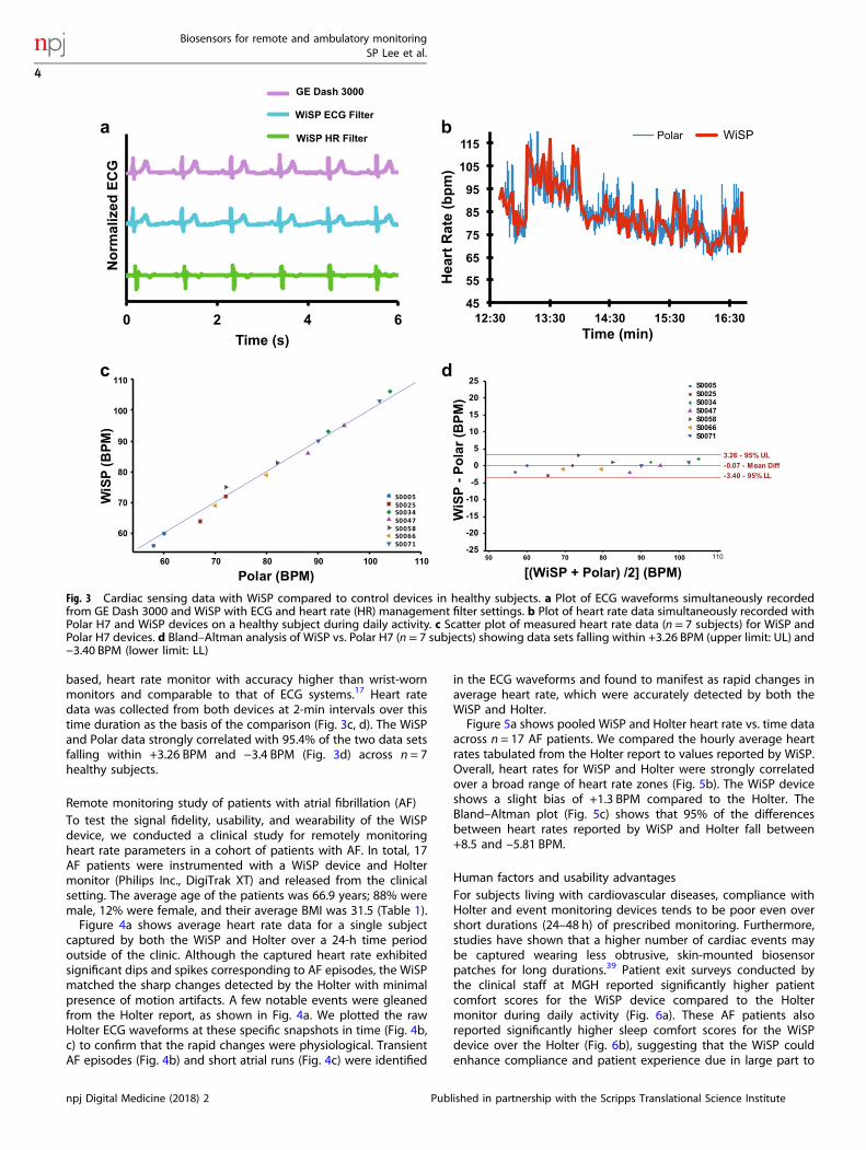
based, heart rate monitor with accuracy higher than wrist-wornmonitors and comparable to that of ECG systems.17 Heart ratedata was collected from both devices at 2-min intervals over thistime duration as the basis of the comparison (Fig. 3c, d). The WiSPand Polar data strongly correlated with 95.4% of the two data setsfalling within +3.26 BPM and −3.4 BPM (Fig. 3d) across n = 7healthy subjects.
Remote monitoring study of patients with atrial fibrillation (AF)To test the signal fidelity, usability, and wearability of the WiSPdevice, we conducted a clinical study for remotely monitoringheart rate parameters in a cohort of patients with AF. In total, 17AF patients were instrumented with a WiSP device and Holtermonitor (Philips Inc., DigiTrak XT) and released from the clinicalsetting. The average age of the patients was 66.9 years; 88% weremale, 12% were female, and their average BMI was 31.5 (Table 1).Figure 4a shows average heart rate data for a single subject
captured by both the WiSP and Holter over a 24-h time periodoutside of the clinic. Although the captured heart rate exhibitedsignificant dips and spikes corresponding to AF episodes, the WiSPmatched the sharp changes detected by the Holter with minimalpresence of motion artifacts. A few notable events were gleanedfrom the Holter report, as shown in Fig. 4a. We plotted the rawHolter ECG waveforms at these specific snapshots in time (Fig. 4b,c) to confirm that the rapid changes were physiological. TransientAF episodes (Fig. 4b) and short atrial runs (Fig. 4c) were identified
in the ECG waveforms and found to manifest as rapid changes inaverage heart rate, which were accurately detected by both theWiSP and Holter.Figure 5a shows pooled WiSP and Holter heart rate vs. time data
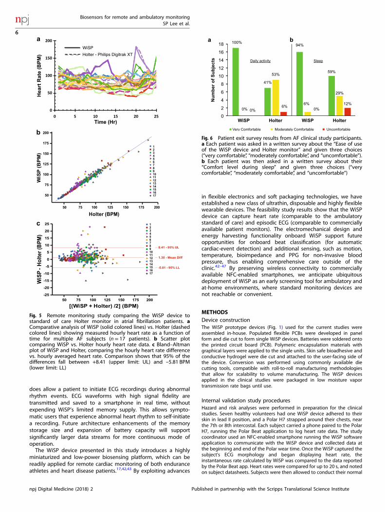
across n = 17 AF patients. We compared the hourly average heartrates tabulated from the Holter report to values reported by WiSP.Overall, heart rates for WiSP and Holter were strongly correlatedover a broad range of heart rate zones (Fig. 5b). The WiSP deviceshows a slight bias of +1.3 BPM compared to the Holter. TheBland–Altman plot (Fig. 5c) shows that 95% of the differencesbetween heart rates reported by WiSP and Holter fall between+8.5 and –5.81 BPM.
Human factors and usability advantagesFor subjects living with cardiovascular diseases, compliance withHolter and event monitoring devices tends to be poor even overshort durations (24–48 h) of prescribed monitoring. Furthermore,studies have shown that a higher number of cardiac events maybe captured wearing less obtrusive, skin-mounted biosensorpatches for long durations.39 Patient exit surveys conducted bythe clinical staff at MGH reported significantly higher patientcomfort scores for the WiSP device compared to the Holtermonitor during daily activity (Fig. 6a). These AF patients alsoreported significantly higher sleep comfort scores for the WiSPdevice over the Holter (Fig. 6b), suggesting that the WiSP couldenhance compliance and patient experience due in large part to
Fig. 3 Cardiac sensing data with WiSP compared to control devices in healthy subjects. a Plot of ECG waveforms simultaneously recordedfrom GE Dash 3000 and WiSP with ECG and heart rate (HR) management filter settings. b Plot of heart rate data simultaneously recorded withPolar H7 and WiSP devices on a healthy subject during daily activity. c Scatter plot of measured heart rate data (n= 7 subjects) for WiSP andPolar H7 devices. d Bland–Altman analysis of WiSP vs. Polar H7 (n= 7 subjects) showing data sets falling within +3.26 BPM (upper limit: UL) and−3.40 BPM (lower limit: LL)
Biosensors for remote and ambulatory monitoringSP Lee et al.
4
npj Digital Medicine (2018) 2 Published in partnership with the Scripps Translational Science Institute
its imperceptible mechanical properties, small surface area, andultrathin form factor.
DISCUSSIONThe unique form factor, ultralow mass, flexible mechanics, andhigh signal fidelity of WiSP represent a major advance overexisting wearable sensors. The multi-layered design of WiSP(Fig. 1a), in particular, was chosen to be compatible with standardhybrid electronics and roll-to-roll manufacturing processes toachieve disposability as a desired goal, with potential for highvolume scale-up at low cost. Compared to existing packagedelectronics technologies for wearable devices, the WiSP hardwareis unique in its highly efficient antenna design and coordinatedpower management for energy harvesting and compatibility withmost smartphones (Fig. 1c), elastic multi-layers of encapsulation(e.g., polyurethane) (Fig. 1d, e) and low mass, highly flexiblemechanical interface with the skin (Fig. 2).In field studies, the WiSP device compares favorably to the Polar
H7 (Fig. 3), which was found to be more accurate than othercompeting photoplethysmogram-based wrist-worn monitors.17 Inambulatory care settings for AF patients, the WiSP results furthershow heart rate signals that match the measurements of theHolter device, with superior comfort scores (as shown in Figs. 5, 6).Taken together, these studies highlight the WiSP’s functionalcapabilities and enhanced wearability for remote cardiac monitor-ing outside of clinical environments.Because the WiSP device is designed for daily wear and has
onboard NFC capability, there are important security and wirelessconnectivity advantages over existing monitoring technologies.The miniaturized antenna design is compatible with manycommercially available NFC-enabled smartphone models (Supple-mentary Table S1), whereby users can upload their encryptedcardiac data wirelessly to a cloud server, without having to returnthe WiSP devices to the hospital. Logged heart rate data andinstantaneous ECG waveforms captured onboard the WiSP in thehome and/or ambulatory environments are thus readily trans-mitted to care providers from anywhere in the world, withoutrequiring wired connections to the wearable patch or in-personfollow-up visits to the clinic, providing an economic benefit toboth users and health-care providers.The configurability of WiSP signal processing filters uniquely
supports both episodic ECG and heart rate logging. There aremany clinical applications where low-cost early screening of heartrate and episodic ECG waveforms could be utilized to significantlyimprove patient outcomes.40 For example, monitoring of heartrate signals and episodic ECG waveforms using a disposable
device platform could make early or community screeningservices more reachable for those with limited geographical oreconomic access to care.41 Moreover, the energy-harvestingfeature of the WiSP device allows multiple uses, even after thebattery is fully depleted. The NFC chip onboard the WiSP deviceharvests additional wireless power from NFC-enabled smart-phones once the smartphone is brought in close proximity tothe WiSP device. As a result, the WiSP device can provide somedegree of utility (episodic ECG and episodic heart rate measure-ments) in remote locations irrespective of battery usage.While the small size, configurability, and conformal mechanics
of WiSP make it comfortable and attractive as remote cardiacmonitoring solution, the standards of care in cardiac monitoringinvolve more than just heart rate and ambulatory ECG measure-ments. Holter monitors provide 24-h recordings of ECG waveformsthat skilled technicians could interpret, albeit after a few days, toquantify normal and abnormal beats. Furthermore, loop recorderdevices perform beat-to-beat analysis in real time, record whenabnormal rhythms are detected, and allow users to initiate arecording. Although the WiSP architecture supports a limitedamount of data storage compared to Holters and loop recorders, it
Table 1. Patient demographics for 24-h clinical study comparing WiSPdevice and Holter monitor
Variables Subjects (n= 17)
Age (years) Average 66.88
Std. Dev 9.72
Min 41
Max 82
Gender Male 15 88%
Female 2 12%
BMI Average 31.5
Std. Dev 6.22
Min 21.47
Max 42.04
Fig. 4 Clinical study comparing the WiSP device to standard of careHolter monitor with annotation. a WiSP heart rate data (2-minresolution) compared to Holter in a subject with atrial fibrillation.Annotations are detailed in sub-figures b and c. b Holter ECGsnapshot of atrial fibrillation corresponding to the steep heart ratedrop from 120 to 50 BPM. c Holter ECG snapshot highlighting aseries of “atrial runs” corresponding to elevated heart rate
Biosensors for remote and ambulatory monitoringSP Lee et al.
5
Published in partnership with the Scripps Translational Science Institute npj Digital Medicine (2018) 2
does allow a patient to initiate ECG recordings during abnormalrhythm events. ECG waveforms with high signal fidelity aretransmitted and saved to a smartphone in real time, withoutexpending WiSP’s limited memory supply. This allows sympto-matic users that experience abnormal heart rhythm to self-initiatea recording. Future architecture enhancements of the memorystorage size and expansion of battery capacity will supportsignificantly larger data streams for more continuous mode ofoperation.The WiSP device presented in this study introduces a highly
miniaturized and low-power biosensing platform, which can bereadily applied for remote cardiac monitoring of both enduranceathletes and heart disease patients.17,42,43 By exploiting advances
in flexible electronics and soft packaging technologies, we haveestablished a new class of ultrathin, disposable and highly flexiblewearable devices. The feasibility study results show that the WiSPdevice can capture heart rate (comparable to the ambulatorystandard of care) and episodic ECG (comparable to commerciallyavailable patient monitors). The electromechanical design andenergy harvesting functionality onboard WiSP support futureopportunities for onboard beat classification (for automaticcardiac-event detection) and additional sensing, such as motion,temperature, bioimpedance and PPG for non-invasive bloodpressure, thus enabling comprehensive care outside of theclinic.42–47 By preserving wireless connectivity to commerciallyavailable NFC-enabled smartphones, we anticipate ubiquitousdeployment of WiSP as an early screening tool for ambulatory andat-home environments, where standard monitoring devices arenot reachable or convenient.
METHODSDevice constructionThe WiSP prototype devices (Fig. 1) used for the current studies wereassembled in-house. Populated flexible PCBs were developed in panelform and die cut to form single WiSP devices. Batteries were soldered ontothe printed circuit board (PCB). Polymeric encapsulation materials withgraphical layers were applied to the single units. Skin safe bioadhesive andconductive hydrogel were die cut and attached to the user-facing side ofthe device. Conversion was performed using commonly available diecutting tools, compatible with roll-to-roll manufacturing methodologiesthat allow for scalability to volume manufacturing. The WiSP devicesapplied in the clinical studies were packaged in low moisture vaportransmission rate bags until use.
Internal validation study proceduresHazard and risk analyses were performed in preparation for the clinicalstudies. Seven healthy volunteers had one WiSP device adhered to theirskin in lead II position, and a Polar H7 strapped around their chests, nearthe 7th or 8th intercostal. Each subject carried a phone paired to the PolarH7, running the Polar Beat application to log heart rate data. The studycoordinator used an NFC-enabled smartphone running the WiSP softwareapplication to communicate with the WiSP device and collected data atthe beginning and end of the Polar wear time. Once the WiSP captured thesubject's ECG morphology and began displaying heart rate, theinstantaneous rate calculated by WiSP was compared to the data reportedby the Polar Beat app. Heart rates were compared for up to 20 s, and notedon subject datasheets. Subjects were then allowed to conduct their normal
Fig. 5 Remote monitoring study comparing the WiSP device tostandard of care Holter monitor in atrial fibrillation patients. aComparative analysis of WiSP (solid colored lines) vs. Holter (dashedcolored lines) showing measured hourly heart rate as a function oftime for multiple AF subjects (n= 17 patients). b Scatter plotcomparing WiSP vs. Holter hourly heart rate data. c Bland–Altmanplot of WiSP and Holter, comparing the hourly heart rate differencevs. hourly averaged heart rate. Comparison shows that 95% of thedifferences fall between +8.41 (upper limit: UL) and –5.81 BPM(lower limit: LL)
Fig. 6 Patient exit survey results from AF clinical study participants.a Each patient was asked in a written survey about the “Ease of useof the WiSP device and Holter monitor” and given three choices(“very comfortable”, “moderately comfortable”, and “uncomfortable”).b Each patient was then asked in a written survey about their“Comfort level during sleep” and given three choices (“verycomfortable”, “moderately comfortable”, and “uncomfortable”)
Biosensors for remote and ambulatory monitoringSP Lee et al.
6
npj Digital Medicine (2018) 2 Published in partnership with the Scripps Translational Science Institute
daily activities. Each subject returned to the study coordinator for check-insand removal of the Polar H7 approximately 4 h later. In addition to theinstantaneous heart rates calculated by both devices, logged heart ratesthroughout the wear time were analyzed and compared after studycompletion. Internal validation was performed by MC10 and informedconsent was obtained from each volunteer prior to the MGH study.
Clinical feasibility study proceduresWe conducted a clinical study at the Massachusetts General HospitalCardiac Arrhythmia Unit using the WiSP devices to measure heart ratesimultaneously for 24 h with a Philips DigiTrak XT Holter monitor. AFpatients who were scheduled for a Holter monitor were asked if theywould like to participate in this study (2015P002439 approved by theMassachusetts General Hospital Institutional Review Board). All methodswere performed in accordance with the relevant guidelines andregulations of the MGH. This study ran from 30 December 2015 to 25October 2016. Informed consent was obtained from each of the patientsprior to the study. In total, 21 patients with existing clinical diagnoses of AFenrolled in this study and 17 patients completed the study. Four enrolleeswithdrew after signing the consent because the device did not functionproperly (e.g., provide good signal quality) immediately after skin-prep.These patients chose not to wait for re-application of the WiSP device bythe Holter lab technician. All of the enrolled patients had existing clinicaldiagnoses of AF and were not compensated for their participation.After obtaining consent from AF patients, clinical study staff prepared
the patient’s skin on the torso region for WiSP device placement byabrading the skin location and cleaning with protective wipe. The WiSPdevice was applied to the patient in either ECG lead I or lead IIconfiguration. The study staff used an NFC-enabled smartphone to activatethe WiSP and assess the ECG signal confirming correct device placement.This initialization step powered on the WiSP and simultaneously initiatedWiSP heart rate recording. With the WiSP correctly applied and assessedfor high signal fidelity by the study staff, clinical technicians then appliedthe Holter monitor to the patient and initiated device recording.To facilitate data stream alignment for the WiSP and Holter outputs, a
timestamped photograph was taken by study staff of the recording Holtermonitor device clock using the WiSP smartphone camera. As a finalassessment step, the study staff held the NFC-enabled smartphoneadjacent to the WiSP device to display cardiac data and visually confirmfunctional heart rate logging.Patients departed the clinic and returned after 24 h in compliance with
their scheduled appointment to remove the Holter monitor. At this time,the study staff held the NFC-enabled smartphone adjacent to the WiSPdevice to initiate wireless data transfer from the WiSP device to the phone.Study staff carefully removed the WiSP device from the patient and storedit in the supplied packaging. Patients were asked to complete an exitsurvey before leaving the clinic. Smartphone data was then pushed to acloud server for analysis. Raw data files were also transferred directly fromthe smartphone for further analysis.
Data analysisHolter heart rate data was aligned with WiSP device heart rate data bymatching the timestamp in the photograph taken in clinic. Because theHolter display only shows time to the minute, there may be up to ±30 s oftime offset in the data. To correct for this, the WiSP heart rate data timesare shifted en masse within a ±30 s window of the photograph timestamp.The WiSP device reports heart rate at 2-min intervals, whereas the Holterreports at a higher rate. To perform statistical computation, the Holter’sheart rate values at the closest time points to the 2-min cadence wereselected and compared (as shown for an example subject in Fig. 4a).The Holter continually records ECG data and lists hourly heart rate values
in summary reports. The 2-min heart rate data measured by WiSP wasaveraged and compared to the Holter hourly data. Because multiplemeasurements are made per subject and there are n = 17 subjects in total,the data comprises a mixture of between and within-individual informa-tion. The data collected in this study is of an instantaneous value of achanging quantity, i.e., heart rate changes over 24 h. The 95.4% confidenceintervals for the Bland–Altman Limits of Agreement were found bycomputing a one-way ANOVA and calculating the estimated variance ofmultiple between-method differences for the same subject and differencesbetween subjects.48 The variance is computed as σ2d ¼ σ2dI þ σ2dw, whereσ2dw is the within-subject variance, mean-square residual and σ2dI is thevariance interaction term, computed from the one-way ANOVA.49
This variance term, σ2dI, is defined by the following expression:
σ2dI ¼ ðMSsubject�MSresidualÞΣmið Þ2�Σm2
in�1ð ÞΣmi
, where m is the number of observations on subject
i. The standard deviation is defined as the square root of σ2d and used toyield the 95.4% confidence intervals in Figs. 3d and 5d.
Data availabilityThe data that support the findings of this study are available on requestfrom the corresponding author [M.M.]. The data are not publicly availabledue to participant privacy/consent.
ACKNOWLEDGEMENTSWe thank Nick Kubasti, Kurt Jaisle, Brandon Suleski, Adam Leech, Briana Morey, JakePhillips, and Kirsten Seagers for valuable discussions and feedback on the studydesign and prototype testing. Milan Raj and Bryan McGrane provided valuableguidance on the near field antenna design. Y.M. acknowledges support from theNational Natural Science Foundation of China (Grant No. 11402135). Y.H. acknowl-edges support from NSF (Grant Nos. DMR-1121262, CMMI-1300846, CMMI-1400169,and CMMI-1534120).
AUTHOR CONTRIBUTIONSS.P.L., J.A.W., N.S., J.A.R., Y.H., M.M., and R.G. designed the research. D.E.W., E.S.G., P.C.B., G.L.H., H.M., Y.M., K.L., W.L., and S.P.L. constructed the prototypes, performed pre-clinical experiments, and analyzed the data. G.H., D.E.W., N.R.H., E.S.G., and M.J.performed the clinical experiments. All authors contributed to the writing of themanuscript.
ADDITIONAL INFORMATIONSupplementary information accompanies the paper on the npj Digital Medicinewebsite (https://doi.org/10.1038/s41746-017-0009-x).
Competing interests: The authors declare that patent application nos.US20160338646 and US20150194817 covering this work were filed with the UnitedStates Patent Office and Trademark Office on 20 May 2016 and 3 January 2015,respectively. Y.Y., M.J., and M.M. are clinical and scientific advisors to MC10 Inc. Theremaining authors declare no competing financial interests.
Publisher's note: Springer Nature remains neutral with regard to jurisdictional claimsin published maps and institutional affiliations.
REFERENCES1. Piwek, L., Ellis, D. A., Andrews, S. & Joinson, A. The rise of consumer health
wearables: promises and barriers. PLoS Med. 13, e1001953 (2016).2. Dorsey, E. R. & Topol, E. J. State of telehealth. N. Engl. J. Med. 375, 154–161 (2016).3. Pantelopoulos, A. & Bourbakis, N. G. A survey on wearable sensor-based systems
for health monitoring and prognosis. IEEE Trans. Syst. Man Cybern. 40, 1–10(2010).
4. Gatzoulis, L. & Iakovidis, I. Wearable and portable ehealth systems. IEEE Eng. Med.Biol. Mag. 26, 51–56 (2007).
5. Bonato, P. Wearable sensors/systems and their impact on biomedical engineer-ing. IEEE Eng. Med. Biol. Mag. 22, 18–20 (2003).
6. DiMarco, J. P. & Philbrick, J. T. Use of ambulatory electrocardiographic (Holter)monitoring. Ann. Intern. Med. 113, 53–68 (1990).
7. Arnold, J. M., Fitchett, D. H., Howlett, J. G., Lonn, E. M. & Tardif, J.-C. Resting heartrate: a modifiable prognostic indicator of cardiovascular risk and outcomes? Can.J. Cardiol. 24, 3A–8A (2008).
8. Mauss, O. et al. Bedside risk stratification after acute myocardial infarction: pro-spective evaluation of the use of heart rate and left ventricular function. J.Electrocardiol. 38, 106–112 (2005).
9. Aboyans, V. & Criqui, M. H. Can we improve cardiovascular risk prediction beyondrisk equations in the physician's office? J. Clin. Epidemiol. 59, 547–558 (2006).
10. Spodick, D. H., Raju, P., Bishop, R. L. & Rifkin, R. D. Operational definition of normalsinus heart rate. Am. J. Cardiol. 69, 1245–1246 (1992).
11. Chen, L. Y. et al. Feasibility of using a leadless patch heart rhythm monitor tomeasure atrial fibrillation burden in community-based epidemiological studies:the multiethnic study of atherosclerosis (MESA). Circulation 132, A11721 (2015).
12. Fung, E. et al. Electrocardiographic patch devices and contemporary wirelesscardiac monitoring. Front. Physiol. 6, 149 (2015).
Biosensors for remote and ambulatory monitoringSP Lee et al.
7
Published in partnership with the Scripps Translational Science Institute npj Digital Medicine (2018) 2
13. Raupp, G. B. Flexible thin film transistor arrays as an enabling platform tech-nology: opportunities and challenges. ECS Trans. 37, 229–240 (2011).
14. Barrett, P. M. et al. Comparison of 24-hour Holter monitoring with 14-day noveladhesive patch electrocardiographic monitoring. Am. J. Med. 127, 95.e11–95.e17(2014).
15. Sherr, D. et al. Prospective comparison of the diagnostic utility of a standardevent monitor versus a “leadless” portable ECG monitor in the evaluation ofpatients with palpitations. J. Interv. Card. Electrophysiol. 22, 39–44 (2008).
16. Ackermans, P. et al. A user-friendly integrated monitor-adhesive patch for long-term ambulatory electrocardiogram monitoring. J. Electrocardiol. 45, 148–153(2012).
17. Wang, R. et al. Accuracy of wrist-worn heart rate monitors. JAMA Cardiol. 2,104–106 (2017).
18. Hertzman, A. B. The blood supply of various skin areas as estimated by thephotoelectric plethysmograph. Am. J. Physiol. 124, 328–340 (1938).
19. Banerjee, R., Sinha, A., Pal, A. & Kumar, A. Estimation of ECG parameters usingphotoplethysmography. 13th IEEE International Conference on BioInformatics andBioEngineering, 1–5 (IEEE, Chania, Greece, 2013).
20. Kim, D.-H. et al. Epidermal electronics. Science 333, 838–843 (2011).21. Tee, B. C.-K. et al. A skin-inspired organic digital mechanoreceptor. Science 350,
313–316 (2015).22. Kim, J. et al. Stretchable silicon nanoribbon electronics for skin prosthesis. Nat.
Commun. 5, 5747 (2014).23. Lee, S. et al. A transparent bending-insensitive pressure sensor. Nat. Nanotech. 11,
472–478 (2016).24. Webb, R. C. et al. Ultrathin conformal devices for precise and continuous thermal
characterization of human skin. Nat. Mater. 12, 938–944 (2013).25. Xu, S. et al. Soft microfluidic assemblies of sensors, circuits, and radios for the
skin. Science 344, 70–74 (2014).26. Kim, J. et al. Epidermal electronics with advanced capabilities in near-field
communication. Small 11, 906–912 (2015).27. Dagdeviren, C. et al. Conformal piezoelectric systems for clinical and experimental
characterization of soft tissue biomechanics. Nat. Mater. 14, 728–736 (2015).28. Rogers, J. A. Electronics for the human body. J. Am. Med. Assoc. 313, 561–562 (2015).29. Kaltenbrunner, M. et al. An ultra-lightweight design for imperceptible plastic
electronics. Nature 499, 458–463 (2013).30. Lipomi, D. J. et al. Skin-like pressure and strain sensors based on transparent
elastic films of carbon nanotubes. Nat. Nanotech. 6, 788–792 (2011).31. Jeong, J.-W. et al. Materials and optimized designs for human-machine interfaces
via epidermal electronics. Adv. Mat. 25, 6839–6846 (2013).32. Gao, L. et al. Epidermal photonic devices for quantitative imaging of temperature
and thermal transport characteristics of the skin. Nat. Commun. 5, 4938 (2014).33. Kim, D.-H., Ghaffari, R., Lu, N. & Rogers, J. A. Flexible and stretchable electronics
for biointegrated devices. Annu. Rev. Biomed. Eng. 14, 113–128 (2012).34. Lee, C. H. et al. Soft core/shell packages for stretchable electronics. Adv. Funct.
Mater. 25, 3698–3704 (2015).35. Puurtinen, M., Viik, J. & Hyttinen, J. Best electrode locations for a small bipolar
ECG device: signal strength analysis of clinical data. Ann. Biomed. Eng. 37,331–336 (2009).
36. ABAQUS Analysis User’s Manual V6.10 (2010). http://abaqusdoc.ucalgary.ca/pdf_books/ANALYSIS_1.pdf
37. Wang, S. et al. Mechanics of epidermal electronics. J. Appl. Mech. 79, 031022-1–031022-6 (2012).
38. Koh, A. et al. A soft, wearable microfluidic device for the capture, storage, andcolorimetric sensing of sweat. Sci. Transl. Med. 8, 366ra165 (2016).
39. Shultz, K. et al. Abstract: the days of the Holter monitor are numbered: extendedcontinuous rhythm monitoring detects more clinically significant arrhythmias inadults with congenital heart disease. Circulation 134, A13416 (2016).
40. Tarakji, K. G. et al. Using a novel wireless system for monitoring patients after theatrial fibrillation ablation procedure: the iTransmit study. Heart Rhythm 12,554–559 (2015).
41. Chan, N. & Choy, C. Screening for atrial fibrillation in 13,122 Hong Kong citizenswith smartphone electrocardiogram. Heart 103, 24–31 (2016).
42. Edwards, J. D. et al. Underutilization of ambulatory ECG monitoring after strokeand transient ischemic attack. Stroke 47, 1982–1989 (2016).
43. Go, A. S. et al. Prevalence of diagnosed atrial fibrillation in adults: nationalimplications for rhythm management and stroke prevention: the anticoagulationand risk factors in atrial fibrillation (ATRIA) study. J. Am. Med. Assoc. 285,2370–2375 (2001).
44. Higgins, P. et al. Noninvasive cardiac event monitoring to detect atrial fibrillationafter ischemic stroke: a randomized, controlled trial. Stroke 44, 2525–2531 (2013).
45. Yong, J. H. E. et al. Clinical and cost-effectiveness of 30 day ECG monitoring foratrial fibrillation detection in patients with cryptogenic stroke or TIA. Int. J. Stroke10, 1–20 (2015).
46. Mehrotra, A. The convenience revolution for treatment of low-acuity conditions.J. Am. Med. Assoc. 310, 35–36 (2013).
47. Ekeland, A. G., Bowes, A. & Flottorp, S. Effectiveness of telemedicine: a systematicreview of reviews. Int. J. Med. Inform. 79, 736–771 (2010).
48. Bland, J. M. & Altman, D. G. Agreement between methods of measurement withmultiple observations per individual. J. Biopharm. Stat. 17, 571–582 (2007).
49. Bland, J. M. & Altman, D. G. Measuring agreement in method comparisons stu-dies. Stat. Methods Med. Res. 8, 135–160 (1999).
Open Access This article is licensed under a Creative CommonsAttribution 4.0 International License, which permits use, sharing,
adaptation, distribution and reproduction in anymedium or format, as long as you giveappropriate credit to the original author(s) and the source, provide a link to the CreativeCommons license, and indicate if changes were made. The images or other third partymaterial in this article are included in the article’s Creative Commons license, unlessindicated otherwise in a credit line to the material. If material is not included in thearticle’s Creative Commons license and your intended use is not permitted by statutoryregulation or exceeds the permitted use, you will need to obtain permission directlyfrom the copyright holder. To view a copy of this license, visit http://creativecommons.org/licenses/by/4.0/.
© The Author(s) 2018
Biosensors for remote and ambulatory monitoringSP Lee et al.
8
npj Digital Medicine (2018) 2 Published in partnership with the Scripps Translational Science Institute