Embed Size (px)
Citation preview

Research
Hippocampal efferents to retrosplenial cortex andlateral septum are required for memory acquisition
Ashley N. Opalka and Dong V. WangDepartment of Neurobiology and Anatomy, Drexel University College of Medicine, Philadelphia, Pennsylvania 19129, USA
Learning and memory involves a large neural network of many brain regions, including the notable hippocampus along
with the retrosplenial cortex (RSC) and lateral septum (LS). Previous studies have established that the dorsal hippocampus
(dHPC) plays a critical role during the acquisition and retrieval/expression of episodic memories. However, the role of
downstream circuitry from the dHPC, including the dHPC-to-RSC and dHPC-to-LS pathways, has come under scrutiny
only recently. Here, we used an optogenetic approach with contextual fear conditioning in mice to determine whether
the above two pathways are involved in acquisition and expression of contextual fear memory. We found that a selective
inhibition of the dHPC neuronal terminals in either the RSC or LS during acquisition impaired subsequent memory per-
formance, suggesting that both the dHPC-to-RSC and dHPC-to-LS pathways play a critical role in memory acquisition.
We also selectively inhibited the two dHPC efferent pathways during memory retrieval and found a differential effect
on memory performance. These results indicate the intricacies of memory processing and that hippocampal efferents to
cortical and subcortical regions may be differentially involved in aspects of physiological and cognitive memory processes.
[Supplemental material is available for this article.]
Processing of memory entails multiple stages: acquisition, the ini-tial encoding of memory; consolidation, the transformation ofnewly acquired information into long-lasting memory; retrieval,the recall of stored memory; and expression, the behavioral read-out of recalledmemory. Extensive research has shown that the dor-sal hippocampus (dHPC) plays an important role in each of thesememory processing stages (Scoville and Milner 1957; Maren andFanselow 1997; McEchron et al. 1998; Lee and Kesner 2004;Misane et al. 2005; Carr et al. 2011; Goshen et al. 2011; Piersonet al. 2015; Ocampo et al. 2017). However, how the dHPC commu-nicates with downstream cortical and subcortical regions duringthese memory stages remains unclear. Recent studies have shownthat a number of cortical regions display increased activity duringmemory acquisition and expression stages (Takata et al. 2015;Miller et al. 2017; DeNardo et al. 2019) and that hippocampal-cortical communications are required for memory acquisitionand consolidation (Kitamura et al. 2017; DeNardo et al. 2019;Yamawaki et al. 2019a). Thus, recruitment of cortical brain regionsduring the initial acquisition stage may be necessary for subse-quent consolidation and retrieval/expression of memories(Lesburguères et al. 2011; Kitamura et al. 2017; DeNardo et al.2019; de Sousa et al. 2019). Additionally, recruitment of hippocam-pal projections to subcortical regions appears to be necessary dur-ing these memory processes as well (Olton et al. 1978; Hunsakeret al. 2009; Roy et al. 2017; Besnard et al. 2019, 2020).
The dHPCprojects directly to a few cortical and subcortical re-gions, notably the granular retrosplenial cortex (RSC) and the lat-eral septum (LS) (Van Groen and Wyss 1990; Jinno et al. 2007;Miyashita and Rockland 2007; Kwapis et al. 2015; Takata et al.2015; Todd and Bucci 2015). However, the function of these twodHPC projections during memory processes has come under scru-tiny only recently (Yamawaki et al. 2019a,b; Besnard et al. 2020;Nitzan et al. 2020; Opalka et al. 2020). Previous studies revealed
that the RSC contributes to contextual memory (Keene and Bucci2008; Corcoran et al. 2011; Cowansage et al. 2014; Kwapis et al.2015; Robinson et al. 2018; de Sousa et al. 2019), spatial memory(Cooper et al. 2001; Czajkowski et al. 2014; Mao et al. 2017;Milczarek et al. 2018; Vann and Aggleton 2002), inhibitory avoid-ance memory (Katche et al. 2013; Katche and Medina 2017), andmultisensory association (Robinson et al. 2011, 2014). Moreover,the RSC is one of few cortical regions that receive direct projectionsfrom the dHPC, positioning the RSC as a potential bridge in con-necting the hippocampus with other cortical regions, such as theanterior cingulate cortex, medial prefrontal cortex, and secondarysensory cortices, to support long-term memory (Frankland et al.2004; Maviel et al. 2004; Todd and Bucci 2015; Wang andIkemoto 2016; Kitamura et al. 2017; DeNardo et al. 2019).Therefore, a better understanding of this dHPC-to-RSC connectionwill shed light on memory acquisition and retrieval/expressionprocesses.
Further, previous literature reports the LS as essential for pro-cessing multiple forms of memory, such as contextual memory(Calandreau et al. 2007, 2010; Besnard et al. 2019; Besnard et al.2020), social encounter and memory (Dantzer et al. 1988; Evertsand Koolhaas 1997; Leroy et al. 2018), and addiction memory(McGlinchey and Aston-Jones 2018). Due to the hippocampal-septal involvement in theta oscillation (Colgin 2016), thedHPC-to-LS pathway has been recently investigated for its role inspatial navigation and spatial memory (Tingley and Buzsáki2018). In particular, the firing of LS neurons phase-locked todHPC theta oscillation (Mondragón-Rodríguez et al. 2019) posi-tions this dHPC-to-LS pathway as a candidate for memory process-ing. To test whether the dHPC-to-RSC and dHPC-to-LS neural
Corresponding author: [email protected]
# 2020 Opalka andWang This article is distributed exclusively by Cold SpringHarbor Laboratory Press for the first 12 months after the full-issue publicationdate (see http://learnmem.cshlp.org/site/misc/terms.xhtml). After 12 months,it is available under a Creative Commons License (Attribution-NonCommercial4.0 International), as described at http://creativecommons.org/licenses/by-nc/4.0/.Article is online at http://www.learnmem.org/cgi/doi/10.1101/lm.051797.120.
27:310–318; Published by Cold Spring Harbor Laboratory PressISSN 1549-5485/20; www.learnmem.org
310 Learning & Memory
Cold Spring Harbor Laboratory Press on January 30, 2022 - Published by learnmem.cshlp.orgDownloaded from

pathways are involved in the acquisition and retrieval/expressionof memory, we used an optogenetic approach and contextualfear conditioning, a procedure widely used to assess hippocampus-dependent memory. Our results provide direct evidence that boththe cortical (RSC) and subcortical (LS) projections from the dHPCare required during acquisition of the fear memory. These hippo-campal efferents to downstream cortical and subcortical regionsmay process distinct features in representing a memory in itsentirety.
Results
We bilaterally injected AAV viruses that encode fluorescent eYFPunder the promoter CaMKII into the medial portion of the dHPCregion (includingmedial CA1 and dorsal subiculum; SupplementalFigs. 1, 2). Consistentwith previouswork (Oh et al. 2014;Wyss andVan Groen 1992), our results showed that this dHPC region pro-jected primarily to the granular RSC layer 3, midline LS, entorhinalcortex, and mammillary area. In the present study, we optogeneti-cally inhibited two dHPC efferent pathways: the dHPC-to-RSC anddHPC-to-LS pathways (Supplemental Fig. 1). Repeated measurestwo-way ANOVAs were conducted on freezing behavior for eachof the following experiments followed by Bonferroni post-hocanalyses. The between-subjects variables included two levels oftreatment: halorhodopsin (Halo) or eYFP (Ctrl) viral injections.The within-subjects variables included two levels of time: recent(day 1) or remote (day 31) memory tests.
Optogenetic inhibition of dHPC-to-RSC pathway during
memory acquisitionMice received bilateral dHPC injections, targeting medial CA1 androstral-dorsal subiculum (Supplemental Figs. 1, 2), of eitherAAV-Halo (Halo) or AAV-eYFP (Ctrl). Meanwhile, an optical fiber
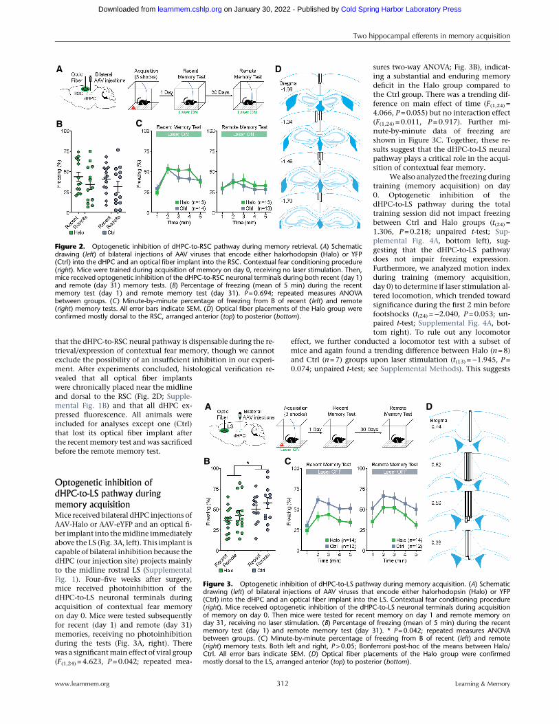
was implanted in the midline of the RSC, capable of inhibitingdHPC projections from both hemispheres because the dHPC pro-jects predominantly to RSC layer 3, located close to the midline(Fig. 1A, left; Supplemental Fig. 1). Four–five weeks after surgery,mice received photoinhibition of the dHPC-to-RSC neuronal ter-minals during acquisition of contextual fear memory on day 0.Mice were tested subsequently for recent memory in 1 d and re-mote memory in 31 d, receiving no photoinhibition during thetests (Fig. 1A, right). There was a significant main effect of viralgroup (F(1,46) = 9.384, P=0.004; repeated measures two-wayANOVA; Fig. 1B), indicating a substantial and enduring memorydeficit of the Halo group compared to the Ctrl group. In addition,there was a main effect of time (F(1,46) = 6.317, P=0.016), indicat-ing a difference in freezing between recent and remote memorytests, but there was no interaction effect (F(1,46) = 0.266, P=0.608). Further minute-by-minute data of freezing are shown inFigure 1C. These results suggest that the dHPC-to-RSC neural path-way plays a critical role in acquisition of contextual fear memory.Note that Figure 1B,C combined two data sets (n=24 per dataset, n=12 per group; see Materials and Methods for details andSupplemental Fig. 3 for data/statistics).
On the day of training (memory acquisition; day 0), we alsoanalyzed freezing and, additionally, motion index to determineif the laser stimulation impacted locomotion. We found thatboth freezing during the total training session (t(46) =−0.091, P=0.928; unpaired t-test; Supplemental Fig. 4A, top left) and motionindex during the first 2 min before footshocks (t(46) =−0.207, P=0.837; Supplemental Fig. 4A, top right) did not differ betweenthe Halo and Ctrl groups. This suggests that the laser stimulationdid not affect freezing expression or general locomotion. Aftercompletion of the above experiments, histological verification ofoptical fiber placements and viral injections revealed that all opti-cal fiber implants were chronically placed around the midline anddorsal to the RSC (Fig. 1D; Supplemental Fig. 1B) and that all dHPC
expressed fluorescence. Therefore, all ani-mals were included for analyses.
Optogenetic inhibition of
dHPC-to-RSC pathway during
memory retrievalSimilarly, mice received bilateral dHPCinjections of either AAV-Halo or AAV-eYFP and a RSC optical fiber implantation(Fig. 2A, left). Four–five weeks after sur-gery, mice were trained for acquisitionof contextual fear memory on day 0 andwere tested subsequently for recent (day1) and remote (day 31) memories. Micereceived photoinhibition of the dHPC-to-RSC neuronal terminals during bothtesting days (Fig. 2A, right). Therewas no main effect of group (F(1,24) =0.159, P=0.694; repeated measures two-way ANOVA; Fig. 2B) or interaction effect(F(1,46) = 0.001, P=0.973), but a main ef-fect of time (F(1,24) = 7.053, P=0.014), in-dicating a difference in freezing betweenrecent and remote memory tests. Furtherminute-by-minute data of freezing areshown in Figure 2C. In addition, therewas no significant difference in meanfreezing between Halo and Ctrl viralgroups during acquisition (day 0; t(46) =0.648, P= 0.523; unpaired t-test; Supple-mental Fig. 4B, top). These results suggest
B
A
C
D
Figure 1. Optogenetic inhibition of dHPC-to-RSC pathway during memory acquisition. (A) Schematicdrawing (left) of bilateral injections of AAV viruses that encode either halorhodopsin (Halo) or YFP (Ctrl)into the dHPC and an optical fiber implant into the RSC. Contextual fear conditioning procedure (right).Mice received optogenetic inhibition of the dHPC-to-RSC neuronal terminals during acquisition ofmemory on day 0. Then mice were tested for recent memory on day 1 and remote memory on day31, receiving no laser stimulation. (B) Percentage of freezing (mean of 5 min) during the recentmemory test (day 1) and remote memory test (day 31). (**) P=0.004, repeated measures ANOVAbetween groups. (C) Minute-by-minute percentage of freezing from B of recent (left) and remote(right) memory tests. Both left and right, P<0.05; Bonferroni post-hoc of the means between Halo/Ctrl. All error bars indicate standard error of the mean (SEM). (D) Optical fiber placements of theHalo group were confirmed mostly dorsal to the RSC, arranged anterior (top) to posterior (bottom).Adapted from Franklin and Paxinos 2008.
Two hippocampal efferents in memory acquisition
www.learnmem.org 311 Learning & Memory
Cold Spring Harbor Laboratory Press on January 30, 2022 - Published by learnmem.cshlp.orgDownloaded from
that the dHPC-to-RSC neural pathway is dispensable during the re-trieval/expression of contextual fear memory, though we cannotexclude the possibility of an insufficient inhibition in our experi-ment. After experiments concluded, histological verification re-vealed that all optical fiber implantswere chronically placed near the midlineand dorsal to the RSC (Fig. 2D; Supple-mental Fig. 1B) and that all dHPC ex-pressed fluorescence. All animals wereincluded for analyses except one (Ctrl)that lost its optical fiber implant afterthe recentmemory test and was sacrificedbefore the remote memory test.
Optogenetic inhibition of
dHPC-to-LS pathway during
memory acquisitionMice received bilateral dHPC injections ofAAV-Halo or AAV-eYFP and an optical fi-ber implant into themidline immediatelyabove the LS (Fig. 3A, left). This implant iscapable of bilateral inhibition because thedHPC (our injection site) projects mainlyto the midline rostral LS (SupplementalFig. 1). Four–five weeks after surgery,mice received photoinhibition of thedHPC-to-LS neuronal terminals duringacquisition of contextual fear memoryon day 0. Mice were tested subsequentlyfor recent (day 1) and remote (day 31)memories, receiving no photoinhibitionduring the tests (Fig. 3A, right). Therewas a significantmain effect of viral group(F(1,24) = 4.623, P=0.042; repeated mea-
sures two-way ANOVA; Fig. 3B), indicat-ing a substantial and enduring memorydeficit in the Halo group compared tothe Ctrl group. There was a trending dif-ference on main effect of time (F(1,24) =4.066, P=0.055) but no interaction effect(F(1,24) = 0.011, P=0.917). Further mi-nute-by-minute data of freezing areshown in Figure 3C. Together, these re-sults suggest that the dHPC-to-LS neuralpathway plays a critical role in the acqui-sition of contextual fear memory.
We also analyzed the freezing duringtraining (memory acquisition) on day0. Optogenetic inhibition of thedHPC-to-LS pathway during the totaltraining session did not impact freezingbetween Ctrl and Halo groups (t(24) =1.306, P=0.218; unpaired t-test; Sup-plemental Fig. 4A, bottom left), sug-gesting that the dHPC-to-LS pathwaydoes not impair freezing expression.Furthermore, we analyzed motion indexduring training (memory acquisition,day 0) to determine if laser stimulation al-tered locomotion, which trended towardsignificance during the first 2 min beforefootshocks (t(24) =−2.040, P=0.053; un-paired t-test; Supplemental Fig. 4A, bot-tom right). To rule out any locomotor
effect, we further conducted a locomotor test with a subset ofmice and again found a trending difference between Halo (n=8)and Ctrl (n=7) groups upon laser stimulation (t(13) =−1.945, P=0.074; unpaired t-test; see Supplemental Methods). This suggests
B
A
C
D
Figure 2. Optogenetic inhibition of dHPC-to-RSC pathway during memory retrieval. (A) Schematicdrawing (left) of bilateral injections of AAV viruses that encode either halorhodopsin (Halo) or YFP(Ctrl) into the dHPC and an optical fiber implant into the RSC. Contextual fear conditioning procedure(right). Mice were trained during acquisition of memory on day 0, receiving no laser stimulation. Then,mice received optogenetic inhibition of the dHPC-to-RSC neuronal terminals during both recent (day 1)and remote (day 31) memory tests. (B) Percentage of freezing (mean of 5 min) during the recentmemory test (day 1) and remote memory test (day 31). P=0.694; repeated measures ANOVAbetween groups. (C) Minute-by-minute percentage of freezing from B of recent (left) and remote(right) memory tests. All error bars indicate SEM. (D) Optical fiber placements of the Halo group wereconfirmed mostly dorsal to the RSC, arranged anterior (top) to posterior (bottom).
B
A
C
D
Figure 3. Optogenetic inhibition of dHPC-to-LS pathway during memory acquisition. (A) Schematicdrawing (left) of bilateral injections of AAV viruses that encode either halorhodopsin (Halo) or YFP(Ctrl) into the dHPC and an optical fiber implant into the LS. Contextual fear conditioning procedure(right). Mice received optogenetic inhibition of the dHPC-to-LS neuronal terminals during acquisitionof memory on day 0. Then mice were tested for recent memory on day 1 and remote memory onday 31, receiving no laser stimulation. (B) Percentage of freezing (mean of 5 min) during the recentmemory test (day 1) and remote memory test (day 31). * P=0.042; repeated measures ANOVAbetween groups. (C) Minute-by-minute percentage of freezing from B of recent (left) and remote(right) memory tests. Both left and right, P>0.05; Bonferroni post-hoc of the means between Halo/Ctrl. All error bars indicate SEM. (D) Optical fiber placements of the Halo group were confirmedmostly dorsal to the LS, arranged anterior (top) to posterior (bottom).
Two hippocampal efferents in memory acquisition
www.learnmem.org 312 Learning & Memory
Cold Spring Harbor Laboratory Press on January 30, 2022 - Published by learnmem.cshlp.orgDownloaded from
that optogenetic inhibition of the dHPC-to-LS pathway has no orlimited effect on locomotor activity. After completion of theabove experiments, histological verification revealed that all opti-cal fibers were chronically placed around the midline and dorsalto the rostral LS (Fig. 3D; Supplemental Fig. 1C) and that alldHPC expressed fluorescence. Therefore, all animals were includedfor analyses.
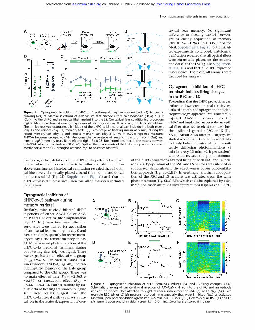
Optogenetic inhibition of
dHPC-to-LS pathway during
memory retrievalSimilarly, mice received bilateral dHPCinjections of either AAV-Halo or AAV-eYFP and a LS optical fiber implantation(Fig. 4A, left). Four–five weeks after sur-gery, mice were trained for acquisitionof contextual fear memory on day 0 andwere tested subsequently for recentmem-ory on day 1 and remote memory on day31. Mice received photoinhibition of thedHPC-to-LS neuronal terminals duringboth testing days (Fig. 4A, right). Therewas a significantmain effect of viral group(F(1,25) = 9.818, P=0.004; repeated mea-sures two-way ANOVA; Fig. 4B), indicat-ing impaired memory of the Halo groupcompared to the Ctrl group. There wasno main effect of time (F(1,25) = 2.363, P=0.137) or interaction effect (F(1,25) =0.933, P=0.343). Further minute-by-mi-nute data of freezing are shown in Figure4C. These results suggest that thedHPC-to-LS neural pathway plays a criti-cal role in the retrieval/expression of con-
textual fear memory. No significantdifference of freezing existed betweengroups during acquisition of memory(day 0; t(25) = 0.943, P=0.335; unpairedt-test; Supplemental Fig. 4B, bottom). Af-ter experiments concluded, histologicalverification revealed that all optical fiberswere chronically placed on the midlineand dorsal to the LS (Fig. 4D; Supplemen-tal Fig. 1C) and that all dHPC expressedfluorescence. Therefore, all animals wereincluded for analyses.
Optogenetic inhibition of dHPC
terminals induces firing changes
in the RSC and LSTo confirm that the dHPCprojections caninfluence downstream neural activity, weutilized a combined optogenetic and elec-trophysiology approach: we unilaterallyinjected AAV-Halo viruses into thedHPC and implanted an optrode (an opti-cal fiber attached to eight tetrodes) intothe ipsilateral granular RSC or LS (Fig.5A,D). About 3 wk after the surgery, westarted recording RSC or LS spike activityin freely behaving mice while intermit-tently delivering photoinhibitions (5min in every 15 min; ∼2 h per session).Our results revealed that photoinhibition
of the dHPC projections affected firing of both RSC and LS neu-rons. A subpopulation of the RSC and LS neurons was silenced orsuppressed, demonstrating the effectiveness of our photoinhibi-tion approach (Fig. 5B,C,E,F). Interestingly, another subpopula-tion of the RSC and LS neurons was activated upon the samephotoinhibition (Fig. 5B,C,E,F), which could be explained by a dis-inhibition mechanism via local interneurons (Opalka et al. 2020)
B
A
C
D
Figure 4. Optogenetic inhibition of dHPC-to-LS pathway during memory retrieval. (A) Schematicdrawing (left) of bilateral injections of AAV viruses that encode either halorhodopsin (Halo) or YFP(Ctrl) into the dHPC and an optical fiber implant into the LS. Contextual fear conditioning procedure(right). Mice were trained during acquisition of memory on day 0, receiving no laser stimulation.Then, mice received optogenetic inhibition of the dHPC-to-LS neuronal terminals during both recent(day 1) and remote (day 31) memory tests. (B) Percentage of freezing (mean of 5 min) during therecent memory test (day 1) and remote memory test (day 31). (**) P=0.004; repeated measuresANOVA between groups. (C) Minute-by-minute percentage of freezing from B of recent (left) andremote (right) memory tests. Both left and right, P<0.05; Bonferroni post-hoc of the means betweenHalo/Ctrl. All error bars indicate SEM. (D) Optical fiber placements of the Halo group were confirmedmostly dorsal to the LS, arranged anterior (top) to posterior (bottom).
E F
BA C
D
Figure 5. Optogenetic inhibition of dHPC terminals induces RSC and LS firing changes. (A,D)Schematic drawing of unilateral viral injection of AAV-CaMKII-Halo into the dHPC and an optrodeimplant, an optical fiber attached to eight tetrodes, into either the RSC (A) or LS (D). (B,E) Twoexample RSC (B) or LS (E) neurons recorded simultaneously that were inhibited (top) or activated(bottom) upon photoinhibition (green bar, 0–5 min; bin, 10 sec). (C,F ) Heatmap of all RSC (C) and LS(F) neurons upon photoinhibition (green bar, 0–5 min). Color bars, z-scored firing rate.
Two hippocampal efferents in memory acquisition
www.learnmem.org 313 Learning & Memory
Cold Spring Harbor Laboratory Press on January 30, 2022 - Published by learnmem.cshlp.orgDownloaded from

or a network feedback disinhibition. Notably, these results also re-vealed a functional recovery of the RSC and LS neurons, albeit asmall rebound activation in a subset of neurons after the cessationof the photoinhibition (Fig. 5B,C,E,F).
Discussion
Utilizing an optogenetic approach and contextual fear condition-ing paradigm, we provided evidence that the dHPC-to-RSC path-way is critical for memory acquisition, whereas the dHPC-to-LSpathway is critical for both memory acquisition and expression.In both instances, the memory impairments were similar betweenrecent and remotememory tests (onemonth apart), indicating thereliability of optogenetic manipulation on memory acquisitionand expression. Notably, this optogenetic approach enabled tran-sient and reversible inhibition of hippocampal efferents comparedto lesions or drug infusions that often cause permanent damages orlong-lasting neural alterations. Additionally, our use of the CaMKIIpromotor likely targeted hippocampal pyramidal neurons but notinhibitory neurons (Liu and Murray 2012; Stark et al. 2012; Wanget al. 2013; Zhu et al. 2014; Besnard et al. 2020); although, futureimmunostaining or in situ hybridization characterizations areneeded to confirm the specificity.
With our optogenetic approach, there was spread of AAV vi-ruses to unintended brain regions. Inside the dHPC, AAV virusesmainly infected the medial CA1 and dorsal subiculum. In somecases, the virus spread partially to the DG, but minimally to theCA3. Given that the DG does not project to the RSC or LS (VanGroen and Wyss 1990; Risold and Swanson 1997; Witter 2007),our observed memory deficits should be mainly attributed to theinactivation of dorsal CA1 and subicular efferents. It should be not-ed that the LS receives a dense projection from both the CA1 andsubiculum, whereas the RSC receives mainly subicular projection(Wyss and Van Groen 1992; Oh et al. 2014; Opalka et al. 2020).Outside of the dHPC, AAV viruses spread mainly along the injec-tion needle track, including the somatosensory and visual anteriorarea of parietal cortices. Those regions do not project to the LS, al-though part of the visual area projects to RSC and could be partiallyresponsible for contextual learning. We also noticed some AAVspread to the ventral RSC close to the corpus callosum(Supplemental Fig. 2B), which is unexpected given its long dis-tance from the needle track. Nonetheless, neurons in this ventralRSC were sparsely labeled and, thus, were unlikely to be responsi-ble for the observed memory deficits.
dHPC-to-RSC pathway is critical for memory acquisitionPrevious studies have shown that the RSC plays a critical role dur-ing acquisition of multiple forms of memories, including acquisi-tion of contextual fear memory (Corcoran et al. 2011;Cowansage et al. 2014; Kwapis et al. 2015; Todd et al. 2016; de Sou-sa et al. 2019), inhibitory avoidance memory (Katche and Medina2017) and trace fear memory (Kwapis et al. 2015). Moreover, usingchemogenetic methods, two recent studies found that thedHPC-to-RSC excitatory and inhibitory projections play opposingroles in facilitating and inhibiting memory acquisition, respective-ly (Yamawaki et al. 2019a,b). Consistently, by using optogeneticmethods, we provided evidence that the dHPC-to-RSC projectionplays a critical role during memory acquisition. In contrast, theentorhinal-to-RSC pathway appears dispensable during memoryacquisition (Kitamura et al. 2017), indicating a functional differen-tiation of RSC connections. Together, our results add to the grow-ing evidence that hippocampal-cortical communication duringmemory acquisition is critical for the formation of both recentand remote memories (Kitamura et al. 2017; DeNardo et al. 2019).
Correspondingly, the RSC has been shown to play a criticalrole during retrieval/expression of multiple forms of memories, in-cluding contextual fear memory (Corcoran et al. 2011; Cowansageet al. 2014; Kwapis et al. 2015; Todd et al. 2016), inhibitory avoid-ance memory (Katche et al. 2013), cued fear memory (Todd et al.2016), and autobiographical memory (Svoboda et al. 2006).However, it remains unclear which upstream regions control theRSC and support memory retrieval/expression, given that theRSC receives direct inputs from a number of memory associated re-gions, such as the dHPC, thalamus and cingulate or motor cortices(Oh et al. 2014; Todd et al. 2016). A recent study reported that che-mogenetic inhibition of dHPC glutamatergic (VGLUT1) terminalsin the RSC impaired the expression of recent but not remotemem-ory (Yamawaki et al. 2019a), whereas our optogenetic inhibition ofthe dHPC-to-RSC pathway had little effect on the expression of re-cent or remote memories. One justification for this discrepancycould be due to technical differences, such as spatial restrictionand temporal sensitivity of the optogenetic approach comparedto chemogenetics. This discrepancy may also be due to differentvolume viral injections: we injected 0.2 µL per hemisphere, limit-ing viral expression primarily to the medial (distal) CA1 androstral-dorsal subiculum, whereas their larger injection of 0.5 µLper hemisphere likely infected a broader dHPC area that includesthe lateral (proximal) CA1 and caudal subiculum. These alternativelateral CA1 and caudal subiculum efferents may support memoryretrieval, whereas the medial CA1 and rostral subiculum efferentsmay support memory acquisition (Nakazawa et al. 2016; Royet al. 2017).
We noticed that the freezing levels were relatively low duringphotoinhibition of the dHPC-to-RSC pathway (Fig. 2). However,average freezing still reached ∼50% between 2–4 min (Fig. 2C,left panel), whereas the baseline freezing level was close to 0% be-fore training (Supplemental Fig. 4), indicating room for reductionbetween groups. Therefore, afloor effectwas unlikely during the re-cent memory test (Fig. 2C, left panel); although, there could be apotential floor effect during the remotememory test (Fig. 2C, rightpanel). Another caveat regards laser stimulation in the RSC poten-tially penetrating the dHPC region, given the proximity of thesetwo regions. To rule out this possibility, we included a secondgroup of mice that received a lower power photoinhibition (∼0.3mW; see Materials and Methods) during acquisition, which re-vealed a significant difference in freezing between the Halo andCtrl mice (F(1,22) = 7.342, P=0.013; repeated measures two-wayANOVA). This power of 0.3 mW is very low, which most likelyonly inhibited the dHPC terminals in the RSC, further confirmingthe function of this dHPC-to-RSC communication in memoryacquisition.
dHPC-to-LS pathway is critical for memory acquisition
and expressionWe found that transient optogenetic inhibition of dHPC projec-tions to the LS during contextual fear acquisition hindered subse-quent memory performances, indicating the importance of thedHPC-to-LS pathway in memory acquisition. This is consistentwith a previous study showing that inactivation of the LS impairedcontextual fear acquisition (Calandreau et al. 2007). Additionally,we found that optogenetic inhibition of dHPC projections to theLS during contextual fear retrieval disrupted fear memory perfor-mance, indicating the importance of this dHPC-to-LS pathway inmemory expression as well, corroborating with another earlierstudy (Olton et al. 1978). Together, our results suggest that thedHPC-to-LS pathway is critical for both memory acquisition andexpression. In support of this, a previous study showed that tran-sected hippocampal efferents to the subcortex impaired both theacquisition and expression of memory (Hunsaker et al. 2009).
Two hippocampal efferents in memory acquisition
www.learnmem.org 314 Learning & Memory
Cold Spring Harbor Laboratory Press on January 30, 2022 - Published by learnmem.cshlp.orgDownloaded from

A recent study reported that the dHPC CA3-to-LS pathway iscritical for context discrimination but not retrieval (Besnard et al.2020), while we found that the dHPC (CA1 and subiculum)-to-LSpathway was critical for context retrieval. Anatomically, the dorsalCA1 andCA3project to themidline and dorsolateral LS, respective-ly (Oh et al. 2014). Together, these results suggest that theCA1-to-LS (midline) and CA3-to-LS (dorsolateral) pathways playdifferential roles, namely, memory retrieval (recalling more gener-alized context information) andmemory discrimination (recallingmore detailed context information), respectively. How the down-stream regions may integrate this context information would beimportant for future investigation.
One caveat regards the potential photoinhibition of LS pass-ing fibers (from the dHPC) to additional downstream subcorticalregions, such as the diagonal band of Broca and mammillary re-gions. Future experiments that specifically target these dHPC pro-jections are needed to understand their functions. Additionally,our observed deficit during memory expression may be confound-ed by reduced freezing expression because the dHPC-to-LS and oth-er LS input pathways have been implicated in processing animal’slocomotor speed (Bender et al. 2015; Wirtshafter and Wilson2019), despite that manipulating the CA1 itself does not affect lo-comotion (Goshen et al. 2011). In our experiments, inhibition ofthe dHPC-to-LS pathway did not affect freezing during trainingbut slightly increased locomotion (Supplemental Fig. 4).Therefore, additional experiments that do not rely on freezing asa measurement of memory are necessary to validate the functionalrole of this dHPC-to-LS pathway in memory retrieval/expression.
Recent and remote memoriesOur results showed that memory impairments were similar be-tween recent and remote memory tests (one month apart).Notably, there was a slight increase of freezing between recentand remote memory tests for mice without optical fiber attach-ment (inhibition during acquisition; Figs. 1B, 3B). This is consis-tent with previous studies that reported strengthened remotememory compared to recent memory, which can be explainedby a reconsolidation mechanism that enhances memories after re-trieval (Fukushima et al. 2014). On the other hand, there was adecrease of freezing between recent and remote memory tests formice that had an optical fiber attachment (inhibition during re-trieval; Figs. 2B, 4B). This reattachment of the optical fiber duringthe remote memory tests may have distracted the mice and affect-ed their expression of freezing, given that the mice had not experi-enced the fiber attachment for one month. Furthermore, it isunlikely that the laser leakage from the optical fiber distractedthe mice because the optical fiber attachment was covered by atight sleeve to minimize laser leakage. Additionally, house lightswere on during the entire test duration, so the chamber environ-ment was lightened to further minimize any distraction from laserleakage. Nonetheless, the fiber attachment seemed to have little ef-fect during the recent memory tests because animals were habitu-ated to the fiber attachment 24 h prior to the test.
Together, our study along with others reveal the intricatefunctional selectivity of memory pathways and provide evidencethat multiple memory systems, involving select cortical and sub-cortical regions during varying stages of memory, may activate inparallel or, conversely, compete under certain circumstances. ThedHPC efferents to both cortical (RSC) and subcortical (LS) regionsplay an important role in memory acquisition. This similar roleof the dHPC-to-RSC and dHPC-to-LS projections in fear memoryacquisition suggests that the two parallel pathwaysmay contributeto the same or represent different aspects of the fear memory, suchas freezing, stress hormones and blood pressure (Roy et al. 2017).Disruption of these pathways during acquisition may also prevent
subsequent consolidationof thememory, resulting in the observedmemory deficits. On the other hand, the differential roles of thedHPC-to-RSC and dHPC-to-LS pathways involved in fear memoryexpression reveal either functional disassociation or compensationof multiple memory pathways. These findings convey the impor-tance of further exploration on the neural mechanisms of thesepathways to better understand functional neuroanatomy for po-tential clinical application.
Materials and Methods
MiceMale C57BL/6J mice (n=131; 25–32 g; 10–13 wk old at the time ofsurgery) purchased from Jackson Laboratories were used for behav-ioral tests. After surgery, mice were dually housed (except foroptrode mice that were singly housed; Fig. 5) in standard mousecages (25×15× 15 cm) that contained bedding and environmentalenrichment (cotton and wood sticks) on a 12 h light–dark cyclewith ad libitum access to water and food. Mice were removedfrom the study if the optical fiber headcap fell off before or duringexperiments (n=3). All experimental procedures were approved byand in accordance with the Institutional Animal Care and UseCommittee at Drexel University College of Medicine.
Viral vectorsAddgene or the University of Pennsylvania Penn Vector Core pro-duced the adeno-associated virus serotype-1 (AAV1) encoding hal-orhodopsin (eNpHR3.0), enhanced yellow fluorescent protein(eYFP) or enhanced green fluorescent protein (eGFP): AAV1.CaMKIIa.eNpHR3.0.eYFP.WPRE.hGH (Addgene, #26971), AAV1.CaMKII.eYFP. WPRE.hGH (Addgene, #105622), AAV1.Syn.eGFP.WPRE.bGH (Penn Vector Core, #CS1221), respectively. The finalviral concentrations were 2.66×10¹³ GC (genome copies)/mL,1.00×1013 GC/mL and 2.80×10¹³ GC/mL, respectively.
Stereotaxic surgeryFor optogenetic experiments, pairs of mice were randomly as-signed to either experimental (Halo) or control (Ctrl) groups andreceived the corresponding bilateral AAV viral injections into thedHPC (0.2 µL per injection site; Halo group: AAV1.CaMKII.eNpHR3.0.eYFP, n=64; Ctrl group: AAV1.CaMKII.eYFP, n=22; orAAV1.Syn.eGFP, n=42).Micewere anesthetized by ketamine/xyla-zine solution (∼100/10 mg/kg, i.p., Vedco, Inc.) and placed into aKopf stereotaxic instrument. Breathing wasmonitored throughoutthe duration of the surgery to ensure that anesthesia was main-tained. After leveling Bregma with Lambda, three ∼340-µm holeswere drilled: two bilaterally above the dHPC and one above themidline of either the RSC or LS, depending on experimental group.After drilling, viruses (200 nL) were microinjected into the dHPCby a syringe pump (World Precision Instruments, WPI) over4 min (50 nL/min), with an addition of 5 min before removal ofthe injection needle (34 gauge, beveled; WPI). The coordinatesfor the dHPC viral injections were AP −2.0 mm, ML ±0.9 mm,DV−1.6mm.Mice also received an opticalfiber (200-µmdiameter;ThorLabs, Inc.) implanted into either the RSC or LS that was se-cured with biocompatible ionomer (DenMat Geristore). The coor-dinates for the RSC optical fiber placement were AP −1.5 mm, ML0.0mm,DV−0.7mm,while the coordinates for the LS opticalfiberplacementwere AP 0.6mm,ML0.0mm,DV−2.05mm. Behavioralexperiments occurred 4–5 wk after surgery to allow geneexpression.
For optogenetics combinedwith electrophysiology recording,an additional four mice were used. These mice received AAV injec-tions into the dHPC (0.2 µL; AAV1.CaMKII.eNpHR3.0.eYFP) withthe same dHPC coordinates as above. Mice underwent a similarsurgical procedure as above, except an optrode, an optical fiber at-tached to a bundle of eight tetrodes, was implanted into either theRSC (n =2) or LS (n=2). This optrode was coupled with a micro-drive to gradually drive the electrodes to deeper recording sites
Two hippocampal efferents in memory acquisition
www.learnmem.org 315 Learning & Memory
Cold Spring Harbor Laboratory Press on January 30, 2022 - Published by learnmem.cshlp.orgDownloaded from

post-surgery, as seen in our previous publications (Wang et al.2015; Opalka et al. 2020). The coordinates for the RSC and LSwere slightly adjusted more laterally in order to record cell bodies:the coordinates for the RSC optrode placement were AP −1.5 mm,ML 0.3 mm, DV −0.9, while the LS optrode placement were AP 0.6mm, ML 0.2, DV −2.0 mm. Recording occurred at least 3 wk aftersurgery to allow gene expression of eNpHR3.0 on hippocampalterminals.
Experimental designFour–fiveweeks after surgery, mice received 2 d of handling, 10–15min on the first day and∼5min on the second day. After handling,micewere trainedwith a contextual fear conditioning procedure infour identical footshock chambers (32×25×25 cm) illuminated bylights inside sound-attenuating cubicles (64×75×36 cm; MedAssociates, Inc.), silencing the dHPC projection either during ac-quisition or retrieval/expression of recent and remote contextualmemory. To minimize laser leaking from the optical fiber attach-ment, a tight sleeve (black shrinking tube) covered this attach-ment. To reduce the mouse’s time spent in the footshockchamber before recording began, two chambers were used whenone experimenter was present, or four chambers were used whentwo experimenters were present. All behavioral procedures beganaround 4:00 P.M. During acquisition and retrieval tests, micewere counterbalanced among pairs and tested in separate cham-bers. After each mouse was tested, the chamber was cleaned with70% ethanol.
Optogenetic inhibition during contextual fear acquisitionDuring acquisition (day 0), the optical fiber implant of eachmousewas connected to a 532 nanometer (nm) green laser (Opto EngineLLC). Then, mice were placed into the footshock chamber (MedAssociates, Inc.) and allowed to explore for a total of 270 seconds(s), receiving laser stimulation throughout the entire duration.Three footshocks (2 sec, 0.75 mA) were delivered at 120, 180, and240 sec. After acquisition, mice were tested for recent memorythe next day (day 1) and remote memory in a month (day 31).Each memory test included a 300 s exploration period in thesame footshock chamber as day 0 but with no laser stimulation.Videos of behavior and freezing scores were collected utilizingvideo-tracking software (VideoFreeze; Med Associates, Inc.) todetermine freezing durations among groups. For laser stimulation,we used a power of ∼6 milliwatts (mW) throughout experimentsexcept for a subset of mice used in Figure 1 in which two RSCdata sets of mice were tested (24 per set; n=12 per group). The firstset received ∼6 mW laser stimulation, whereas the second set re-ceived∼0.3mW laser stimulation (tominimize any leakage of laserinto hippocampal regions given the proximity of the RSC anddHPC). Despite the large difference of the two laser powers used,the results from the two groups were similar (Supplemental Fig.3); thus, we combined the two data sets for analysis (Fig. 1).
Optogenetic inhibition during contextual fear retrievalDuring acquisition (day 0), the optical fiber implant of eachmousewas connected to a 532 nm green laser. Then, mice were placedinto the footshock chamber (Med Associates, Inc.) and allowedto explore for a total of 270 sec, receiving no laser stimulation.Three footshocks (2 sec, 0.75 mA) were delivered at 120, 180, and240 sec. After acquisition, mice were tested for recent memory onthe next day (day 1) and remote memory in a month (day 31).For each memory test, the optical fiber implant of each mousewas connected to a 532 nm green laser. Mice were then placed inthe same footshock chamber as day 0 for a total of 480 sec, receiv-ing laser stimulation for the first 300 sec. Data from300–480 sec areshown in Supplemental Figure 5. Videos of behavior and freezingscores were collected utilizing video-tracking software(VideoFreeze;MedAssociates, Inc.) to determine freezing durationsamong groups. For laser stimulation, we used a power of ∼6 mWthroughout.
Data analysisFor behavioral experiments without the optical fiber connected tothemouse (contextual fear acquisitionmemory tests), freezing wascalculated using the VideoFreeze software (motion index≤18 andlasted for at least 1 sec was considered freezing) (Anagnostaras et al.2010; Wang et al. 2015). Freezing scores were exported fromVideoFreeze, and then percentage of freezing per minute was further cal-culated inMATLAB. For behavioral experiments with the optical fi-ber connected to the mouse (all training/acquisition andcontextual fear retrievalmemory tests), we noticed that sometimesmice were visibly freezing, but the VideoFreeze software calculatedswaying of the optical fiber tether as mouse movement, renderingthe freezing calculation inaccurate. Thus, we reduced this fibermovement effect by cropping the top 75% of video frames that al-most exclusively recorded fiber but not mouse movement (micewere typically recorded in the bottom 25% of video frames by aside-view camera except during footshocks). We then usedMATLAB to analyze the cut video (bottom 25%) and calculatemouse freezing index with the same parameters as VideoFreeze(motion index≤18 and lasted for at least 1 sec). Repeatedmeasurestwo-wayANOVAs andBonferroni post-hoc tests were used for anal-yses. All statistics were run in SPSS (IBM). All mice were included inthe analysis (n= 128) except those with their headcap detached be-fore or during experiments (n=3).
In vivo electrophysiologyWeused optrodes, optic fibers attached to eight tetrodes for record-ing, similar to that shown previously (Opalka et al. 2020). Each tet-rode consisted of eight wires (90% platinum and 10% iridium;18 µm diameter; California Fine Wire). About 3 wk after surgery,tetrodes were screened for spike activity. Neural signals were pre-amplified, digitized, and recorded using a Neuralynx Digital Lynxacquisition system. Each electrode bundle was lowered by50−100 µm daily until multiple RSC or LS neurons were detected.Spikes were digitized at 30 kHz and filtered at 600–6000 Hz, usingground as the reference for both. All mice received two to five ses-sions of recording when they were freely behaving or sleeping inhome cages (∼2 h). After completion of each recording session,the electrode bundlewas lowered by 50−100 µm to record a deepersite in the RSC or LS: a total of two to five sites were recorded fromeach mouse. Five-minute continuous laser stimulations (∼6 mW;532 nm; Opto Engine LLC) were used in each session (∼2 h),with an intertrial interval of 15 min.
HistologyAfter completion of remotememory tests (or locomotor tests whenapplicable), mice received pentobarbital overdose and were per-fused transcardially with 1× PBS for ∼4 min followed by 10% for-malin at a rate of 1.3 mL/min until muscle contractionsoccurred. Then, 10% formalin was perfused for 30 min at a rateof 0.3 mL/min. Brains were stored in 10% formalin for at least24 h before vibratome sectioning (Leica VT1000 S). Coronal sec-tions (50 µm) were collected to verify all injection sites, viral ex-pression, and optical fiber placements.
Competing interest statement
The authors declare no competing interests.
AcknowledgmentsWe thank the Histology and Imaging Facilities at Drexel UniversityDepartment of Neurobiology and Anatomy for their generoushelp. We also thank Dr. Jed Shumsky and Dr. Rodrigo España fortheir advice on statistics and Candace Rizzi-Wise for her editingof our manuscript. This research was supported by DrexelUniversity College of Medicine and NIMH/NIH (R01 MH119102).
Author contributions: A.N.O. and D.V.W. designed andconducted the experiments, analyzed the data, and wrote themanuscript.
Two hippocampal efferents in memory acquisition
www.learnmem.org 316 Learning & Memory
Cold Spring Harbor Laboratory Press on January 30, 2022 - Published by learnmem.cshlp.orgDownloaded from

ReferencesAnagnostaras S, Wood S, Shuman T, Cai D, LeDuc A, Zurn K, Zurn JB, Sage J,
Herrera G. 2010. Automated assessment of Pavlovian conditionedfreezing and shock reactivity inmice using the video freeze system. FrontBehav Neurosci 4: 158. doi:10.3389/fnbeh.2010.00158
Bender F, Gorbati M, Cadavieco MC, Denisova N, Gao X, Holman C,Korotkova T, Ponomarenko A. 2015. Theta oscillations regulate thespeed of locomotion via a hippocampus to lateral septum pathway. NatCommun 6: 8521. doi:10.1038/ncomms9521
Besnard A, Gao Y, TaeWoo Kim M, Twarkowski H, Reed AK, Langberg T,Feng W, Xu X, Saur D, Zweifel LS, et al. 2019. Dorsolateral septumsomatostatin interneurons gate mobility to calibrate context-specificbehavioral fear responses. Nat Neurosci 22: 436–446. doi:10.1038/s41593-018-0330-y
Besnard A, Miller SM, Sahay A. 2020. Distinct dorsal and ventralhippocampal CA3 outputs govern contextual fear discrimination. CellRep 30: 2360–2373 e2365. doi:10.1016/j.celrep.2020.01.055
Calandreau L, Jaffard R, Desmedt A. 2007. Dissociated roles for the lateralandmedial septum in elemental and contextual fear conditioning. LearnMem 14: 422–429. doi:10.1101/lm.531407
Calandreau L, Desgranges B, Jaffard R, Desmedt A. 2010. Switching fromcontextual to tone fear conditioning and vice versa: the key role of theglutamatergic hippocampal-lateral septal neurotransmission. LearnMem17: 440–443. doi:10.1101/lm.1859810
CarrMF, Jadhav SP, Frank LM. 2011. Hippocampal replay in the awake state:a potential substrate for memory consolidation and retrieval. NatNeurosci 14: 147. doi:10.1038/nn.2732
Colgin LL. 2016. Rhythms of the hippocampal network.Nat RevNeurosci17:239. doi:10.1038/nrn.2016.21
Cooper BG,Manka TF,Mizumori SJ. 2001. Finding your way in the dark: theretrosplenial cortex contributes to spatial memory and navigationwithout visual cues. Behav Neurosci 115: 1012–1028. doi:10.1037/0735-7044.115.5.1012
Corcoran KA, Donnan MD, Tronson NC, Guzmán YF, Gao C, Jovasevic V,Guedea AL, Radulovic J. 2011. NMDA receptors in retrosplenial cortexare necessary for retrieval of recent and remote context fear memory. JNeurosci 31: 11655–11659. doi:10.1523/JNEUROSCI.2107-11.2011
Cowansage KK, Shuman T, Dillingham BC, Chang A, Golshani P,Mayford M. 2014. Direct Reactivation of a Coherent NeocorticalMemory of Context. Neuron 84: 432–441. doi:10.1016/j.neuron.2014.09.022
Czajkowski R, Jayaprakash B,Wiltgen B, Rogerson T, Guzman-KarlssonMC,Barth AL, Trachtenberg JT, Silva AJ. 2014. Encoding and storage ofspatial information in the retrosplenial cortex. Proc Natl Acad Sci 111:8661–8666. doi:10.1073/pnas.1313222111
Dantzer R, Koob GF, Bluthé R-M, Le Moal M. 1988. Septal vasopressinmodulates social memory in male rats. Brain Res 457: 143–147. doi:10.1016/0006-8993(88)90066-2
DeNardo LA, Liu CD, Allen WE, Adams EL, Friedmann D, Fu L,Guenthner CJ, Tessier-Lavigne M, Luo L. 2019. Temporal evolution ofcortical ensembles promoting remotememory retrieval.NatNeurosci 22:460–469. doi:10.1038/s41593-018-0318-7
de Sousa AF, Cowansage KK, Zutshi I, Cardozo LM, Yoo EJ, Leutgeb S,MayfordM. 2019. Optogenetic reactivation ofmemory ensembles in theretrosplenial cortex induces systems consolidation. Proc Natl Acad Sci116: 8576–8581. doi:10.1073/pnas.1818432116
Everts HGJ, Koolhaas JM. 1997. Lateral septal vasopressin in rats: role insocial and object recognition? Brain Res 760: 1–7. doi:10.1016/S0006-8993(97)00269-2
Frankland PW, Bontempi B, Talton LE, Kaczmarek L, Silva AJ. 2004. Theinvolvement of the anterior cingulate cortex in remote contextual fearmemory. Science 304: 881–883. doi:10.1126/science.1094804
Franklin K, Paxinos G. 2008. The mouse brain in stereotaxic coordinates,Compact 3rd ed. Academic, New York.
Fukushima H, Zhang Y, Archbold G, Ishikawa R, Nader K, Kida S. 2014.Enhancement of fearmemory by retrieval through reconsolidation. Elife3: e02736. doi:10.7554/eLife.02736
Goshen I, Brodsky M, Prakash R, Wallace J, Gradinaru V, Ramakrishnan C,Deisseroth K. 2011. Dynamics of retrieval strategies for remotememories. Cell 147: 678–689. doi:10.1016/j.cell.2011.09.033
Hunsaker MR, Tran GT, Kesner RP. 2009. A behavioral analysis of the role ofCA3 and CA1 subcortical efferents during classical fear conditioning.Behav Neurosci 123: 624–630. doi:10.1037/a0015455
Jinno S, Klausberger T, Marton LF, Dalezios Y, Roberts JDB, Fuentealba P,Bushong EA, Henze D, Buzsáki G, Somogyi P. 2007. Neuronal diversityin GABAergic long-range projections from the hippocampus. J Neurosci27: 8790–8804. doi:10.1523/JNEUROSCI.1847-07.2007
Katche C, Medina JH. 2017. Requirement of an early activation of BDNF/c-Fos cascade in the retrosplenial cortex for the persistence of along-lasting aversivememory.Cereb Cortex 27: 1060–1067. doi:10.1093/cercor/bhv284
Katche C, Dorman G, Slipczuk L, Cammarota M, Medina JH. 2013.Functional integrity of the retrosplenial cortex is essential for rapidconsolidation and recall of fear memory. LearnMem 20: 170–173. doi:10.1101/lm.030080.112
Keene CS, Bucci DJ. 2008. Contributions of the retrosplenial and posteriorparietal cortices to cue-specific and contextual fear conditioning. BehavNeurosci 122: 89–97. doi:10.1037/0735-7044.122.1.89
Kitamura T, Ogawa SK, Roy DS, Okuyama T, Morrissey MD, Smith LM,Redondo RL, Tonegawa S. 2017. Engrams and circuits crucial for systemsconsolidation of a memory. Science 356: 73. doi:10.1126/science.aam6808
Kwapis JL, Jarome TJ, Lee JL, Helmstetter FJ. 2015. The retrosplenial cortex isinvolved in the formation of memory for context and trace fearconditioning. Neurobiol Learn Mem 123: 110–116. doi:10.1016/j.nlm.2015.06.007
Lee I, Kesner RP. 2004. Differential contributions of dorsal hippocampalsubregions to memory acquisition and retrieval in contextualfear-conditioning. Hippocampus 14: 301–310. doi:10.1002/hipo.10177
Leroy F, Park J, Asok A, Brann DH, Meira T, Boyle LM, Buss EW, Kandel ER,Siegelbaum SA. 2018. A circuit from hippocampal CA2 to lateral septumdisinhibits social aggression. Nature 564: 213–218. doi:10.1038/s41586-018-0772-0
Lesburguères E, Gobbo OL, Alaux-Cantin S, Hambucken A, Trifilieff P,Bontempi B. 2011. Early tagging of cortical networks is required for theformation of enduring associative memory. Science 331: 924. doi:10.1126/science.1196164
Liu XB, Murray KD. 2012. Neuronal excitability and calcium/calmodulin-dependent protein kinase type II: location, location,location. Epilepsia 53: 45–52.
Mao D, Kandler S, McNaughton BL, Bonin V. 2017. Sparse orthogonalpopulation representation of spatial context in the retrosplenial cortex.Nat Commun 8: 243. doi:10.1038/s41467-017-00180-9
Maren S, Fanselow MS. 1997. Electrolytic lesions of the fimbria/fornix,dorsal hippocampus, or entorhinal cortex produce anterograde deficitsin contextual fear conditioning in rats. Neurobiol Learn Mem 67: 142–149. doi:10.1006/nlme.1996.3752
Maviel T, Durkin TP, Menzaghi F, Bontempi B. 2004. Sites of neocorticalreorganization critical for remote spatial memory. Science 305: 96.doi:10.1126/science.1098180
McEchron MD, Bouwmeester H, Tseng W, Weiss C, Disterhoft JF. 1998.Hippocampectomy disrupts auditory trace fear conditioning andcontextual fear conditioning in the rat. Hippocampus 8: 638–646. doi:10.1002/(SICI)1098-1063(1998)8:6<638::AID-HIPO6>3.0.CO;2-Q
McGlinchey EM, Aston-Jones G. 2018. Dorsal hippocampus drivescontext-induced cocaine seeking via inputs to lateral septum.Neuropsychopharmacology 43: 987–1000. doi:10.1038/npp.2017.144
Milczarek MM, Vann SD, Sengpiel F. 2018. Spatial memory engram in themouse retrosplenial cortex.Curr Biol 28: 1975–1980 e1976. doi:10.1016/j.cub.2018.05.002
Miller AMP, Frick BJ, Smith DM, Radulovic J, Corcoran KA. 2017. Networkoscillatory activity driven by context memory processing is differentlyregulated by glutamatergic and cholinergic neurotransmission.Neurobiol Learn Mem 145: 59–66. doi:10.1016/j.nlm.2017.08.010
Misane I, Tovote P, Meyer M, Spiess J, Ögren SO, Stiedl O. 2005.Time-dependent involvement of the dorsal hippocampus in trace fearconditioning in mice. Hippocampus 15: 418–426. doi:10.1002/hipo.20067
Miyashita T, Rockland KS. 2007. GABAergic projections from thehippocampus to the retrosplenial cortex in the rat. Eur J Neurosci 26:1193–1204. doi:10.1111/j.1460-9568.2007.05745.x
Mondragón-Rodríguez S, Gu N, Fasano C, Peña-Ortega F, Williams S. 2019.Functional connectivity between hippocampus and lateral septum isaffected in very young Alzheimer’s transgenic mouse model.Neuroscience 401: 96–105. doi:10.1016/j.neuroscience.2018.12.042
Nakazawa Y, Pevzner A, Tanaka KZ, Wiltgen BJ. 2016. Memory retrievalalong the proximodistal axis of CA1. Hippocampus 26: 1140–1148.doi:10.1002/hipo.22596
Nitzan N, McKenzie S, Beed P, English DF, Oldani S, Tukker JJ, Buzsaki G,Schmitz D. 2020. Propagation of hippocampal ripples to the neocortexby way of a subiculum-retrosplenial pathway. Nat Commun 11: 1947.doi:10.1038/s41467-020-15787-8
Ocampo AC, Squire LR, Clark RE. 2017. Hippocampal area CA1 and remotememory in rats. Learn Mem 24: 563–568. doi:10.1101/lm.045781.117
Oh SW, Harris JA, Ng L, Winslow B, Cain N, Mihalas S, Wang Q, Lau C,Kuan L, Henry AM, et al. 2014. A mesoscale connectome of the mousebrain. Nature 508: 207–214. doi:10.1038/nature13186
Olton DS, Walker JA, Gage FH. 1978. Hippocampal connections and spatialdiscrimination. Brain Res 139: 295–308. doi:10.1016/0006-8993(78)90930-7
Opalka AN, HuangWQ, Liu J, LiangH,WangDV. 2020. Hippocampal ripplecoordinates retrosplenial inhibitory neurons during slow-wave sleep.Cell Rep 30: 432–441 e433. doi:10.1016/j.celrep.2019.12.038
Two hippocampal efferents in memory acquisition
www.learnmem.org 317 Learning & Memory
Cold Spring Harbor Laboratory Press on January 30, 2022 - Published by learnmem.cshlp.orgDownloaded from

Pierson JL, Pullins SE, Quinn JJ. 2015. Dorsal hippocampus infusions ofCNQX into the dentate gyrus disrupt expression of trace fearconditioning. Hippocampus 25: 779–785. doi:10.1002/hipo.22413
Risold PY, Swanson LW. 1997. Connections of the rat lateral septal complex.Brain Res Rev 24: 115–195. doi:10.1016/S0165-0173(97)00009-X
Robinson S, Keene CS, Iaccarino HF, Duan D, Bucci DJ. 2011. Involvementof retrosplenial cortex in forming associations betweenmultiple sensorystimuli. Behav Neurosci 125: 578–587. doi:10.1037/a0024262
Robinson S, Todd TP, Pasternak AR, Luikart BW, Skelton PD, Urban DJ,Bucci DJ. 2014. Chemogenetic silencing of neurons in retrosplenialcortex disrupts sensory preconditioning. J Neurosci 34: 10982–10988.doi:10.1523/JNEUROSCI.1349-14.2014
Robinson S, Adelman JS, Mogul AS, Ihle PCJ, Davino GM. 2018. Putting fearin context: elucidating the role of the retrosplenial cortex in contextdiscrimination in rats. Neurobiol Learn Mem 148: 50–59. doi:10.1016/j.nlm.2017.12.009
Roy DS, Kitamura T, Okuyama T, Ogawa SK, Sun C, Obata Y, Yoshiki A,Tonegawa S. 2017. Distinct neural circuits for the formation andretrieval of episodic memories. Cell 170: 1000–1012 e1019. doi:10.1016/j.cell.2017.07.013
Scoville WB, Milner B. 1957. Loss of recent memory after bilateralhippocampal lesions. J Neurol Neurosurg Psychiatry 20: 11–21. doi:10.1136/jnnp.20.1.11
Stark E, Koos T, Buzsaki G. 2012. Diode probes for spatiotemporal opticalcontrol of multiple neurons in freely moving animals. J Neurophysiol108: 349–363. doi:10.1152/jn.00153.2012
Svoboda E, McKinnon MC, Levine B. 2006. The functional neuroanatomyof autobiographical memory: a meta-analysis. Neuropsychologia 44:2189–2208. doi:10.1016/j.neuropsychologia.2006.05.023
Takata N, Yoshida K, Komaki Y, Xu M, Sakai Y, Hikishima K, Mimura M,Okano H, Tanaka KF. 2015. Optogenetic activation of CA1 pyramidalneurons at the dorsal and ventral hippocampus evokes distinctbrain-wide responses revealed by mouse fMRI. PLoS One 10: e0121417.doi:10.1371/journal.pone.0121417
Tingley D, Buzsáki G. 2018. Transformation of a spatial map across thehippocampal-lateral septal circuit. Neuron 98: 1229–1242.e1225. doi:10.1016/j.neuron.2018.04.028
Todd TP, Bucci DJ. 2015. Retrosplenial cortex and long-term memory:molecules to behavior. Neural Plast 2015: 9. doi:10.1155/2015/414173
Todd TP, Mehlman ML, Keene CS, DeAngeli NE, Bucci DJ. 2016.Retrosplenial cortex is required for the retrieval of remote memory forauditory cues. Learn Mem 23: 278–288. doi:10.1101/lm.041822.116
Van Groen T, Wyss JM. 1990. Extrinsic projections from area CA1 of the rathippocampus: olfactory, cortical, subcortical, and bilateral hippocampalformation projections. J Comp Neurol 302: 515–528. doi:10.1002/cne.903020308
Vann SD, Aggleton JP. 2002. Extensive cytotoxic lesions of the ratretrosplenial cortex reveal consistent deficits on tasks that tax allocentricspatialmemory. BehavNeurosci116: 85–94. doi:10.1037/0735-7044.116.1.85
Wang DV, Ikemoto S. 2016. Coordinated interaction between hippocampalsharp-wave ripples and anterior cingulate unit activity. J Neurosci 36:10663. doi:10.1523/JNEUROSCI.1042-16.2016
Wang X, Zhang C, Szabo G, Sun QQ. 2013. Distribution of CaMKIIalphaexpression in the brain in vivo, studied byCaMKIIalpha-GFPmice. BrainRes 1518: 9–25. doi:10.1016/j.brainres.2013.04.042
Wang DV, Yau HJ, Broker CJ, Tsou JH, Bonci A, Ikemoto S. 2015.Mesopontine median raphe regulates hippocampal ripple oscillationand memory consolidation. Nat Neurosci 18: 728–735. doi:10.1038/nn.3998
Wirtshafter HS, Wilson MA. 2019. Locomotor and hippocampal processingconverge in the lateral septum. Curr Biol 29: 3177–3192 e3173. doi:10.1016/j.cub.2019.07.089
Witter MP. 2007. Intrinsic and extrinsic wiring of CA3: indications forconnectional heterogeneity. Learn Mem 14: 705–713. doi:10.1101/lm.725207
Wyss JM, Van Groen T. 1992. Connections between the retrosplenial cortexand the hippocampal formation in the rat: a review. Hippocampus 2: 1–11. doi:10.1002/hipo.450020102
Yamawaki N, Corcoran KA, Guedea AL, Shepherd GMG, Radulovic J. 2019a.Differential contributions of glutamatergic hippocampal–>retrosplenialcortical projections to the formation and persistence of contextmemories. Cereb Cortex 29: 2728–2736. doi:10.1093/cercor/bhy142
Yamawaki N, Li X, Lambot L, Ren LY, Radulovic J, Shepherd GMG. 2019b.Long-range inhibitory intersection of a retrosplenial thalamocorticalcircuit by apical tuft-targeting CA1 neurons. Nat Neurosci 22: 618–626.doi:10.1038/s41593-019-0355-x
Zhu H, Pleil KE, Urban DJ, Moy SS, Kash TL, Roth BL. 2014. Chemogeneticinactivation of ventral hippocampal glutamatergic neurons disruptsconsolidation of contextual fear memory. Neuropsychopharmacology 39:1880–1892. doi:10.1038/npp.2014.35
Received April 9, 2020; accepted in revised form June 10, 2020.
Two hippocampal efferents in memory acquisition
www.learnmem.org 318 Learning & Memory
Cold Spring Harbor Laboratory Press on January 30, 2022 - Published by learnmem.cshlp.orgDownloaded from

10.1101/lm.051797.120Access the most recent version at doi: 27:2020, Learn. Mem.
Ashley N. Opalka and Dong V. Wang are required for memory acquisitionHippocampal efferents to retrosplenial cortex and lateral septum
Material
Supplemental
http://learnmem.cshlp.org/content/suppl/2020/07/09/27.8.310.DC1
References
http://learnmem.cshlp.org/content/27/8/310.full.html#ref-list-1
This article cites 70 articles, 17 of which can be accessed free at:
License
Commons Creative
.http://creativecommons.org/licenses/by-nc/4.0/described at a Creative Commons License (Attribution-NonCommercial 4.0 International), as
). After 12 months, it is available underhttp://learnmem.cshlp.org/site/misc/terms.xhtmlfirst 12 months after the full-issue publication date (see This article is distributed exclusively by Cold Spring Harbor Laboratory Press for the
ServiceEmail Alerting
click here.top right corner of the article or
Receive free email alerts when new articles cite this article - sign up in the box at the
© 2020 Opalka and Wang; Published by Cold Spring Harbor Laboratory Press
Cold Spring Harbor Laboratory Press on January 30, 2022 - Published by learnmem.cshlp.orgDownloaded from