Embed Size (px)
Citation preview
RESEARCH
Hippocampal Formation Size in Normal Human Aging: A Correlate of Delayed Secondary Memory Performance James Golomb, 1'5 Alan Kluger, 2 Mony J. de Leon, 2 Steven H. Ferris, 2
Antonio Convit, 2 Mary S. Mittelman, 2 Jacob Cohen, 3 Henry Rusinek, 4
Susan De Santi, 2 and Ajax E. George 4 XDepartments of Neurology, 2Psychiatry, 3Psych010gy, and 4Radi010gy Aging and Dementia Research Center New York University Medical Center New York, New York 10016
Abstract
Al though mi ld progress ive m e m o r y i m p a i r m e n t is c o m m o n l y associated wi th n o r m a l h u m a n aging, it is u n c l e a r w h e t h e r this p h e n o m e n o n can be expla ined by specific s t ruc tura l bra in changes . In a r e sea rch sample of 54 medical ly hea l thy and cognit ively n o r m a l e lder ly pe r sons (ages 55-87, 2g =69.0+_7.9), magne t ic r e s o n a n c e imaging (MRI) was u sed to derive head-s ize-adjus ted m e a s u r e m e n t s of the h ippocampa l format ion (HF) (denta te gyms, h i p p o c a m p u s p roper , alveus, fimbria, sub i cu lum) , the supe r io r t empora l g y m s (STG), and the suba rachno id ce rebrosp ina l f luid (CSF) (to est imate genera l i zed cerebra l a t rophy) . Subjects were admin i s t e red tests of p r imary m e m o r y (digit span) and tests of secondary m e m o r y wi th immedia te and de layed recall c o m p o n e n t s (paragraph, pa i red associate, fist recall; facial recogni t ion) . Separate composi te scores for the immedia te and delayed c o m p o n e n t s we re crea ted by combin ing , wi th equa l weight ing , the subtests of each category. The WAIS vocabulary subtes t was used as a cont ro l m e a s u r e for l anguage and in te l l igence . A h igh ly signif icant cor re la t ion (P < 0.001), i n d e p e n d e n t of age, gender , and genera l i zed cerebra l a t rophy was found b e t w e e n HF size and delayed m e m o r y
SCorresponding author.
per fo rmance . No signif icant cor re la t ions were found b e t w e e n HF size and p r imary or immedia te m e m o r y pe r fo rmance . STG size was no t significantly cor re la ted wi th any of the composi te m e m o r y variables. These resul ts suggest that HF a t rophy may play an impor tan t i n d e p e n d e n t role in con t r ibu t ing to the m e m o r y loss expe r i enced by m a n y aging adults.
Introduct ion
A decline in the ability to remember recently presented information following a time delay (de- layed secondary memory) is recognized to occur in normal human aging (Fozard 1985; Poon 1985). Although the precise neuroanatomic basis for this phenomenon is unknown, progressive dysfunction of the hippocampal formation (HF) has been pro- posed as a possible mechanism (Moscovitch and Winocur 1992). The HF is a central component of the medial temporal lobe memory system, and its structural integrity is necessary for effective de- layed declarative memory processing (Squire and Zola-Morgan 1991). Neuroradiographic evidence for HF tissue loss has been described in human disease states associated with severe memory im- pairment (de Leon et al. 1989; Press et al. 1989; Jack et al. 1992). Similar changes have been ob- served in patients with mild Alzheimer's disease as well as in elderly individuals exhibiting cognitive impairment suggestive of questionable dementia (Convit et al. 1993; de Leon et al. 1993; Killiany et al. 1993). Nevertheless, it is uncertain to what ex-
LEARNING & MEMORY 1:45-54 © 1994 by Cold Spring Harbor Laboratory Press ISSN1072-0502/94 $5.00
&~ 45
L E A R N I N G M E M 0 R Y
Cold Spring Harbor Laboratory Press on June 15, 2020 - Published by learnmem.cshlp.orgDownloaded from
Golomb et al.
tent atrophy of this structure can account for the milder decline in memory performance experi- enced by many neurologically healthy older per- s o n s .
Using subjective techniques of X-ray com- puted tomography (CT) and magnetic resonance imaging (MRI) interpretation, we recently showed that medial temporal lobe atrophy in the region of the hippocampus is common in cognitively nor- mal elderly individuals and that the presence of such atrophy is associated with diminished perfor- mance on tests of delayed memory (Golomb et al. 1993). Nevertheless, because diffuse gyral shrink- age also occurs with aging (Kemper 1984), these findings do not support a "unique" association be- tween HF atrophy and the observed memory change. Techniques of MRI morphometry make it possible to obtain quantitative in vivo size mea- surements of the HF and other brain structures (Jack et al. 1990; Convit et al. 1993). Using these techniques, we now provide evidence that HF size is a significant independent predictor of delayed memory function in normal human aging.
In studies of age-related structural brain change, the demonstration of relationships be- tween specific anatomic regions and behavior re- quires an experimental design that controls for the effects of diffuse cerebral atrophy. Radiologic stud- ies based on sulcal cerebrospinal fluid (CSF) mea- surements suggest that the Sylvian fissure and fr- ontoparietal vertex sulci are sensitive regions for detecting age-associated gyral shrinkage (Pfeffer- baum et al. 1990). Histopathologic studies of nor- mal aging support this observation by demonstrat- ing a significant loss (or shrinkage) of large neu- rons within the precentral, postcentral, and superior temporal gyri (Kemper 1984). Compara- ble levels of age-associated neuronal change have also been described for the lateral temporal lobe, a site of early neocortical pathology in Alzheimer's disease (Kemper 1984). Because these regions have no primary memory function, but neverthe- less show considerable atrophy, they are ideally suited to test the specificity of a possible relation- ship between HF size and memory performance.
In this study our objective was to investigate whether in normal human aging a significant cor- relation exists between HF size and delayed mem- ory performance that is independent of the age- associated atrophy affecting brain regions without direct memory processing significance. To this end, we used statistical methods of multiple linear regression to test two related hypotheses. (1) In a
sample of cognitively normal elderly persons, HF size is a significant predictor of delayed memory performance even after partialling out the com- bined effects of age, gender, and diffuse cerebral atrophy. (2) The size of a lateral temporal lobe structure without direct memory processing im- portance is not a significant predictor of memory performance. The superior temporal gyrus (STG) was chosen for this latter purpose because of four considerations. First, its neuropsychologic role is believed to be relatively independent of memory function (Kolb and Whishaw 1990). Second, his- topathologic studies of normal aging suggest that it may be among the most severely affected gyri in terms of neuronal loss (Brody 1955; Terry et al. 1987). Third, it is easily identified, and fourth, its long axis runs parallel with that of the HF, permit- ting the two structures to be adequately measured on the same set of cross-sectional images.
Materials and Methods
Fifty-four medically healthy and cognitively normal individuals 55 years of age and older (23 males, 31 females, age range 55-87 years, mean=69.0 , S.D.=7.9) were consecutively se- lected from a larger pool of community-dwelling research volunteers participating in a study of nor- mal aging. Forty-four of these subjects participated in our previous study using subjective ratings of medial temporal lobe atrophy (Golomb et al. 1993). Selection was based on the following inclu- sion and exclusion criteria.
INCLUSION CRITERIA
Inclusion was contingent on a rating of 1 or 2 on the Global Deterioration Scale (GDS) (Reisberg et al. 1982), as well as a score of 28 or higher on the Mini Mental Status Examination (MMSE) (Fol- stein et al. 1975). The MMSE is a widely accepted brief screening instrument for serious cognitive impairment with a maximum score of 30. A score of 23 or below is considered suggestive of demen- tia (Cockrell and Folstein 1988). The GDS is a seven-point scale for staging cognitive, psychiatric, and functional impairment in patients with Alzhei- mer's disease. In clinical studies of dementia, GDS ratings of 1 and 2 are used to define samples of cognitively normal control subjects. Such subjects are unlikely to undergo significant cognitive dete- rioriation within a 3- to 4-year interval (Flicker et al. 1993). In this study the inclusion only of cog-
L E A R N I N G & 46
M E M 0 R Y
Cold Spring Harbor Laboratory Press on June 15, 2020 - Published by learnmem.cshlp.orgDownloaded from
HIPPOCAMPAL SIZE AND MEMORY IN HUMAN AGING
nitively normal cases was further ensured by re- quiring MMSE scores of 28 and above.
EXCLUSION CRITERIA
All subjects received comprehensive medical, neurologic, psychiatric, neuroradiologic (brain MRI), and laboratory examinations (blood and urine). Exclusions were made if any of these ex- aminations suggested the presence of a disease state that could affect brain functioning. Specifi- cally, subjects were excluded if there was any clin- ical or radiographic evidence for structural, meta- bolic, or epileptogenic central nervous system ab- normalities, including cerebral infarction (cortical or lacunar), severe leukoaraiosis (Hachinski et al. 1987), or if the modified Hachinski Ischemia Scale rating (Rosen et al. 1980) was >3. Also excluded were persons with more than borderl ine hyper- tension ( > 1 6 0 / 9 0 mm Hg) or significant cardio- vascular, rheumatologic, endocrinologic, hemato- logic, neoplastic, pulmonary, or psychiatric disor- ders. Subjects were also excluded if they received a Hamilton Depression Scale score (Hamilton 1967) of 16 or greater, if there was a history of excessive alcohol intake, or if they were taking any medication affecting cognition. The demographic characteristics of the study are described in Ta- ble I.
MRI EVALUATIONS
For each subject, MRI was used to generate a set of coronally oriented 4-mm-thick, T 1-weighted images through the temporal lobe. All scans were acquired on a 1.5 tesla Philips Gyroscan (Shelton, CT). There was a 10% gap be tween slices, and the scan specifications were as follows: 23-cm FOV, 516/21/2 (TR/TE/excitations), acquisition matrix 204×256 . A set of 18 images angled 90 degrees to the long axis of the hippocampus as determined on a T 1-weighted sagittal scout was obtained
through the temporal lobes. These slices were also perpendicular to the long axis of the STG and in- cluded the CSF anatomy of the Sylvian fissure.
Slices depicting the body of the hippocampus from immediately posterior to the hippocampal head to the level of the posterior pulvinar were used to measure the size of the HF, the STG, and the subarachnoid cerebrospinal fluid (SCSF) com- partment. The SCSF served as an index of general- ized temporal, perisylvian, and frontoparietal gyral atrophy. On each of these slices, the intracranial supratentorial (ST) compar tment was also mea- sured as an estimate of head size. These measure- ments were performed on the digitized images us- ing a Sun work station (Sun Microsystems Inc., Mountain View, CA) and software that permits the tracing of anatomic regions, the selection of pixels based on gray-scale intensity levels, and the deter- mination of a region's size based on the number of pixels so identified (see Fig. 1 ).
In this study the right and left HF region was defined by inclusion of the following anatomic structures: alveus, fimbria, dentate gyrus, hippoc- ampus proper, and the section of subiculum occu- pying the most superior port ion of the parahippoc- ampal gyrus. Tl-weighted MRIs permit gray and white matter structures to be differentiated, but histologic features separating the cortical subdivi- sions of the parahippocampal gyrus cannot be dis- cerned. These considerations combined with lim- itations in MRI spatial resolution prevented us from reliably identifying such regions as the en- torhinal cortex and made it necessary to opera- tionaUy define the medial extent of the HF region as described.
With reference to a neuroanatomic atlas, the right and left STG was also outlined. This region was drawn to include a wedge-shaped area defined by the center of the temporal horn and the supe- rior and inferior sulcal borders of the STG (Convit et al. 1993; see Fig. 1). The intracranial (ST) com- par tment was defined by tracing the inner table of the calvarium and the tentor ium cerebelli. On
Table 1: Demographic and clinical characteristics (n = 54)
Male (%) Subjective memory complaints (GDS 2) (%) Mean (--S.D.) age (years) Mean (--S.D.) years of education Mean (--S.D.) MMSE score Mean (+--S.D.) WAIS vocabulary score
42.6 90.1 69.0(7.9) 15.0(2.7) 29.4 (0.6) 66.0 (11.4)
& 47
L E A R N I N G M M
Cold Spring Harbor Laboratory Press on June 15, 2020 - Published by learnmem.cshlp.orgDownloaded from
Golomb et al.
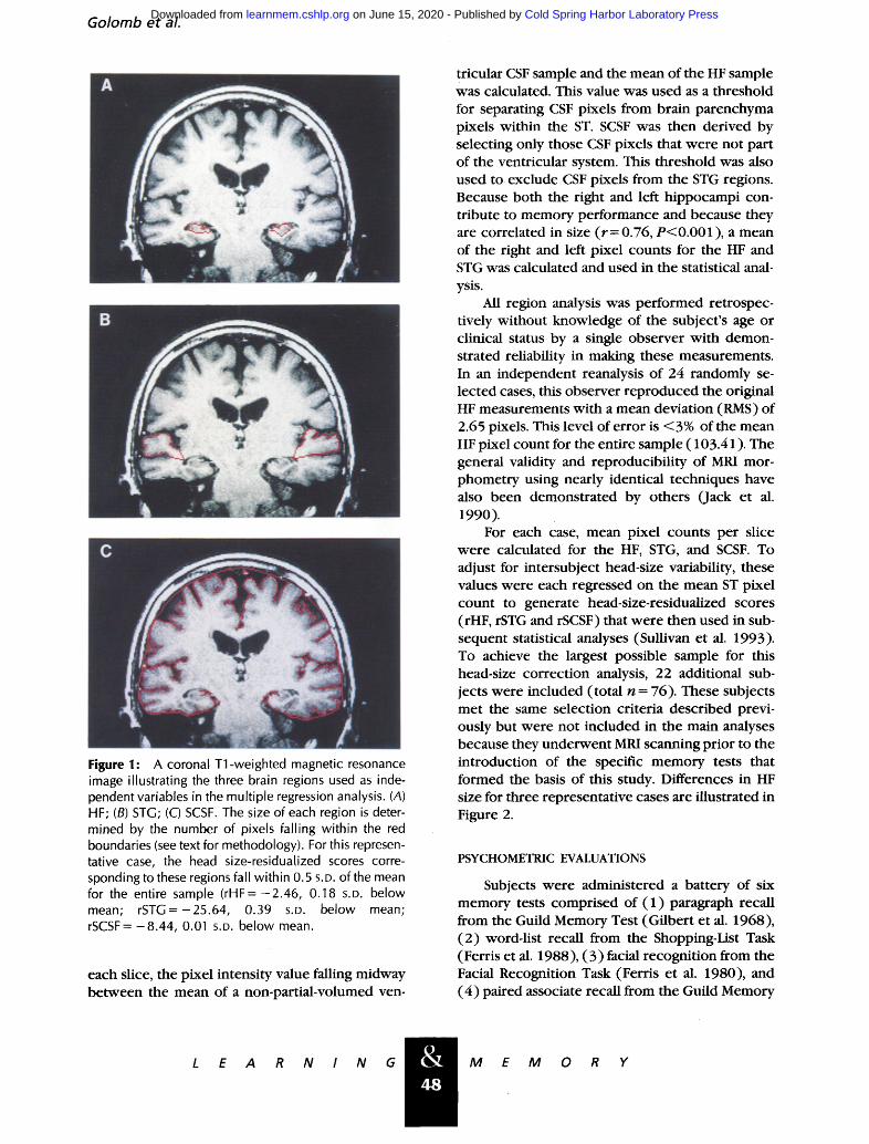
Figure 1: A coronal Tl-weighted magnetic resonance image illustrating the three brain regions used as inde- pendent variables in the multiple regression analysis. (A) HF; (B) STG; (C) SCSF. The size of each region is deter- mined by the number of pixels falling within the red boundaries (see text for methodology). For this represen- tative case, the head size-residualized scores corre- sponding to these regions fall within 0.5 S.D. of the mean for the entire sample ( rHF=-2 .46, 0.18 S.D. below mean; rSTG=-25.64, 0.39 S.D. below mean; rSCSF= -8.44, 0.01 S.D. below mean.
each slice, the pixel intensity value falling midway between the mean of a non-partial-volumed ven-
tricular CSF sample and the mean of the HF sample was calculated. This value was used as a threshold for separating CSF pixels from brain parenchyma pixels within the ST. SCSF was then derived by selecting only those CSF pixels that were not part of the ventricular system. This threshold was also used to exclude CSF pixels from the STG regions. Because both the right and left hippocampi con- tribute to memory performance and because they are correlated in size ( r = 0.76, P<O.O01), a mean of the right and left pixel counts for the HF and STG was calculated and used in the statistical anal- ysis.
All region analysis was performed retrospec- tively without knowledge of the subject 's age or clinical status by a single observer with demon- strated reliability in making these measurements. In an independent reanalysis of 24 randomly se- lected cases, this observer reproduced the original HF measurements with a mean deviation (RMS) of 2.65 pixels. This level of error is < 3% of the mean HF pixel count for the entire sample ( 103.41 ). The general validity and reproducibili ty of MRI mor- phometry using nearly identical techniques have also been demonstrated by others (Jack et al. 1990).
For each case, mean pixel counts per slice were calculated for the HF, STG, and SCSF. To adjust for intersubject head-size variability, these values were each regressed on the mean ST pixel count to generate head-size-residualized scores (rHF, rSTG and rSCSF) that were then used in sub- sequent statistical analyses (Sullivan et al. 1993). To achieve the largest possible sample for this head-size correction analysis, 22 additional sub- jects were included (total n - 76). These subjects met the same selection criteria described previ- ously but were not included in the main analyses because they underwent MRI scanning prior to the introduction of the specific memory tests that formed the basis of this study. Differences in HF size for three representative cases are illustrated in Figure 2.
PSYCHOMETRIC EVALUATIONS
Subjects were administered a battery of six memory tests comprised of (1 ) paragraph recall from the Guild Memory Test (Gilbert et al. 1968), (2 ) word-list recall from the Shopping-List Task (Ferris et al. 1988), (3 ) facial recognition from the Facial Recognition Task (Ferris et al. 1980), and (4 ) paired associate recall from the Guild Memory
& 48
L E A R N I N G M E M 0 R Y
Cold Spring Harbor Laboratory Press on June 15, 2020 - Published by learnmem.cshlp.orgDownloaded from
HIPPOCAMPAL SIZE AND MEMORY IN HUMAN AGING
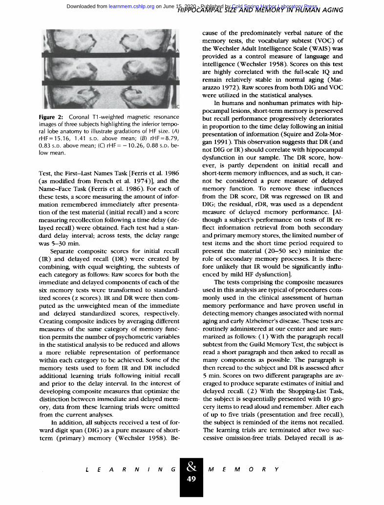
Figure 2: Coronal Tl-weighted magnetic resonance images of three subjects highlighting the inferior tempo- ral lobe anatomy to illustrate gradations of HF size. (A) rHF=15.16, 1.41 S.D. above mean; (B) rHF=8.79, 0.83 S.D. above mean; (C) rHF= - 10.26, 0.88 S.D. be- low mean.
Test, the First-Last Names Task [Ferris et al. 1986 (as modified from French et al. 1974)], and the Name-Face Task (Ferris et al. 1986). For each of these tests, a score measuring the amount of infor- mation remembered immediately after presenta- tion of the test material (initial recall) and a score measuring recollection following a time delay (de- layed recall) were obtained. Each test had a stan- dard delay interval; across tests, the delay range was 5-30 min.
Separate composite scores for initial recall (IR) and delayed recall (DR) were created by combining, with equal weighting, the subtests of each category as follows: Raw scores for both the immediate and delayed components of each of the six memory tests were transformed to standard- ized scores (z scores). IR and DR were then com- puted as the unweighted mean of the immediate and delayed standardized scores, respectively. Creating composite indices by averaging different measures of the same category of memory func- tion permits the number of psychometric variables in the statistical analysis to be reduced and allows a more reliable representation of performance within each category to be achieved. Some of the memory tests used to form IR and DR included additional learning trials following initial recall and prior to the delay interval. In the interest of developing composite measures that optimize the distinction between immediate and delayed mem- ory, data from these learning trials were omitted from the current analyses.
In addition, all subjects received a test of for- ward digit span (DIG) as a pure measure of short- term (primary) memory (Wechsler 1958). Be-
cause of the predominately verbal nature of the memory tests, the vocabulary subtest (VOC) of the Wechsler Adult Intelligence Scale (WAIS) was provided as a control measure of language and intelligence (Wechsler 1958). Scores on this test are highly correlated with the full-scale IQ and remain relatively stable in normal aging (Mat- arazzo 1972). Raw scores from both DIG and VOC were utilized in the statistical analyses.
In humans and nonhuman primates with hip- pocampal lesions, short-term memory is preserved but recall performance progressively deteriorates in proportion to the time delay following an initial presentation of information (Squire and Zola-Mor- gan 1991 ). This observation suggests that DR (and not DIG or IR) should correlate with hippocampal dysfunction in our sample. The DR score, how- ever, is partly dependent on initial recall and short-term memory influences, and as such, it can- not be considered a pure measure of delayed memory function. To remove these influences from the DR score, DR was regressed on IR and DIG; the residual, rDR, was used as a dependent measure of delayed memory performance. [Al- though a subject's performance on tests of IR re- flect information retrieval from both secondary and primary memory stores, the limited number of test items and the short time period required to present the material (20-50 sec) minimize the role of secondary memory processes. It is there- fore unlikely that IR would be significantly influ- enced by mild HF dysfunction].
The tests comprising the composite measures used in this analysis are typical of procedures com- monly used in the clinical assessment of human memory performance and have proven useful in detecting memory changes associated with normal aging and early Alzheimer's disease. These tests are routinely administered at our center and are sum- marized as follows: (1) With the paragraph recall subtest from the Guild Memory Test, the subject is read a short paragraph and then asked to recall as many components as possible. The paragraph is then reread to the subject and DR is assessed after 5 min. Scores on two different paragraphs are av- eraged to produce separate estimates of initial and delayed recall. (2) With the Shopping-List Task, the subject is sequentially presented with 10 gro- cery items to read aloud and remember. After each of up to five trims (presentation and free recall), the subject is reminded of the items not recalled. The learning trims are terminated after two suc- cessive omission-free trials. Delayed recall is as-
& 49
L E A R N / N G M E M 0 R Y
Cold Spring Harbor Laboratory Press on June 15, 2020 - Published by learnmem.cshlp.orgDownloaded from
Golomb et al.
sessed 30 min after the last trial. (3) With the Facial Recognition Task, the subject is sequentially presented with 10 photographs of unfamiliar faces. The photographs are then represented along with 10 new photographs in random order. Recogni- tion for the target photographs is tested over three trials, followed by a delayed trial 30 min later. (4) With the Paired Associate Recall of the Guild Memory Test, the subject is sequentially read 10 word pairs that vary in associative value. The first word of each pair is then given and the second word must be recalled. Subjects are reinforced for a correct response and are cued with the appro- priate second word for incorrect recall. A delayed recall trial is administered after 5 min. (5) With the Paired Associate Recall of the First-Last Names Task, a list of four first-last name pairs is sequen- tially presented. The subject is then tested for recall of the first name in response to the presen- tation of the last name. Up to three trials are ad- ministered, with a learning criterion of two con- secutive error-free trials. A delayed recall trial is administered after 30 min. (6) The Paired Associ- ate Recall of the Name-Face Task is identical to the First-Last Names Task described above, except that the paired associates consist of a first name and a photograph of a unfamiliar face. Test trials involve presentation of the faces alone, and the subject must recall the correct name.
Results
In a series of linear regression analyses, we first examined the relationship of age to each of the head-size-residualized brain regions. Although the age effect for rSTG was considerably weaker
than that seen for the other brain regions, these analyses revealed significant negative correlations for both parenchymal structures and a significant positive correlation for rSCSF (rrsTG = --0.27, P<O.05; rrHr=--0.46, P<O.O001; rrscsr=0.55, P<O.O001 ). These results are consistent with the widely recognized phenomenon of brain volume loss with subarachnoid space expansion that ac- companies normal aging.
To examine the specific effect of rHF on mem- ory performance while controlling for the influ- ence of age, sex, language/intelligence, and gener- alized atrophy, a hierarchical multiple regression statistical model was employed (Cohen and Cohen 1983). With rDR as the dependent variable, age, sex, VOC, and rSCSF were entered at step 1. In accord with our hypothesis, rHF but not rSTG re- sulted in a significant improvement in R 2 when added to the equation at step 2 (AR 2 = O. 19, P < 0.001). In parallel analyses with IR and DIG as dependent variables, both rHF and rSTG failed to result in significant R z changes at step 2. The dis- tributions of the dependent variables were similar in shape and revealed no evidence for ceiling or floor effects.
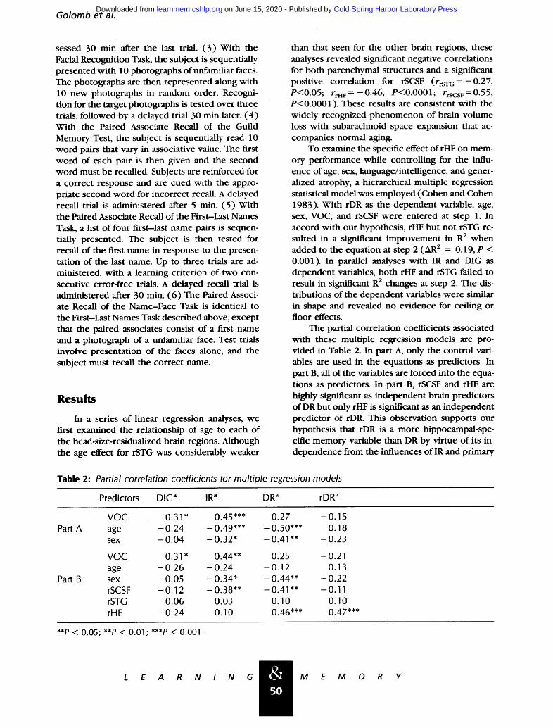
The partial correlation coefficients associated with these multiple regression models are pro- vided in Table 2. In part A, only the control vari- ables are used in the equations as predictors. In part B, all of the variables are forced into the equa- tions as predictors. In part B, rSCSF and rHF are highly significant as independent brain predictors of DR but only rHF is significant as an independent predictor of rDR. This observation supports our hypothesis that rDR is a more hippocampal-spe- cific memory variable than DR by virtue of its in- dependence from the influences of IR and primary
Table 2: Partial correlation coefficients for multiple regression models
Predictors D I G a IR a DR a rDR a
Part A
Part B
VOC 0.31" 0.45*** 0.27 - 0 . 1 5 age - 0 . 2 4 - 0 . 4 9 * * * - 0 . 5 0 * * * 0.18 sex - 0 . 0 4 - 0 . 3 2 * - 0 . 4 1 " * - 0 . 2 3
VOC 0.31" 0.44** 0.25 - 0 . 2 1 age - 0 . 2 6 - 0 . 2 4 - 0 . 1 2 0.13 sex - 0 . 0 5 - 0 . 3 4 * - 0 . 4 4 * * - 0 . 2 2 rSCSF - 0 . 1 2 - 0 . 3 8 * * - 0 . 4 1 " * - 0 . 1 1 rSTG 0.06 0.03 0.10 0.10 rHF - 0 . 2 4 0.10 0.46*** 0.47***
a*P < 0.05; **P < 0.01; ***P < 0.001.
& SO
L E A R N I N G M E M
Cold Spring Harbor Laboratory Press on June 15, 2020 - Published by learnmem.cshlp.orgDownloaded from
HIPPOCAMPAL SIZE AND MEMORY IN HUMAN AGING
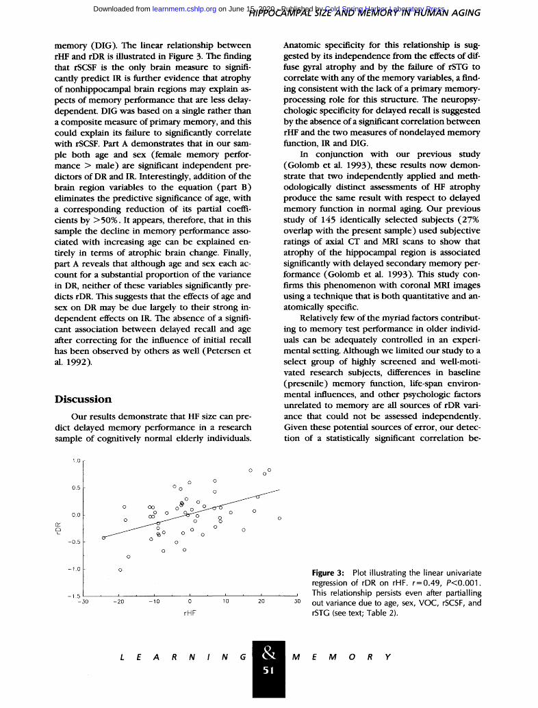
memory (DIG). The linear relationship be tween rHF and rDR is illustrated in Figure 3. The finding that rSCSF is the only brain measure to signifi- cantly predict IR is further evidence that atrophy of nonhippocampal brain regions may explain as- pects of memory performance that are less delay- dependent. DIG was based on a single rather than a composite measure of primary memory, and this could explain its failure to significantly correlate with rSCSF. Part A demonstrates that in our sam- ple both age and sex (female memory perfor- mance > male) are significant independent pre- dictors of DR and IR. Interestingly, addition of the brain region variables to the equation (part B) eliminates the predictive significance of age, with a corresponding reduct ion of its partial coeffi- cients by >50%. It appears, therefore, that in this sample the decline in memory performance asso- ciated with increasing age can be explained en- tirely in terms of atrophic brain change. Finally, part A reveals that although age and sex each ac- count for a substantial propor t ion of the variance in DR, nei ther of these variables significantly pre- dicts rDR. This suggests that the effects of age and sex on DR may be due largely to their strong in- dependent effects on IR. The absence of a signifi- cant association be tween delayed recall and age after correcting for the influence of initial recall has been observed by others as well (Petersen et al. 1992).
D i s c u s s i o n
Our results demonstrate that HF size can pre- dict delayed memory performance in a research sample of cognitively normal elderly individuals.
Anatomic specificity for this relationship is sug- gested by its independence from the effects of dif- fuse gyral atrophy and by the failure of rSTG to correlate with any of the memory variables, a find- ing consistent with the lack of a primary memory- processing role for this structure. The neuropsy- chologic specificity for delayed recall is suggested by the absence of a significant correlation be tween rHF and the two measures of nondelayed memory function, IR and DIG.
In conjunction with our previous study (Golomb et al. 1993), these results now demon- strate that two independent ly applied and meth- odologically distinct assessments of HF atrophy produce the same result with respect to delayed memory function in normal aging. Our previous study of 145 identically selected subjects (27% overlap with the present sample) used subjective ratings of axial CT and MRI scans to show that atrophy of the hippocampal region is associated significantly with delayed secondary memory per- formance (Golomb et al. 1993). This study con- firms this phenomenon with coronal MRI images using a technique that is both quantitative and an- atomically specific.
Relatively few of the myriad factors contribut- ing to memory test performance in older individ- uals can be adequately controlled in an experi- mental setting. Although we limited our study to a select group of highly screened and well-moti- vated research subjects, differences in baseline (presenile) memory function, life-span environ- mental influences, and other psychologic factors unrelated to memory are all sources of rDR vari- ance that could not be assessed independently. Given these potential sources of error, our detec- tion of a statistically significant correlation be-
1.0
0.5
0.0
-0 .5
- 1 . 0
- 1 . 5 - 30
o o
o
o o 0
o ° ~o o o o o
o o
o o
o
/
r i I i
- 2 0 - 1 0 0 10
rHF
i i
20 50
Figure 3: Plot illustrating the linear univariate regression of rDR on rHF. r=0.49, P<0.001. This relationship persists even after partialling out variance due to age, sex, VOC, rSCSF, and rSTG (see text; Table 2).
L E A R N I N G & 51
M E M 0 R Y
Cold Spring Harbor Laboratory Press on June 15, 2020 - Published by learnmem.cshlp.orgDownloaded from
G o l o m b e t al.
tween HF size and memory performance supports the robustness of this relationship.
Practical constraints limited the scope of our study to a relatively circumscribed set of objec- tives. In particular, we did not at tempt to test dou- ble dissociation hypotheses concerning structure/ function relationships of the HF and STG. Further- more, our study was not designed to examine whether the sizes of brain regions other than the HF and STG correlate with memory performance. Although necessary, an intact HF does not ensure effective delayed memory function. For example, age-associated degenerative processes affecting the frontal lobe may also contribute to memory impairment (Moscovitch and Winocur 1992). Re- latedly, it must be emphasized that atrophy of other components of the medial temporal lobe memory system may be even more important de- terminates of memory function than the HF itself. The entorhinal and perirhinal cortices, along with portions of the parahippocampal cortex, also play an important role in memory processing, and these regions may undergo comparable degrees of involutional change with age. Therefore, our find- ing of a significant correlation be tween HF size and memory performance does not necessarily im- ply a causal role for the HF and may actually reflect degeneration of other medial temporal lobe struc- tures with which the HF shares variance. Although we were unable to measure these other structures in this study, the relative contr ibution of medial temporal lobe subregions to memory performance in normal aging should be the focus of future re- search using higher resolution MRI techniques.
Finally, the goal of this study was not to ex- amine the relationship between HF size and mod- els of hippocampal-specific memory processing but, rather, to test whe ther HF atrophy was related to the general phenomenon of age-associated memory loss. Because delayed secondary memory impairment is a principal and clinically well-doc- umented feature of this phenomenon with clear relevance to hippocampal function, we sought to develop a measure that would effectively repre- sent it as a cognitive construct. By combining sev- eral paradigms commonly used clinically to assess memory performance in older adults and applying a statistical method of correct ion for nondelayed recall, the rDR variable represents an intuitively valid estimate of delay-specific memory function. In addition to nondelayed and delayed memory performance, future research should investigate whether implicit/explicit dissociations and other
cognitive features specific to hippocampal mem- ory processing are associated with HF atrophy in normal aging.
This study was based on the examination of carefully screened research volunteers, and cau- tion must be exercised when applying its conclu- sions to the general elderly community. Neverthe- less, the present observations have implications for the evaluation of older individuals with symp- toms of mild memory dysfunction and suggest that hippocampal morphometry could play a role in the differential diagnosis of such complaints. Of particular relevance to these findings may be the recently defined clinical syndrome of Age Associ- ated Memory Impairment (AAMI) (Crook et al. 1986). Forty-four percent of our subjects met cri- teria for this diagnosis on the basis of their subjec- tive impression of a decline in memory perfor- mance over a 5- to lO-year interval [Global Dete- rioration Scale (GDS)= 2] and a score of >1 S.D. below the mean for a young adult reference group on the delayed paired associate and delayed para- graph recall subtests of the Guild Memory Test (Gilbert et al. 1968). Interestingly, even when these AAMI subjects were excluded from the anal- ysis, a significant partial correlation was still ob- tained be tween rHF and rDR ( r - 0 . 4 3 , P<O.O5).
Our findings suggest that the well-recognized loss of parenchymal brain volume that accompa- nies human aging may result in quantifiable behav- ioral manifestations if structures mediating spe- cific neuropsychologic functions are affected se- verely enough. Further research must examine the histopathologic basis for hippocampal atrophy in normal aging and determine whether this phe- nomenon constitutes a significant risk for acceler- ated intellectual decline.
Acknowledgments This work was funded in part by grants IP30AG08051,
IR29MH44697, IP30MH4386, IR01MH43965, and IR01AG03051 from the National Institutes of Health. We are indebted to Martha Helmers and Anthony Jalandoni for their expert assistance in preparing the figures.
The publication costs of this article were defrayed in part by payment of page charges. This article must therefore be hereby marked "advertisement" in accordance with 18 USC section 1734 solely to indicate this fact.
References Brody, H. 1955. Organization of the cerebral cortex III. A study of aging in the human cerebral cortex. J. Comp. Neurol. 102:511-556.
& 52
L E A R N I N G M E M O R Y
Cold Spring Harbor Laboratory Press on June 15, 2020 - Published by learnmem.cshlp.orgDownloaded from
HIPPOCAMPAL SIZE A N D M E M O R Y IN H U M A N A G I N G
Cockrell, J.R. and M.F. Folstein. 1988. Mini-mental state examination (MMSE). Psychopharmacol. Bull. 24: 689-692.
Cohen, J. and P. Cohen. 1983. Applied multiple regression/correlation analysis for the behavioral sciences. Lawrence Erlbaum Associates, Hillsdale, NJ.
Convit, A., M.J. de Leon, J. Golomb, A.E. George, C.Y. Tarshish, M. Bobinski, W. Tsui, S. de Santi, J. Wegiel, and H. Wisniewski. 1993. Hippocampal atrophy in early AIzheimer's disease, anatomic specificity and validation. Psychiatr. Q. 64: 371-387.
Crook, T., R.T. Bartus, S.H. Ferris, P. Whitehouse, G.D. Cohen, and S. Gershon. 1986. Age-associated memory impairment: Proposed diagnostic criteria and measures of clinical change--Report of a NIMH Work Group. Dev. Neuropsychol. 2: 261-276.
de Leon, M.J., A.E. George, L.A. Stylopoulos, G. Smith, and D.C. Miller. 1989. Early marker for Alzheimer's disease: The atrophic hippocampus. Lancet 2" 672-673.
de Leon, M.J., J. Golomb, A.E. George, A. Convit, C.Y. Tarshish, T. McRae, S. de Santi, G. Smith, and S.H. Ferris. 1993. The radiologic prediction of AIzheimer's disease: The atrophic hippocampal formation. Am. J. Neuroradiol. 14" 897-906.
Ferris, S.H., T. Crook, E. Clark, M. McCarthy, and D. Rae. 1980. Facial recognition memory deficits in normal aging and senile dementia. J. Gerontol. 35: 707-714.
Ferris, S.H., T. Crook, C. Flicker, B. Reisberg, and R.T. Bartus. 1986. Assessing cognitive impairment and evaluating treatment effects: Psychometric performance tests. In The handbook for clinical memory assessment of older adults (ed. L.W. Poon), pp. 139-148. American Psychological Association, Washington, D.C.
Ferris, S.H., C. Flicker, and B. Reisberg. 1988. NYU computerized test battery for assessing cognition in aging and dementia. Psychopharmacol. Bull. 24" 699-702.
Flicker, C., S.H. Ferris, and B. Reisberg. 1993. A longitudinal study of cognitive function in elderly persons with subjective memory complaints. J. Am. Geriatr. Soc. 41" 1071-1074.
Folstein, M.F., S.E. Folstein, and P.R. McHugh. 1975. Mini-mental state: A practical method for grading the cognitive state of patients for the clinician. I. Psychiatr. Res. 12: 189-198.
Fozard, J.L. 1985. Psychology of aging--Normal and pathological age differences in memory. In Textbook of geriatric medicine and gerontology (ed. J.C. Brocklehurst), pp. 122-144. Churchill/Livingstone, Edinburgh, Scotland.
French, J.W., R.B. Ekstrom, and A. Leighton. 1974. First and last names test. In Kit of reference tests for cognitive factors (rev. ed.). Educational Testing Service, Princeton, NJ.
& 53
Gilbert, J.G., R.F. Levee, and F.L. Catalano. 1968. A preliminary report on a new memory scale. Percept. Mot. Skills 27: 277-278.
Golomb, J., M.J. de Leon, A. Kluger, A.E. George, C. Tarshish, and S.H. Ferris. 1993. Hippocampal atrophy in normal aging: An association with recent memory impairment. Arch. Neurol. 50" 967-976.
Hachinski, V.C., P. Potter, and H. Mersky. 1987. Leuko-araiosis. Arch. Neurol. 44-21-23.
Hamilton, M. 1967. Development of a rating scale for primary depression illness Bri. J. Soc. Clin. Psychol. 6" 278-296.
Jack, C., M. Bently, C. Twomey, and A. Zinsmeister. 1990. MR imaging-based volume measurements of the hippocampal formation and anterior temporal lobe: Validation studies. Radiology 176: 205-209.
Jack, C.R. Jr., R.C. Petersen, P.C. O'Brien, and E.G. Tangalos. 1992. MR-based hippocampal volumetry in the diagnosis of Alzheimer's disease. Neurology 42" 183-188.
Kemper, T. 1984. Neuroanatomical and neuropathological changes in normal aging and in dementia. In Clinical neurology of aging (ed. M.L. Albert), pp. 9-52. Oxford University Press, New York.
Killiany, R.J., M.B. Moss, M.S. Albert, T. Sandor, J. Tieman, and F. Jolesz. 1993. Temporal lobe regions on magnetic resonance imaging identify patients with early Alzheimer's disease. Arch. Neurol. 50- 949-954.
Kolb, B. and I.Q. Whishaw. 1990. Head trauma and degenerative diseases. In Fundamentals of human neuropsychology, pp. 830-832. W.H. Freeman, New York.
Matarazzo, J.D. 1972. Wechsler's measurement and appraisal of adult intelligence, 5th ed. Williams and Wilkins, Baltimore, MD.
Moscovitch, M. and G. Winocur. 1992. The neuropsychology of memory and aging. In The handbook of aging and cognition (ed. F.I.M. Craik and T.A. Salthouse), pp. 315-372. Lawrence Erlbaum Associates, Hillsdale, NJ.
Petersen, R.C., G. Smith, E. Kokmen, R.J. Ivnik, and E.G. Tangalos. 1992. Memory function in normal aging. Neurology 42: 396-401.
Pfefferbaum, A., E.V. Sullivan, T.L. Jernigan, R.B. Zipursky, M.J. Rosenbloom, J.A. Yesavage, and J.R. Tinklenberg. 1990. A quantitative analysis of CT and cognitive measures in normal aging and Alzheimer's disease. Psychiatr. Res. Neuroimaging 35:115-136.
Poon, L.W. 1985. Differences in human memory with aging: Nature, causes, and clinical implications. In Handbook of the psychology of aging (ed. J.E. Birren and K.W. Schaie), pp. 427-462. Van Nostrand Reinhold, New York.
L E A R N I N G M E M 0 R Y
Cold Spring Harbor Laboratory Press on June 15, 2020 - Published by learnmem.cshlp.orgDownloaded from

Golomb et al.
Press, G.A., D.G. Amaral, and L.R. Squire. 1989. Hippocampal abnormalities in amnesic patients revealed by high-resolution magnetic resonance imaging. Nature 341 : 54-57.
Reisberg, B., S.H. Ferris, M.J. de Leon, and T. Crook. 1982. The global deterioration scale for assessment of primary degenerative dementia. Am. J. Psychiatr. 139:1136-1139.
Rosen, W.G., R.D. Terry, P.A. Fuld, R. Katzman, and A. Peck. 1980. Pathological verification of ischemia score in differentiation of dementias. Ann. Neurol. 7: 486-488.
Squire, L.R. and S. Zola-Morgan. 1991. The medial temporal lobe memory system. Science 20:1380-1386.
Sullivan, E.V., P.K. Shear, D.H. Mathalon, L.O. Kelvin, J.A. Yesavage, J.R. Tinklenberg, and A. Pfefferbaum. 1993. Greater abnormalities of brain cerebrospinal fluid volumes in younger than in older patients with Alzheimer's disease. Arch. Neurol. 50: 359-373.
Terry, R.D., R. DeTeresa, and L.A. Hansen. 1987. Neocortical cell counts in normal human adult aging. Ann. Neurol. 21: 530-539.
Wechsler, D.A. 1958. The measurement and appraisal of adult intelligence. Williams and Wilkins, Baltimore, MD.
Received December 10, 1993; accepted in revised form March 4, 1994.
&~ 54
L E A R N I N G M E M O R Y
Cold Spring Harbor Laboratory Press on June 15, 2020 - Published by learnmem.cshlp.orgDownloaded from

10.1101/lm.1.1.45Access the most recent version at doi: 1:1994, Learn. Mem.
J Golomb, A Kluger, M J de Leon, et al. of delayed secondary memory performance.Hippocampal formation size in normal human aging: a correlate
References
http://learnmem.cshlp.org/content/1/1/45.full.html#ref-list-1
This article cites 25 articles, 3 of which can be accessed free at:
License
ServiceEmail Alerting
click here.the top right corner of the article or
Receive free email alerts when new articles cite this article - sign up in the box at
Copyright © Cold Spring Harbor Laboratory Press
Cold Spring Harbor Laboratory Press on June 15, 2020 - Published by learnmem.cshlp.orgDownloaded from