Embed Size (px)
Citation preview

HIS BUNDLE PACING: TELL ME MORE Transcript of the live webcast – February 8, 2016
Featuring:
Faculty Kenneth Ellenbogen, MD, FACC, FHRS Kontos Professor of Cardiology Chairman, Division of Cardiology Virginia Commonwealth University Richmond, Virginia
Daniel Lustgarten, MD, PhD
Assistant Professor of Medicine Cardiovascular Medicine, Dept. of Medicine University of Vermont Burlington, Vermont
Pugazhendi Vijayaraman, MD Director, Electrophysiology Dept. of Cardiology & Interventional Cardiology Geisinger Wyoming Valley Wilkes-Barre, Pennsylvania
Gopi Dandamudi, MD, FHRS Program Director, IU Health Atrial Fibrillation Center Clinical Assistant Professor of Medicine Indiana University School of Medicine Indianapolis, Indiana
DISCLAIMER
This video is provided for general educational purposes only and should not be considered the exclusive source for this type of information. If applicable, patient information (names, serial numbers, date, etc.) has been changed or removed to protect the privacy of the patients referenced in this video. At all times, it is the professional responsibility of the practitioner to exercise independent clinical judgment in a particular situation. Changes in a patient’s disease and/or medications may alter the efficacy of a device’s programmed parameters or related features and results may vary.
COMPENSATION This faculty is being paid as a consultant for the services being provided in accordance with the Sunshine Act.
CAUTION STATEMENT The content and any case study data in this video is provided by physician faculty and not all comments are the opinions of Medtronic.
This information is intended only for users in markets where Medtronic products and therapies are approved or available for use as indicated within the respective product manuals. Content on specific Medtronic products and therapies is not intended for users in markets that do not have authorization for use.

Ellenbogen: Welcome to Global Grand Rounds. Tonight, we’re going to talk about HIS Bundle Pacing: Tell me more. I am Dr. Kenneth Ellenbogen, chairman, Division of Cardiology Virginia Commonwealth University, Richmond, Virginia. I’m pleased to introduce our faculty tonight. Dr. Gopi Dandamudi who is program director at the IU Health Atrial Fibrillation Center. He's a Clinical Assistant Professor in Medicine at the Indiana University School of Medicine; Dr. Pugazhendhi Vijayaraman, Director, Electrophysiology at the Geisinger Heart Institute in the Geisinger Wyoming Valley campus in Wilkes-Barre, Pennsylvania; and Dr. Daniel Lustgarten, Associate Professor of Medicine in the Cardiovascular Division from the Department of Medicine at the University of Vermont Medical Center in Burlington, Vermont.
Ellenbogen: Now, our first speaker, Dr. Gopi Dandamudi will talk about current evidence for ventricular pacing. I will point out that for each of the speakers tonight, the views and opinions included during this program are related to the methods and outcomes of His bundle pacing are based solely on the clinical experience of each physician and do not represent those of Medtronic. Gopi, go ahead, please.
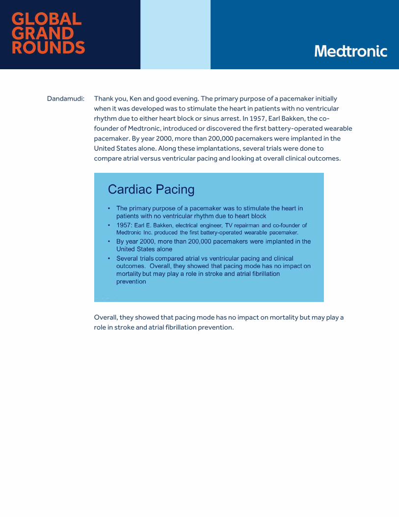
Dandamudi: Thank you, Ken and good evening. The primary purpose of a pacemaker initially when it was developed was to stimulate the heart in patients with no ventricular rhythm due to either heart block or sinus arrest. In 1957, Earl Bakken, the co-founder of Medtronic, introduced or discovered the first battery-operated wearable pacemaker. By year 2000, more than 200,000 pacemakers were implanted in the United States alone. Along these implantations, several trials were done to compare atrial versus ventricular pacing and looking at overall clinical outcomes.
Overall, they showed that pacing mode has no impact on mortality but may play a role in stroke and atrial fibrillation prevention.
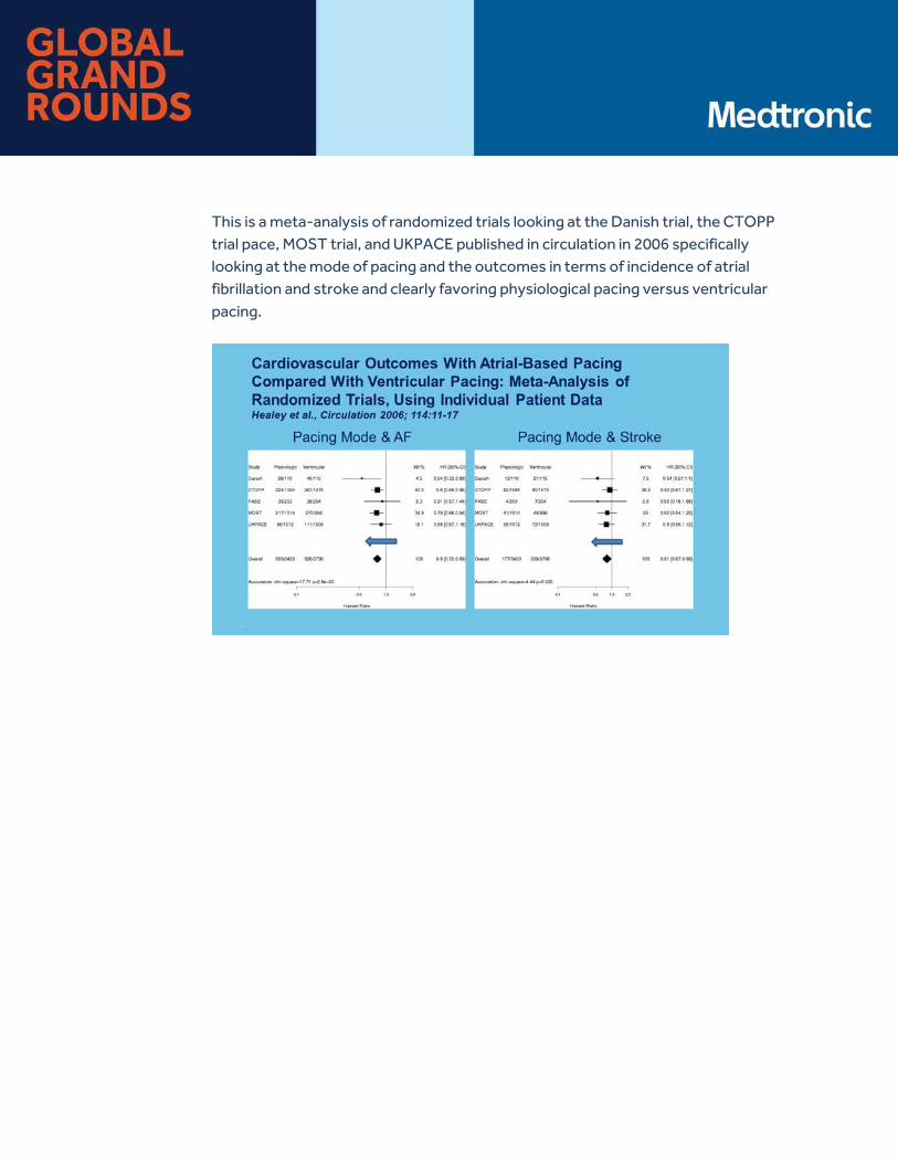
This is a meta-analysis of randomized trials looking at the Danish trial, the CTOPP trial pace, MOST trial, and UKPACE published in circulation in 2006 specifically looking at the mode of pacing and the outcomes in terms of incidence of atrial fibrillation and stroke and clearly favoring physiological pacing versus ventricular pacing.
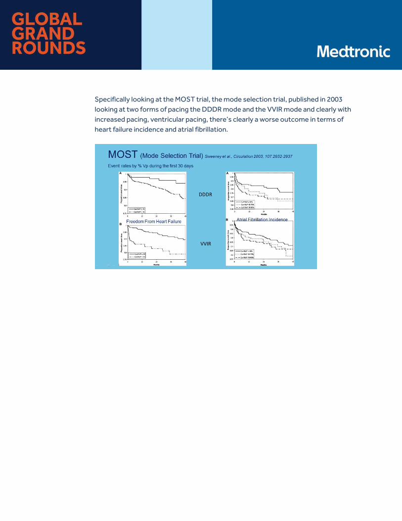
Specifically looking at the MOST trial, the mode selection trial, published in 2003 looking at two forms of pacing the DDDR mode and the VVIR mode and clearly with increased pacing, ventricular pacing, there’s clearly a worse outcome in terms of heart failure incidence and atrial fibrillation.
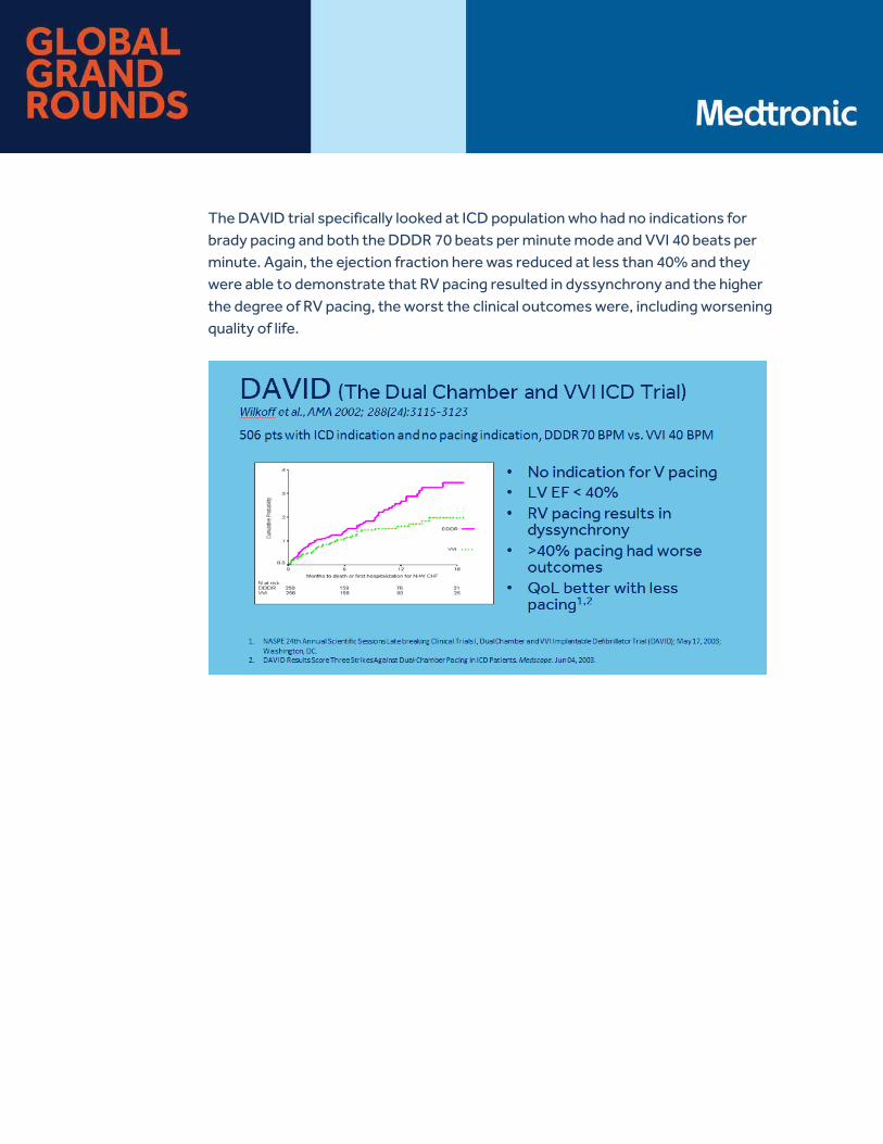
The DAVID trial specifically looked at ICD population who had no indications for brady pacing and both the DDDR 70 beats per minute mode and VVI 40 beats per minute. Again, the ejection fraction here was reduced at less than 40% and they were able to demonstrate that RV pacing resulted in dyssynchrony and the higher the degree of RV pacing, the worst the clinical outcomes were, including worsening quality of life.
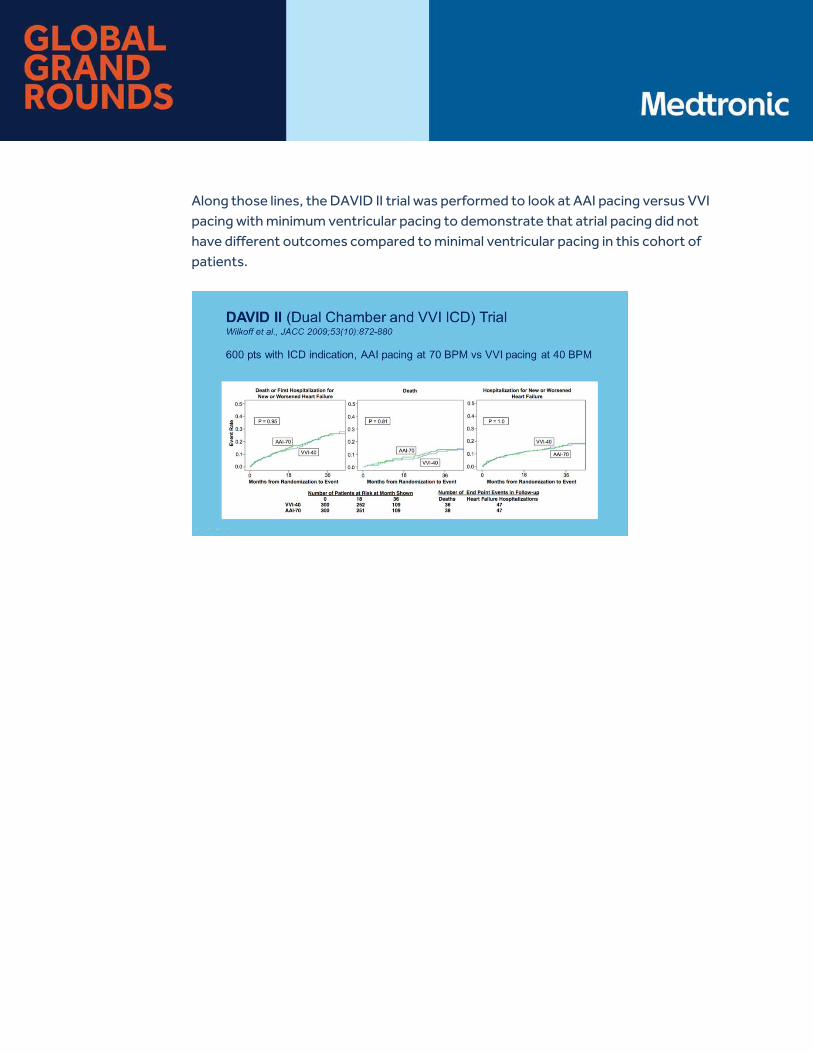
Along those lines, the DAVID II trial was performed to look at AAI pacing versus VVI pacing with minimum ventricular pacing to demonstrate that atrial pacing did not have different outcomes compared to minimal ventricular pacing in this cohort of patients.
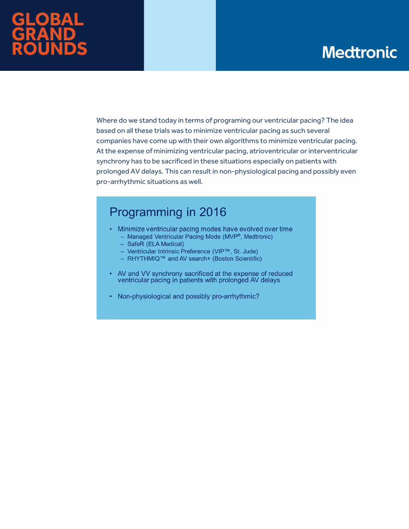
Where do we stand today in terms of programing our ventricular pacing? The idea based on all these trials was to minimize ventricular pacing as such several companies have come up with their own algorithms to minimize ventricular pacing. At the expense of minimizing ventricular pacing, atrioventricular or interventricular synchrony has to be sacrificed in these situations especially on patients with prolonged AV delays. This can result in non-physiological pacing and possibly even pro-arrhythmic situations as well.
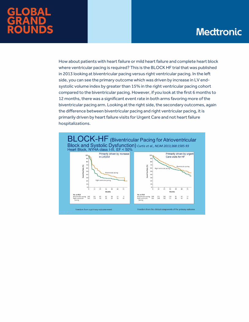
How about patients with heart failure or mild heart failure and complete heart block where ventricular pacing is required? This is the BLOCK HF trial that was published in 2013 looking at biventricular pacing versus right ventricular pacing. In the left side, you can see the primary outcome which was driven by increase in LV end-systolic volume index by greater than 15% in the right ventricular pacing cohort compared to the biventricular pacing. However, if you look at the first 6 months to 12 months, there was a significant event rate in both arms favoring more of the biventricular pacing arm. Looking at the right side, the secondary outcomes, again the difference between biventricular pacing and right ventricular pacing, it is primarily driven by heart failure visits for Urgent Care and not heart failure hospitalizations.
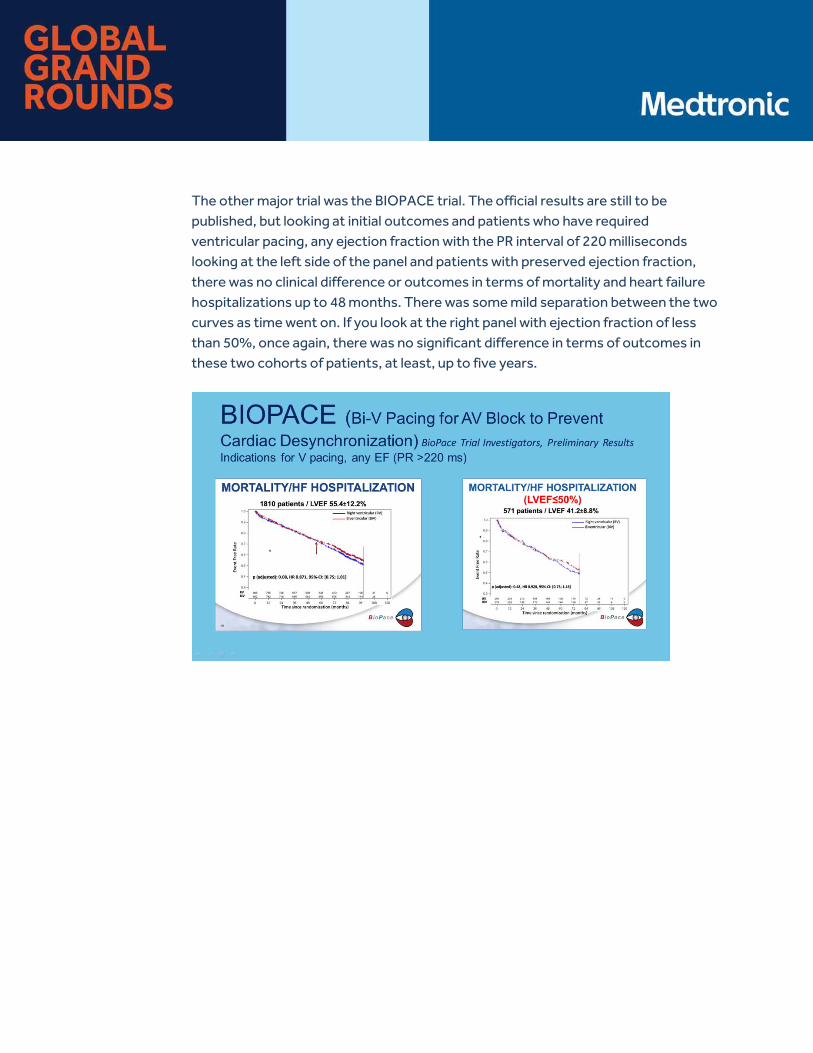
The other major trial was the BIOPACE trial. The official results are still to be published, but looking at initial outcomes and patients who have required ventricular pacing, any ejection fraction with the PR interval of 220 milliseconds looking at the left side of the panel and patients with preserved ejection fraction, there was no clinical difference or outcomes in terms of mortality and heart failure hospitalizations up to 48 months. There was some mild separation between the two curves as time went on. If you look at the right panel with ejection fraction of less than 50%, once again, there was no significant difference in terms of outcomes in these two cohorts of patients, at least, up to five years.

How about RV alternate site pacing? Pacing from areas outside of the RV apex including the outflow track, the high, mid, and apical septum. Clearly, there are limitations in this form of pacing due to difficult anatomy and inability to accurately predict the site of pacing due to fluoroscopic limitations. This has resulted in variable results.
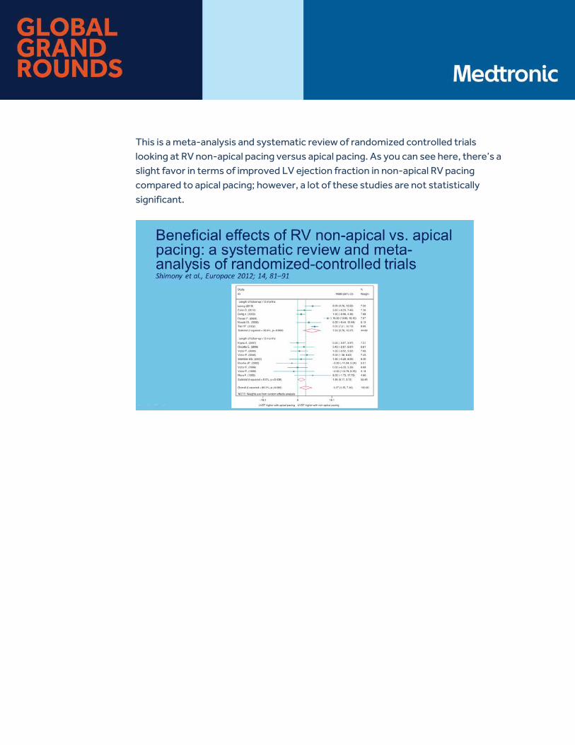
This is a meta-analysis and systematic review of randomized controlled trials looking at RV non-apical pacing versus apical pacing. As you can see here, there’s a slight favor in terms of improved LV ejection fraction in non-apical RV pacing compared to apical pacing; however, a lot of these studies are not statistically significant.
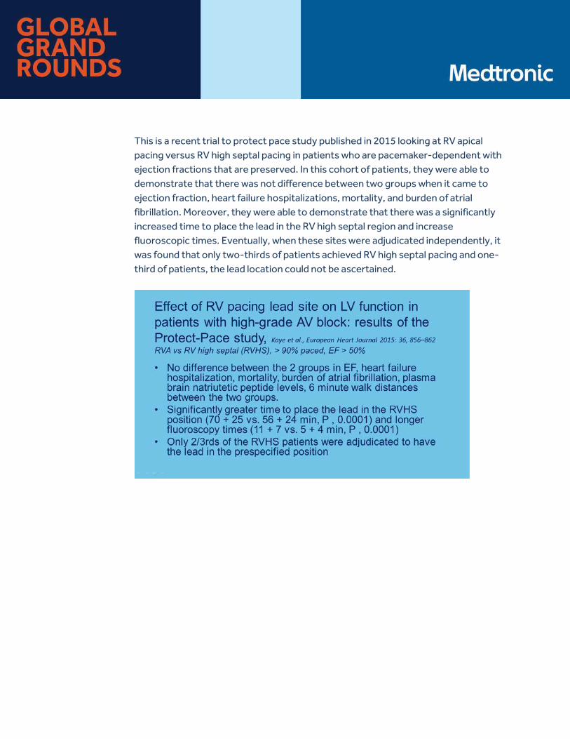
This is a recent trial to protect pace study published in 2015 looking at RV apical pacing versus RV high septal pacing in patients who are pacemaker-dependent with ejection fractions that are preserved. In this cohort of patients, they were able to demonstrate that there was not difference between two groups when it came to ejection fraction, heart failure hospitalizations, mortality, and burden of atrial fibrillation. Moreover, they were able to demonstrate that there was a significantly increased time to place the lead in the RV high septal region and increase fluoroscopic times. Eventually, when these sites were adjudicated independently, it was found that only two-thirds of patients achieved RV high septal pacing and one-third of patients, the lead location could not be ascertained.

Why should we consider His bundle pacing? First and foremost, it replicates human physiology. There is a reason why evolution has selected His-Purkinje system as the most efficient way to activate the ventricles. Normal QRS complex width is around 60 to 80 milliseconds. A lead tip and the body potentially are within the right atrium. You will hear some examples later of looking at the lead tip itself. This could result in reduced tricuspid regurgitation and even reducing the incidence of valve perforation with lead placement.
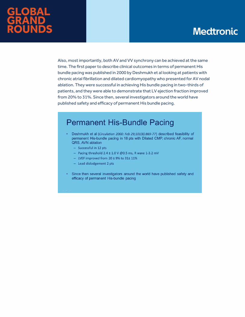
Also, most importantly, both AV and VV synchrony can be achieved at the same time. The first paper to describe clinical outcomes in terms of permanent His bundle pacing was published in 2000 by Deshmukh et al looking at patients with chronic atrial fibrillation and dilated cardiomyopathy who presented for AV nodal ablation. They were successful in achieving His bundle pacing in two-thirds of patients, and they were able to demonstrate that LV ejection fraction improved from 20% to 31%. Since then, several investigators around the world have published safety and efficacy of permanent His bundle pacing.
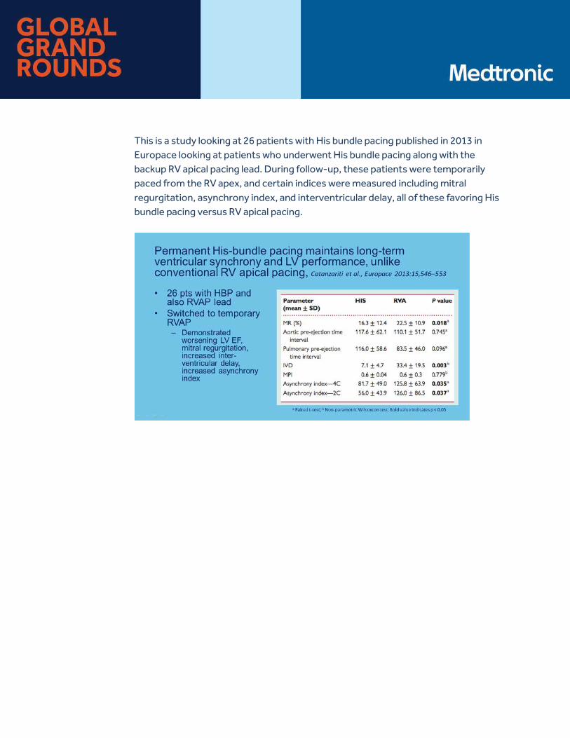
This is a study looking at 26 patients with His bundle pacing published in 2013 in Europace looking at patients who underwent His bundle pacing along with the backup RV apical pacing lead. During follow-up, these patients were temporarily paced from the RV apex, and certain indices were measured including mitral regurgitation, asynchrony index, and interventricular delay, all of these favoring His bundle pacing versus RV apical pacing.
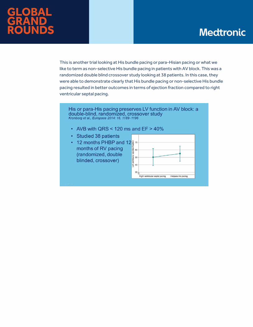
This is another trial looking at His bundle pacing or para-Hisian pacing or what we like to term as non-selective His bundle pacing in patients with AV block. This was a randomized double blind crossover study looking at 38 patients. In this case, they were able to demonstrate clearly that His bundle pacing or non-selective His bundle pacing resulted in better outcomes in terms of ejection fraction compared to right ventricular septal pacing.
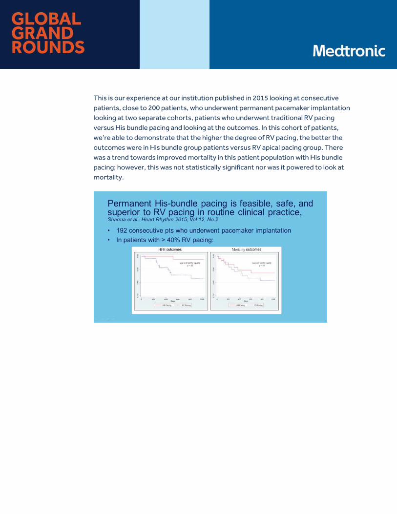
This is our experience at our institution published in 2015 looking at consecutive patients, close to 200 patients, who underwent permanent pacemaker implantation looking at two separate cohorts, patients who underwent traditional RV pacing versus His bundle pacing and looking at the outcomes. In this cohort of patients, we’re able to demonstrate that the higher the degree of RV pacing, the better the outcomes were in His bundle group patients versus RV apical pacing group. There was a trend towards improved mortality in this patient population with His bundle pacing; however, this was not statistically significant nor was it powered to look at mortality.
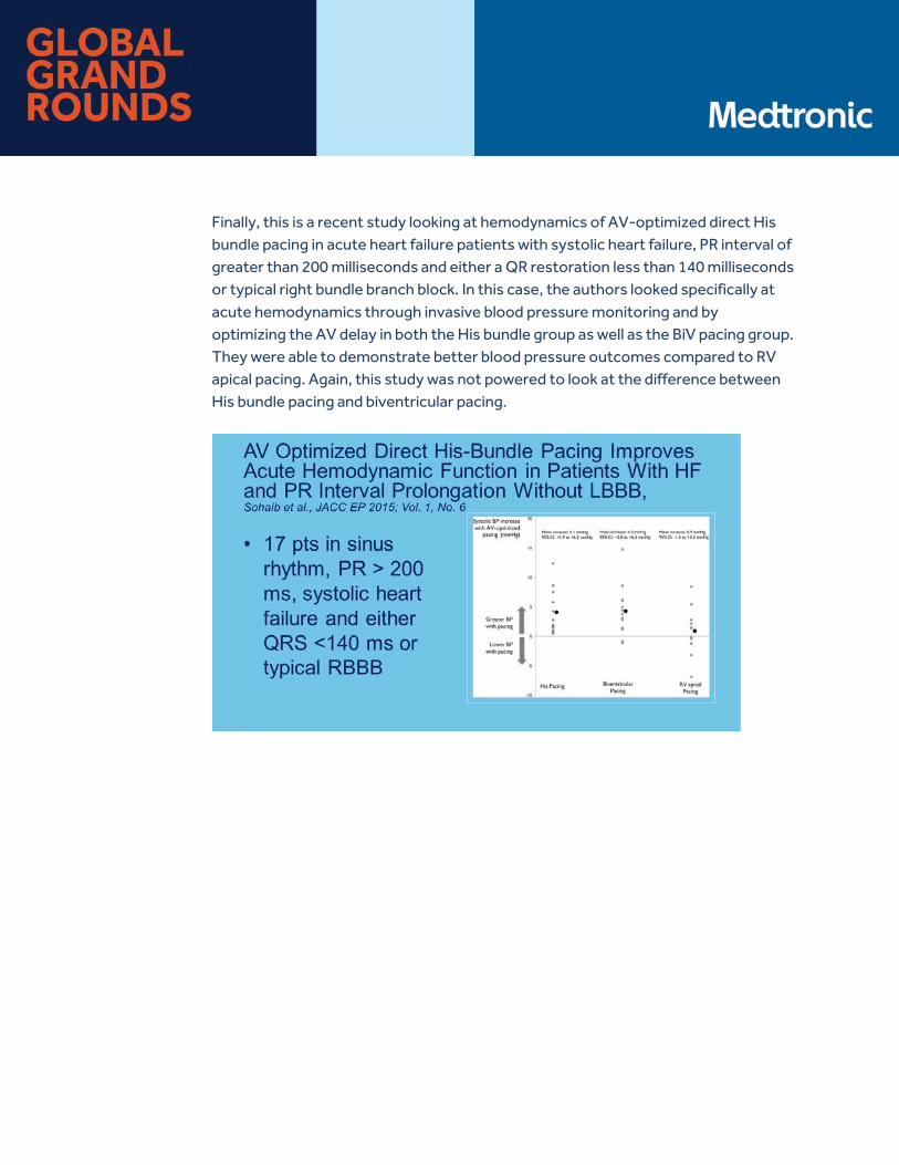
Finally, this is a recent study looking at hemodynamics of AV-optimized direct His bundle pacing in acute heart failure patients with systolic heart failure, PR interval of greater than 200 milliseconds and either a QR restoration less than 140 milliseconds or typical right bundle branch block. In this case, the authors looked specifically at acute hemodynamics through invasive blood pressure monitoring and by optimizing the AV delay in both the His bundle group as well as the BiV pacing group. They were able to demonstrate better blood pressure outcomes compared to RV apical pacing. Again, this study was not powered to look at the difference between His bundle pacing and biventricular pacing.
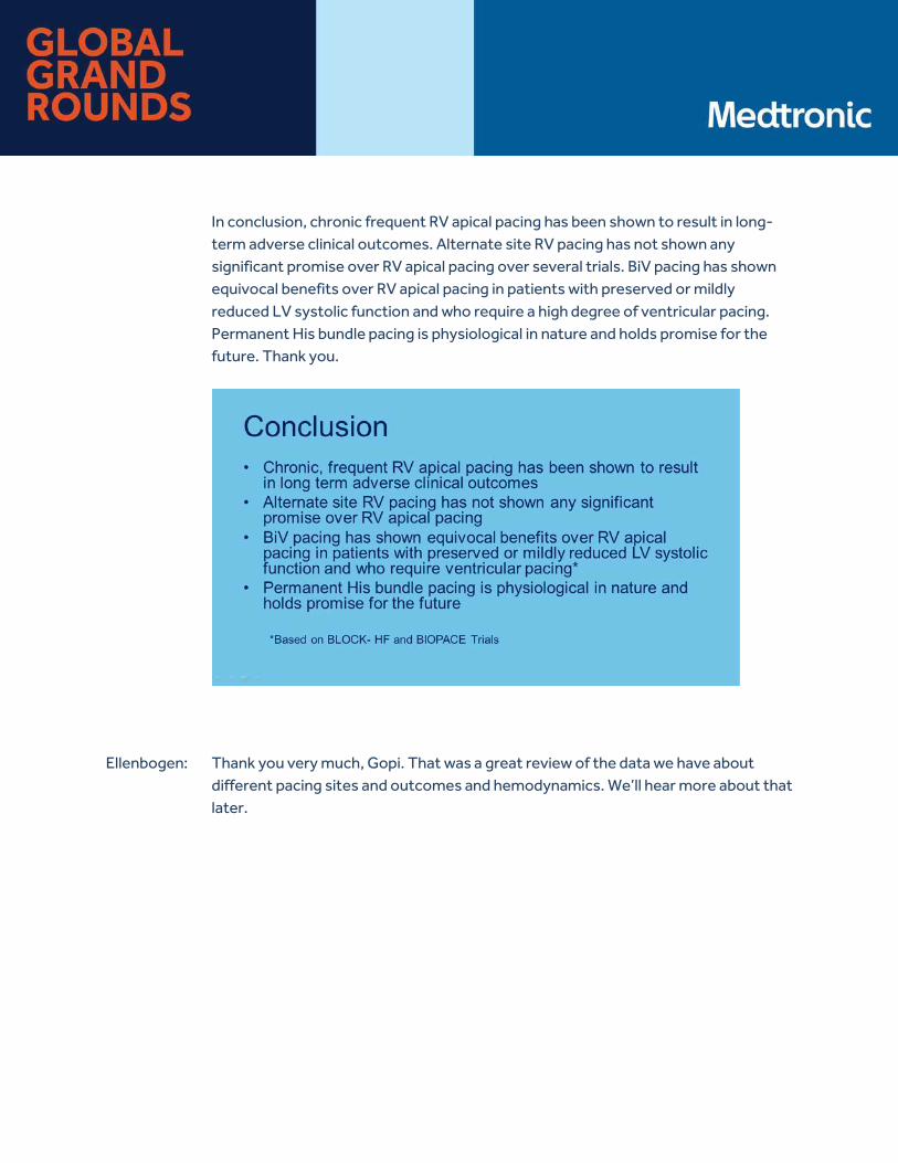
In conclusion, chronic frequent RV apical pacing has been shown to result in long-term adverse clinical outcomes. Alternate site RV pacing has not shown any significant promise over RV apical pacing over several trials. BiV pacing has shown equivocal benefits over RV apical pacing in patients with preserved or mildly reduced LV systolic function and who require a high degree of ventricular pacing. Permanent His bundle pacing is physiological in nature and holds promise for the future. Thank you.
Ellenbogen: Thank you very much, Gopi. That was a great review of the data we have about different pacing sites and outcomes and hemodynamics. We’ll hear more about that later.
Ellenbogen: Our next speaker is Dr. Pugazhendhi Vijayaraman, and he will discuss how to do His bundle pacing. His talk is entitled Methods and Outcomes of His bundle pacing.
Vijayaraman: Thank you, Ken.
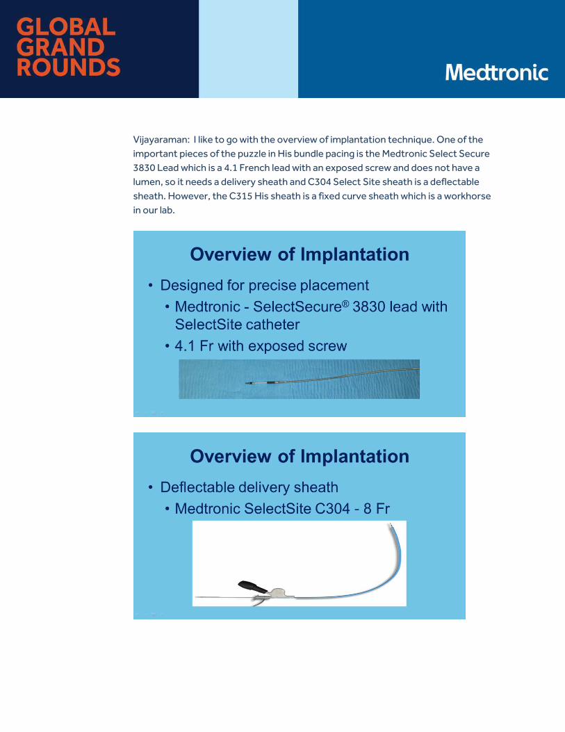
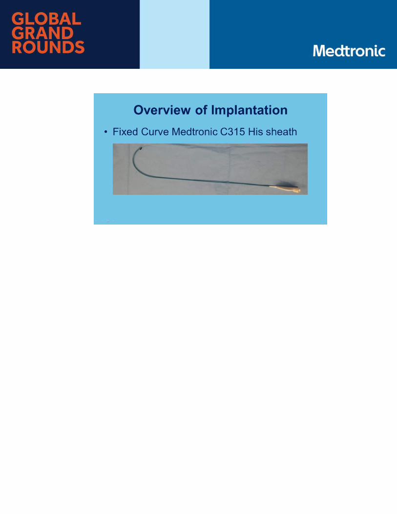
Vijayaraman: I like to go with the overview of implantation technique. One of the important pieces of the puzzle in His bundle pacing is the Medtronic Select Secure 3830 Lead which is a 4.1 French lead with an exposed screw and does not have a lumen, so it needs a delivery sheath and C304 Select Site sheath is a deflectable sheath. However, the C315 His sheath is a fixed curve sheath which is a workhorse in our lab.
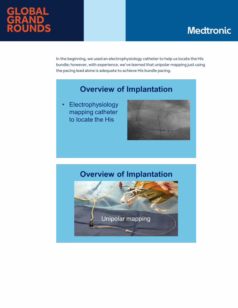
In the beginning, we used an electrophysiology catheter to help us locate the His bundle; however, with experience, we’ve learned that unipolar mapping just using the pacing lead alone is adequate to achieve His bundle pacing.
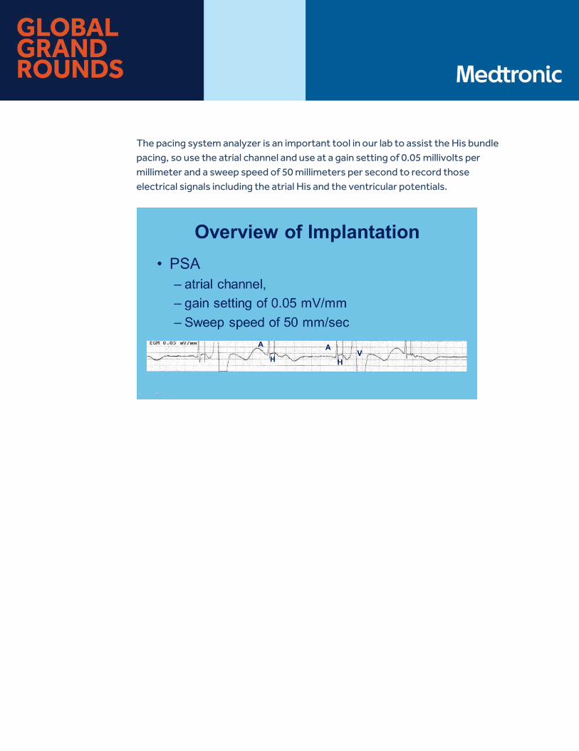
The pacing system analyzer is an important tool in our lab to assist the His bundle pacing, so use the atrial channel and use at a gain setting of 0.05 millivolts per millimeter and a sweep speed of 50 millimeters per second to record those electrical signals including the atrial His and the ventricular potentials.
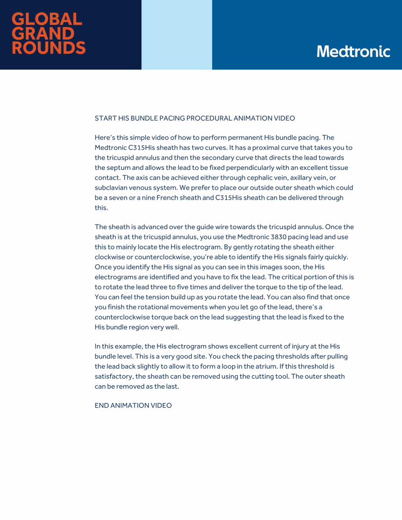
START HIS BUNDLE PACING PROCEDURAL ANIMATION VIDEO
Here’s this simple video of how to perform permanent His bundle pacing. The Medtronic C315His sheath has two curves. It has a proximal curve that takes you to the tricuspid annulus and then the secondary curve that directs the lead towards the septum and allows the lead to be fixed perpendicularly with an excellent tissue contact. The axis can be achieved either through cephalic vein, axillary vein, or subclavian venous system. We prefer to place our outside outer sheath which could be a seven or a nine French sheath and C315His sheath can be delivered through this.
The sheath is advanced over the guide wire towards the tricuspid annulus. Once the sheath is at the tricuspid annulus, you use the Medtronic 3830 pacing lead and use this to mainly locate the His electrogram. By gently rotating the sheath either clockwise or counterclockwise, you’re able to identify the His signals fairly quickly. Once you identify the His signal as you can see in this images soon, the His electrograms are identified and you have to fix the lead. The critical portion of this is to rotate the lead three to five times and deliver the torque to the tip of the lead. You can feel the tension build up as you rotate the lead. You can also find that once you finish the rotational movements when you let go of the lead, there’s a counterclockwise torque back on the lead suggesting that the lead is fixed to the His bundle region very well.
In this example, the His electrogram shows excellent current of injury at the His bundle level. This is a very good site. You check the pacing thresholds after pulling the lead back slightly to allow it to form a loop in the atrium. If this threshold is satisfactory, the sheath can be removed using the cutting tool. The outer sheath can be removed as the last.
END ANIMATION VIDEO
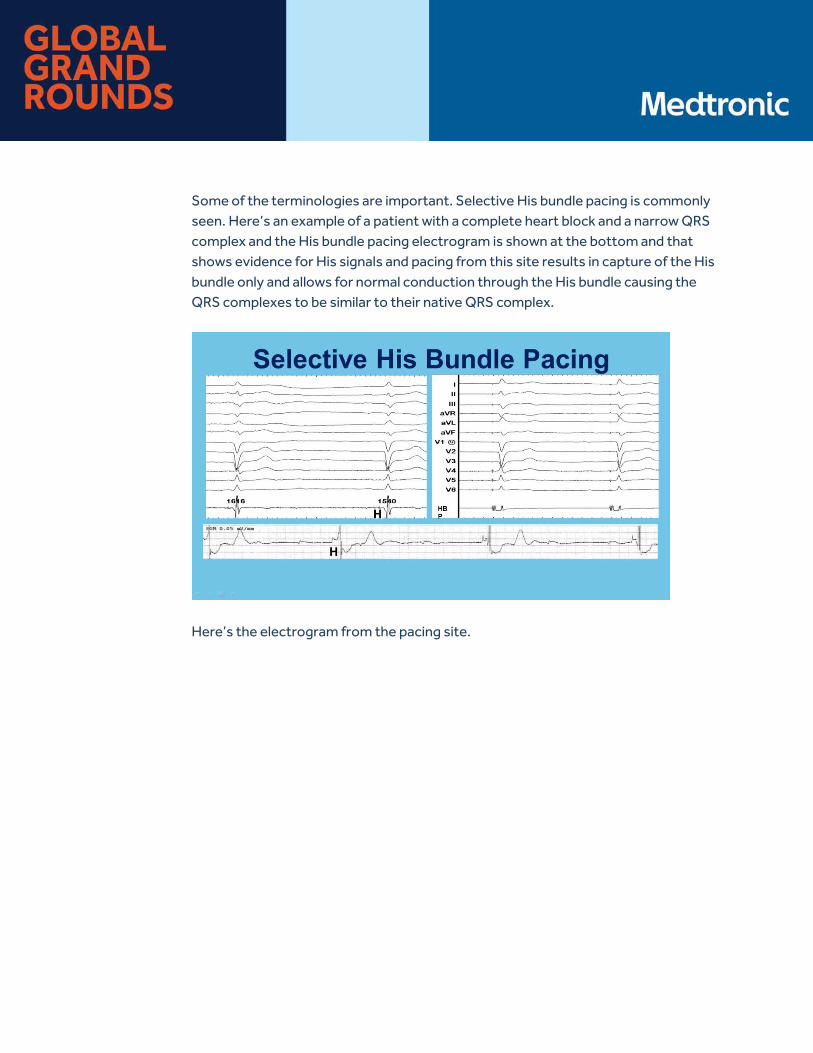
Some of the terminologies are important. Selective His bundle pacing is commonly seen. Here’s an example of a patient with a complete heart block and a narrow QRS complex and the His bundle pacing electrogram is shown at the bottom and that shows evidence for His signals and pacing from this site results in capture of the His bundle only and allows for normal conduction through the His bundle causing the QRS complexes to be similar to their native QRS complex.
Here’s the electrogram from the pacing site.
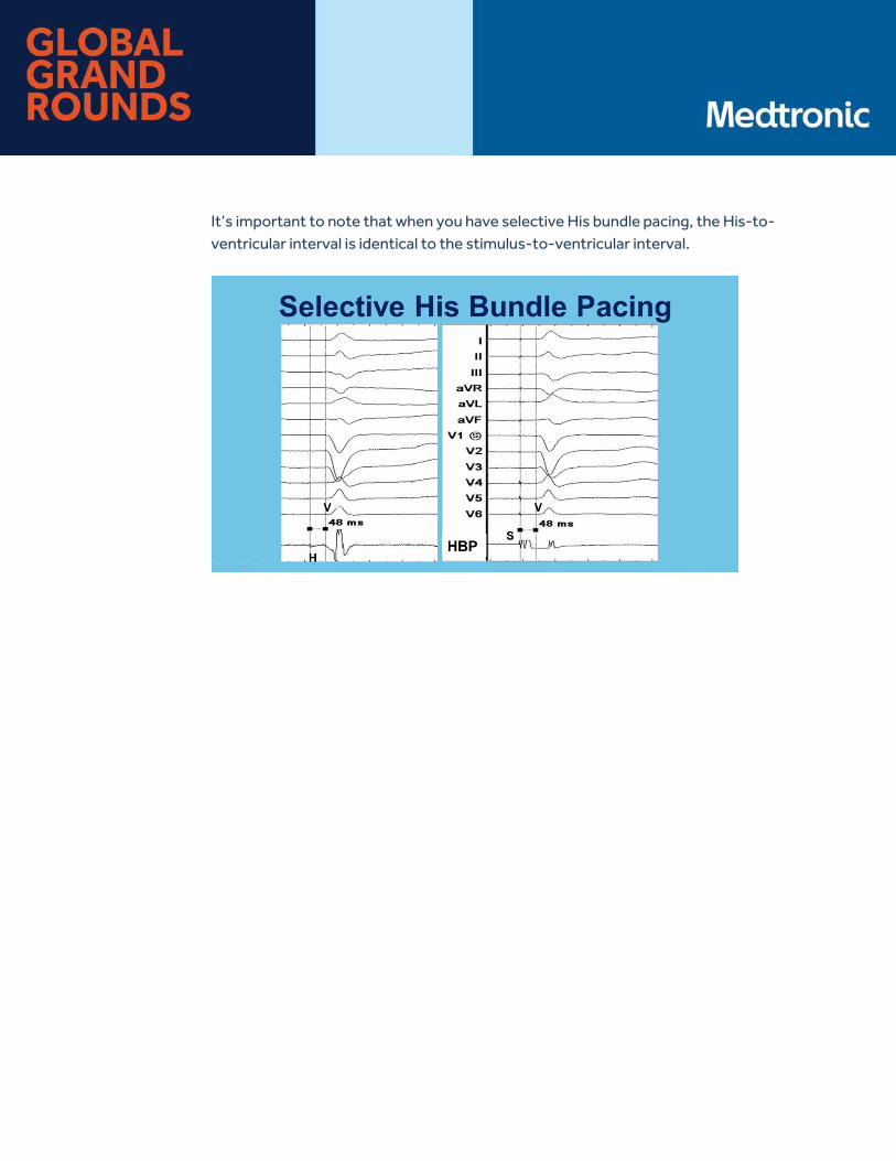
It’s important to note that when you have selective His bundle pacing, the His-to-ventricular interval is identical to the stimulus-to-ventricular interval.

The other situation would be non-selective His bundle pacing. Here’s an example of baseline 12-lead cardiogram and during pacing at 2Vat 0.5 milliseconds. You have evidence for His bundle capture causing the QRS morphology to be similar to the baseline QRS but it also has additional RV capture with certain degree of fusion. As you come down on the output, you lose the His bundle capture and you have a wider right ventricular septal pace QRS morphology.
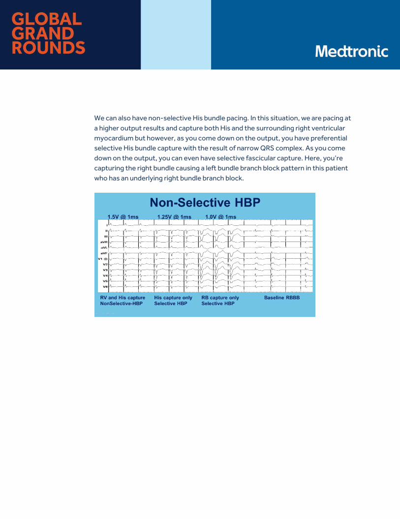
We can also have non-selective His bundle pacing. In this situation, we are pacing at a higher output results and capture both His and the surrounding right ventricular myocardium but however, as you come down on the output, you have preferential selective His bundle capture with the result of narrow QRS complex. As you come down on the output, you can even have selective fascicular capture. Here, you’re capturing the right bundle causing a left bundle branch block pattern in this patient who has an underlying right bundle branch block.
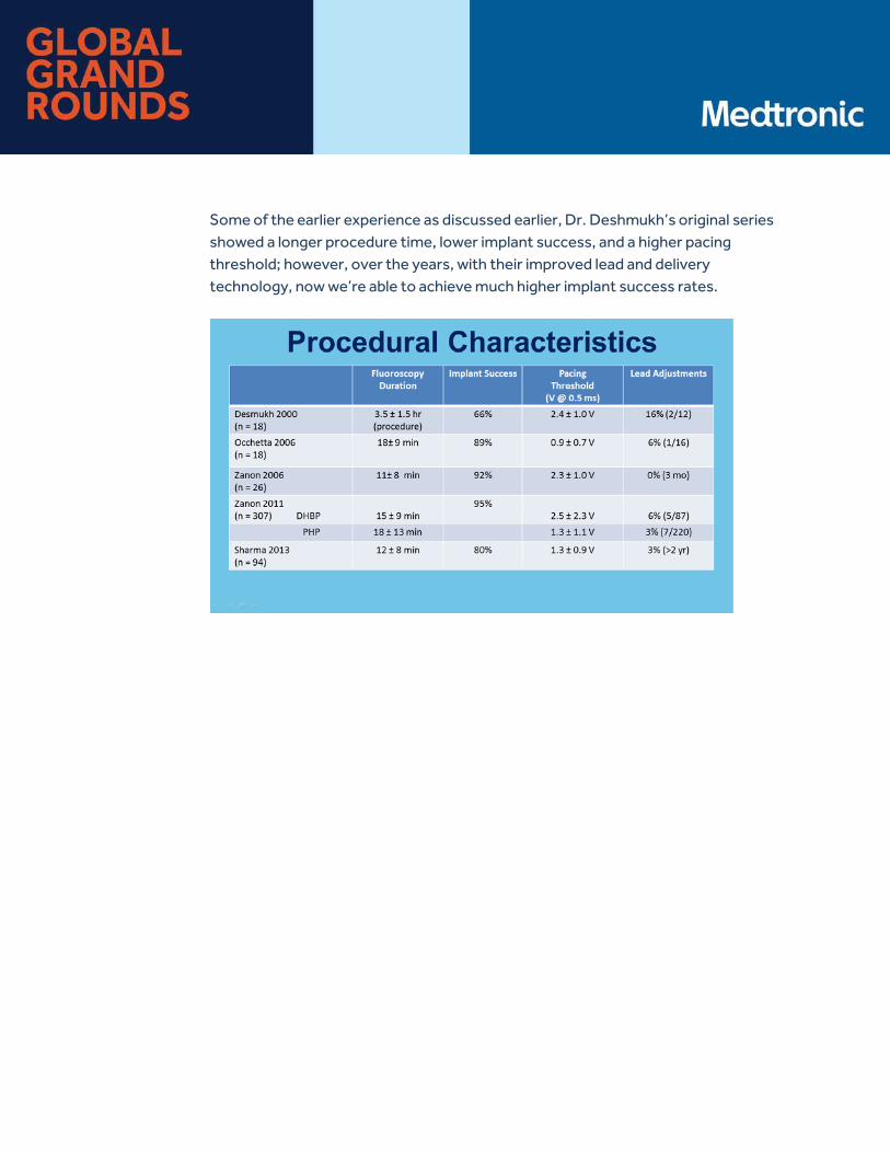
Some of the earlier experience as discussed earlier, Dr. Deshmukh’s original series showed a longer procedure time, lower implant success, and a higher pacing threshold; however, over the years, with their improved lead and delivery technology, now we’re able to achieve much higher implant success rates.
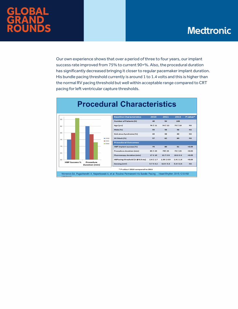
Our own experience shows that over a period of three to four years, our implant success rate improved from 75% to current 90+%. Also, the procedural duration has significantly decreased bringing it closer to regular pacemaker implant duration. His bundle pacing threshold currently is around 1 to 1.4 volts and this is higher than the normal RV pacing threshold but well within acceptable range compared to CRT pacing for left ventricular capture thresholds.
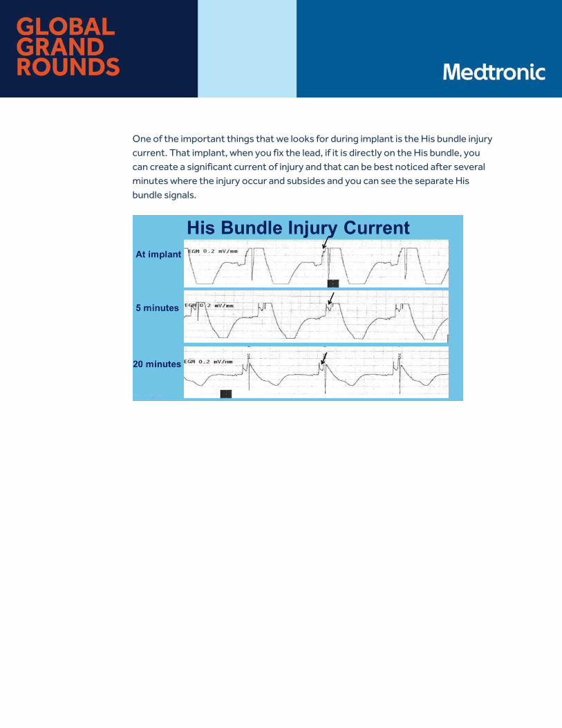
One of the important things that we looks for during implant is the His bundle injury current. That implant, when you fix the lead, if it is directly on the His bundle, you can create a significant current of injury and that can be best noticed after several minutes where the injury occur and subsides and you can see the separate His bundle signals.
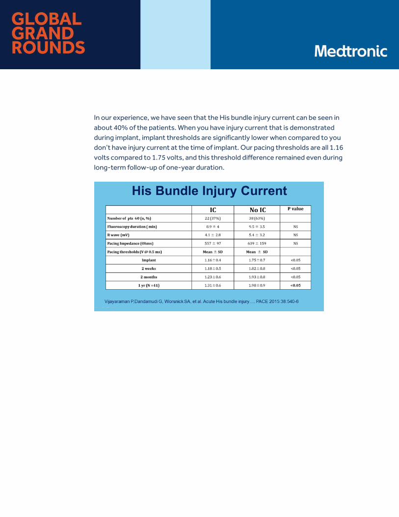
In our experience, we have seen that the His bundle injury current can be seen in about 40% of the patients. When you have injury current that is demonstrated during implant, implant thresholds are significantly lower when compared to you don’t have injury current at the time of implant. Our pacing thresholds are all 1.16 volts compared to 1.75 volts, and this threshold difference remained even during long-term follow-up of one-year duration.
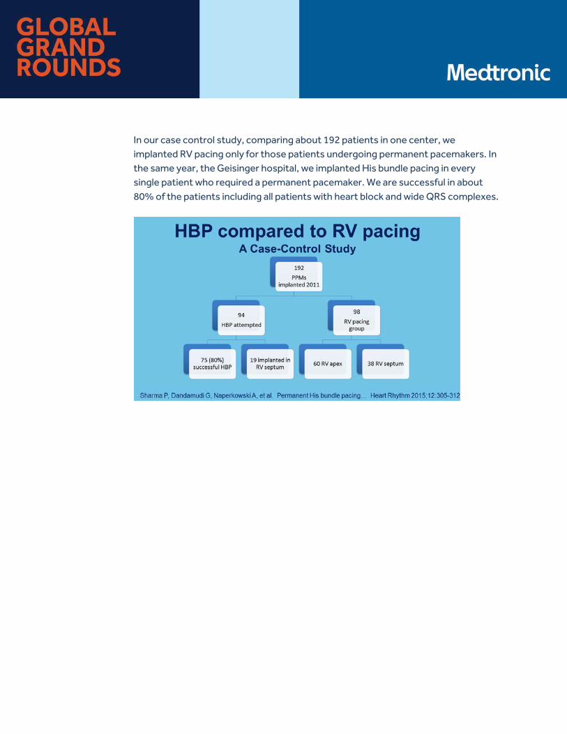
In our case control study, comparing about 192 patients in one center, we implanted RV pacing only for those patients undergoing permanent pacemakers. In the same year, the Geisinger hospital, we implanted His bundle pacing in every single patient who required a permanent pacemaker. We are successful in about 80% of the patients including all patients with heart block and wide QRS complexes.
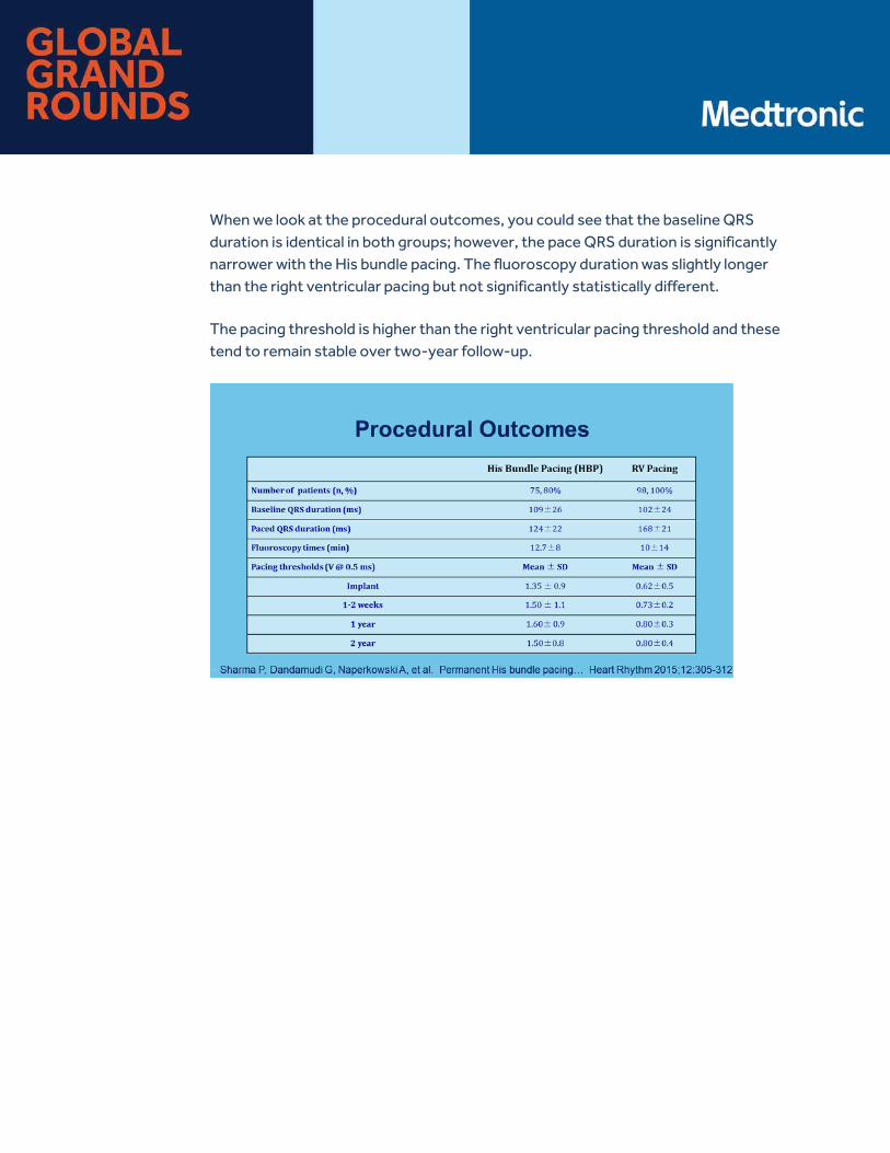
When we look at the procedural outcomes, you could see that the baseline QRS duration is identical in both groups; however, the pace QRS duration is significantly narrower with the His bundle pacing. The fluoroscopy duration was slightly longer than the right ventricular pacing but not significantly statistically different.
The pacing threshold is higher than the right ventricular pacing threshold and these tend to remain stable over two-year follow-up.
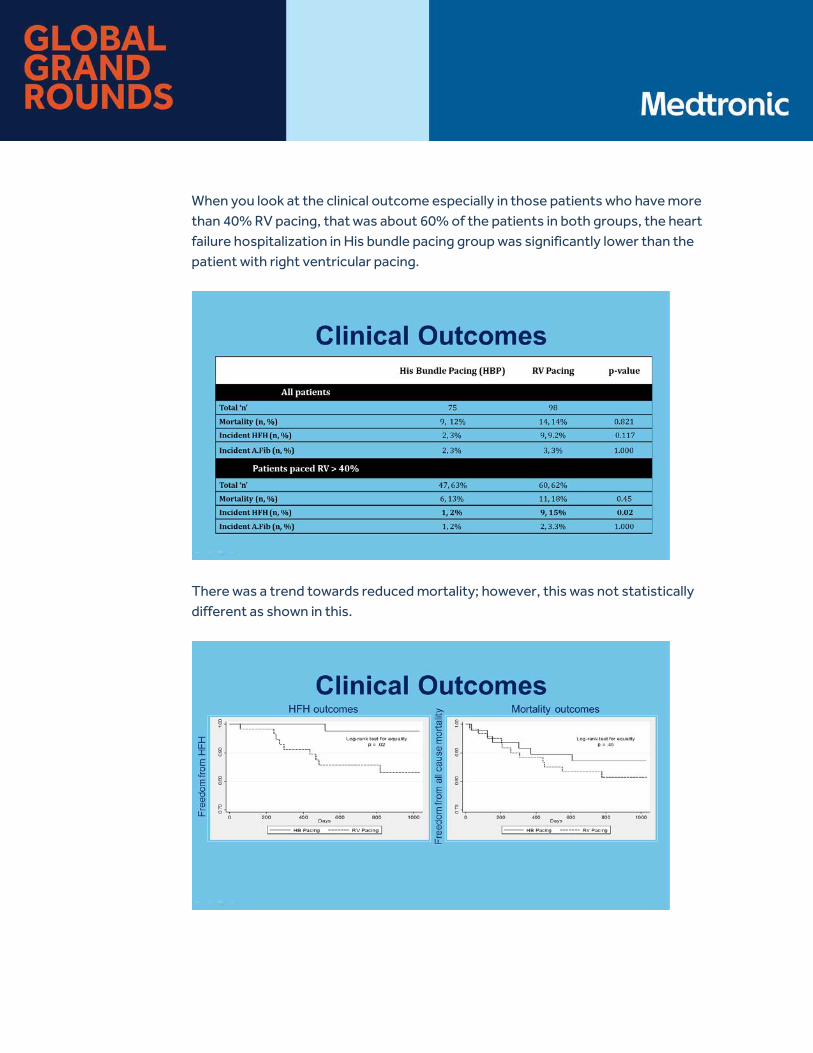
When you look at the clinical outcome especially in those patients who have more than 40% RV pacing, that was about 60% of the patients in both groups, the heart failure hospitalization in His bundle pacing group was significantly lower than the patient with right ventricular pacing.
There was a trend towards reduced mortality; however, this was not statistically different as shown in this.
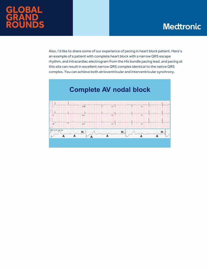
Also, I’d like to share some of our experience of pacing in heart block patient. Here’s an example of a patient with complete heart block with a narrow QRS escape rhythm, and intracardiac electrogram from the His bundle pacing lead, and pacing at this site can result in excellent narrow QRS complex identical to the native QRS complex. You can achieve both atrioventricular and interventricular synchrony.
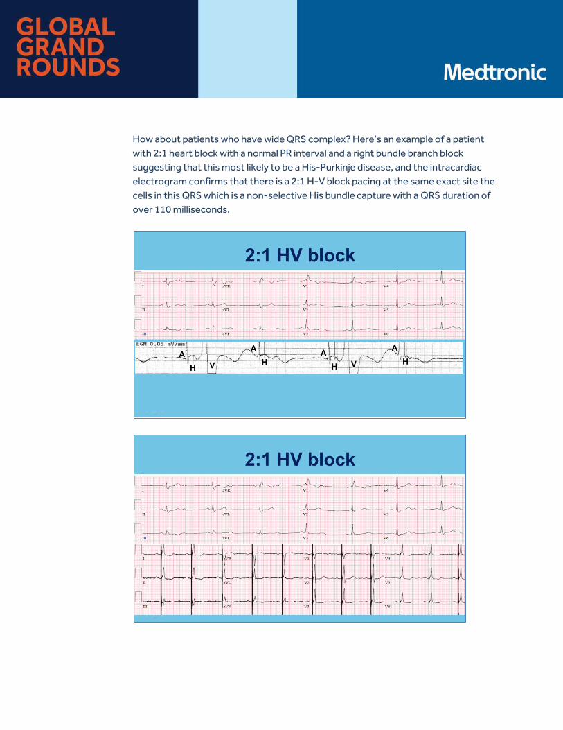
How about patients who have wide QRS complex? Here’s an example of a patient with 2:1 heart block with a normal PR interval and a right bundle branch block suggesting that this most likely to be a His-Purkinje disease, and the intracardiac electrogram confirms that there is a 2:1 H-V block pacing at the same exact site the cells in this QRS which is a non-selective His bundle capture with a QRS duration of over 110 milliseconds.
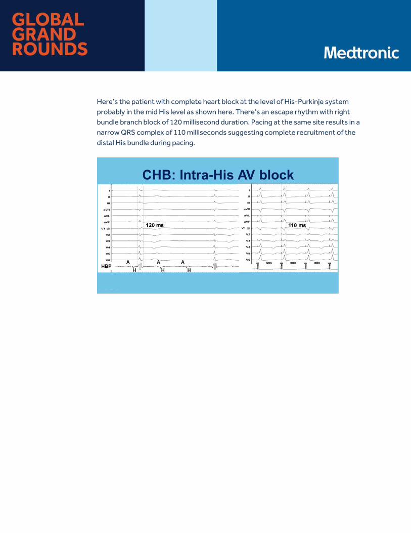
Here’s the patient with complete heart block at the level of His-Purkinje system probably in the mid His level as shown here. There’s an escape rhythm with right bundle branch block of 120 millisecond duration. Pacing at the same site results in a narrow QRS complex of 110 milliseconds suggesting complete recruitment of the distal His bundle during pacing.
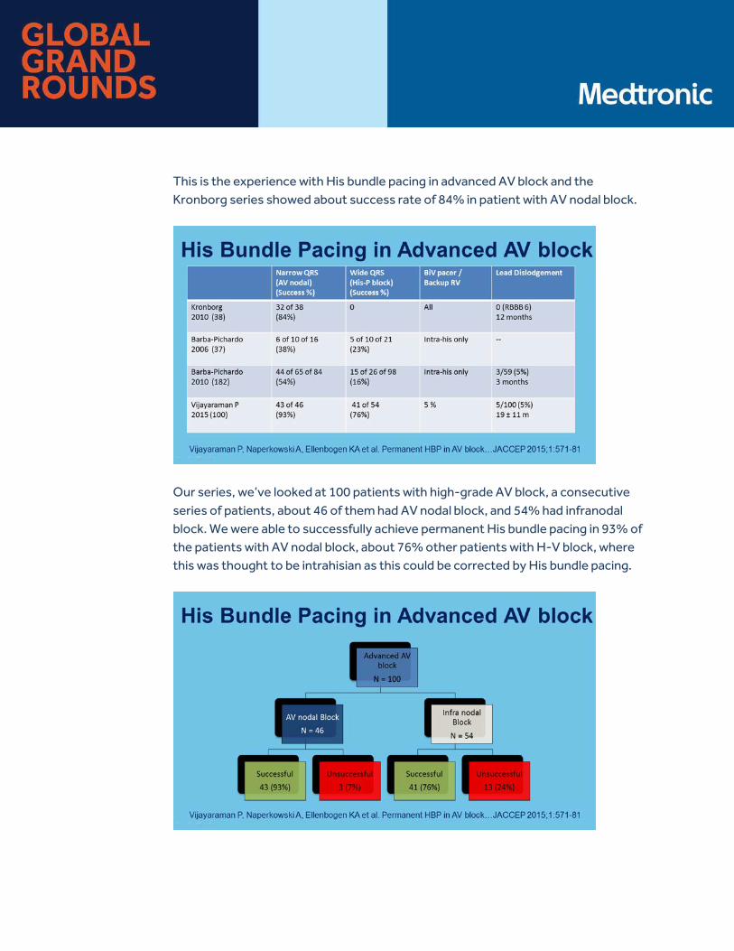
This is the experience with His bundle pacing in advanced AV block and the Kronborg series showed about success rate of 84% in patient with AV nodal block.
Our series, we’ve looked at 100 patients with high-grade AV block, a consecutive series of patients, about 46 of them had AV nodal block, and 54% had infranodal block. We were able to successfully achieve permanent His bundle pacing in 93% of the patients with AV nodal block, about 76% other patients with H-V block, where this was thought to be intrahisian as this could be corrected by His bundle pacing.
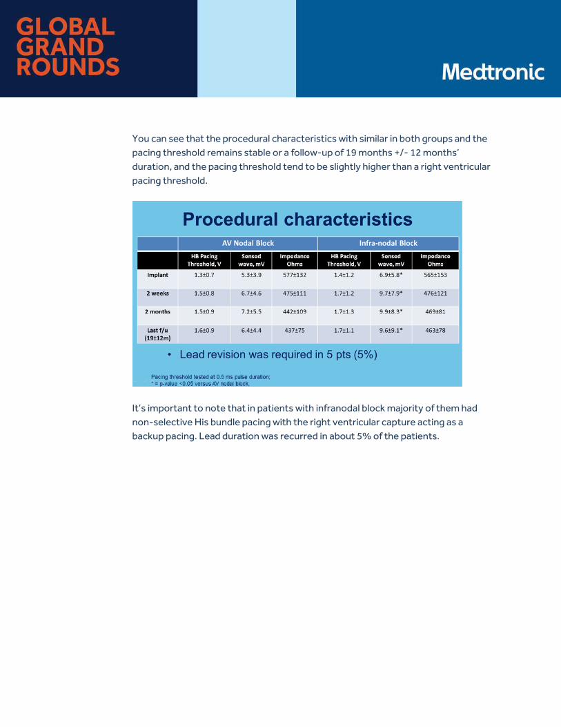
You can see that the procedural characteristics with similar in both groups and the pacing threshold remains stable or a follow-up of 19 months +/- 12 months’ duration, and the pacing threshold tend to be slightly higher than a right ventricular pacing threshold.
It’s important to note that in patients with infranodal block majority of them had non-selective His bundle pacing with the right ventricular capture acting as a backup pacing. Lead duration was recurred in about 5% of the patients.

How about experience in patients with chronic bundle branch block? Here’s an example of a patient with chronic left bundle branch block of four years’ duration. You have the His bundle pacing lead demonstrating nice His electrograms, and pacing at this site at 1.5 volts results in excellent selective His bundle capture with correction of the left bundle branch block. As you lower the output, there is an output-dependent capture of only the right bundle.
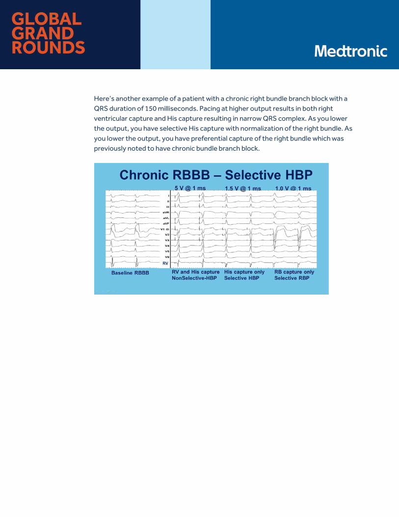
Here’s another example of a patient with a chronic right bundle branch block with a QRS duration of 150 milliseconds. Pacing at higher output results in both right ventricular capture and His capture resulting in narrow QRS complex. As you lower the output, you have selective His capture with normalization of the right bundle. As you lower the output, you have preferential capture of the right bundle which was previously noted to have chronic bundle branch block.
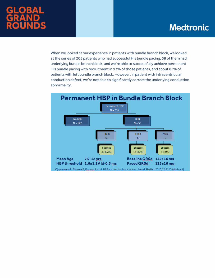
When we looked at our experience in patients with bundle branch block, we looked at the series of 205 patients who had successful His bundle pacing, 58 of them had underlying bundle branch block, and we’re able to successfully achieve permanent His bundle pacing with recruitment in 93% of those patients, and about 82% of patients with left bundle branch block. However, in patient with intraventricular conduction defect, we’re not able to significantly correct the underlying conduction abnormality.

There are several limitations to His bundle pacing which need to be taken note of. Failure to implant can occur in 10% to 20% of patients which is predominantly in patients with infrahisian AV block. High thresholds can be a problem in 10% to 15% of patients. Lead revisions may be required in about 3% of patients. Ventricular undersensing, far field atrial oversensing, and occasionally atrial capture can be an issue.
Important thing to remember is that you can also cause acute injury to the His bundle especially persistent right bundle branch block can occur in 2% to 3% of patients. However, pacing at this site can invariably correct the right bundle branch block created by the pacing lead.
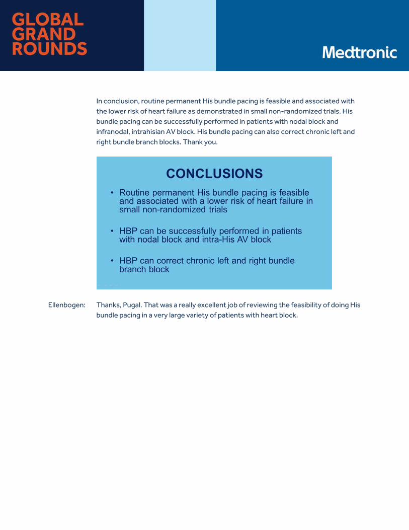
In conclusion, routine permanent His bundle pacing is feasible and associated with the lower risk of heart failure as demonstrated in small non-randomized trials. His bundle pacing can be successfully performed in patients with nodal block and infranodal, intrahisian AV block. His bundle pacing can also correct chronic left and right bundle branch blocks. Thank you.
Ellenbogen: Thanks, Pugal. That was a really excellent job of reviewing the feasibility of doing His bundle pacing in a very large variety of patients with heart block.
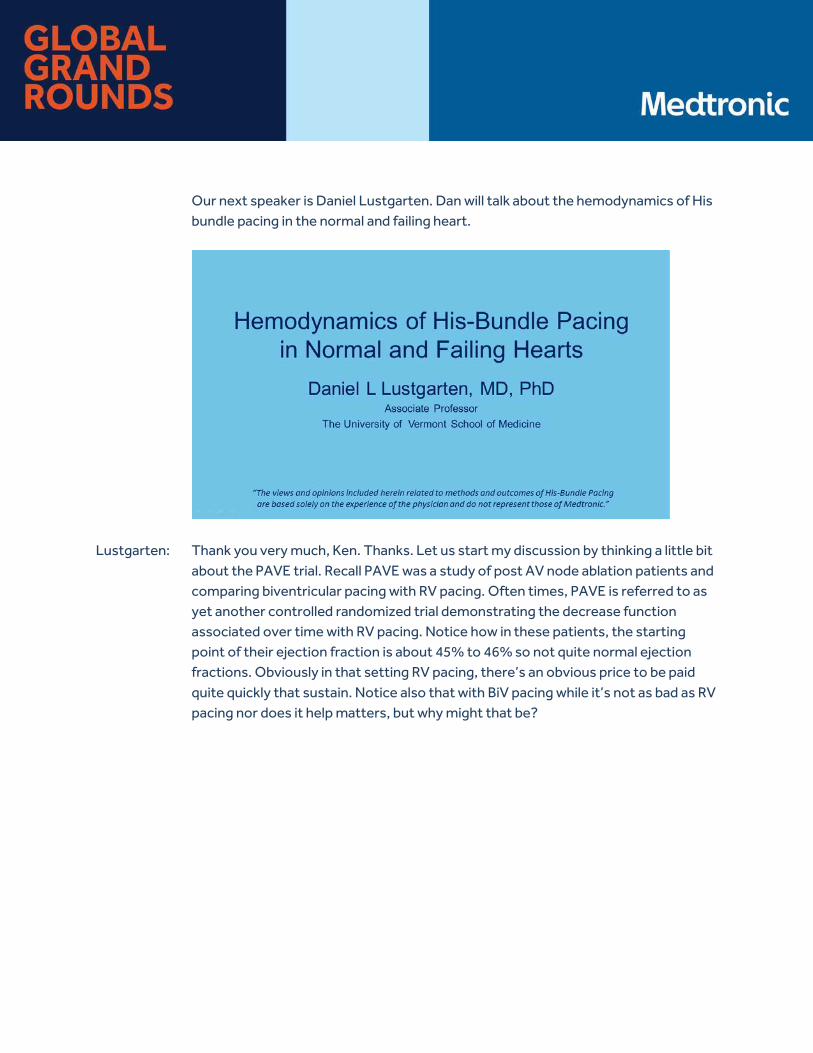
Our next speaker is Daniel Lustgarten. Dan will talk about the hemodynamics of His bundle pacing in the normal and failing heart.
Lustgarten: Thank you very much, Ken. Thanks. Let us start my discussion by thinking a little bit about the PAVE trial. Recall PAVE was a study of post AV node ablation patients and comparing biventricular pacing with RV pacing. Often times, PAVE is referred to as yet another controlled randomized trial demonstrating the decrease function associated over time with RV pacing. Notice how in these patients, the starting point of their ejection fraction is about 45% to 46% so not quite normal ejection fractions. Obviously in that setting RV pacing, there’s an obvious price to be paid quite quickly that sustain. Notice also that with BiV pacing while it’s not as bad as RV pacing nor does it help matters, but why might that be?
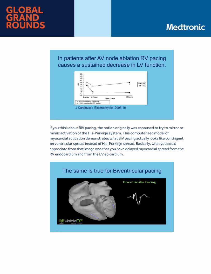
If you think about BiV pacing, the notion originally was espoused to try to mirror or mimic activation of the His-Purkinje system. This computerized model of myocardial activation demonstrates what BiV pacing actually looks like contingent on ventricular spread instead of His-Purkinje spread. Basically, what you could appreciate from that image was that you have delayed myocardial spread from the RV endocardium and from the LV epicardium.
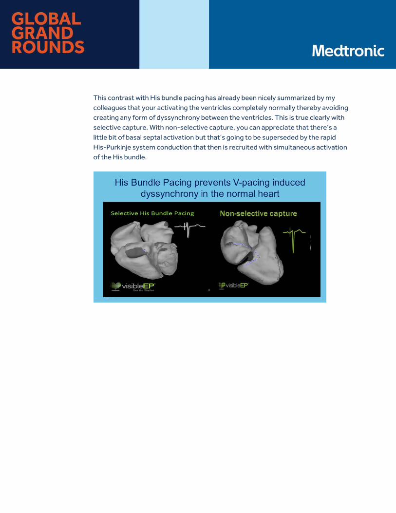
This contrast with His bundle pacing has already been nicely summarized by my colleagues that your activating the ventricles completely normally thereby avoiding creating any form of dyssynchrony between the ventricles. This is true clearly with selective capture. With non-selective capture, you can appreciate that there’s a little bit of basal septal activation but that’s going to be superseded by the rapid His-Purkinje system conduction that then is recruited with simultaneous activation of the His bundle.
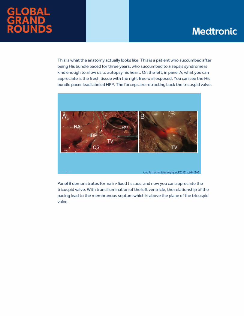
This is what the anatomy actually looks like. This is a patient who succumbed after being His bundle paced for three years, who succumbed to a sepsis syndrome is kind enough to allow us to autopsy his heart. On the left, in panel A, what you can appreciate is the fresh tissue with the right free wall exposed. You can see the His bundle pacer lead labeled HPP. The forceps are retracting back the tricuspid valve.
Panel B demonstrates formalin-fixed tissues, and now you can appreciate the tricuspid valve. With transillumination of the left ventricle, the relationship of the pacing lead to the membranous septum which is above the plane of the tricuspid valve.
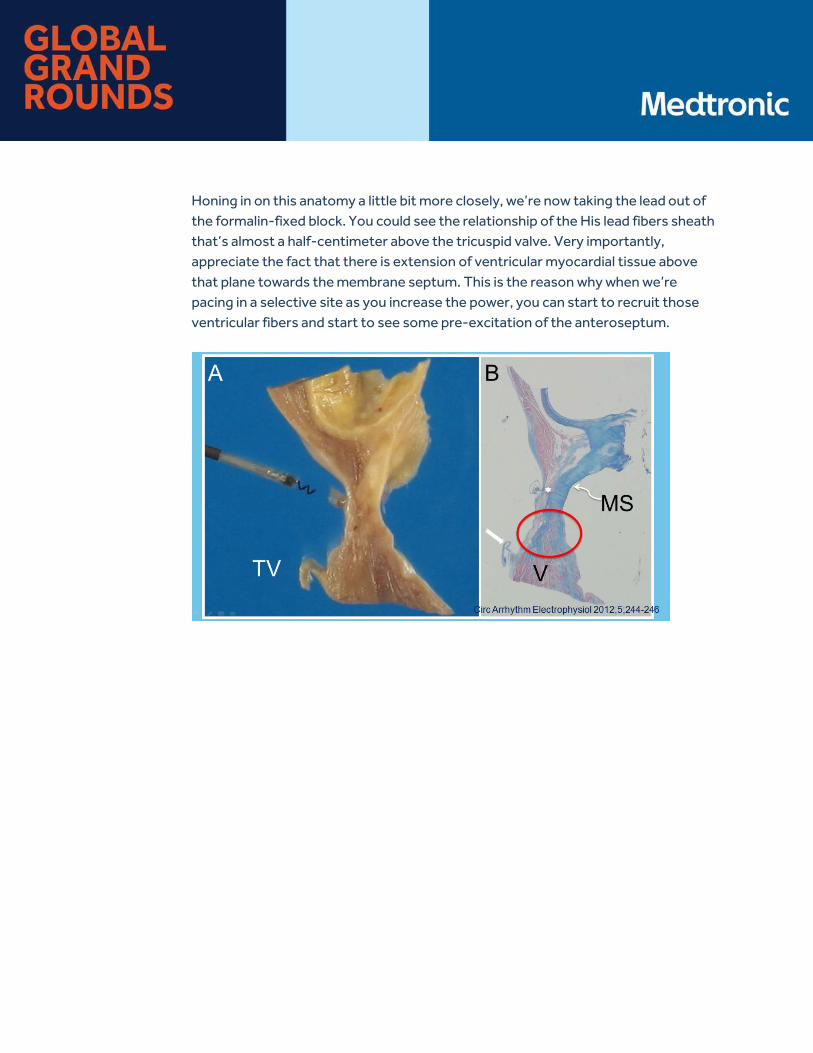
Honing in on this anatomy a little bit more closely, we’re now taking the lead out of the formalin-fixed block. You could see the relationship of the His lead fibers sheath that’s almost a half-centimeter above the tricuspid valve. Very importantly, appreciate the fact that there is extension of ventricular myocardial tissue above that plane towards the membrane septum. This is the reason why when we’re pacing in a selective site as you increase the power, you can start to recruit those ventricular fibers and start to see some pre-excitation of the anteroseptum.
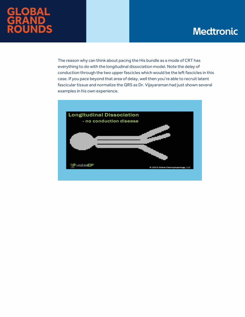
The reason why can think about pacing the His bundle as a mode of CRT has everything to do with the longitudinal dissociation model. Note the delay of conduction through the two upper fascicles which would be the left fascicles in this case. If you pace beyond that area of delay, well then you’re able to recruit latent fascicular tissue and normalize the QRS as Dr. Vijayaraman had just shown several examples in his own experience.
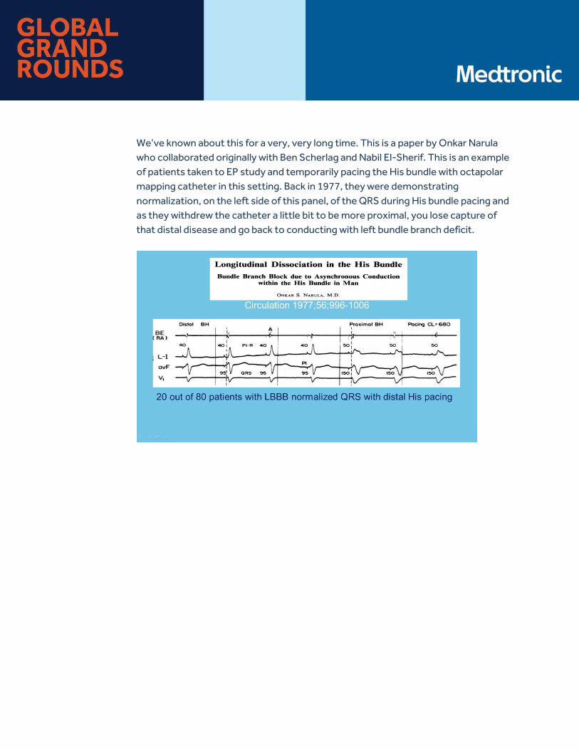
We’ve known about this for a very, very long time. This is a paper by Onkar Narula who collaborated originally with Ben Scherlag and Nabil El-Sherif. This is an example of patients taken to EP study and temporarily pacing the His bundle with octapolar mapping catheter in this setting. Back in 1977, they were demonstrating normalization, on the left side of this panel, of the QRS during His bundle pacing and as they withdrew the catheter a little bit to be more proximal, you lose capture of that distal disease and go back to conducting with left bundle branch deficit.
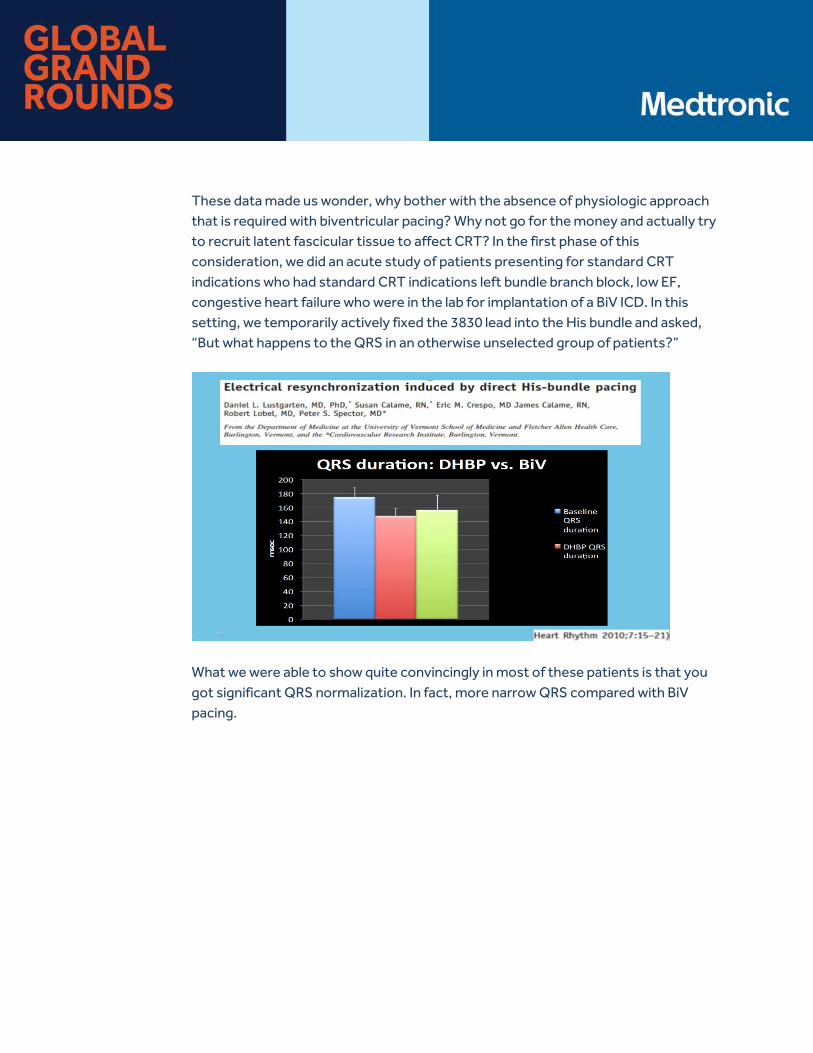
These data made us wonder, why bother with the absence of physiologic approach that is required with biventricular pacing? Why not go for the money and actually try to recruit latent fascicular tissue to affect CRT? In the first phase of this consideration, we did an acute study of patients presenting for standard CRT indications who had standard CRT indications left bundle branch block, low EF, congestive heart failure who were in the lab for implantation of a BiV ICD. In this setting, we temporarily actively fixed the 3830 lead into the His bundle and asked, “But what happens to the QRS in an otherwise unselected group of patients?”
What we were able to show quite convincingly in most of these patients is that you got significant QRS normalization. In fact, more narrow QRS compared with BiV pacing.
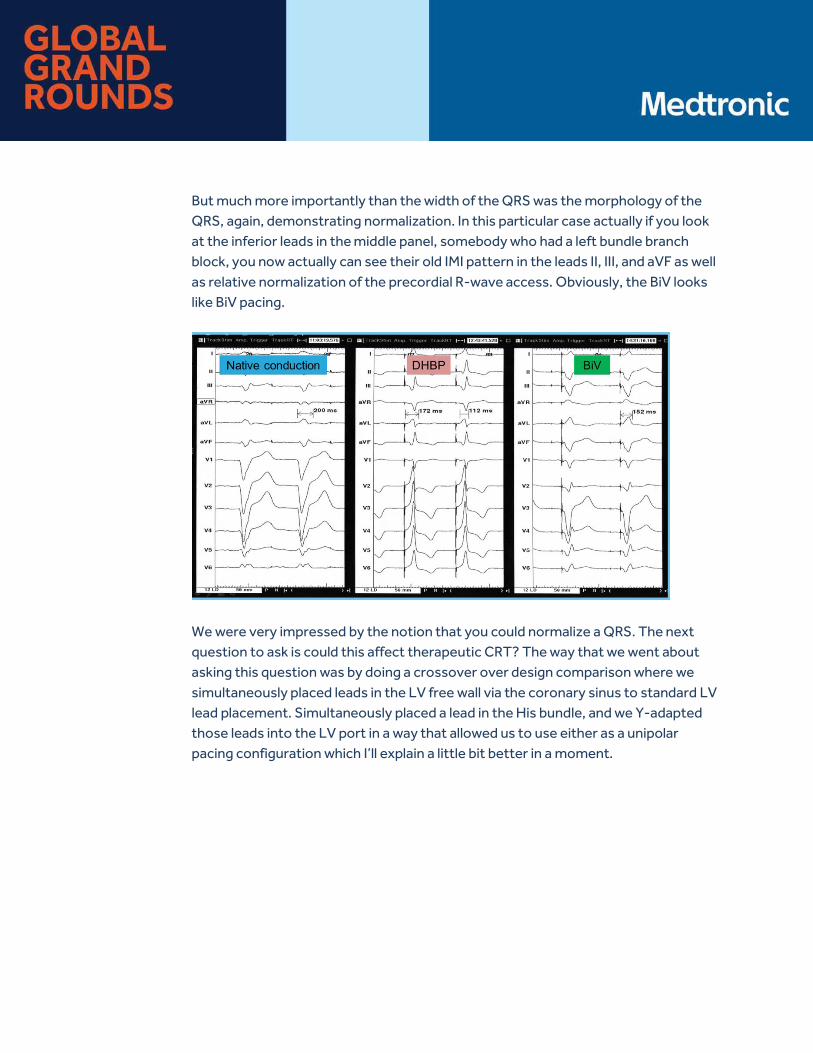
But much more importantly than the width of the QRS was the morphology of the QRS, again, demonstrating normalization. In this particular case actually if you look at the inferior leads in the middle panel, somebody who had a left bundle branch block, you now actually can see their old IMI pattern in the leads II, III, and aVF as well as relative normalization of the precordial R-wave access. Obviously, the BiV looks like BiV pacing.
We were very impressed by the notion that you could normalize a QRS. The next question to ask is could this affect therapeutic CRT? The way that we went about asking this question was by doing a crossover over design comparison where we simultaneously placed leads in the LV free wall via the coronary sinus to standard LV lead placement. Simultaneously placed a lead in the His bundle, and we Y-adapted those leads into the LV port in a way that allowed us to use either as a unipolar pacing configuration which I’ll explain a little bit better in a moment.
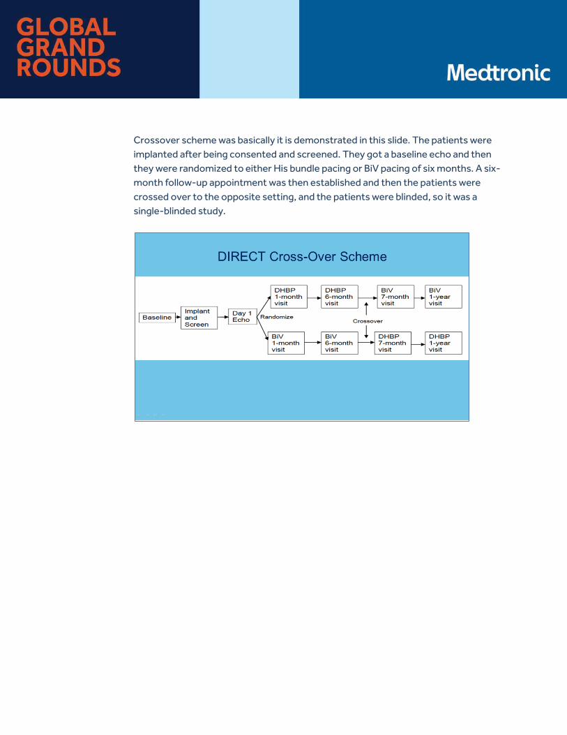
Crossover scheme was basically it is demonstrated in this slide. The patients were implanted after being consented and screened. They got a baseline echo and then they were randomized to either His bundle pacing or BiV pacing of six months. A six-month follow-up appointment was then established and then the patients were crossed over to the opposite setting, and the patients were blinded, so it was a single-blinded study.
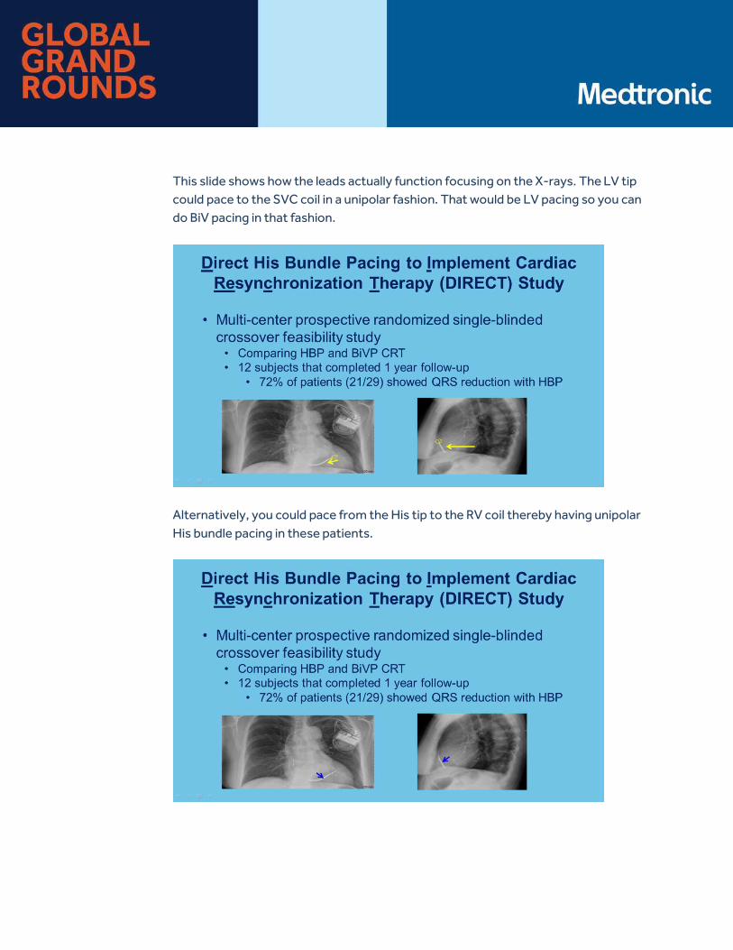
This slide shows how the leads actually function focusing on the X-rays. The LV tip could pace to the SVC coil in a unipolar fashion. That would be LV pacing so you can do BiV pacing in that fashion.
Alternatively, you could pace from the His tip to the RV coil thereby having unipolar His bundle pacing in these patients.
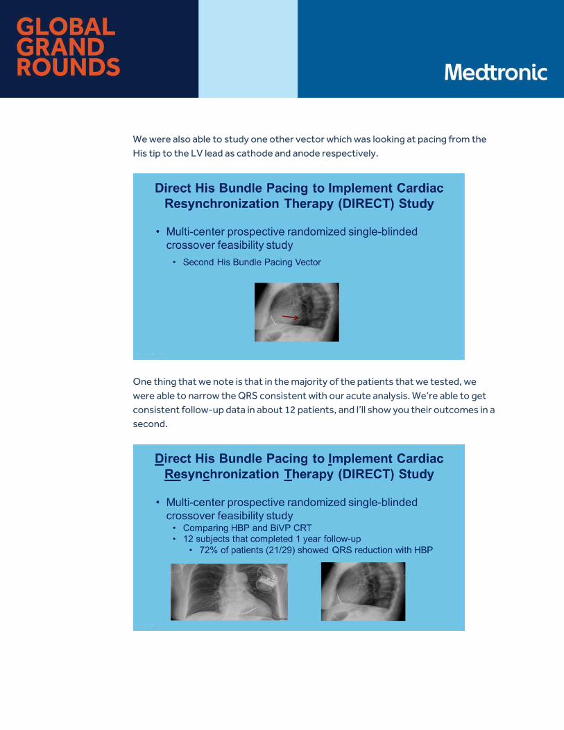
We were also able to study one other vector which was looking at pacing from the His tip to the LV lead as cathode and anode respectively.
One thing that we note is that in the majority of the patients that we tested, we were able to narrow the QRS consistent with our acute analysis. We’re able to get consistent follow-up data in about 12 patients, and I’ll show you their outcomes in a second.
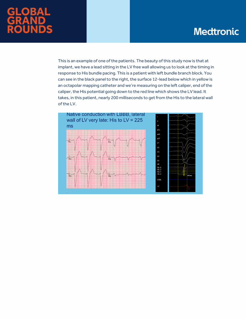
This is an example of one of the patients. The beauty of this study now is that at implant, we have a lead sitting in the LV free wall allowing us to look at the timing in response to His bundle pacing. This is a patient with left bundle branch block. You can see in the black panel to the right, the surface 12-lead below which in yellow is an octapolar mapping catheter and we’re measuring on the left caliper, end of the caliper, the His potential going down to the red line which shows the LV lead. It takes, in this patient, nearly 200 milliseconds to get from the His to the lateral wall of the LV.
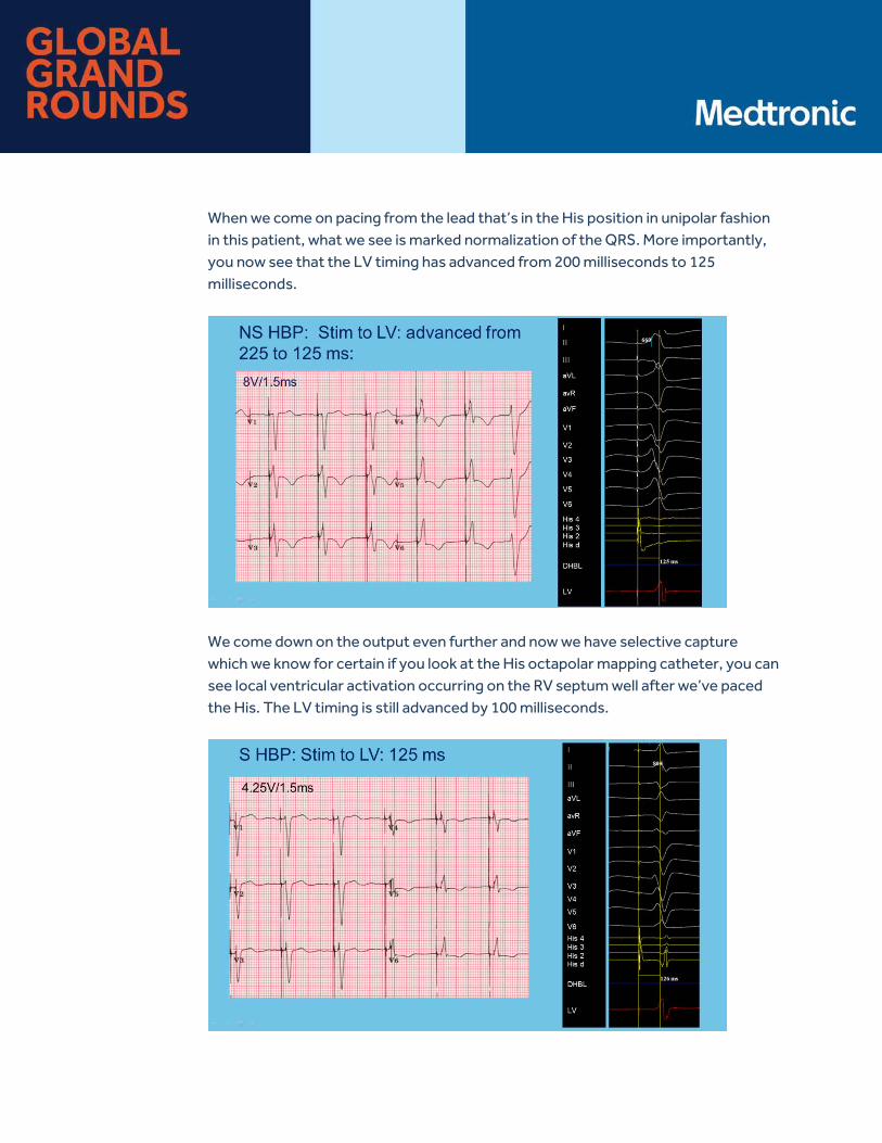
When we come on pacing from the lead that’s in the His position in unipolar fashion in this patient, what we see is marked normalization of the QRS. More importantly, you now see that the LV timing has advanced from 200 milliseconds to 125 milliseconds.
We come down on the output even further and now we have selective capture which we know for certain if you look at the His octapolar mapping catheter, you can see local ventricular activation occurring on the RV septum well after we’ve paced the His. The LV timing is still advanced by 100 milliseconds.
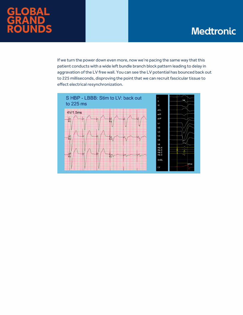
If we turn the power down even more, now we’re pacing the same way that this patient conducts with a wide left bundle branch block pattern leading to delay in aggravation of the LV free wall. You can see the LV potential has bounced back out to 225 milliseconds, disproving the point that we can recruit fascicular tissue to effect electrical resynchronization.
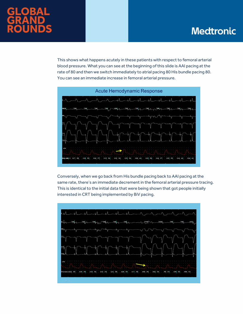
This shows what happens acutely in these patients with respect to femoral arterial blood pressure. What you can see at the beginning of this slide is AAI pacing at the rate of 80 and then we switch immediately to atrial pacing 80 His bundle pacing 80. You can see an immediate increase in femoral arterial pressure.
Conversely, when we go back from His bundle pacing back to AAI pacing at the same rate, there’s an immediate decrement in the femoral arterial pressure tracing. This is identical to the initial data that were being shown that got people initially interested in CRT being implemented by BiV pacing.
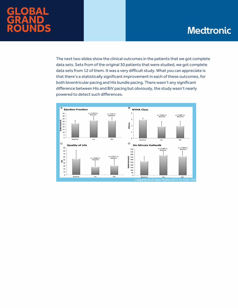
The next two slides show the clinical outcomes in the patients that we got complete data sets. Sets from of the original 30 patients that were studied, we got complete data sets from 12 of them. It was a very difficult study. What you can appreciate is that there’s a statistically significant improvement in each of these outcomes, for both biventricular pacing and His bundle pacing. There wasn’t any significant difference between His and BiV pacing but obviously, the study wasn’t nearly powered to detect such differences.
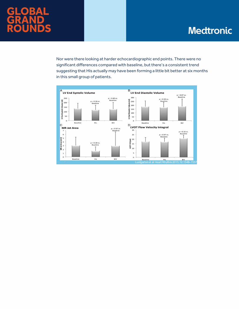
Nor were there looking at harder echocardiographic end points. There were no significant differences compared with baseline, but there’s a consistent trend suggesting that His actually may have been forming a little bit better at six months in this small group of patients.
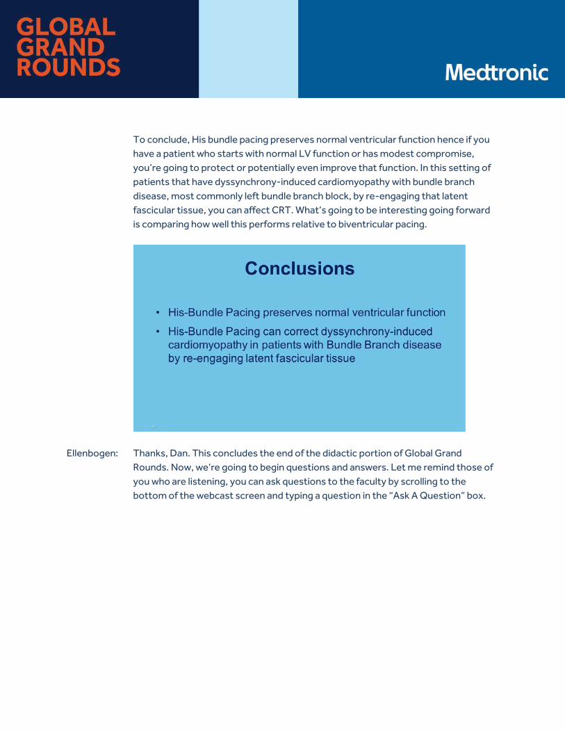
To conclude, His bundle pacing preserves normal ventricular function hence if you have a patient who starts with normal LV function or has modest compromise, you’re going to protect or potentially even improve that function. In this setting of patients that have dyssynchrony-induced cardiomyopathy with bundle branch disease, most commonly left bundle branch block, by re-engaging that latent fascicular tissue, you can affect CRT. What’s going to be interesting going forward is comparing how well this performs relative to biventricular pacing.
Ellenbogen: Thanks, Dan. This concludes the end of the didactic portion of Global Grand Rounds. Now, we’re going to begin questions and answers. Let me remind those of you who are listening, you can ask questions to the faculty by scrolling to the bottom of the webcast screen and typing a question in the “Ask A Question” box.
Ellenbogen: I’m going to start off this session by asking the panel a couple of questions that come up fairly often among people who are starting to learn how to do this new technique. Dan, I wanted to start off with you and ask you something that comes up quite frequently because this technique establishing physiologic pacing. Often people employ this in patients. They are thinking about ablating the AV node. I want to ask you are there issues or things you can tell us about ablating the AV node without damaging the His bundle pacing site in patients undergoing His bundle pacing?
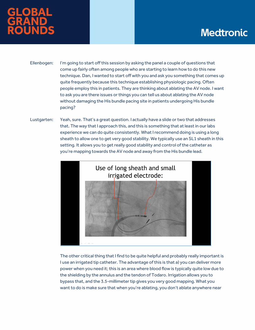
Lustgarten: Yeah, sure. That’s a great question. I actually have a slide or two that addresses that. The way that I approach this, and this is something that at least in our labs experience we can do quite consistently. What I recommend doing is using a long sheath to allow one to get very good stability. We typically use an SL1 sheath in this setting. It allows you to get really good stability and control of the catheter as you’re mapping towards the AV node and away from the His bundle lead.
The other critical thing that I find to be quite helpful and probably really important is I use an irrigated tip catheter. The advantage of this is that a) you can deliver more power when you need it; this is an area where blood flow is typically quite low due to the shielding by the annulus and the tendon of Todaro. Irrigation allows you to bypass that, and the 3.5-millimeter tip gives you very good mapping. What you want to do is make sure that when you’re ablating, you don’t ablate anywhere near
the tip of the His lead. In most, in the vast majority of cases, there is no His potential when we’re ablating.
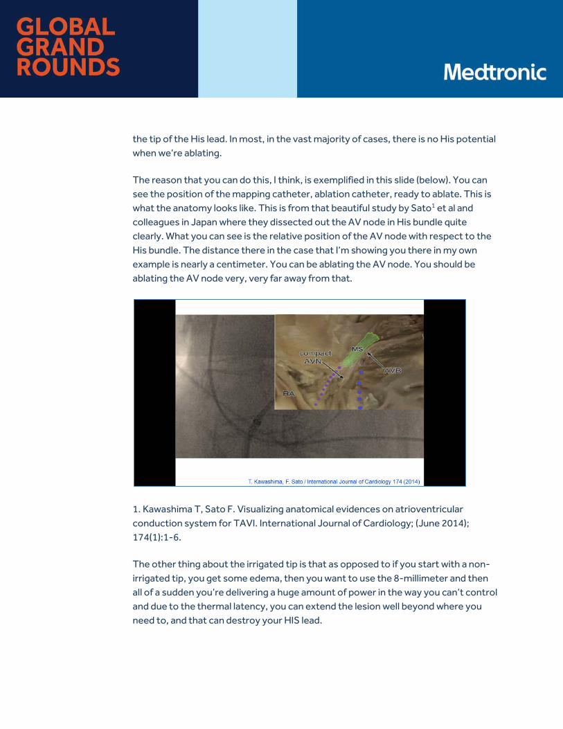
The reason that you can do this, I think, is exemplified in this slide (below). You can see the position of the mapping catheter, ablation catheter, ready to ablate. This is what the anatomy looks like. This is from that beautiful study by Sato1 et al and colleagues in Japan where they dissected out the AV node in His bundle quite clearly. What you can see is the relative position of the AV node with respect to the His bundle. The distance there in the case that I’m showing you there in my own example is nearly a centimeter. You can be ablating the AV node. You should be ablating the AV node very, very far away from that.
1. Kawashima T, Sato F. Visualizing anatomical evidences on atrioventricular conduction system for TAVI. International Journal of Cardiology; (June 2014); 174(1):1-6.
The other thing about the irrigated tip is that as opposed to if you start with a non-irrigated tip, you get some edema, then you want to use the 8-millimeter and then all of a sudden you’re delivering a huge amount of power in the way you can’t control and due to the thermal latency, you can extend the lesion well beyond where you need to, and that can destroy your HIS lead.
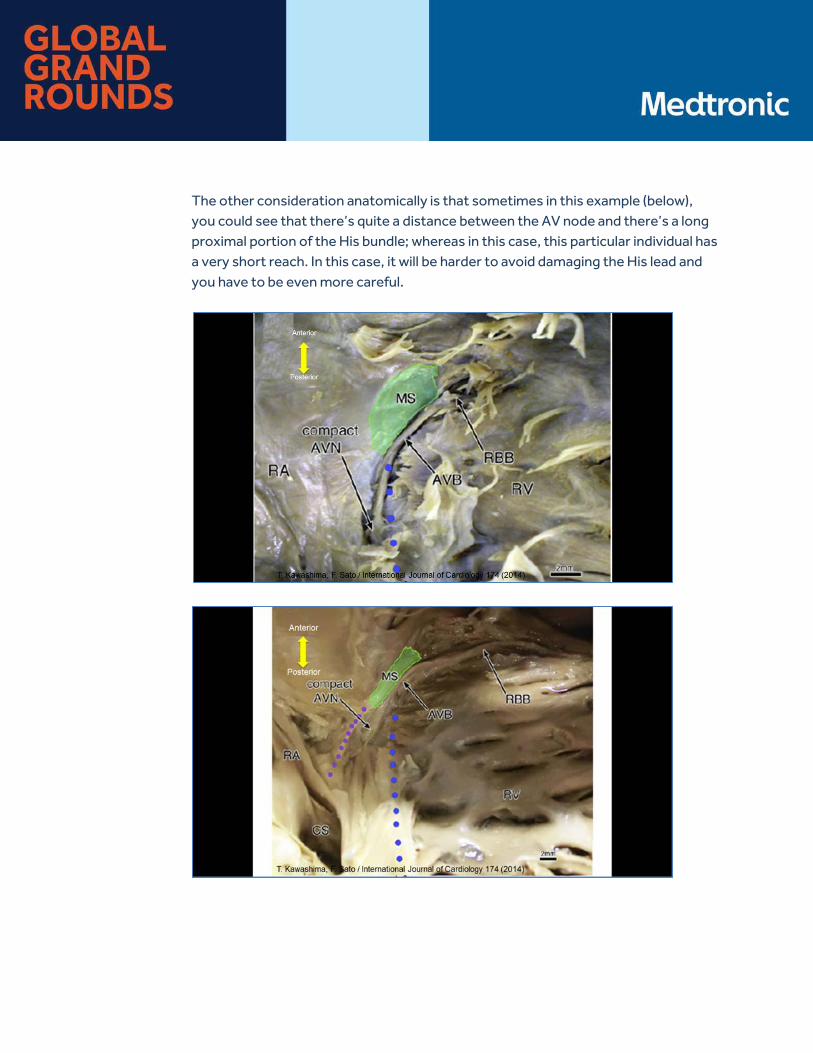
The other consideration anatomically is that sometimes in this example (below), you could see that there’s quite a distance between the AV node and there’s a long proximal portion of the His bundle; whereas in this case, this particular individual has a very short reach. In this case, it will be harder to avoid damaging the His lead and you have to be even more careful.
Ellenbogen: Thanks, Dan, very much. Pugal, can you talk to us a little bit about experience with right-sided versus left-sided access? Are there any tricks you can tell us or any advantages, disadvantages?
Vijayaraman: Yes. Technically, the left-sided implants are a lot easier because the current implantation tools are geared towards implanting from the left side, but however, the right side implant can also be done. We have performed about 25 patients with right-sided implant. We’ve been successful in almost all of them. The implantation is a little bit harder. You have to torque the sheath especially. We use the C315His sheath. You have to torque the sheath to maintain the septal cohesion. If you even let go of the sheath a little bit, it tends to fall away from this. The stability of the sheath is the difficult thing.
Secondly, we have tried using the deflectable sheath and that’s a lot harder to control and the movements are more difficult and the sheath is a little bit more aggressive at the tips, so we’d like to avoid using that sheath.
Ellenbogen: I’m going to ask you, Gopi. What situations have you used to deflect to both sheaths, the C304? Any particular clinical situation where you found that maybe helpful?
Dandamudi: Sure. In majority of cases, to begin with, we always start out with the fixed curved sheath. That tends to be highly successful in terms of reaching the region near the septal isthmus to map the His bundle. In cases where we tend to struggle are large, dilated right atria where the septal reach is because of a fixed curved with a sheath, you don’t have enough of a reach of that primary curve to reach these areas that are displaced more anteriorly. In those situations, we tend to try using the deflectable curve.
It’s important to understand that in this situation, sometimes it’s very hard to place it high up on the AV groove, and it tends to be a little bit more inferior. However, with some manipulation, it’s not uncommon that we can reach the area of the His bundle and actually pace from sites.
Ellenbogen: I have a question for Dan that came up. Do you know of any information comparing LV-only pacing to His bundle pacing? You’ve heard of any studies where they looked at that or are planning on looking at that?
Lustgarten: No, but I think it’s very interesting if you think about LV-only pacing, the place for that is relevant, is where there’s intact right bundle branch conduction. Really what you’re doing there is a hybrid of pacing the LV prematurely. My guess is that LV pacing is going to be a step towards this, but if you’re able to effect resynchronization by His bundle pacing, I would anticipate that it will be shown that it’s probably better that way. I’m not aware specifically that that’s being studied.
Ellenbogen: I just want to remind our audience if you happen to send in a question and we don’t have time to address it today, we will try to email you directly a response.
Now, Dr. Dandamudi, for starters, is there a specific patient population that you think this is ideal for? We’ve seen a lot of data today about a lot of patient population. Is there one particular group where you think this is specifically or uniquely helpful? Right bundle branch block, left bundle branch block.
Dandamudi: In terms of approaching the patient obviously, our experience is biased because we’ve been implanting this in every single patient who comes for a pacemaker indication. If you are starting out for the first time, we recommend that you try this in the sinus node dysfunction patients. Partly, there is a learning curve of at least 20 to 25 cases of getting a good idea about how to rotate the sheath and how to fix the lead itself and what is acceptable in terms of pacing thresholds and sensing issues. Once you gain that experience, I think it’s okay to move on to patients where ventricular pacing is going to performed at a higher degree such as second-degree heart block patients, intermittent heart block patients, and eventually to complete heart block patients where the most benefit is derived in those particular patients.
Finally, as experience, as you will get more experience trying to implant within patients with right bundle branch block and left bundle branch block as well.
Ellenbogen: I’m going to ask the panel. Has anyone had any experience extracting this lead, the 3830? Dr. Vijayaraman.
Vijayaraman: We’ve done probably about five to six leads duration of six months to eight years. Interestingly, these leads are rather easier to extract. Most of the leads come back very easily with counterclockwise rotation and gentle traction. There was one patient where I had to use a lead extraction to and we’re able to successfully get it off.

Ellenbogen: Okay. I’m going to ask the panel another question. Let's start off with Dan on this one. Dan, can you comment on saying where you think we’re going to be dangerous to ask this question in a couple of years, do you think this is going to become a standard in terms of we have to be pacing patients from the His bundle?
Lustgarten: Yeah. I think and I suspect my colleagues would join me in this notion that when you start doing this, the first question you ask yourself is why haven't I been doing this all along. In the majority of patients in whom we try to do this, you can get very good thresholds and you are effecting a totally normal QRS at the same time being able to mandate ventricular pacing. I think that the limitations as they currently exist are impressive to the extent at which they’re really not that bad given that not much effort has really been put into developing systems specific to doing this. I think very subtle modifications in the systems can be anticipated to make this much more to democratize it, if you will. I can’t imagine this won’t be the standard of pacing in five to seven years.
Ellenbogen: Do you agree?
Vijayaraman: I completely agree with that notion. Often people ask, “Why do His bundle pacing?” Our answer has been, “Why not His bundle pacing? That should be the standard.” That is the normal physiological activation and that’s what we should be doing in every single patient provided the limitations can be corrected. I think we’re getting there. We should be able to get there with a little more investment in technology and ability to achieve His bundle pacing in almost all patients.
Ellenbogen: Gopi, I’m going to start off with a question for you. Again, we can go down the aisle and see if other people have other opinions.
In your opinion, do you think to learn how to do this, what do you think is the right way to learn how to do this? Do you think an experienced operator can learn on their own or they should be precepted or they can go somewhere and watch a case or two? How much effort do you think it takes to learn how to do this?
Dandamudi: Sure. It is always helpful to have somebody who’s experienced in this be at least at your side or to go to a center where these have been done. There are some pitfalls depending on how you start out on your patient selection, so it’s always nice to hear other people’s experiences and what to look for and what to avoid. We made
mistakes as we started out, so it’s easier not to make those mistakes by learning from people who already the mistakes.
I would say again we have preached that at least about 20 cases to get a good fix on how to manipulate the lead and the sheath and especially targeting sinus node dysfunction patients where if patient thresholds do go up or lead dislodgements do occur, patients are not dependent, and it is always easier to learn in those situations rather than learning it the hard way in pacer-dependent patients.
Ellenbogen: Any other comments?
Vijayaraman: I completely agree with that; however, I think as Gopi mentioned, the learning process is important. We have gone through a huge learning process, and a lot of it can be imparted fairly easily. You don’t have to repeat some of those things and learn slowly. This is a good way to be precepted or go look at a center that does His bundle pacing to learn.
Ellenbogen: This question came up, “Ask Dan and/or Pugal to comment on this. Are either of you aware of any large outcome trials like block heart failure using the His bundle pacing technique? Do you know of any trials that have underway or people are planning?
Vijayaraman: This has been our interest and now the challenge is we have been pushing for it. We are working on some of these trials. Hopefully, we’ll get through within the next year or two.
Ellenbogen: Let me go down the aisle. We’ll keep out answer short. We’ll over here with Gopi. What pacing parameters are unacceptable for His bundle pacing? If you can’t get a better threshold than X, give up and move on to something else.
Dandamudi: Sure. Again, this is very patient-specific. Patients who are dependent or they have heart block. It is very important to not accept high pacing thresholds even if you’ve achieved pure His bundle pacing. Based on data and our own experience, there is a chance that pacing thresholds can rise over time. In those situations either putting a backup lead, a backup RV pacing lead, or accepting a different position is the way to go. There are also other situations such as pacing left bundle branch block or right bundle branch block where high pacing thresholds result in recruitment of this.

Again over time, these patient thresholds can rise and I wouldn’t recommend in those situations.
Ellenbogen: Dan, do you want to give us a number?
Lustgarten: I think Gopi is exactly right. Clinical contact is everything. That being said, I get nervous if I see thresholds around 3, 3.5 at one.
Ellenbogen: Okay. That’s very helpful. Pugal, any comment?
Vijayaraman: I agree. We tend to have a threshold number for most patient, 2.5 at one. Independent patient, we may accept a higher His bundle capture threshold especially if we have non-selective His capture, and so you have an RV backup from the same lead.
Ellenbogen: That’s a very good point. I think that's a very important point. If you don’t have His bundle capture, you may have some of that RV tissue nearby.
During office follow-up, do each of you do a 12-lead EKG when checking His bundle pacing thresholds to ensure physiologic lead position? Why don’t we start off with Dan. When you see these people in your follow-up clinic, 12-lead EKG, yes or no?
Lustgarten: Yes, I always do it. I think the reason I do it is just to please myself actually, but yes, I always do it.
Ellenbogen: Pugal, how about you?
Vijayaraman: We did that early on, and we would do it in some challenging cases especially with the bundle branch block patient; otherwise, we would have at least two or three of the surface leads on, and we have trained our staff to look for His capture letting them learn selective, non-selective His capture, and also give them idea how to program thresholds. Our goal is to always have programming output at slightly higher than the His bundle correction threshold rather than the RV threshold. That’s important.
Ellenbogen: It’s educational, it’s personally rewarding. Gopi, how about you?
Dandamudi: Yes. Again, it’s personally rewarding, so there are patients where I will do it myself and do different degrees of output to show output-dependent pacing. Physiologically, it’s a marvel when you see that. We’ve also trained our device clinic nurses to look at surface leads at least off of the PSA and looking at different degrees of fusion as we change outputs to suggest that we are, in fact, recruiting the His bundle.
Ellenbogen: Dan.
Lustgarten: One thing we really have talked about is you can intentionally go distally into what would be classically considered a para-Hisian position. If that’s the case, you have to make certain that their output is high enough to capture the His tissues, the opposite when you’re above the tricuspid annulus. That’s one reason if you have somebody with non-selective capture or ventricular capture only. You want to makes sure that the threshold of the His capture hasn’t gotten higher than their currently programmed at.
Ellenbogen: Again, we’ll try to get to all your questions. If you we don’t respond to them, we’ll try to email you response. Anyone performed this in a child? His bundle pacing. I know there are some centers where their pediatric electrophysiologists that are starting to do that. Have any of you three had experienced with a child?
Lustgarten: I haven't done it in a child. I’ve done it in patients who’ve been paced for congenital AV block and have developed cardiomyopathy, but then are now young adults and corrected that with …
Ellenbogen: Pugal?
Vijayaraman: I’ve performed in patients 16 to 20 years of age, and I’ve helped pediatric electrophysiologist perform in a 12-year-old so my experience is limited.
Ellenbogen: When performing AV node ablations in patients with His bundle pacing lead, are you always, sometimes, or never placing an additional backup lead in the right ventricle? Good fair question. Dan, you can start with your answer. We’ll move down the panel.
Lustgarten: I always do. I don’t think I need to but I always do.
Vijayaraman: In our lab, we haven't been doing that routinely, so I would say that 95% of our patients do not have a backup RV lead.
Dandamudi: That’s our similar practice as well. In this situation when targeting the AV node, I usually tend to start near the slow pathway region and slowly migrate up towards the His bundle and not start high up on the septum.
Ellenbogen: Probably 25% of our patients, we’ll put an RV, extra RV lead.
Ellenbogen: Can you talk a little bit about- we’ll start off with Dan since you’ve done some of the hemodynamic studies- about how you program the paced AV delay and the sensed AV delay in patients with His bundle pacing.
Lustgarten: If you have a backup RV lead, you want to make sure that that’s inhibited. In that setting, you have to ascertain the timing between His pacing and RV sensing and make sure that the AV delay is programmed beyond that.
An interesting scenario is when you have a very active person whose primary indication for pacing is AV delay. If they are athletic, try to get the AV hysteresis or rate adaptive AV response to be appropriate to their level of activity, and that can be very challenging. I have found experientially that in those settings it’s critically important to actually work with the patient usually using treadmills and things like that to actually in real time modify the extent or the aggressiveness of the AV response. Those are two critical issues off the top of my head.
Vijayaraman: Just one important point there is that when you program AV delays so you can’t program the normal ones you would program. You have to accommodate for the 40 or 50 milliseconds HV delay that you have, so you have to shorten your AV delay at least by 40 or 50 milliseconds.
Ellenbogen: Good point. That’s really worth reminding the audience You have to subtract that 40 or so, 40 to 50 milliseconds from the AV intervals.
There is a question here about reaching the His from the RV site. Can you speak a little bit more about that? Pugal, will you make some more specific comments about RV.
Vijayaraman: Thanks. Can you repeat the question?
Ellenbogen: Putting a His bundle pacemaker from the right side.
Vijayaraman: Okay. The challenge has been to push on the sheath at His bundle level. When you implant a lead from where the sheath is pointing more laterally, so in order to bring it to the septum, you have to torque the sheath quite a bit towards the septum so that’s usually I’m trying to do it myself not remembering how to do. It’s more counterclockwise to get it to the septal level. It will be a significant amount of torque.
Normally, when we pace from the left side, the movement of the sheath is very minimal, so little movement will move the sheath quite a bit away from the annulus, away from the septum. From the right side, you have to do a significant amount of rotation to get to the septum. Once you get to the septum, you have to hold the sheath in place and that’s the critical part. Once you are able to master that, then it’s easy to do the His bundle pacing.
Lustgarten: Be very careful not to advance it to the right atrial appendage.
Vijayaraman: Taking the usual precautions.
Ellenbogen: I think that’s what they say. Gopi, I’m going to start off with you. How long do you try and obviously, this is going to be patient-specific but just give us a range for people who are listening. How long will you try before you realize it’s unlikely to be successful and need to move on?
Dandamudi: Usually, at least, my policy is in patients with normal right atrial size, right ventricular size. If you can’t get the His bundle within the first five minutes usually that's a struggle for me thereafter because these preformed sheaths and normal-shaped right atrial and right ventricles can easily reach the summit and actually map the His bundle so within five minutes. If it’s a patient who is dependent that I’m really trying hard to place His bundle lead, I will spend maybe up to 10 minutes or 12 minutes, but usually my ceiling is about 15 minutes with any particular patient in general.
Ellenbogen: Any further comments?
Vijayaraman: Just to expand on that, I think want we find is … finding the His bundle is not often the challenge. You can get there in first five minutes. Fixing the lead is often the challenge. Once you’ve seen the His bundle, once you’ve tested that you can
narrow the QRS, it’s hard for me to give up, so I may go up to 20, 25 minutes before I give up.
Lustgarten: Something that I’ve struggled with experientially is if you find … You have excellent capture but the threshold is a little bit high, you take it out, you go for another site. At what point? I always start out with a fixed sheath as well. At some point if I’m having a hard time with that, I will then go over to the deflectable sheath. Where those points are in real time, I haven't quantified but certainly, not more than 20 or 25 minutes.
Vijayaraman: I would like to expand on that one. We’ve learned from some of our Chinese colleagues, Dr. Weijing Huang that when you have one of those locations, your threshold is high, and so moving the lead, you can leave that lead as a marker and bring another lead and map around it. He's been successful in getting it. We have used that in a few situations. It helped us tremendously.
Lustgarten: And place the prior lead in the atrium if you’re doing a dual chamber pacemaker.
Ellenbogen: That’s a great, great little trick. I think we’re close to the end here. We’ve had a pretty thorough discussion of His bundle pacing. I want to take this opportunity to, first of all, remind people that a recorded version of this presentation will be available soon at medtronicacademy.com/globalgrandrounds. Secondly, for His bundle pacing proctorships, please email Medtronic Procedural Training [email protected].
I want to thank our incredibly experienced and excellent faculty, Dr. Lustgarten, Dr. Vijayaraman, and Dr. Dandamudi that it was a really great tour de force of physiology, how to do it, and the clinical trials that led us to the place we are now. Thank you everyone for your time and patience, and thank you to the faculty.
Doctors: Thank you.
Product Safety Information Brief Statement: IPGs, CRT IPGs, ICDs, and CRT ICDs
Indications
Implantable Pulse Generators (IPGs) are indicated for rate adaptive pacing in patients who may benefit from increased pacing rates concurrent with increases in activity. Pacemakers are also indicated for dual chamber and atrial tracking modes in patients who may benefit from maintenance of AV synchrony. Dual chamber modes are specifically indicated for treatment of conduction disorders that require restoration of both rate and AV synchrony, which include various degrees of AV block to maintain the atrial contribution to cardiac output and VVI intolerance (e.g. pacemaker syndrome) in the presence of persistent sinus rhythm. See device manuals for the accepted patient conditions warranting chronic cardiac pacing. Antitachycardia pacing (ATP) is indicated for termination of atrial tachyarrhythmias in patients with one or more of the above pacing indications. For the MR Conditional IPGs, a complete SureScan® pacing system, which consists of an approved combination (see http://www.mrisurescan.com) MRI SureScan device with SureScan lead(s), is required for use in the MR environment.
Cardiac Resynchronization Therapy (CRT) IPGs are indicated for NYHA Functional Class III and IV patients who remain symptomatic despite stable, optimal heart failure medical therapy and have a LVEF ≤ 35% and a prolonged QRS duration and for NYHA Functional Class I, II, or III patients who have a LVEF ≤ 50%, are on stable, optimal heart failure medical therapy if indicated and have atrioventricular block (AV block) that are expected to require a high percentage of ventricular pacing that cannot be managed with algorithms to minimize right ventricular pacing. Optimization of heart failure medical therapy that is limited due to AV block or the urgent need for pacing should be done post implant. Rate adaptive pacing is provided for those patients developing a bradycardia indication who might benefit from increased pacing rates concurrent with increases in activity. Dual chamber and atrial tracking modes are indicated for patients who may benefit from maintenance of AV synchrony. Antitachycardia pacing (ATP) is indicated for termination of atrial tachyarrhythmias in patients with one or more of the above pacing indications.
Implantable cardioverter defibrillators (ICDs) are indicated to provide ventricular antitachycardia pacing and ventricular defibrillation for automated treatment of life-threatening ventricular arrhythmias. Notes on some features in ICDs: The clinical value of the OptiVol fluid monitoring diagnostic feature has not been assessed in those patients who do not have fluid retention related symptoms due to heart failure. Additional notes for DR ICDs: The use of the device has not been demonstrated to decrease the morbidity related to atrial tachyarrhythmias. The effectiveness of high-frequency burst pacing (atrial 50 Hz Burst therapy) in terminating device classified atrial tachycardia (AT) was found to be 17%, and in terminating device classified atrial fibrillation (AF) was found to be 16.8%, in the VT/AT patient population studied. The effectiveness of high-frequency burst pacing (atrial 50 Hz Burst therapy) in terminating device classified atrial tachycardia (AT) was found to be 11.7%, and in terminating device classified atrial fibrillation (AF) was found to be 18.2% in the AF-only patient population studied.
CRT ICDs are indicated for ventricular antitachycardia pacing and ventricular defibrillation for automated treatment of life-threatening ventricular arrhythmias and for providing cardiac resynchronization therapy in heart failure patients on stable, optimal heart failure medical therapy if indicated, and meet any of the following classifications: New York Heart Association (NYHA) Functional Class III or IV and who have a left ventricular ejection fraction < 35% and a prolonged QRS duration. Left bundle branch block (LBBB) with a QRS duration > 130 ms, left ventricular ejection fraction < 30%, and NYHA Functional Class II. NYHA Functional Class I, II, or III and who have left ventricular ejection fraction ≤ 50% and atrioventricular block (AV block) that are expected to require a high percentage of ventricular pacing that cannot be managed with algorithms to minimize right ventricular pacing. Optimization of heart failure medical therapy that is limited due to AV block or the urgent need for pacing should be done post implant. Some ICDs and CRT ICDs are also indicated for use in patients with atrial tachyarrhythmias, or those patients who are at significant risk for developing atrial tachyarrhythmias. The RV Lead Integrity Alert (LIA) feature is intended primarily for patients who have a Medtronic ICD or CRT-D device and a Sprint Fidelis lead (Models 6949, 6948, 6931, and 6930), based on performance data. The RV LIA feature may not perform as well with a St. Jude Riata/Durata lead or a Boston Scientific Endotak lead as it does when used with a Medtronic Sprint Fidelis lead. This is because different lead designs may have different failure signatures and conditions that may or may not be detected early by the RV LIA feature.
Contraindications
IPGs and CRT IPGs are contraindicated for concomitant implant with another bradycardia device and concomitant implant with an implantable cardioverter defibrillator. There are no known contraindications for the use of pacing as a therapeutic modality to control heart rate. The patient’s age and medical condition, however, may dictate the particular pacing system, mode of operation, and implant procedure used by the physician. Rate-responsive modes may be contraindicated in those patients who cannot tolerate pacing rates above the programmed Lower Rate. Dual chamber sequential pacing is contraindicated in patients with chronic or persistent supraventricular tachycardias, including atrial fibrillation or flutter. Asynchronous pacing is contraindicated in the presence (or likelihood) of competition between paced and intrinsic rhythms. Single chamber atrial pacing is contraindicated in patients with an AV conduction disturbance. Anti-tachycardia pacing (ATP) therapy is contraindicated in patients with an accessory antegrade pathway.
ICDs and CRT ICDs are contraindicated in patients experiencing tachyarrhythmias with transient or reversible causes including, but not limited to, the following: acute myocardial infarction, drug intoxication, drowning, electric shock, electrolyte imbalance, hypoxia, or sepsis; patients who have a unipolar pacemaker implanted, patients with incessant ventricular tachycardia (VT) or ventricular fibrillation (VF), and patients whose primary disorder is chronic atrial tachyarrhythmia with no concomitant VT or VF. Warnings/Precautions
Changes in a patient’s disease and/or medications may alter the efficacy of the device’s programmed parameters. Patients should avoid sources of magnetic and electromagnetic radiation to avoid possible underdetection, inappropriate sensing and/or therapy delivery, tissue damage, induction of an arrhythmia, device electrical reset or device damage. Do not place transthoracic defibrillation paddles directly over the device.
Additionally, for CRT ICDs and CRT IPGs, certain programming and device operations may not provide cardiac resynchronization. Also for CRT IPGs, Elective Replacement Indicator (ERI) results in the device switching to VVI pacing at 65 ppm. In this mode, patients may experience loss of cardiac resynchronization therapy and / or loss of AV synchrony. For this reason, the device should be replaced prior to ERI being set. Use of the device should not change the application of established anticoagulation protocols.
For MR Conditional IPG Systems, before performing an MRI scan, refer to the Surescan pacing system technical manual for additional information, patients and their implanted systems must be screened to meet the MRI Conditions of Use. Do not scan patients who do not have a complete SureScan pacing system consisting of an approved combination MRI SureScan device with SureScan lead(s); patients who have broken, abandoned or intermittent leads; or patients who have a lead impedance value of < 200 Ω or > 1,500 Ω.
Potential complications
Potential complications include, but are not limited to, rejection phenomena, erosion through the skin, muscle or nerve stimulation, oversensing, failure to detect and/or terminate arrhythmia episodes, and surgical complications such as hematoma, infection, inflammation, and thrombosis.
An additional complication for ICDs and CRT ICDs is the acceleration of ventricular tachycardia.
SureScan systems have been designed to minimize potential complications in the MRI environment. Potential MRI complications include, but are not limited to, lead electrode heating and tissue damage resulting in loss of sensing or capture or both, or induced currents on leads resulting in continuous capture, VT/VF, and/or hemodynamic collapse.
See the device manual for detailed information regarding the implant procedure, indications, contraindications, warnings, precautions, and potential complications/adverse events. For further information, please call Medtronic at 1-800-328-2518 and/or consult Medtronic’s website at www.medtronic.com.
Caution: Federal law (USA) restricts these devices to sale by or on the order of a physician.
Updated Nov 10 2015
Brief Statement: Medtronic Leads
Indications
Medtronic leads are used as part of a cardiac rhythm disease management system. Leads are intended for pacing and sensing and/or defibrillation. Defibrillation leads have application for patients for whom implantable cardioverter defibrillation is indicated
Contraindications
Medtronic leads are contraindicated for the following:
• ventricular use in patients with tricuspid valvular disease or a tricuspid mechanical heart valve. • patients for whom a single dose of 1.0 mg of dexamethasone sodium phosphate or dexamethasone acetate may be
contraindicated. (includes all leads which contain these steroids) • Epicardial leads should not be used on patients with a heavily infarcted or fibrotic myocardium.
The SelectSecure Model 3830 Lead is also contraindicated for the following:
• patients for whom a single dose of 40.µg of beclomethasone dipropionate may be contraindicated. • patients with obstructed or inadequate vasculature for intravenous catheterization.
Warnings/Precautions
People with metal implants such as pacemakers, implantable cardioverter defibrillators (ICDs), and accompanying leads should not receive diathermy treatment. The interaction between the implant and diathermy can cause tissue damage, fibrillation, or damage to the device components, which could result in serious injury, loss of therapy, or the need to reprogram or replace the device.
For the SelectSecure Model 3830 lead, total patient exposure to beclomethasone 17,21-dipropionate should be considered when implanting multiple leads. No drug interactions with inhaled beclomethasone 17,21-dipropionate have been described. Drug interactions of beclomethasone 17,21-dipropionate with the Model 3830 lead have not been studied.
Potential Complications
Potential complications include, but are not limited to, valve damage, fibrillation and other arrhythmias, thrombosis, thrombotic and air embolism, cardiac perforation, heart wall rupture, cardiac tamponade, muscle or nerve stimulation, pericardial rub, infection, myocardial irritability, and pneumothorax. Other potential complications related to the lead may include lead dislodgement, lead conductor fracture, insulation failure, threshold elevation or exit block.
See the device manual for detailed information regarding the implant procedure, indications, contraindications, warnings, precautions, and potential complications/adverse events. For further information, please call Medtronic at 1-800-328-2518 and/or consult Medtronic’s website at www.medtronic.com.
Caution: Federal law (USA) restricts this device to sale by or on the order of a physician.


![His-Bundle Pacing in a Patient with Transcatheter Aortic ...pacing at the His bundle presumably due to the recruitment of fibres distal to the site of block [15]. In our case, these](https://img.pdfslide.net/doc/110x75/60bf56e592563363970bece8/his-bundle-pacing-in-a-patient-with-transcatheter-aortic-pacing-at-the-his-bundle.jpg)