Embed Size (px)
Citation preview
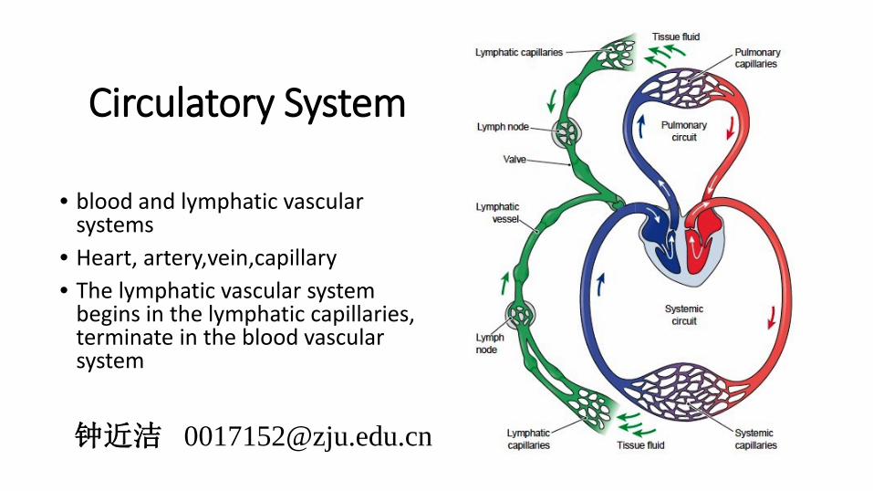
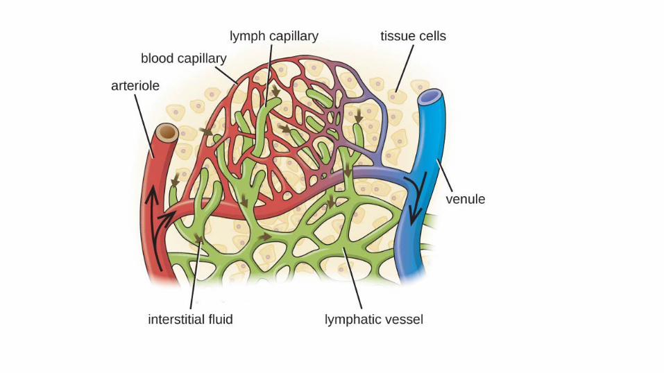
Circulatory System
• blood and lymphatic vascular systems
• Heart, artery,vein,capillary• The lymphatic vascular system
begins in the lymphatic capillaries, terminate in the blood vascular system
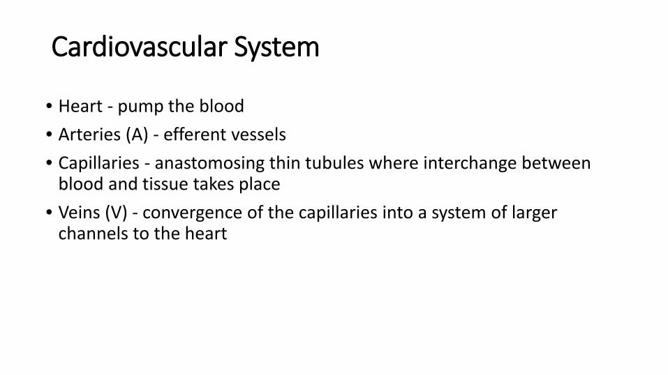
Cardiovascular System
• Heart - pump the blood• Arteries (A) - efferent vessels• Capillaries - anastomosing thin tubules where interchange between
blood and tissue takes place• Veins (V) - convergence of the capillaries into a system of larger
channels to the heart

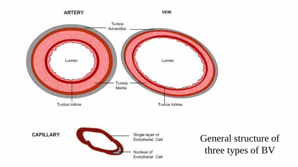
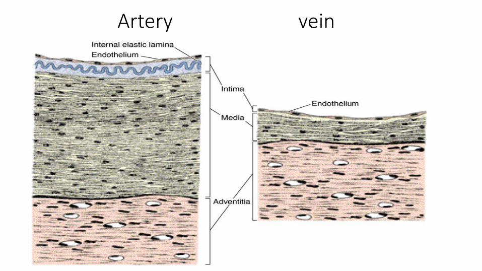
General structure of three types of BV
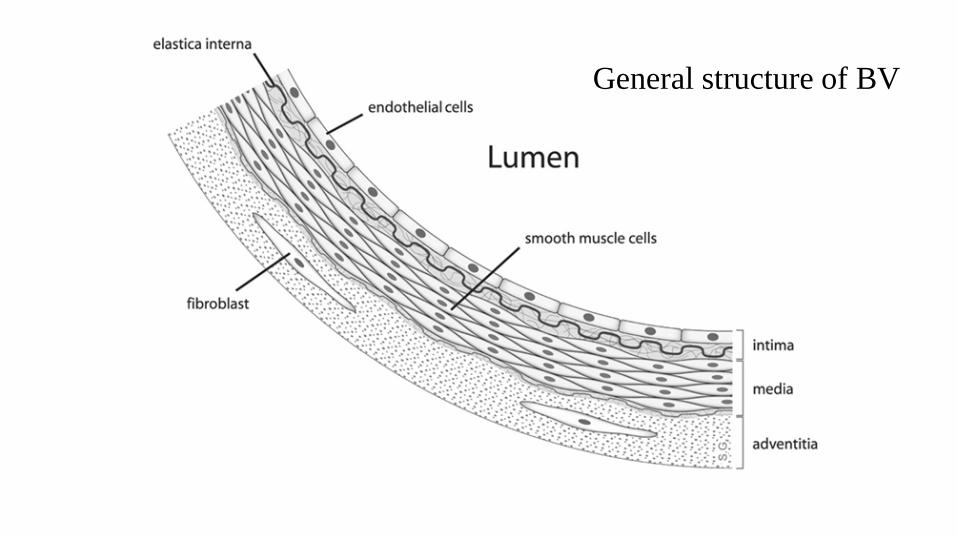
General structure of BV
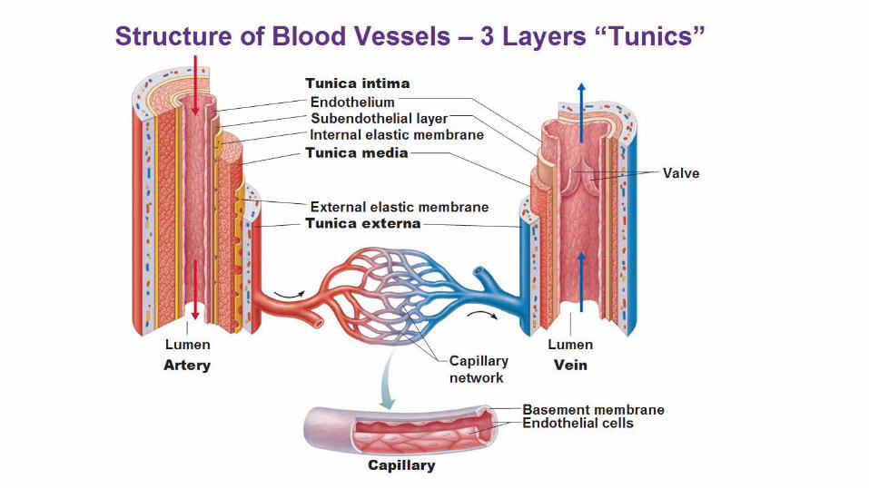
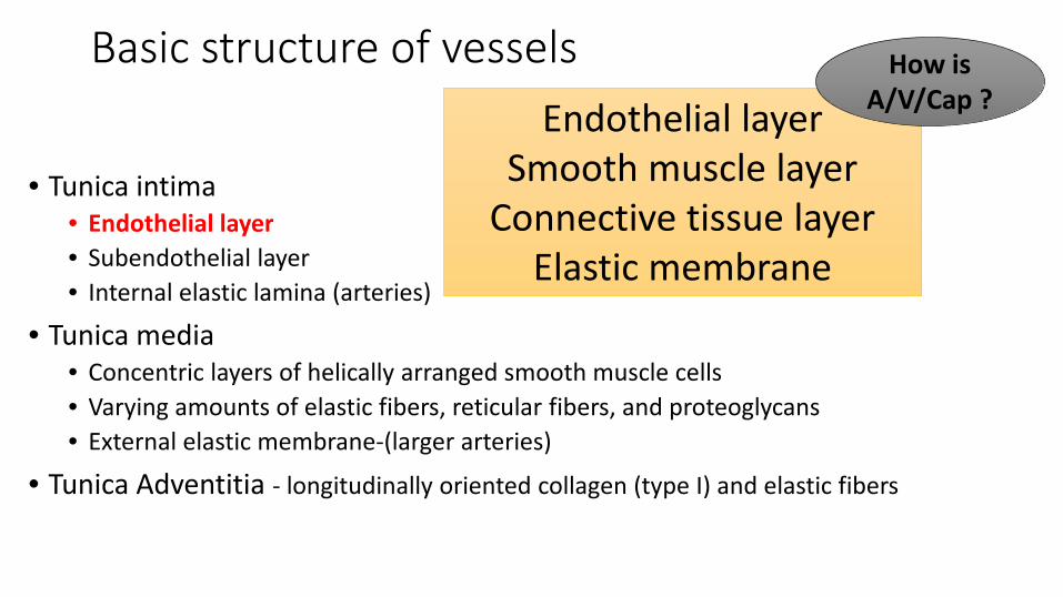
Basic structure of vessels
• Tunica intima• Endothelial layer• Subendothelial layer• Internal elastic lamina (arteries)
• Tunica media• Concentric layers of helically arranged smooth muscle cells• Varying amounts of elastic fibers, reticular fibers, and proteoglycans• External elastic membrane-(larger arteries)
• Tunica Adventitia - longitudinally oriented collagen (type I) and elastic fibers
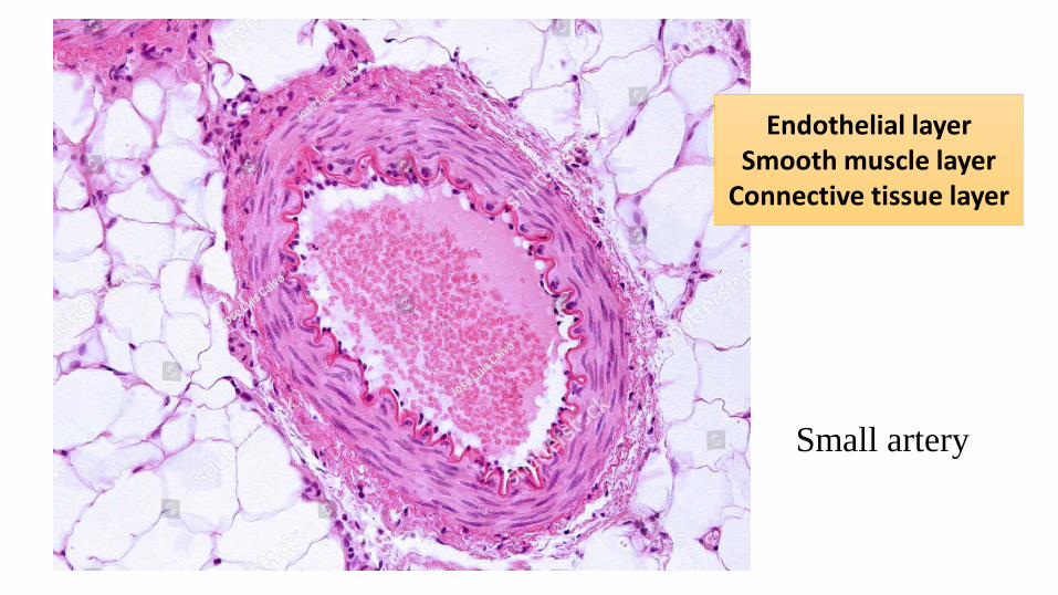
Endothelial layerSmooth muscle layer
Connective tissue layerElastic membrane
How is A/V/Cap ?
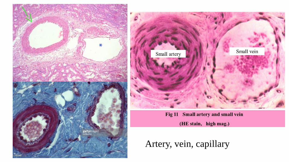
Artery, vein, capillary
Endothelial layerSmooth muscle layer
Connective tissue layer
Small artery
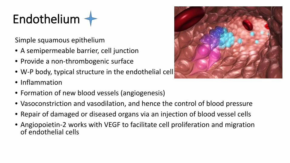
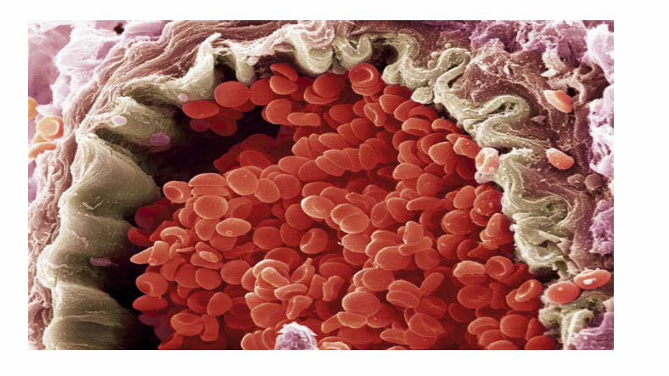
EndotheliumSimple squamous epithelium• A semipermeable barrier, cell junction• Provide a non-thrombogenic surface• W-P body, typical structure in the endothelial cell• Inflammation• Formation of new blood vessels (angiogenesis)• Vasoconstriction and vasodilation, and hence the control of blood pressure• Repair of damaged or diseased organs via an injection of blood vessel cells• Angiopoietin-2 works with VEGF to facilitate cell proliferation and migration
of endothelial cells
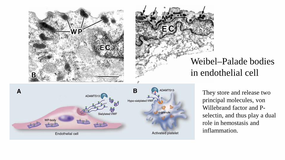
Weibel–Palade bodies in endothelial cell
They store and release two principal molecules, von Willebrand factor and P-selectin, and thus play a dual role in hemostasis and inflammation.
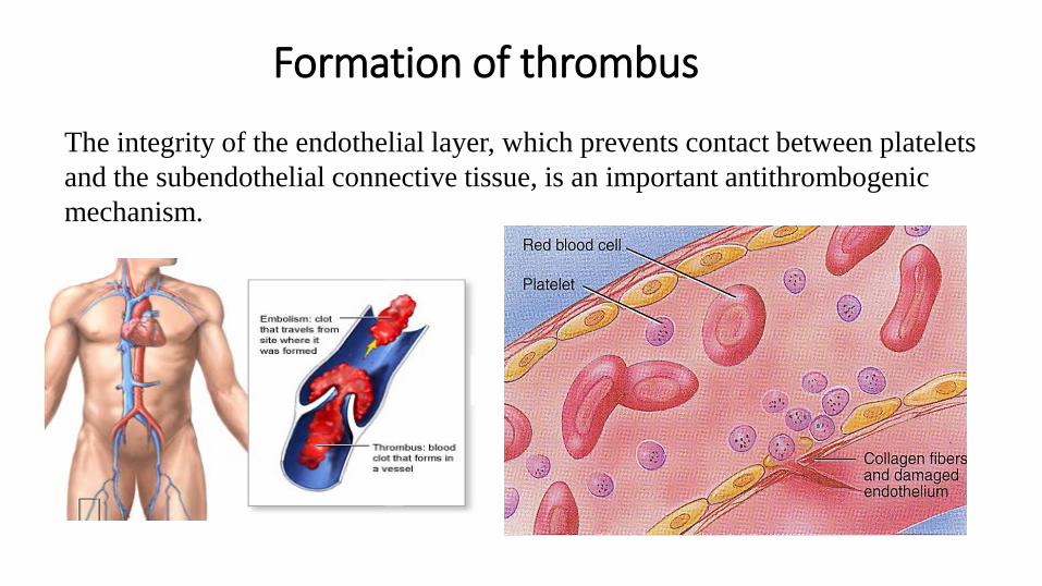
Formation of thrombus
The integrity of the endothelial layer, which prevents contact between platelets and the subendothelial connective tissue, is an important antithrombogenic mechanism.
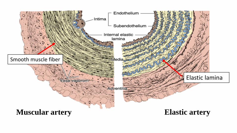
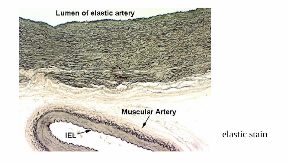
Muscular artery Elastic artery
Elastic lamina
Smooth muscle fiber
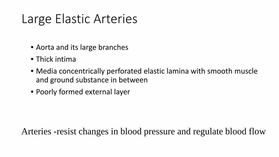
Large Elastic Arteries
• Aorta and its large branches• Thick intima • Media concentrically perforated elastic lamina with smooth muscle
and ground substance in between• Poorly formed external layer
Arteries -resist changes in blood pressure and regulate blood flow
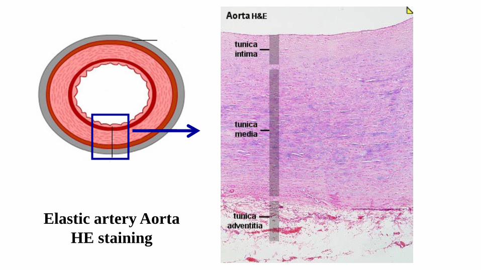
Elastic artery AortaHE staining
elastic stain

Elastic artery- elastic stain↓ internal elastic lamina smooth muscle layer external elastic lamina
Muscular Arteries
• Most arteries in the body• Thin intimal layer• Well developed internal elastic lamina• Muscle layer up to 40 layers• Varying intermingled elastic fibers• adventitia consists of nerves, vessels, collagen, elastic fibers, fibroblasts
and adipose cells
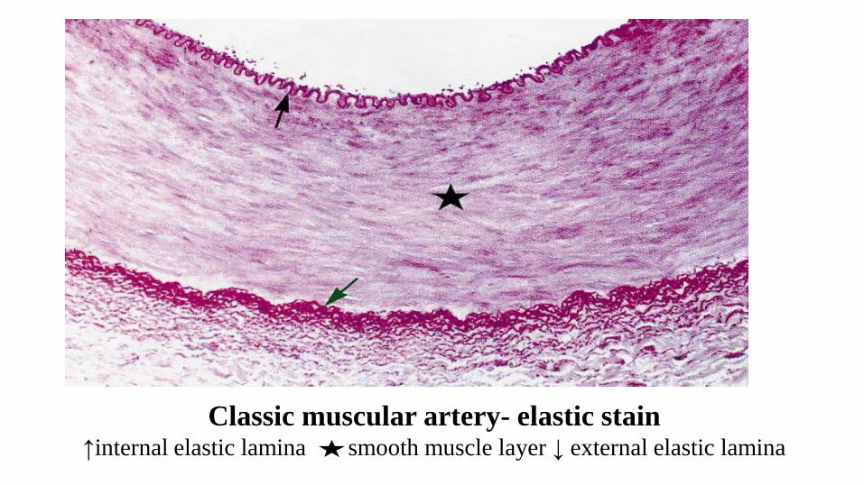
Classic muscular artery- elastic stain↑internal elastic lamina smooth muscle layer ↓ external elastic lamina
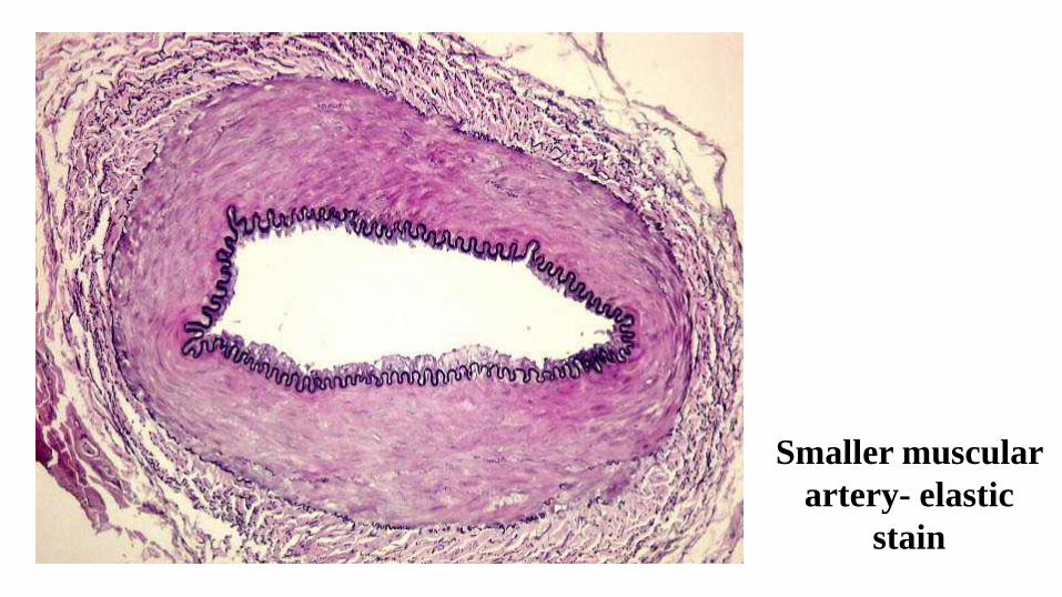
Smaller muscular artery- elastic
stain
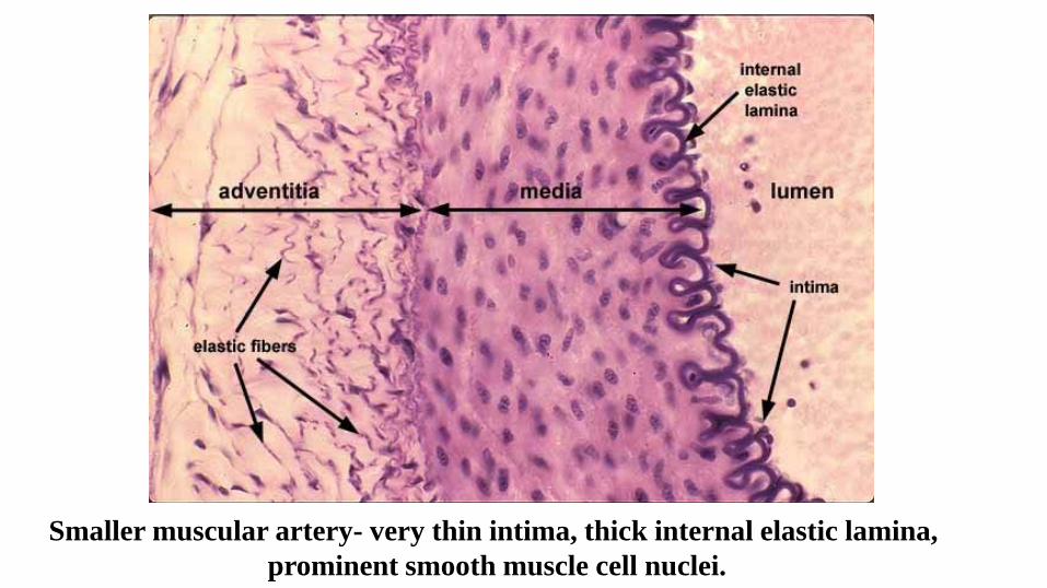
Smaller muscular artery- very thin intima, thick internal elastic lamina,prominent smooth muscle cell nuclei.
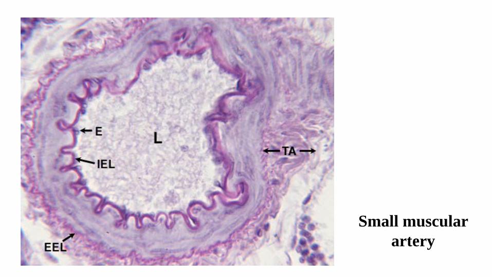
Small muscular artery
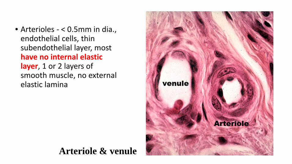
• Arterioles - < 0.5mm in dia., endothelial cells, thin subendothelial layer, most have no internal elastic layer, 1 or 2 layers of smooth muscle, no external elastic lamina
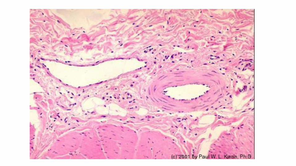
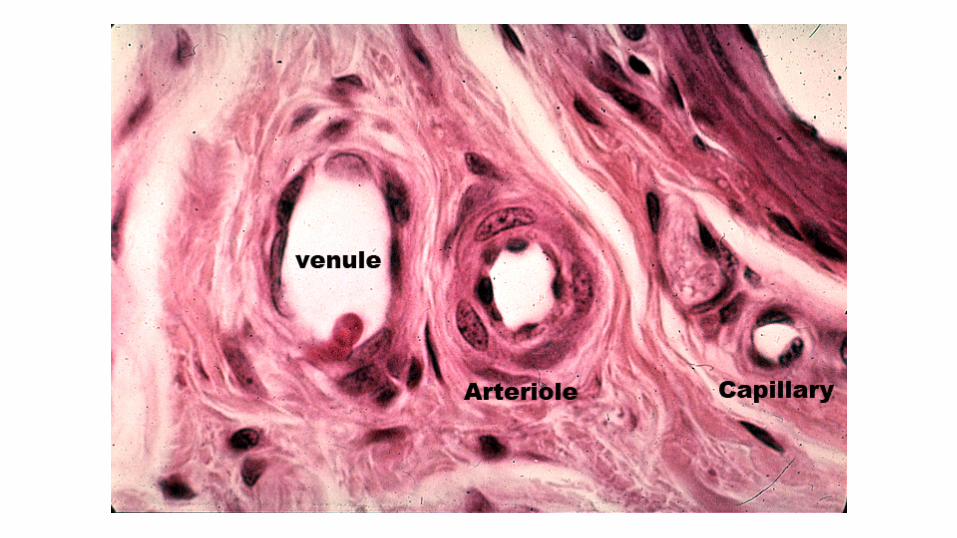
Arteriole & venule
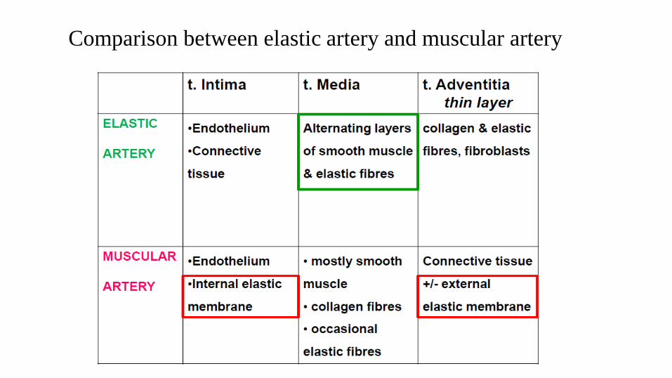
Comparison between elastic artery and muscular artery
Artery vein
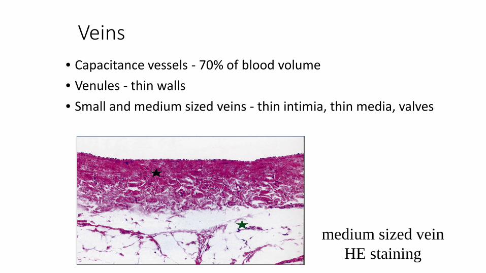
Veins• Capacitance vessels - 70% of blood volume• Venules - thin walls • Small and medium sized veins - thin intimia, thin media, valves
medium sized veinHE staining
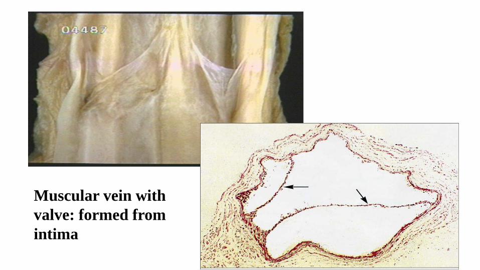
Muscular vein with valve: formed from intima
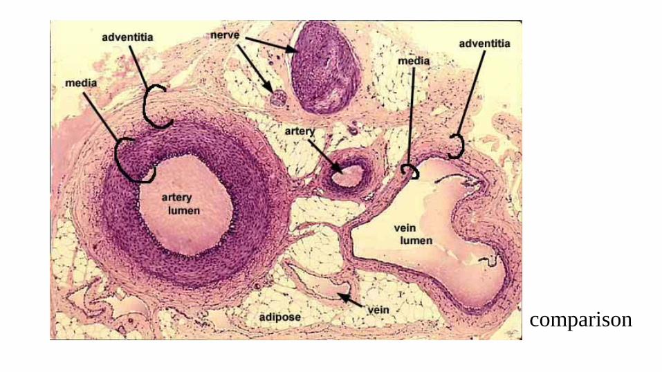
comparison
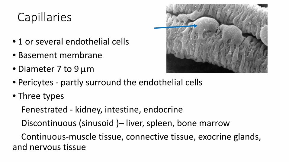
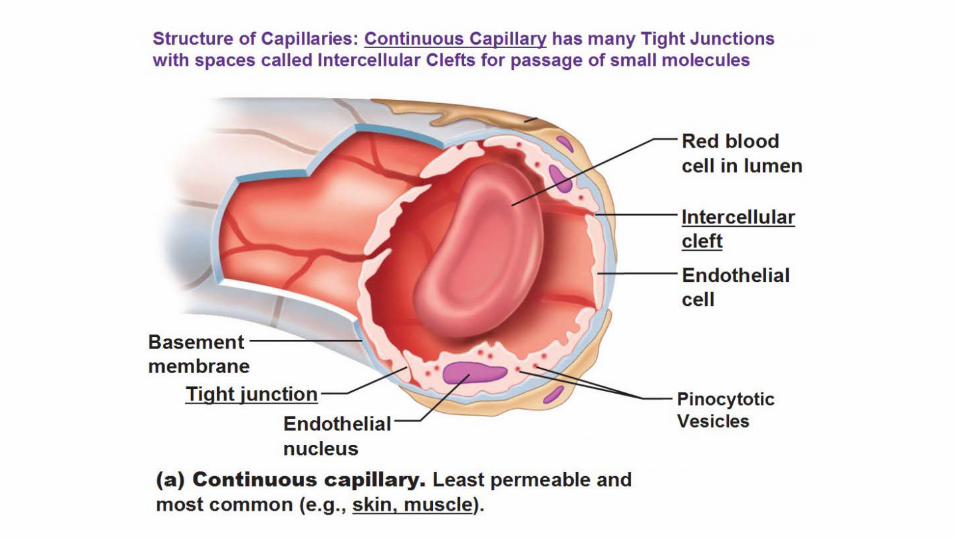
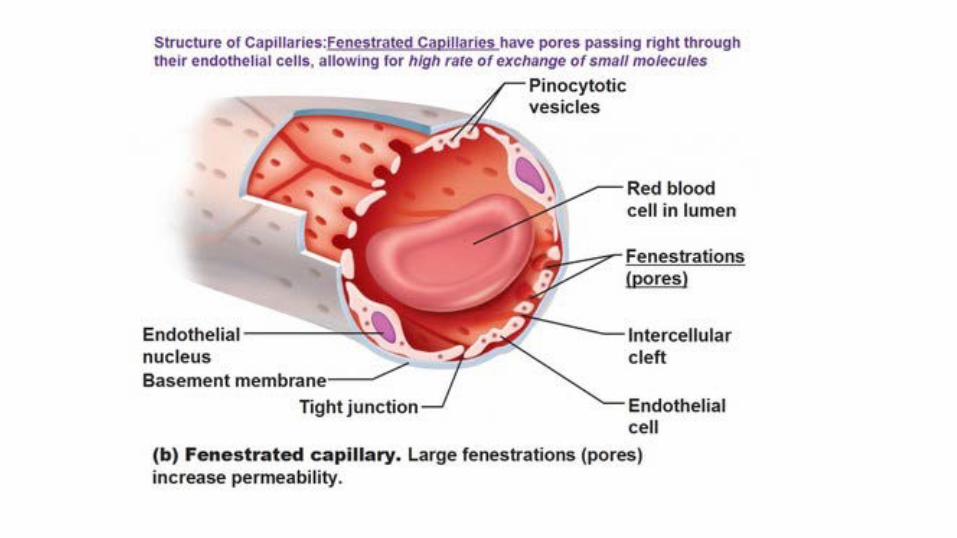
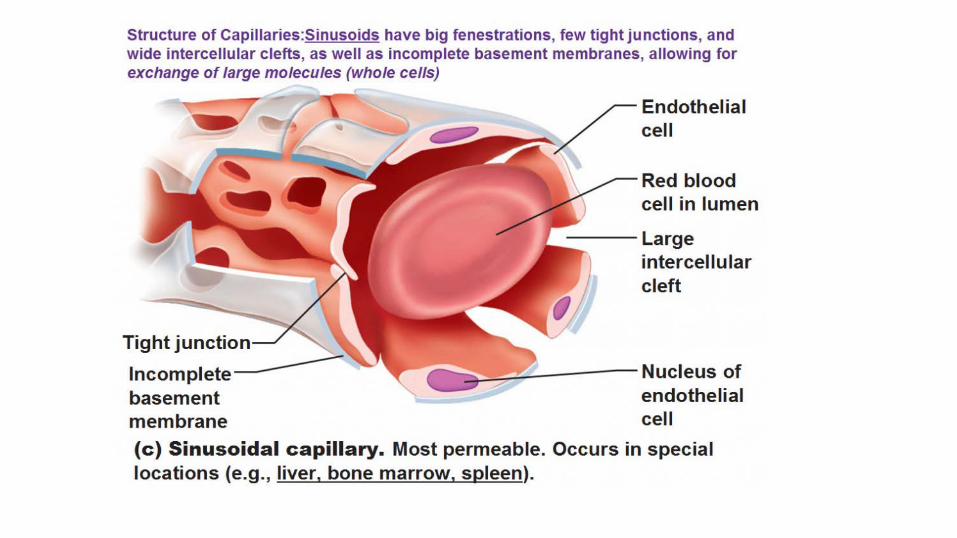
Capillaries
• 1 or several endothelial cells• Basement membrane• Diameter 7 to 9 µm• Pericytes - partly surround the endothelial cells• Three types
Fenestrated - kidney, intestine, endocrineDiscontinuous (sinusoid )– liver, spleen, bone marrowContinuous-muscle tissue, connective tissue, exocrine glands,
and nervous tissue
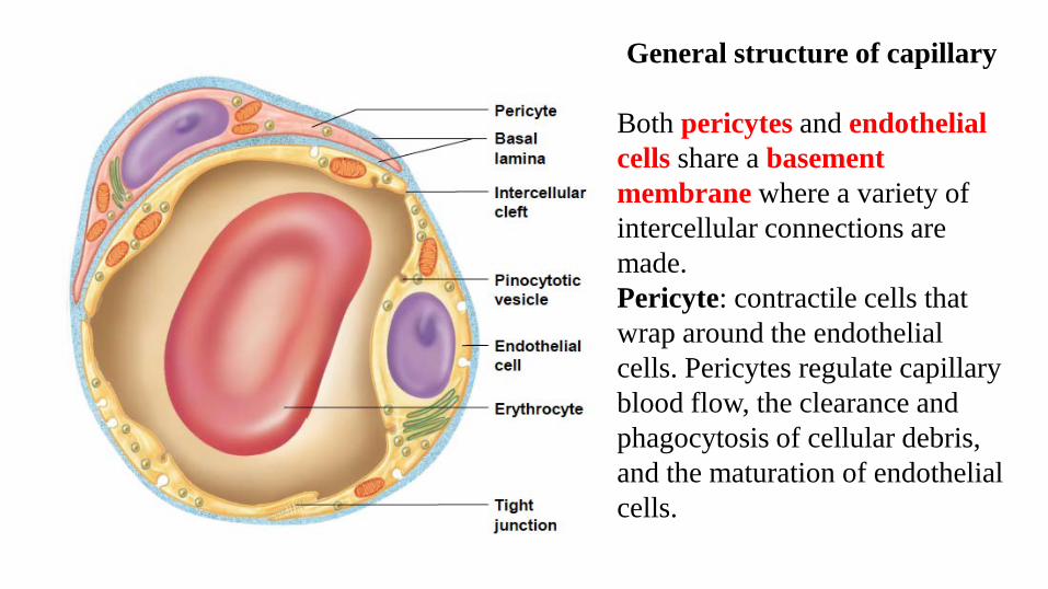
General structure of capillary
Both pericytes and endothelial cells share a basement membrane where a variety of intercellular connections are made.Pericyte: contractile cells that wrap around the endothelial cells. Pericytes regulate capillary blood flow, the clearance and phagocytosis of cellular debris, and the maturation of endothelial cells.

Capillary bed
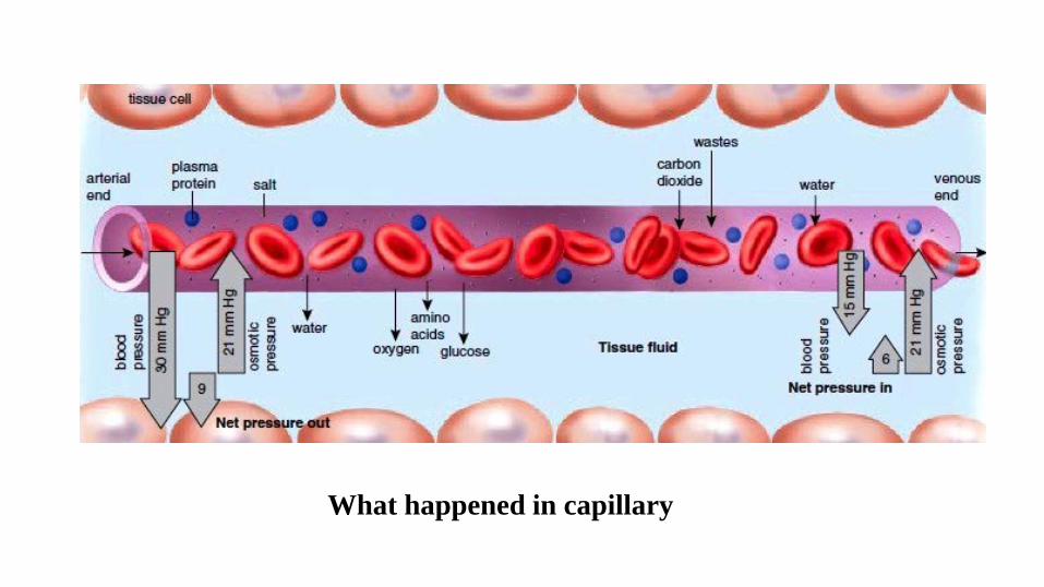
What happened in capillary
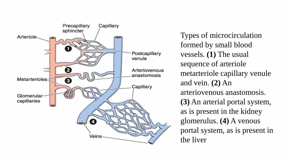
Types of microcirculation formed by small blood vessels. (1) The usual sequence of arteriole metarteriole capillary venule and vein. (2) An arteriovenous anastomosis. (3) An arterial portal system, as is present in the kidney glomerulus. (4) A venous portal system, as is present in the liver
2018/10/29 XJMU 40
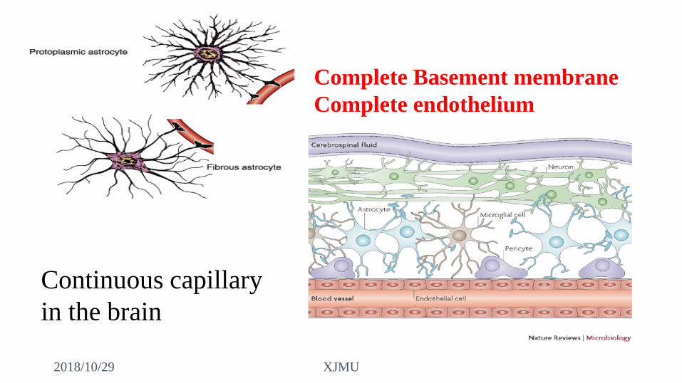
Continuous capillary in the brain
Complete Basement membraneComplete endothelium
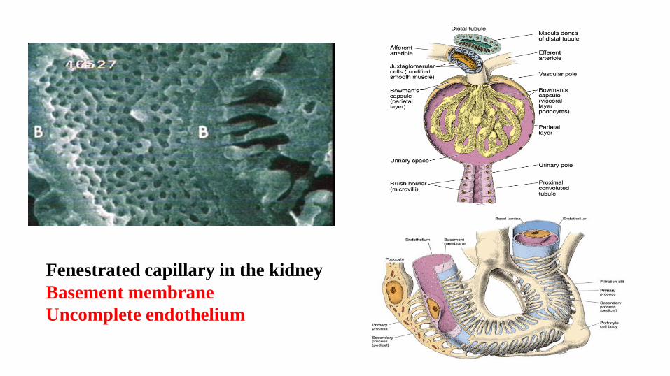
Fenestrated capillary in the kidneyBasement membraneUncomplete endothelium
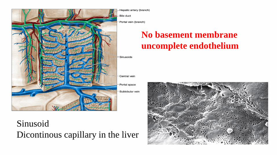
SinusoidDicontinous capillary in the liver
No basement membraneuncomplete endothelium
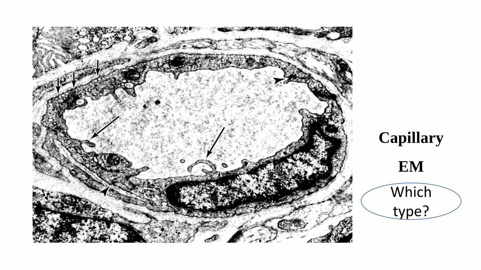
Capillary
EM Which type?
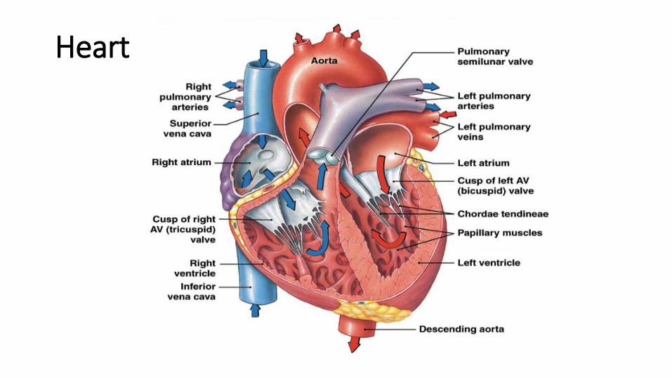
Heart
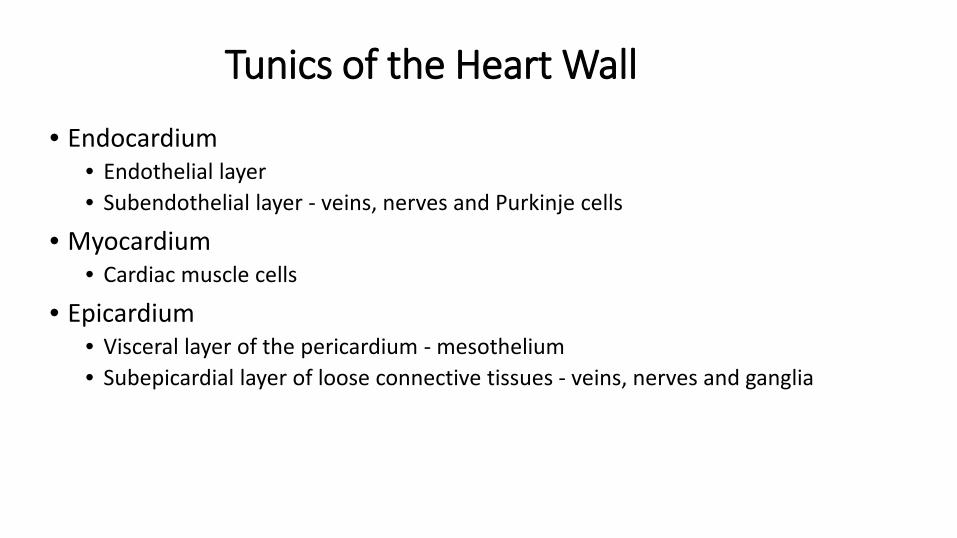
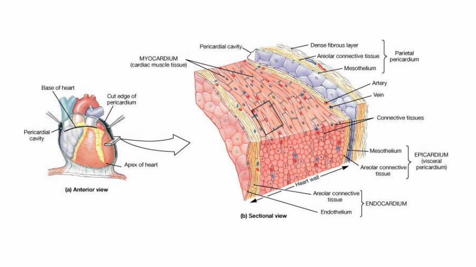
Tunics of the Heart Wall
• Endocardium • Endothelial layer• Subendothelial layer - veins, nerves and Purkinje cells
• Myocardium • Cardiac muscle cells
• Epicardium • Visceral layer of the pericardium - mesothelium• Subepicardial layer of loose connective tissues - veins, nerves and ganglia
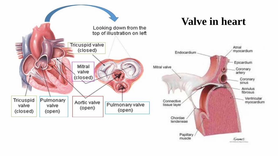
Valve in heart
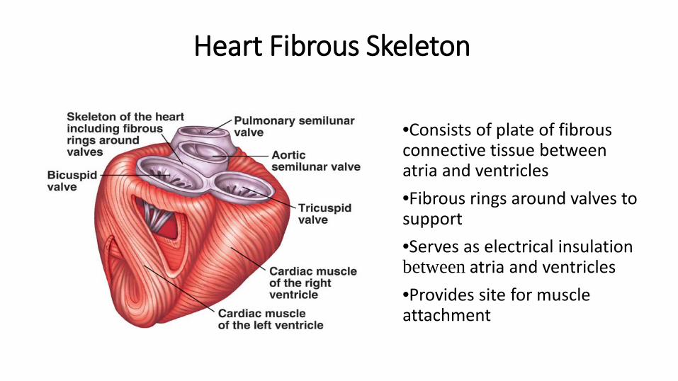
Heart Fibrous Skeleton
•Consists of plate of fibrous connective tissue between atria and ventricles•Fibrous rings around valves to support•Serves as electrical insulation between atria and ventricles•Provides site for muscle attachment
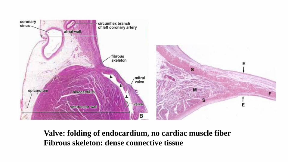
Valve: folding of endocardium, no cardiac muscle fiberFibrous skeleton: dense connective tissue
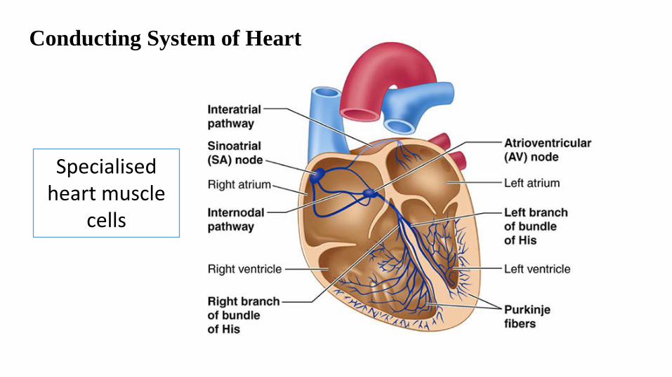
Conducting System of Heart
Specialisedheart muscle
cells
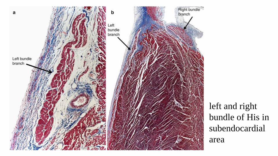
left and right bundle of His in subendocardial area
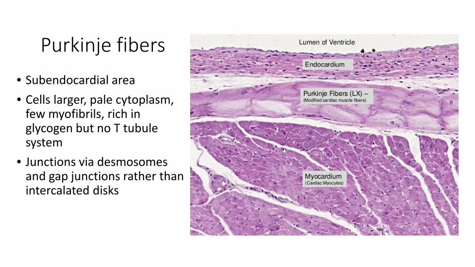
Purkinje fibers• Subendocardial area• Cells larger, pale cytoplasm,
few myofibrils, rich in glycogen but no T tubule system
• Junctions via desmosomes and gap junctions rather than intercalated disks
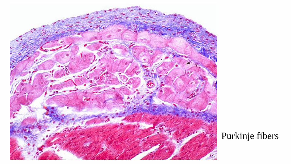
Purkinje fibers