Embed Size (px)
Citation preview

Histology of theHistology of the
Female Genital SystemFemale Genital System
Histology of theHistology of the
Female Genital SystemFemale Genital System

The Female Genital SystemThe Female Genital SystemThe Female Genital SystemThe Female Genital SystemThe female genital system consists of:The female genital system consists of:
1.1. Primary sex organPrimary sex organ: two ovaries.: two ovaries.
2.2. Accessory sex organsAccessory sex organs::
1.1. two oviducts (Fallopian tubes).two oviducts (Fallopian tubes).
2.2. a uterus.a uterus.
3.3. a vagina.a vagina.
4.4. external genitalia.external genitalia.
5.5. two mammary glands.two mammary glands.
The female genital system consists of:The female genital system consists of:
1.1. Primary sex organPrimary sex organ: two ovaries.: two ovaries.
2.2. Accessory sex organsAccessory sex organs::
1.1. two oviducts (Fallopian tubes).two oviducts (Fallopian tubes).
2.2. a uterus.a uterus.
3.3. a vagina.a vagina.
4.4. external genitalia.external genitalia.
5.5. two mammary glands.two mammary glands.

THE OVARIESTHE OVARIESTHE OVARIESTHE OVARIES

The OvariesThe OvariesThe OvariesThe OvariesThe ovary is a flattened almond-shaped The ovary is a flattened almond-shaped small body, divided into peripheral small body, divided into peripheral cortexcortex and central and central medullamedulla..The cortexThe cortex is broad and contains the is broad and contains the ovarian follicles separated by the inter-ovarian follicles separated by the inter-follicular tissue.follicular tissue.The medullaThe medulla consists of highly vascular consists of highly vascular connective tissue, having elastic fibers, connective tissue, having elastic fibers, smooth muscle fibers, lymphatics and smooth muscle fibers, lymphatics and nerves.nerves.
The ovary is a flattened almond-shaped The ovary is a flattened almond-shaped small body, divided into peripheral small body, divided into peripheral cortexcortex and central and central medullamedulla..The cortexThe cortex is broad and contains the is broad and contains the ovarian follicles separated by the inter-ovarian follicles separated by the inter-follicular tissue.follicular tissue.The medullaThe medulla consists of highly vascular consists of highly vascular connective tissue, having elastic fibers, connective tissue, having elastic fibers, smooth muscle fibers, lymphatics and smooth muscle fibers, lymphatics and nerves.nerves.

Stroma of The OvariesStroma of The OvariesStroma of The OvariesStroma of The OvariesTunica albugeniaTunica albugenia is the covering is the covering connective tissue capsule of the ovary. It connective tissue capsule of the ovary. It is formed of white collagenous is formed of white collagenous connective tissue fibers.connective tissue fibers.
Stromal cellsStromal cells: are fusiform cells with oval : are fusiform cells with oval nuclei similar to fibroblasts. They are nuclei similar to fibroblasts. They are present between the ovarian follicles.present between the ovarian follicles.
Reticular connective tissueReticular connective tissue: present in-: present in-between the ovarian follicles.between the ovarian follicles.
Tunica albugeniaTunica albugenia is the covering is the covering connective tissue capsule of the ovary. It connective tissue capsule of the ovary. It is formed of white collagenous is formed of white collagenous connective tissue fibers.connective tissue fibers.
Stromal cellsStromal cells: are fusiform cells with oval : are fusiform cells with oval nuclei similar to fibroblasts. They are nuclei similar to fibroblasts. They are present between the ovarian follicles.present between the ovarian follicles.
Reticular connective tissueReticular connective tissue: present in-: present in-between the ovarian follicles.between the ovarian follicles.

Parenchyma of The OvariesParenchyma of The OvariesParenchyma of The OvariesParenchyma of The Ovaries1.1. The ovarian folliclesThe ovarian follicles. In different stages . In different stages
of development and degeneration.of development and degeneration.2.2. The endocrine cellsThe endocrine cells: polygonal in shape, : polygonal in shape,
with central rounded nuclei. Their with central rounded nuclei. Their cytoplasm is rich in lipoid granules. In cytoplasm is rich in lipoid granules. In animals, they secrete female hormones, animals, they secrete female hormones, but not in humans.but not in humans.The germinal epitheliumThe germinal epithelium: it covers the : it covers the ovary from outside. It is simple cuboidal ovary from outside. It is simple cuboidal in young females and simple squamous in young females and simple squamous in adults.in adults.
1.1. The ovarian folliclesThe ovarian follicles. In different stages . In different stages of development and degeneration.of development and degeneration.
2.2. The endocrine cellsThe endocrine cells: polygonal in shape, : polygonal in shape, with central rounded nuclei. Their with central rounded nuclei. Their cytoplasm is rich in lipoid granules. In cytoplasm is rich in lipoid granules. In animals, they secrete female hormones, animals, they secrete female hormones, but not in humans.but not in humans.The germinal epitheliumThe germinal epithelium: it covers the : it covers the ovary from outside. It is simple cuboidal ovary from outside. It is simple cuboidal in young females and simple squamous in young females and simple squamous in adults.in adults.

The Ovarian FolliclesThe Ovarian FolliclesThe Ovarian FolliclesThe Ovarian FolliclesThey are present mainly in the cortex of the They are present mainly in the cortex of the ovary under the tunica albuginea. They are:ovary under the tunica albuginea. They are:
1.1. Primordial follicles.Primordial follicles.2.2. Primary follicles.Primary follicles.3.3. Secondary follicles.Secondary follicles.4.4. Mature follicles.Mature follicles.
At birth, the average number of 1ry follicles is At birth, the average number of 1ry follicles is 4000, only 400 ova are produced during the 4000, only 400 ova are produced during the reproductive period of the adult female. The reproductive period of the adult female. The remaining follicles degenerate and change to remaining follicles degenerate and change to atretic follicles which are converted to white atretic follicles which are converted to white connective tissue bodies.connective tissue bodies.
They are present mainly in the cortex of the They are present mainly in the cortex of the ovary under the tunica albuginea. They are:ovary under the tunica albuginea. They are:
1.1. Primordial follicles.Primordial follicles.2.2. Primary follicles.Primary follicles.3.3. Secondary follicles.Secondary follicles.4.4. Mature follicles.Mature follicles.
At birth, the average number of 1ry follicles is At birth, the average number of 1ry follicles is 4000, only 400 ova are produced during the 4000, only 400 ova are produced during the reproductive period of the adult female. The reproductive period of the adult female. The remaining follicles degenerate and change to remaining follicles degenerate and change to atretic follicles which are converted to white atretic follicles which are converted to white connective tissue bodies.connective tissue bodies.

(1) Primordial Follicles(1) Primordial Follicles(1) Primordial Follicles(1) Primordial FolliclesThey are derived from the primordial germ They are derived from the primordial germ cells in the yolk sac and then migrate to the cells in the yolk sac and then migrate to the developing ovary.developing ovary.They are formed of They are formed of central primary oocytescentral primary oocytes which are which are surrounded bysurrounded by a single layer of a single layer of simple squamous cells called simple squamous cells called follicular follicular cellscells..The oocyteThe oocyte is a large cell with large is a large cell with large eccentric vesicular nucleus with a large eccentric vesicular nucleus with a large nucleolus. Its cytoplasm contains a well-nucleolus. Its cytoplasm contains a well-developed Golgi apparatus, RER, developed Golgi apparatus, RER, mitochondria and lipid droplets.mitochondria and lipid droplets.
They are derived from the primordial germ They are derived from the primordial germ cells in the yolk sac and then migrate to the cells in the yolk sac and then migrate to the developing ovary.developing ovary.They are formed of They are formed of central primary oocytescentral primary oocytes which are which are surrounded bysurrounded by a single layer of a single layer of simple squamous cells called simple squamous cells called follicular follicular cellscells..The oocyteThe oocyte is a large cell with large is a large cell with large eccentric vesicular nucleus with a large eccentric vesicular nucleus with a large nucleolus. Its cytoplasm contains a well-nucleolus. Its cytoplasm contains a well-developed Golgi apparatus, RER, developed Golgi apparatus, RER, mitochondria and lipid droplets.mitochondria and lipid droplets.

(2) Primary Follicle(2) Primary Follicle(2) Primary Follicle(2) Primary FollicleThe oocyte enlarges and develops Golgi The oocyte enlarges and develops Golgi apparatus, ribosomes and mitochondria.apparatus, ribosomes and mitochondria.It becomes surrounded by a thick highly It becomes surrounded by a thick highly acidophilic glycoprotein coat, called acidophilic glycoprotein coat, called zona zona pellucidapellucida..The flat follicular cells becomes cuboidal The flat follicular cells becomes cuboidal and multiply to give rise several layers and multiply to give rise several layers (stratified) called (stratified) called granulosa cellsgranulosa cells..The connective tissue surrounding the The connective tissue surrounding the follicle condenses and forms two layers; follicle condenses and forms two layers; inner highly vascular secretory layer, inner highly vascular secretory layer, theca theca internainterna and outer fibrous layer, and outer fibrous layer, theca theca externaexterna..
The oocyte enlarges and develops Golgi The oocyte enlarges and develops Golgi apparatus, ribosomes and mitochondria.apparatus, ribosomes and mitochondria.It becomes surrounded by a thick highly It becomes surrounded by a thick highly acidophilic glycoprotein coat, called acidophilic glycoprotein coat, called zona zona pellucidapellucida..The flat follicular cells becomes cuboidal The flat follicular cells becomes cuboidal and multiply to give rise several layers and multiply to give rise several layers (stratified) called (stratified) called granulosa cellsgranulosa cells..The connective tissue surrounding the The connective tissue surrounding the follicle condenses and forms two layers; follicle condenses and forms two layers; inner highly vascular secretory layer, inner highly vascular secretory layer, theca theca internainterna and outer fibrous layer, and outer fibrous layer, theca theca externaexterna..

(3) Secondary Follicle(3) Secondary Follicle(3) Secondary Follicle(3) Secondary FollicleThe granulosa cells reach 6-12 layers and The granulosa cells reach 6-12 layers and start to secrete fluid which forms irregular start to secrete fluid which forms irregular spaces between the granulosa cells.spaces between the granulosa cells.The spaces gradually fuse to form a The spaces gradually fuse to form a crescentic space called crescentic space called antrumantrum which which contains contains liquor folliculiliquor folliculi..The liquor folliculi contains growth factors, The liquor folliculi contains growth factors, steroid and gonadotrophic hormones.steroid and gonadotrophic hormones.The oocyte is eccentric and is surrounded by The oocyte is eccentric and is surrounded by a mass of granulosa cells called a mass of granulosa cells called cumulus cumulus oophorusoophorus..The oocyte starts its The oocyte starts its first meiotic divisionfirst meiotic division and remains in the prophase until ovulation.and remains in the prophase until ovulation.
The granulosa cells reach 6-12 layers and The granulosa cells reach 6-12 layers and start to secrete fluid which forms irregular start to secrete fluid which forms irregular spaces between the granulosa cells.spaces between the granulosa cells.The spaces gradually fuse to form a The spaces gradually fuse to form a crescentic space called crescentic space called antrumantrum which which contains contains liquor folliculiliquor folliculi..The liquor folliculi contains growth factors, The liquor folliculi contains growth factors, steroid and gonadotrophic hormones.steroid and gonadotrophic hormones.The oocyte is eccentric and is surrounded by The oocyte is eccentric and is surrounded by a mass of granulosa cells called a mass of granulosa cells called cumulus cumulus oophorusoophorus..The oocyte starts its The oocyte starts its first meiotic divisionfirst meiotic division and remains in the prophase until ovulation.and remains in the prophase until ovulation.

(4) Mature Graafian Follicle(4) Mature Graafian Follicle(4) Mature Graafian Follicle(4) Mature Graafian FollicleThe primordial follicle reaches maturity in The primordial follicle reaches maturity in 10-14 days and occupies the whole 10-14 days and occupies the whole thickness of the cortex and bulges out on thickness of the cortex and bulges out on the free surface of the ovary.the free surface of the ovary.The liquor folliculi accumulates between The liquor folliculi accumulates between the cells of cumulus oophorus freeing the the cells of cumulus oophorus freeing the oocyte from the cells except for one layer oocyte from the cells except for one layer called corona radiata cells.called corona radiata cells.The granulosa cells secrete estrogen.The granulosa cells secrete estrogen.The ovum is the largest cell in the body. The ovum is the largest cell in the body. It has a large, rounded eccentric nucleusIt has a large, rounded eccentric nucleus
The primordial follicle reaches maturity in The primordial follicle reaches maturity in 10-14 days and occupies the whole 10-14 days and occupies the whole thickness of the cortex and bulges out on thickness of the cortex and bulges out on the free surface of the ovary.the free surface of the ovary.The liquor folliculi accumulates between The liquor folliculi accumulates between the cells of cumulus oophorus freeing the the cells of cumulus oophorus freeing the oocyte from the cells except for one layer oocyte from the cells except for one layer called corona radiata cells.called corona radiata cells.The granulosa cells secrete estrogen.The granulosa cells secrete estrogen.The ovum is the largest cell in the body. The ovum is the largest cell in the body. It has a large, rounded eccentric nucleusIt has a large, rounded eccentric nucleus
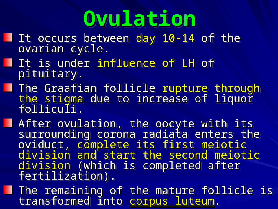
OvulationOvulationOvulationOvulationIt occurs between It occurs between day 10-14day 10-14 of the ovarian of the ovarian cycle.cycle.It is under It is under influence of LHinfluence of LH of pituitary. of pituitary.The Graafian follicle The Graafian follicle rupture through the rupture through the stigmastigma due to increase of liquor folliculi. due to increase of liquor folliculi.After ovulation, the oocyte with its After ovulation, the oocyte with its surrounding corona radiata enters the surrounding corona radiata enters the oviduct, oviduct, complete its first meiotic division complete its first meiotic division and start the second meiotic divisionand start the second meiotic division (which (which is completed after fertilization).is completed after fertilization).The remaining of the mature follicle is The remaining of the mature follicle is transformed into transformed into corpus luteumcorpus luteum..
It occurs between It occurs between day 10-14day 10-14 of the ovarian of the ovarian cycle.cycle.It is under It is under influence of LHinfluence of LH of pituitary. of pituitary.The Graafian follicle The Graafian follicle rupture through the rupture through the stigmastigma due to increase of liquor folliculi. due to increase of liquor folliculi.After ovulation, the oocyte with its After ovulation, the oocyte with its surrounding corona radiata enters the surrounding corona radiata enters the oviduct, oviduct, complete its first meiotic division complete its first meiotic division and start the second meiotic divisionand start the second meiotic division (which (which is completed after fertilization).is completed after fertilization).The remaining of the mature follicle is The remaining of the mature follicle is transformed into transformed into corpus luteumcorpus luteum..

Corpus LuteumCorpus LuteumCorpus LuteumCorpus LuteumIt is considered as a It is considered as a temporary endocrinetemporary endocrine organ.organ.Basement membrane between theca Basement membrane between theca interna and granulosa cells dissolves, and interna and granulosa cells dissolves, and capillaries grow in-between granulosa cells.capillaries grow in-between granulosa cells.After ovulation, granulosa cells enlarge and After ovulation, granulosa cells enlarge and accumulate lipid droplets and called accumulate lipid droplets and called granulosa lutein cellsgranulosa lutein cells..Under influence of LH, the granulosa cell Under influence of LH, the granulosa cell secrete progesterone and estrogensecrete progesterone and estrogen..The same also happens to theca interna The same also happens to theca interna cells which are now called cells which are now called theca lutein cellstheca lutein cells
It is considered as a It is considered as a temporary endocrinetemporary endocrine organ.organ.Basement membrane between theca Basement membrane between theca interna and granulosa cells dissolves, and interna and granulosa cells dissolves, and capillaries grow in-between granulosa cells.capillaries grow in-between granulosa cells.After ovulation, granulosa cells enlarge and After ovulation, granulosa cells enlarge and accumulate lipid droplets and called accumulate lipid droplets and called granulosa lutein cellsgranulosa lutein cells..Under influence of LH, the granulosa cell Under influence of LH, the granulosa cell secrete progesterone and estrogensecrete progesterone and estrogen..The same also happens to theca interna The same also happens to theca interna cells which are now called cells which are now called theca lutein cellstheca lutein cells

Fate of Corpus LuteumFate of Corpus LuteumFate of Corpus LuteumFate of Corpus LuteumIf pregnancy occursIf pregnancy occurs, it enlarges , it enlarges and continues to function for 3 and continues to function for 3 moths till the placenta is formed moths till the placenta is formed and take the gob.and take the gob.
If pregnancy does not occurIf pregnancy does not occur, it , it degenerates and is transformed degenerates and is transformed into fibrous tissue called into fibrous tissue called corpus corpus albicansalbicans..
If pregnancy occursIf pregnancy occurs, it enlarges , it enlarges and continues to function for 3 and continues to function for 3 moths till the placenta is formed moths till the placenta is formed and take the gob.and take the gob.
If pregnancy does not occurIf pregnancy does not occur, it , it degenerates and is transformed degenerates and is transformed into fibrous tissue called into fibrous tissue called corpus corpus albicansalbicans..

THE THE
OVIDUCTOVIDUCT
THE THE
OVIDUCTOVIDUCT

The OviductThe OviductThe OviductThe OviductIt extends from the ovary to the uterus.It extends from the ovary to the uterus.It is divided into 4 segments:It is divided into 4 segments:
1.1. InfundibulumInfundibulum: funnel-shaped : funnel-shaped opening, having finger-like processes opening, having finger-like processes (fimbriae).(fimbriae).
2.2. AmpullaAmpulla: the widest part, where : the widest part, where fertilization usually occurs.fertilization usually occurs.
3.3. IsthmusIsthmus: narrow part near to uterus.: narrow part near to uterus.4.4. Intramural partIntramural part: traverses the uterine : traverses the uterine
wallwall
It extends from the ovary to the uterus.It extends from the ovary to the uterus.It is divided into 4 segments:It is divided into 4 segments:
1.1. InfundibulumInfundibulum: funnel-shaped : funnel-shaped opening, having finger-like processes opening, having finger-like processes (fimbriae).(fimbriae).
2.2. AmpullaAmpulla: the widest part, where : the widest part, where fertilization usually occurs.fertilization usually occurs.
3.3. IsthmusIsthmus: narrow part near to uterus.: narrow part near to uterus.4.4. Intramural partIntramural part: traverses the uterine : traverses the uterine
wallwall

Histology of the OviductHistology of the OviductHistology of the OviductHistology of the Oviduct1.1. MucosaMucosa: highly folded and is formed of: highly folded and is formed of
A.A. EpitheliumEpithelium: simple columnar, partly : simple columnar, partly ciliated (to moved the ovum toward the ciliated (to moved the ovum toward the uterus) and partly non-ciliated uterus) and partly non-ciliated (secretory peg cells, nutritive to the (secretory peg cells, nutritive to the ovum).ovum).
B.B. Lamina propriaLamina propria: connective tissue rich : connective tissue rich in blood vessels.in blood vessels.
2.2. MusculosaMusculosa: inner circular and outer : inner circular and outer longitudinal smooth muscle fibers.longitudinal smooth muscle fibers.
3.3. SerosaSerosa: areolar connective tissue : areolar connective tissue covered by simple squamous covered by simple squamous mesothelium.mesothelium.
1.1. MucosaMucosa: highly folded and is formed of: highly folded and is formed ofA.A. EpitheliumEpithelium: simple columnar, partly : simple columnar, partly
ciliated (to moved the ovum toward the ciliated (to moved the ovum toward the uterus) and partly non-ciliated uterus) and partly non-ciliated (secretory peg cells, nutritive to the (secretory peg cells, nutritive to the ovum).ovum).
B.B. Lamina propriaLamina propria: connective tissue rich : connective tissue rich in blood vessels.in blood vessels.
2.2. MusculosaMusculosa: inner circular and outer : inner circular and outer longitudinal smooth muscle fibers.longitudinal smooth muscle fibers.
3.3. SerosaSerosa: areolar connective tissue : areolar connective tissue covered by simple squamous covered by simple squamous mesothelium.mesothelium.

THE UTERUSTHE UTERUSTHE UTERUSTHE UTERUS

The UterusThe UterusThe UterusThe UterusIt is a thick-walled pear-shaped It is a thick-walled pear-shaped organ which has a narrow lumen.organ which has a narrow lumen.It is formed of body and cervix.It is formed of body and cervix.The body is formed of three layers:The body is formed of three layers:1.1. EndometriumEndometrium..2.2. MyometriumMyometrium..3.3. PerimetriumPerimetrium..
It is a thick-walled pear-shaped It is a thick-walled pear-shaped organ which has a narrow lumen.organ which has a narrow lumen.It is formed of body and cervix.It is formed of body and cervix.The body is formed of three layers:The body is formed of three layers:1.1. EndometriumEndometrium..2.2. MyometriumMyometrium..3.3. PerimetriumPerimetrium..

The EndometriumThe EndometriumThe EndometriumThe EndometriumIt is lined by It is lined by simple columnar simple columnar epitheliumepithelium..Its lamina propria contains Its lamina propria contains simple simple tubular mucous glandstubular mucous glands lined by lined by columnar epithelium and may reach to columnar epithelium and may reach to the myometrium.the myometrium.It undergoes It undergoes cyclic changescyclic changes in in response to ovarian hormones.response to ovarian hormones.It is divided into superficial layer It is divided into superficial layer ((stratum functionalisstratum functionalis) and deep layer ) and deep layer ((stratum basalisstratum basalis).).
It is lined by It is lined by simple columnar simple columnar epitheliumepithelium..Its lamina propria contains Its lamina propria contains simple simple tubular mucous glandstubular mucous glands lined by lined by columnar epithelium and may reach to columnar epithelium and may reach to the myometrium.the myometrium.It undergoes It undergoes cyclic changescyclic changes in in response to ovarian hormones.response to ovarian hormones.It is divided into superficial layer It is divided into superficial layer ((stratum functionalisstratum functionalis) and deep layer ) and deep layer ((stratum basalisstratum basalis).).

The MyometriumThe MyometriumThe MyometriumThe MyometriumThree layers of smooth muscle Three layers of smooth muscle fibers and connective tissue.fibers and connective tissue.They are not easily distinguished They are not easily distinguished from each other.from each other.The middle layer is called stratum The middle layer is called stratum vascularis and contains numerous vascularis and contains numerous large blood vessels.large blood vessels.During pregnancy, the smooth During pregnancy, the smooth muscle fibers increase in length.muscle fibers increase in length.
Three layers of smooth muscle Three layers of smooth muscle fibers and connective tissue.fibers and connective tissue.They are not easily distinguished They are not easily distinguished from each other.from each other.The middle layer is called stratum The middle layer is called stratum vascularis and contains numerous vascularis and contains numerous large blood vessels.large blood vessels.During pregnancy, the smooth During pregnancy, the smooth muscle fibers increase in length.muscle fibers increase in length.

The perimetriumThe perimetriumThe perimetriumThe perimetriumIt is formed of areolar It is formed of areolar connective tissue, blood connective tissue, blood vessels and covered by vessels and covered by simple squamous simple squamous mesothelium.mesothelium.
It is formed of areolar It is formed of areolar connective tissue, blood connective tissue, blood vessels and covered by vessels and covered by simple squamous simple squamous mesothelium.mesothelium.

Cyclic changes of the endometriumCyclic changes of the endometriumCyclic changes of the endometriumCyclic changes of the endometrium
1.1.Menstrual stage (from day 1 Menstrual stage (from day 1 to day 5).to day 5).
2.2.Proliferative phase (from day Proliferative phase (from day 6 to day 16).6 to day 16).
3.3.Secretory phase (from day Secretory phase (from day 17 to day 26).17 to day 26).
4.4.Premenstrual stage (from Premenstrual stage (from day 27 to day 28).day 27 to day 28).

(1) Menstrual Stage(1) Menstrual Stage(1) Menstrual Stage(1) Menstrual StageDue to hormonal deficiency specially Due to hormonal deficiency specially progesterone.progesterone.Constriction of coiled arteries for long Constriction of coiled arteries for long periods causes ischemia and rupture of periods causes ischemia and rupture of capillaries.capillaries.
Glands fragment and uterine fluid, Glands fragment and uterine fluid, tissue debris and blood are tissue debris and blood are sloughed out and discharged sloughed out and discharged through vagina.through vagina.Stratum functionalis is lostStratum functionalis is lost
Due to hormonal deficiency specially Due to hormonal deficiency specially progesterone.progesterone.Constriction of coiled arteries for long Constriction of coiled arteries for long periods causes ischemia and rupture of periods causes ischemia and rupture of capillaries.capillaries.
Glands fragment and uterine fluid, Glands fragment and uterine fluid, tissue debris and blood are tissue debris and blood are sloughed out and discharged sloughed out and discharged through vagina.through vagina.Stratum functionalis is lostStratum functionalis is lost

(2) Proliferative Stage(2) Proliferative Stage(2) Proliferative Stage(2) Proliferative StageOccurs during maturation of follicles till Occurs during maturation of follicles till ovulation.ovulation.Under the effect of estrogen secreted by the Under the effect of estrogen secreted by the follicles.follicles.It is the stage of regeneration of the stratum It is the stage of regeneration of the stratum functionalis from stratum basalis.functionalis from stratum basalis.Epithelium of basal glands recovers the raw Epithelium of basal glands recovers the raw surface.surface.Glands increase in length, become straight Glands increase in length, become straight and uniform in diameter.and uniform in diameter.Endometrium increases in thickness from 0.5 Endometrium increases in thickness from 0.5 mm to 2-3 mm.mm to 2-3 mm.
Occurs during maturation of follicles till Occurs during maturation of follicles till ovulation.ovulation.Under the effect of estrogen secreted by the Under the effect of estrogen secreted by the follicles.follicles.It is the stage of regeneration of the stratum It is the stage of regeneration of the stratum functionalis from stratum basalis.functionalis from stratum basalis.Epithelium of basal glands recovers the raw Epithelium of basal glands recovers the raw surface.surface.Glands increase in length, become straight Glands increase in length, become straight and uniform in diameter.and uniform in diameter.Endometrium increases in thickness from 0.5 Endometrium increases in thickness from 0.5 mm to 2-3 mm.mm to 2-3 mm.

(3) Secretory Stage(3) Secretory Stage(3) Secretory Stage(3) Secretory StageRelated to the formation of corpus Related to the formation of corpus luteum.luteum.Under the effect of progesterone and Under the effect of progesterone and estrogen secreted by the luteal cells.estrogen secreted by the luteal cells.Endometrium becomes hypertrophied Endometrium becomes hypertrophied and vascular to reaches its full thickness and vascular to reaches its full thickness (4-5 mm).(4-5 mm).Endometrial glands become coiled (cork-Endometrial glands become coiled (cork-screw).screw).The glandular lumen contains secretions The glandular lumen contains secretions and rich in glycogen.and rich in glycogen.
Related to the formation of corpus Related to the formation of corpus luteum.luteum.Under the effect of progesterone and Under the effect of progesterone and estrogen secreted by the luteal cells.estrogen secreted by the luteal cells.Endometrium becomes hypertrophied Endometrium becomes hypertrophied and vascular to reaches its full thickness and vascular to reaches its full thickness (4-5 mm).(4-5 mm).Endometrial glands become coiled (cork-Endometrial glands become coiled (cork-screw).screw).The glandular lumen contains secretions The glandular lumen contains secretions and rich in glycogen.and rich in glycogen.

(4) Premenstrual Stage(4) Premenstrual Stage(4) Premenstrual Stage(4) Premenstrual StageRelated to the involution of the corpus Related to the involution of the corpus luteum and formation of corpus albicans.luteum and formation of corpus albicans.Spiral arteries undergo periodic constriction Spiral arteries undergo periodic constriction leading to stasis in capillaries and periods leading to stasis in capillaries and periods of ischemia.of ischemia.Glands stop secretion leading to shrinkage Glands stop secretion leading to shrinkage of stratum functionalis due to water loss.of stratum functionalis due to water loss.The thickness of the endometrium The thickness of the endometrium decreases.decreases.Stratum functionalis appears deeply Stratum functionalis appears deeply stained because of the closely packed stained because of the closely packed stromal cells.stromal cells.
Related to the involution of the corpus Related to the involution of the corpus luteum and formation of corpus albicans.luteum and formation of corpus albicans.Spiral arteries undergo periodic constriction Spiral arteries undergo periodic constriction leading to stasis in capillaries and periods leading to stasis in capillaries and periods of ischemia.of ischemia.Glands stop secretion leading to shrinkage Glands stop secretion leading to shrinkage of stratum functionalis due to water loss.of stratum functionalis due to water loss.The thickness of the endometrium The thickness of the endometrium decreases.decreases.Stratum functionalis appears deeply Stratum functionalis appears deeply stained because of the closely packed stained because of the closely packed stromal cells.stromal cells.

The CervixThe CervixThe CervixThe CervixIt consists of:It consists of:
1.1. EndometriumEndometrium::– Epithelium: simple columnar mucous-Epithelium: simple columnar mucous-
secreting epithelium.secreting epithelium.– Lamina propria: contains branched tubular Lamina propria: contains branched tubular
glands secreting mucous.glands secreting mucous.– The cervical endometrium does not change The cervical endometrium does not change
during the menstrual cycle, only the amount during the menstrual cycle, only the amount and consistency of the mucous chnge.and consistency of the mucous chnge.
2.2. MyometriumMyometrium: dense connective tissue and : dense connective tissue and few amount of smooth muscle fibers.few amount of smooth muscle fibers.
3.3. AdventitiaAdventitia: connective tissue (no peritoneum): connective tissue (no peritoneum)
It consists of:It consists of:1.1. EndometriumEndometrium::
– Epithelium: simple columnar mucous-Epithelium: simple columnar mucous-secreting epithelium.secreting epithelium.
– Lamina propria: contains branched tubular Lamina propria: contains branched tubular glands secreting mucous.glands secreting mucous.
– The cervical endometrium does not change The cervical endometrium does not change during the menstrual cycle, only the amount during the menstrual cycle, only the amount and consistency of the mucous chnge.and consistency of the mucous chnge.
2.2. MyometriumMyometrium: dense connective tissue and : dense connective tissue and few amount of smooth muscle fibers.few amount of smooth muscle fibers.
3.3. AdventitiaAdventitia: connective tissue (no peritoneum): connective tissue (no peritoneum)

THE VAGINATHE VAGINATHE VAGINATHE VAGINA

The VaginaThe VaginaThe VaginaThe VaginaIt consists of:It consists of:
1.1. MucosaMucosa::– Epithelium: stratified squamous non-Epithelium: stratified squamous non-
keratinized epithelium rich in keratinized epithelium rich in glycogen.glycogen.
– Lamina propria: contains lymphatic Lamina propria: contains lymphatic nodules but no glands.nodules but no glands.
2.2. MusculosaMusculosa: longitudinal muscle fibers : longitudinal muscle fibers continuous with uterine muscles.continuous with uterine muscles.
3.3. AdventitiaAdventitia: dense connective tissue.: dense connective tissue.
It consists of:It consists of:1.1. MucosaMucosa::
– Epithelium: stratified squamous non-Epithelium: stratified squamous non-keratinized epithelium rich in keratinized epithelium rich in glycogen.glycogen.
– Lamina propria: contains lymphatic Lamina propria: contains lymphatic nodules but no glands.nodules but no glands.
2.2. MusculosaMusculosa: longitudinal muscle fibers : longitudinal muscle fibers continuous with uterine muscles.continuous with uterine muscles.
3.3. AdventitiaAdventitia: dense connective tissue.: dense connective tissue.

MAMMARY MAMMARY
GLANDSGLANDS
MAMMARY MAMMARY
GLANDSGLANDS

Mammary GlandsMammary GlandsMammary GlandsMammary GlandsIt consists of lobes and lobules, It consists of lobes and lobules, separated by septa formed of separated by septa formed of dense connective tissue and dense connective tissue and elastic fibers.elastic fibers.Fat tissue runs between lobes and Fat tissue runs between lobes and lobules.lobules.Non-lactating gland (inactive): the Non-lactating gland (inactive): the gland parenchyma is formed of gland parenchyma is formed of little amount of lactiferous ducts little amount of lactiferous ducts only, with no alveoli.only, with no alveoli.
It consists of lobes and lobules, It consists of lobes and lobules, separated by septa formed of separated by septa formed of dense connective tissue and dense connective tissue and elastic fibers.elastic fibers.Fat tissue runs between lobes and Fat tissue runs between lobes and lobules.lobules.Non-lactating gland (inactive): the Non-lactating gland (inactive): the gland parenchyma is formed of gland parenchyma is formed of little amount of lactiferous ducts little amount of lactiferous ducts only, with no alveoli.only, with no alveoli.

Lactating Mammary GlandsLactating Mammary GlandsLactating Mammary GlandsLactating Mammary GlandsStroma: connective tissue septa and adipose Stroma: connective tissue septa and adipose tissue are reduced.tissue are reduced.Parenchyma: extensive system of secretory Parenchyma: extensive system of secretory alveoli budding from the ducts.alveoli budding from the ducts.The alveoli are lined with simple cuboidal cells The alveoli are lined with simple cuboidal cells with basal myoepithelial cells.with basal myoepithelial cells.The intralobar ducts are lined with simple The intralobar ducts are lined with simple cuboidal epithelium.cuboidal epithelium.The main ducts are lined with stratified The main ducts are lined with stratified squamous epithelium.squamous epithelium.Mammary gland is an Mammary gland is an apocrineapocrine gland. gland.ProlactinProlactin stimulate secretion of milk. stimulate secretion of milk.OxytocinOxytocin stimulate ejection of milk. stimulate ejection of milk.
Stroma: connective tissue septa and adipose Stroma: connective tissue septa and adipose tissue are reduced.tissue are reduced.Parenchyma: extensive system of secretory Parenchyma: extensive system of secretory alveoli budding from the ducts.alveoli budding from the ducts.The alveoli are lined with simple cuboidal cells The alveoli are lined with simple cuboidal cells with basal myoepithelial cells.with basal myoepithelial cells.The intralobar ducts are lined with simple The intralobar ducts are lined with simple cuboidal epithelium.cuboidal epithelium.The main ducts are lined with stratified The main ducts are lined with stratified squamous epithelium.squamous epithelium.Mammary gland is an Mammary gland is an apocrineapocrine gland. gland.ProlactinProlactin stimulate secretion of milk. stimulate secretion of milk.OxytocinOxytocin stimulate ejection of milk. stimulate ejection of milk.