Embed Size (px)
Citation preview
Histology Pre-lab. # 10
Intestines – Jejunum, Ileum, Colon & Appendix
By Prof. Dr. Ansari
( MBBS semester I ) Wednesday, April 19, 2023 1
Objectives of the lab.
• To identify the microscopic structure of Jejunum, Ileum, colon & appendix.
• To differentiate the small intestine from the large intestine microscopically.
• To learn the structure of a Villus.• To learn the GALT.
2
Jejunum is part of small intestine next to duodenum
• Microscopy of jejunum is similar to any part of GIT.
• It shows tunica mucosa having simple columnar epithelium, Plica circularis, Villi and goblet cells.
• The tunica sub mucosa having blood vessel & nerve endings.
• Tunica Muscularis externa having circular and longitudinal smooth muscles.
3
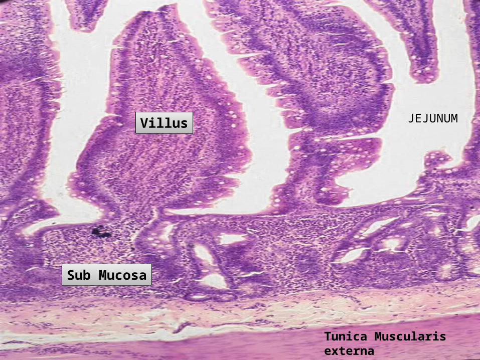
4Tunica Muscularisexterna
VillusVillus
Sub MucosaSub Mucosa
JEJUNUM
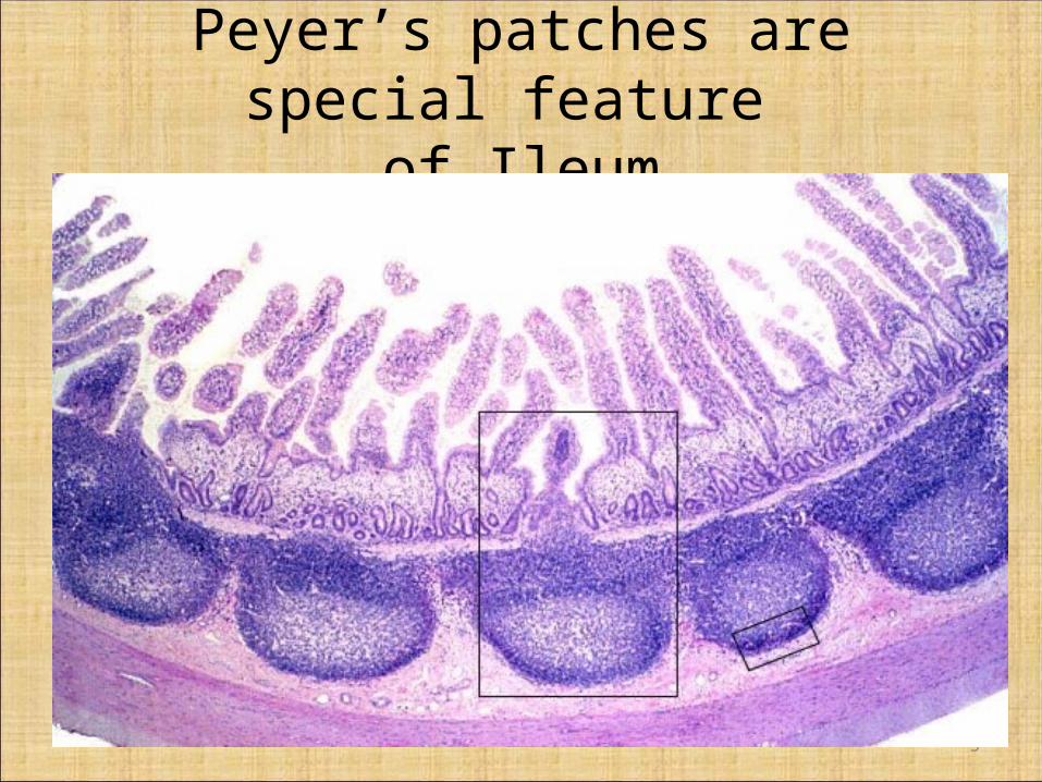
Peyer’s patches are special feature of Ileum
5
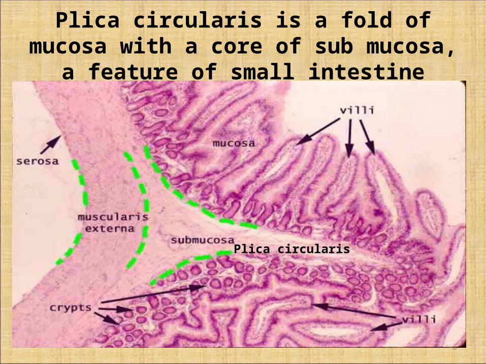
Plica circularis is a fold of mucosa with a core of sub mucosa, a feature of small intestine
6
Plica circularis
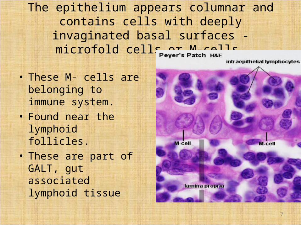
The epithelium appears columnar and contains cells with deeply invaginated basal surfaces - microfold cells
or M-cells.
• These M- cells are belonging to immune system.
• Found near the lymphoid follicles.
• These are part of GALT, gut associated lymphoid tissue
7
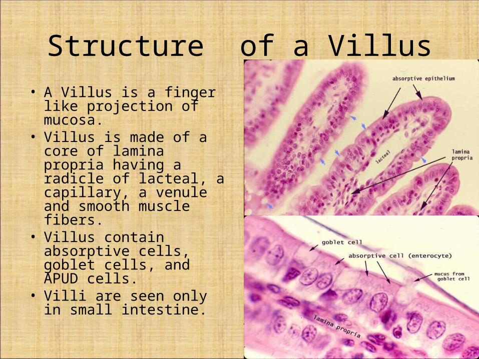
Structure of a Villus• A Villus is a finger like
projection of mucosa.• Villus is made of a core of
lamina propria having a radicle of lacteal, a capillary, a venule and smooth muscle fibers.
• Villus contain absorptive cells, goblet cells, and APUD cells.
• Villi are seen only in small intestine.
8
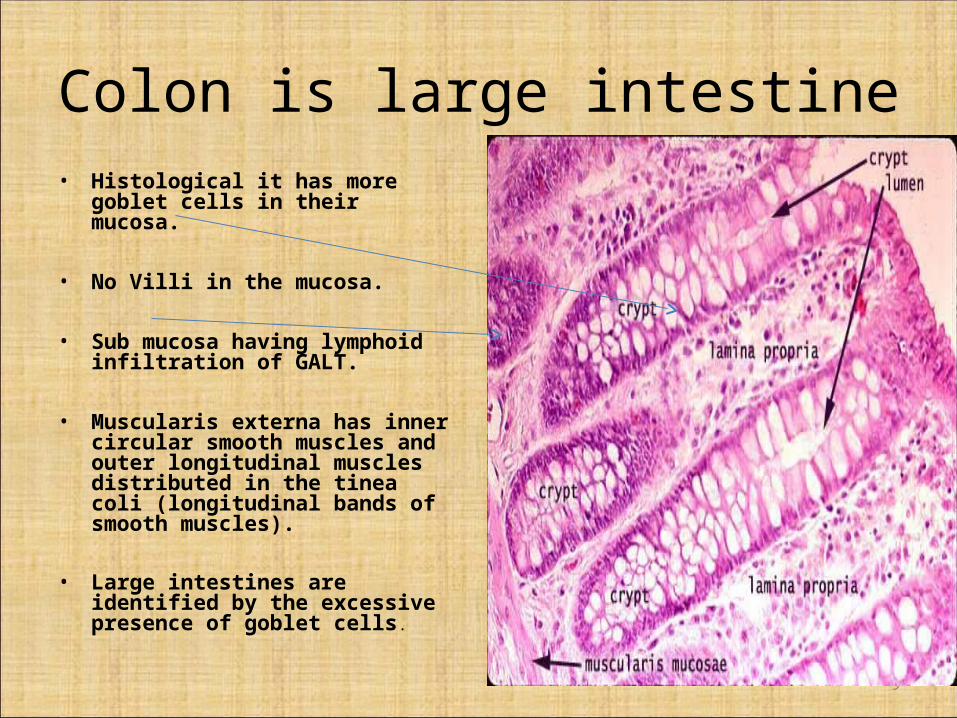
Colon is large intestine• Histological it has more goblet cells in
their mucosa.
• No Villi in the mucosa.
• Sub mucosa having lymphoid infiltration of GALT.
• Muscularis externa has inner circular smooth muscles and outer longitudinal muscles distributed in the tinea coli (longitudinal bands of smooth muscles).
• Large intestines are identified by the excessive presence of goblet cells.
9
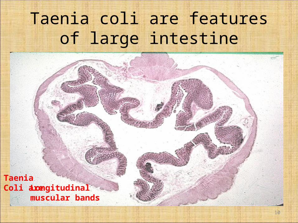
Taenia coli are features of large intestine
10
TaeniaColi are Longitudinal
muscular bands
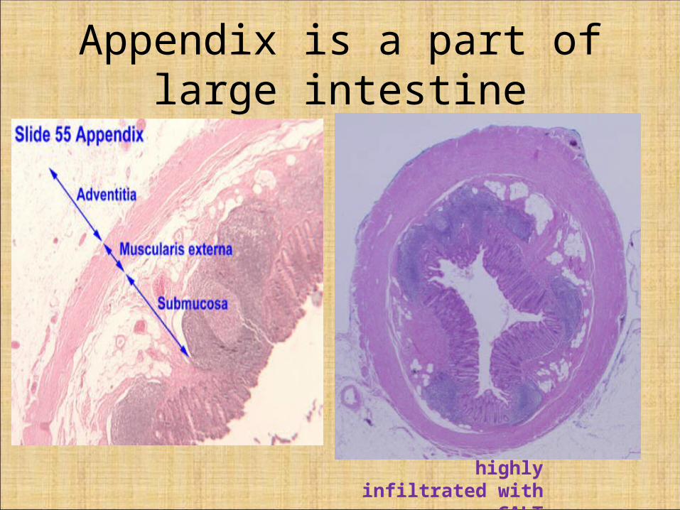
Appendix is a part of large intestine
Sub mucosa is highly infiltrated with GALT
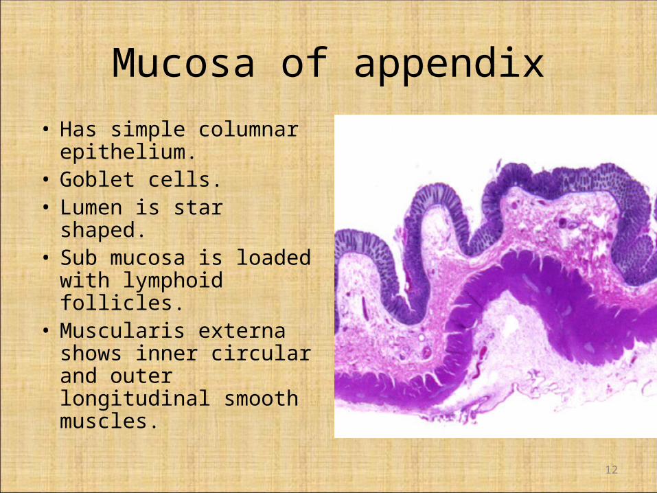
Mucosa of appendix
• Has simple columnar epithelium.
• Goblet cells.• Lumen is star shaped.• Sub mucosa is loaded
with lymphoid follicles.• Muscularis externa
shows inner circular and outer longitudinal smooth muscles.
12
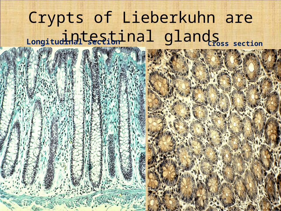
Crypts of Lieberkuhn are intestinal glands
13
Longitudinal section Cross section
Applied anatomy
• Typhoid fever is the enteric fever, complicating to perforation of bowel.
• Colitis is the inflamation of the colon.• Appendicitis is the inflamation of appendix.• Meckle’s diverticulum is a developmental
anomaly of vitellointestinal duct.• Anal hemorrhoids are venous engorgements of
rectal veins leading to bleeding piles.• Colonoscopy is the endoscopy of colon.
14