Embed Size (px)
Citation preview
MOLECULAR AND CELLULAR BIOLOGY, Jan. 2010, p. 399–412 Vol. 30, No. 20270-7306/10/$12.00 doi:10.1128/MCB.00907-09Copyright © 2010, American Society for Microbiology. All Rights Reserved.
Histone Deacetylase 7 and FoxA1 in Estrogen-MediatedRepression of RPRM�†
Simeen Malik,1,2 Shiming Jiang,1 Jason P. Garee,1,2 Eric Verdin,4,5 Adrian V. Lee,1,2,3
Bert W. O’Malley,2 Mao Zhang,6 Narasimhaswamy S. Belaguli,6and Steffi Oesterreich1,2,3*
Lester and Sue Smith Breast Center,1 Department of Molecular and Cellular Biology,2 and Department of Medicine,3 Baylor College ofMedicine, Houston, Texas 77030; Gladstone Institute of Virology and Immunology4 and Department of Medicine,5 University of
California, San Francisco, California 94158; and Michael E. DeBakey Department of Surgery,Michael E. DeBakey Veterans Affairs Medical Center and Baylor College of
Medicine, Houston, Texas 770306
Received 10 July 2009/Returned for modification 5 August 2009/Accepted 2 November 2009
Activation of estrogen receptor � (ER�) results in both induction and repression of gene transcription; whilemechanistic details of estrogen induction are well described, details of repression remain largely unknown. Wecharacterized several ER�-repressed targets and examined in detail the mechanism for estrogen repression ofReprimo (RPRM), a cell cycle inhibitor. Estrogen repression of RPRM is rapid and robust and requires atripartite interaction between ER�, histone deacetylase 7 (HDAC7), and FoxA1. HDAC7 is the critical HDACneeded for repression of RPRM; it can bind to ER� and represses ER�’s transcriptional activity—thisrepression does not require HDAC7’s deacetylase activity. We further show that the chromatin pioneer factorFoxA1, well known for its role in estrogen induction of genes, is recruited to the RPRM promoter, is necessaryfor repression of RPRM, and interacts with HDAC7. Like other FoxA1 recruitment sites, the RPRM promoteris characterized by H3K4me1/me2. Estrogen treatment causes decreases in H3K4me1/me2 and release of RNApolymerase II (Pol II) from the RPRM proximal promoter. Overall, these data implicate a novel role forHDAC7 and FoxA1 in estrogen repression of RPRM, a mechanism which could potentially be generalized tomany more estrogen-repressed genes and hence be important in both normal physiology and pathologicalprocesses.
Estrogen is essential for the growth and development offemale reproductive tissues and is a known potent mitogen inbreast cancer. The pleiotropic effects of 17-�-estradiol (E2),the most potent estrogen, are mediated through the � and �estrogen receptors (ER� and ER�), which contain an ago-nist-independent transcriptional activation function (AF-1),a DNA binding domain (DBD), a hinge region, and anagonist-dependent transcriptional activation function (AF-2). ER� can regulate gene expression directly by bindingDNA at perfect or imperfect estrogen response elements(EREs) (37) and half-ERE sites or indirectly by tethering toother DNA-bound transcription factors like AP-1, Sp1, andNF-�B (40, 60). ER� coordinates the assembly of chromatinremodeling factors, p160 coactivators (SRC1, SRC2, andSRC3), histone acetyltransferases (HATs) (p300, CBP, andthe p300/CBP-associated factor pCAF), histone methyl-transferases, histone deacetylases, general transcription fac-tors, the mediator complex, and RNA polymerase II (Pol II) tothe promoters of induced genes in an ordered and cyclicalfashion (50, 63).
Although ER� has been mostly studied as a transcriptional
activator, recent studies have shown that it can also negativelymodulate gene expression. For example, gene expression pro-filing has demonstrated that greater than 50% of ER� targetgenes are downregulated upon E2 treatment in MCF7 breastcancer cells as well as in breast tumors (5, 8, 16, 53). We (31,54) and others (1, 3, 19, 21, 52, 58, 59, 65, 70) have shown thatcritical genes like those coding for CD24, E-cadherin, BASE,interleukin-6 (IL-6), IR, retinoblastoma protein (Rb), ERBB2,vascular endothelial growth factor (VEGF), tumor necrosisfactor alpha (TNF-�), and CD36 are repressed by E2. Many ofthese E2-repressed genes are cell cycle inhibitors like cyclinG2 (CCNG2) (66), proapoptotic genes, or tumor suppressorgenes, and thus their repression could be a critical step inaugmenting the growth and survival of a tumor and thereby inthe development and/or progression of breast cancer. Whilethe mechanisms of E2-mediated induction of genes like theTrefoil factor 1 gene (TFF1/pS2) have been studied in greatdetail, the mechanisms regulating E2-mediated repressionof genes are virtually unknown. One potential mechanism issquelching (i.e., titration of limiting amounts of essentialtranscription factors by the abundance of an overexpressedtranscriptional regulator), which would result in a loss ofbasal transcription (5). However, active recruitment of re-pressive complexes—for example, NCoR, (nuclear receptorcorepressor), histone deacetylase 1 (HDAC1), and CtBP1 tothe CCNG2 promoter (66, 67); NCoR and SMRT (silencingmediator of RAR and TR) to the VEGFR2 promoter (25);and NCoR and TAB2 to the BMP7, ABCG2, and BCL3
* Corresponding author. Mailing address: One Baylor Plaza, MS600, Houston, TX 77030. Phone: (713) 798-1623. Fax: (713) 798-1642.E-mail: [email protected].
† Supplemental material for this article may be found at http://mcb.asm.org/.
� Published ahead of print on 16 November 2009.
399
Dow
nloa
ded
from
http
s://j
ourn
als.
asm
.org
/jour
nal/m
cb o
n 20
Oct
ober
202
1 by
183
.214
.104
.228
.
promoters (74)—has been shown. Genomewide analysis ofER� binding sites has implicated the involvement of the core-pressor NRIP1 (nuclear receptor interacting protein 1) in theE2-mediated repression of genes like BCAS4, IRX4, GUSB,and MUC1, which are repressed at late time points (5). Thesegenes are most likely secondary rather than direct targets ofER�, as they appear to require the E2 induction of NRIP1 fortheir repression.
Given the paucity of information, it is of particular interestto further explore the mechanism of estrogen repression of aprimary ER� target gene. We focused on a target gene which(i) is directly and robustly repressed by estrogen in breastcancer cell lines and (ii) is involved in mediating estrogen’smitogenic effects in breast cancer cells. Our data revealedthat the cell cycle inhibitor and tumor suppressor Reprimo(RPRM) gene fits these criteria. Rather unexpectedly, we dis-covered a unique role for HDAC7 in E2 repression of RPRM.HDAC7, ER�, and FoxA1 are necessary for repression andare recruited to the promoter, ultimately resulting in release ofRNA Pol II and decrease of transcription. In summary, wepropose a novel model for E2 repression which might also beexploited for repression of other E2-regulated genes and thusplay an important role in hormone response.
MATERIALS AND METHODS
Cell culture. Human breast cancer cells (MCF7, T47D, and ZR-75-1) weremaintained in Dulbecco’s modified Eagle’s medium (DMEM; Invitrogen LifeTechnologies) supplemented with 5% characterized fetal bovine serum(HyClone), 2 mM glutamine, 100 IU/ml penicillin, and 100 �g/ml streptomy-cin. Three days before E2 treatment, the cells were switched to improved min-imal essential medium (IMEM) that was supplemented with 5% charcoal-dext-ran-treated fetal bovine serum (HyClone). The medium was changed on day 2,and the cells were treated with either vehicle or 10 nM E2 (Sigma-Aldrich Corp),unless otherwise specified, 1 �M 4-OH-tamoxifen (4-OH-tam), or 1 �M ICI182,780 (Imperial Chemical Industries), on day 3 for 4, 8, or 24 h. The serum-freemedium (SFM) consisted of phenol-red-free IMEM (Invitrogen), 2 mM glu-tamine, 10 mM HEPES, 1 �g/ml fibronectin (Invitrogen), trace elements(Biosources), and 1 �g/ml transferrin (Invitrogen). For experiments in whichcycloheximide (CHX) was used, CHX at 10 �g/ml was added at the same timeas E2 for the entire 4- or 8-h period. For experiments in which actinomycinD (ActD) was used, ActD at 2 �g/ml was added 30 min before E2 treatmentand RNA was extracted at the �0.5-, 0-, 4-, 8-, and 24-h time points.
RNA extraction, qRT-PCR, and PCR. Total RNA was isolated using theRNeasy RNA isolation kit (Qiagen) as recommended by the supplier. TriplicateRNA samples were prepared for each treatment group. RNA was subjected toDNase treatment (Roche) prior to being reverse transcribed using SuperScript IIreverse transcriptase (Invitrogen) in accordance with manufacturer’s instruc-tions. The SYBR green or TaqMan PCR was then carried out on an ABI PRISM7700 sequence detector (Applied Biosystems), using the SYBR green master mix(Applied Biosystems) or the TaqMan PCR mix comprised of 300 nM (each) theforward and reverse primers, 100 nM probe, 0.025 U/�l of Taq polymerase(Invitrogen), 1� Rox dye (Invitrogen), 125 �M (each) deoxynucleotide triphos-phate (Invitrogen), 5 mM MgCl2 (Invitrogen), and 1� Taq polymerase buffer(Invitrogen). The cycling conditions were 94°C for 1 min, followed by 40 cyclesat 94°C for 15 s and 60°C for 30 s. Primer Express 2.0 software (AppliedBiosystems) was used to design all of the primers and probes. The sequences ofthe primers and probes for each of the genes tested are shown in Table S1 in thesupplemental material. The fold change for each gene was calculated using thecycle threshold (��CT) method as described in reference 46, and data arerepresented as E2 fold change over vehicle, unless otherwise stated. For eachsample, real-time quantitative reverse transcription-PCRs (qRT-PCRs) weredone in triplicate for each gene of interest and the reference gene (�-actin) tonormalize for input cDNA. Methylation-specific PCR (MSP) was performed asdescribed previously (68).
Nuclear run-on assays. Nuclear run-on assays were performed as describedpreviously (57, 67). Briefly, 6 � 107 MCF7 cells were treated with vehicle or E2for 0.5 and 7 h. The cells were washed with phosphate-buffered saline (PBS),
harvested, and lysed in lysis buffer (0.5% NP-40, 10 mM KCl, 10 mM MgCl2, 10mM HEPES [pH 7.9], and 0.5 mM �-mercaptoethanol [�-ME]) on ice until thecells had uniformly lysed and the nuclei appeared free of cytoplasmic material(�1 h). The nuclei were centrifuged at 500 � g, washed with lysis buffer withoutNP-40, and resuspended in 100 �l storage buffer (50 mM Tris-HCl, 5 mM MgCl2,0.5 mM �-ME, 40% glycerol) before being frozen at �80°C. A 100-�l amount oftranscription buffer (10 mM Tris-HCl [pH 8.0], 0.3 M MgCl2, 5 mM dithiothre-itol, 40 U RNase inhibitor [Roche], 1� biotin labeling mix [Roche]) was addedto the nuclei, and the reaction mixture was incubated at 30°C for 45 min. RNAwas isolated by using Trizol reagent (Invitrogen). A 50-�l amount of streptavi-din-conjugated magnetic beads (Invitrogen) resuspended in binding buffer (10mM Tris-HCl [pH 7.5], 1 mM EDTA, 100 mM NaCl) was added to each sampleand incubated for 2 h at room temperature. Beads were washed twice in 500 �lof 2� SSC (1� SSC is 0.15 M NaCl plus 0.015 M sodium citrate) and 15%formamide for 15 min and once in 500 �l of 2� SSC for 5 min and then dissolvedin 30 �l of diethyl pyrocarbonate-water. RNA was reverse transcribed, and qPCRreactions were carried out as described above.
siRNA. MCF7 cells (2 � 105) cells were plated in DMEM, and medium wasswitched to IMEM the next day. The cells were then transfected with 50 nMsmall interfering RNA (siRNA) using DharmaFECT1 (Thermo Fisher Scien-tific). The siRNAs used were as follows: siGENOME nontargeting siRNA 2,ER�, HDACs 1 to 10 (Dharmacon), and FoxA1 (4). NCoR, SMRT, LCoR, andNRIP1 siRNA experiments were performed as described previously (31).
Luc assays. Dual-luciferase (Luc) reporter assays were performed as previ-ously described (69), with some changes described here. Briefly, MCF7 cells (4 �105) were plated in 6-well plates in DMEM 1 day before transfection with 200ng/well ERE-thymidine kinase (Tk)-Luc, 100 ng/well pRL-Null (a Renilla Lucconstruct for normalizing of transfection efficiency), and SAFB1 (used as apositive control) or various concentrations of HDAC7 or HDAC7 deletionconstructs using Lipofectamine 2000. Smaller amounts of Flag-HDAC7(438–912) were transfected to keep the protein level relatively similar to that ofFlag-HDAC7, as seen in the Western blot (see Fig S5B in the supplementalmaterial). The amounts used were as follows: 100 ng/well of Flag-HDAC7 orFlag-HDAC7(1–487) or Flag-HDAC7 (H670A) or 10 ng/well of Flag-HDAC7(438–912). For the RPRM promoter reporter assays, MCF7 cells were trans-fected with 500 ng/well of the RPRM promoter and 100 ng/well of pRL-Null.Medium was changed to IMEM 16 h after transfection, cells were treated withvehicle or E2 for 24 h, and luciferase assays were performed as previouslydescribed (30).
Plasmids. The RPRM promoter was amplified by PCR from bacterial artificialchromosome (BAC) clone 389H5 and primer pairs containing BamHI/NcoIdigestion sites: RPRM-BamHI-F1 (GTCGGATCCGATTCATATTTTTGTGCAACCATCA) and RPRM-NcoI-R1 (GTCCCATGGTCATTATGTACAGGCTACGCTCGTC). The 5,566-bp PCR product was digested with BamHI and NcoIand inserted into the pGL3-basic luciferase vector. The HDAC7 constructs havebeen previously published (14). The Flag-HDAC7 (H670A) construct was clonedby subcloning into the pcDNA3.1-Flag-HDAC7 construct a piece of the mutatedconstruct from a mouse stem cell virus (MSCV)-internal ribosome entry site(IRES)-green fluorescent protein (GFP)-HDAC7(437–912) H657A constructusing the NaeI site. The histidine mutated in hHDAC7-H670A is shown inboldface in the sequence 665-RPPGHHADHST-675.
Coimmunoprecipitations. 293T cells were plated in 15-cm dishes, transfectedwith Flag-HDAC7, and the medium was switched to IMEM. The cells weretreated with either vehicle or 10 nM E2 for 16 h. After being washed with ice-coldPBS twice, the cells were collected into 1 ml of ice-cold PBS. They were thencentrifuged at 800 � g for 2 min at 4°C, and the cell pellet was resuspended in600 �l of lysis buffer (150 mM NaCl, 1% NP-40, and 50 mM Tris) and incubatedfor 20 min on ice. The lysate was centrifuged for 20 min at 14,000 � g at 4°C, andthe supernatant containing 1 mg of protein was incubated with the ER� antibodyH-184 (Santa Cruz) overnight or with Flag-M2 beads (Sigma-Aldrich) for 1 h.Twenty microliters of protein G-Sepharose was added to the ER� sample, andthe incubation was continued for 1 h. The Sepharose and Flag beads werecentrifuged and washed 4 times in 1 ml of lysis buffer. The washed beads wereboiled with Laemmli buffer and subjected to SDS-PAGE followed by immuno-blotting with anti-ER� 6F11 (Novocastra Laboratories) and anti-Flag (M2F1804; Sigma Aldrich) antibodies. A similar procedure was used for the immu-noprecipitation in Ly2 cells, where ER� and HDAC7 were immunoprecipitatedwith anti-ER� HC-20 (Santa Cruz Biotechnology) and anti-HDAC7 H-273(Santa Cruz Biotechnology), respectively, and immunoblotted with anti-ER�D-12 and F-10 (Santa Cruz Biotechnology) and anti-HDAC7 H00051564 (Ab-nova Corporation). Coimmunoprecipitation experiments for HDAC7 andFoxA1, in MCF7 cells, were done using a similar procedure in which HDAC7was immunoprecipitated with anti-HDAC7 H-273 (Santa Cruz Biotechnology)
400 MALIK ET AL. MOL. CELL. BIOL.
Dow
nloa
ded
from
http
s://j
ourn
als.
asm
.org
/jour
nal/m
cb o
n 20
Oct
ober
202
1 by
183
.214
.104
.228
.
and ab53101 (Abcam) antibodies and FoxA1 was immunoprecipitated with anti-FoxA1 ab5089 (Abcam) and C20 (Santa Cruz) antibodies. The membranes wereimmunoblotted with anti-HDAC7 H-273 (Santa Cruz) and anti-FoxA1 ab5089(Abcam).
ChIP assays. Chromatin immunoprecipitation (ChIP) assays were performedas described before (64). Briefly, MCF7 cells (3 � 106) were plated in 15-cmdishes in DMEM. After 24 h, the cells were washed with PBS twice and thenswitched to IMEM. The medium was changed on day 2 of culture, and on day 3of culture, the cells were treated with either vehicle, 10 nM E2, or 1 �M4-OH-tam for 45 min. After DNA purification (QIAquick spin kit; Qiagen), thepromoter regions were amplified by qRT-PCR using the primers described inTable S2 in the supplemental material. The ChIP antibodies used were ER�antibody H-184 and HC20 (Santa Cruz Biotechnology), FoxA1 antibodiesab5089 (Abcam) and C20 (Santa Cruz Biotechnology), H3K9ac (Jiemin Wong),H3K27me3 antibody (Upstate), H3K4me1 antibody ab8895 (Abcam), H3K4me2antibody 07-030 (Millipore), H3K4me3 antibody MC315 (Millipore), Pol IIantibody 8WG16 (Covance), HDAC7 antibody ab53101 (Abcam), p300 an-tibody N-15 (Santa Cruz Biotechnology), H3K9me2 antibody ab1220 (Ab-cam), H3K9me3 antibody ab8898 (Abcam), H4K16ac antibody 07-329 (Up-state), and HA antibody HA.11 (Covance). qPCR was used to calculateenrichment, which is displayed as “% Input” on the y axis or log2 E2 foldchange compared to vehicle control. Primers are presented in the supple-mental material.
Immunofluorescent staining and confocal microscopy. 293 cells (7.5 � 104)were plated on poly-D-lysine-coated coverslips in DMEM in 24-well plates. Oneday after plating, cells were washed twice with PBS, fixed in 4% formaldehydediluted in PBS for 10 min at room temperature, washed twice with PBS, perme-abilized with 0.5% Triton X-100 diluted in PBS for 5 min, washed twice withPBS, and then incubated with 1% bovine serum albumin (BSA) in PBS for 1 h.Following this, the cells were incubated with primary antibody (anti-Flag, diluted1:100 in 1% BSA in PBS) for 1 h at room temperature, washed three times withPBS, incubated with secondary antibody (Alexa 488-conjugated goat anti-mouseIgG, diluted 1:1,000 in 1% BSA in PBS) for 1 h at room temperature in the dark,washed three times with PBS, incubated with the DNA marker TOPRO-3 (di-luted 1:80 in PBS) for 30 min at room temperature in the dark, washed threetimes with PBS, and then mounted with mounting medium. Confocal images
were obtained using a Nikon confocal Eclipse E1000 scanning laser microscope(Nikon) with a �60 objective lens.
RESULTS
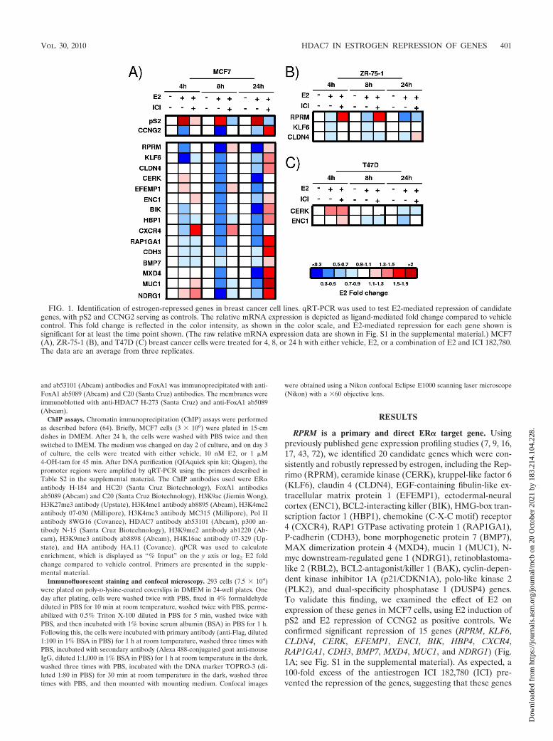
RPRM is a primary and direct ER� target gene. Usingpreviously published gene expression profiling studies (7, 9, 16,17, 43, 72), we identified 20 candidate genes which were con-sistently and robustly repressed by estrogen, including the Rep-rimo (RPRM), ceramide kinase (CERK), kruppel-like factor 6(KLF6), claudin 4 (CLDN4), EGF-containing fibulin-like ex-tracellular matrix protein 1 (EFEMP1), ectodermal-neuralcortex (ENC1), BCL2-interacting killer (BIK), HMG-box tran-scription factor 1 (HBP1), chemokine (C-X-C motif) receptor4 (CXCR4), RAP1 GTPase activating protein 1 (RAP1GA1),P-cadherin (CDH3), bone morphogenetic protein 7 (BMP7),MAX dimerization protein 4 (MXD4), mucin 1 (MUC1), N-myc downstream-regulated gene 1 (NDRG1), retinoblastoma-like 2 (RBL2), BCL2-antagonist/killer 1 (BAK), cyclin-depen-dent kinase inhibitor 1A (p21/CDKN1A), polo-like kinase 2(PLK2), and dual-specificity phosphatase 1 (DUSP4) genes.To validate this finding, we examined the effect of E2 onexpression of these genes in MCF7 cells, using E2 induction ofpS2 and E2 repression of CCNG2 as positive controls. Weconfirmed significant repression of 15 genes (RPRM, KLF6,CLDN4, CERK, EFEMP1, ENCI, BIK, HBP4, CXCR4,RAP1GA1, CDH3, BMP7, MXD4, MUC1, and NDRG1) (Fig.1A; see Fig. S1 in the supplemental material). As expected, a100-fold excess of the antiestrogen ICI 182,780 (ICI) pre-vented the repression of the genes, suggesting that these genes
FIG. 1. Identification of estrogen-repressed genes in breast cancer cell lines. qRT-PCR was used to test E2-mediated repression of candidategenes, with pS2 and CCNG2 serving as controls. The relative mRNA expression is depicted as ligand-mediated fold change compared to vehiclecontrol. This fold change is reflected in the color intensity, as shown in the color scale, and E2-mediated repression for each gene shown issignificant for at least the time point shown. (The raw relative mRNA expression data are shown in Fig. S1 in the supplemental material.) MCF7(A), ZR-75-1 (B), and T47D (C) breast cancer cells were treated for 4, 8, or 24 h with either vehicle, E2, or a combination of E2 and ICI 182,780.The data are an average from three replicates.
VOL. 30, 2010 HDAC7 IN ESTROGEN REPRESSION OF GENES 401
Dow
nloa
ded
from
http
s://j
ourn
als.
asm
.org
/jour
nal/m
cb o
n 20
Oct
ober
202
1 by
183
.214
.104
.228
.
are in fact regulated by ER. Some targets were repressed onlyat the late time point, such as MXD4, suggesting indirectregulation: for example, through an NRIP1-dependent path-way (5). However, timing of repression needs to be interpretedwith caution, since late repression could also simply be a resultof a long mRNA half-life. We tested repression of a subset ofthe genes (RPRM, KLF6, CLDN4, CERK, EFEMP1, andENC1) in the ER�-positive breast cancer cell lines ZR-75-1(Fig. 1B; see Fig. S2A in the supplemental material) and T47D(Fig. 1C; also see Fig. S2B in the supplemental material) andfound cell-type-specific differences which may be attributed tothe absence and/or differential recruitment of coregulators.
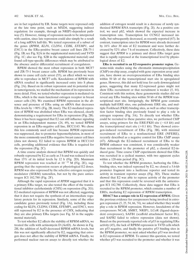
RPRM showed the most robust repression in both MCF7and ZR-75-1 breast cancer cells. RPRM has previously beenshown to cause cell cycle arrest (55), an effect which we wereable to reproduce in MCF7 cells. Knockdown of RPRM withsiRNA resulted in significantly increased entry into S phase(Fig. 2A). Based on its robust repression and its potential rolein tumorigenesis, we studied the mechanism of its repression inmore detail. First, we tested whether repression is mediated viaER�, which is the main functional estrogen receptor in breastcancer cells (38). We examined RPRM repression in the ab-sence and presence of ER� using an siRNA that decreasesER� levels by 90% (Fig. 2B, inset). The E2-mediated repres-sion of RPRM was abolished when ER� was silenced, clearlydemonstrating a requirement for ER� in repression (Fig. 2B).Since it has been suggested that E2 can still influence signalingin an ER�-independent manner (24, 49), we tested RPRMrepression in an ER�-negative cell line, HCC1143. We chosethis less commonly used cell line because RPRM expressionwas suppressed, due to promoter hypermethylation, in most ofthe more commonly used ER�-negative breast cancer cell lines(data not shown). E2 failed to repress RPRM in HCC1143cells, providing additional evidence that ER� is required forthe repression (Fig. 2C).
A time course analysis showed that RPRM was quickly androbustly repressed by E2 as early as 3 h and was reduced to lessthan 15% of its initial levels by 12 h (Fig. 2D). MaximumRPRM repression was reached at 10�10 M (Fig. 2E), sug-gesting that the repression occurs at physiological E2 doses.RPRM was also repressed by the selective estrogen receptormodulator (SERM) tamoxifen, but not by the pure anties-trogen ICI 182,780 (Fig. 2F).
Although the rapid repression of RPRM suggested that it isa primary ER� target, we also tested the effect of the transla-tional inhibitor cycloheximide (CHX) on repression (Fig. 2G).E2-mediated repression of RPRM was not affected, suggestingthat it does not require the synthesis of an intermediate regu-latory protein for its repression. Similarly, some of the othercandidate genes previously tested (Fig. 1A), including thosecoding for KLF6, CLDN4, CERK, EFEMP1, and ENC1, werestill repressed by E2 in the presence of CHX, indicating thatthey are also primary ER� targets (see Fig. S3 in the supple-mental material).
To test whether E2 affects the stability of RPRM mRNA, wetreated the cells with actinomycin D (ActD). As shown in Fig.2H, the addition of ActD decreased RPRM mRNA levels, butthis was not significantly affected by E2, suggesting that estro-gen does not affect the stability of RPRM mRNA. Finally, weperformed nuclear run-on assays to directly test whether the
addition of estrogen would result in a decrease of newly syn-thesized RPRM RNA transcripts (Fig. 2I). As a positive con-trol, we used pS2, which showed the expected increase intranscription rate. Transcription for CCNG2 increased ini-tially, but subsequently decreased, as recently reported (67). Incontrast, transcription rates for RPRM were already decreasedby 16% after 30 min of E2 treatment and were further de-creased by 52% after 7 h of treatment. Collectively, these datasuggest that RPRM is a primary and direct ER� target genethat is rapidly repressed at the transcriptional level by physio-logical doses of E2.
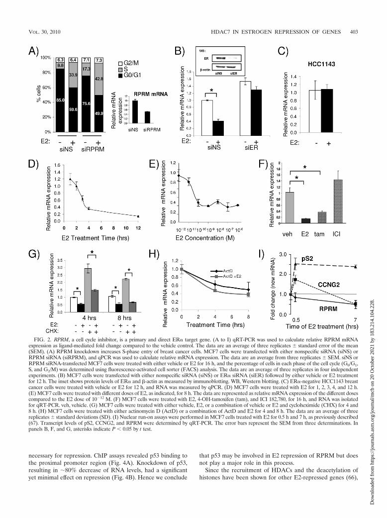
ER� is recruited to an E2-responsive promoter region. Ge-nome-wide studies using the chromatin immunoprecipitation(ChIP)-on-chip assay, coupled with gene expression array anal-ysis, have shown an overrepresentation of ER� binding siteswithin 50 kb of the transcriptional start site in upregulatedgenes. However, this did not hold true for early downregulatedgenes, suggesting that many E2-repressed genes might notshow ER� recruitment or that recruitment is weaker (5, 44).Consistent with this notion, these genomewide studies did notreveal any ER� binding sites within 50 kb of the RPRM tran-scriptional start site. Intriguingly, the RPRM gene containsmultiple half-ERE sites, one palindromic ERE site, and mul-tiple Forkhead (FKH) binding sites, as well as other transcrip-tion factor binding sites previously found to play a role inestrogen response (Fig. 3A). To directly test whether ER�could be recruited to these putative sites, we performed ChIPassays, using primers for the ERE/FKH site (�4.8 kb) andthe transcriptional start site (0.1 kb). They showed estro-gen-induced recruitment of ER� (Fig. 3B), with minimalrecruitment of ER� to a nonfunctional ERE (NFERE),recently described as a negative control for ER� ChIP assays(47). Although the E2-induced recruitment of ER� to theRPRM enhancer was consistent, it was considerably weakerthan recruitment to the promoter of pS2, a classical E2-in-duced gene. Time course ChIP experiments showed that ER�was recruited in a cyclical manner, with two apparent cycleswithin a 120-min period (Fig. 3C).
To test whether the RPRM promoter, harboring the ER�-binding sites, was indeed repressed by E2, we cloned a 5.6-kbpromoter fragment into a luciferase reporter and tested itsactivity in transient reporter assays (Fig. 3D). These studiesshowed that E2 was able to repress activity of the promoterand that this repression could be reversed with the antiestro-gen ICI 182,780. Collectively, these data suggest that ER� isrecruited to the RPRM promoter, which contains a number ofEREs, mediating E2 repression of RPRM transcription.
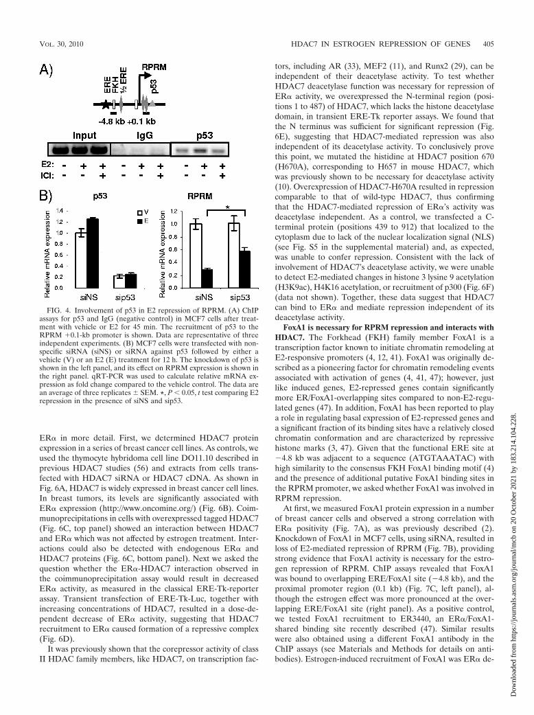
HDAC7 has a unique role in E2 repression of RPRM. Giventhe previous evidence for corepressors being involved in estro-gen repression (5, 25, 54, 66, 74), we asked whether they wouldplay a role in RPRM repression. However, knockdown of thecorepressors NCoR, SMRT, NRIP1, LCoR (ligand-depen-dent corepressor), SAFB1 (scaffold attachment factor B1),and SAFB2 failed to relieve repression (data not shown).Based on the previously reported role of p53 in regulating ER�activity (45), the lack of RPRM repression in T47D cells whichare p53 negative, and finally the putative p53 binding sites inthe RPRM promoter, we next asked whether p53 was involvedin E2 repression of RPRM. To answer this question, we testedwhether p53 was recruited to the promoter and whether it was
402 MALIK ET AL. MOL. CELL. BIOL.
Dow
nloa
ded
from
http
s://j
ourn
als.
asm
.org
/jour
nal/m
cb o
n 20
Oct
ober
202
1 by
183
.214
.104
.228
.
necessary for repression. ChIP assays revealed p53 binding tothe proximal promoter region (Fig. 4A). Knockdown of p53,resulting in �80% decrease of RNA levels, had a significantyet minimal effect on repression (Fig. 4B). Hence we conclude
that p53 may be involved in E2 repression of RPRM but doesnot play a major role in this process.
Since the recruitment of HDACs and the deacetylation ofhistones have been shown for other E2-repressed genes (66),
FIG. 2. RPRM, a cell cycle inhibitor, is a primary and direct ER� target gene. (A to I) qRT-PCR was used to calculate relative RPRM mRNAexpression as ligand-mediated fold change compared to the vehicle control. The data are an average of three replicates � standard error of the mean(SEM). (A) RPRM knockdown increases S-phase entry of breast cancer cells. MCF7 cells were transfected with either nonspecific siRNA (siNS) orRPRM siRNA (siRPRM), and qPCR was used to calculate relative mRNA expression. The data are an average from three replicates � SEM. siNS orRPRM siRNA-transfected MCF7 cells were treated with either vehicle or E2 for 16 h, and the percentage of cells in each phase of the cell cycle (G0/G1,S, and G2/M) was determined using fluorescence-activated cell sorter (FACS) analysis. The data are an average of three replicates in four independentexperiments. (B) MCF7 cells were transfected with either nonspecific siRNA (siNS) or ER� siRNA (siER) followed by either vehicle or E2 treatmentfor 12 h. The inset shows protein levels of ER� and �-actin as measured by immunoblotting. WB, Western blotting. (C) ER�-negative HCC1143 breastcancer cells were treated with vehicle or E2 for 12 h, and RNA was measured by qPCR. (D) MCF7 cells were treated with E2 for 1, 2, 3, 4, and 12 h.(E) MCF7 cells were treated with different doses of E2, as indicated, for 8 h. The data are represented as relative mRNA expression of the different dosescompared to the E2 dose of 10�12 M. (F) MCF7 cells were treated with E2, 4-OH-tamoxifen (tam), and ICI 182,780, for 16 h, and RNA was isolatedfor qRT-PCR. veh, vehicle. (G) MCF7 cells were treated with either vehicle, E2, or a combination of vehicle or E2 and cycloheximide (CHX) for 4 and8 h. (H) MCF7 cells were treated with either actinomycin D (ActD) or a combination of ActD and E2 for 4 and 8 h. The data are an average of threereplicates � standard deviations (SD). (I) Nuclear run-on assays were performed in MCF7 cells treated with E2 for 0.5 h and 7 h, as previously described(67). Transcript levels of pS2, CCNG2, and RPRM were determined by qRT-PCR. The error bars represent the SEM from three determinations. Inpanels B, F, and G, asterisks indicate P � 0.05 by t test.
VOL. 30, 2010 HDAC7 IN ESTROGEN REPRESSION OF GENES 403
Dow
nloa
ded
from
http
s://j
ourn
als.
asm
.org
/jour
nal/m
cb o
n 20
Oct
ober
202
1 by
183
.214
.104
.228
.
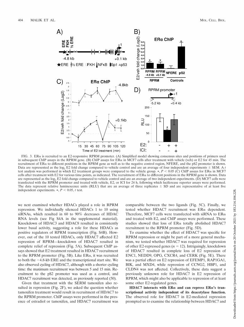
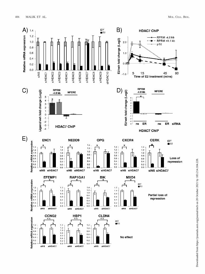
we next examined whether HDACs played a role in RPRMrepression. We individually silenced HDACs 1 to 10 usingsiRNAs, which resulted in 60 to 90% decreases of HDACRNA levels (see Fig S4A in the supplemental material).Knockdown of HDAC3 and HDAC8 resulted in consistentlylower basal activity, suggesting a role for these HDACs aspositive regulators of RPRM transcription (Fig. S4B). How-ever, out of the 10 tested HDACs, only HDAC7 affected E2repression of RPRM—knockdown of HDAC7 resulted incomplete relief of repression (Fig. 5A). Subsequent ChIP as-says showed that E2 treatment resulted in HDAC7 recruitmentto the RPRM promoter (Fig. 5B). Like ER�, it was recruitedto both the �4.8-kb ERE and the transcriptional start site. Wealso observed cycling of HDAC7, although with a shorter cyclingtime: the maximum recruitment was between 5 and 15 min. Re-cruitment to the pS2 promoter was used as a control, andHDAC7 recruitment was detected, as previously reported (50).
Given that treatment with the SERM tamoxifen also re-sulted in repression (Fig. 2F), we asked the question whethertamoxifen treatment would result in recruitment of HDAC7 tothe RPRM promoter. ChIP assays were performed in the pres-ence of estradiol or tamoxifen, and HDAC7 recruitment was
comparable between the two ligands (Fig. 5C). Finally, wetested whether HDAC7 recruitment was ER� dependent.Therefore, MCF7 cells were transfected with siRNA to ER�and treated with E2, and ChIP assays were performed. Thesestudies showed that loss of ER� totally abolished HDAC7recruitment to the RPRM promoter (Fig. 5D).
To examine whether the effect of HDAC7 was specific forRPRM repression or might be part of a more general mecha-nism, we tested whether HDAC7 was required for repressionof other E2-repressed genes (n 12). Intriguingly, knockdownof HDAC7 resulted in complete loss of E2 repression ofENC1, NEDD9, OPG, CXCR4, and CERK (Fig. 5E). Therewas a partial effect on E2 repression of EFEMP1, RAP1GA1,BIK, and MXD4, while repression of CCNG2, HBP1, andCLDN4 was not affected. Collectively, these data suggest apreviously unknown role for HDAC7 in E2 repression ofRPRM, which might also be applicable to repression of at leastsome other E2-regulated genes.
HDAC7 interacts with ER� and can repress ER�’s tran-scriptional activity independent of its deacetylase function.The observed role for HDAC7 in E2-mediated repressionprompted us to examine the relationship between HDAC7 and
FIG. 3. ER� is recruited to an E2-responsive RPRM promoter. (A) Simplified model showing consensus sites and positions of primers usedin subsequent ChIP assays in the RPRM gene. (B) ChIP assays for ER� in MCF7 cells after treatment with vehicle (veh) or E2 for 45 min. Therecruitment of ER� to different positions in the RPRM gene as well as to the negative control region, NFERE, and the pS2 promoter is shown.Data are represented as the log2 E2 fold change compared to vehicle control and are an average of four independent experiments � SEM. A ttest analysis was performed in which E2 treatment groups were compared to the vehicle group. *, P � 0.05 (C) ChIP assays for ER� in MCF7cells after treatment with E2 for various time points, as indicated. The recruitment of ER� to different positions in the RPRM gene is shown. Dataare represented as the log2 E2 fold change compared to vehicle control and are an average of two independent experiments. (D) MCF7 cells weretransfected with the RPRM promoter and treated with vehicle, E2, or ICI for 24 h, following which luciferease reporter assays were performed.The data represent relative luminescence units (RLU) that are an average of three replicates � SD and are representative of at least fiveindependent experiments. *, P � 0.05, t test.
404 MALIK ET AL. MOL. CELL. BIOL.
Dow
nloa
ded
from
http
s://j
ourn
als.
asm
.org
/jour
nal/m
cb o
n 20
Oct
ober
202
1 by
183
.214
.104
.228
.
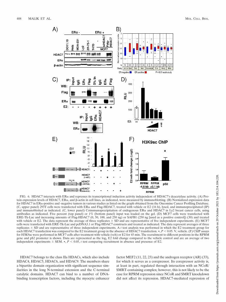
ER� in more detail. First, we determined HDAC7 proteinexpression in a series of breast cancer cell lines. As controls, weused the thymocyte hybridoma cell line DO11.10 described inprevious HDAC7 studies (56) and extracts from cells trans-fected with HDAC7 siRNA or HDAC7 cDNA. As shown inFig. 6A, HDAC7 is widely expressed in breast cancer cell lines.In breast tumors, its levels are significantly associated withER� expression (http://www.oncomine.org/) (Fig. 6B). Coim-munoprecipitations in cells with overexpressed tagged HDAC7(Fig. 6C, top panel) showed an interaction between HDAC7and ER� which was not affected by estrogen treatment. Inter-actions could also be detected with endogenous ER� andHDAC7 proteins (Fig. 6C, bottom panel). Next we asked thequestion whether the ER�-HDAC7 interaction observed inthe coimmunoprecipitation assay would result in decreasedER� activity, as measured in the classical ERE-Tk-reporterassay. Transient transfection of ERE-Tk-Luc, together withincreasing concentrations of HDAC7, resulted in a dose-de-pendent decrease of ER� activity, suggesting that HDAC7recruitment to ER� caused formation of a repressive complex(Fig. 6D).
It was previously shown that the corepressor activity of classII HDAC family members, like HDAC7, on transcription fac-
tors, including AR (33), MEF2 (11), and Runx2 (29), can beindependent of their deacetylase activity. To test whetherHDAC7 deacetylase function was necessary for repression ofER� activity, we overexpressed the N-terminal region (posi-tions 1 to 487) of HDAC7, which lacks the histone deacetylasedomain, in transient ERE-Tk reporter assays. We found thatthe N terminus was sufficient for significant repression (Fig.6E), suggesting that HDAC7-mediated repression was alsoindependent of its deacetylase activity. To conclusively provethis point, we mutated the histidine at HDAC7 position 670(H670A), corresponding to H657 in mouse HDAC7, whichwas previously shown to be necessary for deacetylase activity(10). Overexpression of HDAC7-H670A resulted in repressioncomparable to that of wild-type HDAC7, thus confirmingthat the HDAC7-mediated repression of ER�’s activity wasdeacetylase independent. As a control, we transfected a C-terminal protein (positions 439 to 912) that localized to thecytoplasm due to lack of the nuclear localization signal (NLS)(see Fig. S5 in the supplemental material) and, as expected,was unable to confer repression. Consistent with the lack ofinvolvement of HDAC7’s deacetylase activity, we were unableto detect E2-mediated changes in histone 3 lysine 9 acetylation(H3K9ac), H4K16 acetylation, or recruitment of p300 (Fig. 6F)(data not shown). Together, these data suggest that HDAC7can bind to ER� and mediate repression independent of itsdeacetylase activity.
FoxA1 is necessary for RPRM repression and interacts withHDAC7. The Forkhead (FKH) family member FoxA1 is atranscription factor known to initiate chromatin remodeling atE2-responsive promoters (4, 12, 41). FoxA1 was originally de-scribed as a pioneering factor for chromatin remodeling eventsassociated with activation of genes (4, 41, 47); however, justlike induced genes, E2-repressed genes contain significantlymore ER/FoxA1-overlapping sites compared to non-E2-regu-lated genes (47). In addition, FoxA1 has been reported to playa role in regulating basal expression of E2-repressed genes anda significant fraction of its binding sites have a relatively closedchromatin conformation and are characterized by repressivehistone marks (3, 47). Given that the functional ERE site at�4.8 kb was adjacent to a sequence (ATGTAAATAC) withhigh similarity to the consensus FKH FoxA1 binding motif (4)and the presence of additional putative FoxA1 binding sites inthe RPRM promoter, we asked whether FoxA1 was involved inRPRM repression.
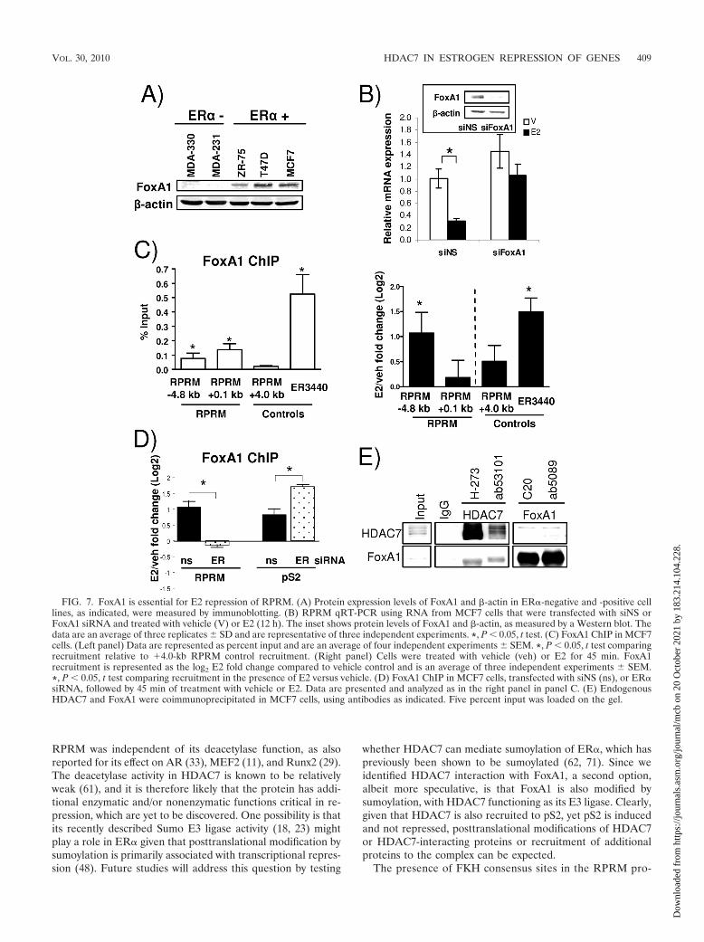
At first, we measured FoxA1 protein expression in a numberof breast cancer cells and observed a strong correlation withER� positivity (Fig. 7A), as was previously described (2).Knockdown of FoxA1 in MCF7 cells, using siRNA, resulted inloss of E2-mediated repression of RPRM (Fig. 7B), providingstrong evidence that FoxA1 activity is necessary for the estro-gen repression of RPRM. ChIP assays revealed that FoxA1was bound to overlapping ERE/FoxA1 site (�4.8 kb), and theproximal promoter region (0.1 kb) (Fig. 7C, left panel), al-though the estrogen effect was more pronounced at the over-lapping ERE/FoxA1 site (right panel). As a positive control,we tested FoxA1 recruitment to ER3440, an ER�/FoxA1-shared binding site recently described (47). Similar resultswere also obtained using a different FoxA1 antibody in theChIP assays (see Materials and Methods for details on anti-bodies). Estrogen-induced recruitment of FoxA1 was ER� de-
FIG. 4. Involvement of p53 in E2 repression of RPRM. (A) ChIPassays for p53 and IgG (negative control) in MCF7 cells after treat-ment with vehicle or E2 for 45 min. The recruitment of p53 to theRPRM 0.1-kb promoter is shown. Data are representative of threeindependent experiments. (B) MCF7 cells were transfected with non-specific siRNA (siNS) or siRNA against p53 followed by either avehicle (V) or an E2 (E) treatment for 12 h. The knockdown of p53 isshown in the left panel, and its effect on RPRM expression is shown inthe right panel. qRT-PCR was used to calculate relative mRNA ex-pression as fold change compared to the vehicle control. The data arean average of three replicates � SEM. *, P � 0.05, t test comparing E2repression in the presence of siNS and sip53.
VOL. 30, 2010 HDAC7 IN ESTROGEN REPRESSION OF GENES 405
Dow
nloa
ded
from
http
s://j
ourn
als.
asm
.org
/jour
nal/m
cb o
n 20
Oct
ober
202
1 by
183
.214
.104
.228
.
406 MALIK ET AL. MOL. CELL. BIOL.
Dow
nloa
ded
from
http
s://j
ourn
als.
asm
.org
/jour
nal/m
cb o
n 20
Oct
ober
202
1 by
183
.214
.104
.228
.
pendent—knockdown of ER� by siRNA totally abolishedFoxA1 recruitment (Fig. 7D). In contrast, loss of ER� did notaffect FoxA1 recruitment to the pS2 promoter, an estrogen-in-duced gene, suggesting differences in FoxA1 recruitment betweenestrogen-repressed and -induced genes. Finally, ChIP assays re-vealed that HDAC7 recruitment was not a prerequisite for FoxA1recruitment to the RPRM promoter (data not shown).
Given the binding of HDAC7 and FoxA1 to the sameRPRM promoter binding sites and their shared requirementfor E2 repression, we asked whether the two proteins interact.This was tested in coimmunoprecipitations, using two indepen-dent antibodies for both HDAC7 and FoxA1 (Fig. 7E). Per-forming the immunoprecipitation with the HDAC7 antibodies,we observed strong interactions with FoxA1. The interaction,which was estrogen independent (data not shown), was weakerin the reciprocal coimmunoprecipitation, but could still bedetected. The interaction between ER� and HDAC7 was notsignificantly affected by different amounts of FoxA1 (data notshown), suggesting that HDAC7 and FoxA1 do not competewith each other for binding to ER�.
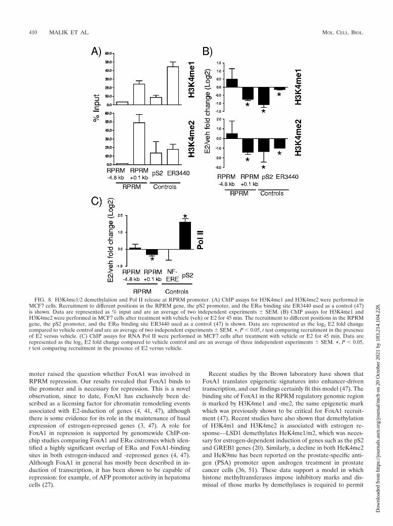
Ligand-dependent H3K4 demethylation is associated withrelease of RNA Pol II. FoxA1 occupies only a very small per-centage of its potential recognition motifs (less than 4%) (47),and recent studies have shown that its recruitment is guided byhistone H3 lysine 4 (H3K4) methylation, especially H3K4me1/me2 (47). Analysis of these specific histone modifications atthe FoxA1 recruitment sites in the RPRM promoter revealeda significant and strong presence of H3K4me1 and H3K4me2at the proximal ER/FoxA1 binding site at 0.1 kb (Fig. 8A).We again used ER3440, the recently described ER�/FoxA1-shared binding site (47), and pS2 as controls, where we alsofound H3K4me1/me2 present, as expected. The removal ofthese marks was recently shown to be necessary for E2 induc-tion of genes (20), and we hence sought to determine if de-crease of H3K4me1/me2 recruitment was also associated withE2 repression of target genes. ChIP assays were performed,and significant decreases of H3K4me1/me2 were observed forER3440 and pS2 and importantly also for the proximal RPRMpromoter region (Fig. 8B). Finally, we sought to determinewhether this was associated with dissociation of RNA Pol II. Inagreement with transcriptional repression of RPRM, RNA PolII was significantly released upon E2 treatment (Fig. 8C). To-gether, these results provide compelling evidence for the roleof an interplay between FoxA1 and H3K4me1/me2 in regula-tion of E2-repressed genes, in addition to the previously re-ported role in gene induction.
DISCUSSION
Here we report a novel mechanism for estrogen-mediatedrepression of gene expression involving an interplay betweenER�, HDAC7, and FoxA1. Over the last 2 decades, inductionof genes by ER� has been a subject of intense investigation,but recent studies have shown that over half of E2-regulatedgenes are actually repressed. It is likely that an understandingof E2-mediated repression of transcription will allow a morethorough understanding of how ER� mediates its downstreamactions, such as regulation of cell proliferation. Transient re-pression of cell cycle inhibitors such as CCNG2 and RPRM islikely to play a vital role in mediating estrogen’s effects inhormone-responsive cells, thus contributing both to estrogenaction in normal physiology and to various pathological pro-cesses such as breast cancer. For example, ER� repressesErbB2 expression, and loss of this control may lead to aggres-sive ErbB2-positive tumors with poor outcome. Surprisingly,despite the long-held knowledge of the inverse correlationbetween ER� and ErbB2 in breast tumors, few studies havedirectly investigated how ER� downregulates ErbB2 (28, 52).It is therefore critical that we unravel the full diversity ofmechanistic programs for ER�-mediated gene repression.
Currently, there is evidence for three models of estrogenrepression, which are not mutually exclusive. In the first model,repression is simply the result of loss of activation, due tosquelching of coactivators from promoters or enhancers (5,32). Second, corepressors can compete with coactivators at theregulatory region of the repressed gene, with competition ofPAX2 with the ER� coactivator AIB-1/SRC-3 for binding andregulation of ErbB2 transcription being a recent example (28).The third model involves active recruitment of repressive com-plexes, including NCoR, SMRT, and HDACs (25, 35, 66, 74).Our data provide strong evidence for “active repression” play-ing a crucial role for a significant subset of estrogen-repressedgenes.
Here we show estrogen-dependent recruitment of ER�,HDAC7, and FoxA1 to the proximal and also more distalpromoter in the RPRM gene. We also find a modest role forp53 in RPRM repression, and there might be other factors thatplay a role in this process that have yet to be identified. Pre-vious studies have shown that HDACs, such as HDAC1 andHDAC4, can interact with ER� and repress its transcriptionalactivity (34, 42). Moreover, various ER�-associated corepres-sors, including NCoR, SMRT, NRIP1, LCoR, MTA1, REA,and SAFB1/2 have been shown to recruit HDACs (13, 15, 26,39, 73). However, this is the first time that a unique role ofHDAC7 in estrogen repression has been described.
FIG. 5. HDAC7 is necessary for repression of RPRM and other E2-repressed genes. (A) RPRM qRT-PCR was performed using RNA fromMCF7 cells transfected with nonspecific siRNA (siNS) or siRNA against HDACs 1 to 10 and treated with vehicle (V) or E2 (12 h). (B) HDAC7ChIP in MCF7 cells treated with vehicle (veh) or E2 for 5, 15, 45, and 60 min. Data are represented as the log2 E2 fold change compared to vehiclecontrol and are an average of two independent experiments � SEM. (C) HDAC7 ChIP in MCF7 cells treated with vehicle, E2, or 4-OH-tamoxifenfor 15 min. Nonfunctional ERE was used as negative control. *, P � 0.05, t test comparing recruitment in the presence of E2 versus vehicle.(D) HDAC7 ChIP in MCF7 cells, transfected with siNS (ns) or ER� (ER) siRNA, followed by 45 min of treatment with vehicle or E2. *, P �0.05, t test comparing recruitment in the presence of siNS and ER� siRNA. (E) The E2-mediated repression of candidate genes was tested usingRNA from MCF7 cells transfected with siNS or HDAC7 siRNA. The data are an average of three replicates � SD and are representative of twoindependent experiments. *, P � 0.05, t test.
VOL. 30, 2010 HDAC7 IN ESTROGEN REPRESSION OF GENES 407
Dow
nloa
ded
from
http
s://j
ourn
als.
asm
.org
/jour
nal/m
cb o
n 20
Oct
ober
202
1 by
183
.214
.104
.228
.
HDAC7 belongs to the class IIa HDACs, which also includeHDAC4, HDAC5, HDAC6, and HDAC9. The members sharea bipartite domain organization with significant sequence sim-ilarities in the long N-terminal extension and the C-terminalcatalytic domains. HDAC7 can bind to a number of DNA-binding transcription factors, including the myocyte enhancer
factor MEF2 (11, 22, 23) and the androgen receptor (AR) (33),for which it serves as a corepressor. Its corepressor activity is,at least in part, regulated through interaction with an NCoR/SMRT-containing complex; however, this is not likely to be thecase for RPRM repression since NCoR and SMRT knockdowndid not affect its repression. HDAC7-mediated repression of
FIG. 6. HDAC7 interacts with ER� and represses its transcriptional induction activity independent of HDAC7’s deacetylase activity. (A) Pro-tein expression levels of HDAC7, ER�, and �-actin in cell lines, as indicated, were measured by immunoblotting. (B) Normalized expression datafor HDAC7 in ER�-positive and -negative tumors in various studies as listed on the graph obtained from the Oncomine Cancer Profiling Database.(C, upper panel) 293T cells were transfected with ER� and Flag-HDAC7, treated with vehicle or E2 (16 h), lysed, and immunoprecipitated (IP)and immunoblotted as indicated. (C, lower panel) Coimmunoprecipitation of endogenous ER� and HDAC7 in Ly2 breast cancer cells, usingantibodies as indicated. Five percent (top panel) or 1% (bottom panel) input was loaded on the gel. (D) MCF7 cells were transfected withERE-Tk-Luc and increasing amounts of Flag-HDAC7 (0, 50, 100, and 250 ng) or SAFB1 (250 ng [used as a positive control]) (30) and treatedwith vehicle or E2. The data represent the average of three replicates � SD and are representative of five independent experiments. (E) MCF7cells were transfected with ERE-Tk-Luc and pcDNA3.1 or Flag-HDAC7 constructs and treated as indicated. The data represent averages of threereplicates � SD and are representative of three independent experiments. A t test analysis was performed in which the E2 treatment group foreach HDAC7 transfection was compared to the E2 treatment group in the absence of HDAC7 transfection. *, P � 0.05. V, vehicle. (F) ChIP assaysfor H3K9ac were performed in MCF7 cells after treatment with vehicle (veh) or E2 for 45 min. The recruitment to different positions in the RPRMgene and pS2 promoter is shown. Data are represented as the log2 E2 fold change compared to the vehicle control and are an average of twoindependent experiments � SEM. *, P � 0.05, t test comparing recruitment in absence and presence of E2.
408 MALIK ET AL. MOL. CELL. BIOL.
Dow
nloa
ded
from
http
s://j
ourn
als.
asm
.org
/jour
nal/m
cb o
n 20
Oct
ober
202
1 by
183
.214
.104
.228
.
RPRM was independent of its deacetylase function, as alsoreported for its effect on AR (33), MEF2 (11), and Runx2 (29).The deacetylase activity in HDAC7 is known to be relativelyweak (61), and it is therefore likely that the protein has addi-tional enzymatic and/or nonenzymatic functions critical in re-pression, which are yet to be discovered. One possibility is thatits recently described Sumo E3 ligase activity (18, 23) mightplay a role in ER� given that posttranslational modification bysumoylation is primarily associated with transcriptional repres-sion (48). Future studies will address this question by testing
whether HDAC7 can mediate sumoylation of ER�, which haspreviously been shown to be sumoylated (62, 71). Since weidentified HDAC7 interaction with FoxA1, a second option,albeit more speculative, is that FoxA1 is also modified bysumoylation, with HDAC7 functioning as its E3 ligase. Clearly,given that HDAC7 is also recruited to pS2, yet pS2 is inducedand not repressed, posttranslational modifications of HDAC7or HDAC7-interacting proteins or recruitment of additionalproteins to the complex can be expected.
The presence of FKH consensus sites in the RPRM pro-
FIG. 7. FoxA1 is essential for E2 repression of RPRM. (A) Protein expression levels of FoxA1 and �-actin in ER�-negative and -positive celllines, as indicated, were measured by immunoblotting. (B) RPRM qRT-PCR using RNA from MCF7 cells that were transfected with siNS orFoxA1 siRNA and treated with vehicle (V) or E2 (12 h). The inset shows protein levels of FoxA1 and �-actin, as measured by a Western blot. Thedata are an average of three replicates � SD and are representative of three independent experiments. *, P � 0.05, t test. (C) FoxA1 ChIP in MCF7cells. (Left panel) Data are represented as percent input and are an average of four independent experiments � SEM. *, P � 0.05, t test comparingrecruitment relative to 4.0-kb RPRM control recruitment. (Right panel) Cells were treated with vehicle (veh) or E2 for 45 min. FoxA1recruitment is represented as the log2 E2 fold change compared to vehicle control and is an average of three independent experiments � SEM.*, P � 0.05, t test comparing recruitment in the presence of E2 versus vehicle. (D) FoxA1 ChIP in MCF7 cells, transfected with siNS (ns), or ER�siRNA, followed by 45 min of treatment with vehicle or E2. Data are presented and analyzed as in the right panel in panel C. (E) EndogenousHDAC7 and FoxA1 were coimmunoprecipitated in MCF7 cells, using antibodies as indicated. Five percent input was loaded on the gel.
VOL. 30, 2010 HDAC7 IN ESTROGEN REPRESSION OF GENES 409
Dow
nloa
ded
from
http
s://j
ourn
als.
asm
.org
/jour
nal/m
cb o
n 20
Oct
ober
202
1 by
183
.214
.104
.228
.
moter raised the question whether FoxA1 was involved inRPRM repression. Our results revealed that FoxA1 binds tothe promoter and is necessary for repression. This is a novelobservation, since to date, FoxA1 has exclusively been de-scribed as a licensing factor for chromatin remodeling eventsassociated with E2-induction of genes (4, 41, 47), althoughthere is some evidence for its role in the maintenance of basalexpression of estrogen-repressed genes (3, 47). A role forFoxA1 in repression is supported by genomewide ChIP-on-chip studies comparing FoxA1 and ER� cistromes which iden-tified a highly significant overlap of ER� and FoxA1-bindingsites in both estrogen-induced and -repressed genes (4, 47).Although FoxA1 in general has mostly been described in in-duction of transcription, it has been shown to be capable ofrepression: for example, of AFP promoter activity in hepatomacells (27).
Recent studies by the Brown laboratory have shown thatFoxA1 translates epigenetic signatures into enhancer-driventranscription, and our findings certainly fit this model (47). Thebinding site of FoxA1 in the RPRM regulatory genomic regionis marked by H3K4me1 and -me2, the same epigenetic markwhich was previously shown to be critical for FoxA1 recruit-ment (47). Recent studies have also shown that demethylationof H3K4m1 and H3K4me2 is associated with estrogen re-sponse—LSD1 demethylates HeK4me1/m2, which was neces-sary for estrogen-dependent induction of genes such as the pS2and GREB1 genes (20). Similarly, a decline in both HeK4me2and HeK9me has been reported on the prostate-specific anti-gen (PSA) promoter upon androgen treatment in prostatecancer cells (36, 51). These data support a model in whichhistone methyltransferases impose inhibitory marks and dis-missal of those marks by demethylases is required to permit
FIG. 8. H3K4me1/2 demethylation and Pol II release at RPRM promoter. (A) ChIP assays for H3K4me1 and H3K4me2 were performed inMCF7 cells. Recruitment to different positions in the RPRM gene, the pS2 promoter, and the ER� binding site ER3440 used as a control (47)is shown. Data are represented as % input and are an average of two independent experiments � SEM. (B) ChIP assays for H3K4me1 andH3K4me2 were performed in MCF7 cells after treatment with vehicle (veh) or E2 for 45 min. The recruitment to different positions in the RPRMgene, the pS2 promoter, and the ER� binding site ER3440 used as a control (47) is shown. Data are represented as the log2 E2 fold changecompared to vehicle control and are an average of two independent experiments � SEM. *, P � 0.05, t test comparing recruitment in the presenceof E2 versus vehicle. (C) ChIP assays for RNA Pol II were performed in MCF7 cells after treatment with vehicle or E2 for 45 min. Data arerepresented as the log2 E2 fold change compared to vehicle control and are an average of three independent experiments � SEM. *, P � 0.05,t test comparing recruitment in the presence of E2 versus vehicle.
410 MALIK ET AL. MOL. CELL. BIOL.
Dow
nloa
ded
from
http
s://j
ourn
als.
asm
.org
/jour
nal/m
cb o
n 20
Oct
ober
202
1 by
183
.214
.104
.228
.
recruitment of coactivators and other factors necessary forgene induction. Our studies add an additional layer of com-plexity, showing that decrease of these marks is not only asso-ciated with transcriptional activation but also with genes tar-geted for repression.
Finally, we show that recruitment of ER�, HDAC7, andFoxA1 to the RPRM promoter was associated with dissocia-tion of RNA Pol II from the RPRM promoter and with de-creased rates of transcription. Although this was expected, arecent study showed that estrogen-mediated repression ofCCNG2 was associated with a transient increase in transcrip-tion and recruitment of p300, followed by subsequent recruit-ment of corepressors (67). This data reflects the existence ofdiverse mechanisms which might act in a nonredundant fash-ion to ensure repression of growth-inhibitory signals.
In summary, we have revealed a unique role for HDAC7,which we show is required not only for repression of RPRM,but also for other E2-repressed genes we tested, suggestingthat HDAC7-mediated repression may be a common mecha-nism of repression for a subset of E2-repressed genes. Giventhe relevance of ER� and its target genes in a number ofhormone-dependent diseases, we suggest that this interactionbetween ER�, HDAC7, and FoxA1 could be interrogated as anovel target for prevention and treatment of such diseases,including breast cancer.
ACKNOWLEDGMENTS
This work was supported by a Department of Defense Breast CancerResearch Program grant (BC043880) (S. Malik), NIH grants R01CA097213 and P01030195 (S. Oesterreich), a SPORE pilot grant(CA58183) (S. Oesterreich), and a Nancy Owen Foundation grant (S.Oesterreich).
We thank Herb Kasler (UCSF) for helpful discussions, Jiemin Wongfor the H3K9ac antibody, Chad Creighton for help with the OncomineCancer Profiling Database, Steven Johnsen (University of Gottingen)for technical support, and Gary Chamness for critical review of themanuscript.
REFERENCES
1. An, J., R. C. Ribeiro, P. Webb, J. A. Gustafsson, P. J. Kushner, J. D. Baxter,and D. C. Leitman. 1999. Estradiol repression of tumor necrosis factor-alphatranscription requires estrogen receptor activation function-2 and is en-hanced by coactivators. Proc. Natl. Acad. Sci. U. S. A. 96:15161–15166.
2. Badve, S., D. Turbin, M. A. Thorat, A. Morimiya, T. O. Nielsen, C. M. Perou,S. Dunn, D. G. Huntsman, and H. Nakshatri. 2007. FOXA1 expression inbreast cancer—correlation with luminal subtype A and survival. Clin. CancerRes. 13:4415–4421.
3. Bretschneider, N., H. Brand, N. Miller, A. J. Lowery, M. J. Kerin, F. Gan-non, and S. Denger. 2008. Estrogen induces repression of the breast cancerand salivary gland expression gene in an estrogen receptor alpha-dependentmanner. Cancer Res. 68:106–114.
4. Carroll, J. S., X. S. Liu, A. S. Brodsky, W. Li, C. A. Meyer, A. J. Szary, J.Eeckhoute, W. Shao, E. V. Hestermann, T. R. Geistlinger, E. A. Fox, P. A.Silver, and M. Brown. 2005. Chromosome-wide mapping of estrogen recep-tor binding reveals long-range regulation requiring the forkhead proteinFoxA1. Cell 122:33–43.
5. Carroll, J. S., C. A. Meyer, J. Song, W. Li, T. R. Geistlinger, J. Eeckhoute,A. S. Brodsky, E. K. Keeton, K. C. Fertuck, G. F. Hall, Q. Wang, S.Bekiranov, V. Sementchenko, E. A. Fox, P. A. Silver, T. R. Gingeras, X. S.Liu, and M. Brown. 2006. Genome-wide analysis of estrogen receptor bind-ing sites. Nat. Genet. 38:1289–1297.
6. Reference deleted.7. Cicatiello, L., C. Scafoglio, L. Altucci, M. Cancemi, G. Natoli, A. Facchiano,
G. Iazzetti, R. Calogero, N. Biglia, M. De Bortoli, C. Sfiligoi, P. Sismondi, F.Bresciani, and A. Weisz. 2004. A genomic view of estrogen actions in humanbreast cancer cells by expression profiling of the hormone-responsive tran-scriptome. J. Mol. Endocrinol. 32:719–775.
8. Creighton, C. J., K. E. Cordero, J. M. Larios, R. S. Miller, M. D. Johnson,A. M. Chinnaiyan, M. E. Lippman, and J. M. Rae. 2006. Genes regulated byestrogen in breast tumor cells in vitro are similarly regulated in vivo in tumorxenografts and human breast tumors. Genome Biol. 7:R28.
9. Cunliffe, H. E., M. Ringner, S. Bilke, R. L. Walker, J. M. Cheung, Y. Chen,and P. S. Meltzer. 2003. The gene expression response of breast cancer togrowth regulators: patterns and correlation with tumor expression profiles.Cancer Res. 63:7158–7166.
10. Downes, M., P. Ordentlich, H. Y. Kao, J. G. Alvarez, and R. M. Evans. 2000.Identification of a nuclear domain with deacetylase activity. Proc. Natl. Acad.Sci. U. S. A. 97:10330–10335.
11. Dressel, U., P. J. Bailey, S. C. Wang, M. Downes, R. M. Evans, and G. E.Muscat. 2001. A dynamic role for HDAC7 in MEF2-mediated muscle dif-ferentiation. J. Biol. Chem. 276:17007–17013.
12. Eeckhoute, J., J. S. Carroll, T. R. Geistlinger, M. I. Torres-Arzayus, and M.Brown. 2006. A cell-type-specific transcriptional network required for estro-gen regulation of cyclin D1 and cell cycle progression in breast cancer. GenesDev. 20:2513–2526.
13. Fernandes, I., Y. Bastien, T. Wai, K. Nygard, R. Lin, O. Cormier, H. S. Lee,F. Eng, N. R. Bertos, N. Pelletier, S. Mader, V. K. Han, X. J. Yang, and J. H.White. 2003. Ligand-dependent nuclear receptor corepressor LCoR func-tions by histone deacetylase-dependent and -independent mechanisms. Mol.Cell 11:139–150.
14. Fischle, W., F. Dequiedt, M. Fillion, M. J. Hendzel, W. Voelter, and E.Verdin. 2001. Human HDAC7 histone deacetylase activity is associated withHDAC3 in vivo. J. Biol. Chem. 276:35826–35835.
15. Fischle, W., F. Dequiedt, M. J. Hendzel, M. G. Guenther, M. A. Lazar, W.Voelter, and E. Verdin. 2002. Enzymatic activity associated with class IIHDACs is dependent on a multiprotein complex containing HDAC3 andSMRT/N-CoR. Mol. Cell 9:45–57.
16. Frasor, J., J. M. Danes, B. Komm, K. C. Chang, C. R. Lyttle, and B. S.Katzenellenbogen. 2003. Profiling of estrogen up- and down-regulated geneexpression in human breast cancer cells: insights into gene networks andpathways underlying estrogenic control of proliferation and cell phenotype.Endocrinology 144:4562–4574.
17. Frasor, J., F. Stossi, J. M. Danes, B. Komm, C. R. Lyttle, and B. S. Kat-zenellenbogen. 2004. Selective estrogen receptor modulators: discriminationof agonistic versus antagonistic activities by gene expression profiling inbreast cancer cells. Cancer Res. 64:1522–1533.
18. Gao, C., C. C. Ho, E. Reineke, M. Lam, X. Cheng, K. J. Stanya, Y. Liu, S.Chakraborty, H. M. Shih, and H. Y. Kao. 2008. Histone deacetylase 7promotes PML sumoylation and is essential for PML nuclear body forma-tion. Mol. Cell. Biol. 28:5658–5667.
19. Garcia-Arencibia, M., S. Molero, N. Davila, M. C. Carranza, and C. Calle.2005. 17beta-Estradiol transcriptionally represses human insulin receptorgene expression causing cellular insulin resistance. Leuk. Res. 29:79–87.
20. Garcia-Bassets, I., Y. S. Kwon, F. Telese, G. G. Prefontaine, K. R. Hutt, C. S.Cheng, B. G. Ju, K. A. Ohgi, J. Wang, L. Escoubet-Lozach, D. W. Rose, C. K.Glass, X. D. Fu, and M. G. Rosenfeld. 2007. Histone methylation-dependentmechanisms impose ligand dependency for gene activation by nuclear re-ceptors. Cell 128:505–518.
21. Gottardis, M. M., M. Saceda, P. Garcia-Morales, Y. K. Fung, H. Solomon,P. F. Sholler, M. E. Lippman, and M. B. Martin. 1995. Regulation ofretinoblastoma gene expression in hormone-dependent breast cancer. En-docrinology 136:5659–5665.
22. Gregoire, S., L. Xiao, J. Nie, X. Zhang, M. Xu, J. Li, J. Wong, E. Seto, andX. J. Yang. 2007. Histone deacetylase 3 interacts with and deacetylatesmyocyte enhancer factor 2. Mol. Cell. Biol. 27:1280–1295.
23. Gregoire, S., and X. J. Yang. 2005. Association with class IIa histone deacety-lases upregulates the sumoylation of MEF2 transcription factors. Mol. Cell.Biol. 25:2273–2287.
24. Hammes, S. R., and E. R. Levin. 2007. Extranuclear steroid receptors: natureand actions. Endocr. Rev. 28:726–741.
25. Higgins, K. J., S. Liu, M. Abdelrahim, K. Vanderlaag, X. Liu, W. Porter, R.Metz, and S. Safe. 2008. Vascular endothelial growth factor receptor-2 ex-pression is down-regulated by 17beta-estradiol in MCF-7 breast cancer cellsby estrogen receptor alpha/Sp proteins. Mol. Endocrinol. 22:388–402.
26. Huang, E. Y., J. Zhang, E. A. Miska, M. G. Guenther, T. Kouzarides, andM. A. Lazar. 2000. Nuclear receptor corepressors partner with class II his-tone deacetylases in a Sin3-independent repression pathway. Genes Dev.14:45–54.
27. Huang, M. C., K. K. Li, and B. T. Spear. 2002. The mouse alpha-fetoproteinpromoter is repressed in HepG2 hepatoma cells by hepatocyte nuclear fac-tor-3 (FOXA). DNA Cell Biol. 21:561–569.
28. Hurtado, A., K. A. Holmes, T. R. Geistlinger, I. R. Hutcheson, R. I. Nichol-son, M. Brown, J. Jiang, W. J. Howat, S. Ali, and J. S. Carroll. 2008.Regulation of ERBB2 by oestrogen receptor-PAX2 determines response totamoxifen. Nature 456:663–666.
29. Jensen, E. D., T. M. Schroeder, J. Bailey, R. Gopalakrishnan, and J. J.Westendorf. 2008. Histone deacetylase 7 associates with Runx2 and re-presses its activity during osteoblast maturation in a deacetylation-indepen-dent manner. J. Bone Miner. Res. 23:361–372.
30. Jiang, S., R. Meyer, K. Kang, C. K. Osborne, J. Wong, and S. Oesterreich.2006. Scaffold attachment factor SAFB1 suppresses estrogen receptor alpha-mediated transcription in part via interaction with nuclear receptor core-pressor. Mol. Endocrinol. 20:311–320.
VOL. 30, 2010 HDAC7 IN ESTROGEN REPRESSION OF GENES 411
Dow
nloa
ded
from
http
s://j
ourn
als.
asm
.org
/jour
nal/m
cb o
n 20
Oct
ober
202
1 by
183
.214
.104
.228
.
31. Kaipparettu, B. A., S. Malik, S. D. Konduri, W. Liu, M. Rokavec, H. van derKuip, R. Hoppe, S. Hammerich-Hille, P. Fritz, W. Schroth, I. Abele, G. M.Das, S. Oesterreich, and H. Brauch. 2008. Estrogen-mediated downregula-tion of CD24 in breast cancer cells. Int. J. Cancer 123:66–72.
32. Kalaitzidis, D., and T. D. Gilmore. 2005. Transcription factor cross-talk: theestrogen receptor and NF-kappaB. Trends Endocrinol. Metab. 16:46–52.
33. Karvonen, U., O. A. Janne, and J. J. Palvimo. 2006. Androgen receptorregulates nuclear trafficking and nuclear domain residency of corepressorHDAC7 in a ligand-dependent fashion. Exp. Cell Res. 312:3165–3183.
34. Kawai, H., H. Li, S. Avraham, S. Jiang, and H. K. Avraham. 2003. Overex-pression of histone deacetylase HDAC1 modulates breast cancer progres-sion by negative regulation of estrogen receptor alpha. Int. J. Cancer 107:353–358.
35. Kelley, K. M., B. G. Rowan, and M. Ratnam. 2003. Modulation of the folatereceptor alpha gene by the estrogen receptor: mechanism and implications intumor targeting. Cancer Res. 63:2820–2828.
36. Kim, J., L. Jia, W. D. Tilley, and G. A. Coetzee. 2003. Dynamic methylationof histone H3 at lysine 4 in transcriptional regulation by the androgenreceptor. Nucleic Acids Res. 31:6741–6747.
37. Klinge, C. M. 2001. Estrogen receptor interaction with estrogen responseelements. Nucleic Acids Res. 29:2905–2919.
38. Kurebayashi, J., T. Otsuki, H. Kunisue, K. Tanaka, S. Yamamoto, and H.Sonoo. 2000. Expression levels of estrogen receptor-alpha, estrogen recep-tor-beta, coactivators, and corepressors in breast cancer. Clin. Cancer Res.6:512–518.
39. Kurtev, V., R. Margueron, K. Kroboth, E. Ogris, V. Cavailles, and C. Seiser.2004. Transcriptional regulation by the repressor of estrogen receptor activ-ity via recruitment of histone deacetylases. J. Biol. Chem. 279:24834–24843.
40. Kushner, P. J., D. A. Agard, G. L. Greene, T. S. Scanlan, A. K. Shiau, R. M.Uht, and P. Webb. 2000. Estrogen receptor pathways to AP-1. J. SteroidBiochem. Mol. Biol. 74:311–317.
41. Laganiere, J., G. Deblois, C. Lefebvre, A. R. Bataille, F. Robert, and V.Giguere. 2005. Location analysis of estrogen receptor alpha target promotersreveals that FOXA1 defines a domain of the estrogen response. Proc. Natl.Acad. Sci. U. S. A. 102:11651–11656.
42. Leong, H., J. R. Sloan, P. D. Nash, and G. L. Greene. 2005. Recruitment ofhistone deacetylase 4 to the N-terminal region of estrogen receptor alpha.Mol. Endocrinol. 19:2930–2942.
43. Lin, C. Y., A. Strom, V. B. Vega, S. L. Kong, A. L. Yeo, J. S. Thomsen, W. C.Chan, B. Doray, D. K. Bangarusamy, A. Ramasamy, L. A. Vergara, S. Tang,A. Chong, V. B. Bajic, L. D. Miller, J. A. Gustafsson, and E. T. Liu. 2004.Discovery of estrogen receptor alpha target genes and response elements inbreast tumor cells. Genome Biol. 5:R66.
44. Lin, C. Y., V. B. Vega, J. S. Thomsen, T. Zhang, S. L. Kong, M. Xie, K. P.Chiu, L. Lipovich, D. H. Barnett, F. Stossi, A. Yeo, J. George, V. A.Kuznetsov, Y. K. Lee, T. H. Charn, N. Palanisamy, L. D. Miller, E. Cheung,B. S. Katzenellenbogen, Y. Ruan, G. Bourque, C. L. Wei, and E. T. Liu. 2007.Whole-genome cartography of estrogen receptor alpha binding sites. PLoSGenet. 3:e87.
45. Liu, W., S. D. Konduri, S. Bansal, B. K. Nayak, S. A. Rajasekaran, S. M.Karuppayil, A. K. Rajasekaran, and G. M. Das. 2006. Estrogen receptor-alpha binds p53 tumor suppressor protein directly and represses its function.J. Biol. Chem. 281:9837–9840.
46. Livak, K. J., and T. D. Schmittgen. 2001. Analysis of relative gene expressiondata using real-time quantitative PCR and the 2(�Delta Delta C(T))method. Methods 25:402–408.
47. Lupien, M., J. Eeckhoute, C. A. Meyer, Q. Wang, Y. Zhang, W. Li, J. S.Carroll, X. S. Liu, and M. Brown. 2008. FoxA1 translates epigenetic signa-tures into enhancer-driven lineage-specific transcription. Cell 132:958–970.
48. Lyst, M. J., and I. Stancheva. 2007. A role for SUMO modification intranscriptional repression and activation. Biochem. Soc. Trans. 35:1389–1392.
49. Madak-Erdogan, Z., K. J. Kieser, S. H. Kim, B. Komm, J. A. Katzenellen-bogen, and B. S. Katzenellenbogen. 2008. Nuclear and extranuclear pathwayinputs in the regulation of global gene expression by estrogen receptors. Mol.Endocrinol. 22:2116–2127.
50. Metivier, R., G. Penot, M. R. Hubner, G. Reid, H. Brand, M. Kos, and F.Gannon. 2003. Estrogen receptor-alpha directs ordered, cyclical, and com-binatorial recruitment of cofactors on a natural target promoter. Cell 115:751–763.
51. Metzger, E., M. Wissmann, N. Yin, J. M. Muller, R. Schneider, A. H. Peters,T. Gunther, R. Buettner, and R. Schule. 2005. LSD1 demethylates repressivehistone marks to promote androgen-receptor-dependent transcription. Na-ture 437:436–439.
52. Newman, S. P., N. P. Bates, D. Vernimmen, M. G. Parker, and H. C. Hurst.2000. Cofactor competition between the ligand-bound oestrogen receptorand an intron 1 enhancer leads to oestrogen repression of ERBB2 expressionin breast cancer. Oncogene 19:490–497.
53. Nishidate, T., T. Katagiri, M. L. Lin, Y. Mano, Y. Miki, F. Kasumi, M.Yoshimoto, T. Tsunoda, K. Hirata, and Y. Nakamura. 2004. Genome-widegene-expression profiles of breast-cancer cells purified with laser microbeammicrodissection: identification of genes associated with progression and me-tastasis. Int. J. Oncol. 25:797–819.
54. Oesterreich, S., W. Deng, S. Jiang, X. Cui, M. Ivanova, R. Schiff, K. Kang,D. L. Hadsell, J. Behrens, and A. V. Lee. 2003. Estrogen-mediated down-regulation of E-cadherin in breast cancer cells. Cancer Res. 63:5203–5208.
55. Ohki, R., J. Nemoto, H. Murasawa, E. Oda, J. Inazawa, N. Tanaka, and T.Taniguchi. 2000. Reprimo, a new candidate mediator of the p53-mediatedcell cycle arrest at the G2 phase. J. Biol. Chem. 275:22627–22630.
56. Parra, M., T. Mahmoudi, and E. Verdin. 2007. Myosin phosphatase dephos-phorylates HDAC7, controls its nucleocytoplasmic shuttling, and inhibitsapoptosis in thymocytes. Genes Dev. 21:638–643.
57. Patrone, G., F. Puppo, R. Cusano, M. Scaranari, I. Ceccherini, A. Puliti, andR. Ravazzolo. 2000. Nuclear run-on assay using biotin labeling, magneticbead capture and analysis by fluorescence-based RT-PCR. Biotechniques29:1012–1014, 1016–1017.
58. Perissi, V., N. Menini, E. Cottone, D. Capello, M. Sacco, F. Montaldo, andM. De Bortoli. 2000. AP-2 transcription factors in the regulation of ERBB2gene transcription by oestrogen. Oncogene 19:280–288.
59. Ray, P., S. K. Ghosh, D. H. Zhang, and A. Ray. 1997. Repression of inter-leukin-6 gene expression by 17 beta-estradiol: inhibition of the DNA-bindingactivity of the transcription factors NF-IL6 and NF-kappa B by the estrogenreceptor. FEBS Lett. 409:79–85.
60. Safe, S. 2001. Transcriptional activation of genes by 17 beta-estradiolthrough estrogen receptor-Sp1 interactions. Vitam. Horm. 62:231–252.
61. Schuetz, A., J. Min, A. Allali-Hassani, M. Schapira, M. Shuen, P. Loppnau,R. Mazitschek, N. P. Kwiatkowski, T. A. Lewis, R. L. Maglathin, T. H.McLean, A. Bochkarev, A. N. Plotnikov, M. Vedadi, and C. H. Arrowsmith.2008. Human HDAC7 harbors a class IIa histone deacetylase-specific zincbinding motif and cryptic deacetylase activity. J. Biol. Chem. 283:11355–11363.
62. Sentis, S., M. Le Romancer, C. Bianchin, M. C. Rostan, and L. Corbo. 2005.Sumoylation of the estrogen receptor alpha hinge region regulates its tran-scriptional activity. Mol. Endocrinol. 19:2671–2684.
63. Shang, Y., X. Hu, J. DiRenzo, M. A. Lazar, and M. Brown. 2000. Cofactordynamics and sufficiency in estrogen receptor-regulated transcription. Cell103:843–852.
64. Shang, Y., M. Myers, and M. Brown. 2002. Formation of the androgenreceptor transcription complex. Mol. Cell 9:601–610.
65. Stoner, M., F. Wang, M. Wormke, T. Nguyen, I. Samudio, C. Vyhlidal, D.Marme, G. Finkenzeller, and S. Safe. 2000. Inhibition of vascular endothelialgrowth factor expression in HEC1A endometrial cancer cells through inter-actions of estrogen receptor alpha and Sp3 proteins. J. Biol. Chem. 275:22769–22779.
66. Stossi, F., V. S. Likhite, J. A. Katzenellenbogen, and B. S. Katzenellenbogen.2006. Estrogen-occupied estrogen receptor represses cyclin G2 gene expres-sion and recruits a repressor complex at the cyclin G2 promoter. J. Biol.Chem. 281:16272–16278.
67. Stossi, F., Z. Madak-Erdogan, and B. S. Katzenellenbogen. 2009. Estrogenreceptor alpha represses transcription of early target genes via p300 andCtBP1. Mol. Cell. Biol. 29:1749–1759.
68. Takahashi, T., M. Suzuki, H. Shigematsu, N. Shivapurkar, C. Echebiri, M.Nomura, V. Stastny, M. Augustus, C. W. Wu, I. I. Wistuba, S. J. Meltzer, andA. F. Gazdar. 2005. Aberrant methylation of Reprimo in human malignan-cies. Int. J. Cancer 115:503–510.
69. Townson, S. M., K. M. Dobrzycka, A. V. Lee, M. Air, W. Deng, K. Kang, S.Jiang, N. Kioka, K. Michaelis, and S. Oesterreich. 2003. SAFB2, a newscaffold attachment factor homolog and estrogen receptor corepressor.J. Biol. Chem. 278:20059–20068.
70. Uray, I. P., Y. Liang, and S. M. Hyder. 2004. Estradiol down-regulates CD36expression in human breast cancer cells. Cancer Lett. 207:101–107.
71. Vu, E. H., R. J. Kraus, and J. E. Mertz. 2007. Phosphorylation-dependentsumoylation of estrogen-related receptor alpha1. Biochemistry 46:9795–9804.
72. Watanabe, H., A. Suzuki, T. Mizutani, S. Khono, D. B. Lubahn, H. Handa,and T. Iguchi. 2002. Genome-wide analysis of changes in early gene expres-sion induced by oestrogen. Genes Cells 7:497–507.
73. Wei, L. N., M. Farooqui, and X. Hu. 2001. Ligand-dependent formation ofretinoid receptors, receptor-interacting protein 140 (RIP140), and histonedeacetylase complex is mediated by a novel receptor-interacting motif ofRIP140. J. Biol. Chem. 276:16107–16112.
74. Zhu, P., S. H. Baek, E. M. Bourk, K. A. Ohgi, I. Garcia-Bassets, H. Sanjo,S. Akira, P. F. Kotol, C. K. Glass, M. G. Rosenfeld, and D. W. Rose. 2006.Macrophage/cancer cell interactions mediate hormone resistance by a nu-clear receptor derepression pathway. Cell 124:615–629.
412 MALIK ET AL. MOL. CELL. BIOL.
Dow
nloa
ded
from
http
s://j
ourn
als.
asm
.org
/jour
nal/m
cb o
n 20
Oct
ober
202
1 by
183
.214
.104
.228
.