Embed Size (px)
Citation preview
1999, 65(4):1670. Appl. Environ. Microbiol.
Geoffrey P. Lin Cereghino and Michael H. GoldMaarten D. Sollewijn Gelpke, Mary Mayfield-Gambill,
chrysosporiumPhanerochaeteLignin Peroxidase in
Homologous Expression of Recombinant
http://aem.asm.org/content/65/4/1670Updated information and services can be found at:
These include:
REFERENCEShttp://aem.asm.org/content/65/4/1670#ref-list-1at:
This article cites 37 articles, 14 of which can be accessed free
CONTENT ALERTS more»articles cite this article),
Receive: RSS Feeds, eTOCs, free email alerts (when new
http://journals.asm.org/site/misc/reprints.xhtmlInformation about commercial reprint orders: http://journals.asm.org/site/subscriptions/To subscribe to to another ASM Journal go to:
on February 21, 2013 by P
EN
N S
TA
TE
UN
IVhttp://aem
.asm.org/
Dow
nloaded from
APPLIED AND ENVIRONMENTAL MICROBIOLOGY,0099-2240/99/$04.0010
Apr. 1999, p. 1670–1674 Vol. 65, No. 4
Copyright © 1999, American Society for Microbiology. All Rights Reserved.
Homologous Expression of Recombinant Lignin Peroxidasein Phanerochaete chrysosporium
MAARTEN D. SOLLEWIJN GELPKE, MARY MAYFIELD-GAMBILL,GEOFFREY P. LIN CEREGHINO, AND MICHAEL H. GOLD*
Department of Biochemistry and Molecular Biology, Oregon Graduate Instituteof Science and Technology, Portland, Oregon 97291-1000
Received 13 October 1998/Accepted 21 January 1999
The glyceraldehyde-3-phosphate dehydrogenase (gpd) promoter was used to drive expression of lip2, the geneencoding lignin peroxidase (LiP) isozyme H8, in primary metabolic cultures of Phanerochaete chrysosporium.The expression vector, pUGL, also contained the Schizophyllum commune ura1 gene as a selectable marker.pUGL was used to transform a P. chrysosporium Ura11 auxotroph to prototrophy. Ura1 transformants werescreened for peroxidase activity in liquid cultures containing high-carbon and high-nitrogen medium. Recom-binant LiP (rLiP) was secreted in active form by the transformants after 4 days of growth, whereas endogenouslip genes were not expressed under these conditions. Approximately 2 mg of homogeneous rLiP/liter wasobtained after purification. The molecular mass, pI, and optical absorption spectrum of rLiPH8 were essen-tially identical to those of the wild-type LiPh8 (wt LiPH8), indicating that heme insertion, folding, andsecretion functioned normally in the transformant. Steady-state and transient-state kinetic properties for theoxidation of veratryl alcohol between wtLiPH8 and rLiPH8 were also identical.
The white rot basidiomycete Phanerochaete chrysosporiumhas been the focus of numerous studies on the degradation oflignin (6, 15, 22) and aromatic pollutants (5, 17). Two perox-idases, manganese peroxidase (MnP) and lignin peroxidase(LiP), along with an extracellular H2O2-generating system, arethought to be the major extracellular components of the lignin-degrading system (14, 18, 22) of this organism. Both MnP andLiP occur as a series of isozymes encoded by a family of geneswhich are expressed under secondary metabolic growth condi-tions (9, 12, 14). The major isozymes, MnP1 (H3) and LiPH8,have been characterized in detail (14), and the X-ray structuresof MnP1 (38) and LiPH8 (30, 31) have been reported. Inaddition, a homologous expression system (28) and severalheterologous expression systems for MnP have been estab-lished (37, 41), allowing structure-function studies of mutantMnPs (24, 25, 42).
In contrast, the efficient expression of active recombinantLiPH8 (rLiPH8) has not been achieved. The use of Escherichiacoli as a LiP expression host has resulted in expression; how-ever, refolding of denatured LiP from E. coli inclusion bodiesresulted in the isolation of active rLiPH8 (10) and rLiPH2 (29)in relatively low yield. In addition, neither isozyme was glyco-sylated and, in one case, the recombinant protein containedseven extra N-terminal amino acids (10).
In this paper, we report the first successful homologousexpression of rLiPH8 in P. chrysosporium and the character-ization of the recombinant enzyme.
MATERIALS AND METHODS
Organisms. P. chrysosporium wild-type strain OGC-101 (3), auxotrophic strainOGC316-7 (Ura11) (1), and prototrophic transformants were maintained asdescribed previously (2). E. coli DH5a/F9 was used for subcloning of plasmids.
Construction of the Ura transformation plasmid. A 1.5-kb blunt-endedBspMI-EcoRI fragment of pEF1 (1, 11), containing the Schizophyllum commune
ura1 gene, was ligated into the blunt-ended EcoO109 site of pUC18 (GibcoBRL)to obtain pUB. This P. chrysosporium transformation plasmid contains the com-plete pUC18 plasmid and the full S. commune ura1 coding region, including 200bp of the promoter region.
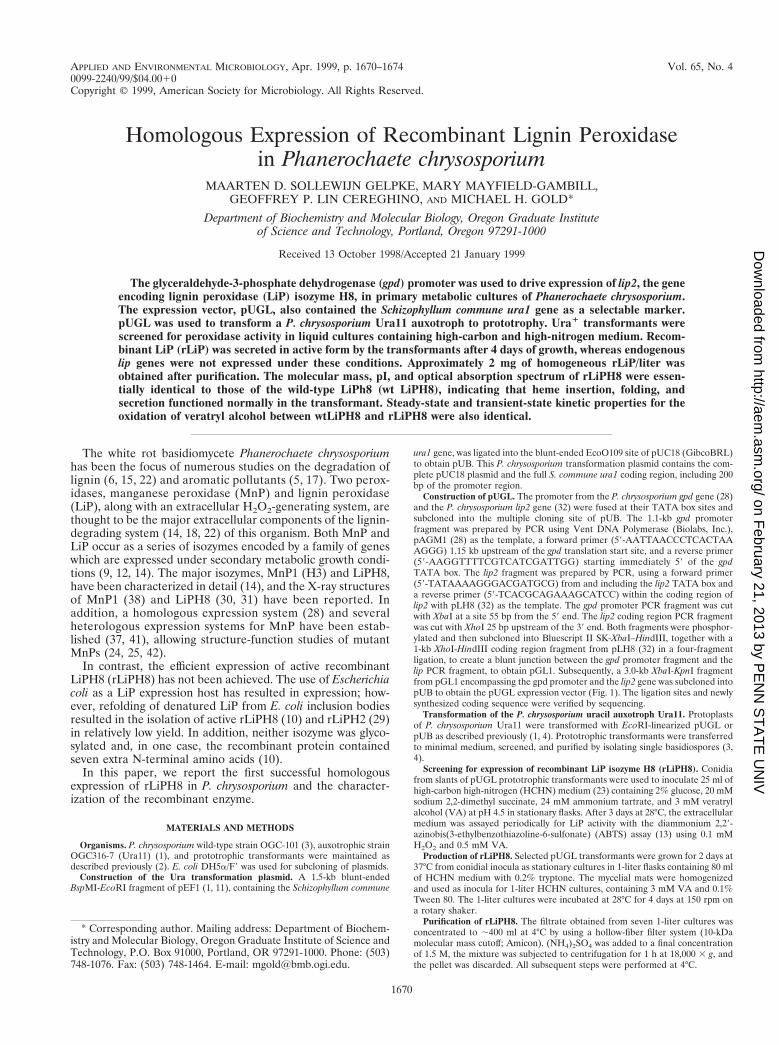
Construction of pUGL. The promoter from the P. chrysosporium gpd gene (28)and the P. chrysosporium lip2 gene (32) were fused at their TATA box sites andsubcloned into the multiple cloning site of pUB. The 1.1-kb gpd promoterfragment was prepared by PCR using Vent DNA Polymerase (Biolabs, Inc.),pAGM1 (28) as the template, a forward primer (59-AATTAACCCTCACTAAAGGG) 1.15 kb upstream of the gpd translation start site, and a reverse primer(59-AAGGTTTTCGTCATCGATTGG) starting immediately 59 of the gpdTATA box. The lip2 fragment was prepared by PCR, using a forward primer(59-TATAAAAGGGACGATGCG) from and including the lip2 TATA box anda reverse primer (59-TCACGCAGAAAGCATCC) within the coding region oflip2 with pLH8 (32) as the template. The gpd promoter PCR fragment was cutwith XbaI at a site 55 bp from the 59 end. The lip2 coding region PCR fragmentwas cut with XhoI 25 bp upstream of the 39 end. Both fragments were phosphor-ylated and then subcloned into Bluescript II SK-XbaI–HindIII, together with a1-kb XhoI-HindIII coding region fragment from pLH8 (32) in a four-fragmentligation, to create a blunt junction between the gpd promoter fragment and thelip PCR fragment, to obtain pGL1. Subsequently, a 3.0-kb XbaI-KpnI fragmentfrom pGL1 encompassing the gpd promoter and the lip2 gene was subcloned intopUB to obtain the pUGL expression vector (Fig. 1). The ligation sites and newlysynthesized coding sequence were verified by sequencing.
Transformation of the P. chrysosporium uracil auxotroph Ura11. Protoplastsof P. chrysosporium Ura11 were transformed with EcoRI-linearized pUGL orpUB as described previously (1, 4). Prototrophic transformants were transferredto minimal medium, screened, and purified by isolating single basidiospores (3,4).
Screening for expression of recombinant LiP isozyme H8 (rLiPH8). Conidiafrom slants of pUGL prototrophic transformants were used to inoculate 25 ml ofhigh-carbon high-nitrogen (HCHN) medium (23) containing 2% glucose, 20 mMsodium 2,2-dimethyl succinate, 24 mM ammonium tartrate, and 3 mM veratrylalcohol (VA) at pH 4.5 in stationary flasks. After 3 days at 28°C, the extracellularmedium was assayed periodically for LiP activity with the diammonium 2,29-azinobis(3-ethylbenzothiazoline-6-sulfonate) (ABTS) assay (13) using 0.1 mMH2O2 and 0.5 mM VA.
Production of rLiPH8. Selected pUGL transformants were grown for 2 days at37°C from conidial inocula as stationary cultures in 1-liter flasks containing 80 mlof HCHN medium with 0.2% tryptone. The mycelial mats were homogenizedand used as inocula for 1-liter HCHN cultures, containing 3 mM VA and 0.1%Tween 80. The 1-liter cultures were incubated at 28°C for 4 days at 150 rpm ona rotary shaker.
Purification of rLiPH8. The filtrate obtained from seven 1-liter cultures wasconcentrated to ;400 ml at 4°C by using a hollow-fiber filter system (10-kDamolecular mass cutoff; Amicon). (NH4)2SO4 was added to a final concentrationof 1.5 M, the mixture was subjected to centrifugation for 1 h at 18,000 3 g, andthe pellet was discarded. All subsequent steps were performed at 4°C.
* Corresponding author. Mailing address: Department of Biochem-istry and Molecular Biology, Oregon Graduate Institute of Science andTechnology, P.O. Box 91000, Portland, OR 97291-1000. Phone: (503)748-1076. Fax: (503) 748-1464. E-mail: [email protected].
1670
on February 21, 2013 by P
EN
N S
TA
TE
UN
IVhttp://aem
.asm.org/
Dow
nloaded from
Phenyl Sepharose chromatography. The concentrated culture filtrate was ap-plied to a Phenyl Sepharose CL-4B (Pharmacia) column (100 ml) equilibratedwith 20 mM sodium acetate (pH 4.5) containing 1.5 M (NH4)2SO4. The columnwas washed with 200 ml of 20 mM sodium acetate (pH 4.5) containing 0.8 M(NH4)2SO4, and the protein was eluted with a gradient of 0.8 to 0.2 M(NH4)2SO4 in 20 mM sodium acetate (pH 4.5). Fractions with LiP activity werepooled and concentrated to ;2 ml by membrane ultrafiltration.
Size exclusion chromatography. The Phenyl Sepharose fraction was applied toa 100-ml Sephadex G-100 column equilibrated with 20 mM sodium succinatebuffer (pH 4.5), and protein was eluted with the same buffer. Fractions with LiPactivity were desalted and concentrated.
Anion-exchange FPLC. The pooled, concentrated Sephadex G-100 proteinfraction was applied to a Mono Q HR 5/5 column (Pharmacia) equilibrated with10 mM sodium acetate (pH 6.0) in a fast protein liquid chromatography (FPLC)system. The protein was eluted with a nonlinear gradient (0.01 to 0.5 M sodiumacetate; pH 6.0). Fractions containing LiP activity were desalted and concen-trated.
SDS-PAGE and IEF. Sodium dodecyl sulfate-polyacrylamide gel electrophore-sis (SDS-PAGE) was performed using a 12% Tris-glycine gel system (27) and aMiniprotean II apparatus (Bio-Rad). The gels were stained with Coomassie blue(16). The Bio-Rad SDS-PAGE Low Range standard mix was used for compar-ison. Isoelectric focusing (IEF) electrophoresis was performed using the Phar-macia Phastsystem with IEF Phastgels (pH 3 to 9). The gels were stained withCoomassie blue. The Sigma IEF MIX 3.6-6.6 marker kit was used as a standard.
Spectroscopic and kinetic procedures. Enzyme absorption spectra and assayswere determined with a Shimadzu UV-260 spectrophotometer at room temper-ature, using a 1-cm light path cuvette. LiP oxidation of VA to veratraldehyde wasperformed as described previously (15, 22) and followed at 310 nm. LiP oxidationof ABTS was carried out in sodium succinate (pH 3.0) in the presence of 0.1 mMH2O2 and 0.5 mM VA and followed at 415 nm as described previously (13). Forsteady-state kinetic measurements, VA oxidation was determined in the pres-ence of various H2O2 concentrations at a constant VA concentration (0.5 mM)or with various VA concentrations at a constant H2O2 concentration (0.1 mM)in 20 mM sodium-succinate (pH 3.0) with 1 mg of enzyme/ml.
Transient-state kinetics. Kinetic measurements were conducted at 25°C usingan Applied Photophysics stopped-flow reaction analyzer (SX.18MV) with se-quential mixing. LiP compound I formation was measured at 397 nm. Native LiP(2 mM) was mixed with a 10- to 50-fold excess of H2O2 in 20 mM sodiumsuccinate (pH 3.0). LiP compound I reduction was measured by first mixing 4 mMenzyme and 1 equiv of H2O2 in H2O. Then, VA in 40 mM sodium succinate (pH3.0) was added, and compound I reduction was measured at 416 nm. LiP com-pound II reduction was measured at 397 nm by sequential mixing of 4 mMenzyme, 1 eq of ferrocyanide, and 1 eq of H2O2 in H2O and then by adding VAin 40 mM sodium succinate (pH 3.0).
RESULTS
Expression of rLiP. Transformation of the Ura11 auxotrophwith 2 mg of the linearized LiPH8 expression construct pUGL(Fig. 1) resulted in the isolation of 35 Ura1 transformants.Twelve transformants were grown in HCHN stationary cul-tures and screened for extracellular LiP activity using theABTS assay in the presence of VA. Total RNA was extractedfrom 4-day-old cells, and Northern blotting was carried out onthe same sample of transformants. Hybridization with a lip H8cDNA probe revealed that LiP mRNA was present in all trans-formants, although in various amounts (data not shown). Twoof the pUGL transformants with the highest rLiP activity inthis initial screening were purified by isolating single basidio-spores (3) and were analyzed further in large liquid shakecultures. A Southern blot of DNA from one of the transfor-mants probed with pUC18 showed that the transforming DNAwas chromosomally integrated (data not shown).
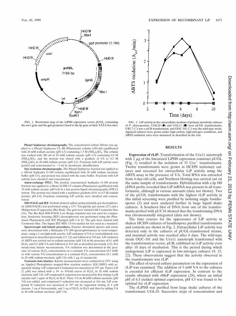
The time courses for the appearance of LiP activity inshaken HCHN cultures of basidiospore purified transformantsand controls are shown in Fig. 2. Extracellular LiP activity wasdetected only in the cultures of pUGL-transformed strains,and maximal activity was reached after 6 days. The wild-typestrain OGC-101 and the Ura11 auxotroph transformed withthe transformation vector, pUB, exhibited no LiP activity evenafter 10 days of incubation. This is the period during whichendogenous LiP is expressed in low-nitrogen cultures (9, 15,22). These observations suggest that the activity observed inthe transformants was rLiP.
The effect of several culture parameters on the expression ofrLiP was examined. The addition of 3 mM VA to the culturesis essential for efficient rLiP expression. In contrast to theresults obtained with rMnP expression (28), where an initialpH of 6.5 yielded optimal expression, pH 4.5 was found to beoptimal for rLiP expression.
The rLiPH8 was purified from large shake cultures of thetransformant UGL10. Successive steps of concentration and
FIG. 1. Restriction map of the LiPH8 expression vector pUGL, containingthe ura1 gene and the gpd promoter fused to the lip gene at their TATA box sites.
FIG. 2. LiP activity in the extracellular medium of primary metabolic culturesof P. chrysosporium. UGL10 (F) and UGL11 (■) were pUGL transformants,UB1.7 (E) was a pUB transformant, and OGC-101 (h) was the wild-type strain.Agitated cultures were grown under high-carbon, high-nitrogen conditions, andABTS oxidation rates were measured as described in the text.
VOL. 65, 1999 EXPRESSION OF RECOMBINANT LiP 1671
on February 21, 2013 by P
EN
N S
TA
TE
UN
IVhttp://aem
.asm.org/
Dow
nloaded from
hydrophobic-interaction, size exclusion, and anion-exchangechromatography were performed. Mono Q anion-exchangechromatography demonstrated that rLiP activity eluted asa single peak corresponding to that of wild-type LiPH8(wtLiPH8) (data not shown).
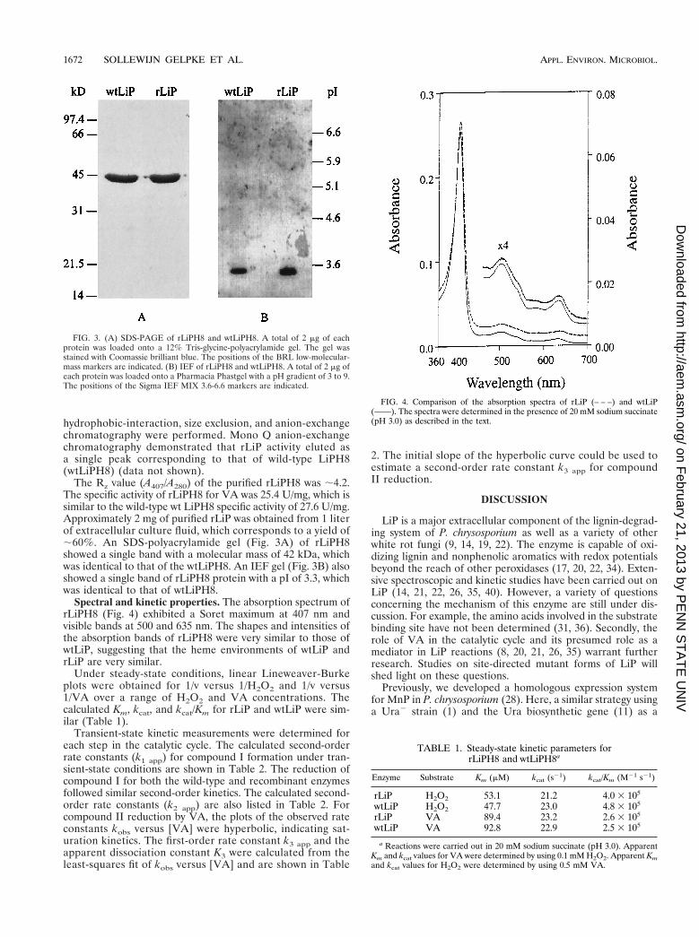
The Rz value (A407/A280) of the purified rLiPH8 was ;4.2.The specific activity of rLiPH8 for VA was 25.4 U/mg, which issimilar to the wild-type wt LiPH8 specific activity of 27.6 U/mg.Approximately 2 mg of purified rLiP was obtained from 1 literof extracellular culture fluid, which corresponds to a yield of;60%. An SDS-polyacrylamide gel (Fig. 3A) of rLiPH8showed a single band with a molecular mass of 42 kDa, whichwas identical to that of the wtLiPH8. An IEF gel (Fig. 3B) alsoshowed a single band of rLiPH8 protein with a pI of 3.3, whichwas identical to that of wtLiPH8.
Spectral and kinetic properties. The absorption spectrum ofrLiPH8 (Fig. 4) exhibited a Soret maximum at 407 nm andvisible bands at 500 and 635 nm. The shapes and intensities ofthe absorption bands of rLiPH8 were very similar to those ofwtLiP, suggesting that the heme environments of wtLiP andrLiP are very similar.
Under steady-state conditions, linear Lineweaver-Burkeplots were obtained for 1/v versus 1/H2O2 and 1/v versus1/VA over a range of H2O2 and VA concentrations. Thecalculated Km, kcat, and kcat/Km for rLiP and wtLiP were sim-ilar (Table 1).
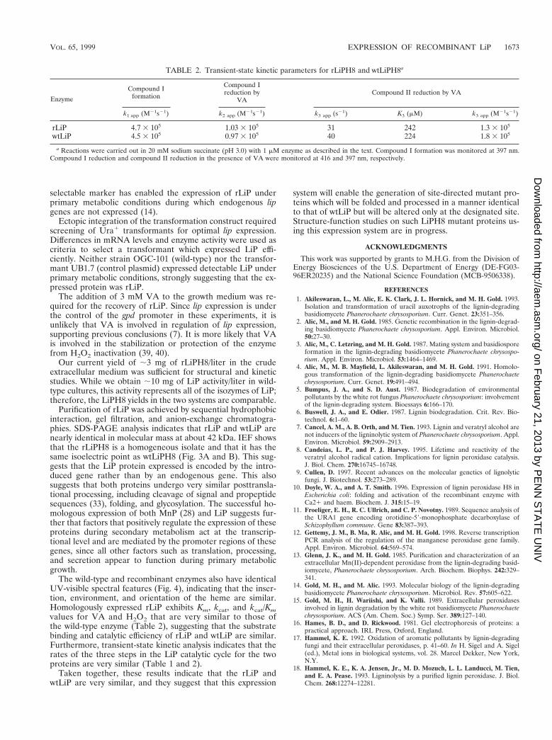
Transient-state kinetic measurements were determined foreach step in the catalytic cycle. The calculated second-orderrate constants (k1 app) for compound I formation under tran-sient-state conditions are shown in Table 2. The reduction ofcompound I for both the wild-type and recombinant enzymesfollowed similar second-order kinetics. The calculated second-order rate constants (k2 app) are also listed in Table 2. Forcompound II reduction by VA, the plots of the observed rateconstants kobs versus [VA] were hyperbolic, indicating sat-uration kinetics. The first-order rate constant k3 app and theapparent dissociation constant K3 were calculated from theleast-squares fit of kobs versus [VA] and are shown in Table
2. The initial slope of the hyperbolic curve could be used toestimate a second-order rate constant k3 app for compoundII reduction.
DISCUSSION
LiP is a major extracellular component of the lignin-degrad-ing system of P. chrysosporium as well as a variety of otherwhite rot fungi (9, 14, 19, 22). The enzyme is capable of oxi-dizing lignin and nonphenolic aromatics with redox potentialsbeyond the reach of other peroxidases (17, 20, 22, 34). Exten-sive spectroscopic and kinetic studies have been carried out onLiP (14, 21, 22, 26, 35, 40). However, a variety of questionsconcerning the mechanism of this enzyme are still under dis-cussion. For example, the amino acids involved in the substratebinding site have not been determined (31, 36). Secondly, therole of VA in the catalytic cycle and its presumed role as amediator in LiP reactions (8, 20, 21, 26, 35) warrant furtherresearch. Studies on site-directed mutant forms of LiP willshed light on these questions.
Previously, we developed a homologous expression systemfor MnP in P. chrysosporium (28). Here, a similar strategy usinga Ura2 strain (1) and the Ura biosynthetic gene (11) as a
FIG. 3. (A) SDS-PAGE of rLiPH8 and wtLiPH8. A total of 2 mg of eachprotein was loaded onto a 12% Tris-glycine-polyacrylamide gel. The gel wasstained with Coomassie brilliant blue. The positions of the BRL low-molecular-mass markers are indicated. (B) IEF of rLiPH8 and wtLiPH8. A total of 2 mg ofeach protein was loaded onto a Pharmacia Phastgel with a pH gradient of 3 to 9.The positions of the Sigma IEF MIX 3.6-6.6 markers are indicated.
FIG. 4. Comparison of the absorption spectra of rLiP (– – –) and wtLiP(——). The spectra were determined in the presence of 20 mM sodium succinate(pH 3.0) as described in the text.
TABLE 1. Steady-state kinetic parameters forrLiPH8 and wtLiPH8a
Enzyme Substrate Km (mM) kcat (s21) kcat/Km (M21 s21)
rLiP H2O2 53.1 21.2 4.0 3 105
wtLiP H2O2 47.7 23.0 4.8 3 105
rLiP VA 89.4 23.2 2.6 3 105
wtLiP VA 92.8 22.9 2.5 3 105
a Reactions were carried out in 20 mM sodium succinate (pH 3.0). ApparentKm and kcat values for VA were determined by using 0.1 mM H2O2. Apparent Kmand kcat values for H2O2 were determined by using 0.5 mM VA.
1672 SOLLEWIJN GELPKE ET AL. APPL. ENVIRON. MICROBIOL.
on February 21, 2013 by P
EN
N S
TA
TE
UN
IVhttp://aem
.asm.org/
Dow
nloaded from
selectable marker has enabled the expression of rLiP underprimary metabolic conditions during which endogenous lipgenes are not expressed (14).
Ectopic integration of the transformation construct requiredscreening of Ura1 transformants for optimal lip expression.Differences in mRNA levels and enzyme activity were used ascriteria to select a transformant which expressed LiP effi-ciently. Neither strain OGC-101 (wild-type) nor the transfor-mant UB1.7 (control plasmid) expressed detectable LiP underprimary metabolic conditions, strongly suggesting that the ex-pressed protein was rLiP.
The addition of 3 mM VA to the growth medium was re-quired for the recovery of rLiP. Since lip expression is underthe control of the gpd promoter in these experiments, it isunlikely that VA is involved in regulation of lip expression,supporting previous conclusions (7). It is more likely that VAis involved in the stabilization or protection of the enzymefrom H2O2 inactivation (39, 40).
Our current yield of ;3 mg of rLiPH8/liter in the crudeextracellular medium was sufficient for structural and kineticstudies. While we obtain ;10 mg of LiP activity/liter in wild-type cultures, this activity represents all of the isozymes of LiP;therefore, the LiPH8 yields in the two systems are comparable.
Purification of rLiP was achieved by sequential hydrophobicinteraction, gel filtration, and anion-exchange chromatogra-phies. SDS-PAGE analysis indicates that rLiP and wtLiP arenearly identical in molecular mass at about 42 kDa. IEF showsthat the rLiPH8 is a homogeneous isolate and that it has thesame isoelectric point as wtLiPH8 (Fig. 3A and B). This sug-gests that the LiP protein expressed is encoded by the intro-duced gene rather than by an endogenous gene. This alsosuggests that both proteins undergo very similar posttransla-tional processing, including cleavage of signal and propeptidesequences (33), folding, and glycosylation. The successful ho-mologous expression of both MnP (28) and LiP suggests fur-ther that factors that positively regulate the expression of theseproteins during secondary metabolism act at the transcrip-tional level and are mediated by the promoter regions of thesegenes, since all other factors such as translation, processing,and secretion appear to function during primary metabolicgrowth.
The wild-type and recombinant enzymes also have identicalUV-visible spectral features (Fig. 4), indicating that the inser-tion, environment, and orientation of the heme are similar.Homologously expressed rLiP exhibits Km, kcat, and kcat/Kmvalues for VA and H2O2 that are very similar to those ofthe wild-type enzyme (Table 2), suggesting that the substratebinding and catalytic efficiency of rLiP and wtLiP are similar.Furthermore, transient-state kinetic analysis indicates that therates of the three steps in the LiP catalytic cycle for the twoproteins are very similar (Table 1 and 2).
Taken together, these results indicate that the rLiP andwtLiP are very similar, and they suggest that this expression
system will enable the generation of site-directed mutant pro-teins which will be folded and processed in a manner identicalto that of wtLiP but will be altered only at the designated site.Structure-function studies on such LiPH8 mutant proteins us-ing this expression system are in progress.
ACKNOWLEDGMENTS
This work was supported by grants to M.H.G. from the Division ofEnergy Biosciences of the U.S. Department of Energy (DE-FG03-96ER20235) and the National Science Foundation (MCB-9506338).
REFERENCES1. Akileswaran, L., M. Alic, E. K. Clark, J. L. Hornick, and M. H. Gold. 1993.
Isolation and transformation of uracil auxotrophs of the lignin-degradingbasidiomycete Phanerochaete chrysosporium. Curr. Genet. 23:351–356.
2. Alic, M., and M. H. Gold. 1985. Genetic recombination in the lignin-degrad-ing basidiomycete Phanerochaete chrysosporium. Appl. Environ. Microbiol.50:27–30.
3. Alic, M., C. Letzring, and M. H. Gold. 1987. Mating system and basidiosporeformation in the lignin-degrading basidiomycete Phanerochaete chrysospo-rium. Appl. Environ. Microbiol. 53:1464–1469.
4. Alic, M., M. B. Mayfield, L. Akileswaran, and M. H. Gold. 1991. Homolo-gous transformation of the lignin-degrading basidiomycete Phanerochaetechrysosporium. Curr. Genet. 19:491–494.
5. Bumpus, J. A., and S. D. Aust. 1987. Biodegradation of environmentalpollutants by the white rot fungus Phanerochaete chrysosporium: involvementof the lignin-degrading system. Bioessays 6:166–170.
6. Buswell, J. A., and E. Odier. 1987. Lignin biodegradation. Crit. Rev. Bio-technol. 6:1–60.
7. Cancel, A. M., A. B. Orth, and M. Tien. 1993. Lignin and veratryl alcohol arenot inducers of the ligninolytic system of Phanerochaete chrysosporium. Appl.Environ. Microbiol. 59:2909–2913.
8. Candeias, L. P., and P. J. Harvey. 1995. Lifetime and reactivity of theveratryl alcohol radical cation. Implications for lignin peroxidase catalysis.J. Biol. Chem. 270:16745–16748.
9. Cullen, D. 1997. Recent advances on the molecular genetics of lignolyticfungi. J. Biotechnol. 53:273–289.
10. Doyle, W. A., and A. T. Smith. 1996. Expression of lignin peroxidase H8 inEscherichia coli: folding and activation of the recombinant enzyme withCa21 and haem. Biochem. J. 315:15–19.
11. Froeliger, E. H., R. C. Ullrich, and C. P. Novotny. 1989. Sequence analysis ofthe URA1 gene encoding orotidine-59-monophosphate decarboxylase ofSchizophyllum commune. Gene 83:387–393.
12. Gettemy, J. M., B. Ma, R. Alic, and M. H. Gold. 1998. Reverse transcriptionPCR analysis of the regulation of the manganese peroxidase gene family.Appl. Environ. Microbiol. 64:569–574.
13. Glenn, J. K., and M. H. Gold. 1985. Purification and characterization of anextracellular Mn(II)-dependent peroxidase from the lignin-degrading basid-iomycete, Phanerochaete chrysosporium. Arch. Biochem. Biophys. 242:329–341.
14. Gold, M. H., and M. Alic. 1993. Molecular biology of the lignin-degradingbasidiomycete Phanerochaete chrysosporium. Microbiol. Rev. 57:605–622.
15. Gold, M. H., H. Wariishi, and K. Valli. 1989. Extracellular peroxidasesinvolved in lignin degradation by the white rot basidiomycete Phanerochaetechrysosporium. ACS (Am. Chem. Soc.) Symp. Ser. 389:127–140.
16. Hames, B. D., and D. Rickwood. 1981. Gel electrophoresis of proteins: apractical approach. IRL Press, Oxford, England.
17. Hammel, K. E. 1992. Oxidation of aromatic pollutants by lignin-degradingfungi and their extracellular peroxidases, p. 41–60. In H. Sigel and A. Sigel(ed.), Metal ions in biological systems, vol. 28. Marcel Dekker, New York,N.Y.
18. Hammel, K. E., K. A. Jensen, Jr., M. D. Mozuch, L. L. Landucci, M. Tien,and E. A. Pease. 1993. Ligninolysis by a purified lignin peroxidase. J. Biol.Chem. 268:12274–12281.
TABLE 2. Transient-state kinetic parameters for rLiPH8 and wtLiPH8a
Enzyme
Compound Iformation
Compound Ireduction by
VACompound II reduction by VA
k1 app (M21s21) k2 app (M21s21) k3 app (s21) K3 (mM) k3 app (M21s21)
rLiP 4.7 3 105 1.03 3 105 31 242 1.3 3 105
wtLiP 4.5 3 105 0.97 3 105 40 224 1.8 3 105
a Reactions were carried out in 20 mM sodium succinate (pH 3.0) with 1 mM enzyme as described in the text. Compound I formation was monitored at 397 nm.Compound I reduction and compound II reduction in the presence of VA were monitored at 416 and 397 nm, respectively.
VOL. 65, 1999 EXPRESSION OF RECOMBINANT LiP 1673
on February 21, 2013 by P
EN
N S
TA
TE
UN
IVhttp://aem
.asm.org/
Dow
nloaded from
19. Hatakka, A. 1994. Lignin-modifying enzymes from selected white-rot fungi:production and role in lignin degradation. FEMS Microbiol. Rev. 13:125–135.
20. Joshi, D. K., and M. H. Gold. 1996. Oxidation of dimethoxylated aromaticcompounds by lignin peroxidase from Phanerochaete chrysosporium. Eur.J. Biochem. 237:45–57.
21. Khindaria, A., I. Yamazaki, and S. D. Aust. 1996. Stabilization of the veratrylalcohol cation radical by lignin peroxidase. Biochemistry 35:6418–6424.
22. Kirk, T. K., and R. L. Farrell. 1987. Enzymatic “combustion”: the microbialdegradation of lignin. Annu. Rev. Microbiol. 41:465–505.
23. Kirk, T. K., E. Schultz, W. J. Connors, L. F. Lorenz, and J. G. Zeikus. 1978.Influence of culture parameters on lignin metabolism by Phanerochaetechrysosporium. Arch. Microbiol. 177:277–285.
24. Kishi, K., D. P. Hildebrand, M. Kusters-van Someren, J. Gettemy, A. G.Mauk, and M. H. Gold. 1997. Site-directed mutations at phenylalanine-190of manganese peroxidase: effects on stability, function, and coordination.Biochemistry 36:4268–4277.
25. Kishi, K., M. Kusters-van Someren, M. B. Mayfield, J. Sun, T. M. Loehr,and M. H. Gold. 1996. Characterization of manganese(II) binding site mu-tants of manganese peroxidase. Biochemistry 35:8986–8994.
26. Koduri, R. S., and M. Tien. 1995. Oxidation of guaiacol by lignin peroxidase.Role of veratryl alcohol. J. Biol. Chem. 270:22254–22258.
27. Laemmli, U. K. 1970. Cleavage of structural proteins during the assembly ofthe head of bacteriophage T4. Nature 227:680–685.
28. Mayfield, M. B., K. Kishi, M. Alic, and M. H. Gold. 1994. Homologousexpression of recombinant manganese peroxidase in Phanerochaete chryso-sporium. Appl. Environ. Microbiol. 60:4303–4309.
29. Nie, G., N. S. Reading, and S. D. Aust. 1998. Expression of the ligninperoxidase H2 gene from Phanerochaete chrysosporium in Escherichia coli.Biochem. Biophys. Res. Commun. 249:146–150.
30. Piontek, K., T. Glumoff, and K. Winterhalter. 1993. Low pH crystal structureof glycosylated lignin peroxidase from Phanerochaete chrysosporium at 2.5 A
resolution. FEBS Lett. 315:119–124.31. Poulos, T. L., S. L. Edwards, H. Wariishi, and M. H. Gold. 1993. Crystallo-
graphic refinement of lignin peroxidase at 2 Å. J. Biol. Chem. 268:4429–4440.32. Ritch, T. G., Jr., and M. H. Gold. 1992. Characterization of a highly ex-
pressed lignin peroxidase-encoding gene from the basidiomycete Phanero-chaete chrysosporium. Gene 118:73–80.
33. Ritch, T. G., Jr., V. J. Nipper, L. Akileswaran, A. J. Smith, D. G. Pribnow,and M. H. Gold. 1991. Lignin peroxidase from the basidiomycete Phanero-chaete chrysosporium is synthesized as a preproenzyme. Gene 107:119–126.
34. Schoemaker, H. E. 1990. On the chemistry of lignin biodegradation. Recl.Trav. Chim. Pays-Bas 109:255–272.
35. Sheng, D., and M. H. Gold. 1998. Irreversible oxidation of ferricytochromec by lignin peroxidase. Biochemistry 37:2029–2036.
36. Smith, A. T., and N. C. Veitch. 1998. Substrate binding and catalysis in hemeperoxidases. Curr. Opin. Chem. Biol. 2:269–278.
37. Stewart, P., R. E. Whitwam, P. J. Kersten, D. Cullen, and M. Tien. 1996.Efficient expression of a Phanerochaete chrysosporium manganese peroxidasegene in Aspergillus oryzae. Appl. Environ. Microbiol. 62:860–864.
38. Sundaramoorthy, M., K. Kishi, M. H. Gold, and T. L. Poulos. 1994. Thecrystal structure of manganese peroxidase from Phanerochaete chrysosporiumat 2.06-A resolution. J. Biol. Chem. 269:32759–32767.
39. Tonon, F., and E. Odier. 1988. Influence of veratryl alcohol and hydrogenperoxide on ligninase activity and ligninase production by Phanerochaetechrysosporium. Appl. Environ. Microbiol. 54:466–472.
40. Wariishi, H., and M. H. Gold. 1990. Lignin peroxidase compound III. Mech-anism of formation and decomposition. J. Biol. Chem. 265:2070–2077.
41. Whitwam, R., and M. Tien. 1996. Heterologous expression and reconstitu-tion of fungal Mn peroxidase. Arch. Biochem. Biophys. 333:439–446.
42. Whitwam, R. E., K. R. Brown, M. Musick, M. J. Natan, and M. Tien. 1997.Mutagenesis of the Mn21-binding site of manganese peroxidase affects ox-idation of Mn21 by both compound I and compound II. Biochemistry 36:9766–9773.
1674 SOLLEWIJN GELPKE ET AL. APPL. ENVIRON. MICROBIOL.
on February 21, 2013 by P
EN
N S
TA
TE
UN
IVhttp://aem
.asm.org/
Dow
nloaded from


![Hormographiella aspergillata: an emerging basidiomycete in ...filamentous basidiomycete found in numerous substrates including soils, leaves, pressmud compost and in the air [ 3, 4]](https://img.pdfslide.net/doc/110x75/6123c989ca7b815f6634ae2f/hormographiella-aspergillata-an-emerging-basidiomycete-in-filamentous-basidiomycete.jpg)