Embed Size (px)
Citation preview

Hormone sensitive lipase:structure, function and
regulation
Jeroen de MeijerMrt-1998/Mei-1998


Front page illustration:Hormone sensitive lipase (kindly provided by J.A. Contreras and C. Holm)

Hormone sensitivelipase: structure,
function andregulation

Hormone sensitivelipase: structure,
function andregulation
A thesis writen at the Biochemical Physiology Research Group,
Department of Experimental Zoology, University of Utrecht, under
supervision of dr. W. J. A. van Marrewijk
J. de Meijer 1998
6Abstract
AAbstract
Hormone sensitive lipase (HSL) is the key enzyme in the regula-
tion of lipid stores. It is the rate limiting enzyme in the degradation
of triacylglycerol (TAG) to diacylglycerol (DAG) and free fatty
acids (FFA). In addition, it has hydrolyzing activity against choles-
terol esters. In this sight it is not remarkable that HSL is not only
found in adipose tissue, but also in tissues that store cholesterol
esters. In these tissues HSL plays a key role in the cholesterol me-
tabolism.
HSL is regulated by reversible phosphorylation on four residues.
Phosphorylation alone, however, is not enough to activate HSL. It
probably also involves conformational changes and a translocation
from the cytoplasm to the lipid droplet.
HSL is a product of the HSL gene, located in human on chromo-
some 19, which contains 9 exons. Every exon (or exons) gives rise
to a distinct domain of HSL suggesting that the HSL gene is a mo-
saic gene. The promoter of the HSL gene has only recently been
characterized and has no TATA or CCAAT box. It does contain
the consensus sequences common in TATA-less promoters like a
GC-rich region, an AT-rich region and an initiator region. The regu-
lation of the gene remains obscure at this moment.
The HSL protein has an α/β hydrolase fold conformation. It con-
tains a catalytic triad commonly found in lipases. The regulatory
phosphorylation sites are located on a loop protruding the protein.
The carboxyl terminal and/or the amino terminal can have a role in
lipid binding.
The knowledge about HSL is still premature but is growing rap-
idly. Not remarkably, as knowledge about lipid metabolism is of
crucial importance in several health issues today.

7Contents
C ContentsAbstract .................................................... 6Contents .................................................... 71 Introduction .......................................... 82 Physiological role of HSL................... 103 Regulation of HSL .............................. 124 Structure of HSL gene and protein... 144.1 Homology ............................................................................ 144.2 Gene structure ..................................................................... 154.3 Protein structure ................................................................... 17
5 Phosphorylation of HSL .................... 216 Translocation ...................................... 247 Perilipins.............................................. 268 Artherosclerosis .................................. 279 Model ................................................... 3010 Concluding remarks .......................... 32References............................................... 35Acknowledgements................................. 42

Introduction 8
1Introduction
Every organism continuously uses energy; not only for move-
ment or to perform labor, but also for maintenance, regulation and
almost any other process. Organisms derive their energy from the
breakdown of nutrients they take up. The type of nutrients can
roughly be divided in three groups: proteins, carbohydrates and
lipids. During breakdown of these nutrients energy is produced in
the form of ATP, which is the universal energy carrier in organ-
isms. Most nutrients, however, are not directly used for energy, but
are stored in tissues in an appropriate form for later use.
Proteins are normally not used for energy but broken down into
amino acids which can be used to construct new proteins. Only in
case of a severe lack of energy sources proteins are used for en-
ergy. In that case the proteins are broken down into alanine and
glutamine and processed into glucose by the liver and the kidney
respectively. This is important in the brain as this tissue can not
utilize lipids as an energy source.
Carbohydrates are, however, a major energy source. Used carbo-
hydrates are mostly sugars like glucose or trehalose. These sugars
a b
Introduction 9
Figure 1;a) The glycolytic pathway by whichglucose is degraded to twomolecules of pyruvate. Along theway two ATP molecules and twomolecules NADH+H+ are formed.Pyruvate is subsequently convertedinto acetyl-CoA by the pyruvatedehydrogenase complex (not infigure). In this process onemolecule of NADH+H+ is formedand one molecule CO
2.
b) The citric acid cycle. A twocarbon acetyl residue from acetyl-CoA condenses with oxaloacetate(1) to form citrate. In a cascade ofreactions (2-9) citrate is convertedin oxaloacetate which cansubsequently condense again witha two carbon acetyl residue fromacetyl-CoA. Every cycle producesthree molecules NADH+H+, onemolecule FADH
2, one molecule
GTP, and 2 molecules CO2.
NADH+H+, FADH2 and GTP are
used to produce ATP, the overallenergy carrier in the cell. (Adaptedfrom Lodish et al. 1995)
are, in mammals, stored as glycogen in the liver. When needed, the
glycogen is broken down into sugars and released to the circulation
system. A key regulator in this breakdown is the enzyme glycogen
phosphorylase. Carbohydrates provide a quick energy source and
are degraded in the well known glycolytic pathway and the subse-
quent citric acid cycle (Figure 1). The pool of available carbohy-
drates is, however, quickly exhausted, making carbohydrates in-
sufficient as an energy source for long endurances. And this is where
lipids come into play.
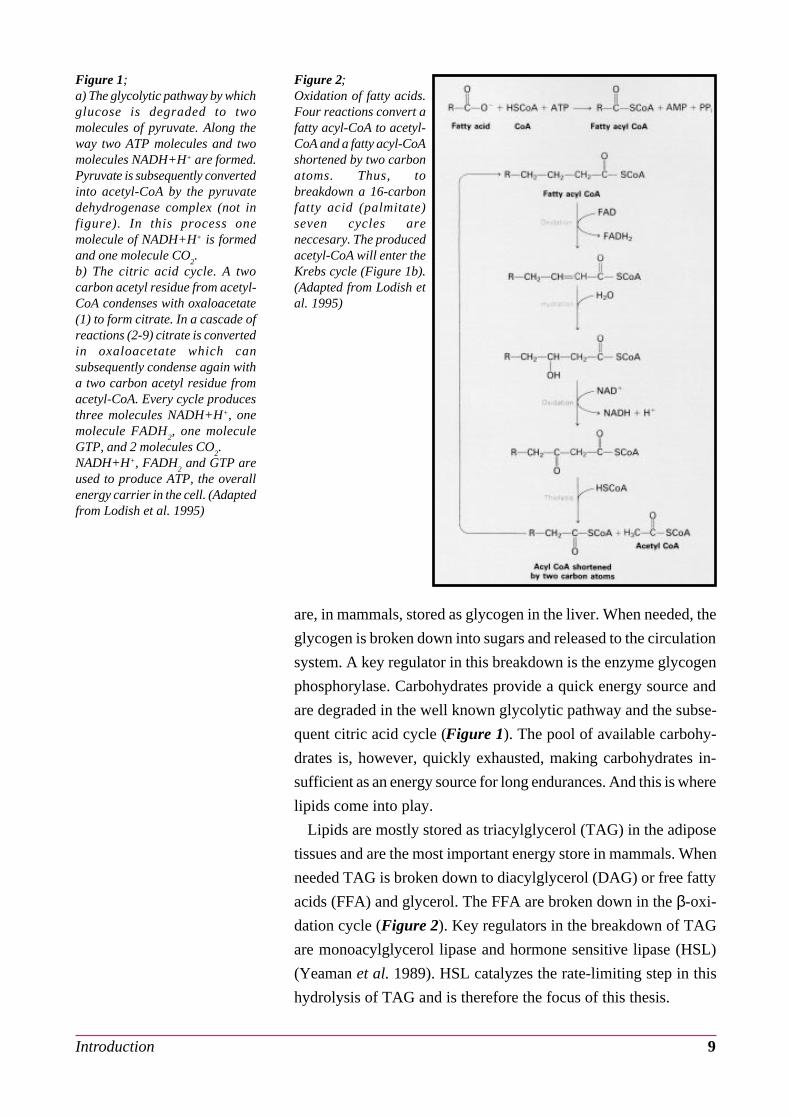
Lipids are mostly stored as triacylglycerol (TAG) in the adipose
tissues and are the most important energy store in mammals. When
needed TAG is broken down to diacylglycerol (DAG) or free fatty
acids (FFA) and glycerol. The FFA are broken down in the β-oxi-
dation cycle (Figure 2). Key regulators in the breakdown of TAG
are monoacylglycerol lipase and hormone sensitive lipase (HSL)
(Yeaman et al. 1989). HSL catalyzes the rate-limiting step in this
hydrolysis of TAG and is therefore the focus of this thesis.
Figure 2;Oxidation of fatty acids.Four reactions convert afatty acyl-CoA to acetyl-CoA and a fatty acyl-CoAshortened by two carbonatoms. Thus, tobreakdown a 16-carbonfatty acid (palmitate)seven cycles areneccesary. The producedacetyl-CoA will enter theKrebs cycle (Figure 1b).(Adapted from Lodish etal. 1995)

Physiological role of hormone sensitive lipase 10
2Physiological role ofhormone sensitive lipase
The use of energy stores is, to meet the continuously changing
demand for energy, tightly regulated depending mainly on neural
and hormonal signals.
During activity energy sources are modulated to ensure the avail-
ability of the appropriate form of energy. For instance, during long
endurances, the source switches from the "fast" energy (carbohy-
drates) to the "slow" energy (lipids). Upon starvation this switch is
also seen. Only the brain keeps using carbohydrates as it can not
metabolize lipids. When starvation proceeds, proteins are used for
energy and also ketone bodies are formed which the brain can use
for energy. It will be clear that regulation is of crucial importance.
Deregulation can have a serious impact on the health of the organ-
ism, for instance obesity or diabetes.
As stated before, HSL is a key enzyme in the regulation of the
most important energy source: lipids. HSL is, as the name implies,
hormonally regulated. In response to various lipolytic hormones
HSL is phosphorylated and activated. This phosphorylation is re-
versible, for instance in response to anti-lipolytic hormones like
insulin (Yeaman 1990).
TAG has three ester bonds which can be hydrolyzed, resulting in
FFAs (Figure 3). Although HSL is capable of fully hydrolyzing
TAG to glycerol and FFA, HSL has a marked specificity for the
1(3)-ester bond (Fredrikson et al. 1983). There is evidence that the
Figure 3;Triacylglycerol consists of aglycerol backbone and three lipidresidues. The bond can behydrolyzed to produce free fattyacids and glycerol. (Adapted fromLodish et al. 1995)

Physiological role of hormone sensitive lipase 11
2-ester bond is hydrolyzed by monoacylglycerol lipase and that
this enzyme is required for an efficient complete hydrolysis of TAG
(Fredrikson et al. 1986).
HSL, however, not only possesses activity against TAG, DAG,
and monoacylglycerol (MAG) but also against the long chain es-
ters of cholesterol. Remarkably, the activity of HSL against choles-
terol is approximately equal to its activity against TAG (Fredrikson
et al. 1981). This suggests an additional role for HSL in cholesterol
metabolism, beside the hydrolysis of lipids.
As one would aspect from its dual role, HSL is not only present
in adipose tissues but is also found in tissues in which cholesterol
esters are stored. For instance adrenal cortex, ovaries, heart (Small
et al. 1989b), muscle and macrophages. The first evidence for this
was found when 84 kDa neutral cholesterol ester hydrolase was
found in bovine adrenal cortex tissue (Cook et al. 1981). The activ-
ity of this hydrolase, like HSL, increased in the presence of cyclic
AMP-dependent protein kinase (Beckett et al. 1977). Later it was
confirmed that this hydrolase was indeed HSL (Cook et al. 1982).
In addition, HSL was found in the corpus luteum which is also a
tissue that produces steroid hormones (Cook et al. 1983). The theory
is postulated that the role of HSL in these steroid producing tissues
is the supply of free cholesterol which is the precursor for steroido-
genesis. The role of HSL in macrophages will be discussed later.
Summarizing, roughly three roles can be distinguished:
- lipid metabolism
- cholesterol metabolism
- steroidogenesis

Regulation of hormone sensitive lipase 12
3Regulation of hormonesensitive lipase
In contrast to the regulation of the energy stored in carbohydrates,
much less is known about the regulation of the lipid energy stores.
HSL plays a key role in this lipid metabolism. This hydrolyzing
enzyme is regulated by reversible phosphorylation. This reversible
phosphorylation is under tight hormonal control, and as a result so
is HSL. When rat adipocytes were treated with lipolytic agents (like
noradrenaline) HSL phosphorylation was increased (Belfrage et al.
1980). When these adipocytes were treated with the known anti-
lipolytic agent, insulin, HSL showed a decrease in phosphorylation
(Nilsson et al. 1980). Phosphorylation of HSL will be discussed in
more detail in chapter 5.
The regulation of HSL is not solely on the level of protein phos-
phorylation as phosphorylation of HSL does not seem to be suffi-
cient to obtain a high level of HSL activity. There is a dramatic
difference between the activity of phosphorylated HSL in vitro and
in vivo: the activity in vitro being substantially lower (Nilsson et al.
1980, Fredrikson et al. 1981, Cook et al. 1982, Strålfors et al. 1983).
There is evidence that a translocation, possibly induced by phos-
phorylation, is necessary to yield a high HSL activity. This will be
discussed in more detail in chapter 6. In addition, accessory pro-
teins, like perilipin discussed in chapter 7, seem to be involved in
the HSL regulation. Beside this, also the composition of the substrate
droplet seems to effect the activity of HSL. Okuda et al. (1994)
have shown that the activity of HSL is greatly influenced by the
phospholipid content of the lipid droplet surface. They also showed
that hormones can act on endogenous fat as substrate (Okuda et al.
1983, 1986). If hormones change the interfacial properties of the
lipid droplet it could effect the liplytic activity of the fat cell. The
presence of phosphatidylcholine on the droplet surface greatly re-
duced the responsiveness of the lipid droplets to HSL. Phospholi-
pase C seems to restore the responsiveness.
HSL is possibly also subject to regulation at the gene level. In the
mouse HSL gene regulatory elements have been found in the 5'
flanking region that controls expression in specific tissues (Li et al.

Regulation of hormone sensitive lipase 13
1994). In addition a sterol regulatory element has been found in
HSL which regulates the transcription of several genes encoding
proteins involved in cholesterol metabolism (Goldstein et al. 1990).
Very recently, Laurell et al. (1997) showed that HSL is subject to
species-specific alternative splicing, generating a short form of HSL
mRNA. This mRNA is a result of skipping exon 6 which contains a
part of the catalytic triad necessary for HSL activity. They postu-
late that this alternative splicing of HSL mRNA could have a role
in the fine regulation of HSL as this splicing decreases the amount
of mRNA that can be translated into functional HSL and thereby
reduces the amount of functional HSL.
Before investigating the regulation of HSL in more detail, we
will first discuss the structure of the HSL gene and protein.

Structure of the hormone sensitive lipase gene and protein 14
4Structure of thehormone sensitive lipasegene and protein
4.1 HomologyMost enzymes can be categorized in families. At first, however,
HSL did not seem to be a member of the known family of mamma-
lian lipases which includes lipases as lipoprotein lipase , hepatic
lipase and pancreatic lipase (Cordle et al. 1986, Kirchgessner et al.
1986). Virtually no homology was found with any mammalian pro-
tein (Langin et al. 1993, Li et al. 1994). Only one lipase showing
amino acid sequence homology with HSL was found so far: the
cold-adapted lipase 2 of Moraxella TA144 (Langin et al. 1993a).
This suggests that HSL could also have cold-adaptability proper-
ties. This idea is supported by the observation that HSL shows a
relatively high activity at low temperatures (Langin et al. 1993a).
There are, however, also similarities found, in the same regions as
with Moraxella TA144, between HSL and a prokaryotic enzyme
which was found in hot springs Bacillus acidocaldarius (Langin et
al. 1993b). Recently, however, Contreras et al. (1996) found a re-
markable secundary structure homology of the HSL protein with
the family of lipases and esterases. Recently it is thought that HSL
is a member of an esterase subfamily of a newly described
superfamily of lipases/esterases, described by Hemilä et al. (1994).
Figure 4;Organization of the human HSLgene and amino acid sequence ofhuman HSL. The top partillustrates the exon-intronorganization of the human HSLgene. Exons are represented byboxes, intron by lines. Hatchedareas are noncoding regions.Different functional regions areencoded by different exons asindicated. The bottom partillustrates the aminoacid sequenceof human HSL. The catalytic siteserine (solid line), HG dipeptide(dotted line), the regulatory andbasal phosphorylation sites (closedand open circle respectively), thenewly identified phosphorylationsites (closed and open square) andthe putative lipid binding region(overlined) are indicated. Thedeletion in human HSL comparedto rat HSL is indicated with a ^,and the exon 7/8 boundary isindicated by arrows. (Adaptedfrom Holm et al. 1994)

Structure of the hormone sensitive lipase gene and protein 15
4.2 Gene structureThe rat HSL gene encodes a 786 amino-acid polypeptide (Holm
et al. 1988b, Langin et al. 1993a). The polypeptide reveals no mem-
brane spanning region, suggesting it is a free protein not attached
to a membrane (Holm et al. 1988a). The human HSL is composed
of nine exons and is located on chromosome 19 (Holm et al.
1988a)(Figure 4). It is for 83% identical to rat HSL. It is also slightly
larger compared to rat HSL, and has an 12 amino acid deletion
(Langin et al. 1993a). Exon 6 contains the catalytic site serine mo-
tif found in almost all lipases (G-X-S-X-G). This serine is thought
to be part of a catalytic triad found in many lipases together with
Asp703
and His733
(Contreras et al. 1996). Exon 8 contains the two
serine phosphorylation sites. Exon 9 could contain the lipid bind-
ing region as it encodes a hydrophobic stretch (Holm et al. 1994).
Only recently, any attention was payed to the regulation of the
HSL gene. Grober et al. (1997) are probably the first to elucidate
the regulatory structures of the HSL gene. They showed that up-
stream of exon 1, two 5' untranslated regions (UTRs) are located;
exon A and B, respectively, which are mutually exclusive (Figure
5). Exon A containing transcripts have a very low abundance in
adipocytes. They also identified the transcription start site (tss) which
Figure 5;Organization of the human HSLgene. Closed boxes are codingregions whereas open boxes areuntranslated regions. Exon T is atestis specific exon. Exon Acontaining transcripts are found insignificant amounts in HT29 cellsand at very low levels in adipocytes(Adapted from Grober et al. 1997)
Figure 6;Genomic sequence of the regioncontaining exon B. Thetranscription start site is indicatedwith +1. The putative bindingssites for transcription factors areindicated. The start codon foradipocyte human HSL is shown initalics. Capital letters indicateexonic sequences. (Adapted fromGrober et al. 1997)

Structure of the hormone sensitive lipase gene and protein 16
is located at 5' flanking region of exon B (Figure 6). Using trun-
cated promoter transcripts, ranging from -2400 to -31, they deter-
mined that the 5' border of the minimal promoter is located be-
tween -86 and -57 (Figure 7).
Remarkably, the HSL promoter does not contain a TATA-box or
a CCAAT-box, consensus sequences found in almost all promot-
ers. This indicates that the HSL promoter is a TATA-less promoter.
Several consensus sequences generally present in TATA-less pro-
moter could indeed be found (Figure 6). It contains an CAC-box
between -83 and -76 which can bind Sp1 and related transcription
factors which possibly participate in the trans-activation of the hu-
man HSL promoter (Bucher et al. 1990, Boisclair et al. 1993). It
also contains a GC-rich region which can be bound by Sp1 which
probably plays a role in the stabilization of the initiation complex
(Figure 8). It also contains a AT-rich region between -22 and -27
which could serve as a binding region for the replication complex
(a replacement for the TATA-box). Also the initiator (Inr.) consen-
sus sequence could be found in the HSL gene between -3 and +5.
Although the HSL Inr. sequence differs at two positions from the
Inr. consensus sequence (YYCA+1NWYY (Javahery et al. 1994))
this difference does not seem to disturb the promoter activity (Grober
et al. 1997). This recent understanding of the promoter region forms
the basis for further research to get a better understanding of the
HSL gene regulation.
Figure 7;Deletion analysis of human HSLpromoter activity. Cells weretransfected with HSL promoter-luciferase gene fusion constructs.Data are means of luciferaseactivities ± SEM. The data showthat the 5' border of the minimumpromoter is located between -86and -57. (Adapted from Grober etal. 1997)
Figure 8;Coactivator and tethering modelsfor transcriptional activation bySp1.a) A model for trans-activationthrough coactivators. This modelproposes that specific coactivatorsfunction as adapters, each servingto connect different trans-activating domains into a generaltranscription initiation complex.b) Tethering model for Sp1activation of TATA-less promoters.Sp1 correctly positions theinitiation complex, with the use ofa tethering factor. Also otherregions can aid in this positioning(for instance the AT-rich region).(Adapted from Pugh et al. 1990)

Structure of the hormone sensitive lipase gene and protein 17
4.3 Protein structure
Rat HSL is a polypeptide with a molecular mass of approximately
84 kDa (Belfrage et al. 1977, Fredrikson et al. 1981). Smith et al.
(1996) have done elaborate research on the structure of HSL. Struc-
ture analysis by tryptic digestion suggests that HSL consists of sepa-
rately folded domains with short, protease-sensitive, sequences in
between. This analysis revealed a domain of approximately 17.6
kDa which retained its activity against the water soluble p-
nitrophenyl butyrate (PNPB) but lost its activity against lipid
substrates (Tsujita et al. 1989). This domain is located between
333 and 499 of the rat HSL sequence and contains the active site
serine residue. In addition this domain contains the GXSXG motif
between 421 and 425 of the rat HSL sequence which is conserved
in almost all lipases. This all suggests that this domain is the cata-
lytic domain of HSL. Additional evidence for this is provided by
Holm et al. (1994). They have shown that substitution of serine
423 led to the complete abolition of esterase and lipase activity.
Mutation of the other serine residues present in HSL had no effect.
A three-dimensional model of the catalytic domain has been built
by Contreras et al. (1996) (Figure 9). It consists off various α heli-
ces and β sheets folded in a so called α/β hydrolase fold, a central
β sheet surrounded by a variable number of α helices. Two large
differences with homologous enzymes were found. One is the con-
nection between β7 and β8, the other the connection between β6
and β7. The latter has a major insert containing the phosphoryla-
tion sites.
Most lipases show an increase in activity in the presence of a
lipid-water interface. Analysis by crystallography showed that in
these lipases the catalytic triad is buried within the enzyme. On
binding to the lipid interface the lid opens and exposes the active
site, thereby enlarging the non-polar surface and burying the polar
residues. Yeaman et al. (1994) have shown that this might also be
true for HSL, as addition of phospholipid vesicles increased the
HSL-catalyzed hydrolysis of PNPB. Trypsin treated HSL does not
show an enhanced activity against PNPB in the presence of a lipid-
water interface suggesting that HSL possesses a lipid binding do-
main that is susceptible to proteolytic digestion. The helix-loop-
helix that constitutes the lid in many lipases is situated directly in

Structure of the hormone sensitive lipase gene and protein 18
front of β2. In HSL, however, the connecting loop between β2 and
the α helix in front of it (not shown in Figure 9), is too short to
allow the helix to cover the catalytic site. This suggests that this
region is not a functional lid in HSL (Contreras et al. 1996).
HSL has two phosphorylation sites, termed site 1 and site 2 (re-
spectively residue 563 and 565 of rat HSL). Tryptic digestion of
HSL that is phosphorylated with 32P at site 1 revealed an approxi-
mately 11.5 kDa domain which contains both phosphorylation sites
(Smith et al. 1996). This fragment is thought to be the regulatory
Figure 9;a) Schematic representation of thecatalytic domain of HSL. Exonlimits are indicated with dashedlines and the corresponding exonnumbers. Also residue positionsare indicated. The three catalytictriad residues are indicated withSer, Asp and His. The regioncontaining the phosphorylationsites protrudes from the protein.The regulatory site and basal siteare shown with circles. Theestimated position of the newlydiscovered phosphorylation siteare indicated with squares.b) Ribbon representation of themodel for the catalytic domain ofHSL. The strands of the central βsheet are numbered accordingly tothe enzymes of the carboxylesteraseB family. The residues of thecatalytic triad are shown in balland stick representation . Themodel does not include a vast arealocated immediately behind thecatalytic triad, inserted in theprimary sequence between βstrands 6 and 7, that constitues aregulatory module. The N- and C-terminal residues of this regulatorymodule are indicated by a lightgreen and dark green sphere,respectively. (kindly provided by J.A. Contreras and C. Holm)
a
b

Structure of the hormone sensitive lipase gene and protein 19
domain and is located at approximately position 499 to 653 of the
rat HSL sequence. Digestion of HSL labeled at site 2, however, did
not generate a phosphorylated domain. The label was recovered in
a small phosphopeptide suggesting that phosphorylation at site 2
induces a conformational change rendering the regulatory domain
susceptible to proteolysis (Smith et al. 1996).
When HSL is digested with low concentrations of trypsin, result-
ing in a loss of HSL activity against lipids, an approximately 11
kDa polypeptide can be detected. This polypeptide amino terminal
corresponds to residue 658 and probably runs, consistently with its
11 kDa mass, to the carboxyl end of the HSL protein at residue
768. Furthermore, contributing to its possible role as a lipid-bind-
ing region, it contains a hydrophobic region from residue 735 to
742 (FLSLAALC) which could be a possible lipid-binding site
(Smith et al. 1996).
All together, taking into account the total mass of 84 kDa, the last
region to be distinguished is the 35 kDa amino-terminal region.
The role of this region remains to be elucidated but possible roles
could be in the (regulating) interaction with other proteins, like
perilipin.
Summarizing, four domains can be distinguished (Figure 10); an
amino-terminal domain (approx. residue 1-330), a catalytic domain
containing the active site (333 to 499), a regulatory domain con-
taining the two phosphorylation sites (between 499 and 658) and a
possible lipid-binding domain (658 and further). Parts of the cata-
lytic triad, however, are located in this last region arguing against a
role of this last domain in lipid-binding because with limited tryp-
tic digestion, lipid binding is lost but catalytic activity is retained.
This pleads for a role for the amino-terminal domain in lipid inter-
action as suggested by Østerlund et al. (1996).
It should be noted that human HSL is larger then that of rat (88
kDa and 84 kDa respectively) and that of mouse and guinea-pigFigure 10;Proposed domain structure ofhormone sensitive lipase. Fourregions can be distinguished: a 35kDa amino terminal domain, a17.6 kDa catalytic domain, a 11.5kDa regulatory domain containingtwo phosphorylation sites, and a11 kDa putative lipid bindingdomain containing the newlydiscovered phosphorylation sites(not indicated). (Adapted fromSmith et al. 1996)

Structure of the hormone sensitive lipase gene and protein 20
smaller (both 82 kDa)(Holm et al. 1989). This could implicate some
differences between the structure of the various HSL species. The
regulation of human HSL, however, appears to be analogous to
that of rat HSL (Khoo et al. 1974) suggesting that the general struc-
ture is conserved.

Phosphorylation of hormone sensitive lipase 21
5Phosphorylation ofhormone sensitive lipase
Initial peptide mapping studies have shown that HSL possesses
two phosphorylation sites. Site 1 is called the regulatory site and is
thought to have a role in the activation of HSL. This site is phos-
phorylated by the cyclic AMP-dependent protein kinase and is lo-
cated at position 563 in the rat HSL sequence (Strålfors et al. 1983,
1984, Garton et al. 1988) (Figure 11). This kinase is hormonally
controlled (Strålfors et al. 1983, 1984). The hormonal regulation
seems to involve a widely used signal transduction mechanism i.e.
the cyclic AMP pathway. Binding of the hormone activates ade-
nylate cyclase (AC), which results in increased levels of cyclic AMP.
Cyclic AMP on its turn can activate the cyclic AMP dependent
protein kinase (also known as protein kinase A or PKA) which
phosphorylates site 1 of HSL. Evidence for the role of cyclic AMP
in HSL regulation is provided by Salimath et al. (1987). They have
shown that capsaicin inhibits Ca2+ and calmodulin dependent cy-
clic AMP phosphodiesterase, thereby causing a raise in cAMP lev-
els and subsequent HSL activation. The dephosphorylation, how-
ever, does not seem to be hormonally controlled.
Site 2 is called the basal site (Strålfors et al. 1984). This site is
phosphorylated by three protein kinases, Ca2+/calmodulin-depend-
ent kinase, glycogen synthase kinase-4 (Olsson et al. 1986) and
a
b
Figure 11;a) Phosphorylation of theregulatory and basal site. Thekinases known to phosphorylateeither site in vitro are indicated.(Adapted from Yeaman et al. 1992)b) Hypothesis regarding the short-term regulation of HSL. HSL firsthas to be dephosphorylated at thebasal site, and subsequentlyphosphorylated at the regulatorysite to be active. Lipolytic agentsinfluence the phosphorylation ofthe regulatory site by modulatingthe cyclic AMP levels. A possiblenegative feedback loop of acyl-CoAis also indicated. (Adapted fromHolm et al. 1994)

Phosphorylation of hormone sensitive lipase 22
AMP-activated protein kinase (Garton et al. 1989), and is located
at position 565 in the rat HSL sequence (Strålfors et al. 1984, Garton
et al. 1988, 1989) (Figure 11). Phosphorylation on site 2 does not
seem to have a direct effect on HSL activity (Garton et al. 1989).
Interestingly, though, phosphorylation of these two phosphoryla-
tion sites is mutually exclusive (Garton et al. 1989, 1990). This
means that site 2 can possess an anti-lipolytic effect as phosphor-
ylation of site 2 prevents phosphorylation of site 1 and maybe thereby
the activation of HSL. The phosphorylation of site 1 is drastically
increased by lipolytic agents, whereas lipolytic agents seem to have
no effect on the phosphorylation of site 2 (Strålfors et al. 1984,
1989).
In contrast to phosphorylation also dephosphorylation by
phosphatases can play an important role in HSL regulation. There
is evidence that phosphatases are involved in the anti-lipolytic ef-
fect of insulin (Butcher et al. 1966, Manganiello et al. 1973, Wong
et al. 1981, Elks et al. 1983, Londos et al. 1985, Strålfors et al.
1989). Insulin would activate a cyclic AMP phosphodiesterase
(Manganiello et al. 1973, Elks et al. 1983) which would result in a
lower intracellular concentration of cyclic AMP and thereby in-
hibit cyclic AMP dependent protein kinase. However, there is evi-
dence that insulin can also have a cyclic AMP independent effect
on HSL activity. It has been suggested that this would involve the
activation of a protein phosphatase (Londos et al. 1985, Strålfors et
al. 1989).
Several commonly known phosphatases are capable of
dephosphorylating HSL. Protein phosphatases 1, 2A and 2C are
able to do so, although not all to the same extent (Olsson et al.
1987). HSL is a good substrate for protein phosphatases 2A and
2C, but not for phosphatase 1. Interestingly, all three phosphatases
show a preference for site 2 (the basal site).
A model for the short-term regulation is presented in Figure 11b
(Holm et al. 1994). HSL is phosphorylated on residue 563 by PKA
resulting in active HSL. The activity of PKA is depenedent on cy-
clic AMP which levels are modulated by lipolytic hormones. Lipo-
lytic hormones like catecholamines induce an increase in cyclic
AMP levels by activating AC. Anti-lipolytic hormones like insulin
induce a decrease in the cyclic AMP levels probably by the activa-
tion of a cyclic AMP phosphodiesterase. Phosphorylation on resi-
due 565 inhibits phosphorylation on residue 563 and thereby pre-

Phosphorylation of hormone sensitive lipase 23
vents HSL activation, and is phosphorylated by AMP activated pro-
tein kinase. This kinase is activated by phosphorylation by a kinase
kinase. FFA generated in the breakdown of TAG by HSL are con-
verted into acyl-CoA and it is suggested that acyl-CoA activates
this kinase kinase and thereby inhibits its own formation by inacti-
vating HSL.
However, reversible phosphorylation does not seem to be the only
mechanism of HSL regulation as the activity of HSL against DAG
is not influenced by phosphorylation of HSL (Fredrikson et al.
1981). In addition, as discussed later, HSL protein itself is
downregulated in lipid laden macrophages (Jepson et al. 1996),
suggesting there is additional regulation at the transcriptional or
translational level.
Recently, Anthonsen et al. (1998) found two additional phos-
phorylation sites in rat HSL (Figure 12). They have shown that
HSL is phosphorylated at residues 659 and 660 upon treatment of
adipocytes, mutated at residues 563 and 565, with protein kinase
A. They also showed that site directed mutagenesis of residue 563
or 563,565 did not abolish HSL activity. Site directed mutagenesis
of residue 565 did increase HSL activity slightly, which could be
expected, as removal of this phosphorylation site lifts its inhibiting
effect on the phosphorylation of residue 563. In addition, mutation
of both residue 659 and 660 was necessary to abolish HSL activity.
This suggests that these new phosphorylation sites are the major
sites for HSL regulation, even more as has been shown that phos-
phorylation of the regulatory site (563) is not sufficient for activa-
tion of HSL.Figure 12;Activation of HSL mutated atresidue 563, 565, 659 and/or 660.Cells transfected with expressionvectors encoding HSL (mutated ornot). Homogenates were treatedwith PKA and the activity of HSLmeasured. Data is indicatedrelative to non-phosphorylatedHSL. Site directed mutagenesis ofresidue 563 has no effect on HSLactivity. Mutation of residue 565results in an increase in activitydue to the loss of inhibition onphosphorylation of residue 563.Mutation of both residue 659 and660 abolishes HSL activity.(Adapted from Anthonsen et al.1998)

Translocation 24
6Translocation
A discrepancy was found between HSL in adipose tissues which
was activated maximally by lipolytic agents and in vitro phosphor-
ylation of HSL by cyclic AMP-dependent protein kinase. In the
first case a 50 to 100 fold increase in HSL activity was observed
(Nilsson et al. 1980) whereas in the second case only a 2 to 3 fold
increase of HSL activity could be seen (Fredrikson et al. 1981,
Cook et al. 1982, Strålfors et al. 1983). This indicates that phos-
phorylation alone is not enough to fully activate HSL.
Several theories have been proposed to explain this. It could be
due to a difference in the mode of presentation of the substrate to
the enzyme (Belfrage et al. 1984) or because the basal site is al-
ready (partly) phosphorylated in the purified enzyme used in in
vitro assays, especially because Garton et al. (1988) have shown
that the purified HSL is partly phosphorylated. This could prevent
further phosphorylation of HSL, as phosphorylation of the regula-
tory and basal site is mutually exclusive (Garton et al. 1989).
A plausible explanation is that upon activation HSL translocates
from the cytoplasm to the fat globule. So phosphorylation not only
activates HSL but also targets it to the TAG substrate. It was found
that upon lipolytic activation the HSL activity in the aqueous frac-
tion (cytosol) decreases whereas the activity in the fat fraction is
increased (Hirsch et al. 1984, Egan et al. 1990, Greenberg et al.
1991). Egan et al. (1992) provided further evidence for this theory.
They showed, with western blotting and a polyclonal antiserum
against HSL, that in non-lipolytically stimulated cells almost all
the HSL was present in the supernatant and almost non in the fat
cake. When the cells were lipolytically stimulated with isoproter-
enol, all HSL was associated with the fat cake (Figure 13). In both
cases, the membrane fraction did not contain any HSL. The fact
that the translocation is quantitative suggests that the amount of
HSL is a rate limiting factor in lipolysis. The role of phosphoryla-
tion in the translocation of HSL has been evidenced by Hirsch et al.
(1984). They showed that addition of 8-Br-cAMP, mimicking an
elevation of cyclic AMP levels, was able to induce the transloca-
tion of the enzyme.
In addition a fat droplet associated protein has been identified
Figure 13;Western blotting of adipocytefractions with antiserum againstHSL. A clear translocation of HSLactivity from the supernatant (S,the cytoplasmatic fraction) to thefat cake (F) upon stimulation withisoproterenol (ISO). (Adapted fromEgan et al. 1992)

Translocation 25
whose phosphorylation and dephosphorylation parallels that of HSL
(Egan et al. 1989, Greenberg et al. 1991). This protein, named
perilipin, may localize HSL to the fat droplet by binding to it or at
least participate in some way in lipolysis.
As a result of these findings Egan et al. (1992) postulated the
idea that HSL is constitutively active, phosphorylated or not, but
that it is just inaccessible to the cellular substrate. This is supported
by the findings of Strålfors et al. (1977) who showed that the dif-
ference in HSL activity of stimulated and unstimulated cells disap-
peared upon sonication of the cells. The role of the phosphoryla-
tion would be to increase the access of HSL to the substrate by
translocating the HSL to the lipid droplet. In addition, HSL is not
freely soluble in the cytosol. HSL could be bound to a cytosolic
factor and be released upon phosphorylation (Londos et al. 1995).
Furthermore, phosphorylation could enhance the interaction between
HSL and the lipid droplet due to, for instance, a change in confor-
mation of HSL.

Perilipins 26
7Perilipins
Perilipins are the most abundantly labeled proteins after lipolytic
activation in adipocytes (Egan et al. 1990, Greenberg et al. 1991,
1993). Most interestingly, their response to lipolytic agents, i.e.
phosphorylation, parallels that of HSL. The perilipin gene gives
rise to multiple isoforms by alternative RNA splicing. They differ
in their C-terminal regions, whereas the N-terminal regions are iden-
tical. Both perilipin A and B contain three phosphorylation sites in
their shared region. Perilipin A has three additional phosphoryla-
tion sites in its unique C-terminal region (Greenberg et al. 1993)
(Figure 14).
Beside adipose tissue, perilipins can, like HSL, also be found in
steroid-hormone producing cells (Yeaman 1990, Londos et al. 1995).
In these cells an additional perilipin could be found, designated
perilipin C (Figure 14). No phosphorylation sites are know yet for
this perilipin (Londos et al. 1995). Although no common lipid bind-
ing motifs can be found in perilipins, they directly associate with
the first formed lipid depositions. In addition, when undifferenti-
ated 3T3-L1 cells are transfected with perilipin A encoding con-
structs, numerous small lipid droplets are formed suggesting that
perilipin might serve as a formation site for lipid droplets (Londos
et al. 1995). This possible role of perilipin in lipid packaging seems
to be in contradiction with the suggested role of perilipin in HSL
mediated lipid hydrolysis. Londos et al. (1995) postulate the possi-
ble explanation that non-phosphorylated perilipin might serve a role
in lipid packaging, and that phosphorylated perilipin has a role in
tethering HSL to the fat droplet.
Figure 14;Schematic view of perilipinsisoforms. Murine adipocytesexpress four species, but only theA and B isoforms are detected inrat fat cells. A and B differ only intheir C-terminal regions. Pindicates PKA phosphorylationsites. The mRNA for C is relativelyscarce in adipocytes and abundantin steroidogenic cells. The N-terminal region of C is similar butnot identical to the N-terminalregion of A and B. D is anabundant mRNA in adipocytes,however, no corresponding proteinhas been identified. (Adapted fromLondos et al. 1995)

Artherosclerosis 27
8Artherosclerosis
Atherosclerosis is an arterial decease in which the formation of
fatty plaques in the blood vessels results in thickening, loss of elas-
ticity and in the end obstruction of the vessel (Figure 15).
Macrophages which are overloaded with cholesterol esters form a
major source of foam cells, lipid laden cells that form the fatty
plaques. Macrophages normally reside in the arterial wall or circu-
late in the blood stream and penetrate sites of damage (Steinberg et
al. 1987). Here macrophages accumulate cholesterol esters by the
uptake of lipoproteins, like LDL which is the major lipoprotein for
cholesterol transport. Internalized cholesterol esters are hydrolyzed
in the lysosomes by an acid hydrolase (Small et al. 1989a, 1990),
and the resulting cholesterol is released into the cytoplasm (Figure
16). In the cytoplasm the cholesterol is either esterified by acyl-
CoA:cholesterol acyl transferase (ACAT) to cholesterol esters
(Brown et al. 1980), or released from the cell to a HDL vesicle, if
present. Stored cholesterol esters can subsequently be hydrolyzed
by a neutral cholesterol ester hydrolase. So cholesterol esters in
macrophages are continuously hydrolyzed and re-esterified by HSL
and ACAT respectively (Brown et al. 1980). The re-esterification,
however, uses ATP making the cycling of cholesterol esters an
eneregy waisting process. When an acceptor for the free choles-
terol is present (like HDL) there is a net hydrolysis of cholesterol
esters. The hydrolyzing reactions is, however, not increased. It is
the esterification reaction that decreases. The reduced availability
of substrate for ACAT might be the reason for this, but it is also
suggested that the reduction of the free cholesterol pool decreases
the catalytic activity of ACAT (Brown et al. 1975, Goldstein et al.
Figure 15;Formation of atheroscleroticlesions in the blood vessel.Endothelial damage initiates theformation of atheroscleroticplaques. The damagedendothelium becomes leaky and ispenetrated by blood platelets andLDP particles (1). Next, the smoothmuscle cells start to multiply andmigrate into the damaged area (2).At the same time macrophagesenter the damaged area and startto ingest and degrade LDL andtransform into foam cells. Whenthe internalized cholesterolaccumulates (becauseesterification exceeds thehydrolysis) the accumulatedcholesterol, cells and debris forman atherosclerotic lesion (3).(Source unknown)

Artherosclerosis 28
1977).
It is evidenced that the cycling of cholesterol esters, besides in
vitro, also occurs in vivo atherosclerotic foam cells (St. Clair et al.
1976). When the rate of re-esterification exceeds the rate of hy-
drolysis, accumulation of cholesterol esters will take place, and foam
cells will develop (Steinberg 1987). Evidence for HSL being re-
sponsible for the neutral cholesterol ester hydrolyzing activity was
provided in immunological studies (Small et al. 1989a). Antibod-
ies against HSL inhibit the neutral cholesterol ester hydrolase ac-
tivity completely and immunoprecipitate a 84 kDa protein from
macrophage extract.
Yeaman et al. (1994) have shown that the activity of HSL is greatly
reduced to totally lost in foam cells. Additionally, Jepson et al.
(1996) have shown, with western blotting, that the decrease in HSL
activity is caused by a decrease in HSL protein and their results
suggest that the level of HSL protein is directly related to the amount
of intracellular sterol esters. The reason for the downregulation of
HSL protein is not yet fully understood. This does, however, sug-
gest that HSL has an important role in the regulation of this choles-
terol ester cycle, and thus in foam cell development.
Beside HSL, also ACAT seems to have a role in the regulation of
this cycle. Lipid laden cells show an increased ACAT activity
(Brown et al. 1980, Tabas et al. 1987, Xu et al. 1991) and thereby
contribute to the cholesterol ester accumulation. Also the acid hy-
drolase in the lysosome can play a role in the accumulation choles-
terol esters. An increase in the acid hydrolase activity could result
in increased levels of free cholesterol in the cytoplasm. This in-
crease in free cholesterol results in an increase in ACAT activity
and subsequently in an increase in cholesterol ester levels. There is,
Figure 16;Model illustrating the cholesterolester cycle. Cholesterol taken upfrom LDL is hydrolyzed in thelysosome and the free cholesterolis secreted into the cytoplasm. Hereit is esterified by ACAT if notloaded onto an external exceptorlike HDL. Cholesterol esters cansubsequently be hydrolyzed by HSLto free cholesterol. (Adapted fromBrown et al. 1980)

Artherosclerosis 29
however, at this moment no direct evidence for this.
Atherosclerosis is, however, far more complex than stated here.
Several other metabolic alterations have to occur to develop athero-
sclerotic lesions, and genetic and environmental factors can modify
one's susceptibility for this disease. A more thorough discussion
about these additional factors necessary to develop atherosclerosis
is beyond the scope of this thesis.

Model 30
9Model
In this chapter it is tried to compose a model of the regulation of
HSL (Figure 17). Upon a lipolytic signal the receptor activates
adenylate cyclase. This results in an increase in the cyclic AMP
levels, resulting in the activation of PKA. This is then capable of
phosphorylating HSL at residue 563. This requires HSL to be
dephosphorylated at residue 565 by a protein phosphatase. PKA
also phosphorylates HSL at residues 659 and 660. At the same time
perilipin is phosphorylated. The phosphorylated HSL is now ac-
tive, possibly due to a conformational change exposing the active
site and/or enhancing its availability to the substrate. In addition,
HSL translocates from the cytoplasm to the lipid droplet, perhaps
aided by perilipin. This activation of HSL can be fine-tuned at sev-
eral levels. Insulin is able to decrease HSL activity, by activating a
cyclic AMP phosphodiesterase resulting in a decrease in cyclic AMP
levels and in this way inhibiting PKA, and/or by activating a phos-
Figure 17;Model for HSL regulation. Redarrows indicate an activation, bluearrows an inhibition and blackarrows indicates another kind ofrelation. The green arrowsrepresent protein kinase activity.

Model 31
phatase that dephosphorylates HSL at residue 563. In addition, an
intermediate in the breakdown of FFA, acyl-CoA, is thought to be
able to provide a negative feedback by enhancing the phosphoryla-
tion of residue 565. As stated in chapter 3, also the lipid droplet
composition can influence HSL activity. The composition of the
droplet can be altered in response to external signals, like hormones.
Another possible important factor in the regulation is the HSL tran-
scription and translation. However, nothing is known yet about the
regulation of the HSL gene.

Concluding remarks 32
10Concluding remarks
The use of energy is tightly regulated in every living organism.
Correct regulation of uptake, storage and release of energy sources
is of vital importance for survival. It is not strange that a tremen-
dous amount of research has been done to reveal how the energy
metabolism functions. The knowledge of the catalytic pathways of
sugars and fatty acids is very detailed. So is the knowledge about
the regulation of carbohydrate sources. However, until recently,
there was a severe lack of knowledge regarding the regulation of
the lipid metabolism. The enzymatic pathways are almost clear,
but their regulation is, for a great part, still a mystery.
A key role in the breakdown of stored lipids, and thereby in the
regulation of energy availability, is played by a rate limiting en-
zyme. This enzyme, called hormone sensitive lipase or HSL, was
the focus of this thesis. This thesis tried to present an overview of
all the aspects of HSL presently known. It will be clear that our
understanding of HSL, and related subjects, although growing, is
still marginal. Not alone because of the added complexity by HSL's
dual role, hydrolysis of lipids and also hydrolysis of cholesterol
esters.
HSL is regulated by reversible phosphorylation which is not seen
with any other known lipase. It can by phosphorylated at 4 residues;
563, 565, 659 and 660. Phosphorylation of residue 565 blocks the
phosphorylation of residue 563. Phosphorylation at the regulatory
site (563) was thought to be the main switch in the activation of
HSL. Anthonsen et al. (1998) however have shown that phosphor-
ylation of the regulatory site is only partly responsible for the acti-
vation of HSL as site directed mutagenesis results in only a small
decrease in activity. They show that phosphorylation of both resi-
due 659 and 660 is necessary for the activation of HSL, as muta-
genesis of both residues abolishes HSL activity. The question that
arises is, what is the role of residues 563 and 565? The role of
phosphorylation on residue 565 will clearly be the fine regulation
of phosphorylation on residue 563. One could speculate that phos-
phorylation of 563 could have a role in the translocation of HSL to
the lipid droplet. Another possibility is that it induces a conforma-
tional change that enhances the availability of the enzyme to the
Concluding remarks 33
substrate. The role of phosphorylation in the translocation can eas-
ily be investigated by a combination of site directed mutagenesis,
as performed by Anthonsen et al. (1998), and HSL localization
studies, as performed by Egan et al. (1992).
The possible role of perilipins in the translocation of HSL also
deserves further investigation. The parallel phosphorylation of
perilipins with HSL in response to lipolytic agents suggests a rela-
tion with the regulation of HSL in some way. Also the affinity of
perilipins for lipid depositions and its ability to induce the forma-
tion of lipid droplets in non-adipose cells (Londos et al. 1995) sug-
gest a role in lipid metabolism. And also the equal distribution in
tissues of perilipin, compared to HSL, adds to this possible role.
However, no direct relation has been evidenced so far. Greenberg
et al. (1991) proposed an interesting possibility, however. They
suggest that perilipin might have a gating and docking function for
HSL, binding HSL to the fat droplet and presenting TAG to HSL.
The HSL gene is regulated by a TATA-less promoter. The fac-
tors regulating this promoter are still largely unknown. Binding
regions for the Sp1 transcription factors have been indicated, which
are commonly seen in household genes. Li et al. (1994) mentioned
the presence of gen regulatory elements in the 5' region, and sug-
gests a role for them in the tissue specificity of HSL. In addition,
Goldstein et al. (1990) have reported the presence of a sterol regu-
latory element. There are at this moment virtually no reports of
other regulatory sequences or enhancer regions. Now that the basal
promoter structure has been identified, our understanding of the
regulation of the HSL promoter will rapidly evolve.
The several distinguishable domains of HSL seem to be located
within their own exon or exons (Figure 4). This strongly suggests
that HSL is a mosaic protein.
Whereas the primary structure and the gene structure do not re-
semble that of any other lipase or cholesterol ester hydrolase, the
secondary structure presents homology with some other enzymes
(Contreras et al. 1996). The HSL structure consists of various αhelixes and β sheets folded in a so called α/β hydrolase fold. As
said, HSL consists of several seperate domains. Smith et al. sug-
gested a structure of HSL with four functional domains; a catalytic
domain, a regulatory domain, a lipid binding domain and an amino
terminal domain with no clear function. The modeling studies of

Concluding remarks 34
Contreras et al. (1996), however, indicate that this four domain
structure is possibly not valid. The proposed catalytic, regulatory
and lipid binding domains fold into one structure (Figure 9a). The
regulatory domain is encompassed between the regions composing
the catalytic triad. The proposed lipid binding domain possesses
two of the three residues that form the catalytic triad (Asp and His).
When, as was suggested by Smith et al. (1996), digestion of HSL
with a low concentration of trypsin would result in the loss of the
proposed lipid binding domain, it would also result in the loss of a
functional catalytic triad, and thereby probably in loss of HSL ac-
tivity. Remarkably, however, Smith et al. (1996) showed that HSL
activity against a water soluble substrate was maintained and their
data do show that the region from residue 658 and up is removed
upon tryptic digestion. The question that arises is how can HSL
have hydrolyzing activity when it doesn't possess a functional cata-
lytic triad? This is a very important question for which there is no
answer yet. Østerlund et al. (1996), however, ascribe the lipid bind-
ing properties of HSL to the amino terminal domain and propose a
two domain structure of HSL. These seemingly contradictory data
clearly needs further investigation.
This thesis tried to provide a general overview of the, though
limited, knowledge of the function, structure and regulation of HSL.
For many years scientists were focussed on the carbohydrate me-
tabolism. Now, one can see a clear increase in the interest in lipid
metabolism, possibly fed by rapidly growing health issues like obes-
ity and atherosclerosis. Their is a whole new unrevealed field in
front of us, waiting to be explored.
Have a nice journey!

35References
RReferencesAnthonsen M.W., Rönnstrand L., Wernstedt C., Degerman E., HolmC. (1998) Identification of novel phosphorylation sites in hormone-sensitive lipase that are phosphorylated in response to isoproterenoland govern activation properties in vivo. J. Biol. Chem. 273:215-221
Beckett G.J., Boyd G.S. (1977) Purification and control of bovineadrenal cortical cholesterol ester hydrolase and evidence for theactivation of the enzyme by a phosphorylation. Eur. J. Biochem.72:223-233
Belfrage P., Jergil B., Strålfors P., Tornqvist H. (1977) Hormone-sensitive lipase of rat adipose tissue: identification and someproperties of the enzyme protein. FEBS Lett. 75:259-264
Belfrage P., Fredrikson G., Nilsson N.O., Strålfors P. (1980)regulation of adipose tissue lipolysis: phosphorylation of hormonessensitive lipase in intact rat adipocytes. FEBS Lett. 111-120-124
Belfrage P., Fredrikson G., Strålfors P., Tornqvist H. (1984) inLipases (B. Borgstrom , H. Brockman, eds.), pp. 365-416, Elseviers,Amsterdam
Boisclair Y.R., Brown A.L., Casola S., Rechler M.M. (1993) Threeclustered Sp1 sites are required for efficient transcription of theTATA-less promoter of the gene for insulin-like growth factor-binding protein-2 from the rat. J. Biol. Chem. 268:24892-24901
Brown M.S., Dana S.E., Goldstein J.L. (1975) Cholesterol esterformation in cultured human fibroblasts. Stimulation by oxygenatedsterols. J. Biol. Chem. 250:4025-4027
Brown M.S., Ho Y.K., Goldstein J.L. (1980) The cholesterol estercycle in macrophage foam cells. Continual hydrolysis and re-esterification of cytoplasmatic cholesterol esters. J. Biol. Chem.255:9344-9352
Bucher P. (1990) Weight matrix descriptions of four eukaryoticRNA polymerase II promoter elements derived from 502 unrelatedpromoter sequences. J. Mol. Biol. 212:563-578
Butcher R.W., Sneyd J.C., Park C.R., Sutherland E.W. (1966) Effectof insulin on adenosine 3',5'-monophosphate in the rat epididymalfat pad. J. Biol. Chem. 241:1651-1653

36References
Contreras J.A., Karlsson M., Osterlund T., Laurell H., SvenssonA., Holm C. (1996) Hormone-sensitive lipase is structurally relatedto acetylcholinesterase, bile salt-stimulated lipase, and several fungallipases: Building of a three-dimensional model for the catalyticdomain of hormone-sensitive lipase. J. Biol. Chem. 271:31426-31430
Cook K.G., Lee F.-T., Yeaman S.J. (1981) Hormone-sensitivecholesterol ester hydrolase of bovine adrenal cortex: identificationof the enzyme protein. FEBS Lett. 132:10-14
Cook K.G., Yeaman S.J., Strålfors P., Fredrikson G., Belfrage P.(1982) Direct evidence that cholesterol ester hydrolase is the sameenzyme as hormone sensitive lipase in adipose tissue. Eur. J.Biochem. 125:245-249
Cook K.G., Colbran R.J., Snee J., Yeaman S.J. (1983) Cytosoliccholesterol ester hydrolase from bovine corpus luteum. Itspurification, identification, and relationship to hormone-sensitivelipase. Biochim. Biophys. Acta 752:46-53
Cordle S.R., Colbran R.J., Yeaman S.J. (1986) Hormone-sensitivelipase from bovine adipose tissue. Biochim. Biophys. Acta 887:51-57
Egan J.J., Chang M.-K., Wek S.A., Greenberg A.S., Londos C.(1989) Diabetes 38, Suppl. 2, 42A (abstr.)
Egan J.J., Greenberg A.S., Chang M.-K., Londos C. (1990) Controlof endogenous phosphorylation of the major cAMP-dependentprotein kinase substrate in adipocytes by insulin and β-adrenergicstimulation. J. Biol. Chem. 265:18769-18775
Egan J.J., Greenberg A.S., Chang M.-K., Wek S.A., Moos M.C.,Londos C. (1992) Mechanism of hormone-stimulated lipolysis inadipocytes: Translocation of hormone-sensitive lipase to the lipidstorage droplet. Proc. Natl. Acad. Sci. USA 89:8537-8541
Elks M.L., Manganiello V.C., Vaughan M. (1983) Hormone-sensitive particulate cAMP phosphodiesterase activity in 3T3-L1adipocytes. Regulation of responsiveness by dexamethasone. J. Biol.Chem. 258:8582-8587
Fredrikson G., Strålfors P., Nilson N.O., Belfrage P. (1981)Hormone-sensitive lipase of rat adipose tissue. Purification and someproperties. J. Biol. Chem. 256:6311-6320

37References
Fredrikson G., Belfrage P. (1983) Positional specificity of hormone-sensitive lipase from rat adipose tissue. J. Biol. Chem. 258:14253-14256
Fredrikson G., Tornqvist H., Belfrage P. (1986) Hormone-sensitivelipase and monoacylglycerol lipase are both required for completedegradation of adipocyte triacylglycerol. Biochim. Biophys. Acta876:288-293
Garton A.J., Campbell D.G., Cohen P., Yeaman S.J. (1988) Primarystructure of the site on bovine hormone sensitive lipasephosphorylated by cyclic AMP-dependent protein kinase. FEBSLett. 229:68-72
Garton A.J., Campbell D.G., Carling D., Hardie D.G., Colbran R.J.,Yeaman S.J. (1989) Phosphorylation of bovine hormone-sensitivelipase by the AMP-activated protein kinase. Eur. J. Biochem.179:249-254
Garton A.J., Yeaman S.J. (1990) Identification and role of the basalphosphorylation site on hormone-sensitive lipase. Eur. J. Biochem.191:245-250
Goldstein J.L., Brown M.S. (1977) The low-density lipoproteinpathway and its relation to atherosclerosis. Annu. Rev. Biochem.46:897-930
Goldstein J.L., Brown M.S. (1990) Regulation of the mevalonatepathway. Nature 343:425-430
Greenberg A.S., Egan J.J., Wek S.A., Garty N.B., Blanchette-Mackie E.J., Londos C. (1991) Perilipin, a major hormonallyregulated adipocyte-specific phosphoprotein associated with theperiphery of lipid storage droplets. J. Biol. Chem. 266:11341-11346
Greenberg A.S., Egan J.J., Wek S.A., Moos M.C., Londos C.,Kimmel A.R. (1993) Isolation of cDNAs for perilipins A and B:sequence and expression of lipid-associated proteins of adipocytes.Proc. Natl. Acad. Sci. USA 90:12035-12039
Grober J., Laurell H., Blaise R., Fabry B., Schaak S., Holm C.,Langin D. (1997) Characterization of the promoter of humanadipocyte hormone-sensitive lipase. Biochem. J. 328:453-461
Hemilä H., Koivula T.T., Palva I. (1994) Hormone-sensitive lipaseis closely related to several bacterial proteins, and distantly relatedto acetylcholinesterase and lipoprotein lipase: identification of asuperfamily of esterases and lipases. Biochim. Biophys. Acta1210:249-253

38References
Hirsch A.H., Rosen O.M. (1984) Lipolytic stimulation modulatesthe subcellular distribution of hormone-sensitive lipase in 3T3-L1cells. J. Lipid Res. 25:665-677
Holm C., Kirchgessner T.G., Svenson K.L., Fredrikson G., NilssonS., Miller C.G., Shivley J.E., Heinzmann C., Sparkes R.S.,Mohandas T., Lusis A.J., Belfrage P., Schotz M.C. (1988a)Hormone-sensitive lipase: sequence, expression, and chromosomallocalization to 19 cent-q13.3. Science 241:1503-1506
Holm C., Kirchgessner T.G., Svenson K.L., Lusis A.J., BelfrageP., Schotz M.C. (1988b) Nucleotide sequence of rat adipose hormonesensitive lipase cDNA. Nucl. Acids Res. 16:9879
Holm C., Belfrage P., Fredrikson G. (1989) Human adipose tissuehormone-sensitive lipase: identification and comparison with otherspecies. Biochim. Biophys. Acta 1006:193-197
Holm C., Belfrage P., Østerlund T., Davis R.C., Schotz M.C., LanginD. (1994) Hormone-sensitive lipase: structure, function, evolutionand overproduction in insect cells using the baculovirus expressionsystem. Protein Eng. 7:537-541
Javahery R., Khachi A., Lo K., Zenzie-Gregory B., Smale S. (1994)DNA sequence requirements for transcriptional initiator activity inmammalian cells. Mol. Cell. Biol. 14:116-127
Jepson C.A., Harrison J.A., Kraemer F.B., Yeaman S.J. (1996)Down-regulation of hormone-sensitive lipase in sterol ester-ladenJ774.2 macrophages. Biochem. J. 318:173-177
Khoo J.C., Aquino A.A., Steinberg D. (1974) The mechanism ofactivation of hormone-sensitive lipase in human adipose tissue. J.Clin. Invest. 53:1124-1131
Kirchgessner T.G., Svenson K.L., Lusis A.J., Schotz M.C. (1987)The sequence of cDNA encoding lipoprotein lipase. A member ofa lipase gene family. J. Biol. Chem. 262:8463-8466
Langin D., Laurell H., Stenson Holst L., Belfrage P., Holm C.(1993a) Gene organization and primary structure of humanhormone-sensitive lipase: possible significance of a sequencehomology with a lipase of Moraxella TA144, an antartic bacterium.Proc. Natl. Acad. Sci. USA 90:49897-49901
Langin D., Holm C. (1993b) Sequence similarities betweenhormone-sensitive lipase and five prokaryotic enzymes. TrendsBiochem. Sci. 18:466-467

39References
Laurel H., Grober J., Vindis C., Lacombe T., Dauzats M., Holm C.(1997) Species-specific alternative splicing generates a catalyticallyinactive form of human hormone-sensitive lipase. Biochem. J.328:137-143
Li Z., Sumida M., Birchbauer A., Schotz M., Reue K (1994)Isolation and characterization of the gene for mouse hormone-sensitive lipase. Genomics 24:259-265
Lodish H., Baltimore D., Berk A., Zipursky S.L., Matsudaira P.,Darnell J. (1995) Molecular cell biology. Third edition, ScientificAmerican Books, New York.
Londos C., Honnor R.C., Dhillon G.S. (1985) cAMP-dependentprotein kinase and lipolysis in rat adipocytes. III. multiple modesof insulin regulation of lipolysis and regulation of insulin responsesby adenylate cyclase regulators. J. Biol. Chem. 260:15139-15145
Londos C., Brasaemle D.L., Gruia-Gray J., Servetnick D.A., SchultzC.J., Levin D.M., Kimmel A.R. (1995) Perilipin: unique proteinsassociated with intracellular neutral lipid droplets in adipocytes andsteroidogenic cells. Biochem. Soc. Trans. 23:611-615
Manganiello V., Vaughan M. (1973) An effect of insulin on cyclicadenosine 3':5'-monophosphate phosphodiesterase activity in fatcells. J. Biol. Chem. 248:7164-7170
Nilsson N.O., Strålfors P., Fredrikson G., Belfrage P. (1980)Regulation of adipose tissue lipolysis: Effects of noradrenaline andinsulin on phosphorylation of hormone-sensitive lipase and onlipolysis in intact rat adipocytes. FEBS Lett. 111:125-130
Olsson H., Strålfors P., Belfrage P. (1986) Phosphorylation of thebasal site of hormone-sensitive lipase by glycogen synthase kinase-4. FEBS Lett. 209:175-180
Olsson H., Belfrage P. (1987) The regulatory and basalphosphorylation sites of hormone-sensitive lipase aredephosphorylated by protein phosphatase-1, 2A and 2C but not byprotein phosphatase-2B. Eur. J. Biochem. 168:399-405
Okuda H., Tsujita T., Sumida M., Takahashi Y., Shimizu S., FujiiS. (1983) Role of endogenous lipid droplets of fat in epinephrine-induced lipolysis. J. Biochem. 93:575-582
Okuda H., Tsujita T., Kinutani M. (1986) Studies on a protein kinaseinhibitor-insensitive, phospholipase C-sensitive pathway of lipolysisin rat adipocytes. Pharmacol. Res. Commun. 18:877-893

40References
Okuda H., Morimoto C., Tsujita T. (1994) Effect of substrates onthe cyclic AMP-dependent lipolytic reaction of hormone-sensitivelipase. J. Lipid Res. 35:1267-1273
Østerlund T., Danielsson B., Degerman E., Contreras J.A.,Edgren G., Davis R.C., Schotz M.C., Holm C. (1996) Domain-structure analysis of recombinant rat hormone-sensitive lipase.Biochem. J. 319:411-420
Pugh B.F., Tjian R. (1990) Mechanism of transcriptional activationby Sp1: evidence for coactivators. Cell 61:1187-1197
Salimath B.P., Satyanarayana M.N. (1987) Inhibition of calciumand calmodulin-dependent phosphodiesterase activity in rats bycapsaicin. Biochem. Biophys. Res. Commun. 148:292-299
Small C.A., Goodacre J.A., Yeaman S.J. (1989a) Hormone-sensitivelipase is responsible for the neutral cholesterol ester hydrolaseactivity in macrophages. FEBS Lett. 247:205-208
Small C.A., Garton A.J., Yeaman S.J. (1989b) The presence androle of hormone-sensitive lipase in heart muscle. Biochem. J. 258:67-72
Small C.A., Rogers M.P., Goodacre J.A., Yeaman S.J. (1990)Phosphorylation and activation of hormone-sensitive lipase inisolated macrophages. FEBS Lett. 279:323-326
Smith G.M., Garton A.J., Aitken A., Yeaman S.J. (1996) Evidencefor a multi-domain structure for hormone-sensitive lipase. FEBSLett. 396:90-94
St. Clair R.W. (1976) Cholesterol ester metabolism in atheroscleroticarterial tissue. Ann. N.Y. Acad. Sci. 275:228-237
Steinberg D. (1987) in Hypercholesterolaemia and Atherosclerosis:Pathogenesis and Prevention (Steinberd D., Olefsky J.M., eds.),pp. 5-23, Churchill Livingstone, Edinburgh
Strålfors P., Olsson H., Belfrage P. (1977) The Enzymes, eds. BoyerP.D., Krebs E.G. (Academic New York), Vol. 8, pp. 147-175
Strålfors P., Belfrage P. (1983) Phosphorylation of hormone-sensitive lipase by cyclic AMP-dependent protein kinase. J. Biol.Chem. 258:15146-15152

41References
Strålfors P., Bjorgell P., Belfrage P. (1984) Hormonal regulationof hormone sensitive lipase in intact adipocytes: Identification ofphosphorylated sites and effects on the phosphorylation by lipolytichormones and insulin. Proc. Natl. Acad. Sci. USA 81:3317-3321
Strålfors P., Honnor R.C. (1989) Insulin-induced dephosphorylationof hormone-sensitive lipase. Eur. J. Biochem. 182:379-385
Tsujita T., Ninomiya H., Okuda H. (1989) p-Nitrophenyl butyratehydrolyzing activity of hormone-sensitive lipase from bovineadipose tissue. J. Lipid Res. 30:997-1004
Wong E.H.-A., Loten E.G. (1981) The antilipolytic action of insulinon adrenocorticotrophin-stimulated rat adipocytes. The roles ofadenosine 3',5'-monophosphate and the protein kinase dependenton adenosine 3',5'-monophosphate. Eur. J. Biochem. 115:17-22
Yeaman S.J. (1990) Hormone sensitive lipase - a multipurposeenzyme in lipid metabolism. Biochim. Biophys. Acta 1052:128-132
Yeaman S.J., Smith G.M., Jepson C.A., Wood S.L., Emmison N.(1994) The multifunctional role of hormone-sensitive lipase in lipidmetabolism. Advan. Enzyme Regul. 34:355-370

42Acknowledgements
AAcknowledgements
A thesis like this is something you actually do almost by your-
self. However, several persons were of help to me in some way.
First, of course, Wil van Marrewijk who supervised this thesis. But
I do not only want to thank him for his advice, but also for reading
and correcting the first version in a very short time. Next, I’d like
to thank the whole Biochemical physiology research group. It was
a great environment to work in, not just regarding work, but also
regarding private issues. I also want to thank Juan A. Contreras and
Cecilia Holm for providing me the pictures of their 3D-model of
HSL.
Well, I will graduate soon. Will this finally be the end of my
attachment with the biochemical physiology research group? I sure
don’t hope so. I’ll certainly keep in touch.
Wil, Dick, Wilbert, Hans, Claudia, Hugo, Astrid, Caroline,
Maarten and everybody else who made my study biology such a
great time, thank you! I am sure going to miss it!
I’m almost past another milestone. I wonder were the road will
lead me.
The future is a convenient place for dreamsANATOLE FRANCE (1844-1924)
Make wisdom your provision for the journey from youth to oldage, for it is a more certain support than all other possessions.BIAS (570 B.C.)