Embed Size (px)
DESCRIPTION
What’s the Diagnosis? is a means for you to test your orthopaedic, rheumatologic and radiology/imaging knowledge. Monthly, new cases will be presented as unknowns. The answers will be available and indexed so that should you want to search on cases representative of a specific topic, you can do so. The cases are from the records of HSS and the teaching files of the Department of Radiology and Imaging. The cases are intended to be representative and informative demonstrating the comprehensive care of Orthopaedics, Rheumatology, Radiology and Imaging and related services at HSS. We know you like to be challenged and hope this section meets your expectations.
Citation preview
0
What’s the Diagnosis – Case 44

1
What’s the Diagnosis – Case 44
2
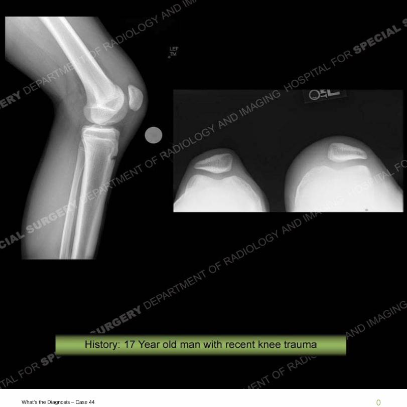
What’s the Diagnosis – Case 44
3
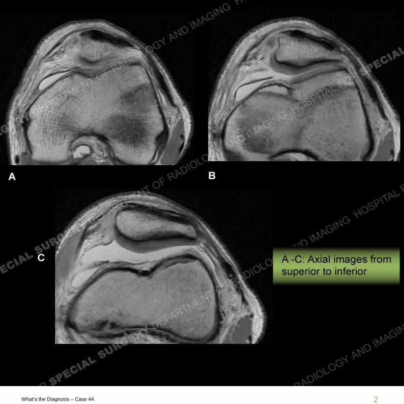
What’s the Diagnosis – Case 44
4
What’s the Diagnosis – Case 44
Findings
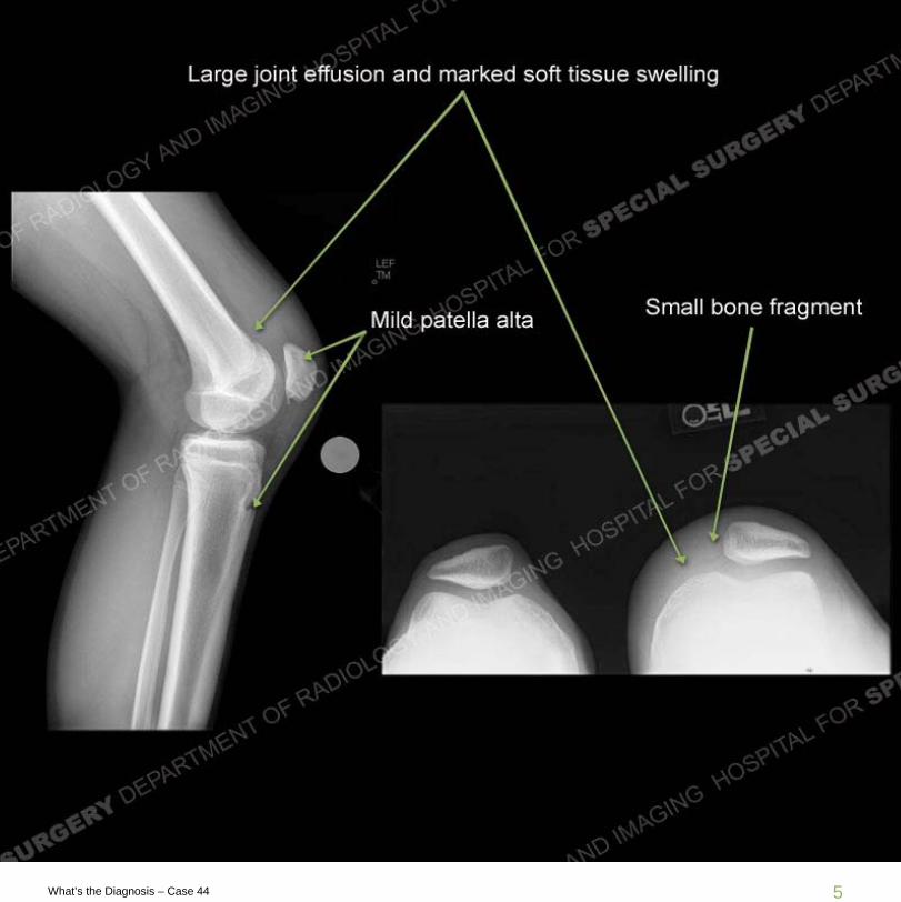
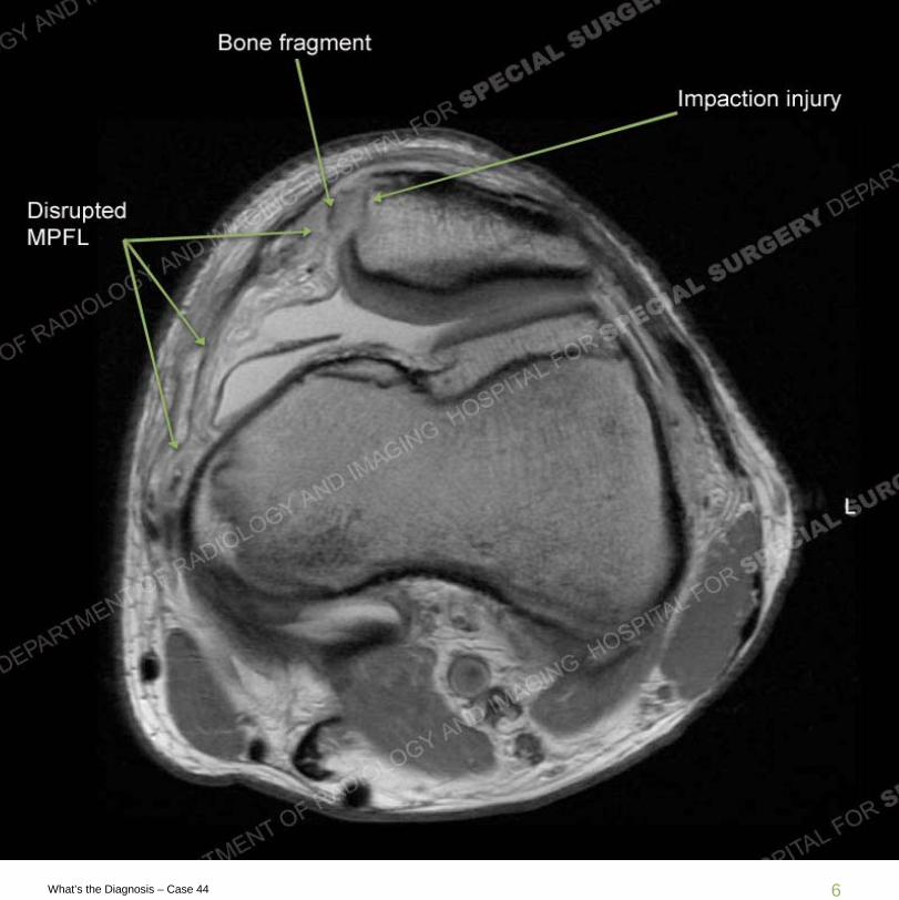
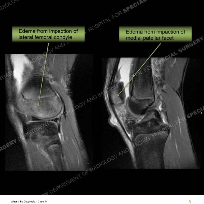
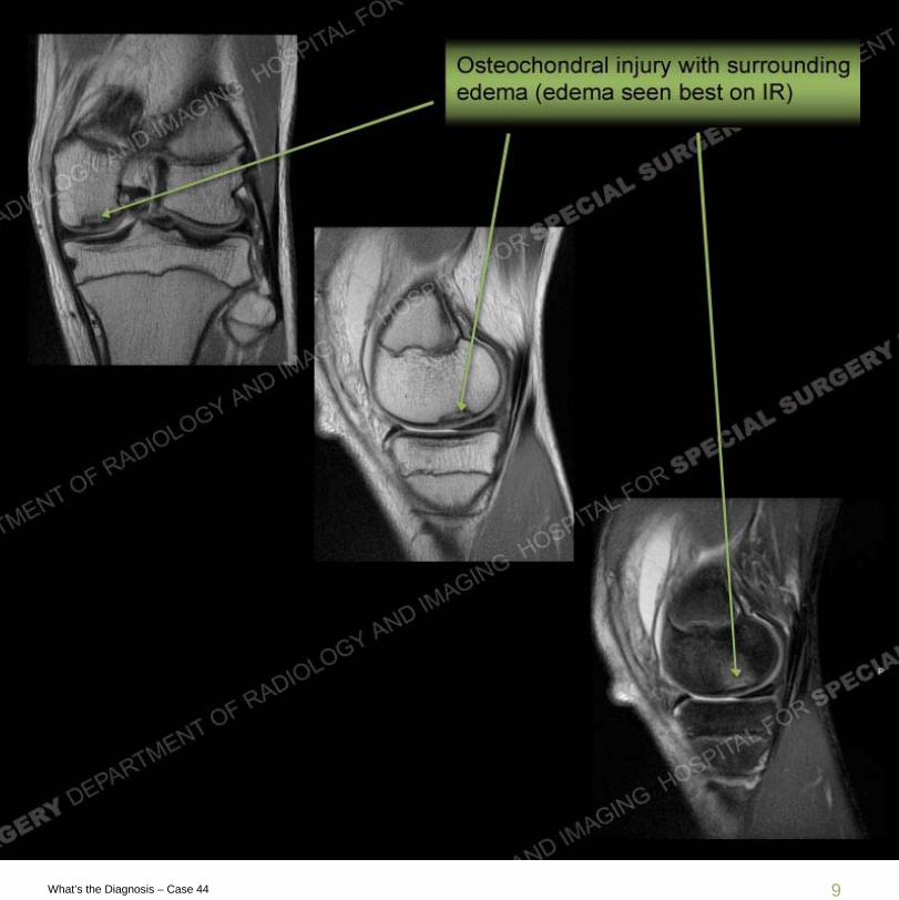
Radiographs demonstrate a large joint effusion with a small bone fragment seen adjacent to an elongated lateral patellar facet. Mild degree of patella alta is present. MRI demonstrates prominent marrow edema pattern of the mid to inferior aspect of the medialpatellar facet with an impaction injury and an impaction injury of the anterior aspect of the lateral femoral condyle. Small bone fragment is seen adjacent to the medial patellar facet where medial patellofemoralligament (MPFL) disruption has been sustained. Further images of the medial condyle demonstrate an osteochondral injury.
5
What’s the Diagnosis – Case 44
6
What’s the Diagnosis – Case 44

7
What’s the Diagnosis – Case 44
8
What’s the Diagnosis – Case 44
9
What’s the Diagnosis – Case 44

10
What’s the Diagnosis – Case 44
Diagnosis
Transient lateral patellar dislocation (LPD) is a well documented injury caused by internal rotation of the femur on a fixed tibia with flexion of the knee and firing of the quadriceps mechanism. This leads to a laterally imparted force on the patella. With relocation of the patella, impaction fractures are seen of the anterior aspect of the lateral femoral condyle and the medial patellar facet. Positioning of the impaction along the central to inferior aspect of the medial patella relates to the degree of flexion of the patella at the time of injury. Along the medial aspect of the knee is a documented trilaminar structure that supports the medial aspect of the knee but with the medial patellofemoralligament being the key stabilizer along the medial

11
What’s the Diagnosis – Case 44
Diagnosis
aspect of the knee. The disruption of the MPFL may be at the patellar, midsubstance, or femoral attachment. Often, as in this case force is transmitted through the entire ligament yielding diffuse injury. Recently, direct MPFL reconstruction has become a more routine procedure for some of these patients. Underlying osseous architecture is a known predisposition for recurrent LPD including trochlear hypoplasia, patella alta, patella tilt, elevated quadriceps angles, and others. Treatment often relates to reconstituting normal osseous relationships to help prevent recurrent LPD and subsequent early cartilage loss. In this case, the additional OCD of the medial condyle is not a classic finding although often MCL and medial meniscal injuries are seen in the setting of LPD.
12
What’s the Diagnosis – Case 44
Resources
http://emedicine.medscape.com/article/90068-overviewMedial patellofemoral ligament: cadaveric investigation of anatomy with
MRI, MR arthrography, and histologic correlation.Dirim B, Haghighi P, Trudell D, Portes G, Resnick D. AJR Am J Roentgenol. 2008 Aug;191(2):490-8.
The supporting structures and layers on the medial side of the knee: an anatomical analysis.Warren RF, Marshall JL.J Bone Joint Surg Am. 1979 Jan;61(1):56-62


![What's New In Glaucoma Surgery [OD CE 2 credit hours] - PPT Slides and Videos](https://img.pdfslide.net/doc/110x75/55bab2d8bb61eba5158b463d/whats-new-in-glaucoma-surgery-od-ce-2-credit-hours-ppt-slides-and-videos.jpg)