Embed Size (px)
DESCRIPTION
Housekeeping. Your performance on the exam has caused me to re-evaluate how homework will be handled I will now be picking up every problem assigned on the Course Schedule It was readily apparent that very few of you actually did the problems - PowerPoint PPT Presentation
Citation preview

Housekeeping
• Your performance on the exam has caused me to re-evaluate how homework will be handled
• I will now be picking up every problem assigned on the Course Schedule– It was readily apparent that very few of you
actually did the problems
• If you are not spending AT LEAST 1 hour a day on this course, you are not going to do very well.
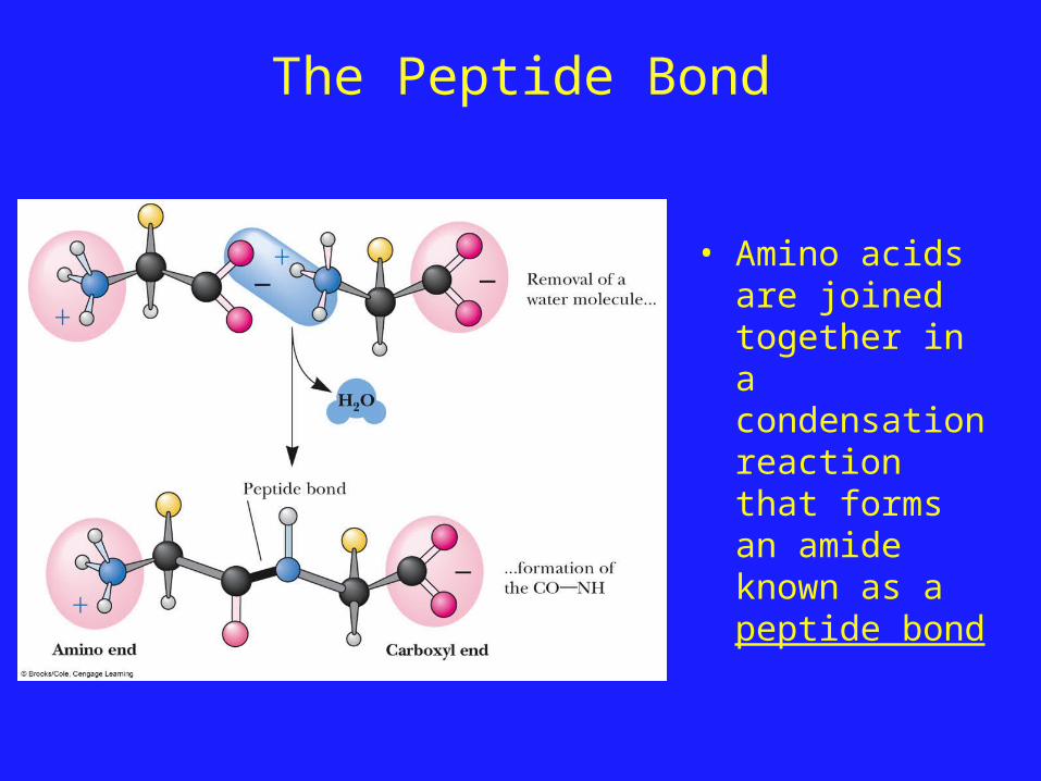
The Peptide Bond
• Amino acids are joined together in a condensation reaction that forms an amide known as a peptide bond

The Peptide Bond
• A peptide bond has planar character due to resonance hybridization of the amide
• This planarity is key to the three dimensional structure of proteins
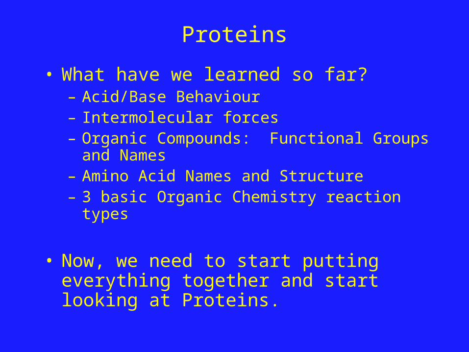
Proteins
• What have we learned so far?– Acid/Base Behaviour– Intermolecular forces– Organic Compounds: Functional Groups and
Names– Amino Acid Names and Structure– 3 basic Organic Chemistry reaction types
• Now, we need to start putting everything together and start looking at Proteins.
Proteins
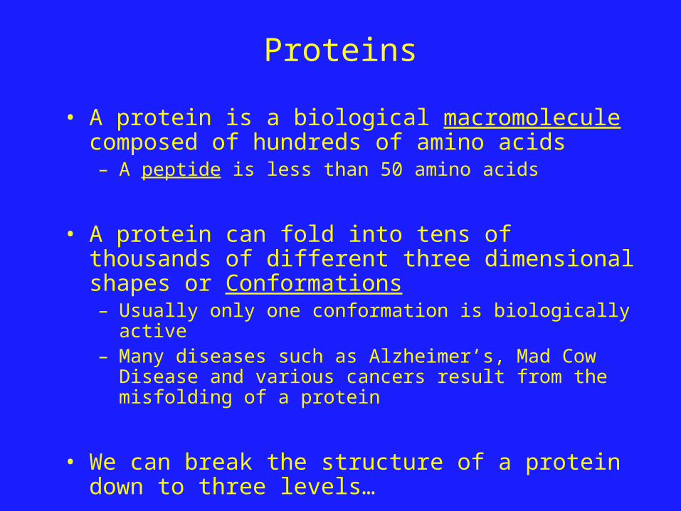
• A protein is a biological macromolecule composed of hundreds of amino acids– A peptide is less than 50 amino acids
• A protein can fold into tens of thousands of different three dimensional shapes or Conformations– Usually only one conformation is biologically active– Many diseases such as Alzheimer’s, Mad Cow Disease and
various cancers result from the misfolding of a protein
• We can break the structure of a protein down to three levels…
Protein Structure: Primary (1°) Structure
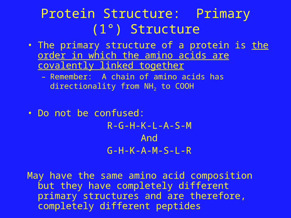
• The primary structure of a protein is the order in which the amino acids are covalently linked together– Remember: A chain of amino acids has directionality from
NH2 to COOH
• Do not be confused:R-G-H-K-L-A-S-M
AndG-H-K-A-M-S-L-R
May have the same amino acid composition but they have completely different primary structures and are therefore, completely different peptides
Proteins: Secondary (2°) Structure
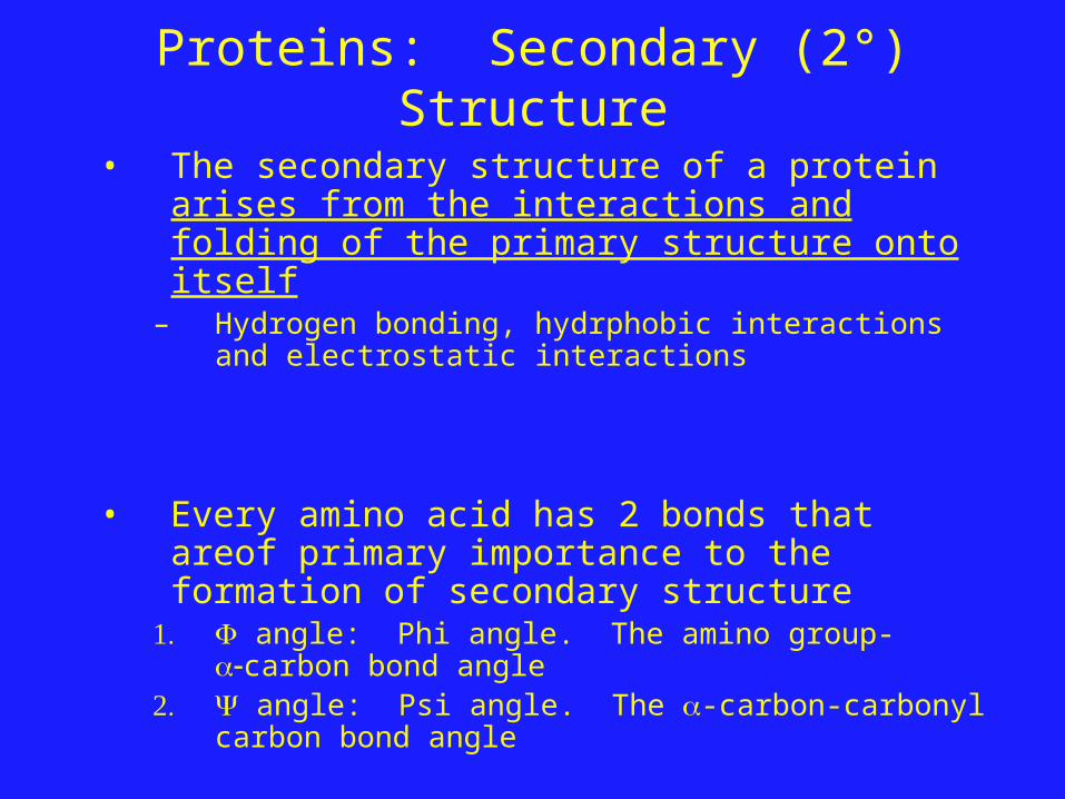
• The secondary structure of a protein arises from the interactions and folding of the primary structure onto itself
– Hydrogen bonding, hydrphobic interactions and electrostatic interactions
• Every amino acid has 2 bonds that areof primary importance to the formation of secondary structure
1. angle: Phi angle. The amino group-carbon bond angle
2. angle: Psi angle. The -carbon-carbonyl carbon bond angle
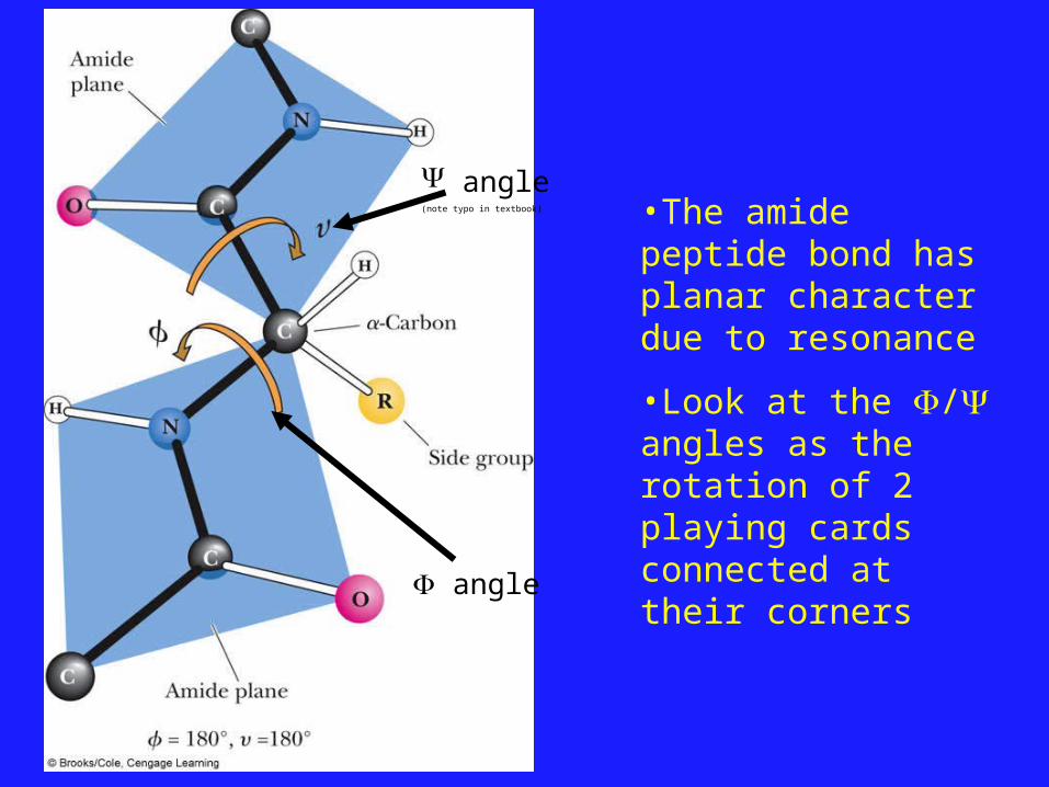
angle(note typo in textbook)
angle
•The amide peptide bond has planar character due to resonance
•Look at the / angles as the rotation of 2 playing cards connected at their corners
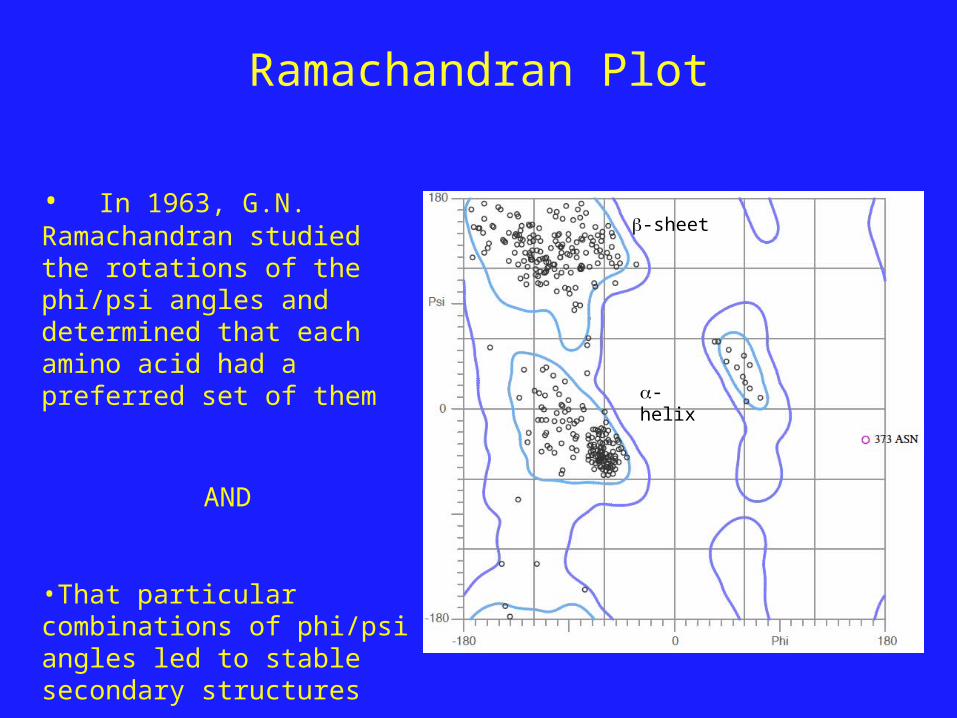
Ramachandran Plot
• In 1963, G.N. Ramachandran studied the rotations of the phi/psi angles and determined that each amino acid had a preferred set of them
AND
•That particular combinations of phi/psi angles led to stable secondary structures
-helix
-sheet
Secondary Structures: -helices and -sheets
• The 2 secondary structures that proteins are primarily composed of ar: -helix: a rod-like coil held together by
hydrogen bonds -helix: A ribbon-like structure held
together by hydrogen bonds
• Both types of structure are Periodic– Their features repeat at regular intervals
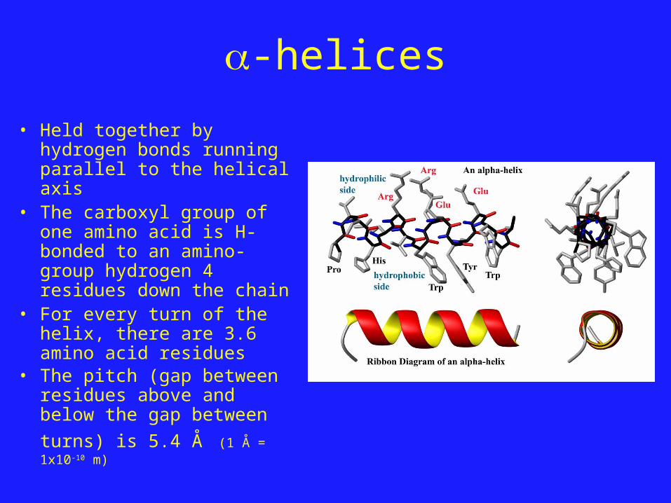
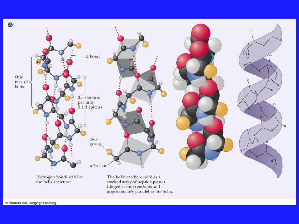
-helices
• Held together by hydrogen bonds running parallel to the helical axis
• The carboxyl group of one amino acid is H-bonded to an amino-group hydrogen 4 residues down the chain
• For every turn of the helix, there are 3.6 amino acid residues
• The pitch (gap between residues above and below the gap between turns) is
5.4 Å (1 Å = 1x10⁻¹⁰ m)
-helices
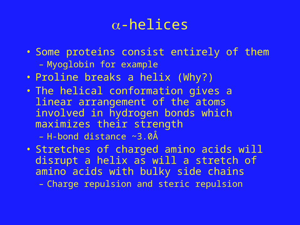
• Some proteins consist entirely of them– Myoglobin for example
• Proline breaks a helix (Why?)• The helical conformation gives a linear
arrangement of the atoms involved in hydrogen bonds which maximizes their strength– H-bond distance ~3.0Å
• Stretches of charged amino acids will disrupt a helix as will a stretch of amino acids with bulky side chains– Charge repulsion and steric repulsion
-sheets
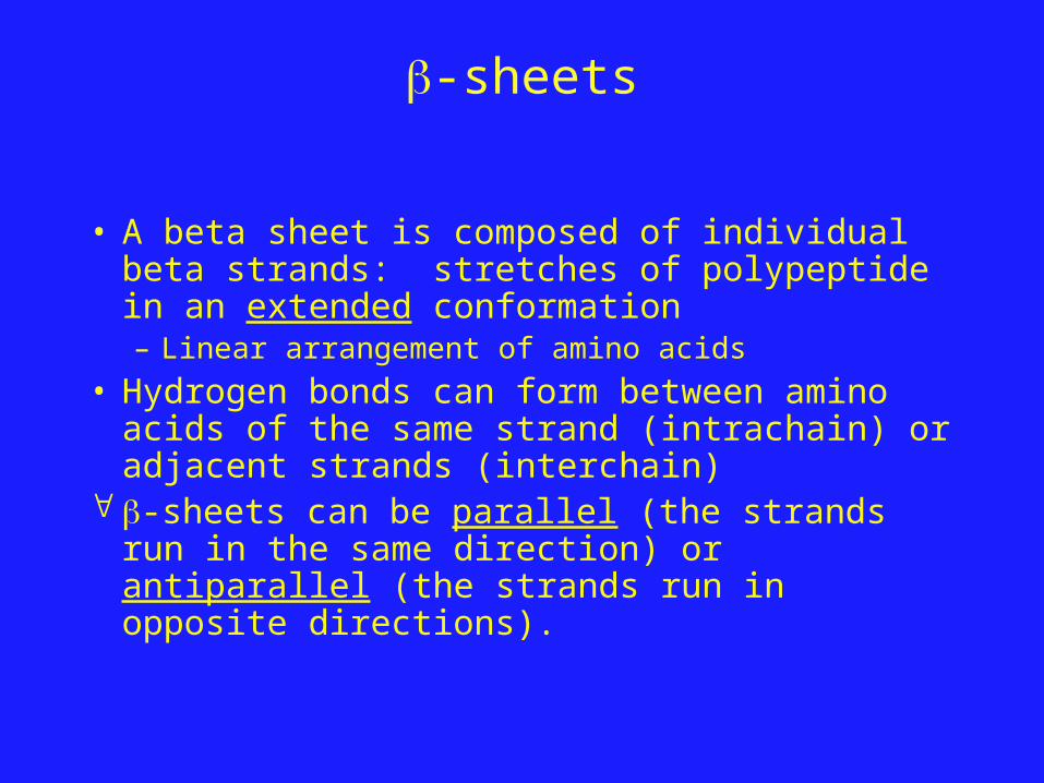
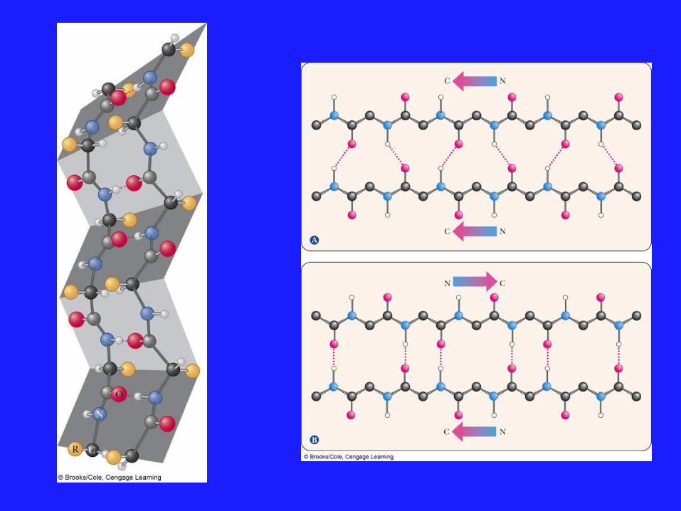
• A beta sheet is composed of individual beta strands: stretches of polypeptide in an extended conformation– Linear arrangement of amino acids
• Hydrogen bonds can form between amino acids of the same strand (intrachain) or adjacent strands (interchain)
-sheets can be parallel (the strands run in the same direction) or antiparallel (the strands run in opposite directions).
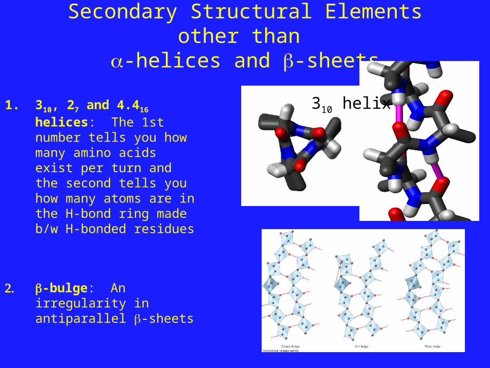
Secondary Structural Elements other than -helices and -sheets
1. 310, 27 and 4.416 helices: The 1st number tells you how many amino acids exist per turn and the second tells you how many atoms are in the H-bond ring made b/w H-bonded residues
2. -bulge: An irregularity in antiparallel -sheets
310 helix
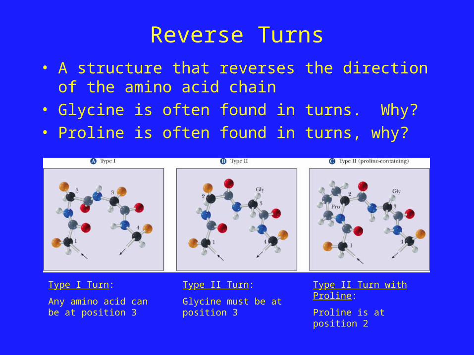
Reverse Turns• A structure that reverses the direction of the amino
acid chain• Glycine is often found in turns. Why?• Proline is often found in turns, why?
Type I Turn:
Any amino acid can be at position 3
Type II Turn:
Glycine must be at position 3
Type II Turn with Proline:
Proline is at position 2
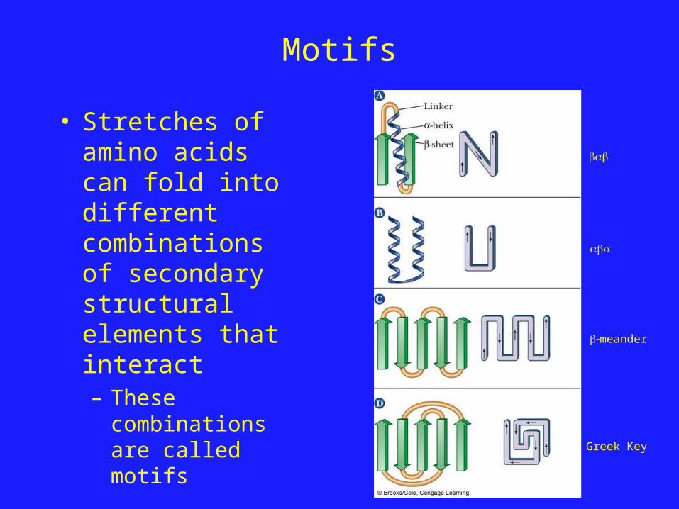
Motifs
• Stretches of amino acids can fold into different combinations of secondary structural elements that interact– These
combinations are called motifs
meander
Greek Key
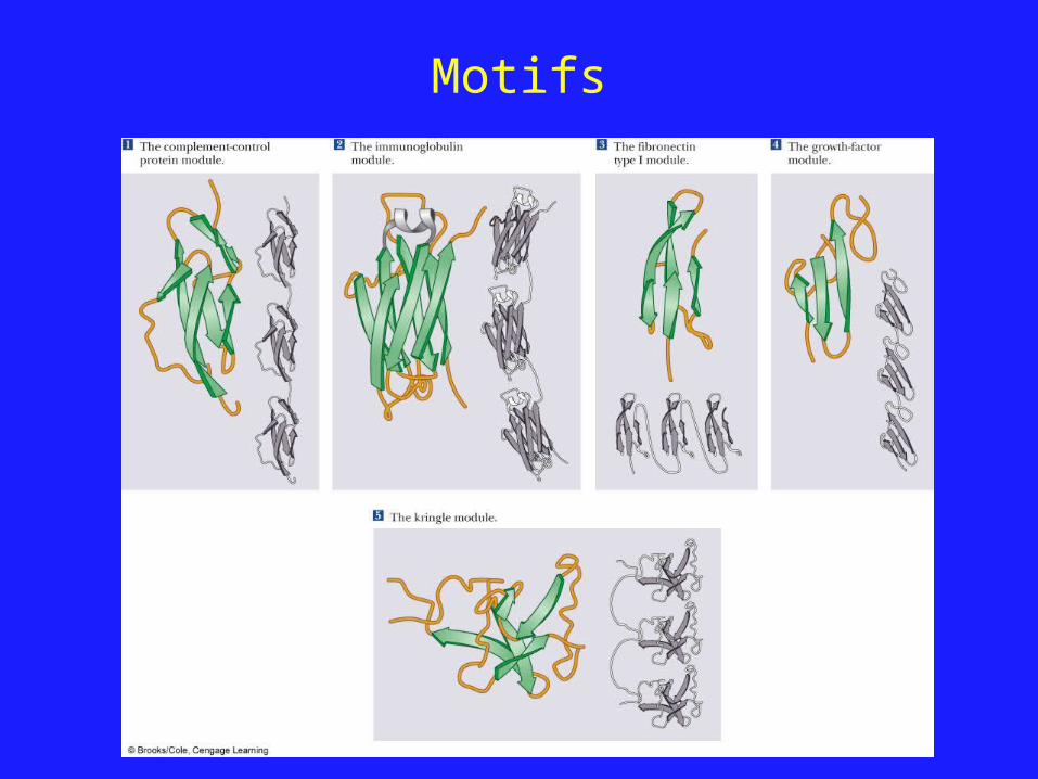
Motifs
Domains and Tertiary Structure
• Several motifs pack together to form Domains– A protein Domain is a stable unit of protein
structure that will fold spontaneously– Domains have similar function in different proteins
• Domains tend to evolve as a unit.
• There are some good websites to look at protein domains:– CATH: www.cathdb.info– SCOP: scop.mrc-lmb.cam.ac.uk/scop/
Tertiary (3°) Structure
• Many all -helix proteins exist– Myoglobin
• The -barrel domain is seen in many proteins– Xylanase C
QuickTime™ and aYUV420 codec decompressor
are needed to see this picture.
QuickTime™ and aYUV420 codec decompressor
are needed to see this picture.
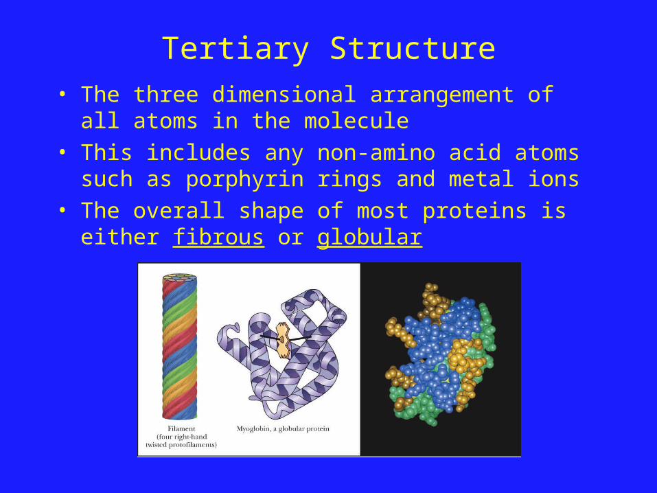
Tertiary Structure• The three dimensional arrangement of all atoms in
the molecule• This includes any non-amino acid atoms such as
porphyrin rings and metal ions• The overall shape of most proteins is either fibrous or
globular
Forces Important in Maintaining Tertiary Structure
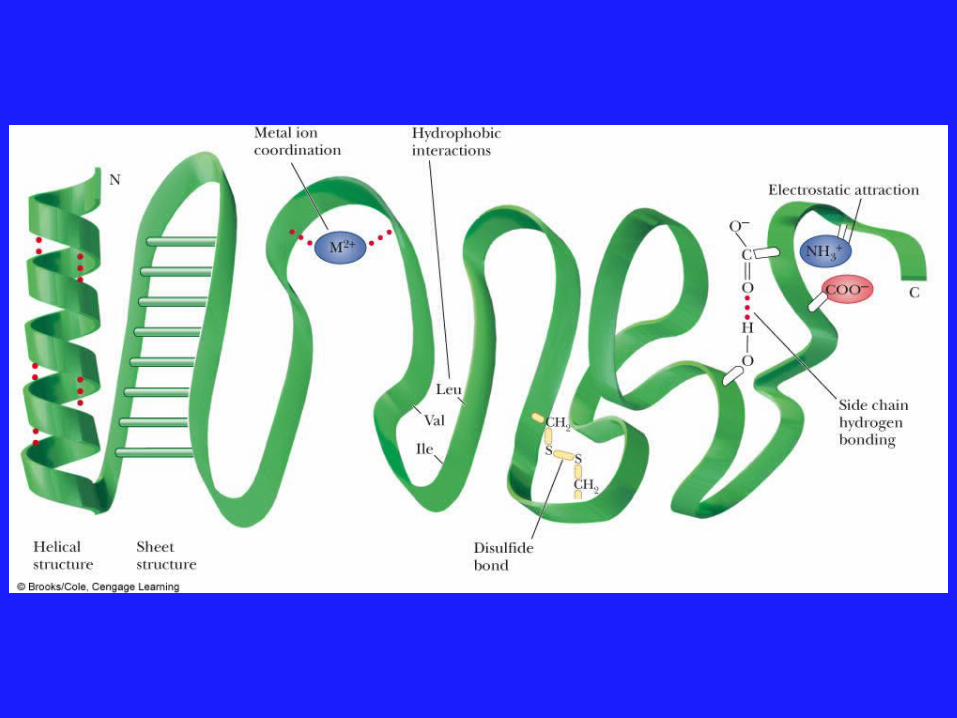
• Peptide bonds = Covalent bonds• 2° and 3° structures = Noncovalent interactions• Let’s look at these non-covalent interactions:
1. Hydrogen bonding: – H-bonds between backbone atoms (C=O and H-N)– H-bonds between sidechains (COO- and -O-H)
2. Hydrophobic interactions:– Nonpolar amino acids tend to be found in the core of the protein due to
phydrophobic interactions
3. Electrostatic Interactions:– Metal/Side Chain interactions– Side chain/Ion interactions
4. Disulfide bonds:– Two cysteine side chains can form S-S bonds, thereby linking two different
sections of the polypeptide chain together– Not every protein has disulfide bonds!
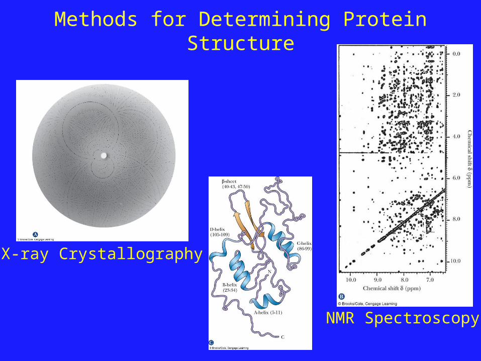
Methods for Determining Protein Structure
X-ray Crystallography
NMR Spectroscopy
Protein Structure: Quaternary Structure
• The quaternary structure of a protein (4°) is the collection of discrete tertiary structures.
• For example: Hemoglobin is a dimeric protein comprised of an and a subunit.
• The functional form of hemoglobin found in red blood cells is actually a dimer of the / dimers.
• The quaternary structure of active hemoglobin is therefore 2subunitsand2subunits.
• Many proteins are monomers; their quaternary structure is the same as their tertiary structure