Embed Size (px)
Citation preview
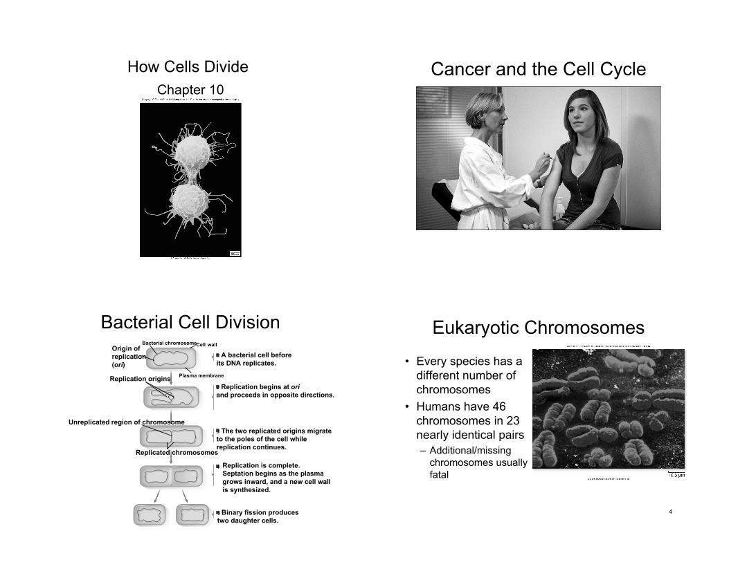
How Cells Divide
Chapter 10
Cancer and the Cell Cycle
Bacterial Cell DivisionBacterial chromosomeCell wall
Plasma membrane
Origin of
replication
(ori)
Replication origins
1 A bacterial cell before
its DNA replicates.
2 Replication begins at ori
and proceeds in opposite directions.
3 The two replicated origins migrate
to the poles of the cell while
replication continues.
4 Replication is complete.
Septation begins as the plasma
grows inward, and a new cell wall
is synthesized.
5 Binary fission produces
two daughter cells.
Unreplicated region of chromosome
Replicated chromosomes
Eukaryotic Chromosomes
• Every species has a
different number of
chromosomes
• Humans have 46
chromosomes in 23
nearly identical pairs
– Additional/missing
chromosomes usually
fatal
4
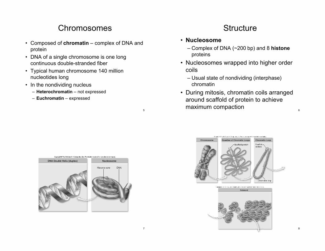
Chromosomes
• Composed of chromatin – complex of DNA and
protein
• DNA of a single chromosome is one long
continuous double-stranded fiber
• Typical human chromosome 140 million
nucleotides long
• In the nondividing nucleus
– Heterochromatin – not expressed
– Euchromatin – expressed
5
Structure
• Nucleosome
– Complex of DNA (~200 bp) and 8 histone
proteins
• Nucleosomes wrapped into higher order
coils
– Usual state of nondividing (interphase)
chromatin
• During mitosis, chromatin coils arranged
around scaffold of protein to achieve
maximum compaction6
7 8
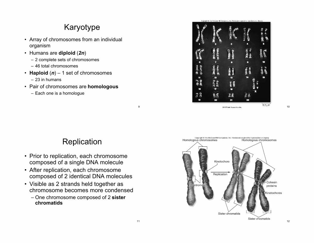
Karyotype
• Array of chromosomes from an individual
organism
• Humans are diploid (2n)
– 2 complete sets of chromosomes
– 46 total chromosomes
• Haploid (n) – 1 set of chromosomes
– 23 in humans
• Pair of chromosomes are homologous
– Each one is a homologue
9 10
Replication
• Prior to replication, each chromosomecomposed of a single DNA molecule
• After replication, each chromosomecomposed of 2 identical DNA molecules
• Visible as 2 strands held together aschromosome becomes more condensed
– One chromosome composed of 2 sisterchromatids
11 12
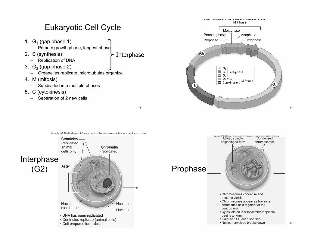
Eukaryotic Cell Cycle
1. G1 (gap phase 1)– Primary growth phase, longest phase
2. S (synthesis)– Replication of DNA
3. G2 (gap phase 2)– Organelles replicate, microtubules organize
4. M (mitosis)– Subdivided into multiple phases
5. C (cytokinesis)– Separation of 2 new cells
13
Interphase
14
15
Interphase
(G2)
16
Prophase
17
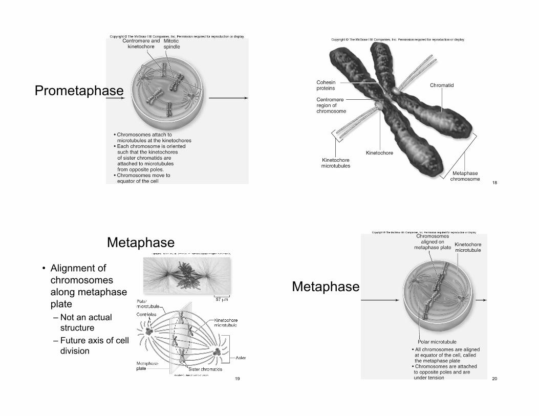
Prometaphase
18
Metaphase
• Alignment of
chromosomes
along metaphase
plate
– Not an actual
structure
– Future axis of cell
division
19 20
Metaphase
21
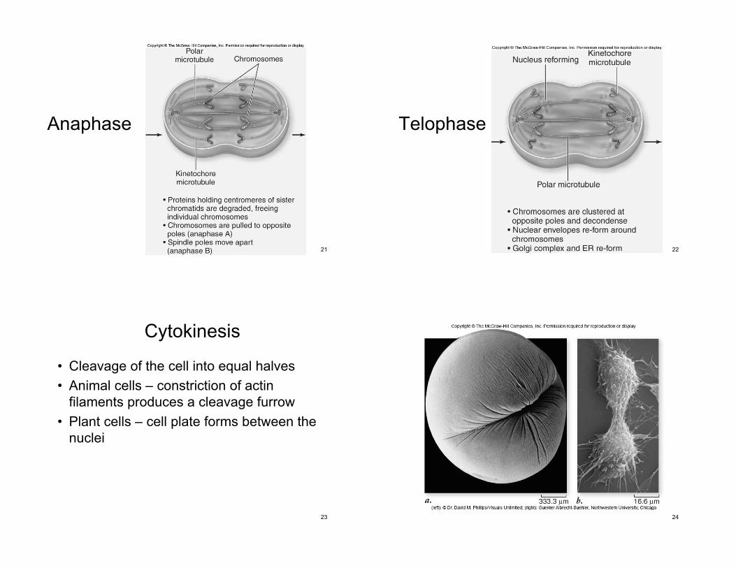
Anaphase
22
Telophase
23
Cytokinesis
• Cleavage of the cell into equal halves
• Animal cells – constriction of actin
filaments produces a cleavage furrow
• Plant cells – cell plate forms between the
nuclei
24
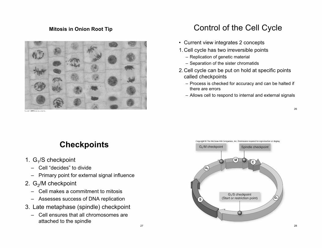
Mitosis in Onion Root Tip
26
Control of the Cell Cycle
• Current view integrates 2 concepts
1.Cell cycle has two irreversible points
– Replication of genetic material
– Separation of the sister chromatids
2.Cell cycle can be put on hold at specific points
called checkpoints
– Process is checked for accuracy and can be halted if
there are errors
– Allows cell to respond to internal and external signals
Checkpoints
1. G1/S checkpoint
– Cell “decides” to divide
– Primary point for external signal influence
2. G2/M checkpoint
– Cell makes a commitment to mitosis
– Assesses success of DNA replication
3. Late metaphase (spindle) checkpoint
– Cell ensures that all chromosomes are
attached to the spindle27 28
29
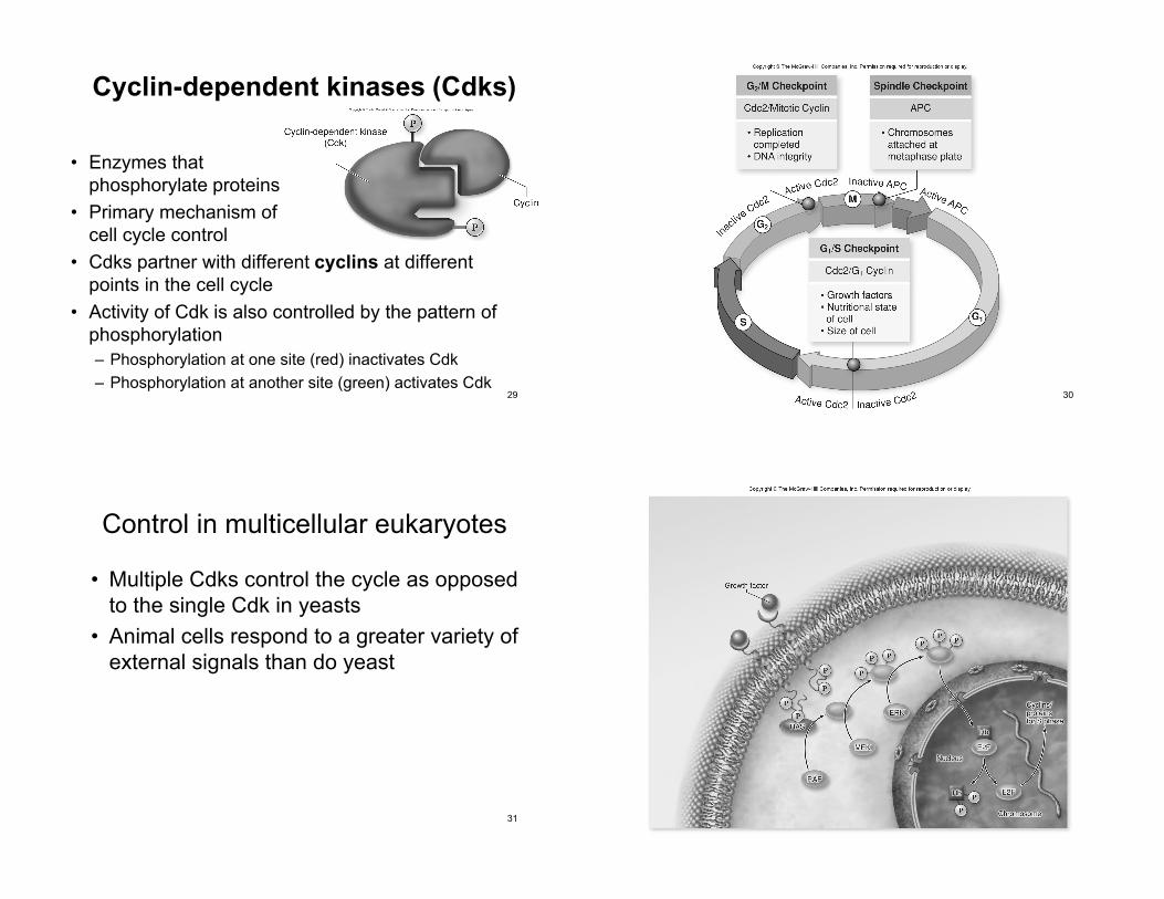
Cyclin-dependent kinases (Cdks)
• Enzymes that
phosphorylate proteins
• Primary mechanism of
cell cycle control
• Cdks partner with different cyclins at different
points in the cell cycle
• Activity of Cdk is also controlled by the pattern of
phosphorylation
– Phosphorylation at one site (red) inactivates Cdk
– Phosphorylation at another site (green) activates Cdk30
31
Control in multicellular eukaryotes
• Multiple Cdks control the cycle as opposed
to the single Cdk in yeasts
• Animal cells respond to a greater variety of
external signals than do yeast
32
33
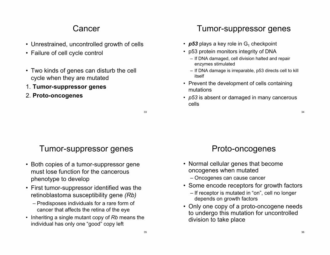
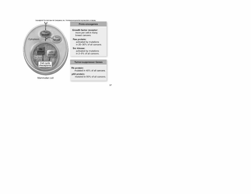
Cancer
• Unrestrained, uncontrolled growth of cells
• Failure of cell cycle control
• Two kinds of genes can disturb the cell
cycle when they are mutated
1. Tumor-suppressor genes
2. Proto-oncogenes
34
Tumor-suppressor genes
• p53 plays a key role in G1 checkpoint
• p53 protein monitors integrity of DNA
– If DNA damaged, cell division halted and repair
enzymes stimulated
– If DNA damage is irreparable, p53 directs cell to kill
itself
• Prevent the development of cells containing
mutations
• p53 is absent or damaged in many cancerous
cells
Tumor-suppressor genes
• Both copies of a tumor-suppressor gene
must lose function for the cancerous
phenotype to develop
• First tumor-suppressor identified was the
retinoblastoma susceptibility gene (Rb)
– Predisposes individuals for a rare form of
cancer that affects the retina of the eye
• Inheriting a single mutant copy of Rb means the
individual has only one “good” copy left
35 36
Proto-oncogenes
• Normal cellular genes that becomeoncogenes when mutated
– Oncogenes can cause cancer
• Some encode receptors for growth factors
– If receptor is mutated in “on”, cell no longerdepends on growth factors
• Only one copy of a proto-oncogene needsto undergo this mutation for uncontrolleddivision to take place
37