Embed Size (px)
Citation preview


HOW DID WE FIND OUT ABOUT THE BRAIN?
Isaac Asimov
Illustrated by Erika W. Kors
Contents
1. Inside the Skull
2. Trouble with the Brain
3. Cells of the Brain
4. The Nerve Impulse
5. Sides and Sleep
1. INSIDE THE SKULL
INSIDE THE HEAD is a soft, grayish mass that weighs just about three pounds. It is wrinkled and looks
a little bit like a large walnut. It is the brain.
It doesn’t seem to do anything. It just sits there. Yet it must be very important since, for one thing,
it is surrounded by a protective shell of bone called the skull. No other part of the body is as well
protected as the brain is.
The heart, for instance, is a very important part of the body. It has the breastbone in front of it, and
a series of ribs. There are spaces between the ribs, however; a knife can be pushed between the ribs
and into the heart, and that would kill a person at once. There are no openings in the skull.
Then, too, the body needs a gas called oxygen (OK-sih-jen), which is found in the air. We are
constantly breathing in order to get the oxygen into our body. The oxygen combines with food to give
us energy.
The muscles need a lot of oxygen, since they use up so much energy. When you use muscles a
great deal, for instance, when you are running or lifting weights or chopping wood, you start to pant.
You breathe quickly and deeply in order to get as much oxygen into your body as possible.
Yet, of all the oxygen you breathe, one-quarter is used by the brain, even though it just seems to sit
there and do nothing. This shows that the brain must be doing something important.
Sometimes a person chokes so that he can’t breathe, and oxygen can’t get into his body. Most
parts of the body can do without oxygen for quite a while, but the brain can last only a few minutes if
its oxygen supply is cut off. After those few minutes, the brain stops doing what it is doing—and the
person dies.

Ancient people didn’t know about the importance of oxygen to the brain. They didn’t even know
what oxygen was. Still, they did know that when a person was hit violently on the skull, he would be
stunned for a while, and even after he came to, he might not behave as he usually did. Something
would be wrong with his thinking and acting.
For this reason, some scholars in ancient times thought that the brain might be the part of the body
that enables human beings to think and to feel. One of these scholars was Plato (PLAY-toh, 427-347
B.C.).
The most influential of the ancient philosophers was a student of Plato’s named Aristotle (AR-is-
TOT-ul, 384-322 B.C.). It seemed to him that the heart was thecenter of thinking and feeling. After all,
the heart speeds up when when you’re excited and slows down when you’re resting. But if the heart
does the thinking and feeling, what does the brain do?
Plato and Aristotle
Aristotle thought the brain was a place where the blood was cooled after it had been heated up by
the heart.
But if that’s all the brain does, why is it so well protected by bone? Why is a blow on the head so
much more likely to knock us out than a blow on the chest? Why is the brain so much larger in human
beings than it is in other animals? Horses and camels, for instance, are much larger than human beings
yet have smaller brains than we do. Only huge elephants and whales have brains that are larger than
ours.
For all these reasons, some scholars were doubtful about Aristotle’s theory about the brain, even
though the philosopher had such a great reputation.

About 290 B.C., a Greek doctor, Herophilus (hee-ROF-ih-lus), became the first person to cut
open dead bodies before students. This is called dissection (dih-SEK-shun).
In this way, students could study the different parts of the body. Such a study is called anatomy
(uh-NAT-uh-mee).
Herophilus studied the brain carefully and decided, as Plato did, that it was the brain that did the
thinking and feeling. He also noticed that there were long, thin, threadlike objects that seemed to
connect to the brain. We call them nerves.
Herophilus found that some nerves extended to muscles and others to the eyes and ears. He
decided this meant that some nerves control motions by making the muscles work and that other
nerves control the senses—what you see, hear and so on.
Different Brain Sizes
Following Herophilus was another Greek doctor, Erasistratus (er-uh-SIS-tru-tus, 304-250 B.C.).
He also studied the brain carefully. He was the first to describe the wrinkles and folds in the brain’s
surface. We now call them convolutions (kon-vuh-LOO-shunz). He pointed out that the human brain
has more convolutions than is found in the brains of other animals. He felt that this was why human
beings are more intelligent than other animals.
pigeon opossum
rabbit cat
monkeychimpanzee
human

He also noticed that the brain is in two parts. In front is a large part that we now call the cerebrum
(SEHR-uh-brum, which is a Latin word for “brain”). Behind and below it is a smaller part that we call
the cerebellum (SEHR-uh-BEL-um, Latin for “little brain”). He also studied the membranes covering
the brain and the spaces within the brain.
Herophilus and Erasistratus did their work in Alexandria, Egypt, which was the center of science in
those days. The Egyptian people, however, objected to dissection. It was against their religion. For
that reason, the very good start these two doctors made was not followed up.
At the time the Roman Empire was at its height, there was another Greek doctor of importance,
Galen (GAY-len, 130-200). He wasn’t allowed to dissect human corpses, so he dissected animals.
Sometimes this misled him, for there are parts of animals such as dogs and pigs that are not exactly
like those of human beings.
The brain
cerebrum
cerebellum
He did work on another part of the brain. Behind the cerebellum, the brain narrows into a part
called the medulla oblongata (meh-DUL-uh-OB-long-GOT-tuh), and this ends in a kind of tail that is
enclosed and protected by the bones of the spine, or backbone. This tail is called the spinal cord, and
nerves are attached to it, just as they are attached to the brain.
Galen studied what happened if he cut an animal’s spinal cord. If it was cut high up, near the brain,
the animal died at once. If it was cut in places lower and lower down, different sets of muscles
stopped working and were paralyzed. This showed that the nerves attached to the spinal cord con-
trolled various muscles.
Unfortunately, science declined under the Romans. After the fall of the Roman Empire, science
declined even further. It was not until a thousand years after Galen that scientists once again began to
dissect the bodies of animals and human beings, so as to study anatomy.
In 1316, an Italian doctor, Mondino de Luzzi (day-LOOT-tsee, 1275-1326), wrote a book that was
entirely on anatomy. It was the first all-anatomy book in history. He managed to get many things
wrong, however, because he depended too much on what Galen had said, and didn’t do enough
dissection of his own. Still, it was a beginning.

To make his art more realistic, an Italian artist, Leonardo da Vinci (da-VEEN-chee, 1452-1519)
dissected at least thirty dead bodies and carefully drew the various parts he observed. He was the first
to show that Galen was wrong in some ways.
However, Leonardo didn’t write about his findings in such a wav that others could learn about
them. He kept his findings to himself, and his notes on the subject were only discovered much later.
The real founder of modern anatomy was a Belgian scientist, Andreas Vesalius (veh-SAY-lee-us,
1514-1564). Like Leonardo, Vesalius dissected many dead bodies. However, he wrote careful de-
scriptions for others to read. In 1543, he published one of the great books of science. Its title, in
English, is On the Structure of the Human Body. Vesalius had it illustrated by one of the best artists
of the time. By then, printing had been invented, and the book was printed and reached all the learned
men of Europe. The book was accurate, and it corrected almost all of Galen’s mistakes.
Vesalius studied the brain and described it very accurately. He repeated the experiments of Galen
on the spinal cord. He decided that the brain, the spinal column, and the nerves attached to them make
up a nervous system.
After Vesalius’s time, people fearlessly dissected human bodies and worked out the details of the
various parts more and more clearly. Many anatomists followed the various nerves to see exactly
where they go.
A Swiss biologist, Albrecht von Haller (1708-1777) showed that muscles can be irritable. That is,
they contract if they are touched. When Haller touched a nerve that led to a muscle that muscle would
contract. In fact, the nerve was more irritable than the muscle itself; a very light touch on the nerve
was enough to make the muscle contract. Haller’s work made it quite clear that the brain (to which the
nerves are attached) controls bodily movements.
Nerve connections to the brain
spinal cord
medulla oblongata

What’s more, Haller showed that all the nerves in the body can be traced back to the brain or to the
spinal cord. Since the brain and spinal cord are attached to each other, this showed there is only one
nervous system.
2. TROUBLE WITH THE BRAIN
ONCE IT BECAME quite clear that the brain and spinal cord control the body’s motions and senses,
it was possible to wonder what would happen if the brain didn’t do its work properly. What if the
brain were disordered or diseased?
If someone’s brain stops working to any considerable extent, he may suddenly fall unconscious.
This is called a stroke, or apoplexy (AP-uh-PLEK-see, from a Greek word meaning “to strike”). After
all, it might seem to people who knew nothing about the brain that someone suffering a stroke had
actually been struck down by some invisible being—a god or a demon, perhaps.
A stroke may lead to death, or it may leave a person alive but paralyze a portion of the body and
make it unable to move. Nowadays we know that a common cause of a stroke is that one of the small
blood vessels in the brain is blocked or breaks, so that part of the brain doesn’t get oxygen and dies.
Sometimes there are less drastic disorders of the brain. Some people occasionally make involun-
tary movements. Suddenly they fall down, twist and turn, make strange sounds, or froth at the mouth.
They may also see and hear things that other people don’t. (To hear or see something that isn’t really
there is now called a hallucination [HAL-loo-sih-NAY-shun].)
In ancient times, people thought that those who hallucinated were actually seeing and hearing an
invisible world. Perhaps they were receiving messages from the gods. Perhaps gods took over the
body and made it move uncontrollably.
This seemed especially true when people who fell down and did all these peculiar things then
recovered and seemed perfectly normal again. Such a condition is sometimes called “the falling
sickness.” The medical name is epilepsy (EP-ih-LEP-see, from a Greek word meaning “seizure”) - It
was as though the gods had seized you and temporarily taken over control.
The ancient Greeks called epilepsy “the sacred disease” and people who suffered from it were
sometimes viewed with awe. Since these people were thought to be given glimpses of the future, they
were often considered to be prophets.
Almost anyone who behaved in a curious fashion, whose speech and repetitious behavior didn’t
seem to make any sense, was treated with similar awe. In later times, such people were called “mad-
men” or “insane.” (Insane is from Latin words meaning “not healthy”; that is, not healthy in one’s way
of behaving and thinking.) These people were also described as “touched;” that is, touched by some
god.
So long as people thought gods were involved, those who suffered from epilepsy or insanity were
treated with kindness and consideration. As time went on, though, more and more people began to
think it wasn’t a god, but a demon, who controlled the bodies of epileptics and madmen. Therefore,
the demons had to be driven out of the body before a person could be restored to health and sanity.

Sometimes the methods thought to be useful in driving out the demons were very cruel. People
who acted insane were beaten, chained up, and in many other ways mistreated. They were locked up
in insane asylums, where their condition would simply grow worse. Sometimes people who consid-
ered themselves sane would go to such asylums just to watch the odd things that mad people did and
to laugh at them.
Of course, not everybody thought that gods or demons had anything to do with what we now call
mental disorders. Throughout the centuries there were occasional doctors who said that insane
people were sick and should be treated with consideration instead of being tortured. The Greek
doctor Hippocrates (hih-POK-ruh-teez, 460-370 B.C.) and those who followed his teachings insisted
that epilepsy was a disease like any other. They said it had natural causes and would be relieved by
medicines rather than by prayers or beatings.
Another doctor who thought this way was a Greek physician named Soranus (soh-RAN-us), who
lived in Roman times and who classified various types of mental disorders.
Hippocrates
For the most part, such reasonable advice was not listened to. People continued to believe in
“demonic possession.”
In 1789, the French people rose against their corrupt and inefficient government in a movement
known as the French Revolution. Over the next few years, the revolutionary leaders were willing to try
a number of new things such as new measuring systems, new calendars, and so on.
They were willing to try a new way of treating the insane, too. A French doctor, Philippe Pinel (pee-
NEL, 1745-1826), was another of those who, like Hippocrates and Soranus, studied mental disorders
and favored gentle treatment for the insane.

In 1793, Pinel was put in charge of a large insane asylum. Most of the unfortunate people in the
asylum were kept chained up, some of them for many years. Pinel took the chains off the patients,
cleaned them up, and tried to make them comfortable. He studied each case and kept records of
developments. It turned out that the insane improved greatly in this way.
Pinel started a new fashion in the treatment of the insane, but things improved slowly. In country
after country, however—in England, in Germany, in the United States—gentle treatment of the insane
caught on. By the end of the 1800s, the old cruel ways had vanished in civilized places.
Meanwhile, how did one treat mental disorders? If the brain wasn’t working properly, was there
any way of making it work better?
Almost anything might make the brain work better.
Philippe Pinel Faith Healer at work
The world is full of sights, sounds, smells, tastes, touches, and even thoughts that reach the brain,
and any of these might soothe it or somehow change its action. If a person believes that something
will make him feel better, that something may really make him feel better.
Even when something is wrong that doesn’t involve the brain, the way the brain works might affect
the disorder. It may make the disorder worse, or better.
Suppose, for instance, that a person has a problem. A friend tells him that there is a special charm
that will cure him or that gods and demons can be called in to cure him. The friend then mumbles
strange words over the sick person, or he makes gestures with his hands, or he just touches him. If
the sick person believes he will be helped—if he has faith that he will be helped—he sometimes is.
This is called faith-healing.
As time went on, and people learned more and more about how the body works, faith-healing
didn’t seem to be enough. People wanted to be cured by something that was scientific.
In the 1700s, scientists were studying the way magnets and electricity behaved. Both magnets and
electrified objects could attract things, and many people believed these objects could be used to
attract diseases and pull them out of the body.

Around 1774, a German doctor named Franz Anton Mesmer (1734-1815), working in Vienna,
Austria, actually tried that. He passed magnets over the bodies of his patients and tried to pull out the
disease. It sometimes seemed to help, and mesmerism became quite fashionable. The magnet, of
course, had no real effect on the disease, so it was only another form of faith-healing.
Mesmer found that he could even leave out the magnets and just pass his hands over the patient.
They might be helped in this way, too. However, a good many patients weren’t helped, and they
complained to the police that Mesmer was a fake. Mesmer was forced to leave Vienna, and in 1778 he
went to Paris.
In Paris, he was quite successful for a while, but then the same thing happened, and in 1785 he was
forced to leave Paris.
Mesmer wasn’t a total fraud, however. He was just too enthusiastic, thinking his methods could
cure any disorder. After his time, there were others who practiced mesmerism.
In 1841, a Scottish doctor, James Braid (1795-1860), attended an exhibition of mesmerism. He
began to wonder if there might not be something to it. He felt that in those cases where it seemed to
help a sick patient, it must do its work by affecting the brain.
Franz Anton Mesmer
He experimented and found that if he had a patient concentrate on a sensation that was dull and
repetitious, the patient’s brain seemed to get tired, and he stopped paying attention. It was as though
the person had gone into a kind of sleep and was no longer aware of many things about him. Braid
called it hypnotism, from a Greek word for “sleep.”

Hypnotism wasn’t real sleep, though; the hypnotized person could still hear what was said to him.
With the conscious mind asleep, the unconscious mind underneath was very receptive to suggestion.
It believed things much more strongly than the conscious mind would.
Thus, if a disease could be helped by addressing the brain, addressing the brain of a hypnotized
person could have even a stronger effect. Doctors began to use hypnotism to treat mental disorders
in particular.
One such doctor was an Austrian, Josef Breuer (BROY-er, 1842-1925). In 1880, he found that a
hypnotized patient seemed to remember events that the patient could not remember when not hypno-
tized. These memories were buried in the unconscious part of the mind, perhaps because they were
too unpleasant or embarrassing to remember under normal conditions.
These unconscious thoughts and memories might influence the patient and make him behave in
strange ways. Breuer believed that if these thoughts and memories could be expressed while the
patient was hypnotized and if he were told to remember them after he came out of hypnotism, then the
patient would know the reason for his strange behavior and could stop.
Breuer discussed this with another Austrian doctor, Sigmund Freud (FROID, 1856-1939). In 1887,
Freud tried this hypnotism technique on his patients and found it worked. In the 1890s, however, he
found that he didn’t have to use hypnotism. If he just let the patient talk about anything he wished, so
that one thing reminded him of another, which reminded him of yet another (this is called free associa-
tion), the hidden memories would eventually pop out.
Freud also thought that the hidden memories could be expressed in dreams, when the conscious
mind was not in control. He felt it was important to remember dreams so that their meaning might be
analyzed. Freud’s methods were called psychoanalysis (SY-koh-uh-NAL-ih-sis, from Greek words
meaning “to take apart the mind”). His work had a great deal of influence on the treatment of mental
disorders, and eventually a number of different psychoanalytic systems were developed by different
psychoanalysts.
Even with the gentlest treatment, people with mental disorders sometimes grew violent and danger-
ous. They would then have to be tied up or treated with strong drugs that put them to sleep.
In 1952, however, a new group of drugs was discovered, called tranquilizers (TRANK-wih-
LYZE-erz). These quieted a person (made him “tranquil”) without putting him to sleep. Such drugs
made it far easier to handle patients who might otherwise be violent.

3. CELLS OF THE BRAIN
BY THE 1700s, the general structure of the brain had been worked out. But what about the tiny details
that are too small for the eye to see? For these, one needs to use a microscope.
In 1665, an English scientist, Robert Hooke (1635-1703), focused his microscope on a thin slice of
cork. Cork is the bark of a tree, and it is a dead material. To the eye, the cork seemed solid, but under
the microscope it was seen to consist of a pattern of tiny rectangular holes. Hooke called these holes
cells, a name given to small rooms.
Other scientists looked at living material under a microscope. A German biologist, Matthias Jakob
Schleiden (SHYL-den, 1804-1881), discovered that thin slices of plant tissue were made up of tiny
structures marked off from each other by partitions. They were like Hooke’s cells, except that they
were filled with a thick liquid. Though the word cell should be applied to empty objects, Schleiden
called these plant structures cells anyway. Once a plant tissue died and became bark, the cells emp-
tied, leaving empty holes behind, and that was what Hooke had seen.
In 1838, Schleiden concluded that all plant life is made up of tiny cells one can see only under a
microscope.
Another German biologist, Theodor Schwann (SHVAHN, 1810-1882), studied thin slices of ani-
mal tissue. He found that these, too, consist of tiny structures marked off from each other by parti-
tions. In 1839 he decided that animals, too, consist of tiny cells one can see only under the micro-
scope.
What Schleiden and Schwann did was to establish the cell theory, which states that all living things
are made up of cells. Some microscopic plants and animals are made up of only a single cell. A
human being, on the other hand, is made up of many trillions of cells.
If the cell theory was correct (and it turned out that it was) then the brain ought to be made up of
cells, too.
Just as the cell theory was being established, in 1838 a Polish doctor, Robert Remak (RAY-mak,
1815-1865), made important studies of nerves under the microscope. He was the first to show that
nerves are not hollow, although, since ancient Greek times, they had been thought to be hollow.
He went on to show that some nerve fibers that are too thin to see without a microscope are
surrounded by a kind of fatty material, and some are not. This fatty material is called a myelin sheath
(MY-uh-lin, from a Greek word for “fatty bone marrow”).
Those parts of the nerves that are covered with a myelin sheath have a whitish appearance. Those
without have a grayish appearance. For this reason we speak of the gray matter and the white matter
of the brain. The outer surface of the brain is gray matter, while the interior is white matter. In the
spinal cord, the white matter is on the outside and the gray matter on the inside.
At about the same time, a Czech biologist, Jan Evangelista Purkinje (POOR-kin-yay, 1787-1869),
was making similar discoveries. In 1837 he described individual nerve cells. He was the first to use the
word protoplasm (PROH-toh-PLAZ-um, from the Greek words meaning “first form”) to describe
the material inside cells.

The nerve cells, which are rather large, are surrounded by a material that supports them and holds
them together. This material is referred to as neuro-glia (nyoo-ROG-lee-uh, from Greek words mean-
ing “nerve glue”). In 1854 a German doctor, Rudolph Carl Virchow (FIR-khov, 1821-1902), showed
that the neu-roglia also contains cells called glial cells which are smaller than nerve cells. The human
brain contains about ten billion nerve cells and ninety billion glial cells.
Purkinje had also shown that nerve cells are irregular in shape. Little fibers stick out from them that
break up into smaller and smaller fibers, like the branches of a tree. These fibers came to be called
dendrites (DEN-drites, from the Greek word for “tree”).
Cross-section of the brain
gray matter
white
matter
Nerve Cell
dendrites
cell body
axon
m y e l i n
sheath
synaptic knob
There are also long thin fibers (sometimes very-long) that are now called axons (from a Greek
word for “axle,” which is also long and thin). It is these axons that are surrounded by a myelin sheath.
This means that the gray matter is made up mostly of nerve and glial cells, while the white matter is
mostly axons.
A Swiss biologist, Rudolf Albert von Kolliker (KOIL-ih-kuhr, 1817-1905), showed, in 1849, that
some axons, at least, are connected to nerve cells.
In 1891, a German biologist, Heinrich Wilhelm Gottfried von Waldeyer-Hartz (VAHL-dy-er-
HAHRTS, 1836-1921), showed that all axons are connected to nerve cells. He called the nerve cells
with the dendrite and axon attachments neurons, from the Greek word for “nerve.”

Waldeyer-Hartz believed that the long axon of one neuron is connected somehow with the den-
drites of other neurons so that the nervous system is made up of a huge interconnection of neurons.
This is called the neuron theory.
In 1873, an Italian biologist, Camillo Golgi (GOLE-jee, 1843-1926), worked out a way of staining
nerve cells with certain chemicals, thus adding color to some tiny structures within the cell and not to
others. In this way, Golgi could see important details inside the cell that other researchers had missed.
Eventually, he was able to show that axons of one neuron do not actually join the dendrites of other
neurons. A tiny gap between them exists. This gap is called a synapse (sih-NAPS, from a Greek word
for “union,” even though it was not really a union).
The synapse
synapse
dendrites
A Spanish biologist, Santiago Ramon y Cajal (rah-MONE-ee-kah-HAHL, 1852-1934), improved
on Golgi’s staining method. He finally showed, without doubt, that Waldeyer-Hartz’s neuron theory
was correct.
4. THE NERVE IMPULSE
IN 1826, A German biologist, Johannes Peter Muller (MYOO-ler, 1801-1858), showed that a par-
ticular nerve could only do one task. The optic nerve, for instance, leads from the eye to the brain.
When a flash of light stimulates the optic nerve, it carries the sensation to the brain that causes us to
experience a flash of light.
However, anything that stimulates the optic nerve, such as pressure, also causes the brain to give
us the experience of a flash of light. That is why when you are hit in the eye you sometimes “see
stars.”
But how do such stimulations travel along a nerve?
By the 1800s, people knew about the way in which electricity travels through a metal wire. Could
there be some kind of electric current traveling along the nerve?
When engineers worked with electricity moving through wires, they were careful to wrap the wires
with something electricity couldn’t pass through, such as silk or rubber. That insulated the wire,
keeping the current from leaking out into another part of the apparatus and making a short-circuit.
Axons are like living wires and they are wrapped in the myelin sheath that acts as insulation. This led
to the notion of an electric current.
Muller could not detect the electric current in nerves, but he was sure there was something of the
sort there. There was some nerve impulse that carried the stimulation along the nerve. He tried to
figure out some way of determining the speed of the nerve impulse, but in the 1830s he gave up. He
decided that the nerve impulse moves so quickly and over so short a distance that it might never be
possible to measure its speed.
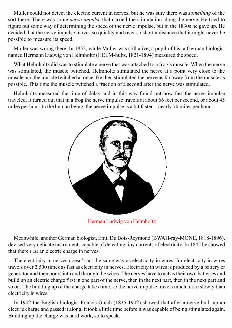
Muller was wrong there. In 1852, while Muller was still alive, a pupil of his, a German biologist
named Hermann Ludwig von Helmholtz (HELM-hults, 1821-1894) measured the speed.
What Helmholtz did was to stimulate a nerve that was attached to a frog’s muscle. When the nerve
was stimulated, the muscle twitched. Helmholtz stimulated the nerve at a point very close to the
muscle and the muscle twitched at once. He then stimulated the nerve as far away from the muscle as
possible. This time the muscle twitched a fraction of a second after the nerve was stimulated.
Helmholtz measured the time of delay and in this way found out how fast the nerve impulse
traveled. It turned out that in a frog the nerve impulse travels at about 66 feet per second, or about 45
miles per hour. In the human being, the nerve impulse is a bit faster—nearly 70 miles per hour.
Herman Ludwig von Helmholtz
Meanwhile, another German biologist, Emil Du Bois-Reymond (BWAH-ray-MONE, 1818-1896),
devised very delicate instruments capable of detecting tiny currents of electricity. In 1845 he showed
that there was an electric charge in nerves.
The electricity in nerves doesn’t act the same way as electricity in wires, for electricity in wires
travels over 2,500 times as fast as electricity in nerves. Electricity in wires is produced by a battery or
generator and then pours into and through the wires. The nerves have to act as their own batteries and
build up an electric charge first in one part of the nerve, then in the next part, then in the next part and
so on. The building up of the charge takes time, so the nerve impulse travels much more slowly than
electricity in wires.
In 1902 the English biologist Francis Gotch (1835-1902) showed that after a nerve built up an
electric charge and passed it along, it took a little time before it was capable of being stimulated again.
Building up the charge was hard work, so to speak.

In 1909, another English biologist, Keith Lucas (1879-1910), found that if a particular nerve was
stimulated very gently then nothing at all might happen. The muscle fiber to which it was attached
didn’t budge. If the nerve was stimulated harder there came a point where the nerve impulse was set
off and the muscle fiber contracted as hard as it could. It was “all or nothing.”
Of course, you can make your arm muscle contract by any amount from a tiny pull to a forceful
one, but that is because the muscle consists of many, many fibers. Each fiber contracts “all or
nothing,” but when only a few fibers contract, the muscle as a whole contracts gently, and as more
and more fibers contract, the muscle contracts more and more forcefully.
Scientists continued to measure the strength and behavior of the nerve impulse in more and more
delicate ways. Finally, two English biologists, Alan Lloyd Hodgkin (born 1914} and Andrew Fielding
Huxley (born 1917), began to work with certain axons in squid, axons that were unusually thick.
Hodgkin and Huxley managed to insert delicate instruments inside the axon. By 1952, they worked
out the way in which certain electrically charged atoms (ions, EYE-onz) move into the axon and out
of it, building up an electric charge.
In 1924, a German doctor, Hans Berger (1873-1941), worked out a system whereby the electric
impulse in the brain cells could be measured. Wires were pasted to the skull, and as an electric current
flowed here and there in the brain, a needle could be made to trace out a wavering line on paper.
The waves changed according to whether the patient’s eyes were open or closed, whether he was
awake or asleep and so on. It is difficult to get the meaning out of the waves for little things, but there
are changes that can mark off the presence of .something serious like a brain tumor or a small
epileptic attack.
The measurement of the brain’s electric
currents is called electroencephalography
(ee-LEK-troh-en-sef-uh-LOG-ruh-fee), or
EEC for short. It comes from the Greek
words meaning “electric writing of the
brain.”
The nerve impulse can run along from
one end of the neuron to the other—from
the dendrites, through the nerve cell itself,
and along the axon to its very end. But then
comes the synapse; a little space between
the end of the axon and the beginning of
the dendrites of the next neuron. How does
the nerve impulse cross the synapse?
Electroencephalography

A number of scientists thought that there might be a chemical that was produced by the nerve
impulse. This chemical would cross the synapse and produce a nerve impulse in the next neuron. But
how could that be proved?
A German-American biologist, Otto Loewi (LOI-vee, 1873-1961), was working on the problem,
and one night in 1921, he woke at 3 A.M., having thought of an experiment he could try. He made a
note on a pad near the bed and went back to sleep. When he woke in the morning, he couldn’t
remember the experiment he had thought of and couldn’t read what he had written when he was half-
asleep.
The next night, he woke at 3 A.M. again and remembered the experiment. This time, he took no
chances. He dressed, went to his laboratory, and got to work.
He took the heart out of a frog and filled it with a mixture of chemicals that kept it beating. To the
heart was attached a nerve that, when stimulated, slowed the heartbeat. Loewi stimulated the nerve
and the heartbeat slowed.
Loewi then took the mixture of chemical from the heart and put it into a second frog heart. The beat
of the second heart slowed at once, even though in that case there was no nerve stimulation. This
meant that the nerve impulse had formed a chemical in the mixture in the first heart and this chemical
did the trick in the second heart. By 5 A.M., Loewi had proved his point.
An English biologist, Henry Hallett Dale (1875-1968), had isolated a chemical from a certain fungus
called ergot and found that it made muscles contract the way a nerve impulse would. In 1914 this
chemical was identified as something chemists call acetylcholine (AS-eh-teel-KOH-leen). Once Dale
heard of Loewi’s experiment, he wondered whether the nerve impulse produced acetylcholine at the
synapse. In 1929 he was able to show that this was so.
Some nerves produce other chemicals that crossed synapses, and the natures of these were worked
out also.
Did different parts of the brain specialize in different tasks? Some people suspected this might be
so. A German doctor, Franz Joseph Gall (GAHL, 1758-1828), thought so. He thought that a certain
part of the brain might control a sense of humor, for instance, and another a tendency to be a criminal.
He thought that bumps in the skull showed the extra growth of the brain underneath. From the
location of the bumps, one could then understand a person’s character, intelligence, and so on.
This technique for reading character is called phrenology (freh-NOLruh-jee, from Greek words
meaning “study of the mind”). For a while, phrenology was quite popular, but it was really worthless.
It was no more useful than reading the future in a crystal ball.
Gall’s thoughts did spur other scientists to work on the problem. They would remove or damage
or stimulate parts of the brains of animals to see what would happen to the body. In 1870 two German
doctors, Julius Eduard Hitzig (1838-1907) and Gustav Fritsch
(1838-1927), worked with dogs. They stimulated certain portions of the brain and noted which
muscles contracted or weakened as a result.

Phrenology of the brain
This work was continued by a Scottish doctor, David Ferrier (1843-1928), who, about 1876, drew
a kind of map of the body on the brain that showed which parts of the brain control which parts of the
body. There was a band around the mid-part that controls various muscles, and a portion near the
back of the brain that receives nerve impulses from the eye. Other places receive impulses from the
other senses.
The map was made even more accurate by an English doctor, Charles Sherrington (1857-1952).
Only a small portion of the surface of the brain seemed to be involved with muscular movement
and with the senses. This led some people to believe that most of the human brain wasn’t used. This
is quite wrong. All of the brain is used. The parts that don’t control muscles or senses must still be
used in storing memories, in making judgments, in thinking new thoughts, and so forth.
The main portion of the brain, the cerebrum, doesn’t take care of all the workings of the body. The
cerebrum is needed when something requires judgment, when you have to make a decision as to what
to do. Sometimes, though, you can’t wait for a decision. If you accidentally touch something hot,
you can’t wait to decide that you had better take your hand away. By that time, you may be badly
hurt. Instead, your hand moves away instantly, before you even have a chance to know you’ve
touched something hot.

This sort of thing was first studied by an English doctor, Marshall Hall (1790-1857). Beginning in
1832, he studied these quick, emergency responses. He called them reflex actions (from Latin words
meaning “to bend back”). The sense impression travels along one nerve, and another impulse bends
back, returning at once to produce the necessary muscular motion. Hall suggested that such reflex
actions are controlled automatically by nerves that go to and from the spinal cord.
Sherrington, whom I mentioned a little while ago, studied other kinds of reflex action. He showed
that though nerves went from the brain to the muscles and stimulated them to contract, other nerves
went from the muscles to the brain. This second set of nerves made it possible for the brain to know
the exact position of each muscle and how contracted it was.
Thus, if you are standing, your brain always knows if you are beginning to lean to one side, and it
can direct a particular muscle to contract a little and keep you upright. When you stand, you are
always adjusting the balance this way and that without knowing it. That is why you get tired when
you’ve been standing a long time, even though it doesn’t seem to you that you’ve been doing
anything.
There are other reflex actions that control your breathing. When you reach for something, a con-
stant adjustment of your arm muscles keeps you from moving your hand too far or too much to one
side, or not far enough. All such reflexes are controlled by the cerebellum.
to the spinal cord spinal cord
muscle
from the spinal cord
A reflex action

The portion of the nervous system that controls all these automatic bits of behavior is called the
auto-nomic nervous system. Autonomic (AW-toh-NOM-ik) means “independent” because that part
of the nervous system works without control by the cerebrum. The name was first used in 1889 by an
English doctor, John Newport Langley (1852-1925).
A Swiss doctor, Walter Rudolf Hess (1881-1973) used thin needles to stimulate tiny portions of
animal brain to tell exactly where the various centers of the autonomic nervous system are located.
Autonomic nervous system
New reflex actions can be developed. This was shown by a Russian biologist, Ivan Petrovich
Pavlov (1849-1936). In the 1920s, he studied dogs, whose mouths water and fill with saliva when they
see food. This is a reflex. For a period of time, Pavlov would ring a bell every time he presented the
dog with food. Eventually, the dog associated the bell with food, and his mouth watered whenever the
bell rang even if there was no food. The dog had developed a conditioned reflex.
If this was repeated over and over with just the bell and no food, the association would be broken
at last. The conditioned reflex would disappear.

5. SIDES AND SLEEP
THE HUMAN BODY is bilaterally symmetrical. That means it has a left side and a right side that are
mirror images of each other. What we have on one side, we usually have on the other. We have two
ears, two eyes, two nostrils, two arms, and two legs. Inside the body, we have two lungs, two
kidneys, and so on.
When we have only one of something, it usually lies along the midline of the body. We have one
nose, one mouth, one chin, one breastbone, one backbone and so on.
What about the brain? It seems we only have one brain and one spinal cord below it, and the spinal
cord runs down the midline of the body inside the backbone.
If you were to look at the brain, however, you would see that it exists in two halves, right and left,
with a thick bundle of nerve connecting them. The connection is the corpus callosum (KAWR-pus-
ka-LOHS-um). If you were to look at an intact walnut kernel, you would see much the same thing.
Corpus callosum
nerve fibrescorpus collosum
In some ways the two halves of the cerebrum are two separate brains. If there is a place on the left
side of the brain which controls the arm muscle on one side of the body, there is a place on the same
spot on the right side of the brain that controls the arm muscle on the other side of the body.
In fact, if you cut the corpus callosum so that there is no connection between the two halves of the
brain, it is possible to train one half of the brain to do something while the other half knows nothing
about it.
Are the two halves exactly the same, or does each side do something the other side can’t, or at
least do it better?
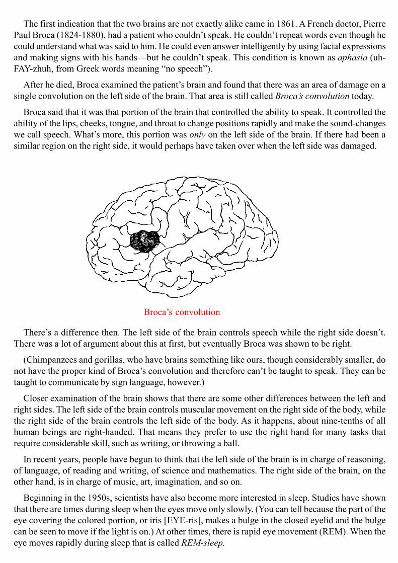
The first indication that the two brains are not exactly alike came in 1861. A French doctor, Pierre
Paul Broca (1824-1880), had a patient who couldn’t speak. He couldn’t repeat words even though he
could understand what was said to him. He could even answer intelligently by using facial expressions
and making signs with his hands—but he couldn’t speak. This condition is known as aphasia (uh-
FAY-zhuh, from Greek words meaning “no speech”).
After he died, Broca examined the patient’s brain and found that there was an area of damage on a
single convolution on the left side of the brain. That area is still called Broca’s convolution today.
Broca said that it was that portion of the brain that controlled the ability to speak. It controlled the
ability of the lips, cheeks, tongue, and throat to change positions rapidly and make the sound-changes
we call speech. What’s more, this portion was only on the left side of the brain. If there had been a
similar region on the right side, it would perhaps have taken over when the left side was damaged.
Broca’s convolution
There’s a difference then. The left side of the brain controls speech while the right side doesn’t.
There was a lot of argument about this at first, but eventually Broca was shown to be right.
(Chimpanzees and gorillas, who have brains something like ours, though considerably smaller, do
not have the proper kind of Broca’s convolution and therefore can’t be taught to speak. They can be
taught to communicate by sign language, however.)
Closer examination of the brain shows that there are some other differences between the left and
right sides. The left side of the brain controls muscular movement on the right side of the body, while
the right side of the brain controls the left side of the body. As it happens, about nine-tenths of all
human beings are right-handed. That means they prefer to use the right hand for many tasks that
require considerable skill, such as writing, or throwing a ball.
In recent years, people have begun to think that the left side of the brain is in charge of reasoning,
of language, of reading and writing, of science and mathematics. The right side of the brain, on the
other hand, is in charge of music, art, imagination, and so on.
Beginning in the 1950s, scientists have also become more interested in sleep. Studies have shown
that there are times during sleep when the eyes move only slowly. (You can tell because the part of the
eye covering the colored portion, or iris [EYE-ris], makes a bulge in the closed eyelid and the bulge
can be seen to move if the light is on.) At other times, there is rapid eye movement (REM). When the
eye moves rapidly during sleep that is called REM-sleep.

Dreams apparently take place only during REM-sleep. If someone is awakened during REM-sleep,
he or she can almost always remember a dream that was just being dreamed.
It is rather a puzzle as to why sleep is needed. It can’t be just in order to rest. You can rest just as
well if you lie quietly and relax with your eyes open. In fact, you are liable to be more active while
sleeping than while relaxing. During sleep, you often change your position, and sometimes you just
thrash about.
And yet relaxing doesn’t substitute for sleep. If you stay awake, even though you relax, you feel a
greater and greater need for sleep. Eventually, if you stay awake long enough, you begin to hallucinate.
Without sleep you would die sooner than if you go without water.
What’s more, it’s REM-sleep that seems to be the necessary part. If a person is deliberately
awakened every time he begins to have REM-sleep, he has more than usual the next night, as though
he has to make up for what he missed.
This would make it seem that it’s dreaming rather than sleeping that is necessary. But why?
No one really knows. My own feeling is that the human brain is so complicated that it needs to take
time out every once in a while in order to get rid of snarls and mixed-up thoughts that have accumu-
lated during the waking hours. The body may accomplish this by dreaming, thus getting rid of mental
“garbage.” It’s a sort of housecleaning that leaves the brain fresh for new thought and activity the next
day.
But I repeat, no one really knows.
In fact, after all the many centuries of studying the brain, we still don’t know very much about it. It
is the most complicated part of the human body, so perhaps that’s not surprising. In fact, it is the
most complicated object we know in any part of the universe we can observe—living or nonliving.
Complexity of nerve
tissue from cerebral cortex
The human brain is the most hopeful thing there is,
for it is through the brain that people think original
thoughts, develop emotions like sympathy and love,
write books, create music and art, and work out solu-
tions for the dangerous problems that constantly face
humanity.
The human brain is also the most dangerous thing
there is, for it is through the brain that we develop
superstitions that steer us in the wrong direction. It is
also through the brain that we develop fears and hates
and suspicions of each other that lead to cruelty, crime,
and war.
We can only hope that continued study of the brain
will teach us how to encourage the helpful aspects of
our thinking and discourage the harmful aspects. Then
maybe we can have a much better, safer, and kinder
world than we seem to have right now.
END