Embed Size (px)
Citation preview
Speaker:
How to Treat Melanoma
Professor Rodney Sinclair MBBS, MD, FACD
University of Melbourne and the Epworth Hospital
Supported by an unrestricted educational grant from Bayer
What is a melanoma
• Melanomas are malignant tumours derived from melanocytes.
• The commonest site of involvement is the skin, although occasionally primary melanoma develops in other organs (eye, oral/nasal mucosa, vulval and anorectal mucosa).
• Melanomas are a major cause of premature death from cancer.
What is a melanoma• Recognised risk factors include personal or family history of melanoma, large numbers of naevi and/or dysplastic naevi, giant congenital melanocytic naevi, fair complexion, a tendency to sunburn, solar‐damaged skin, a history of non‐melanoma skin cancer, and immunodeficiency.
• The commonest sites for melanoma are the legs of women and the backs of men (which are not the sites of greatest sun exposure).
• Early detection improves survival.
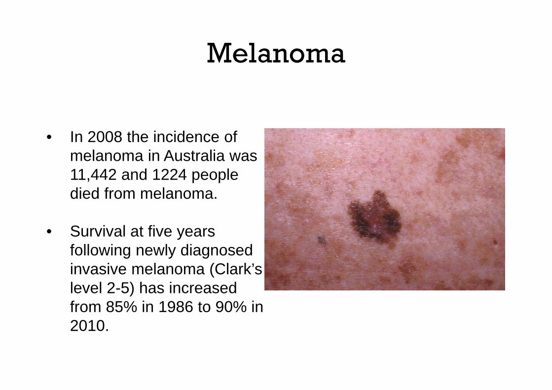
• In 2008 the incidence of melanoma in Australia was 11,442 and 1224 people died from melanoma.
• Survival at five years following newly diagnosed invasive melanoma (Clark’s level 2-5) has increased from 85% in 1986 to 90% in 2010.
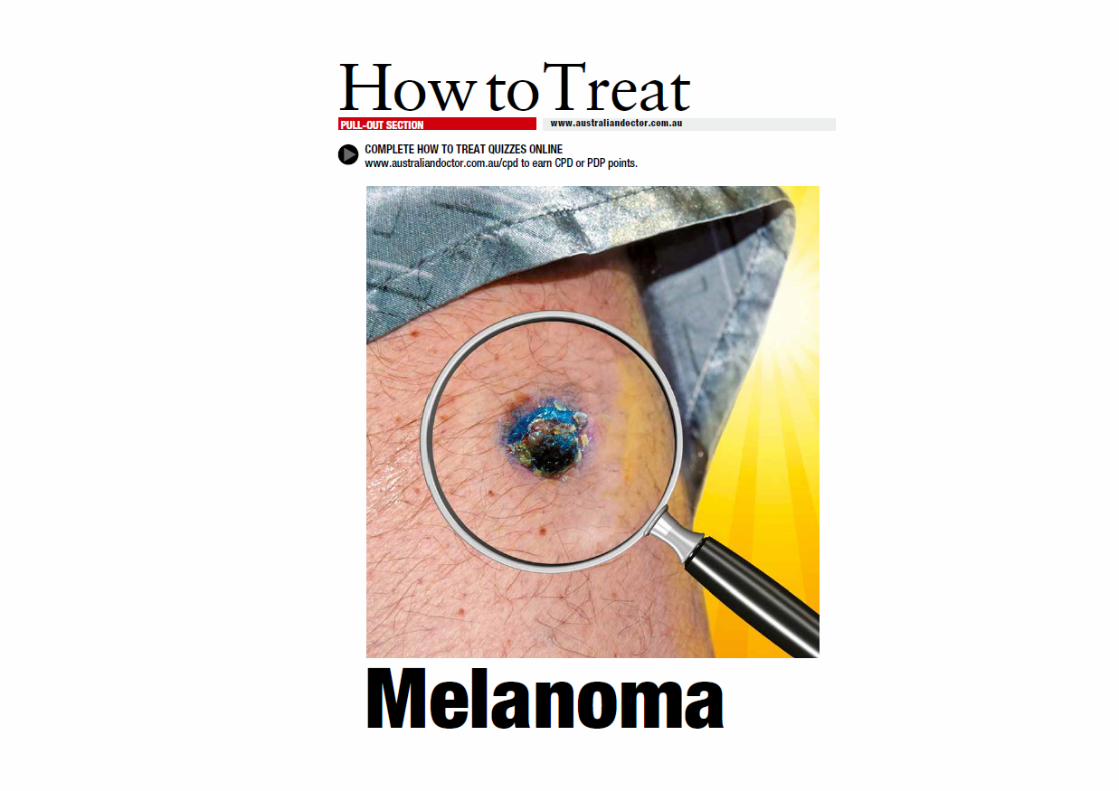
Melanoma
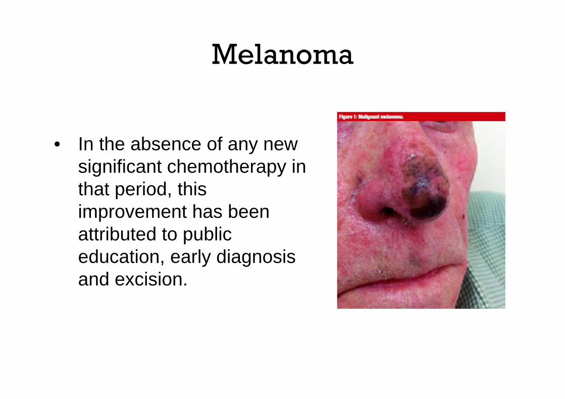
• In the absence of any new significant chemotherapy in that period, this improvement has been attributed to public education, early diagnosis and excision.
Melanoma
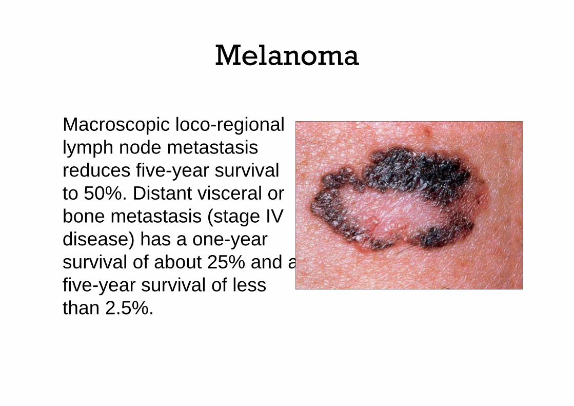
Macroscopic loco-regional lymph node metastasis reduces five-year survival to 50%. Distant visceral or bone metastasis (stage IV disease) has a one-year survival of about 25% and a five-year survival of less than 2.5%.
Melanoma
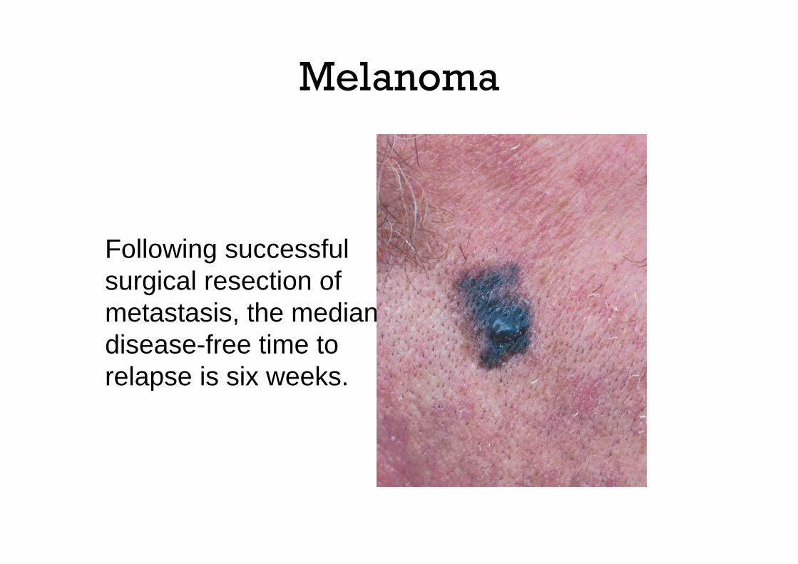
Following successful surgical resection of metastasis, the median disease-free time to relapse is six weeks.
Melanoma

Functional mutations in genes in the mitogen-activated protein (MAP) kinase pathway are commonly detected in melanoma and these mutations influence growth control (figure 1).
Melanoma
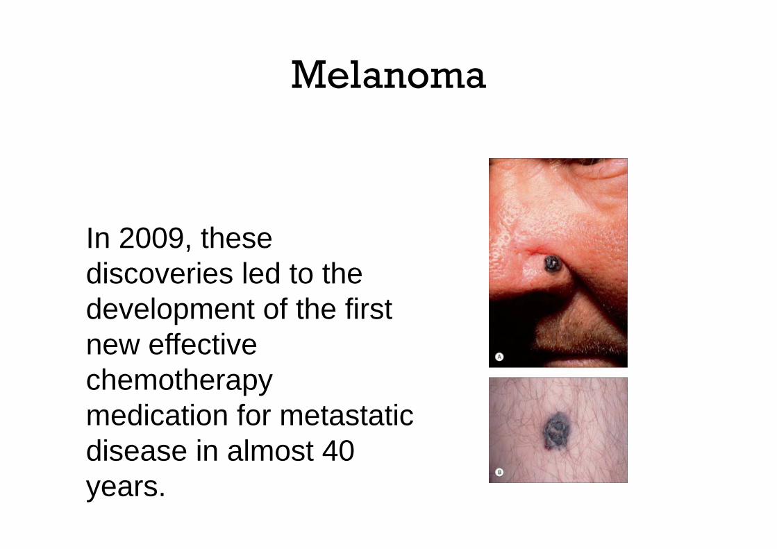
In 2009, these discoveries led to the development of the first new effective chemotherapy medication for metastatic disease in almost 40 years.
Melanoma
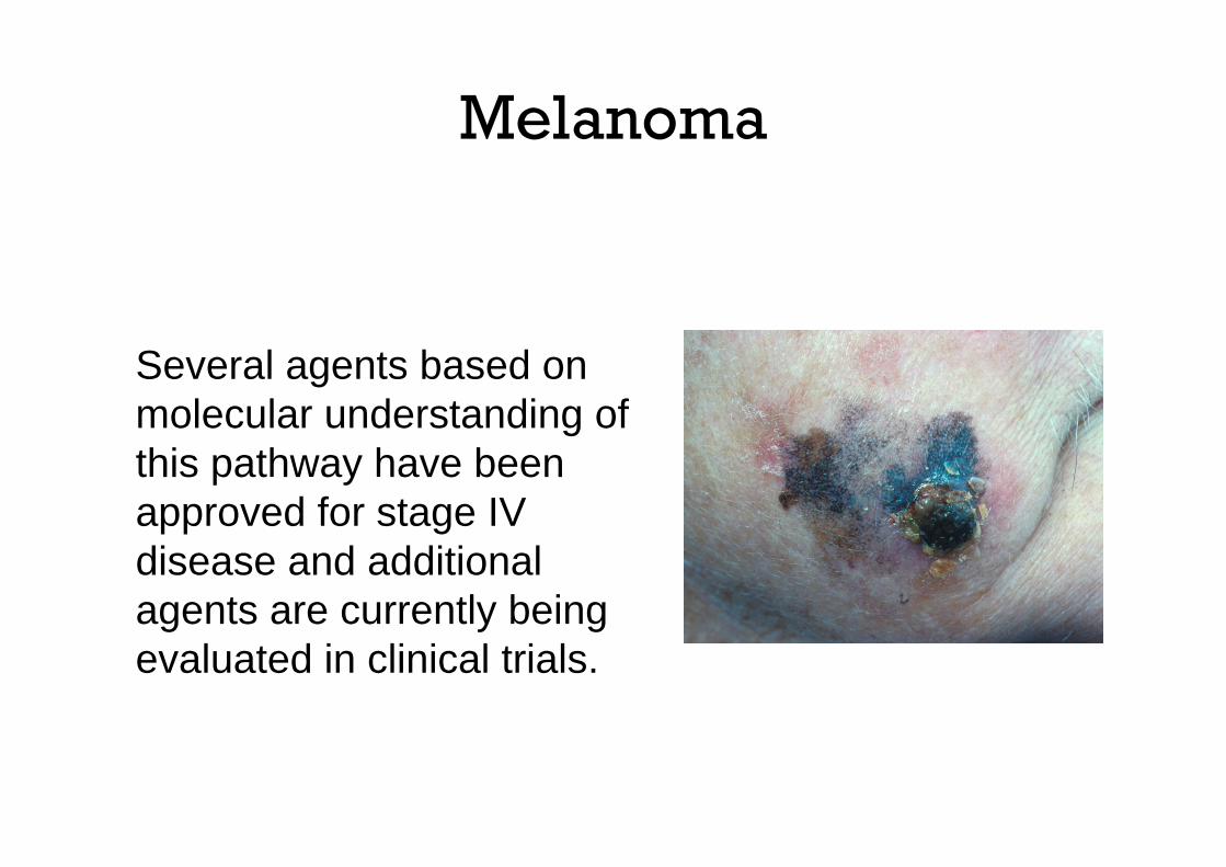
Several agents based on molecular understanding of this pathway have been approved for stage IV disease and additional agents are currently being evaluated in clinical trials.
Melanoma
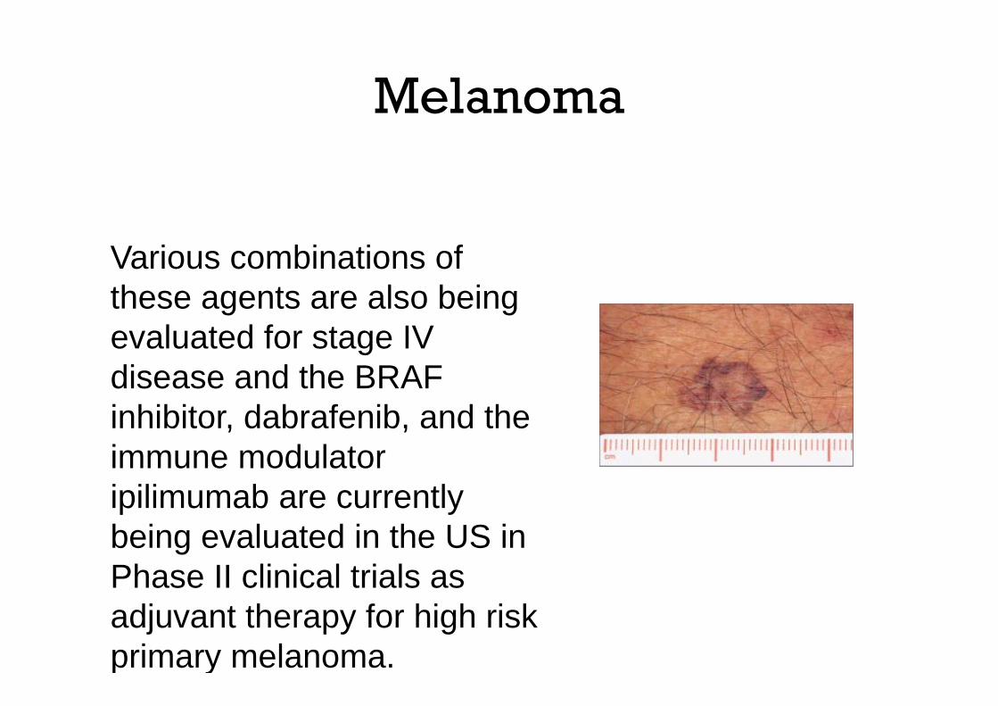
Various combinations of these agents are also being evaluated for stage IV disease and the BRAF inhibitor, dabrafenib, and the immune modulator ipilimumab are currently being evaluated in the US in Phase II clinical trials as adjuvant therapy for high risk primary melanoma.
Melanoma
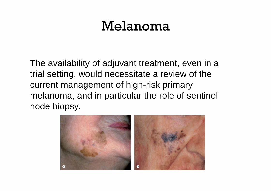
The availability of adjuvant treatment, even in a trial setting, would necessitate a review of the current management of high-risk primary melanoma, and in particular the role of sentinel node biopsy.
Melanoma
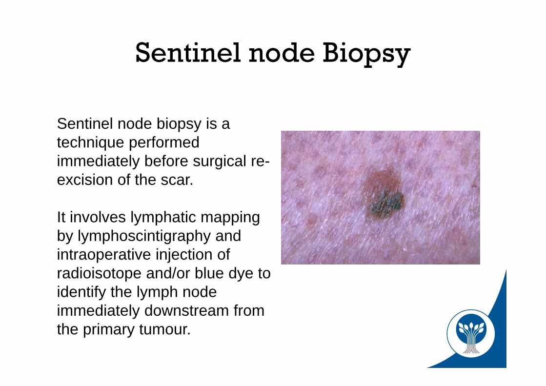
Sentinel node biopsy is a technique performed immediately before surgical re-excision of the scar.
It involves lymphatic mapping by lymphoscintigraphy and intraoperative injection of radioisotope and/or blue dye to identify the lymph node immediately downstream from the primary tumour.
Sentinel node Biopsy
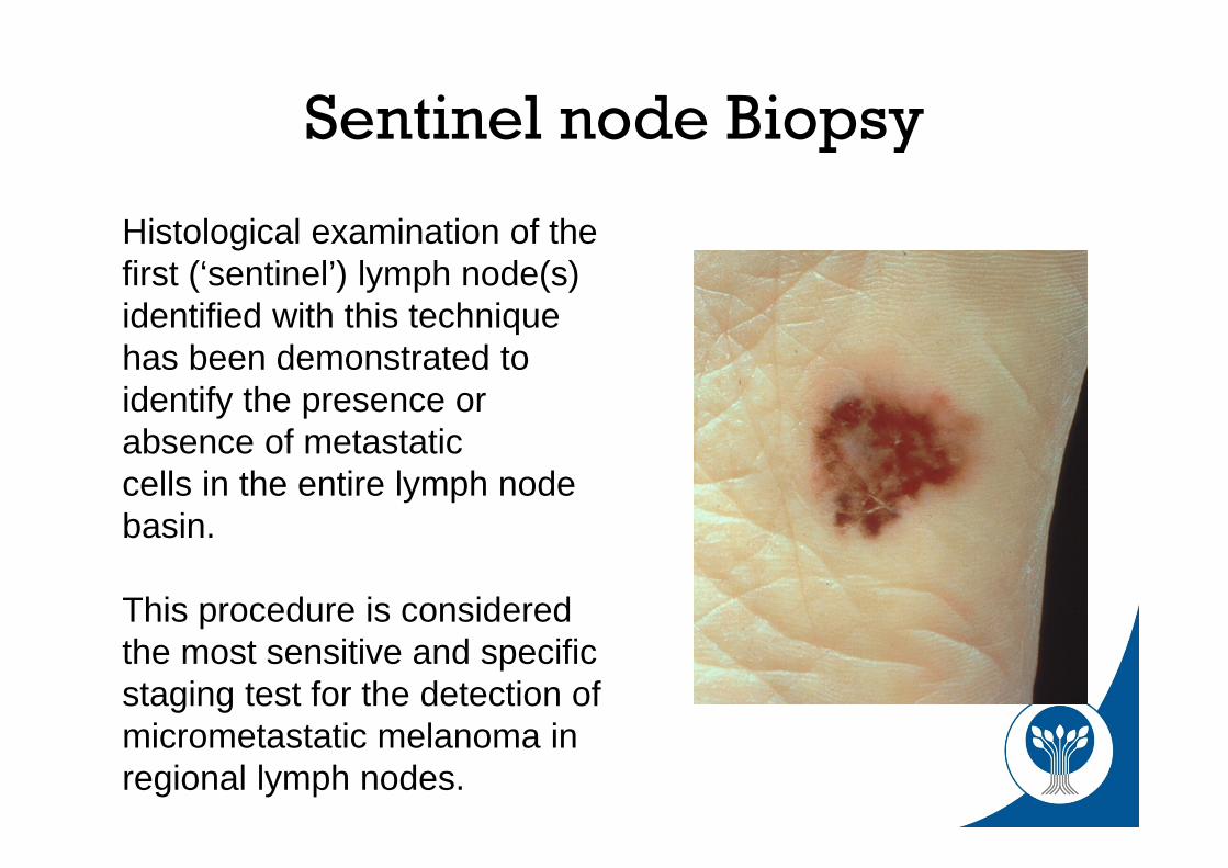
Histological examination of the first (‘sentinel’) lymph node(s) identified with this technique has been demonstrated to identify the presence or absence of metastaticcells in the entire lymph node basin.
This procedure is considered the most sensitive and specific staging test for the detection of micrometastatic melanoma in regional lymph nodes.
Sentinel node Biopsy
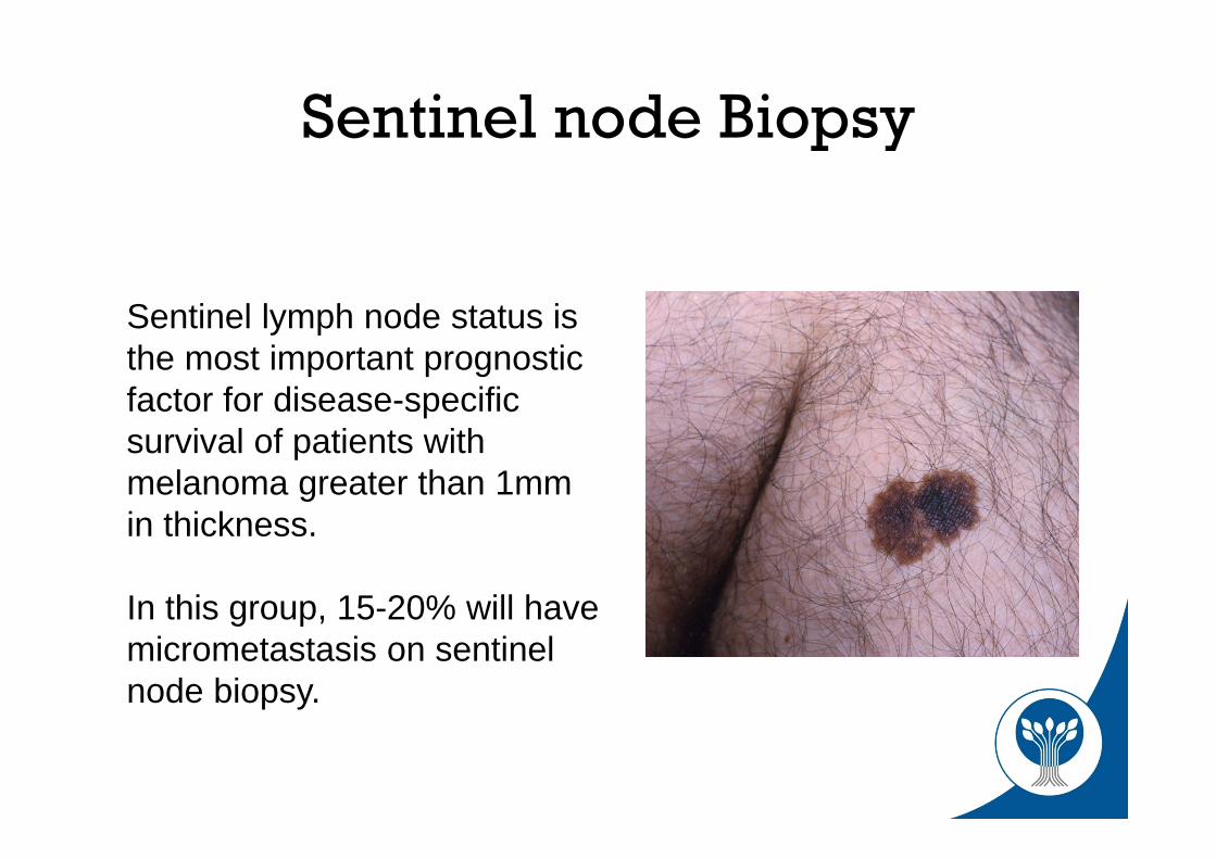
Sentinel lymph node status is the most important prognostic factor for disease-specific survival of patients with melanoma greater than 1mm in thickness.
In this group, 15-20% will have micrometastasis on sentinel node biopsy.
Sentinel node Biopsy
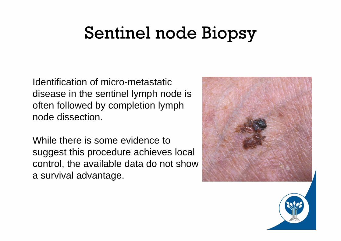
Identification of micro-metastatic disease in the sentinel lymph node is often followed by completion lymph node dissection.
While there is some evidence to suggest this procedure achieves local control, the available data do not show a survival advantage.
Sentinel node Biopsy
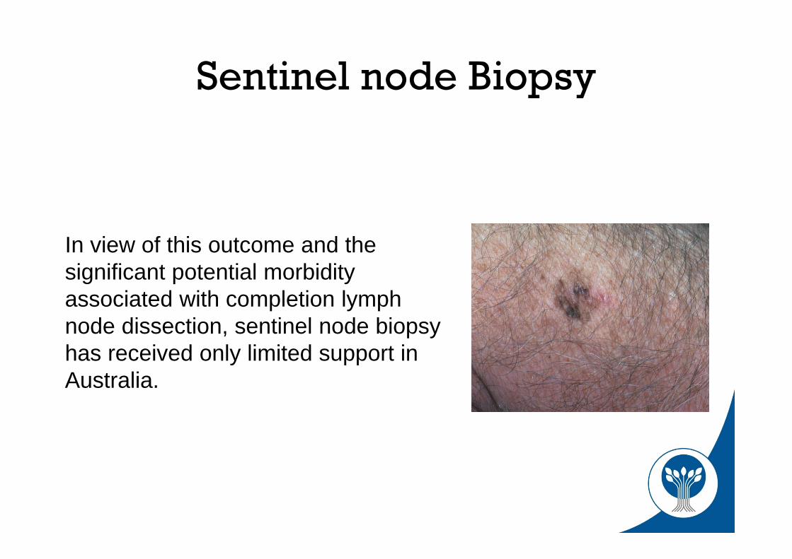
In view of this outcome and the significant potential morbidity associated with completion lymph node dissection, sentinel node biopsy has received only limited support in Australia.
Sentinel node Biopsy
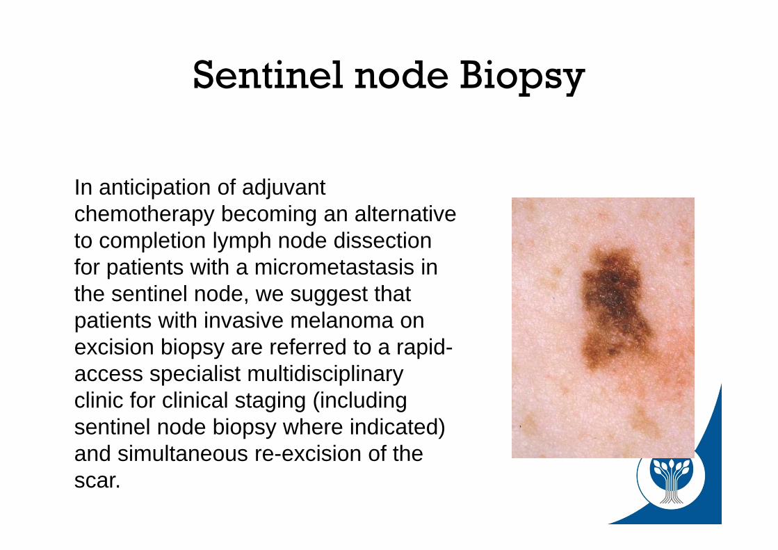
In anticipation of adjuvant chemotherapy becoming an alternative to completion lymph node dissection for patients with a micrometastasis in the sentinel node, we suggest that patients with invasive melanoma on excision biopsy are referred to a rapid-access specialist multidisciplinary clinic for clinical staging (including sentinel node biopsy where indicated) and simultaneous re-excision of the scar.
Sentinel node Biopsy
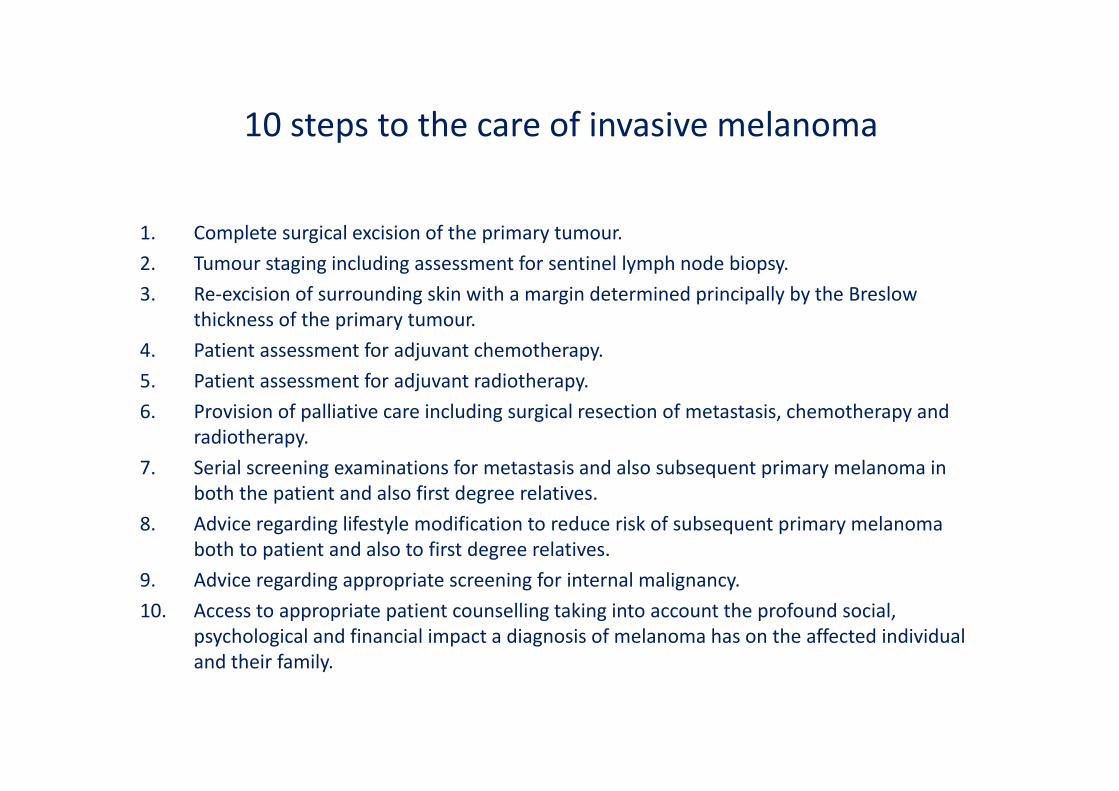
10 steps to the care of invasive melanoma
1. Complete surgical excision of the primary tumour.2. Tumour staging including assessment for sentinel lymph node biopsy. 3. Re‐excision of surrounding skin with a margin determined principally by the Breslow
thickness of the primary tumour. 4. Patient assessment for adjuvant chemotherapy. 5. Patient assessment for adjuvant radiotherapy.6. Provision of palliative care including surgical resection of metastasis, chemotherapy and
radiotherapy.7. Serial screening examinations for metastasis and also subsequent primary melanoma in
both the patient and also first degree relatives.8. Advice regarding lifestyle modification to reduce risk of subsequent primary melanoma
both to patient and also to first degree relatives.9. Advice regarding appropriate screening for internal malignancy.10. Access to appropriate patient counselling taking into account the profound social,
psychological and financial impact a diagnosis of melanoma has on the affected individual and their family.
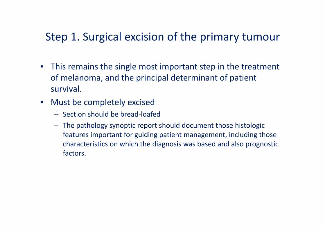
Step 1. Surgical excision of the primary tumour
• This remains the single most important step in the treatment of melanoma, and the principal determinant of patient survival.
• Must be completely excised– Section should be bread‐loafed– The pathology synoptic report should document those histologic
features important for guiding patient management, including those characteristics on which the diagnosis was based and also prognostic factors.
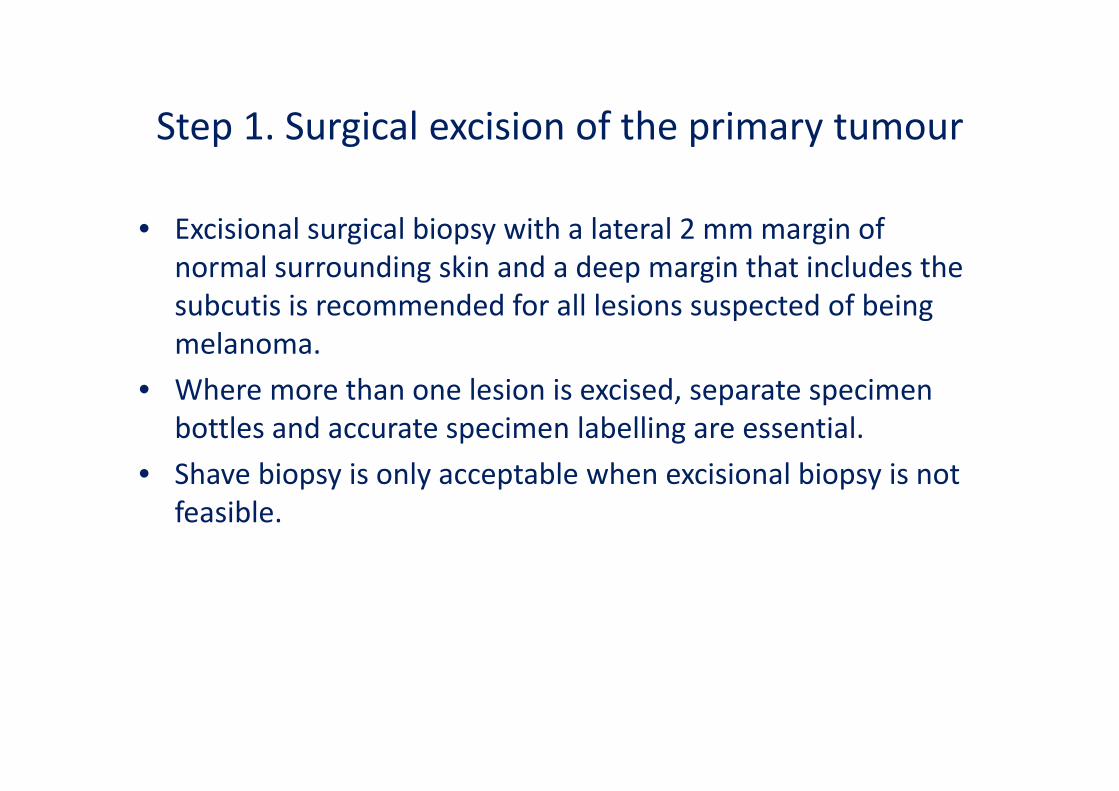
Step 1. Surgical excision of the primary tumour
• Excisional surgical biopsy with a lateral 2 mm margin of normal surrounding skin and a deep margin that includes the subcutis is recommended for all lesions suspected of being melanoma.
• Where more than one lesion is excised, separate specimen bottles and accurate specimen labelling are essential.
• Shave biopsy is only acceptable when excisional biopsy is not feasible.
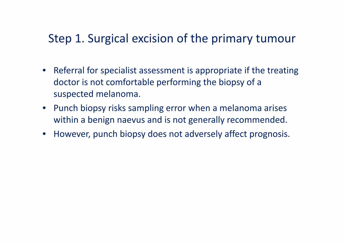
Step 1. Surgical excision of the primary tumour
• Referral for specialist assessment is appropriate if the treating doctor is not comfortable performing the biopsy of a suspected melanoma.
• Punch biopsy risks sampling error when a melanoma arises within a benign naevus and is not generally recommended.
• However, punch biopsy does not adversely affect prognosis.
Step 2. Tumour staging including assessment for sentinel lymph node biopsy.
• Clinical staging– 5 year survival overall is 90%– if macroscopic loco‐regional lymph node metastas, 5 year survival is
50%– If distant visceral or bone metastasis (stage 4 disease), 5 year survival
less than 2.5%.– Following successful surgical resection of metastasis, the median
disease free time to relapse is 6 weeks. – The median 12 months survival rate for Stage 4 melanoma is 25%.

Step 2. Tumour staging including assessment for sentinel lymph node biopsy.
• Clinical staging includes palpation of the regional lymph node basins and examination for hepatomegaly.
• Sentinel node biopsy should be discussed with any patient with the following diagnosis:– invasive melanoma greater than 1mm thick; – Clark level 4 melanoma;– melanoma with more than two mitoses per high‐power field; – Ulcerated melanoma; – melanoma with significant regression; – or melanoma of unknown malignant potential.
Step 3 Re‐excision Margins
• The scar following removal of an invasive melanoma should be re‐excised with a margin of between 1 and 2 cm, with the choice of excision margin determined primarily by the Breslow tumour thickness in mm
melanoma in situ (restricted to epidermis)—margin 5 mm melanoma <1.0 mm thick—margin 1 cm melanoma <1.0 mm thick—minimum margin 1 cm and
maximum 2 cm melanoma >4 mm thick—minimum margin 2 cm
Step 3 Re‐excision Margins
• Wider excision has been demonstrated to reduce the risk of local persistence/recurrence of the tumour and local metastasis but there is no evidence that a margin greater than 1 cm offers additional benefit in terms of patient survival.
Step 4 Adjuvant chemotherapy
• Interferon alpha 2b• Decarbazine• Ipilimumab• Vemarafinib• Debrafinib• Combination chemotherapy
Step 5 adjuvant radiotherapy
• While primary radiotherapy is occasionally used for unresectable lentigo maligna or invasive melanoma, it is more commonly used as adjuvant radiotherapy for cutaneous melanoma likely to recur locally.
• Indications for adjuvant radiotherapy include a Breslow thickness >4mm, satellite nodules or neurotropic spread.
• Adjuvant radiotherapy is also used to prevent recurrence following regional lymph node resection.
• Common indications include more than three nodes with metastasis, a large tumour mass in a single node or extracapsular spread.
Step 6 Adjuvant surgery
• Therapeutic elective lymph node resection is generally not recommended as adjuvant surgical treatment.
• This is because there is no evidence to suggest a survival advantage and there is significant potential surgical morbidity, including postoperative lymphoedema
Step 6 Adjuvant surgery
• Identification of suspicious lymph nodes on clinical examination should be followed by fine‐needle aspiration and ultrasound imaging, MRI or PET.
• If nodal metastasis is confirmed histologically, immediate complete regional lymph node dissection is recommended.
• Cure rates in the order of 30% may be achieved with completion lymphadenectomy for palpable disease.
Step 6 Adjuvant surgery
• Completion lymph node dissection following identification of micrometastasis on sentinel lymph node biopsy is more controversial as the proportion of lymph node micrometastasis that progresses to symptomatic disease is not known
• A prospective randomised multicentre selective lymphadenectomy trial (MSLT‐I) comparing completion lymph node dissection with observation showed no difference in overall survival
• Patients with micrometastasis may be enrolled inthe multicentre sentinel lymph node treatment trial MSLT‐II.1
Step 7. Surgery, chemotherapy and radiotherapy for stage IV disease
• Specialist multi‐disciplinary care• Combination chemotherapy• Improve survival at 1 year• No improved survival at 5 years
Step 8. Follow‐up examination for metastasis and subsequent primary melanoma
• All patients diagnosed with invasive melanoma require periodic follow‐up that includes examination and palpation for local recurrence, in transit metastasis, lymph node metastasis and hepatomegaly.
• Additional examination and investigation should be guided by reported symptoms.
Step 8. Follow‐up examination for metastasis and subsequent primary melanoma
• For the 90% of Australians who survive melanoma, the risk of developing a subsequent primary melanoma is in the order of 10%.
• This risk is also influenced by other factors including family history, skin type, hair colour and the presence of significant solar skin damage including non‐melanoma skin cancer
• It is recommended that all melanoma patients, including with in situ melanoma, have a complete skin check at least once a year for life following the diagnosis of a melanoma.
Step 8. Follow‐up examination for metastasis and subsequent primary melanoma
• As there is a familial tendency to melanoma, all first‐degree relatives of a patient diagnosed with melanoma should be encouraged to attend for a full skin examination to identify a previously unsuspected melanoma and to assess risk of future development of melanoma.
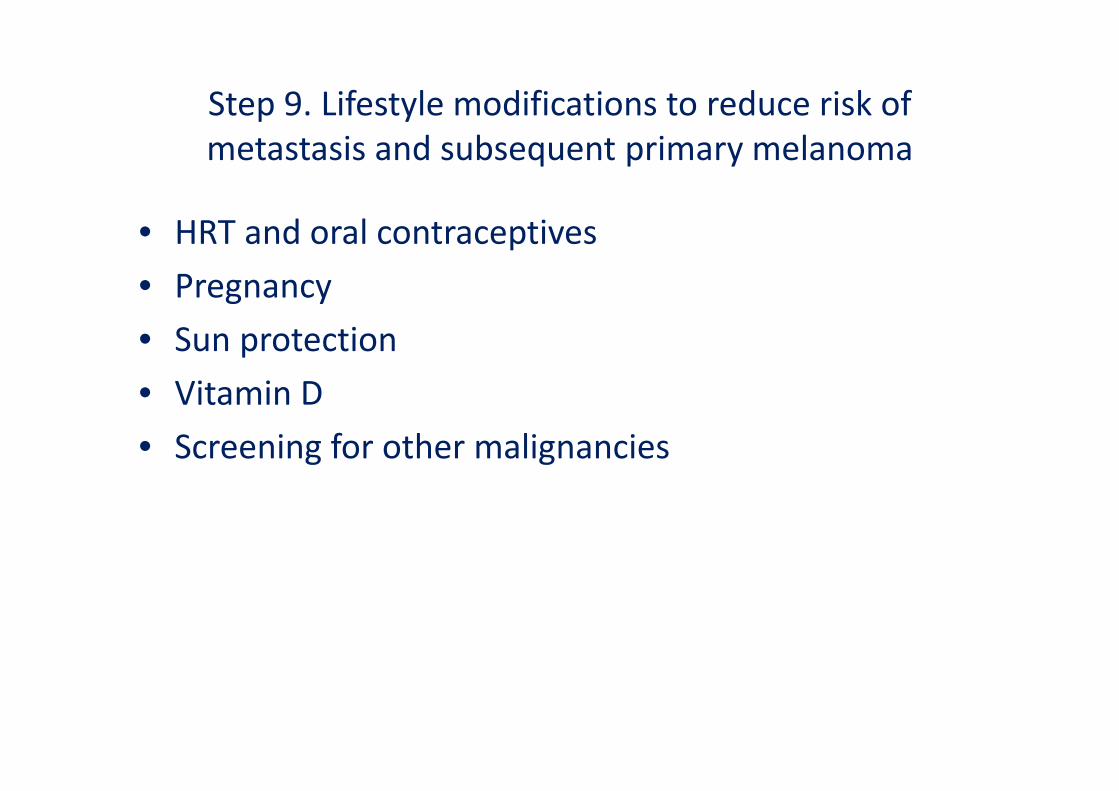
Step 9. Lifestyle modifications to reduce risk of metastasis and subsequent primary melanoma
• HRT and oral contraceptives• Pregnancy• Sun protection• Vitamin D• Screening for other malignancies

Step 10. Counselling
• Patients with invasive melanoma and their families will have complex social, psychological and financial issues.
• Life and income protection insurance may be declined even for people with thin melanomas.
• The nature and intensity of these issues will vary from person to person and with disease severity.
• Psychologists with experience in palliative care should be involved when the patients’ needs and those of the family can no longer be met by the treating physician.