Embed Size (px)
Citation preview

J. clin. Path. (1961), 14, 565
The histological diagnosis oftoxoplasmic lymphadenitis
A. G. STANSFELD
From the Departnment of Pathology, St. Bartholomew's Hospital, London
SYNOPSIS The commonest presenting sign of acquired toxoplasmosis in man is enlargement ofsuperficial lymph nodes. The persistence of the nodes may lead to a suspicion of malignant lymphomaand the diagnosis then hinges on the lymph node biopsy.
Three cases of toxoplasmic lymphadenitis are described in each of which the diagnosis was
unsuspected clinically. The chance discovery of a toxoplasma cyst in the lymph node section ofCase 1 led to the correct diagnosis, after an initial diagnosis of Hodgkin's disease had been made.In the other two, strikingly similar histological changes in the lymph node biopsies suggested thediagnosis, which was confirmed serologically in each case.
The histological changes are described and the clinical and pathological aspects of toxoplasmiclymphadenitis are briefly reviewed, with special reference to the differentiation from malignantlymphomatous conditions and to the specificity of the histological picture. It is concluded that thehistology is, in many instances, sufficiently distinctive for a tentative diagnosis of toxoplasmiclymphadenitis to be made on the lymph node biopsy. The diagnosis should always be confirmedby isolation of the parasite or by serological tests. It is exceedingly rare for toxoplasma cysts to befound in lymph nodes and only one previous observation of this kind has been reported.
Over the past 20 years it has become evident thathuman infection with Toxoplasma gondii is almostworld-wide in distribution and that this remarkablysuccessful parasite has, as alternative hosts, a widerange of mammals, birds, and even reptiles. In man,infection is not nearly as rare as was formerlythought. Not unnaturally, the first cases of acquiredtoxoplasmosis to be diagnosed were all examples ofthe severe, acute form of the disease, ending fatally.This variety is undoubtedly rare, but within the pastdecade a number of reports have been published ofa much commoner manifestation of acquired toxo-plasmosis, characterized by enlargement of super-ficial lymph nodes, often without other conspicuoussymptoms.The importance of toxoplasmic lymphadenitis
lies in its differentiation from much more seriousconditions, especially Hodgkin's disease and lympho-sarcoma, for which it may be mistaken on bothclinical and pathological grounds. Writers on thesubject have expressed different opinions about thespecificity of the histological changes in the lymphnodes in toxoplasmosis. Nevertheless there is inmany of the accounts a striking measure of agree-Received for publication 8 May, 1961
ment about the principal histological features, allow-ing for differences in terminology.The three cases reported here serve to emphasize
the relative frequency of the condition, the constancyof the histological changes in the lymph nodes, andthe importance of lymph node biopsy in arriving atthe diagnosis.
CASE REPORTS
CASE 1 A.M., a married woman of 30, was referred toSt. Bartholomew's Hospital on 13 October 1960 forradiotherapy after a diagnosis of Hodgkin's disease hadbeen made at another hospital on a cervical lymph nodebiopsy.For six to seven weeks she had noticed swellings in the
neck but felt otherwise quite well. Her appetite was goodand weight steady.
Physical examination showed a well nourished woman.Two enlarged lymph nodes, each nearly 2 cm. in diameter,were palpable in the neck: one on the right side, at theupper end of the posterior triangle; the other, in the leftsupraclavicular fossa, lying just below the biopsy incision.There were no enlarged nodes in axillae or groins andneither liver nor spleen were felt on abdominal examina-tion.The results of laboratory investigations were as
565
on April 20, 2020 by guest. P
rotected by copyright.http://jcp.bm
j.com/
J Clin P
athol: first published as 10.1136/jcp.14.6.565 on 1 Novem
ber 1961. Dow
nloaded from

A. G. Stansfeld
follows:-E.S.R. 7 mm. in the first hour, haemoglobin86% (12-7 g. haemoglobin / 100 ml.) W.B.C. 5,000 perc.mm. (polymorph neutrophils 70%, eosinophils 2%,lymphocytes 20%, monocytes 8%), platelets 176,000per c.mm. A radiograph of the chest did not show anyabnormality.The lymph node biopsy section was re-examined at St.
Bartholomew's at the start of radiotherapy and thechanges seen were then interpreted as reactive rather thanneoplastic. In the course of this examination a single cystof toxoplasma was encountered in the section and adiagnosis of toxoplasmic lymphadenitis was made on thestrength of this discovery. The diagnosis was confirmeda few days later by a positive dye test at a titre of 1 in1,000.The patient was subsequently given a 14-day course of
triple sulphonamide and pyrimethamine and, three weeksafter its conclusion, examination failed to disclose anypalpable lymph nodes in the neck or elsewhere. She hasremained well, but it was later reported that her son,aged 2 years, had an asymptomatic enlargement ofcervical lymph nodes.
CASE 2 D.L., a married woman of 40, was referred toanother hospital on 10 November 1960 by her doctor forthe investigation of enlarged cervical lymph nodes.Four months previously she had noticed a swelling
behind the left ear, but this had disappeared spon-taneously. A month later, a swelling appeared behind theleft sternomastoid and grew steadily in size, while asecond smaller swelling appeared on the right side too.She had not had a sore throat. The swelling was slightlytender at first but was not painful, though she experiencedsome difficulty in turning her head to the left. The swell-ings had persisted since. For a period of about threemonths she had also complained of indigestion andepigastric discomfort after food. She had also noticedtiredness and breathlessness but had no cough and herweight was steady.
Physical examination showed a well nourished woman.There were two enlarged, fairly soft nodes on the left sideof the neck and one on the right, while two similar nodeswere also found in the right axilla. The liver and spleenwere not felt. On the basis of these findings and the historyit was thought most likely that she had some form of'lymphomatous neoplasia'.The chest radiograph was normal and Wassermann,
Kahn, and Price's precipitation reactions gave negativeresults. A blood leucocyte count before admission hadshown a total of 5,000 cells per c.mm. (polymorphneutrophils 35 %, lymphocytes 60 %, and monocytes 5 %).The E.S.R. varied between 18 and 25 mm. in one hour andthe haemoglobin was 90% (13-3 g. haemoglobin/100 ml.).A relative lymphocytosis persisted for several weeks,though the total leucocyte count remained low. Noatypical cells were seen in blood smears. A Paul-Bunnellscreening test performed at the beginning of Decemberwas negative.On 16 November biopsy of a cervical lymph node was
carried out. The node, which measured 2-3 x 1-8 x 0-5cm., showed a hard, white cut surface with brown streaks.The pathologist reported that the histological picture
corresponded with that of lympho-histiocytic medullaryreticulosis (Robb-Smith, 1947), which he regarded as areactive response of relatively non-specific type. Thesection was referred to me for my opinion and, in viewof the remarkable similarity of the histological appear-ances to those of Case 1, a diagnosis of toxoplasmiclymphadenitis was suggested. Toxoplasma cysts werenot found in the original section or in sections sub-sequently examined but the diagnosis was confirmedserologically. The patient's serum, taken on 3 December,gave a positive dye test at a titre of 1 in 4,096 and thecomplement-fixation test was positive at 1 in 32.No treatment was given and on 21 December the lymph
nodes were reported to be subsiding.
CASE 3 R.A.L-N., an advertising representative, aged19, was referred by his own doctor to St. Bartholomew'sHospital on 14 November 1960 with a swelling beneathhis chin which was thought to be a thyroglossal cyst.He had first noticed the swelling, which was well to the
left of the midline, two to three months previously andat that time another swelling had appeared on the rightside of the neck also. The latter had disappeared followinga course of sulphonamide treatment. He had had nopain, sore throat, or cough and his weight was steady.Tonsillectomy had been performed at the age of 7.
Physical examination showed a young man of healthyappearance with a visible swelling beneath the chin onthe left side. On palpation, a fairly soft, doubtfullyfluctuant swelling, 1 in. x j in., could be felt in thissituation. A few soft nodes were palpable in the rightaxilla. The liver and spleen could not be felt.He was kept under observation and, two months later,
since the swelling showed no diminution in size, he wasadmitted to hospital for biopsy. Examination now showeda small palpable tonsillar lymph node on the right side,as well as the swelling on the left. Laboratory tests showed:Haemoglobin 102% (15-1 g. haemoglobin/100 ml.),W.B.C. 6,000 per c.mm. (polymorph neutrophils 65%,eosinophils 2%, lymphocytes 24%, monocytes 9%).On 13 January 1961 the swelling beneath the chin was
excised and proved to be an enlarged lymph node,measuring 3 0 x 1-6 x 0-3 cm. The consistency wasfirm and the cut surface of the node showed a faintlydiscernible follicular pattern. The histological appear-ances were sufficiently similar to those of Cases 1 and 2as at once to suggest a diagnosis of toxoplasmic lympha-denitis. Cysts were not found but the diagnosis of toxo-plasmosis was confirmed serologically. The patient'sserum, taken on 18 January, gave a positive dye test at atitre of 1 in 2,048 and the complement-fixation test waspositive at 1 in 64. A course of treatment with triplesulphonamide and pyrimethamine was given subse-quently and when last seen the patient was symptom free.
HISTOLOGY
The diagnosis of toxoplasmosis was made in Case 1by the chance discovery of a toxoplasma cyst in thelymph node section. This section also showed ratherdistinctive reactive changes, and it was the finding of
566
on April 20, 2020 by guest. P
rotected by copyright.http://jcp.bm
j.com/
J Clin P
athol: first published as 10.1136/jcp.14.6.565 on 1 Novem
ber 1961. Dow
nloaded from

The histological diagnosis of toxoplasmic lymphadenitis
very similar appearances in the lymph node biopsysections that led to the diagnosis in Cases 2 and 3.The following description of the histological changesis based upon all three cases.The lymph node in each instance was markedly
enlarged, but apart from this there was nothingremarkable about the macroscopic appearance.
On microscopical examination, the essentialfeatures are those of a subacute to chronic lympha-denitis with well-marked periadenitis. A strikingdegree of lymphoid hyperplasia is present which is,no doubt, mainly responsible for the lymph nodeenlargement. Many of the follicles are large andirregular in outline with active germinal centres(Fig. 1). These are, however, often less clearly definedthan is commonly the case in reactive hyperplasia,and proliferating lymphoblasts sometimes minglewith mature lymphocytes, giving the impression ofan infiltrative lesion. There are numerous mitosesof regular pattern in the germinal centres and a
notable feature is the presence of many freshlynecrotic cells, so that the centres are littered withkaryorrhectic particles of nuclear debris, some ofwhich have been engulfed by macrophages (Figs. 2and 3).
Scattered through the pulp of the node, and some-times invading the follicles, there are small clustersof very conspicuous, pale-staining histiocytes withvesicular nuclei and copious, somewhat eosinophiliccytoplasm (Figs. 3, 4, and 5). These cells resemblethe epithelioid cells of tuberculosis, but multinu-cleate cells are scarce and none simulate Langhansgiant cells. Moreover caseation is absent. The cellclusters are smaller and generally less sharply definedthan the focal collections of similar cells seen insarcoidosis. A further contrast with sarcoidosis isseen in the absence of reticulin in relation to thesecells in a silver impregnation preparation (Fig. 6).The presence of these histiocyte clusters is perhaps
the most distinctive feature of the histologicalpicture in all three cases. They are not regularly dis-tributed, but are chiefly seen towards the peripheryof the node which presents a spotted appearance on
low-power examination (Fig. 4). They appear to beengaged in the phagocytosis of nuclear debrisliberated by the piecemeal necrosis of cells in thefollicles. Infiltration of the follicles by histiocytes iscertainly one reason for the blurring of the follicularoutline.Almost as distinctive a feature as the histiocyte
clusters is the dense packing of many of the lymphsinuses with free macrophages (Fig. 7). In sectionsof each of the three lymph nodes some of the sinusesare so completely filled that they would not be dis-tinguishable, in the haematoxylin- and eosin-stainedsection, from the dense pulp of the node, but for
the smaller size and stronger basophilia of the lym-phocytes in the latter. Both peripheral and centralsinuses may be affected, but the density of thecellular infiltration varies and, in places, discretecells are seen against a background of eosinophiliccoagulum.
High-power examination reveals a mixed popu-lation of cells in the lymph sinuses, including ascattering of neutrophil polymorphs and some largemacrophages laden with cellular debris. The pre-dominant cell, however, is a small macrophage withmany of the characters of a blood monocyte. These'monocytoid' cells have relatively large, often con-torted, nuclei and moderately strongly staining,amphophilic cytoplasm (Fig. 8). They are quitedistinct from the large epithelioid histiocytes des-cribed above (Fig. 7), and also from the proliferatedlittoral cells commonly found in reactive conditions.
Lastly, there is a marked increase of plasma cellsthroughout the pulp of the node, especially in themedullary cords and around the small blood vessels.The node capsule is thickened and infiltrated withcells and this infiltration, which includes manyplasma cells, extends out into the surroundingadipose tissue (Fig. 9). Eosinophil leucocytes arescarce or absent in all three biopsies.Numerous sections, from all three cases, were
searched for organisms and Giemsa's method wasused as well as haematoxylin and eosin staining.Apart from the cyst originally discovered in Case 1,no other definite organisms were found. The cyst,which does not appear to be intracellular, is locatedat the margin of a lymph follicle. It is oval in shape,perhaps due to compression by surrounding cells,and shows a definite external limiting membranewhich stains with eosin. Inside the cyst, a largenumber of very small toxoplasms can be distinctlyseen (Fig. 10).
DISCUSSION
Up to a decade ago, the only known cases of acquiredtoxoplasmosis in man took the form either of asevere illness resembling Rocky Mountain spottedfever (Pinkerton and Weinman, 1940; Pinkerton andHenderson, 1941), or of an almost equally fataldisease characterized mainly by encephalitis (Sabin,1941). It was realized that there was a marked dis-crepancy between the rarity of these adult cases andthe comparative frequency of congenital toxo-plasmosis in babies. With these facts in mind, Siimin Denmark and Magnusson in Sweden began toseek for evidence of a milder form of the disease inadults. In this they were greatly assisted by thenewly introduced cytoplasm-modifying antibodyreaction (dye test) of Sabin and Feldman (1948).
567
on April 20, 2020 by guest. P
rotected by copyright.http://jcp.bm
j.com/
J Clin P
athol: first published as 10.1136/jcp.14.6.565 on 1 Novem
ber 1961. Dow
nloaded from

FIG. 1. Enlarged and irre-gular lymph follicles (Case3). Haematoxylin and eosinx 65.
FIG. 2. Germinal centre offollicle showing mitrosesand necrotic cells (Case 1).Haematoxylin and eosin x400.
FIG. 3. Margin ofenlargedfollicle showing nuclearfragments and invading epi-thelioid histiocytes (Case 1).Haematoxylin and eosin x135.
FIG. 1
FIG. 2 FIG. 3
on April 20, 2020 by guest. P
rotected by copyright.http://jcp.bm
j.com/
J Clin P
athol: first published as 10.1136/jcp.14.6.565 on 1 Novem
ber 1961. Dow
nloaded from
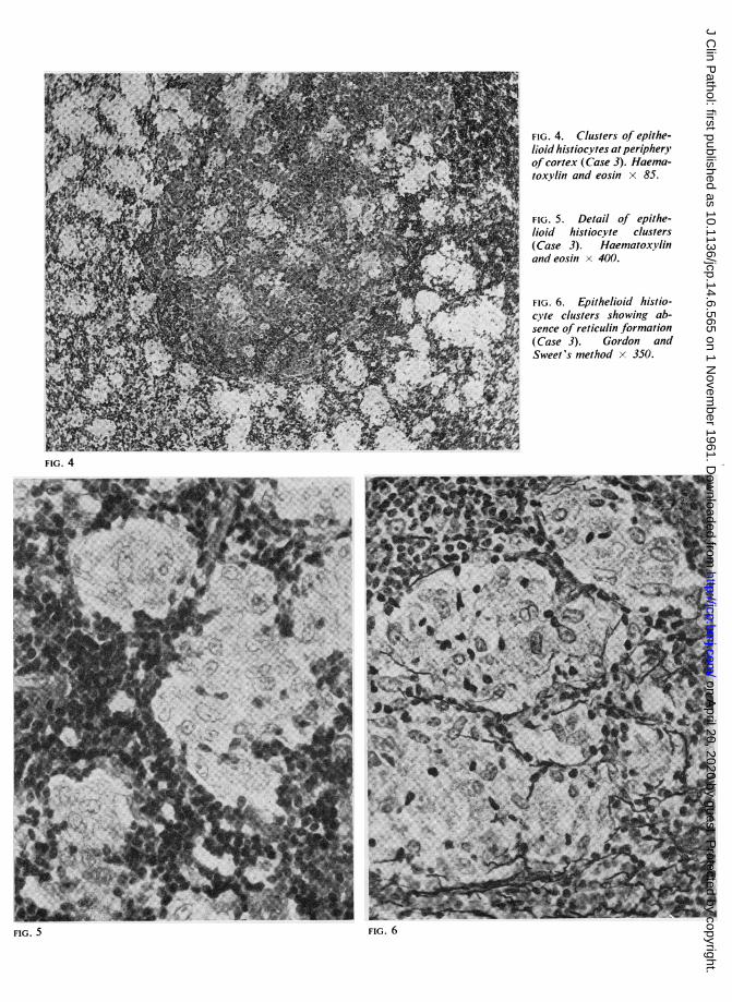
FIG. 4. Clusters of epithe-4. lioidhistiocytes at periphery
of cortex (Case 3). Haema-toxylin and eosin x 85.
FIG. 5. Detail of epithe-lioid histiocyte clusters(Case 3). Haematoxylinand eosin x 400.
$=¢' N FIG. 6. Epithelioid histio-4 cyte clusters showing ab-
b sence of reticulin formation(Case 3). Gordon andSweet's method x 350.
FIG.6~~~~~~~~~~~~~~~. o-
4.....
....w~~~~~.....
FI.6 t'-k
FIG. 4
FIG. 5
on April 20, 2020 by guest. P
rotected by copyright.http://jcp.bm
j.com/
J Clin P
athol: first published as 10.1136/jcp.14.6.565 on 1 Novem
ber 1961. Dow
nloaded from

A. G. Stansfeld
FIG. 7 FIG. 8
FIG. 7. Dense packing oflymph sinuses with monocytoidmacrophages. Note epithelioidhistiocytes at upper left (Case2). Haematoxylin and eosin x125.
FIG. 8. Details of cells fillinglymph sinus (Case 2). Haema-toxylin and eosin x 400.
FIG. 9. Periphery of nodeshowing capsular and extra-capsular infiltration by cells,including many plasma cells(Case 2). Haematoxylin andeosin x 135.
G ... ..
FIG. 9
570
on April 20, 2020 by guest. P
rotected by copyright.http://jcp.bm
j.com/
J Clin P
athol: first published as 10.1136/jcp.14.6.565 on 1 Novem
ber 1961. Dow
nloaded from

The histological diagnosis of toxoplasmic lymphadenitis
FIG. 1O. Toxoplasma cyst at margin offollicle (Case 1).Haematoxylin and eosin x 500.
The first published account of glandular toxo-plasmosis was that of Gard and Magnusson (1951)who described three adult patients with the disease.In the succeeding year or so, several reports ofsimilar cases appeared from Scandinavia (Siim, 1951and 1952; Wahlgren, 1951; Landau, 1951; von
Zeipel and Linder, 1951; Magnusson, 1951; Bang,1953), followed by reports from the United States(Armstrong and MacMurray, 1953; Stanton andPinkerton, 1953). The first account of glandulartoxoplasmosis in this country was that of Skipper,Beverley, and Beattie (1954) and, since that time,there have been a number of further publications on
the subject from many parts of the world.The number of these reports and the fact that I
encountered three cases in the space of a few weekssuggest that the condition is far from rare. Indeed,the frequency of positive serological reactions fortoxoplasma among series of apparently healthyadults indicates that the cases presenting clinicallystill form only a small proportion of the totalnumber of infected individuals. It is probable thatmore cases will be diagnosed in the future, withincreasing awareness of the condition and the
greater use of lymph node biopsy as a diagnosticprocedure.The clinical aspects of toxoplasmic lymphadenitis
have been recently reviewed by Beverley and Beattie(1958) and by Remington, Jacobs, and Kaufman(1960). The disease is commonest in older childrenand young adults. Some European authors (Bang,1953; Piringer-Kuchinka, Martin, and Thalhammer,1958; Saxen and Saxen, 1959) have observed a pre-ponderance of female patients, but in reports fromthis country the sexes have been almost equallyaffected. Saxen and Saxen (1959) noted a seasonalprevalence of the disease in the winter months.For a variety of reasons little is known of the
epidemiology of acquired toxoplasmosis and theonly cases in which the mode of infection has beendefinitely established have been instances of labora-tory infection, where workers handling the organismhave pricked themselves with infected hypodermicneedles (Gard and Magnusson, 1951; Strom, 1951;Beverley, Skipper, and Marshall, 1955; Kayhoe,Jacobs, Beye, and McCullough, 1957). In thenaturally occurring disease, it has been suggestedthat infection may occur most commonly by therespiratory route, with primary involvement of thenasopharyngeal lymphoid tissue (Beattie, 1957;Lainson, 1958). This would agree with the obser-vation that the cervical lymph nodes are commonlyaffected and there is not infrequently a sore throatat the onset ofsymptoms (Beverley and Beattie, 1958).Commonly the lymphadenopathy extends to involveother nodes, especially the axillary and inguinalgroups. Enlarged mediastinal nodes have beenobserved radiographically on several occasions andit has been claimed that the retroperitoneal andmesenteric nodes may be affected (Beverleyand Beattie, 1958; Remington et al., 1960).The chief importance attaching to this generally
mild disease is that it may be mistaken for disordersof a more serious nature. Clinically, one of thecharacteristics which may lead to a suspicion ofmalignancy is the persistence of the lymph nodeenlargement, often for several months and sometimesfor a year or longer. Some patients complain ofmarked fatigue and general debility, but loss ofweight is not a feature. If there is fever at the outset,infectious mononucleosis may be suspected and thepresence of a lymphocytosis with atypical lympho-cytes in the peripheral blood smear may strengthenthis impression, but the Paul-Bunnell reaction isconsistently negative (Beverley and Beattie, 1958).Rarely, the changes in the peripheral blood may beso striking as to lead to a diagnosis of acute lymphaticleukaemia, as in Case 2 of Kayhoe et al. (1957). Itshould be noted that the E.S.R. is seldom muchraised and anaemia is usually absent.
571
on April 20, 2020 by guest. P
rotected by copyright.http://jcp.bm
j.com/
J Clin P
athol: first published as 10.1136/jcp.14.6.565 on 1 Novem
ber 1961. Dow
nloaded from

A. G. Stansfeld
Although the clinical findings may arouse thesuspicion of malignant lymphoma, the establishmentof this diagnosis is dependent upon the findings in alymph node biopsy. That there is a possibility of anerroneous diagnosis at this stage is shown by thehistory of Case 1, where initially a diagnosis ofHodgkin's disease was made on the lymph nodebiopsy. It is understandable that toxoplasmiclymphadenitis might be mistaken histologically forHodgkin's disease, lymphosarcoma, or reticulo-sarcoma, and several authors (Stanton andPinkerton, 1953; Paton, Dick, and Beverley, 1958;Paterson, 1960) mention that the first impressiongained from histological examination of the lymphnode biopsy was one of a malignant condition. Paceand Babando (1960) go so far as to state thatglandular toxoplasmosis cannot with certainty bedistinguished from Hodgkin's disease on the histo-logical findings.
Confusion with malignant lymphomatous condi-tions is liable to arise from (1) the apparent dis-organization of the architecture of the node, (2) theactive proliferation of large cells, variously inter-preted as lymphoblasts or reticulum cells, (3) thesimulation of neoplastic infiltration, and (4) theprominent histiocyte clusters, which resemble thosesometimes found in association with Hodgkin'sdisease and, less often, with lymphosarcoma andreticulosarcoma.The chief points which distinguish toxoplasmic
lymphadenitis from the various forms of malignantlymphoma are:-(I) The lymph node architecture isnot, in fact, completely destroyed; (2) the proliferat-ing cells show normal mitoses only and amongstthese cells there is a high incidence of necrosis,witnessed by the numerous pyknotic nuclear frag-ments; (3) giant reticulum cells characteristic ofHodgkin's disease are absent (Bang, 1957); (4) thelymph sinuses are crowded with macrophages; and(5) the cellular infiltration, which involves thecapsule and extends outside the node, has all thecharacteristics of an inflammatory infiltration.
It is too early yet to decide whether the con-currence of toxoplasmosis and lymphosarcoma, orreticulosarcoma, which has been reported twice (seereferences given by Remington et al., 1960) is morethan a chance association. In the case of Finckh(1954), the diagnosis of toxoplasmosis rested solelyon the morphological interpretation of organismsseen in bone marrow smears. Certainly this proto-zoon does not seem to be as frequently associatedwith the malignant lymphomata as are some fungalinfections.
Whilst it is generally agreed that toxoplasmiclymphadenitis can be distinguished histologicallyfrom the various forms of malignant lymphoma,
there is no unanimity of opinion on the question ofwhether the histological picture is at all diagnosticof toxoplasmosis. Some authors have stated that thelymph node changes are non-specific or inconstant(Gard and Magnusson, 1951; Str6m, 1951; Skipperet al., 1954; Alexander and Callister, 1955; Beverley,Caley, and Warrack, 1958; Paton et al., 1958).According to others, the changes are, at least, highlysuggestive of the disease (Wahlgren, 1951; Siim,1953; Stanton and Pinkerton, 1953; Bang, 1953,1957; Saxen and Saxen, 1959).The prominent histiocyte clusters have attracted
most attention and these have been described byalmost all writers. Some have likened them to'miniature sarcoid lesions' (Stanton and Pinkerton,1953), but, as pointed out above, the cell clustersare generally less well defined than sarcoid lesionsand there is no increased production of collagen orreticulin (Piringer-Kuchinka, 1953). Thus there islittle likelihood of confusion with sarcoidosis.Robb-Smith (1947) described similar reactive
lesions in lymph nodes under the title of lympho-histiocytic medullary reticulosis. He stated that muchthe same appearance was seen in Leishmaniasis, butin that condition Leishman-Donovan bodies couldbe identified in the epithelioid histiocytes. Later,Robb-Smith (quoted by Beverley and Beattie, 1958)examined a number of lymph node biopsies fromproven cases of toxoplasmosis and reported that,in nine out of 11 cases, the appearances correspondedto his description of lympho-histiocytic medullaryreticulosis. He doubted, however, whether thelesion could be regarded as specific for toxoplas-mosis.
Similarly, Piringer-Kuchinka (1953) gave anaccurate account of the histological changes, in-cluding the macrophage infiltration of the lymphsinuses, before she was aware of the occurrence ofthis type of lesion in toxoplasmosis. In 1958, thesame author published an account of 62 cases show-ing this histological picture. Among these, positiveserological tests for toxoplasmosis were obtained in46 out ofthe 49 individuals tested (Piringer-Kuchinkaet al., 1958).Bang (1953 and 1957) drew attention to the 'focal
medullary reticulosis' which he claimed to becharacteristic oftoxoplasmic lymphadenitis, and Siim(1953) found the 'same characteristic histologicalpicture' in all of 16 cases where lymph node biopsieswere performed. More recently Saxen and Saxen(1959) and Roth and Piekarski (1959) have con-firmed the constancy of the histological findings intoxoplasmic lymphadenitis.
Confirmation of the diagnosis has been obtainedin some instances by successful isolation of theorganism from lymph nodes which have shown
572
on April 20, 2020 by guest. P
rotected by copyright.http://jcp.bm
j.com/
J Clin P
athol: first published as 10.1136/jcp.14.6.565 on 1 Novem
ber 1961. Dow
nloaded from

The histological diagnosis of toxoplasmic lymphadenitis
these changes (Lelong, Desmonts, Le Tan Vinh,Nezelof, Satge, and Couvreur, 1954; Siim, 1956). Itmust be admitted, however, that, in most of thepublished series of cases the selection of cases hasbeen made on a histological basis and the diagnosishas been confirmed retrospectively by serologicaltests. Allowance must therefore be made for thepossibility that cases which did not present character-istic histological changes in the lymph node biopsymay have escaped recognition. Nevertheless, thiscannot invalidate the conclusion that, when thelymph node biopsy does show this combination ofchanges, it provides strong presumptive evidence oftoxoplasmic lymphadenitis. This conclusion is sup-ported by the findings in the three cases presentedhere, as well as by the other evidence cited above.The diagnosis should never rest on the histology
alone, for, however characteristic the changes maybe, they can hardly be said to be specific. Ideally,confirmation of the diagnosis should be obtained byisolation of the organism, which may be done byintraperitoneal inoculation of the mouse with freshlymph node suspension (Siim, 1956). Unfortunately,the diagnosis is seldom suspected at the time thelymph node is removed for biopsy and it is hardlyjustifiable to perform a second operation for thispurpose. Moreover, animal inoculations have notproved uniformly successful even when the diagnosishas not been in doubt. Cathie (1954) has successfullyisolated toxoplasma by mouse inoculation with thepatient's saliva.
In the majority of cases the diagnosis of toxoplas-mic lymphadenitis has been confirmed by sero-logical tests, and studies have shown that the resultsof these are sufficiently reliable for practical pur-poses (Cathie, 1957). The demonstration of a risingtitre in the cytoplasm-modifying antibody (dye test)and the complement-fixation test, or the presence ofa high titre in the two tests, is generally taken asevidence of an active infection. These results, takenin conjunction with the characteristic findings in alymph node biopsy, provide sufficient grounds for adiagnosis of toxoplasmic lymphadenitis.Only very rarely are toxoplasma cysts to be found
in lymph node sections. Indeed, a definite cyst witha clearly defined wall, such as was found in Case 1 ofthe present series, has been described in a lymphnode on only one previous occasion (Stanton andPinkerton, 1953). In multiple sections of the node,these authors succeeded in demonstrating two suchcysts, both apparently situated in lymph sinuses.These cysts appear to correspond to the true toxo-plasma cyst, described by Lainson (1958), as occurr-ing in chronic toxoplasma infections in animals.They differ from the pseudocysts, which are foundin more acute infections, in that there is a clearly
defined cyst wall, apparently of parasitic origin. Thecontained toxoplasms are both smaller and muchmore numerous than those in the pseudocyts(Lainson, 1958). Most authors have been under-standably cautious in identifying vegetative forms oftoxoplasma in lymph nodes, in view of their similarityto particles of nuclear debris which are regularly tobe found in large numbers.
I should like to thank Dr. R. Bodley Scott, Mr. P.Stringer, and Mr. M. A. Birnstingl for permission topublish records of their patients, Dr. I. G. Williams andDr. J. I. Pugh for the pathological material from Cases 1and 2, Dr. D. G. Fleck for the results of toxoplasmaserological tests, Mr. J. W. Miller for most of the histo-logical preparations, and Mr. P. Crocker for the photo-micrographs.
REFERENCES
Alexander, C. M., and Callister, J. W. (1955). A.M.A. Arch. Path. 60,563.
Armstrong, C., and MacMurray, F. G. (1953). J. Amer. med. Ass.,151, 1103.
Bang, F. (1953). Bull. Ass. franc. Cancer, 40, 335(1957). Ibid, 44, 60.
Beattie, C. P. (1957). Trans. roy. Soc. trop. Med. Hyg., 51, 96.Beverley, J. K. A., and Beattie, C. P. (1958). Lancet, 2, 379.
Caley, J. P., and Warrack, A. J. N. (1958). J. clin. Path., 11,119.
, Skipper, E., and Marshall, S. C. (1955). Brit. med. J., 1, 577.Cathie, I. A. B. (1954). Lancet, 2, 115.
(1957). Trans. roy. Soc. trop. Med. Hyg., 51, 104.Finckh, E. S. (1954). Med. J. Aust., 2, 965.Gard, S., and Magnusson, J. H. (1951). Acta med. scand., 141, 59.Kayhoe, D. E., Jacobs, L., Beye, H. K., and McCullough, N. B.
(1957). New Engi. J. Med., 257, 1247.Lainson, R. (1958). Trans. roy. Soc. trop. Med. Hyg., 52, 396.Landau, A. (1951). Nord Med., 46, 1575.Lelong, M., Desmonts, G., Le Tan Vinh, N6zelof, C., Satg6, P., and
Couvreur, J. (1954). Arch. franC. Pediat., 11, 1092.Magnusson, J. H. (1951). Nord. Med., 45, 344.Pace, O., and Babando, G. M. (1960). Arch. Sci. med., 109, 536.Paterson, M. W. (1960). Scot. med. J., 5, 467.Paton, J. P. J., Dick, A., and Beverley, J. K. A. (1958). Ibid, 3, 249.Pinkerton, H., and Henderson, R. G. (1941). J. Amer. med. Ass., 116,
807., and Weinman, D. (1940). Arch. Path. (Chicago), 30, 374.
Piringer-Kuchinka, A. (1953). Verh. dtsch. Ges. Path., 1952, 36, 352.Martin, I., and Thalhammer, 0. (1958). Virchows Arch. path.Anat., 331, 522.
Remington, J. S., Jacobs, L., and Kaufman, H. E. (1960). New Engl.J. Med., 262, 180.
Robb-Smith, A. H. T. (1947). In Recent Advances in ClinicalPathology,p. 355, 1st ed., ed. S. C. Dyke. Churchill, London.
Roth, F., and Piekarski, G. (1959). Virchows Arch. path. Anat., 332,181.
Sabin, A. B. (1941). J. Amer. med. Ass., 116, 801.and Feldman, H. A. (1948). Science, 108, 660.
Sax6n, E., and Sax6n, L. (1959). Lab. Invest., 8, 386.Siim, J. C. (1951). J. Amer. med. Ass., 147, 1641.
(1952). Acta path. microbiol. scand., 30, 104.(1953). Schweiz. Z. allg. Path., 16, 506.(1956). Ann. N.Y. Acad. Sci., 64, 185.
Skipper, E., Beverley, J. K. A., and Beattie, C. P. (1954). Lancet, 1,287.
Stanton, M. F., and Pinkerton, H. (1953). Amer. J. clin. Path., 23,1199.
Strom, J. (1951). Acta med. scand., 139, 244.Wahigren, F. (1951). Nord. Med., 45, 349.Zeipel, G. von, and Linder, L. A. (1951). Acta path. microbiol. scand.,
29, 229.
573
on April 20, 2020 by guest. P
rotected by copyright.http://jcp.bm
j.com/
J Clin P
athol: first published as 10.1136/jcp.14.6.565 on 1 Novem
ber 1961. Dow
nloaded from


![Croniconis difficult to differentiate from tuberculous lymphadenitis [1,18]. Clinical Features Tuberculous lymphadenitis most frequently involves the cervical lymph nodes (Figure 1)](https://img.pdfslide.net/doc/110x75/5f6c813fd6b455557074c482/cronicon-is-difficult-to-differentiate-from-tuberculous-lymphadenitis-118-clinical.jpg)