Embed Size (px)
Citation preview
DMD #53223
1
The non-enzymatic reactivity of the acyl-linked metabolites of mefenamic acid towards
amino and thiol functional group bionucleophiles
Howard Horng and Leslie Z. Benet
Department of Bioengineering and Therapeutic Sciences, University of California, San Francisco
DMD Fast Forward. Published on August 23, 2013 as doi:10.1124/dmd.113.053223
Copyright 2013 by the American Society for Pharmacology and Experimental Therapeutics.
This article has not been copyedited and formatted. The final version may differ from this version.DMD Fast Forward. Published on August 23, 2013 as DOI: 10.1124/dmd.113.053223
at ASPE
T Journals on D
ecember 3, 2021
dmd.aspetjournals.org
Dow
nloaded from
DMD #53223
2
Running title: Reactivity of the acyl-linked metabolites of mefenamic acid
Address correspondence to:
Leslie Z. Benet, Ph.D.
University of California, San Francisco
Department of Bioengineering and Therapeutic Sciences
533 Parnassus Ave., Room U-68
San Francisco, CA 94143-0912
Telephone: 415-476-3853 Fax: 415-476-8887 Email: [email protected]
Number of text pages: 22
Number of tables: 1
Number of figures: 8
Number of references: 44
Number of words in Abstract: 250
Number of words in Introduction: 657
Number of words in Discussion: 1498
This article has not been copyedited and formatted. The final version may differ from this version.DMD Fast Forward. Published on August 23, 2013 as DOI: 10.1124/dmd.113.053223
at ASPE
T Journals on D
ecember 3, 2021
dmd.aspetjournals.org
Dow
nloaded from
DMD #53223
3
Non-standard abbreviations used are:
ACN, acetonitrile; ACS, acyl-CoA synthetase; AMP, adenosine monophosphate; ATP,
adenosine triphosphate; CA, cholic acid; CBZ, carbamazepine; CoA, coenzyme A; DMSO,
dimethylsulfoxide; ECF, ethyl chloroformate; ESI, electrospray ionization; Gly, glycine; GSH,
L-glutathione; GST, glutathione-S-transferase; HPLC, high performance liquid chromatography;
IS, internal standard; Kpi, potassium phosphate buffer; LC-MS/MS, liquid chromatography mass
spectrometry; MFA, mefenamic acid; MFA-AMP, mefenamic acid-acyl-adenylate; MFA-CoA,
mefenamic acid-S-acyl-Coenzyme A; MFA-Gly, mefenamic acid-glycine; MFA-GSH,
mefenamic acid-S-acyl-glutathione; MFA-1-O-G, mefenamic acid-1-O-acyl-glucuronide; MFA-
Tau, mefenamic acid-taurine; MFA-NAC, mefenamic acid- N-acetylcysteine; SRM, single
reaction monitoring; NAC, N-acetylcysteine; NSAID, nonsteroidal anti-inflammatory drug; Tau,
taurine; TEA, triethylamine; THF, tetrahydrofuran; Tol, tolmetin; UGT, uridine 5'-diphospho-
glucuronosyltransferase; ZP, zomepirac
This article has not been copyedited and formatted. The final version may differ from this version.DMD Fast Forward. Published on August 23, 2013 as DOI: 10.1124/dmd.113.053223
at ASPE
T Journals on D
ecember 3, 2021
dmd.aspetjournals.org
Dow
nloaded from
DMD #53223
4
Abstract:
Mefenamic acid, (MFA), a carboxylic acid-containing nonsteroidal anti-inflammatory drug
(NSAID) is metabolized into the chemically-reactive, MFA-1-O-acyl-glucuronide (MFA-1-O-
G), MFA-acyl-adenylate (MFA-AMP), and the MFA-S-acyl-CoA (MFA-CoA), all of which are
electrophilic and capable of acylating nucleophilic sites on biomolecules. In this study, we
investigate the non-enzymatic ability of each MFA acyl-linked metabolite to transacylate amino
and thiol functional groups on the acceptor biomolecules glycine (Gly), taurine (Tau),
glutathione (GSH), and N-acetylcysteine (NAC). In vitro incubations with each of the MFA
acyl-linked metabolites (1 μM) in buffer under physiological conditions with Gly, Tau, GSH, or
NAC (10 mM) revealed that MFA-CoA was 11.5- and 19.5-fold more reactive than MFA-AMP
towards the acylation of cysteine-sulfhydryl groups of GSH and NAC, respectively. However,
MFA-AMP was more reactive towards both Gly and Tau, 17.5-fold more reactive towards the N-
acyl-amidation of taurine than its corresponding CoA thioester, while MFA-CoA displayed little
reactivity towards glycine. Additionally, MFA-GSH was 5.6- and 108-fold more reactive
towards NAC than MFA-CoA and MFA-AMP, respectively. In comparison to MFA-AMP and
MFA-CoA, MFA-1-O-G was not significantly reactive towards all four bionucleophiles. MFA-
AMP, MFA-CoA, MFA-1-O-G, MFA-GSH, and MFA-Tau were also detected in rat in vitro
hepatocyte MFA (100 µM) incubations while MFA-Gly was not. These results demonstrate that
MFA-AMP selectively reacts nonenzymatically with the amino functional groups of glycine and
lysine, MFA-CoA selectively reacts nonenzymatically with the thiol functional groups of GSH
and NAC, and MFA-GSH reacts nonenzymatically with the thiol functional group of GSH, all of
which may potentially elicit an idiosyncratic toxicity in vivo.
This article has not been copyedited and formatted. The final version may differ from this version.DMD Fast Forward. Published on August 23, 2013 as DOI: 10.1124/dmd.113.053223
at ASPE
T Journals on D
ecember 3, 2021
dmd.aspetjournals.org
Dow
nloaded from
DMD #53223
5
Introduction
Mefenamic acid (MFA), (2’,3’)-dimethyl-N-phenyl-anthranilic acid, is a carboxylic acid-
containing nonsterodial anti-inflammatory drug (NSAID) associated with a rare, but sometimes
serious idiosyncratic nephrotoxicity (Robertson et al., 1980; Drury et al., 1981; Woods, 1981;
Taha et al., 1985) and possibly hepatotoxicity (Somchit et al., 2004). A proposed mechanism for
the occurrence of these MFA induced toxicities suggests that MFA undergoes bioactivation into
chemically-reactive acyl-linked metabolites that ultimately become covalently bound to tissue
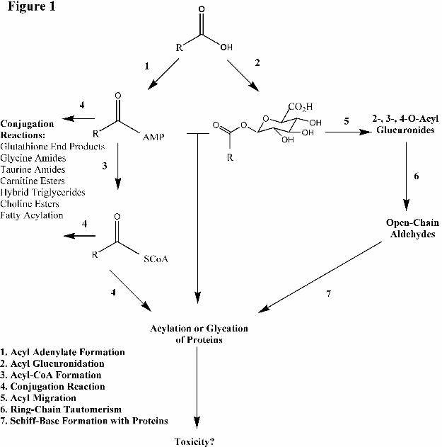
proteins resulting in adverse immunological responses (Figure 1). MFA is metabolized to 3-
hydroxy-MFA (Glazko, 1966) and 3-carboxy-MFA (Sato et al., 1993) via cytochrome P450 2C9.
MFA also undergoes glucuronidation via uridine 5'-diphospho-glucuronosyltransferase (UGT)
into the unstable, reactive acyl glucuronide metabolite, MFA-1-O-acyl-glucuronide (MFA-1-O-
G) (Somchit et al., 2004; McGurk et al, 1996). Acyl glucuronides of acidic drugs are proposed
to bind covalently to protein via a direct transacylation reaction in which protein nucleophiles
react with the facile carbonyl-carbon of the acyl glucuronide, resulting in the liberation of the
glucuronic acid, and via the formation of a drug-protein conjugate or a glycation mechanism
involving prior acyl migration of the drug on the glucuronic acid moiety permitting ring opening
of the sugar resulting in an exposed reactive aldehyde group that reversibly forms an imine
(Schiff’s base) with an amine group on proteins. Subsequent Amadori rearrangement results in a
stable ketoamine derivative in which both the drug and the glucuronic acid moiety become
covalently bound onto the protein (Benet et al., 1993). MFA is also metabolized into the reactive
MFA-acyl-adenylate (MFA-AMP) and MFA-S-acyl-CoA (MFA-CoA) via acyl-CoA synthetase
(ACS), both of which have been demonstrated to form glutathione (GSH)-adducts and are
proposed to play a role in MFA mediated idiosyncratic toxicity (Grillo et al., 2012; Horng and
This article has not been copyedited and formatted. The final version may differ from this version.DMD Fast Forward. Published on August 23, 2013 as DOI: 10.1124/dmd.113.053223
at ASPE
T Journals on D
ecember 3, 2021
dmd.aspetjournals.org
Dow
nloaded from
DMD #53223
6
Benet, 2013). This pathway occurs when the adenosine monophosphate (AMP) moiety of ATP
is covalently transferred to the carboxyl group of MFA to form MFA-AMP, followed by the
displacement of the AMP with coenzyme A (CoA) to form MFA-CoA thioesters (Kelley and
Vessey, 1994; Mano et al., 2001). Upon their formation, the carbonyl carbon of both MFA-AMP
and MFA-CoA increase in electrophilicity enabling them to transacylate the biological
nucleophile GSH (Horng and Benet, 2013). It is proposed that these drug-protein adducts could
act as haptens and are recognized by the immune system as foreign, illiciting an autoimmune
type response resulting in the associated idiosyncratic toxicity (Uetrecht, 2007). Previous in
vitro incubations with the model nucleophile glutathione (GSH) under physiological conditions
showed MFA-AMP to be reactive towards GSH, but 11-fold less reactive than MFA-CoA, while
MFA-1-O-G exhibited little GSH reactivity (Horng and Benet, 2013). In vitro rat hepatocyte
incubations have also resulted in the detection of MFA-AMP, MFA-CoA, MFA-1-O-G, and
MFA-GSH (Horng and Benet, 2013), all of which could be more reactive than MFA per se and
potentially involved in the formation of drug-protein adducts. Additionally, studies involving the
acyl-linked metabolites of the bile acid, cholic acid (CA), revealed that CA-AMP selectively
reacts non-enzymatically with amino functional groups while CA-CoA preferentially reacts
nonenzymatically with thiol functional groups (Mitamura et al., 2011).
The following experiments were designed to characterize the non-enzymatic acylation of the
nucleophilic biomolecules containing the amino functional groups of glycine (Gly) and taurine
(Tau) and the thiol functional groups of GSH and N-acetylcysteine (NAC) by MFA-AMP,
MFA-CoA, MFA-1-O-G, and MFA-GSH (NAC only) as well as the detection of these adducts in
rat hepatocyte in vitro incubations. We propose that MFA-AMP, MFA-CoA, and MFA-1-O-G
This article has not been copyedited and formatted. The final version may differ from this version.DMD Fast Forward. Published on August 23, 2013 as DOI: 10.1124/dmd.113.053223
at ASPE
T Journals on D
ecember 3, 2021
dmd.aspetjournals.org
Dow
nloaded from
DMD #53223
7
are all selective towards their acylation of nucleophilic functional groups on biological
molecules, all of which can contribute to the formation of MFA adducts with amino acids,
peptides, and proteins. We also hypothesize that MFA-GSH is reactive in its own right due to its
structural similarity to MFA-CoA via the thioester bond. If this proposal is correct, the reactive
acyl-linked metabolites of MFA would be anticipated to be selective in their formation of drug-
protein adducts in vivo, which may potentially mediate the idiosyncratic toxicities associated
with MFA and other carboxylic acid-containing drugs.
This article has not been copyedited and formatted. The final version may differ from this version.DMD Fast Forward. Published on August 23, 2013 as DOI: 10.1124/dmd.113.053223
at ASPE
T Journals on D
ecember 3, 2021
dmd.aspetjournals.org
Dow
nloaded from
DMD #53223
8
Materials and Methods
Materials
MFA, AMP, CoA, anhydrous tetrahydrofuran (THF), triethylamine (TEA), ethyl chloroformate
(ECF), N,N’-dicyclohexylcarbodiimide, pyridine, potassium phosphate, potassium carbonate,
dimethyl sulfoxide (DMSO), carbamazepine (CBZ), L-glutathione (GSH), Gly, NAC, and Tau
were all purchased from Sigma-Aldrich Chemical Co (St. Louis, MO). Acetonitrile (ACN),
methanol, acetone, ammonium acetate, and ethyl acetate were all purchased from Fisher
Scientific (Pittsburgh, PA). MFA-1-O-G was purchased from Toronto Research Chemicals
(TRC) Inc. (North York, Ontario). All solvents used for HPLC and LC-MS/MS analysis were of
chromatographic grade. Williams Medium E and L-glutamine were purchased from Gibco
(Grand Island, NY). Male Sprague-Dawley rats were purchased from Charles River
(Wilmington, MA). Stock solutions of MFA-AMP, MFA-CoA, MFA-GSH, MFA-1-O-G,
MFA-Gly, MFA-Tau, and MFA-NAC were all prepared as 1 mM solutions in DMSO.
Instrumentation and Analytical Methods
HPLC/UV analysis was performed on a Hewlett Packard 1100 series binary pump HPLC system
(Santa Clara, CA) coupled to a Hewlett Packard 1100 UV-Vis detector, utilizing HP
Chemstation software for the acquisition of all HPLC/UV data. LC-MS/MS analyses of
synthetic standards and in vitro samples were performed on a Shimadzu LC-20AD (Kyoto,
Japan) HPLC system coupled to an Applied Biosystem/MDS Sciex API (Framingham, MA)
4000 triple quadrupole mass spectrometer outfitted with a Turbo V ion source using positive
ionization mode. All LC-MS/MS analyses were performed on a reverse phase column (XTerra
This article has not been copyedited and formatted. The final version may differ from this version.DMD Fast Forward. Published on August 23, 2013 as DOI: 10.1124/dmd.113.053223
at ASPE
T Journals on D
ecember 3, 2021
dmd.aspetjournals.org
Dow
nloaded from
DMD #53223
9
C-18, 5.0 µm, 4.6 x 150 mm, Milford, MA). The detection of MFA, MFA-AMP, MFA-CoA,
MFA-1-O-G, MFA-GSH, MFA-Gly, MFA-Tau, and MFA-NAC were obtained using
electrospray (ESI) positive ionization and a gradient system of either aqueous ammonium
acetate (10 mM, pH 5.6) and acetonitrile (MFA-CoA) or aqueous solution (0.1% formic acid)
and acetonitrile (0.1% formic acid) (MFA, MFA-AMP, MFA-1-O-G, MFA-GSH, MFA-Gly,
MFA-Tau, and MFA-NAC), 5% ACN to 100%, over 15 min at a flow rate of 0.5 ml/min. The
high pH and ion strength afforded by the aqueous ammonium acetate is necessary to elute MFA-
CoA from the column. Electrospray positive ionization was employed with a needle potential
held at 5.5 kV. MS/MS tandem conditions utilized 2 mTorr argon collision gas and a collision
potential of 89 eV. Mass spectral data were acquired with Analyst software (version 1.5.2, AB
Sciex, Framingham, MA).
Synthesis of MFA-AMP, MFA-Gly, MFA-NAC, and MFA-Tau Derivatives
The synthesis of MFA-AMP, MFA-Gly, MFA-NAC, and MFA-Tau was carried out with a
solution consisting of 110 mg N,N’-dicyclohexylcarbodiimide in 0.4 ml pyridine (Ikegawa et al.,
1999; Horng and Benet, 2013). Briefly, an N,N’-dicyclohexylcarbodiimide solution was added
to a solution containing MFA (0.49 mmol), and either AMP, Gly, Tau, or NAC (0.49 mmol)
separately in 75% pyridine/25% water. The reaction mixture was stirred at 4oC for 7 hr and then
centrifuged at 3000 rpm for 5 min to remove any N-acylurea derivatives. The supernatant was
transferred to another culture tube for precipitation by the addition of acetone (10 ml). The
resulting precipitate was isolated by centrifugation at 3000 rpm for 5 min followed by further
washes with acetone (10 x 10 ml) and acidified water (pH 4-5) (10 x 10 ml). For MFA-AMP,
This article has not been copyedited and formatted. The final version may differ from this version.DMD Fast Forward. Published on August 23, 2013 as DOI: 10.1124/dmd.113.053223
at ASPE
T Journals on D
ecember 3, 2021
dmd.aspetjournals.org
Dow
nloaded from
DMD #53223
10
the precipitate was dissolved in 0.1 M potassium phosphate buffer (pH 6) and underwent
continued liquid-liquid washes with ethyl acetate (10 x 10 ml). Following precipitation via 1M
HCl, the MFA-AMP was further washed with acetone (10 x 10 ml). The MFA-AMP precipitate
was blown down to dryness using N2 gas and weighed out for preparation of a 1 mM MFA-AMP
solution in DMSO. For MFA-Gly, MFA-NAC, and MFA-Tau, the initial acetone derived
precipitate was dissolved in DMSO and subjected to purification via HPLC/UV-mass
spectrometry. The correct HPLC eluent fractions, as determined by UV-MS, of each acyl-linked
metabolite were collected, blown down to dryness, weighed, and then prepared as 1 mM
solutions in DMSO. MFA-AMP eluted at a retention time of 7.6 min and showed no impurities
when analyzed by HPLC/UV (wavelengths: 220, 254, 262, and 280 nm) and LC-MS via reverse-
phase gradient elution (as described above), and 1H NMR (Horng and Benet, 2013). Tandem
LC-MS/MS analysis of MFA-AMP revealed (CID of MH+ ion at m/z 571), m/z (%) yielded: m/z
224([M + H – AMP]+, 100%), m/z 207 ([M + H –364]+, 25%), and m/z 136([M + H - adenine]+,
28%). MFA-Gly eluted at a retention time of 8.7 min (Figure 2C) and showed no impurities
when analyzed by HPLC/UV (wavelengths: 220, 254, 262, and 280 nm) and LC-MS via reverse-
phase gradient elution (as described above). Tandem LC-MS/MS analysis of MFA-Gly (CID of
MH+ ion at m/z 299), m/z (%) : m/z 224([M + H - Gly]+, 99%), m/z 209 ([M + H –90]+, 20%),
m/z 180([M + H – 119]+, 18%), m/z 152([M + H –147]+, 4%), m/z 127([M + H –172]+, 2%), m/z
77([Gly + H]+, 1%) (Figure 2A and 2B). MFA-Tau eluted at a retention time of 9.1 min (Figure
3C) and showed no impurities when analyzed by HPLC/UV (wavelengths: 220, 254, 262, and
280 nm) and LC-MS via reverse-phase gradient elution (as described above). Tandem LC-
MS/MS analysis of MFA-Tau (CID of MH+ ion at m/z 349), m/z (%) : m/z 332 ([M + H – H2O]+,
10%), m/z 224([M + H - Tau]+, 99%), m/z 209 ([M + H –140]+, 25%), m/z 180([M + H – 169]+,
This article has not been copyedited and formatted. The final version may differ from this version.DMD Fast Forward. Published on August 23, 2013 as DOI: 10.1124/dmd.113.053223
at ASPE
T Journals on D
ecember 3, 2021
dmd.aspetjournals.org
Dow
nloaded from
DMD #53223
11
16%), m/z 152([M + H –197]+, 4%), and m/z 126([Tau + H+]+, 2%) (Figure 3A and 3B). MFA-
NAC eluted at a retention time of 9.3 min (Figure 4C) and showed no impurities when analyzed
by HPLC/UV (wavelengths: 220, 254, 262, and 280 nm) and LC-MS via reverse-phase gradient
elution (as described above). Tandem LC-MS/MS analysis of MFA-NAC (CID of MH+ ion at
m/z 387), m/z (%) : m/z 309 ([M + H – 78]+, 30%), m/z 224([M + H - NAC]+, 99%), m/z 209 ([M
+ H –178]+, 18%), m/z 180([M + H - 207]+, 13%), and m/z 165([NAC + H]+, 3%) (Figure 4A
and 4B).
Synthesis of MFA-CoA and MFA GSH Thioester Derivatives
The synthesis of MFA-CoA and MFA-GSH thioesters was accomplished by a method employing
ECF (Stadtman, 1957; Grillo et al., 2002; Horng and Benet, 2013) as described previously.
Briefly, MFA (1.6 mmol) was dissolved in anhydrous THF (25 ml). While stirring at room
temperature, TEA (1.6 mmol) was added to the solution followed by the addition of ECF (1.6
mmol). After 30 min, the resulting triethylamine hydrochloride was removed by passing the
reaction mixture through a glass funnel fitted with a glass wool plug. The filtered solution was
then added to a solution containing CoA (0.13 mmol, 100 mg) or GSH (1 g) and KHCO3 (1.6
mmol) in nanopure water (10 ml) and THF (15 ml). The solution was stirred continuously at
room temperature for 2 hr, after which the reaction was terminated by acidification (pH 4-5)
through the addition of 1 M HCl. THF was then removed by evaporation under N2 gas, followed
by further solvent washes: acidified water (pH 5) (3 x 10 ml) and ethyl acetate (3 x 10 ml) for
MFA-CoA or acetone (3 x 10 ml) for MFA-GSH. MFA-CoA and MFA-GSH precipitate was
blown down to dryness using N2 gas and then weighed out for preparation of a 1 mM MFA-CoA
This article has not been copyedited and formatted. The final version may differ from this version.DMD Fast Forward. Published on August 23, 2013 as DOI: 10.1124/dmd.113.053223
at ASPE
T Journals on D
ecember 3, 2021
dmd.aspetjournals.org
Dow
nloaded from
DMD #53223
12
or 1 mM MFA-GSH solution in DMSO. HPLC analysis of MFA-CoA thioester resulted in an
elution time of 7.3 min and showed no impurities when analyzed by HPLC/UV (wavelengths:
220, 254, 262, and 280 nm) and LC-MS via reverse-phase gradient elution (as described above).
Tandem LC-MS/MS analysis of MFA-CoA standard yielded (CID of MH+ ion at m/z 991), m/z
(%): m/z 582 ([M + H-adenosine diphosphate – H2O]+, 20%), m/z 484([M + H – adenosine
triphosphate]+, 94%), m/z 428([adenosine diphosphate + H+]+, 40%), m/z 382 ([M + H –609]+,
25%), m/z 330([adenosine monophosphate + H – H2O]+, 3%), m/z 224([M + H –CoA]+, 99%).
Synthetic MFA-GSH eluted at a retention time of 7.7 min and showed no detectable impurities
when analyzed by HPLC/UV (wavelengths: 220, 254, 262, and 280 nm) and LC-MS via reverse-
phase gradient elution (as described above). Tandem LC-MS/MS analysis of MFA-GSH
standard yielded product in mass spectrum under CID of the protonated molecular ion at MH+
m/z 531, m/z (%): m/z 456 ([M+H– GSH]+, 10%), m/z 384 ([M + H – pyroglutamic acid –
water]+, 82%), m/z 224 ([MFA + H – H2O]+, 73%).
Stability and Reactivity Incubation Conditions and Quantitative Analysis of Reaction
Products
Chemical stability was assessed by incubating MFA-AMP, MFA-CoA, MFA-GSH, MFA-Gly,
MFA-Tau, and MFA-NAC (1 µM) with CBZ (internal standard) in 0.1 M potassium phosphate
buffer (Kpi) (pH 7.4) in 2 ml HPLC vials (n=3). Each solution was then placed into an HPLC
autosampler warmed to 37oC and injections were taken every 15 min for 3 or 24 hrs for LC-
MS/MS analysis to determine each metabolite’s chemical stability. The stability of each sample
was determined by comparing the analyte peak area to peak area ratios of CBZ, which we
This article has not been copyedited and formatted. The final version may differ from this version.DMD Fast Forward. Published on August 23, 2013 as DOI: 10.1124/dmd.113.053223
at ASPE
T Journals on D
ecember 3, 2021
dmd.aspetjournals.org
Dow
nloaded from
DMD #53223
13
previously found to be stable for at least 72 hours (data not shown). Chemical reactivity
experiments for MFA-AMP, MFA-CoA, and MFA-1-O-G were performed by incubating each
acyl-linked metabolite (1 µM) separately in 0.1 M Kpi (pH 7.4) containing Gly, Tau, GSH, or
NAC and MFA-GSH with NAC (10 mM) (n=3) at 37oC in screw-capped glass vials in a shaking
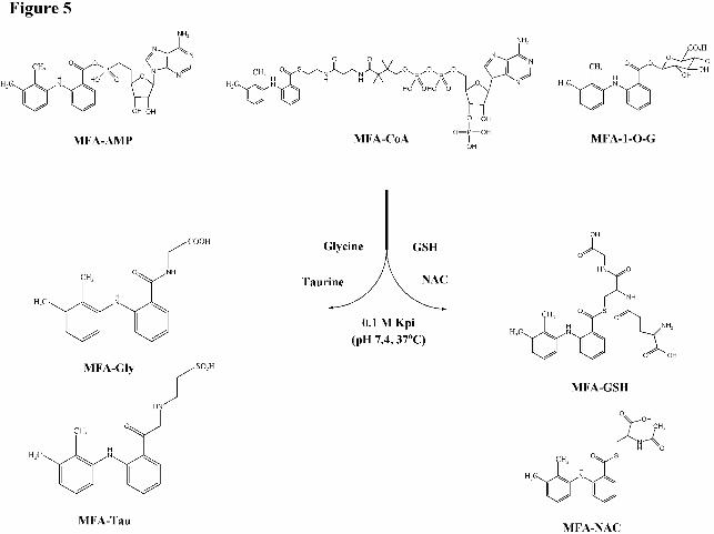
incubator (Figure 5). Aliquots (100 µl) of the incubation mixture were taken at 0, 2, 5, 10, 30,
and 60 min and quenched with 1 µM CBZ/ACN solution and then injected onto the column for
LC-MS/MS analyses. Quantitative measurements were performed by plotting peak area ratios of
MFA-GSH, MFA-Gly, MFA-Tau, or MFA-NAC to CBZ versus the concentration of each acyl-
linked MFA metabolite.
In Vitro Studies with Rat Hepatocytes
Freshly isolated rat (250-300 g, male Sprague-Dawley) hepatocytes were prepared according to
the method of Moldeus et al. (1978) and greater than 85% viability was achieved routinely as
determined by trypan blue exclusion testing. Incubations of hepatocytes (2 million viable
cells/mL) with MFA (100 μM) were performed in Williams Medium E fortified with L-
glutamine (4 mM) in a 50 ml round bottom flask. Incubations (n=3) were performed with
continuous rotation and gassed with 95% O2/5% CO2 at 37oC. Aliquots were taken at 0, 0.2,
0.5, 1, 2, 4, 8, 10, 20, 30, and 60 min and analyzed for MFA-AMP, MFA-CoA, MFA-GSH,
MFA-1-O-G, MFA-Gly, MFA-Tau, and MFA-NAC by LC-MS/MS.
For the analyses of MFA-AMP, MFA-GSH, MFA-1-O-G, MFA-Gly, MFA-Tau, and MFA-
NAC, aliquots (200 μl) of the incubation mixture were added directly into microcentrifuge tubes
This article has not been copyedited and formatted. The final version may differ from this version.DMD Fast Forward. Published on August 23, 2013 as DOI: 10.1124/dmd.113.053223
at ASPE
T Journals on D
ecember 3, 2021
dmd.aspetjournals.org
Dow
nloaded from
DMD #53223
14
(2 ml) followed by quenching with a solution of ACN containing 3% formic acid/2 μM CBZ
(200 μl). Samples were then centrifuged (14,000 rpm, 5 min) and the supernatant fractions (200
μl) were transferred to HPLC autosampler vials for LC-MS/MS analysis.
For the analyses of MFA-CoA formation, aliquots (200 μl) from the same incubations were
transferred directly into microcentrifuge tubes and quenched with a solution of ACN/ 2 μM CBZ
(400 μl) followed by the addition of hexane (600 μl). The samples were vortexed (1 min),
centrifuged (14,000 rpm, 5 min), and aliquots (300 μl) of the aqueous layer were transferred to
an HPLC autosampler vial followed by a 1 hr evaporation of residual hexane under the fume
hood. Samples were then analyzed by LC-MS/MS.
Identification and Quantification of MFA-AMP, MFA-CoA, MFA-GSH, and MFA-1-O-G
MFA treated rat hepatocyte extracts were analyzed by LC-MS/MS for MFA-AMP, MFA-CoA,
MFA-GSH, and MFA-1-O-G as previously described (Horng and Benet, 2013). Single Reaction
Monitoring (SRM) in positive ionization mode with the chromatographic conditions described
above were used for the quantitation with the following mass transitions: MH+ m/z 571 to m/z
224 (MFA-AMP), MH+ m/z 991 to m/z 224 (MFA-CoA), MH+ m/z 531 to m/z 224 (MFA-GSH),
MH+ m/z 418 to m/z 224 (MFA-1-O-G) and MH+ m/z 237 to m/z 194 for CBZ. The elution times
of each acyl-linked metabolite were as follows: 7.6 min (MFA-AMP), 7.3 min (MFA-CoA), 7.7
min (MFA-GSH), 7.9 min (MFA-1-O-G), and 9.3 min (CBZ). No chromatographic peaks
corresponding to each acyl-linked metabolite were detected in blank incubation extracts lacking
This article has not been copyedited and formatted. The final version may differ from this version.DMD Fast Forward. Published on August 23, 2013 as DOI: 10.1124/dmd.113.053223
at ASPE
T Journals on D
ecember 3, 2021
dmd.aspetjournals.org
Dow
nloaded from
DMD #53223
15
MFA. The concentration of each MFA-acyl-linked metabolite was determined by plotting peak
area ratios of each metabolite to CBZ versus the concentration.
Identification and Quantification of MFA-Gly
The identification and quantitation of MFA-Gly by LC-MS/MS was carried out by single
reaction monitoring (SRM) using the mass transitions MH+ m/z 299 to m/z 224 and MH+ m/z 237
to m/z 194 for CBZ detection using ESI positive ionization mode and the chromatographic
methods described above. The elution times of 8.7 min (Figure 2C) and 9.3 min were obtained
for authentic MFA-Gly and CBZ, respectively. No chromatographic peaks corresponding to
MFA-Gly were detected in blank incubation extracts lacking MFA. The concentration of MFA-
Gly was determined by plotting peak area ratios of MFA-Gly to CBZ versus the concentration of
MFA-Gly.
Identification and Quantification of MFA-Tau
The identification and quantitation of MFA-Tau by LC-MS/MS was carried out by SRM using
the mass transitions MH+ m/z 349 to m/z 224 and MH+ m/z 237 to m/z 194 for CBZ detection
using ESI positive ionization mode and the chromatographic methods described above. The
elution times of 9.1 min (Figure 3C) and 9.3 min were obtained for authentic MFA-Tau and
CBZ, respectively. No chromatographic peaks corresponding to MFA-Tau were detected in
blank incubation extracts lacking MFA. The concentration of MFA-Tau was determined by
plotting peak area ratios of MFA-Tau to CBZ versus the concentration of MFA-Tau.
This article has not been copyedited and formatted. The final version may differ from this version.DMD Fast Forward. Published on August 23, 2013 as DOI: 10.1124/dmd.113.053223
at ASPE
T Journals on D
ecember 3, 2021
dmd.aspetjournals.org
Dow
nloaded from
DMD #53223
16
Identification and Quantification of MFA-NAC
The identification and quantitation of MFA-NAC by LC-MS/MS was carried out by SRM using
the mass transitions MH+ m/z 387 to m/z 224 and MH+ m/z 237 to m/z 194 for CBZ detection
using ESI positive ionization mode and the chromatographic methods described above. The
elution times of 9.25 min (Figure 4C) and 9.3 min were obtained for authentic MFA-NAC and
CBZ, respectively. No chromatographic peaks corresponding to MFA-NAC were detected in
blank incubation extracts lacking MFA. The concentration of MFA-NAC was determined by
plotting peak area ratios of MFA-NAC to CBZ versus the concentration of MFA-NAC.
This article has not been copyedited and formatted. The final version may differ from this version.DMD Fast Forward. Published on August 23, 2013 as DOI: 10.1124/dmd.113.053223
at ASPE
T Journals on D
ecember 3, 2021
dmd.aspetjournals.org
Dow
nloaded from
DMD #53223
17
Results
Identification of MFA-AMP, MFA-CoA, MFA-1-O-G, and MFA-GSH
Analysis of rat hepatocyte extracts incubated with MFA by LC-MS/MS detection allowed for the
identification of MFA-AMP, MFA-CoA, MFA-1-O-G, and MFA-GSH formed in incubations
with MFA, as previously described (Horng and Benet, 2013). MFA-AMP, MFA-CoA, MFA-
GSH, and MFA-1-O-G formed in rat hepatocyte extracts and authentic standards coeluted at
retention times of 7.6, 7.3, 7.7, and 7.9 min, respectively, and the product ion spectrum of each
conjugate was consistent with its chemical structure and identical to its corresponding authentic
standard.
Identification of MFA-Tau
Analysis of rat hepatocyte extracts incubated with MFA by LC-MS/MS detection allowed for the
identification of MFA-Tau formed in incubations with MFA. MFA-Tau formed in rat
hepatocyte extracts and authentic standard coeluted at a retention time of 9.1 min (Figure 3C)
and the product ion spectrum of the conjugate was consistent with its chemical structure and
identical to its corresponding authentic standard. MFA-Gly and MFA-NAC were not detected in
MFA rat hepatocyte incubations.
Chemical Stability of MFA-CoA, MFA-AMP, MFA-GSH, MFA-NAC, MFA-Gly, and
MFA-Tau in Buffer
This article has not been copyedited and formatted. The final version may differ from this version.DMD Fast Forward. Published on August 23, 2013 as DOI: 10.1124/dmd.113.053223
at ASPE
T Journals on D
ecember 3, 2021
dmd.aspetjournals.org
Dow
nloaded from
DMD #53223
18
In vitro incubation of each acyl-linked metabolite of MFA in Kpi under physiological conditions
(pH 7.4, 37oC) revealed that MFA-AMP, MFA-CoA, and MFA-GSH were chemically stable for
at least 24 hr while MFA-NAC, MFA-Gly, and MFA-Tau were chemically stable with no
detectable hydrolysis for at least 3 hr of incubation (data not shown). Previous studies carried
out under the same conditions have shown MFA-1-O-G possesses a half life of degradation of
~16 h in buffer under physiological conditions (McGurk et al., 1996; Grillo et al., 2012).
Chemical Reactivity of MFA-CoA, MFA-AMP, MFA-1-O-G, and MFA-GSH with Gly,
Tau, GSH, and NAC
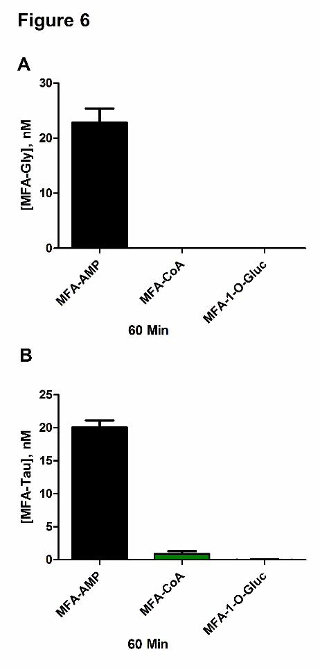
Incubation of MFA-AMP (1 µM) with Gly and Tau (10 mM) in Kpi (0.1 M) under physiological
conditions ( i.e., 37oC, pH 7.4) resulted in the N-amidation of both glycine and taurine,
producing 23.2±4.2 nM and 20.1±1.8 nM of MFA-Gly and MFA-Tau conjugates, respectively,
after 60 min of incubation (Figure 6A and 6B). Incubations of MFA-CoA (1 µM) with glycine
and taurine (10 mM) resulted in minimal N-amidation, producing no MFA-Gly (limit of
detection~ 0.5 nM for all MFA-conjugates) and 0.93±0.65 nM of MFA-Tau at the 60 min time
point. MFA-1-O-G (1 µM) exhibited no reactivity towards both Gly and Tau. In vitro GSH (10
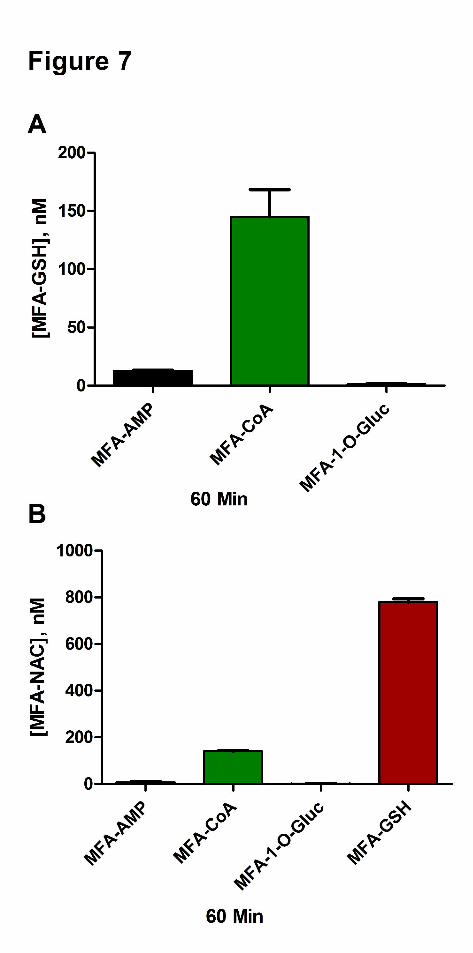
mM) reactivity studies with MFA-AMP, MFA-CoA, and MFA-1-O-G (1 µM) resulted in
12.8±1.0 nM, 145±40 nM, and 1.3±0.97 nM of MFA-GSH formation, respectively at the 60 min
time point (Figure 7A). The incubation of NAC (10 mM) with MFA-AMP, MFA-CoA, and
MFA-GSH (1 µM) under physiological conditions resulted in the formation of 7.45±4.8 nM,
141±4.9 nM, and 780±26 nM of MFA-NAC conjugates, respectively at 60 min (Figure 7B).
While MFA-1-O-G continued to show no reactivity towards the nucleophile NAC.
This article has not been copyedited and formatted. The final version may differ from this version.DMD Fast Forward. Published on August 23, 2013 as DOI: 10.1124/dmd.113.053223
at ASPE
T Journals on D
ecember 3, 2021
dmd.aspetjournals.org
Dow
nloaded from
DMD #53223
19
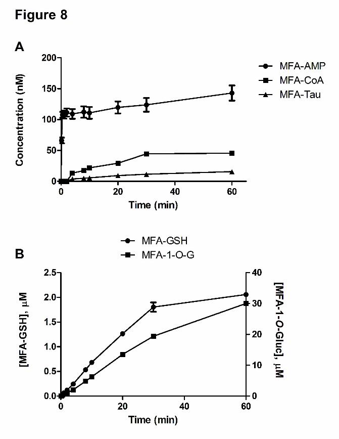
Time course of Formation of MFA-AMP, MFA-Tau, and MFA-Gly in Rat Hepatocyte
Incubations
The incubation of MFA (100 μM) in rat hepatocytes under physiological conditions (37oC, pH
7.4) resulted in the detection of MFA-AMP, MFA-CoA, MFA-GSH, MFA-1-O-G, and MFA-
Tau. The initial rise of MFA-AMP formation was very rapid attaining a concentration of 107.7
nM at ~30 sec (Figure 8A). MFA-CoA was not detectable until the 4 min time point, reaching a
concentration of 45.6 nM at 60 min. MFA-Tau levels were undetectable until 4 min, reaching a
concentration of 15.7 nM at 60 min. The formation of MFA-GSH was linear, reaching a
concentration of 2.1 μM at 60 min, while the formation of MFA-1-O-G increased to a
concentration of 30.0 μM at 60 min (Figure 8B). MFA-Gly and MFA-NAC conjugates were
undetectable during the 60 min incubation period.
This article has not been copyedited and formatted. The final version may differ from this version.DMD Fast Forward. Published on August 23, 2013 as DOI: 10.1124/dmd.113.053223
at ASPE
T Journals on D
ecember 3, 2021
dmd.aspetjournals.org
Dow
nloaded from
DMD #53223
20
Discussion
Carboxylic acid-containing drugs are metabolized into reactive electrophilic acyl-linked
metabolites that can form irreversible adducts with proteins, potentially causing an allergic
reaction in hypersensitive individuals (Stogniew and Fenselau, 1982). MFA is an NSAID
prescribed for its analgesic and anti-inflammatory activity via inhibition of cyclooxygenase-
dependent prostanoid formation (Hawkey, 1999). Commonly used to treat pain, MFA has been
implicated in several cases of hepatic and renal disturbances and hypersensitivity reactions
(Handisurya et al., 2011). These toxicities are proposed to occur via bioactivation of MFA into
reactive acyl-linked metabolites covalently binding onto macromolecules. MFA undergoes
conjugation via the free carboxyl group into the 1-O-acyl glucuronide, MFA-1-O-G (Glazko,
1966; Sato et al., 1993), which has been shown to irreversibly bind to albumin (McGurk et al.,
1996). MFA also undergoes further conjugation into MFA-AMP, MFA-CoA and MFA-GSH
(Grillo et al., 2012; Horng and Benet, 2013) in rat hepatocytes, all of which possess an increased
chemical electrophilicity and are reactive towards protein nucleophiles.
Electrophilicity assessment of metabolites in drug development involve screens utilizing
nucleophilic trapping agents. Glutathione is a commonly used biomarker of reactivity for
bioactivation studies. Presumably, the greater the in vitro nucleophilic adduct formation, the
greater the probability it will covalently bind onto proteins and elicit a toxic reaction. However,
not all protein covalent binding result in the onset of toxicity, and thus the challenge is to identify
those protein targets that are critical for the onset of a drug induced toxicity. In addition to
thioesters (Van Breemen and Fenselau, 1985), acyl-linked metabolites have also been shown to
This article has not been copyedited and formatted. The final version may differ from this version.DMD Fast Forward. Published on August 23, 2013 as DOI: 10.1124/dmd.113.053223
at ASPE
T Journals on D
ecember 3, 2021
dmd.aspetjournals.org
Dow
nloaded from
DMD #53223
21
react with proteins via oxygen ester (Wells and Janssen, 1987) and amide-linkage (van Breemen
and Fenselau, 1986; Mitamura et al., 2011). In the present study, we investigate the selective
non-enzymatic acylation of amino and thiol functional groups of four biological nucleophiles:
Gly, Tau, GSH, and NAC.
In vitro GSH reactivity assessment of MFA-AMP, MFA-CoA, and MFA-1-O-G revealed MFA-
CoA to be 11.5-fold more reactive than MFA-AMP toward the thiol functional group of GSH
(Figure 7A), consistent with our previous studies (Horng and Benet, 2013), while MFA-CoA was
19.5-fold more reactive toward the thiol groups of NAC than its corresponding MFA-AMP
(Figure 7B). Alternatively, incubations with Gly and Tau revealed that MFA-AMP is more
reactive towards N-acyl-amidation than its corresponding MFA-CoA, producing a significant
amount of MFA-Gly while MFA-CoA did not react with Gly (Figure 6A). The amidation of Tau
by MFA-AMP was also 17.5-fold greater than MFA-CoA (Figure 6B). MFA-1-O-G exhibited
little to no reactivity towards all four bionucleophiles. Reactivity data were linear during the
incubation and therefore the 60 min time point was used to calculate the slope of conjugate
formation (Table 1). Reactivity studies utilizing cholic acid have also demonstrated a greater
reactivity of CA-AMP compared to CA-CoA towards the N-acyl-amidation of glycine and
taurine, while CA-CoA exhibited a greater reactivity towards the acylation of the cysteine-
sulfhydryl group of GSH and NAC (Mitamura et al., 2011). Studies in buffer also show that
acyl-adenylates can spontaneously react with the amino groups of substance P (Goto et al.,
2001), lysozomes (Goto et al., 2005), and histones (Mano et al., 2004), further suggesting that
acyl-adenylates have a high reactive affinity towards amino functional groups, and that acyl-
AMPs and acyl-CoAs are selective in their non-enzymatic acylation of amino versus thiol
This article has not been copyedited and formatted. The final version may differ from this version.DMD Fast Forward. Published on August 23, 2013 as DOI: 10.1124/dmd.113.053223
at ASPE
T Journals on D
ecember 3, 2021
dmd.aspetjournals.org
Dow
nloaded from
DMD #53223
22
functional groups. This selectivity may be attributed to differences in the degree of
electrophilicity of MFA-AMP versus MFA-CoA and the nucleophilicity of the amino versus the
thiol functional groups, suggesting different reactivity mechanisms towards protein nucleophilic
sites.
Acyl-GSH conjugates share a common structural moiety, thioester, to that of the acyl-CoA.
Therefore, it is conceivable that MFA-GSH is just as, if not more reactive than its corresponding
acyl-CoA derivative. NAC reactivity assessments show that MFA-GSH is indeed highly
reactive, exhibiting a 5.5-fold and 108-fold greater reactivity towards NAC than MFA-CoA and
MFA-AMP, respectively (Figure 7B). Grillo and Benet (2002) have demonstrated that the
reactive potential of S-acyl-glutathione conjugates with NAC correlates to the degree of
α-carbon substitution of the acyl-linkage, increasing α-carbon substitution results in decreasing
reactivity, which is in agreement with the relative degradation rates associated with acyl
glucuronides and assumed to be identical for that of their respective S-acyl-CoA thioesters.
Therefore, if MFA toxicity is the result of covalent binding by reactive intermediates, then it is
conceivable that the unusually high formation of MFA-GSH in rat hepatocytes (Grillo et al.,
2012; Horng and Benet, 2013), compared to diclofenac (Grillo et al., 2003), zomepirac (Olsen et
al., 2005), (R)-ibuprofen (Grillo and Hua, 2008), and (R)-flunoxaprofen (Grillo et al., 2010),
may be responsible for the tissue injury associated with MFA. This assumption is based on the
high covalent binding values in animals dosed with drugs known to cause hepatotoxicity in
humans, such as isoniazid (Nelson et al., 1978) and acetaminophen (Matthews, 1997).
This article has not been copyedited and formatted. The final version may differ from this version.DMD Fast Forward. Published on August 23, 2013 as DOI: 10.1124/dmd.113.053223
at ASPE
T Journals on D
ecember 3, 2021
dmd.aspetjournals.org
Dow
nloaded from
DMD #53223
23
Acyl-CoA thioester synthesis is a two step reaction. This reaction occurs when the AMP moiety
of ATP is transferred to the acyl group of the carboxylic acid via ACS forming an acyl-adenylate
intermediate. The enzyme bound activated intermediate is then displaced by coenzyme A to
yield the associated acyl-CoA product and free AMP (Vlahcevic et al., 1999). Acyl-CoA and
acyl-AMP metabolites both spontaneously and enzymatically, via glutathione-S-transferase, form
GSH-conjugates (Li et al.,2002; Grillo et al., 2012; Horng and Benet, 2013). In addition to GSH,
glycine (Keller and Keller, 1842) and taurine (James et al., 1971; Hutson and Casida, 1978) are
two of the most commonly cited metabolic amino acid conjugation reactions. Amino acid
conjugation occurs through the transfer of the acyl group from the acyl-CoA to an amino acid via
N-acetyltransferase. However, previous and our current studies have shown that acyl-adenylates
are capable of spontaneously reacting to both Gly and Tau (Mitamura et al., 2011) suggesting
that acyl-adenylate intermediates have a greater inherent chemical affinity towards amino groups
than both the acyl-CoAs and acyl-1-O-G. Rat hepatocyte MFA incubation resulted in rapid
MFA-AMP formation, (Cmax 107 nM at ~30 sec) while MFA-CoA was undetectable until 4
mins, achieving a concentration of 45.6 nM at 60 min (Figure 8A). This sequence is in
agreement with the acyl-CoA biosynthetic pathway. MFA-1-O-G and MFA-GSH were also
shown to increase linearly, achieving concentrations of 30.0 μM and 2.1 μM at 60 min,
respectively (Figure 8B). Our experiments also revealed the presence of MFA-Tau, undetectable
until 4 min, reaching a concentration of 15.7 nM at 60 min. It was not determined if the formed
MFA-Tau primarily occurs nonenzymatically from MFA-AMP or enzymatically via the MFA-
CoA thioester. MFA-Gly was not detected in these incubations, possibly due to an insufficiency
in analytical sensitivity (limit of detection for all MFA-conjugates was ~0.5 nM). Previous
zomepirac (ZP) rat hepatocyte incubations revealed the presence of ZP-CoA , ZP-1-O-G, ZP-
This article has not been copyedited and formatted. The final version may differ from this version.DMD Fast Forward. Published on August 23, 2013 as DOI: 10.1124/dmd.113.053223
at ASPE
T Journals on D
ecember 3, 2021
dmd.aspetjournals.org
Dow
nloaded from
DMD #53223
24
Tau, and ZP-Gly (Olsen et al., 2005). The high concentration of ZP-1-O-G compared to ZP-Gly
and ZP-Tau may be reflective of dose, a determinant of whether or not a drug undergoes
glucuronidation or amino acid conjugation (Hutt and Caldwell, 1990). At lower doses,
carboxylic acids tend to undergo amino acid conjugation while at higher doses, glucuronidation
dominates. Amino acid conjugation is a high-affinity, low capacity system while
glucuronidation is a high-capacity pathway with a broader substrate selectivity. Dose also
influences the type of amino acid conjugate formed (Hirom et al., 1977), with dosage increases
of phenylacetic acid resulting in decreases in glycine:taurine conjugate ratios. Rat liver
experiments with tolmetin (Tol) also identified the acyl-CoA derived conjugates, Tol-Gly and
Tol-Tau conjugates in rat urine (Olsen et al., 2003), whose concentrations were unaffected by
clofibric acid, confirming a high-affinity/low-capacity metabolic pathway (Hutt and Caldwell,
1990). Oral administration of RS-ibuprofen to humans also led to the detection of ibuprofen-Tau
but not the glycine conjugate (Shirley et al., 1994), signifying a minor biotransformation
pathway. Gly and Tau conjugates are believed to be formed enzymatically (N-acyltransferases)
via the acyl-CoA, however we have demonstrated that Gly and Tau conjugates also form
spontaneously via acyl-AMP conjugates. MFA-NAC conjugates were not detected in rat
hepatocyte incubations, due to the lack of detectable N-acylcysteinylglycine and glutamyl
transpeptidase (γ-GT) activity in rat livers (Hinchman and Ballatori, 1990). However, in
humans, NAC formation would be expected to occur.
In conclusion, MFA-AMP selectively reacts nonenzymatically with the amino functional groups
of glycine and lysine, while MFA-CoA selectively reacts nonenzymatically with the thiol
functional groups of GSH and NAC. MFA-GSH is also reactive towards NAC, which may be of
This article has not been copyedited and formatted. The final version may differ from this version.DMD Fast Forward. Published on August 23, 2013 as DOI: 10.1124/dmd.113.053223
at ASPE
T Journals on D
ecember 3, 2021
dmd.aspetjournals.org
Dow
nloaded from
DMD #53223
25
toxicological significance considering that MFA produces a high amount of GSH adducts in
hepatocytes. MFA-1-O-G was not chemically reactive toward both amino and thiol functional
groups. This preferential reactivity between MFA-AMP and MFA-CoA provides MFA the
ability to covalently bind onto a broad range of nucleophilic sites on proteins, which increases its
probability of covalently modifying critical protein targets and inducing a toxic reaction.
Therefore, acyl-linked metabolites may be important in the formation of drug-protein adducts
and the onset of an idiosyncratic toxicity, suggesting that the bioactivation of carboxylic acid-
containing drugs into acyl-linked metabolites should be further evaluated to allow for structural
modification of drug candidates thereby reducing bioactivation and improving drug safety.
This article has not been copyedited and formatted. The final version may differ from this version.DMD Fast Forward. Published on August 23, 2013 as DOI: 10.1124/dmd.113.053223
at ASPE
T Journals on D
ecember 3, 2021
dmd.aspetjournals.org
Dow
nloaded from
DMD #53223
26
Acknowledgements
We would like to thank Dr. Mark Grillo (Amgen Inc., Department of Pharmacokinetics and Drug
Metabolism, South San Francisco, CA) for many helpful discussions during the course of this
work and Chris Her (UCSF Liver Center Cell Biology Core) for his assistance in the rat
hepatocyte preparations.
This article has not been copyedited and formatted. The final version may differ from this version.DMD Fast Forward. Published on August 23, 2013 as DOI: 10.1124/dmd.113.053223
at ASPE
T Journals on D
ecember 3, 2021
dmd.aspetjournals.org
Dow
nloaded from
DMD #53223
27
Authorship Contributions
Participated in research design: Horng, Benet
Conducted experiments: Horng
Performed data analysis: Horng, Benet
Wrote or contributed to the writing of the manuscript: Horng, Benet
This article has not been copyedited and formatted. The final version may differ from this version.DMD Fast Forward. Published on August 23, 2013 as DOI: 10.1124/dmd.113.053223
at ASPE
T Journals on D
ecember 3, 2021
dmd.aspetjournals.org
Dow
nloaded from
DMD #53223
28
References
Benet LZ, Spahn-Langguth H, Iwakawa S, Volland C, Mizuma T, Mayer S, Mutchler E, and Lin
ET (1993) Predictability of the covalent binding of acidic drugs in man. Life Sci 53:
PL141-146.
Drury, PL, Asirdas LG, and Bulger GV (1981). Mefenamic acid nephropathy: further evidence.
Br Med J (Clin Res Ed) 282: 865-866.
Glazko AJ (1966) Experimental observations on flufenamic, mefenamic and meclofenamic acids
metabolic disposition. Ann Phys Med Suppl: 23-36.
Goto J, Nagata M, Mano N, Kobayashi N, Ikegawa S, and Kiyonami R (2001) Bile acid acyl
adenylate: a possible intermediate to produce a protein-bound bile acid. Rapid Commun
Mass Spectrom 15: 104-109.
Goto T, Shibata A, Sasaki D, Suzuki N, Hishinuma T, Kakiyama G, Iida T, Mano N, and Goto J
(2005) Identification of a novel conjugate in human urine: bile acid acyl galactosides.
Steroids 70: 185-192.
Grillo MP and Benet LZ (2002) Studies on the reactivity of clofibryl-S-acyl-CoA thioester with
glutathione in vitro. Drug Metab Dispos 30: 55-62.
Grillo MP and Hua F (2008) Enantioselective formation of ibuprofen-S-acyl-glutathione in vitro
in incubations of ibuprofen with rat hepatocytes. Chem Res Toxicol 21: 1749-1759.
This article has not been copyedited and formatted. The final version may differ from this version.DMD Fast Forward. Published on August 23, 2013 as DOI: 10.1124/dmd.113.053223
at ASPE
T Journals on D
ecember 3, 2021
dmd.aspetjournals.org
Dow
nloaded from
DMD #53223
29
Grillo MP, Hua F, Knutson CG, Ware JA, and Li C (2003) Mechanistic studies on the
bioactivation of diclofenac: identification of diclofenac-S-acyl-glutathione in vitro in
incubations with rat and human hepatocytes. Chem Res Toxicol 16: 1410-1417.
Grillo MP, Tadano Lohr M, and Wait JC (2012) Metabolic activation of mefenamic acid leading
to mefenamyl-S-acyl-glutathione adduct formation in vitro and in vivo in rat. Drug Metab
Dispos 40: 1515-1526.
Grillo MP, Wait JC, Tadano Lohr M, Khera S, and Benet LZ (2010) Stereoselective
flunoxaprofen-S-acyl-glutathione thioester formation mediated by acyl-CoA formation in
rat hepatocytes. Drug Metab Dispos 38: 133-142.
Handisurya A, Moritz KB, Riedl E, Reinisch C, Stingl G, and Wohrl S (2011) Fixed drug
eruption caused by mefenamic acid: a case series and diagnostic algorithms. J Dtsch
Dermatol Ges 9: 374-378.
Hawkey C J (1999) COX-2 inhibitors. Lancet 353: 307-314.
Hinchman CA and Ballatori N (1990). Glutathione-degrading capacities of liver and kidney in
different species. Biochem Pharmacol 40: 1131-1135.
Hirom PC., Idle JR, Millburn P, and Williams RT (1977) Glutamine conjugation of phenylacetic
acid in the ferret. Biochem Soc Trans 5: 1033-5.
Horng H and Benet LZ (2013) Characterization of the acyl-adenylate linked metabolite of
mefenamic acid. Chem Res Toxicol 26: 465-476.
Hutson DH and Casida JE (1978) Taurine conjugation in metabolism of 3-phenoxybenzoic acid
and the pyrethroid insecticide cypermethrin in mouse. Xenobiotica 8: 565-571.
This article has not been copyedited and formatted. The final version may differ from this version.DMD Fast Forward. Published on August 23, 2013 as DOI: 10.1124/dmd.113.053223
at ASPE
T Journals on D
ecember 3, 2021
dmd.aspetjournals.org
Dow
nloaded from
DMD #53223
30
Hutt, A J and Caldwell J (1990). Amino acid conjugation, in: Conjugation Reactions in Drug
Metabolism (Mulder G ed.) Taylor & Francis, New York
Ikegawa SH, Ishikawa H, Oiwa H, Nagata M, Goto J, Kozaki T, Gotowda M, and Asakawa N
(1999) Characterization of cholyl-adenylate in rat liver microsomes by liquid
chromatography/electrospray ionization-mass spectrometry. Anal Biochem 266: 125-132.
James M, Smith RL, and Williams RT (1971) Conjugates of phenylacetic acid with taurine and
other amino acids in various species. Biochem J 124: 15P-16P.
Keller W and Keller M (1842) Conversion of benzoic into hippuric acid. Prov Med J Retrosp
Med Sci, 4: 256-257.
Kelley M and Vessey DA (1994) Determination of the mechanism of reaction for bile acid: CoA
ligase. Biochem J 304: 945-949.
Li C, Benet LZ, and Grillo MP (2002) Studies on the chemical reactivity of 2-phenylpropionic
acid 1-O-acyl glucuronide and S-acyl-CoA thioester metabolites. Chem Res Toxicol 15:
1309-1317.
Mano N, Goto T, Uchida M, Nishimura K, Ando M, Kobayashi, N, and Goto J (2004) Presence
of protein-bound unconjugated bile acids in the cytoplasmic fraction of rat brain. J Lipid
Res 45: 295-300.
Mano N, Uchida M, Okuyama H, Sasaki I, Ikegawa S, and Goto J (2001). Simultaneous
detection of cholyl adenylate and coenzyme A thioester utilizing liquid
chromatography/electrospray ionization mass spectrometry. Anal Sci 17: 1037-1042.
This article has not been copyedited and formatted. The final version may differ from this version.DMD Fast Forward. Published on August 23, 2013 as DOI: 10.1124/dmd.113.053223
at ASPE
T Journals on D
ecember 3, 2021
dmd.aspetjournals.org
Dow
nloaded from
DMD #53223
31
Matthews A M, Hinson JA, Roberts DW, and Pumford NR (1997). Comparison of covalent
binding of acetaminophen and the regioisomer 3'-hydroxyacetanilide to mouse liver
protein. Toxicol Lett 90: 77-82.
McGurk KA, Remmel RP, Hosagrahara VP, Tosh D, and Burchell B (1996). Reactivity of
mefenamic acid 1-O-acyl glucuronide with proteins in vitro and ex vivo. Drug Metab
Dispos 24: 842-849.
Mitamura K, Aoyama E, Sakai T, Iida T, Hogmann AF, and Ikegawa S (2011) Characterization
of non-enzymatic acylation of amino or thiol groups of bionucleophiles by the acyl-
adenylate or acyl-CoA thioester of cholic acid. Anal Bioanal Chem 400: 2253-2259.
Moldeus P, Jones DP, Ormstad K, and Orrenius S (1978). Formation and metabolism of a
glutathione-S-conjugate in isolated rat liver and kidney cells. Biochem Biophys Res
Commun 83: 195-200.
Nelson SD , Mitchell JR, Snodgrass JR, and Timbrell JA (1978). Hepatotoxicity and metabolism
of iproniazid and isopropylhydrazine. J Pharmacol Exp Ther 206: 574-585.
Olsen J, Bjornsdottir I, and Honore Hansen S (2003). Identification of coenzyme A-related
tolmetin metabolites in rats: relationship with reactive drug metabolites. Xenobiotica 33:
561-570.
Olsen J, Li C, Bjornsdottir I, Sidenius U, Hansen SH, and Benet LZ (2005). In vitro and in vivo
studies on acyl-coenzyme A-dependent bioactivation of zomepirac in rats. Chem Res
Toxicol 18: 172917-36.
Robertson CE, Ford MJ, Van Someren V, Dlugolecka M, and Prescott LF (1980). Mefenamic
acid nephropathy. Lancet 2: 232-233.
This article has not been copyedited and formatted. The final version may differ from this version.DMD Fast Forward. Published on August 23, 2013 as DOI: 10.1124/dmd.113.053223
at ASPE
T Journals on D
ecember 3, 2021
dmd.aspetjournals.org
Dow
nloaded from
DMD #53223
32
Sato J, Yamane Y, Ito K, and Bando H (1993). Structures of mefenamic acid metabolites from
human urine. Biol Pharm Bull 16: 811-812.
Shirley MA, Guan X, Kaiser DG, Halstead GW, and Baille TA (1994). Taurine conjugation of
ibuprofen in humans and in rat liver in vitro. Relationship to metabolic chiral inversion. J
Pharmacol Exp Ther 269: 1166-1175.
Somchit N, Sanat F, Gan EH, Shahrin IA, and Zuraini A (2004). Liver injury induced by the
non-steroidal anti-inflammatory drug mefenamic acid. Singapore Med J 45: 530-532.
Stadtman TC and Elliott P (1957). Studies on the enzymic reduction of amino acids. II.
Purification and properties of D-proline reductase and a proline racemase from
Clostridium sticklandii. J Biol Chem 228: 983-97.
Stogniew M and Fenselau C (1982). Electrophilic reactions of acyl-linked glucuronides.
Formation of clofibrate mercapturate in humans. Drug Metab Dispos 10: 609-613.
Taha A, Lenton RJ, Murdoch PS, and Peden NR (1985). Non-oliguric renal failure during
treatment with mefenamic acid in elderly patients: a continuing problem. Br Med J (Clin
Res Ed) 291: 661-662.
Uetrecht J (2007). Idiosyncratic drug reactions: current understanding. Annu Rev Pharmacol
Toxicol 47: 513-539.
Van Breemen RB and Fenselau C (1985). Acylation of albumin by 1-O-acyl glucuronides. Drug
Metab Dispos 13: 318-320.
Van Breemen, RB and Fenselau C (1986). Reaction of 1-O-acyl glucuronides with 4-(p-
nitrobenzyl)pyridine. Drug Metab Dispos 14: 197-201.
This article has not been copyedited and formatted. The final version may differ from this version.DMD Fast Forward. Published on August 23, 2013 as DOI: 10.1124/dmd.113.053223
at ASPE
T Journals on D
ecember 3, 2021
dmd.aspetjournals.org
Dow
nloaded from
DMD #53223
33
Vlahcevic ZR, Pandak WM, and Stravitz RT (1999). Regulation of bile acid biosynthesis.
Gastroenterol Clin North Am 28: 1-25.
Wells DS, Janssen FW, and Ruelius HW (1987). Interactions between oxaprozin glucuronide
and human serum albumin. Xenobiotica 17: 1437-1449.
Woods KL and Michael J (1981) Mefenamic acid nephropathy. Br Med J (Clin Res Ed) 282:
1471.
This article has not been copyedited and formatted. The final version may differ from this version.DMD Fast Forward. Published on August 23, 2013 as DOI: 10.1124/dmd.113.053223
at ASPE
T Journals on D
ecember 3, 2021
dmd.aspetjournals.org
Dow
nloaded from
DMD #53223
34
Footnotes
This work was supported in part by the National Institute of Health Grant [GM36633] and by the
University of California, San Francisc Liver Center Cell Biology Core through National Institute
of Health grant [P30 DK026743]. The authors declare no competing financial interests.
This article has not been copyedited and formatted. The final version may differ from this version.DMD Fast Forward. Published on August 23, 2013 as DOI: 10.1124/dmd.113.053223
at ASPE
T Journals on D
ecember 3, 2021
dmd.aspetjournals.org
Dow
nloaded from
DMD #53223
35
Figure Legends:
Figure 1 Proposed conjugative bioactivation pathways of mefenamic acid.
Figure 2 Proposed identities of the fragment ions of MFA-Gly (A), tandem mass spectrum
(B), and representative reverse-phase gradient LC-MS/MS Single Reaction
Monitoring (SRM) (m/z 299 to m/z 224) (C) of MFA-Gly authentic standard.
Figure 3 Proposed identities of the fragment ion of MFA-Tau (A), tandem mass spectrum
(B), and representative reverse-phase gradient LC-MS/MS SRM (m/z 349 to m/z
224) (C) of MFA-Tau authentic standard.
Figure 4 Proposed identities of the fragment ions of MFA-NAC (A), tandem mass
spectrum (B), and representative reverse-phase gradient LC-MS/MS SRM (m/z
387 to m/z 224) (C) of MFA-NAC authentic standard.
Figure 5 Scheme of the transacylation of MFA-AMP, MFA-CoA, and MFA-1-O-G with
the bionucleophiles Gly, Tau, GSH, and NAC in buffer (0.1 M Kpi (pH 7.4,
37oC)).
This article has not been copyedited and formatted. The final version may differ from this version.DMD Fast Forward. Published on August 23, 2013 as DOI: 10.1124/dmd.113.053223
at ASPE
T Journals on D
ecember 3, 2021
dmd.aspetjournals.org
Dow
nloaded from
DMD #53223
36
Figure 6 Mean reactivity assessment ± standard deviation at 60 min of MFA-AMP, MFA-
CoA, and MFA-1-O-G toward Gly (A) and Tau (B) in buffer (0.1 M Kpi (pH 7.4,
37oC)).
Figure 7 Mean reactivity assessment ± standard deviation at 60 min of MFA-AMP, MFA-
CoA, MFA-1-O-G, and MFA-NAC toward GSH (A) and NAC (B) in buffer
(0.1 M Kpi (pH 7.4, 37oC)).
Figure 8 Mean time-dependent formation ± standard deviation of MFA-AMP, MFA-CoA,
and MFA-Tau (A), MFA-1-O-G (right axis units) and MFA-GSH (left axis units)
(B) in rat hepatocyte incubations.
This article has not been copyedited and formatted. The final version may differ from this version.DMD Fast Forward. Published on August 23, 2013 as DOI: 10.1124/dmd.113.053223
at ASPE
T Journals on D
ecember 3, 2021
dmd.aspetjournals.org
Dow
nloaded from
DMD #53223
37
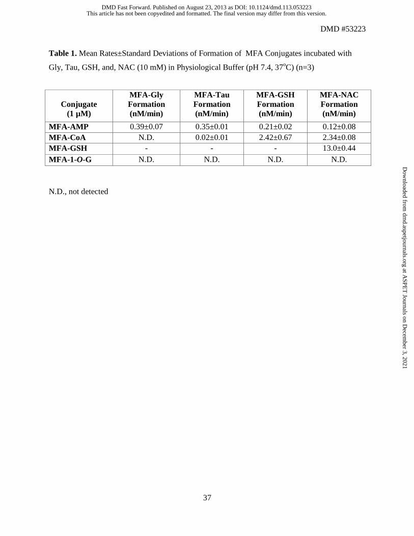
Table 1. Mean Rates±Standard Deviations of Formation of MFA Conjugates incubated with
Gly, Tau, GSH, and, NAC (10 mM) in Physiological Buffer (pH 7.4, 37oC) (n=3)
N.D., not detected
Conjugate
(1 µM)
MFA-Gly Formation (nM/min)
MFA-Tau Formation (nM/min)
MFA-GSH Formation (nM/min)
MFA-NAC Formation (nM/min)
MFA-AMP 0.39±0.07 0.35±0.01 0.21±0.02 0.12±0.08 MFA-CoA N.D. 0.02±0.01 2.42±0.67 2.34±0.08 MFA-GSH - - - 13.0±0.44
MFA-1-O-G N.D. N.D. N.D. N.D.
This article has not been copyedited and formatted. The final version may differ from this version.DMD Fast Forward. Published on August 23, 2013 as DOI: 10.1124/dmd.113.053223
at ASPE
T Journals on D
ecember 3, 2021
dmd.aspetjournals.org
Dow
nloaded from
This article has not been copyedited and formatted. The final version may differ from this version.DMD Fast Forward. Published on August 23, 2013 as DOI: 10.1124/dmd.113.053223
at ASPE
T Journals on D
ecember 3, 2021
dmd.aspetjournals.org
Dow
nloaded from
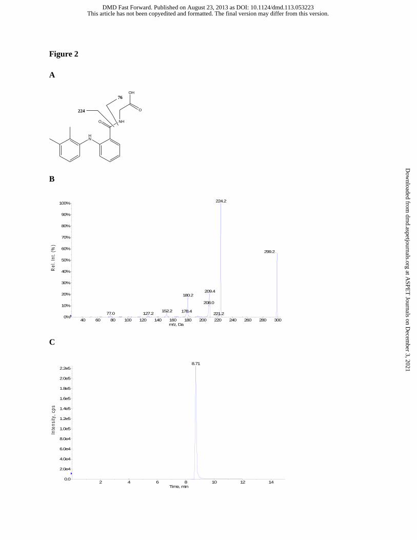
Figure 2
A
HN
NH
OH
O
O224
76
B
C
40 60 80 100 120 140 160 180 200 220 240 260 280 300m/z, Da
0%
10%
20%
30%
40%
50%
60%
70%
80%
90%
100%
Re
l. I
nt.
(%
)
224.2
299.2
209.4180.2
208.0
152.2 178.477.0 127.2 221.2
2 4 6 8 10 12 14Time, min
0.0
2.0e4
4.0e4
6.0e4
8.0e4
1.0e5
1.2e5
1.4e5
1.6e5
1.8e5
2.0e5
2.2e5
Inte
ns
ity
, c
ps
8.71
This article has not been copyedited and formatted. The final version may differ from this version.DMD Fast Forward. Published on August 23, 2013 as DOI: 10.1124/dmd.113.053223
at ASPE
T Journals on D
ecember 3, 2021
dmd.aspetjournals.org
Dow
nloaded from
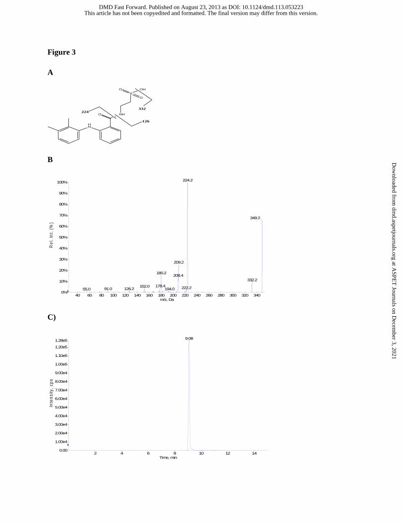
Figure 3
A
HN
NH
S
O
OHO
O
126
224332
B
C)
40 60 80 100 120 140 160 180 200 220 240 260 280 300 320 340m/z, Da
0%
10%
20%
30%
40%
50%
60%
70%
80%
90%
100%
Re
l. I
nt.
(%
)
224.2
349.2
209.2
180.2208.4
332.2
178.4152.0 222.291.0 126.2 194.055.0
2 4 6 8 10 12 14Time, min
0.00
1.00e4
2.00e4
3.00e4
4.00e4
5.00e4
6.00e4
7.00e4
8.00e4
9.00e4
1.00e5
1.10e5
1.20e5
1.28e5
Inte
ns
ity
, c
ps
9.08
This article has not been copyedited and formatted. The final version may differ from this version.DMD Fast Forward. Published on August 23, 2013 as DOI: 10.1124/dmd.113.053223
at ASPE
T Journals on D
ecember 3, 2021
dmd.aspetjournals.org
Dow
nloaded from
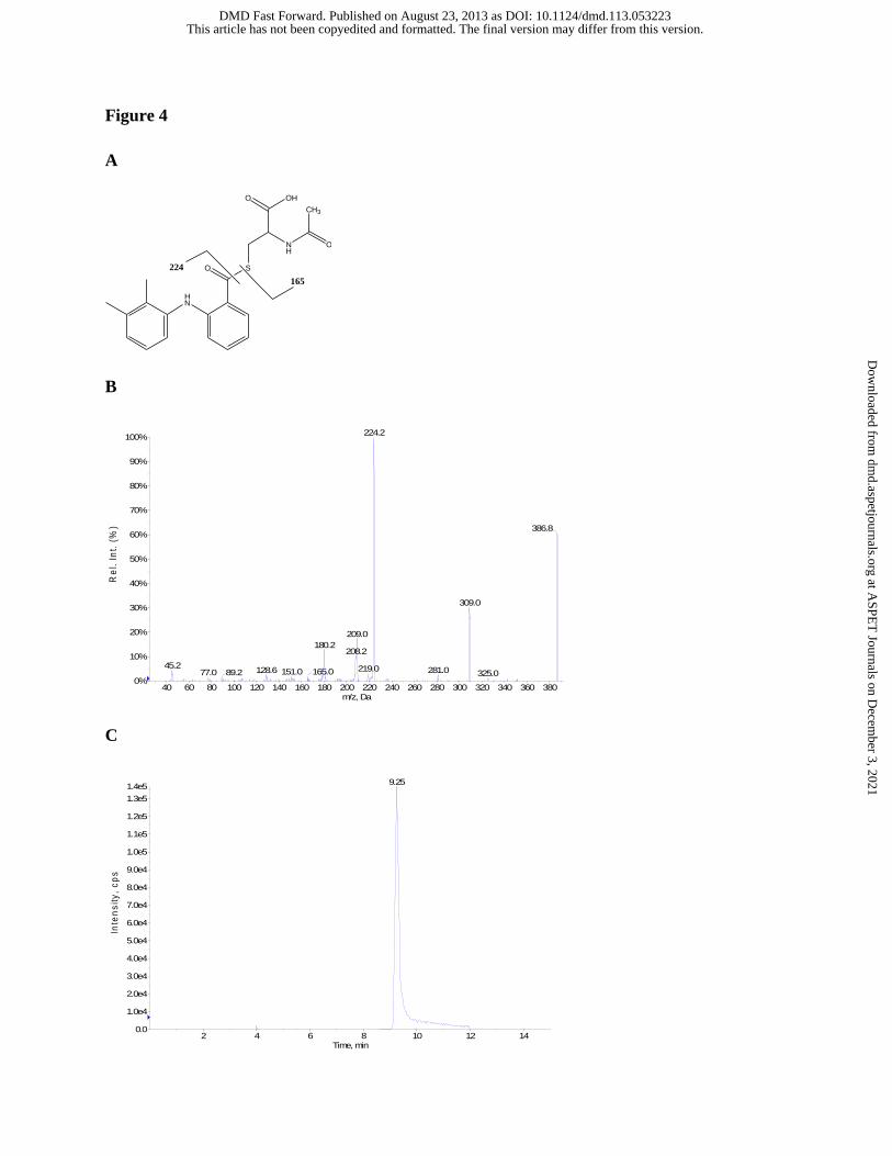
Figure 4
A
HN
S
OH
O
O
NH
CH3
O
224
165
B
C
+MS2 (387.19) CE (52): 26 MCA scans from Sample 1 (TuneSampleName) of ... Max. 3.1e5 cps.
40 60 80 100 120 140 160 180 200 220 240 260 280 300 320 340 360 380m/z, Da
0%
10%
20%
30%
40%
50%
60%
70%
80%
90%
100%
Re
l. I
nt.
(%
)
224.2
386.8
309.0
209.0
180.2208.2
45.2 219.0128.6 281.0151.0 165.077.0 89.2 325.0
XIC of +MRM (9 pairs): 387.186/224.127 Da from Sample 9 (Sample008) of Da... Max. 1.4e5 cps.
2 4 6 8 10 12 14Time, min
0.0
1.0e4
2.0e4
3.0e4
4.0e4
5.0e4
6.0e4
7.0e4
8.0e4
9.0e4
1.0e5
1.1e5
1.2e5
1.3e5
1.4e5
Inte
ns
ity
, c
ps
9.25
This article has not been copyedited and formatted. The final version may differ from this version.DMD Fast Forward. Published on August 23, 2013 as DOI: 10.1124/dmd.113.053223
at ASPE
T Journals on D
ecember 3, 2021
dmd.aspetjournals.org
Dow
nloaded from
This article has not been copyedited and formatted. The final version may differ from this version.DMD Fast Forward. Published on August 23, 2013 as DOI: 10.1124/dmd.113.053223
at ASPE
T Journals on D
ecember 3, 2021
dmd.aspetjournals.org
Dow
nloaded from
This article has not been copyedited and formatted. The final version may differ from this version.DMD Fast Forward. Published on August 23, 2013 as DOI: 10.1124/dmd.113.053223
at ASPE
T Journals on D
ecember 3, 2021
dmd.aspetjournals.org
Dow
nloaded from
This article has not been copyedited and formatted. The final version may differ from this version.DMD Fast Forward. Published on August 23, 2013 as DOI: 10.1124/dmd.113.053223
at ASPE
T Journals on D
ecember 3, 2021
dmd.aspetjournals.org
Dow
nloaded from
This article has not been copyedited and formatted. The final version may differ from this version.DMD Fast Forward. Published on August 23, 2013 as DOI: 10.1124/dmd.113.053223
at ASPE
T Journals on D
ecember 3, 2021
dmd.aspetjournals.org
Dow
nloaded from