Embed Size (px)
Citation preview
The Journal of Neuroscience, March 1993, 13(3): 941-951
HSV-1 Vector-Mediated Gene Transfer of the Human Nerve Growth Factor Receptor ~75 hNGFR Defines High-Affinity NGF Binding
David S. Battleman,’ Alfred I. Geller,* and Moses V. Chaol
‘Department of Cell Biology and Anatomy, Cornell University Medical College, New York, New York 10021 and *Division of Endocrinology, Children’s Hospital, Boston, Massachusetts 02115
A series of recombinant herpes simplex virus (HSV-1) vec- tors have been constructed that encode either the full-length cDNA of the human p75 NGF receptor (~75~~9 or truncated forms of the receptor. Infection of cultured fibroblast cells with viral stocks results in abundant expression of all three cDNAs, as detected by affinity cross-linking, immunoblot analysis, and equilibrium binding. Furthermore, viral infec- tion of primary neuronal cultures gives easily detectable p75 expression by immunofluorescence and affinity cross-link- ing. When p75 was introduced by viral infection into fibro- blast cells expressing the frkproto-oncogene, a new binding site was created, consistent with high-affinity NGF binding. This site is not created by the coexpression of truncated forms of p75 that lack either the extracellular ligand binding domain or the cytoplasmic domain of the receptor, sug- gesting that both of these regions of the receptor are re- quired for the formation of the high-affinity NGF binding site. Hence, these HSV-1 vectors give rise to appropriate NGF receptor binding after viral infection. The application of these HSV-1 constructs to primary neuronal culture and in vivo models of ~75~~~~ function is discussed.
[Key words: NGF receptor, herpes simplex virus, primary culture, trk oncogene, neurotrophin, receptor binding]
NGF signal transduction results in cell survival and mainte- nance of discrete neuronal populations, including sensory neu- rons of neural crest origin and sympathetic neurons (Thoenen and Barde, 1980; Levi-Montalcini, 1987). NGF also regulates neural populations in the brain, most notably cholinergic fibers of the basal forebrain (Gnahn et al., 1983; Hefti, 1986; Mobley et al., 1986). The identification of neurotrophic factors BDNF, NT-3, NT-4, and NT-5 has uncovered a family of related poly- peptide differentiation factors with similar biological effects (Leibrock et al., 1989; Emfors et al., 1990; Hohn et al., 1990; Maisonpierre et al., 1990; Rosenthal et al., 1990; Berkemeier et al., 1991; Hallbook et al., 1991). Furthermore, a primary
Received Apr. 24, 1992; revised July 27, 1992; accepted Sept. 2, 1992. We are grateful to Drs. Howard Federoff and’Roy Silverstein for critical reading
of the manuscript. We thank Dr. Barbara Hempstead and Dr. Margaret Berg for many helpful discussions and technical assistance. Also, we thank both Dr. Nila Patil and Margret Einarson for use of the NGF receptor mutant constructs and Dr. Luis Parada for the trk-3T3 cells. This work was supported by grants from the National Institutes of Health, the American Cancer Society, the Zenith Award from the Alzheimer’s Association, and the Dorothy Rodbell Cohen Foundation to M.V.C. A.I.G. was supported by the American Health Assistance Foundation, the American Parkinson’s Disease Association and Alkermes Inc. A.I.G. thanks Oliver Ashe for technical assistance.
Correspondence should be addressed to Dr. Moses Chao, The Department of Cell Biology and Anatomy, Cornell University Medical College, 1300 York Av- enue, New York, NY 1002 1.
Copyright 0 1993 Society for Neuroscience 0270-6474/93/130941-l 1$05.00/O
signaling mechanism for these factors has been defined with the discovery that the p140proto1rk receptor tyrosine kinase (trk) in- teracts specifically with NGF, resulting in increased tyrosine phosphorylation of cellular substrates in PC1 2 cells (Kaplan et al., 1991 b; Klein et al., 1991a). Other trk family members, in- cluding trkB and trkC, have since been found to interact with multiple neurotrophic ligands (Cordon-Card0 et al., 199 1; Klein et al., 199 la,b; Lambelle et al., 199 1; Soppet et al., 199 1; Squint0 et al., 1991).
An important question concerning the action of neurotro- phins is how the functional receptor complex is encoded and specified. NGF and the other neurotrophin family members are known to interact with a 75,000 Da transmembrane receptor, ~75~~~~ (Johnson et al., 1986; Radeke et al., 1987; Emfors et al., 1990; Rodriquez-Tebar et al., 1990; Squint0 et al., 1991). NGF binds to this receptor with low affinity (Kd = 1O-9 M), but this interaction does not initiate any measurable signal trans- duction in fibroblast cells expressing ~75~~~~ (Hempstead et al., 1989). Only in NGF-responsive cells are high-affinity (Z& = lo-” M) and low-affinity binding sites displayed (Sutter et al., 1979; Schechter and Bothwell, 198 1; Green and Greene, 1986). High-affinity binding is required for functional responses to NGF both in culture and in vivo and can be reconstituted in cultured cells that express p140prototrk (Hempstead et al., 1991; Klein et al., 1991a). Whether this binding requires the concomitant ex- pression of p7YGFR has been the subject of debate (Bothwell, 199 1; Chao, 1992).
Both receptors are known to be colocalized in sensory, sym- pathetic, and central cholinergic neurons during NGF-respon- sive periods (Buck et al., 1987; Hohaman et al., 1992; Schech- terson and Bothwell, 1992), and monoclonal antibodies against ~75 have been shown to display inhibitory effects upon neurite regeneration (Chandler et al., 1984) and c-fos induction in PC1 2 cells (Milbrandt, 1986). Previous equilibrium binding studies have indicated that ~75~~~~ participates in high-affinity NGF binding(Green and Greene, 1986; Hempstead et al., 1989, 199 1; Pleasure et al., 1990; Matsushima and Bogenmann, 199 1) and that ~14.0~~~~~‘~~ binds to NGF with low affinity (Kaplan et al., 199 1 a). Other experiments, however, have reached the opposite conclusion, suggesting that the unique expression of p140pro101rk defines a high-affinity receptor for NGF (Klein et al., 1991a). The involvement of ~75~~~~ in NGF signal transduction was recently discounted with results demonstrating that p 1 40prolo1rk and the other trk receptor tyrosine kinase family members could function independently in fibroblast cells without the coexpres- sion of ~75~~~~ (Cordon-Card0 et al., 199 1; Glass et al., 199 1). Moreover, use of anti-p75NCFR antibodies and site-directed mu- tants of NGF has implied that the biological effects of NGF
942 Battleman et al. + p7PNGFR Defines High-Affinity NGF Binding
may be mediated through the trk receptor alone, without binding to p7YGFR (Weskamp and Reichardt, 199 1; IbZnez et al., 1992).
To address the functional role of the ~75~~~~ in mediating the differentiative effects of NGF action, we have sought to express ~75 cDNAs in defective herpes simplex virus (HSV-1)
virus subsequently harvested, passaged, and titered (8 x lo5 infectious particles/ml) as described previously (Miller and Hyman, 1978).
Northern blot analysis. Cells were grown to 70% confluence on 100 mm tissue culture dishes, infected with 0.4 ml of viral stock at a mul- tinlicitv of infection (MOB of 0.25. and incubated for 6-24 hr at 37°C. and total mRNA was prenared usinn the RNASol B method (Chom: _ _
vectors (Geller and Breakefield, 1988; Geller et al., 199 1). Here czynski and Nicoletta,-1987). For Northern blot analysis, mRNA (20
we report the construction and expression of three novel HSV- 1 &sample) was separated by electrophoresis in 1% agarose gels con-
vectors carrying a full-length human ~75 receptor cDNA as well taining 2.2 M formaldehyde, transferred to nitrocellulose filters, and baked in vacua at 80°C for 4 hr (Sambrook et al., 1989). The presence
as two mutant receptor clones, each lacking an essential domain of mRNA species was confirmed by ethidium bromide visualization. of the receptor. Our results confirm that the coexpression of Filters were then prehybridized in 50% formamide, 5 x SSC, and 5 x
both p75NoFR and ~140~~~‘~~~~ is required for the functional re- Denhardt’s solution with 500 &ml of denatured salmon sperm DNA
constitution of a high-affinity binding site for NGF. The ex- at 42°C for at least 4 hr. Hybridization with radiolabeled probe was
pression of trk with either the ligand binding domain or the carried out at 42°C in 50% formamide, 5 x SSC, and 1 x Denhardt’s solution with 100 pg of denatured salmon sperm DNA. Randomly
cytoplasmic domain of p75, however, was not sufficient to me- primed restriction fragments were generated to specific activity of 5 x
diate this high-affinity interaction. Moreover, expression studies lo8 cpm/pg. Filters were washed to 0.4 x SSC at 68°C and exposed to
in primary culture indicate that the HSV-1 vectors employed x-ray film at -70°C with an intensifying screen.
in this study are capable of faithfully delivering high levels of Immunoblotting. For immunoblot analysis, infected cell cultures were
receptors into neurons, and therefore these HSV- 1 vectors will prepared as above. Infected fibroblasts were washed in phosphate-buf- fered saline (PBS; pH 6.4) and rapidly solubilized in 1 ml of solubili-
be useful in addressing questions about the physiological roles that ~75~~~~ plays in nerve cells.
Materials and Methods Materials. Restriction endonucleases were purchased from New England Biolabs (Beverly, MA), Mouse NGF (p-dimer) was obtained from Bio- products for Science (Indianapolis, IN). Anti-2.5s NGF antiserum, a rabbit anti-mouse polyclonal antibody, was purchased from Collabo- rative Research (Bedford, MA), and anti-p75 antibody, a polyclonal antibody that was generated against the cytoplasmic domain of the low- affinity NGF receptor, was generously supplied by Dr. S. J. Decker (Parke-Davis, Ann Arbor, MI). Antibody ME20.4 against the human NGF receptor (hNGFR; Ross et al., 1984) was harvested from cultured hybridoma cell line supematants (from American Type Culture Collec- tion), and rhodamine-conjugated goat anti-mouse (Fab’), antibody was purchased from Jackson Labs (Bar Harbor, ME). Ethyl-3-(dimethylam- inopropyl) carbodiimide (EDAC) was purchased from Pierce Chemical Co. (Rockford, IL). lZ51-NGF was prepared by the lactoperoxidase meth- od as described previously (Green and Greene, 1986).
CelI culture. PC 12 cells were maintained in Dulbecco’s modified Ea- gle’s medium (DMEM) with 10% fetal bovine serum and 5% horse serum (GIBCO, GrandIsland, NY). NIH-3T3 cells, which overexpress ~140~~~‘~~~~ (3T3-trk cells) (Kaplan et al.. 1991a). were maintained in DMEM with 10% fetal bovineserum and 0.4 mg&tl of G4 18 (GIBCO).
px 1 insert (2.3 kb),’ and the p5A insert (1 .O kb), respectively. The ori-
Construction of vectors and packaging of HSV-I virus particles. ~75 human NGF receptors pSL and pxl were constructed as described pre- viously (Hempstead et al., 1990). For mutant p5A, a 1.5 kilobase (kb) hNGFR cDNA was digested with both StuZ and Sau3A and religated. The resulting 1 .O kb fragment lacking nucleotides 2 15-682 was recov- ered. The pSL, pxl, and p5A cDNA inserts were then isolated from carrier sequence by EcoRI digestion and subcloned into the recipient HSV-1 vector pHSVPrpUC (Geller and Breakefield, 1988). pHSV- hNGFR. DHSV-xl, and DHSV-5A contain the DSL insert (2.3 kb), the
zation buffer [lo mM Tris-HCl (pH 7.4) 1% SDS, and 0.1 mM each of phenylmethylsulfonyl fluoride (PMSF)/aprotinin/leupeptin]. Cell lys- ates were incubated on ice for 15 min and then pelleted at top speed in a microfuge at 4°C for 10 min. Supematants were assayed for protein concentrations with a protein assay kit from Bio-Rad (Richmond, CA). Proteins (200 pg) were immunoprecipitated with anti-p75 antibody, electrophoresed by SDS-PAGE (8%), and electrophoretically transferred to nitrocellulose. Filters were then incubated with anti-p75 antisera followed by lZ51-labeled protein A from Amersham (Arlington Heights, IL). Autoradiography was carried out at -70°C with an intensifying screen.
Affinity cross-linking and immunoprecipitation. 3T3-trk cells infected at an MO1 of 0.25, and uninfected control cells were harvested for affinity cross-linking. NGF receptors were labeled by affinity cross-link- ing methods previously described (Green and Greene, 1986; Hempstead et al., 1989). For EDAC cross-linking to ‘Y-NGF, cells were washed in PBS (pH 6.5) and resuspended to a final concentration of 2 x lo6 cells/ml in PBS containing 1 mg/ml each of glucose and bovine serum albumin. lZ51-NGF was then added to each cell suspension at a final concentration of 25 r&ml, in the presence or absence of 5 wt~ unlabeled NGF for 2 hr on a rotator at 4°C. EDAC was next added to a final con- centration of 4 mM at room temperature for 30 min. Alter cross-linking, cells were centrifuged, washed in PBS-lysine (50 mM), andsolubilized in lysis buffer (10 mM Tris, pH 7.6, 66 mM EDTA, 1% NonidetP40, 0.4% deoxycholate, and 0.1 mM each of PMSF/aprotinin/leupeptin) at 4°C for 20 min. Lysates were immunoprecipitated with anti-2.5s NGF antisera at 4°C for 2 hr and washed extensively in RIPA buffer (10 mM Tris-HCl, pH 8.0, 150 mM NaCl, 10 mM KCl, 1% deoxycholate, 1% Nonidet P-40, 0.1% SDS, and 1 mM EDTA). The samples were then resuspended in sample buffer, boiled for 5 min, and subjected to elec- trophoresis on a 7.5Oh SDS-polyacrylamide gel. The dried gels were exposed to autoradiographic film at - 70°C with an intensifying screen.
_ I assessed by Scatchard analysis (Scatchard, 1949). Only binding values
Equilibrium binding analysis. Infections were carried out as described above. Equilibrium binding of 12’I-NGF to crude cellular membrane preparations was performed as described previously (Hempstead et al., 1989). All measurements were verified in trinlicate. and the data were
entation of each insert was verified by restriction enzyme analysis. pHSVlac contains the 1acZ gene and was constructed as previously described (Geller and Breakefield, 1988).
above 50% specific binding were used in the final analysis. The LIGAND
program was used to analyze the data (Munson and Rodbard, 1980). Primary neuronal cultures and immunohistochemistry. Timed-prea-
The packaging system for HSV- 1 viral particles has been previously reported (Spaete and Frenkel, 1982; Geller, 1988). In brief, 1.5 x 10s CVl cells were plated on 60 mm dishes and transfected with 0.5 ml of a calcium phosphate precipitate containing 1 pg of pHSV- 1 vector DNA and 9 pg of salmon sperm DNA. Four hours later the cells were treated with 15% glycerol (Parker and Stark, 1987) and incubated at 37°C for 24 hr. Cells were then infected with 1.5 x 1 O6 plaque-forming units of HSV-1 strain 17 ts K (Davison et al., 1984) in 100 ~1 of medium. HSV- 1 ts K is a mutant HSV-1 strain that contains a temperature-sensitive mutation in the IE3 gene, resulting in an immediate-early phenotype and no viral replication at the restrictive temperature of 37°C. After 1 hr at room temperature, an additional 5 ml of medium were added to each infected plate. Cultures were then incubated for 72 hr at 3 1°C and
nant Sprague-Dawley rats [embryonic day 16 (E 16)] were purchased from Charles River Labs (Wilmington, MA) and embryos removed under sterile conditions. Brain tissue was isolated and the cerebral cortex was carefully dissected away from hippocampus, basal forebrain, and olfactory bulbs. Meninges were also removed. Cortical cells were then manually dissociated and plated on poly-D-lysine-coated dishes. Cul- tures were enriched for nerve cells by maintaining cultures in serum- free conditions for at least 5 d as described previously (di Porzio et al., 1980).
For immunohistochemistry, primary cortical cells were infected at an MO1 of 0.25 for 8 hr at 37°C. Fresh media was then added and the cells were placed at 37°C for an additional 36 hr mior to fixation. Cells were fixed -in buffered 4% paraformaldehyde for 1 hr on ice, washed with
The Journal of Neuroscience, March 1993, U(3) 943
Construct
bNGFR
Cyeteine Repeat Domains
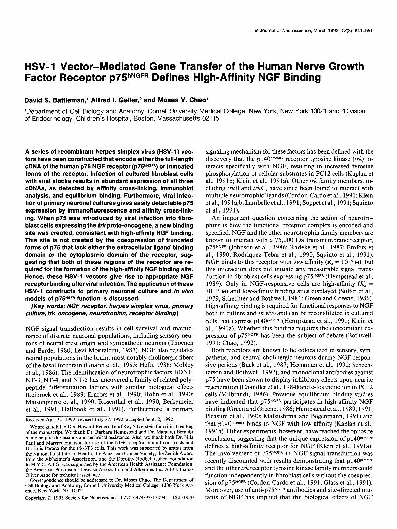
Figure I. Schematic representation of p75hNGFR and receptor mutants. Con- structs were generated as outlined in the Materials and Methods. The hNGFR
serine - Threonine TM Cytoplasmic Domain construct represents the full-length R&Xl cDNA of the human low-affinity NGF
receptor, ~75. Mutant-xl encodes for an intracellular deletion receptor that is truncated 4 amino acids after the trans- membrane domain at amino acid DO-
Mutant-xl
Mutant-SA
sition 248. Mutant-5A encodes for an extracellular deletion mutation that lacks the NGF binding domain due to a large deletion (Welcher et al., 199 1;
NH2b ‘OOH Yan and Chao, 199 1) spanning amino acids 34-191.
PBS, and preincubated in PB buffer (0.1 M PBS, pH 7.4, with 0.1% Triton X-100 and 1 mg/ml BSA) for 1 hr at room temperature. ME20.4 hybridoma supematants were diluted 1:5 in PB buffer and incubated with cells overnight at 4°C. Cells were thoroughly washed and then incubated in a 1:250 dilution of a rhodamine-conjugated goat anti- mouse secondary antibody (Jackson Labs). Cells were washed exten- sively and mounted. Control cultures were assayed in the absence of primary antibody.
Results
hNGFR constructs and HS V- 1 vectors The NGF receptor constructs used in this study are shown sche- matically in Figure 1. The hNGFR construct is a 2.3 kb full- length cDNA of the human NGF receptor that contains the entire coding region of the receptor but lacks most of the 3’- untranslated sequences. The cytoplasmic deletion mutant hNGFR-xl was generated by the site-directed insertion of a universal Xba-termination linker 4 amino acids beyond the transmembrane domain at amino acid position 248 (Hempstead et al., 1990). The extracellular deletion mutant hNGFR-SA con- tains a large deletion in the ligand binding domain (Welcher et al., 199 1; Yan and Chao, 199 I), spanning amino acids 34-l 9 1. Each of these receptor constructs was subcloned into the 4.8 kb
HSV-1 IE 415
p75NGFR gene
SW0 ~01~ A site
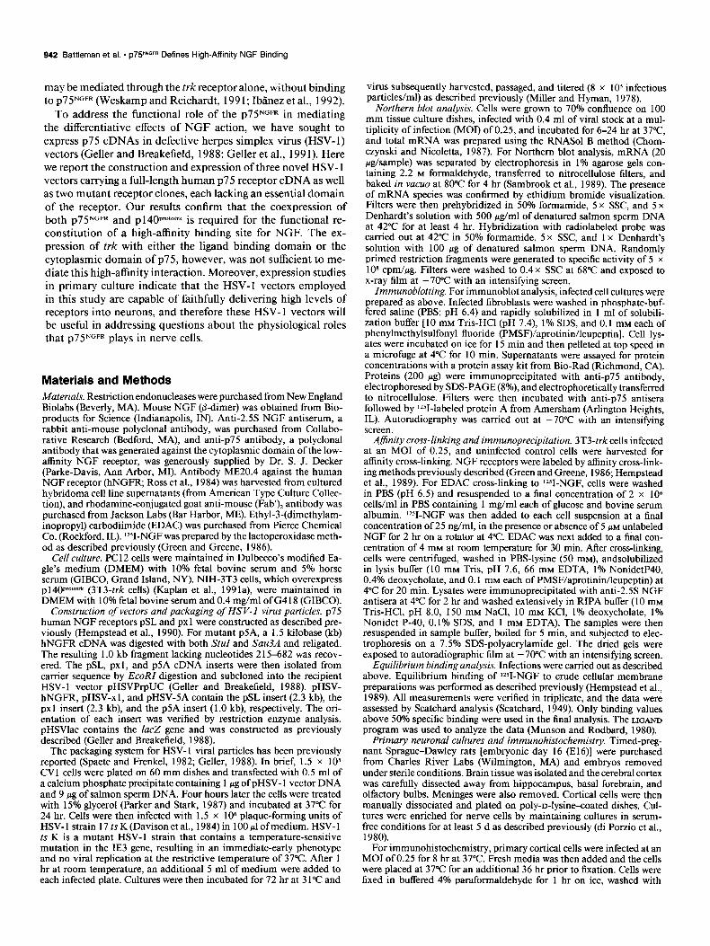
defective HSV-1 vector (Geller and Breakefield, 1988), which is shown in Figure 2, and then packaged into HSV-1 particles as outlined in the Materials and Methods.
Expression studies in cell culture
To determine if the HSV- 1 vectors appropriately express human
P75 NGFR (P75hNGFR ) and mutant receptor constructs, 3T3-t&cells were infected with 8 x lo5 infectious particles/ml of viral stock and analyzed for both mRNA and cell surface protein. These cells were chosen as the recipient cell line because they lack endogenous p7YGFR (Kaplan et al., 199 1 a; Klein et al., 199 la). Expression of all three receptor constructs in fibroblasts is readi- ly detectable by Northern blot analysis (Fig. 3A). In Figure 3A, a prominent 2.3 kb message was detected in pHSV-hNGFR and pHSV-xl lanes (lanes 2 and 3) while in mock-infected 3T3-trk cells, no signal was detected (lane 1).
Cells infected with pHSV-5A also expressed a message of about 2.3 kb in size (Fig. 3A, lane 4). Though the cDNA insert of this construct was approximately 1.0 kb, the increased size of this primary transcript was likely due to an alternative po- lyadenylation event. Unlike the pHSV-hNGFR and pHSV-xl constructs, which contain an endogenous polyadenylation signal in their cDNA insert, the pHSV-5A construct does not. There- fore, pHSV-5A must rely on the SV40 polyadenylation signal
Figure 2. Structure of pHSV-hNGFR. This HSV-1 expression vector is a 4.8 kb plasmid that contains the 2.3 kb hNGFR insert and the following set of genetic elements: (1) sequences from pBR322 that allow for bacterial repli- cation and selection of the plasmid, in- cluding the ColEl origin of DNA rep- lication and the ampicillin-resistance gene; (2) sequences from the HSV- 1 ge- nome that allow for packaging of the plasmid into viral particles, including the HSV-1 packaging site and herpes simplex virus DNA replication ori, (Davison and Wilkie, 198 1; McGeoch et al., 1986); and (3) sequences from both HSV-1 and SV40 to compose el- ements of the transcription unit, in- cluding the HSV-1 IE 4/5 promoter (McGeoch et al., 1986) and the SV40 early region polyA site (Hall et al., 1983).
944 Battleman et al. l ~75”~~~~ Defines High-Affinity NGF Binding
A. Northern Blot
28s
18S
C. Affinity Crosslinking
PC12 3T3- trk uninfected Mock --
B. lmmunoblot
28
3T3- trk 3T3- trk 3T3- trk
HSV-5A HSV-xl HSV-hNGFR 1-- I
+ - +-+-+-+
228
+ IgG
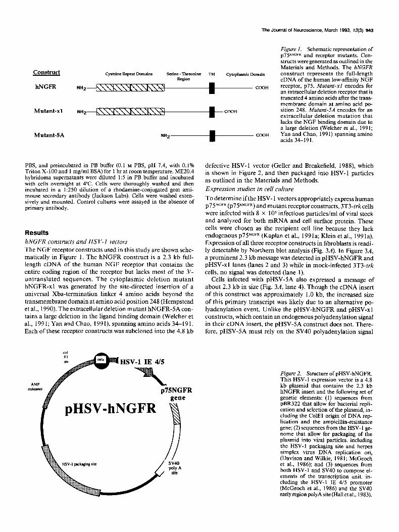
Figure 3. Expression of p7YNGFR and receptor mutants in 3T3-trk cells using recombinant HSV-1 vectors. A, Northern blot analysis of RNA isolated from 3T3-trk cells infected with HSV- 1 viral stocks. Cells were infected for 6-24 hr at 37”C, and RNA was isolated 36 hr later as described in Materials and Methods. Northern blots were probed with radiolabeled B9 fragment, a probe specific to ~75”~~~ cDNA. Each lane contained 20 pg of total RNA. Lane I is RNA harvested from fibroblasts following mock infection. Lanes 2-4 contain RNA from fibroblasts infected with 0.4 ml ofHSV-hNGFR, HSV-x 1, and HSV-SA, respectively. pHSVlac virus, a vector that expresses the E. coli @galactosidase (Geller and Breakefield, 1988) was used for control infection. Positions of 28s and 18s ribosomal markers are indicated. B, Immunoblot analysis with anti-p75 antibody of HSV-l-infected 3T3-trk cells. Proteins (200 fig) from infected cells were immunoprecipitated and blotted with anti-p75 antibody as described in Materials and Methods. Anti-p75 antibody recognizes the cytoplasmic domain of human ~75. Lane I represents mock-infected 3T3-trk cells. Lanes 2-4 are 3T3-trk cells infected with HSV-hNGFR, HSV-x 1, and HSV-SA, respectively. Molecular mass markers (in kilodaltons) are indicated. An unlabeled arrow indicates the position of the HSV-5A protein at about 35 kDa. The broad IgG band (arrow) runs at approximately 50 kDa. pHSVlac virus was used for control infection (Geller and Breakefield, 1988). C, Affinity cross-linking of NGF receptors in cultured cell lines. NGF receptors were cross-linked to 1251-NGF using EDAC in the presence (+) or absence (-) of excess unlabeled NGF as outlined in Materials and Methods. Samples were loaded as indicated above the lanes. pHSVlac virus was used for control infection (Geller and Breakefield, 1988). Molecular mass markers (in kilodaltons) are indicated on the left. Arrows indicate the position of cross-linked ~75 at 100 kDa and the cross-linked HSV-x 1 protein at approximately 68 kDa.
that is present in the HSV- 1 vector about 1.15 kb downstream of the cloning site (Hall et al., 1983).
To verify further that the HSV- l-infected cells appropriately expressed these cDNAs, we assayed both for cell surface protein expression and for ligand binding. Figure 3B shows an immu- noblot of proteins immunoprecipitated with anti-p75 antibody. The polyclonal anti-p75 antibody was raised against the cyto- plasmic domain of ~75~~~~ and was used to confirm the ex- pression ofthe pHSV-5A construct. Cells infected with the HSV- 5A virus demonstrated a major 35 kDa protein of predicted size (Fig. 3B, lane 4). No signal was detected in this range in mock-infected fibroblasts (Fig. 3B, lane 1) or in cells infected
with either the full-length receptor HSV-hNGFR or the intra- cellular deletion mutant HSV-xl (Fig. 3B, lanes 2,3). The broad IgG band is observed across the top of each lane at approxi- mately 50 kDa.
To detect the hNGFR and mutant-xl proteins, an affinity cross-linking assay was used (Fig. 3C). This cross-linking re- action results in a 90-100 kDa labeled species in PC12 cells, human melanoma cells, and rat brain, and has been shown to represent a complex between labeled NGF and the ~75 receptor (Grob et al., 1983; Green and Greene, 1986). Infected 3T3-trk fibroblast and PC1 2 cells were incubated with lZ51-NGF in sus- pension for 2 hr, cross-linked with EDAC, and then immuno-
The Journal of Neuroscience, March 1993, U(3) 945
I ,‘.‘.,‘~‘.l....l.‘..l...~
sw Bouri?(fmole%ng) lmo ’
m Bound (fmoWmg)
I 5. 3lStrk
HSV-xl I
500 loo0 Iyw) m 2m 3mm :
Bound (fmoles/mg)
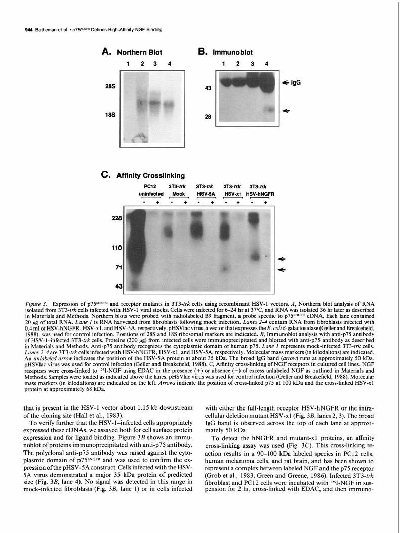
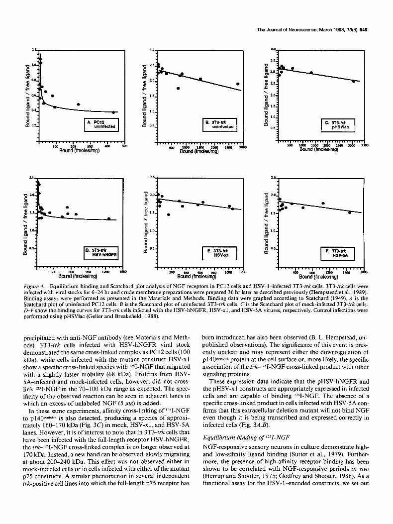
Figure 4. Equilibrium binding and Scatchard plot analysis of NGF receptors in PC 12 cells and HSV- l-infected 3T3-trk cells. 3T3-trk cells were infected with viral stocks for 6-24 hr and crude membrane preparations were prepared 36 hr later as described previously (Hempstead et al., 1989). Binding assays were performed as presented in the Materials and Methods. Binding data were graphed according to Scatchard (1949). A is the Scatchard plot of uninfected PC 12 cells. B is the Scatchard plot of uninfected 3T3-trk cells. C is the Scatchard plot of mock-infected 3T3-trk cells. D-F show the binding curves for 3T3-trk cells infected with the HSV-hNGFB, HSV-xl, and HSV-SA viruses, respectively. Control infections were performed using pHSVlac (Geller and Breakefield, 1988).
precipitated with anti-NGF antibody (see Materials and Meth- ods). 3T3-trk cells infected with HSV-hNGFR viral stock demonstrated the same cross-linked complex as PC1 2 cells (100 kDa), while cells infected with the mutant construct HSV-xl show a specific cross-linked species with lZ51-NGF that migrated with a slightly faster mobility (68 kDa). Proteins from HSV- SA-infected and mock-infected cells, however, did not cross- link lZ51-NGF in the 70-100 kDa range as expected. The spec- ificity of the observed reaction can be seen in adjacent lanes in which an excess of unlabeled NGF (5 PM) is added.
In these same experiments, affinity cross-linking of lZ51-NGF to p140proto1rk is also detected, producing a species of approxi- mately 160-l 70 kDa (Fig. 3C) in mock, HSV-x 1, and HSV-5A lanes. However, it is of interest to note that in 3T3-trk cells that have been infected with the full-length receptor HSV-hNGFR, the trkJ51-NGF cross-linked complex is no longer observed at 170 kDa. Instead, a new band can be observed, slowly migrating at about 200-240 kDa. This effect was not observed either in mock-infected cells or in cells infected with either of the mutant ~75 constructs. A similar phenomenon in several independent trk-positive cell lines into which the full-length ~75 receptor has
been introduced has also been observed (B. L. Hempstead, un- published observations). The significance of this event is pres- ently <unclear and may represent either the downregulation of p 1 40pT0101rk protein at the cell surface or, more likely, the specific association of the trkJZ51-NGF cross-linked product with other signaling proteins.
These expression data indicate that the pHSV-hNGFR and the pHSV-x 1 constructs are appropriately expressed in infected cells and are capable of binding ‘Y-NGF. The absence of a specific cross-linked product in cells infected with HSV-5A con- firms that this extracellular deletion mutant will not bind NGF even though it is being transcribed and expressed correctly in infected cells (Fig. 3AJ).
Equilibrium binding of lz51-NGF NGF-responsive sensory neurons in culture demonstrate high- and low-affinity ligand binding (Sutter et al., 1979). Further- more, the presence of high-affinity receptor binding has been shown to be correlated with NGF-responsive periods in vivo (Herrup and Shooter, 1975; Godfrey and Shooter, 1986). As a functional assay for the HSV-l-encoded constructs, we set out
946 Battleman et al. l p75hNGFR Defines High-Affinity NGF Binding
Table 1. Summary of Scatchard plot analysis
Affinity site 1 Affinity site 2 Site 1: Cell line Infection Temperature Kd (M) Sites/mg of protein Kd (M) Sites/mg of protein Site 2
PC12 Uninfected 30°C 4.15 x 10-P 8.54 x 10”’ 6.62 x lo-” 4.93 x 10”” 17:l 3T3-trk Uninfected 30°C 4.23 x 1O-9 4.53 x lo”2 N.A. 3T3-trk pHSVIac 30°C 5.61 x lo+ 6.76 x lo+‘* N.A. 3T3-trk HSV-hNGFR 30°C 3.99 x 10-g 4.69 x lo+‘* 5.11 x 10-I’ 1.44 x 10”’ 32:l 3T3-trk HSV-x 1 30°C 4.81 x 10e9 3.12 x lo+‘* N.A. 3T3-trk HSV-5A 30°C 10.46 x lo+’ 5.53 x lo+‘2 N.A.
All binding data presented above were analyzed using the WGAND program (Munson and Rodbard, 1980). N.A. indicates that only one kinetic site was identified. Temperatures refer to the equilibrium binding assay conditions.
to determine if we could reconstitute high-affinity NGF binding in 3T3-trk cells, a recipient fibroblast cell line that overexpresses the trk proto-oncogene at high levels (> 200,000 sites per cell). Since the high-affinity NGF binding complex can be distin- guished from low-affinity complex by equilibrium binding, Scat- chard analysis was conducted. A membrane binding assay was employed in order to minimize the effects of internalization of NGF. Crude cell membranes were prepared from PC1 2 cells and HSV- l-infected 3T3-trk cells. PC 12 cell membranes (Fig. 4A) displayed two binding sites for NGF as previously reported (Landreth and Shooter, 1980; Block and Bothwell, 1983). The high-affinity binding site represents only about 5% of the total number of NGF binding sites and demonstrates a Kd of 6.62 x lO-L* M. The low-affinity site has a Kd of 4.15 x 1O-9 M.
Multiple binding studies of the 3T3-trk recipient cell line demonstrate that this cell line contains predominately low-af- finity receptors for NGF. A representative Scatchard plot is shown in Figure 4B. A single binding site with a Kd of 4.23 x 10-P M is observed, consistent with a low-affinity interaction. pHSVlac infections of 3T3-trk cells generated a similar binding
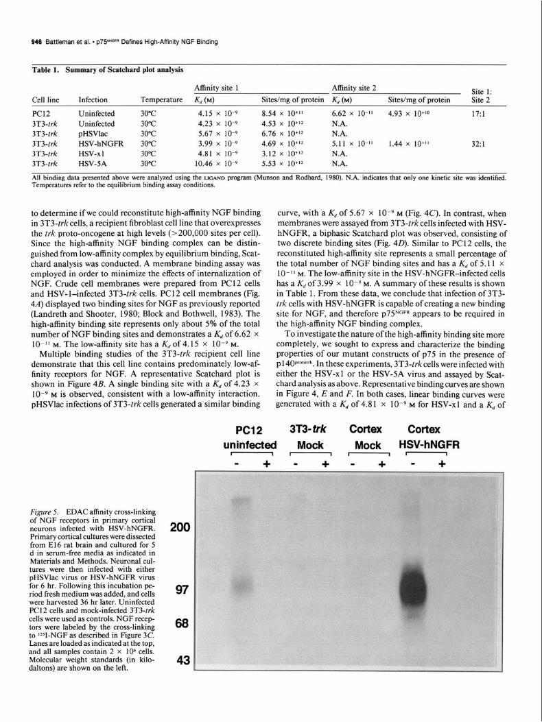
Figure 5. EDAC affinity cross-linking of NGF receptors in primary cortical neurons infected with HSV-hNGFR. Primary cortical cultures were dissected from El6 rat brain and cultured for 5 d in serum-free media as indicated in Materials and Methods. Neuronal cul- tures were then infected with either pHSVlac virus or HSV-hNGFR virus for 6 hr. Following this incubation pe- riod fresh medium was added, and cells were harvested 36 hr later. Uninfected PC1 2 cells and mock-infected 3T3-trk cells were used as controls. NGF recep- tors were labeled by the cross-linking to Y-NGF as described in Figure 3C. Lanes are loaded as indicated at the top, and all samples contain 2 x lo6 cells. Molecular weight standards (in kilo- daltons) are shown on the left.
200
97
68
43
PC12 3T3-frk Cortex Cortex
curve, with a Kd of 5.67 x 1O-9 M (Fig. 4C). In contrast, when membranes were assayed from 3T3-trk cells infected with HSV- hNGFR, a biphasic Scatchard plot was observed, consisting of two discrete binding sites (Fig. 40). Similar to PC12 cells, the reconstituted high-affinity site represents a small percentage of the total number of NGF binding sites and has a Kd of 5.11 x 10-l’ M. The low-affinity site in the HSV-hNGFR-infected cells has a Kd of 3.99 x 1O-9 M. A summary of these results is shown in Table 1. From these data, we conclude that infection of 3T3- trk cells with HSV-hNGFR is capable of creating a new binding site for NGF, and therefore p7SNGFR appears to be required in the high-affinity NGF binding complex.
To investigate the nature of the high-affinity binding site more completely, we sought to express and characterize the binding properties of our mutant constructs of p75 in the presence of pl 40pr0M’rk. In these experiments, 3T3-trkcells were infected with either the HSV-xl or the HSV-SA virus and assayed by Scat- chard analysis as above. Representative binding curves are shown in Figure 4, E and F. In both cases, linear binding curves were generated with a Kd of 4.81 x 1O-9 M for HSV-xl and a Kd of
uninfected Mock Mock HSV-hNGFR I-I-
+ - + - + - +
The Journal of Neuroscience, March 1993, 13(3) 947
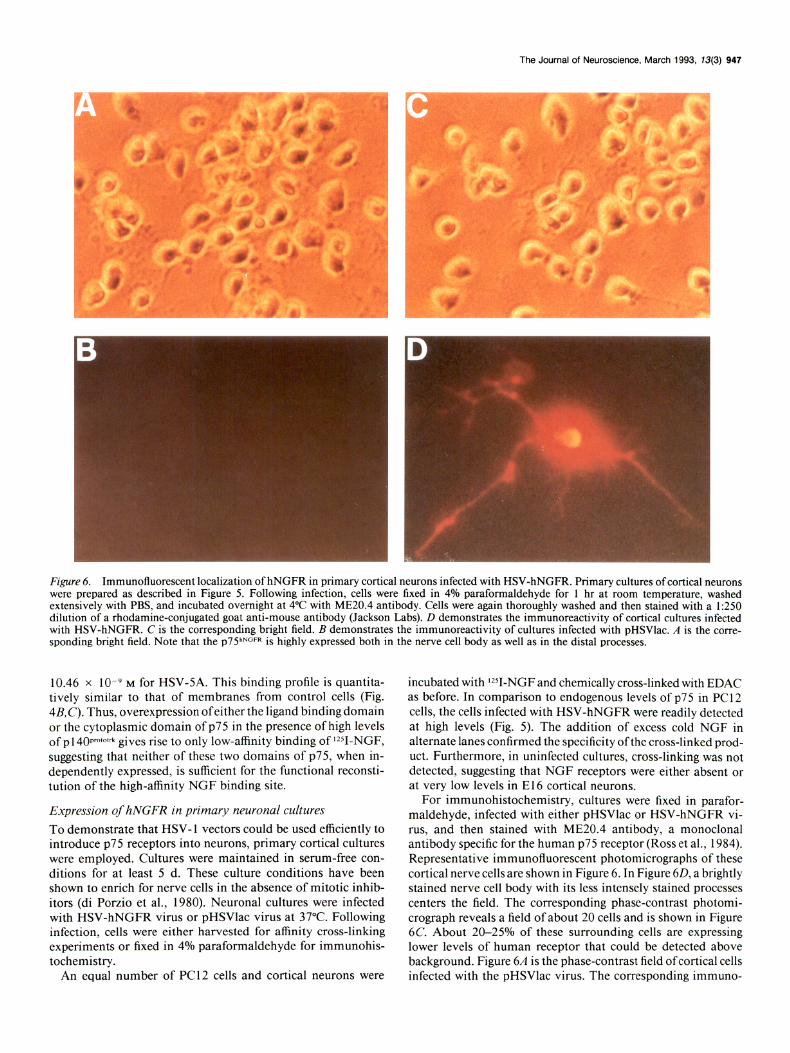
Fzgure 6. Immunofluorescent localization of hNGFR in primary cortical neurons infected with HSV-hNGFR. Primary cultures of cortical neurons were prepared as described in Figure 5. Following infection, cells were fixed in 4% paraformaldehyde for 1 hr at room temperature, washed extensively with PBS, and incubated overnight at 4°C with ME20.4 antibody. Cells were again thoroughly washed and then stained with a 1:250 dilution of a rhodamine-conjugated goat anti-mouse antibody (Jackson Labs). D demonstrates the imrnunoreactivity of cortical cultures infected with HSV-hNGFR. C is the corresponding bright field. B demonstrates the immunoreactivity of cultures infected with pHSVlac. A is the corre- sponding bright field. Note that the ~75”~~~~ is highly expressed both in the nerve cell body as well as in the distal processes.
10.46 x 10 9 M for HSV-SA. This binding profile is quantita- tively similar to that of membranes from control cells (Fig. 4&C). Thus, overexpression ofeither the ligand binding domain or the cytoplasmic domain of ~75 in the presence of high levels of p 140~~~‘~~~~ gives rise to only low-affinity binding of rZ51-NGF, suggesting that neither of these two domains of ~75, when in- dependently expressed, is sufficient for the functional reconsti- tution of the high-affinity NGF binding site.
Expression of hNGFR in primary neuronal cultures
To demonstrate that HSV- 1 vectors could be used efficiently to introduce ~75 receptors into neurons, primary cortical cultures were employed. Cultures were maintained in serum-free con- ditions for at least 5 d. These culture conditions have been shown to enrich for nerve cells in the absence of mitotic inhib- itors (di Porzio et al., 1980). Neuronal cultures were infected with HSV-hNGFR virus or pHSVlac virus at 37°C. Following infection, cells were either harvested for affinity cross-linking experiments or fixed in 4% paraformaldehyde for immunohis- tochemistry.
An equal number of PC12 cells and cortical neurons were
incubated with rZSI-NGF and chemically cross-linked with EDAC as before. In comparison to endogenous levels of p75 in PC 12 cells, the cells infected with HSV-hNGFR were readily detected at high levels (Fig. 5). The addition of excess cold NGF in alternate lanes confirmed the specificity ofthe cross-linked prod- uct. Furthermore, in uninfected cultures, cross-linking was not detected, suggesting that NGF receptors were either absent or at very low levels in E 16 cortical neurons.
For immunohistochemistry, cultures were fixed in parafor- maldehyde, infected with either pHSVlac or HSV-hNGFR vi- rus, and then stained with ME20.4 antibody, a monoclonal antibody specific for the human p75 receptor (Ross et al., 1984). Representative immunofluorescent photomicrographs of these cortical nerve cells are shown in Figure 6. In Figure 60, a brightly stained nerve cell body with its less intcnscly stained processes centers the field. The corresponding phase-contrast photomi- crograph reveals a field of about 20 cells and is shown in Figure 6C. About 20-25% of these surrounding cells are expressing lower levels of human receptor that could be detected above background. Figure 6A is the phase-contrast field ofcortical cells infected with the pHSVlac virus. The corresponding immuno-

848 Battleman et al. * p7PNGFR Defines High-Affinity NGF Binding
fluorescent field is shown in Figure 6B. These results indicate that HSV-mediated expression of ~75~~~~ can be readily ex- tended to primary nerve cell cultures.
Discussion
A central theme that has emerged from the study of NGF is that a limited supply of neurotrophic factor is available for developing neurons during target innervation (Levi-Montalcini, 1987; Barde, 1989; Oppenheim, 1989; Davies, 1991). Com- petition for target-derived factors therefore depends upon the precise interaction of neurotrophins for specific receptors. For NGF, two receptors have been identified, p7SNGFR and the prod- uct of the trk proto-oncogene, p 140 protofrk. The colocalization of both of these receptors in the majority of NGF-responsive cells in viva suggests that the specificity of NGF action must be de- rived from an interaction of NGF with both receptors. Fur- thermore, the equilibrium binding constants of NGF binding to each of these receptors suggest that the sensitivity with which responsive cells bind NGF may also depend upon an interaction between both of these receptors.
Measurements of lZSI-NGF binding to ~75~~~~ reveal a single low-affinity class of receptors with a Kd = 1O-9 M (Chao et al., 1986; Radeke et al., 1987; Rodriquez-Tebar et al., 1990). Sim- ilar measurements of p140pro’01rk indicate that the majority of sites display a low-affinity Kd (Hempstead et al., 199 1; Kaplan et al., 199 la; Klein et al., 199 la). A small percentage of higher- affinity binding sites have been observed for the trk receptor (Klein et al., 199 la). These results have led to several models of high-affinity NGF binding, one including both ~75 and the trk receptor and another in which p 1 40prototrk acts independently as a high-affinity receptor site for NGF (Bothwell, 199 1; Ross, 1991).
In a further attempt to define the requirements for high-af- finity NGF binding, we have expressed ~75 cDNAs in defective HSV-1 vectors (Geller and Breakefield, 1988). Here we have reported the construction of three novel HSV-1 vectors that encode either p75hNGFR or mutant forms of the receptor. pHSV- hNGFR contains the full-length human ~75 cDNA. The two mutant constructs, pHSV-x 1 and pHSV-SA, represent large in- tracellular and extracellular deletions, respectively. Direct in- fection of cultured fibroblasts with each of these three HSV-1 vectors resulted in the transcription and correct processing of receptor RNAs, such that high levels of surface-bound receptor protein were detected. These high levels of expression for ~75~~~~ are required for high-affinity binding.
The binding properties of each virally encoded construct were then assessed by equilibrium binding and Scatchard analysis following direct infection into 3T3-trk cells, a fibroblast cell line that stably overexpress the trk proto-oncogene (> 200,000 sites per cell). HSV-hNGFR-infected cells revealed a distinctive two site Scatchard plot, consistent with both high- affinity (Kd = 5.11 x 10-l’ M) and low-affinity (Kd = 3.99 x 1O-9 M) NGF binding. This binding curve parallels that of PC12 cells and sensory neurons, which are known to possess both classes of receptors (Schecter and Bothwell, 1981; Green and Greene, 1986). The total number of high-affinity sites observed represents only a small proportion of the total number of NGF binding sites. However, from our studies it is difficult to assess the absolute number of each of these two NGF binding sites on a per cell basis because not every cell was infected with vector. At the MO1 used in these experiments, 20-25% of the cells were ex- pressing the transgene as detected by immunofluorescence.
Therefore, the percentage of reconstituted high-affinity binding sites per cell may be significantly underestimated. For this rea- son, the relative number of high- and low-affinity sites for each cell line was reported as sites per milligram of protein (Table 1).
Scatchard analysis of 3T3-trk cells infected with either HSV- xl or HSV-5A has served to characterize further the nature of the high-affinity NGF binding site. When either of these two constructs were expressed to high levels in the presence of p140proto’rk, only low-affinity NGF binding was observed, sug- gesting that an intact ~75 receptor is required to form the high- affinity complex. We can also conclude from these experiments that ~75~~~~ is not merely functioning as a binding protein for NGF. If p75 served to present NGF to the high-affinity receptor, then the HSV-x 1 construct, which binds NGF, would have been predicted to reconstitute a high-affinity binding site. The extra- cellular domain of ~75 alone is not sufficient to mediate this high-affinity interaction. The lack of high-affinity binding in HSV-x l-infected cells was not due to insufficient expression of this construct at the cell surface, since a lo-fold increase in the MO1 did not change the Kd (data not shown). Moreover, the uninfected parental cell line, 3T3-trk fibroblasts, as well as con- trol cells infected with pHSVlac virus displayed only a single low-affinity (Kd = 10e9 M) binding site.
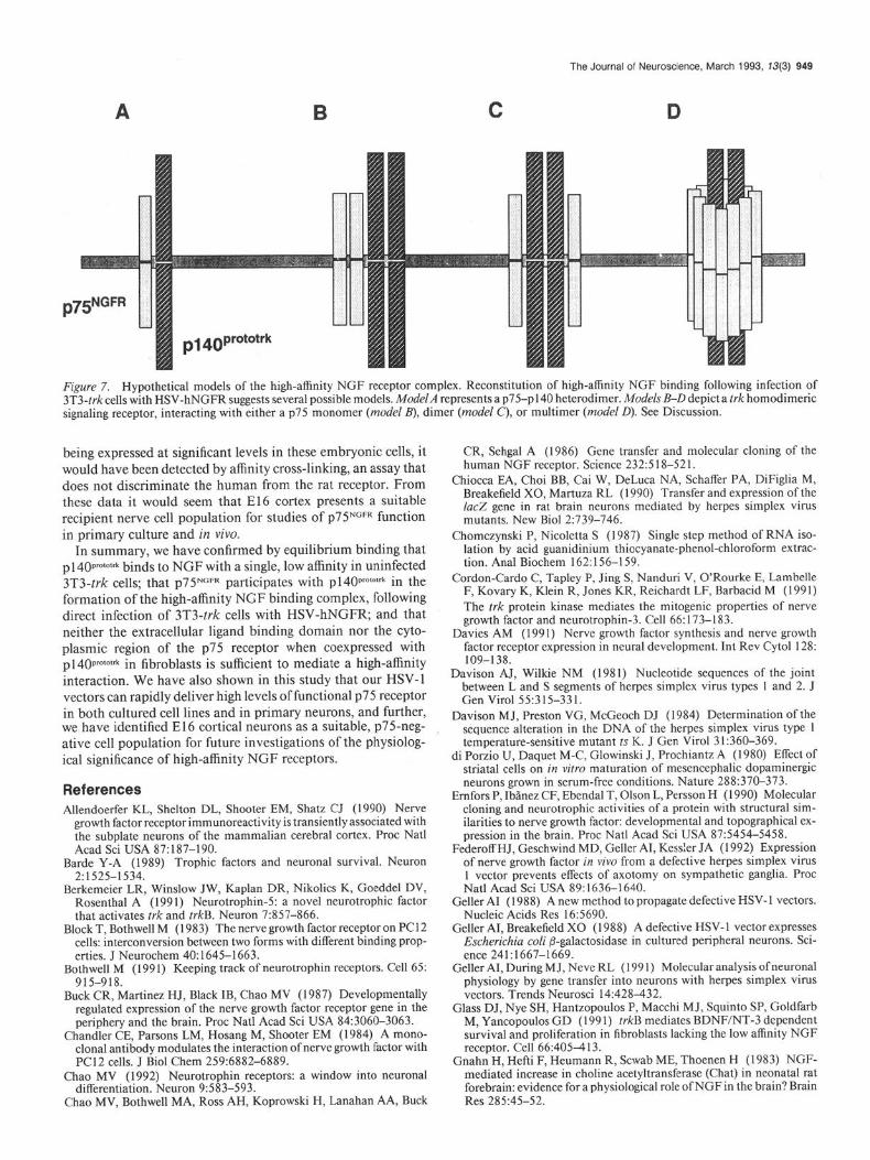
From these observations we can propose several hypothetical models to describe the high-affinity receptor complex. Four of these models are shown in Figure 7. Model A demonstrates a p75NGFR-p 140 prototrk heterodimer. This model assumes that pl40~~~‘~“~ signals as a monomer. Models B-D assume that p 140~ro101rk is activated, like other receptor tyrosine kinases (Ull- rich and Schlessinger, 1990), as a homodimer interacting with ~75~~~~. Whether this interaction requires a ~75~~~~ monomer (model B), dimer (model C), or multimer (model D) is presently unclear. In PC12 cells, ~75~~~~ appears to be in excess of the trk receptor. Furthermore, membrane fusion data has suggested that the stoichiometric ratio of ~75~~~~ to p140pro101rk for the reconstitution of high-affinity binding is about 10: 1 (Hempstead et al., 1991). A trk dimer-p75 multimer (model D) may be consistent with these observations.
An important next step toward the understanding of the phys- iological role of p75NGFR will be to assess the function of this receptor in neurons, rather than in heterologous cultured cell lines. HSV- 1 vectors offer an effective method to introduce cloned genes in nerve cells (Geller, 1988; Geller and Breakefield, 1988; Chiocca et al., 1990; Geller et al., 1991; Federoff et al., 1992). In fact, recombinant HSV-1 vectors provide the only method for direct gene transfer and stable gene expression in postmitotic cells. Here, we have also demonstrated that the HSV-1 vectors employed in this study can faithfully deliver high levels of NGF receptors into neurons in primary culture with minimal cyto- toxicity.
Infection of cortical neurons in culture with HSV-hNGFR (MO1 = 0.25) resulted in high levels of receptor expression as detected by both affinity cross-linking and immunofluorescence. In mock-infected cortical neurons, ~75~~~~ was not detectable, suggesting that endogenous receptor was either expressed at very low levels or not present at this developmental stage in rat cortex. Previous work in feline cortex has demonstrated a tran- sient immunohistochemical staining of p75 in subplate neurons during brain development (Allendoerfer et al., 1990). However, Yan and Johnson (1988) failed to detect ~75 expression in the developing rat cortex. Moreover, if endogenous receptor were
The Journal of Neuroscience, March 1993, 33) 949
Figure 7. Hypothetical models of the high-affinity NGF receptor complex. Reconstitution of high-affinity NGF binding following infection of 3T3-t&cells with HSV-hNGFR suggests several possible models. ModelA represents a p75-~140 heterodimer. Models B-D depict a trk homodimeric signaling receptor, interacting with either a p75 monomer (model B), dimer (model C), or multimer (model 0). See Discussion.
being expressed at significant levels in these embryonic cells, it would have been detected by affinity cross-linking, an assay that does not discriminate the human from the rat receptor. From these data it would seem that El6 cortex presents a suitable recipient nerve cell population for studies of p7YGFR function in primary culture and in vivo.
In summary, we have confirmed by equilibrium binding that p 14Opm101* binds to NGF with a single, low affinity in uninfected 3T3-trk cells; that p7YGFR participates with p140~~~~~‘* in the formation of the high-affinity NCF binding complex, following direct infection of 3T3-trk cells with HSV-hNGFR; and that neither the extracellular ligand binding domain nor the cyto- plasmic region of the ~75 receptor when coexpressed with p140~0101rk in fibroblasts is sufficient to mediate a high-affinity interaction. We have also shown in this study that our HSV-1 vectors can rapidly deliver high levels of functional p7 5 receptor in both cultured cell lines and in primary neurons, and further, we have identified E 16 cortical neurons as a suitable, p75-neg- ative cell population for future investigations of the physiolog- ical significance of high-affinity NGF receptors.
References Allendoerfer KL, Shelton DL, Shooter EM, Shatz CJ (1990) Nerve
growth factor receptor immunoreactivity is transiently associated with the subplate neurons of the mammalian cerebral cortex. Proc Nat1 Acad Sci USA 87:187-190.
Barde Y-A (1989) Trophic factors and neuronal survival. Neuron 2:1525-1534.
Berkemeier LR, Winslow JW, Kaplan DR, Nikolics K, Goeddel DV, Rosenthal A (1991) Neurotrophin-5: a novel neurotrophic factor that activates trk and trkB. Neuron 7:857-866.
Block T, Bothwell M ( 1983) The nerve growth factor receptor on PC1 2 cells: interconversion between two forms with different binding prop- erties. J Neurochem 40: 1645-l 663.
Bothwell M (199 1) Keeping track of neurotrophin receptors. Cell 65: 915-918.
Buck CR, Martinez HJ, Black IB, Chao MV (1987) Developmentally regulated expression of the nerve growth factor receptor gene in the periphery and the brain. Proc Nat1 Acad Sci USA 84:3060-3063.
Chandler CE, Parsons LM, Hosang M, Shooter EM (1984) A mono- clonal antibody modulates the interaction of nerve growth factor with PC12 cells. J Biol Chem 259:6882-6889.
Chao MV (1992) Neurotrophin receptors: a window into neuronal differentiation. Neuron 9:583-593.
Chao MV, Bothwell MA, Ross AH, Koprowski H, Lanahan AA, Buck
CR, Sehgal A (1986) Gene transfer and molecular cloning of the human NGF receptor. Science 233.518-521. -.__- _-..
Chiocca EA, Choi BB, Cai W, DeLuca NA, Schaffer PA, DiFiglia M, Breakefield X0, Martuza RL (1990) Transfer and expression of the IucZ gene in rat brain neurons mediated by herpes simplex virus mutants. New Biol 2:739-746.
Chomczynski P, Nicoletta S (1987) Single step method of RNA iso- lation by acid guanidinium thiocyanate-phenol-chloroform extrac- tion. Anal Biochem 162:156-159.
Cordon-Card0 C, Tapley P, Jing S, Nanduri V, O’Rourke E, Lambelle F, Kovary K, Klein R, Jones KR, Reichardt LF, Barbacid M (199 1) The trk protein kinase mediates the mitogenic properties of nerve growth factor and neurotrophin-3. Cell 66: 173-l 83.
Davies AM (1991) Nerve growth factor synthesis and nerve growth factor receptor expression in neural development. Int Rev Cytol 128: 109-138.
Davison AJ, Wilkie NM (1981) Nucleotide secmences of the ioint between L and S segments of herpes simplex v&us types 1 and 2. J Gen Virol 55:315-331.
Davison MJ, Preston VG, McGeoch DJ (1984) Determination of the sequence alteration in the DNA of the herpes simplex virus type 1 temperature-sensitive mutant ts K. J Gen Virol 3 1:360-369.
di Porzio U, Daquet M-C, Glowinski J, Prochiantz A (1980) Effect of striatal cells on in vitro maturation of mesencephalic dopaminergic neurons grown in serum-free conditions. Nature 288:370-373.
Ernfors P, Ibanez CF, Ebendal T, Olson L, Persson H (1990) Molecular cloning and neurotrophic activities of a protein with structural sim- ilarities to nerve growth factor: developmental and topographical ex- pression in the brain. Proc Nat1 Acad Sci USA 87:5454-5458.
Federoff HJ, Geschwind MD, Geller AI, Kess!er JA (1992) Expression of nerve growth factor in vivo from a defective herpes simplex virus 1 vector prevents effects of axotomy on sympathetic ga&lia. Proc Nat1 Acad Sci USA 89:1636-1640.
Geller AI (1988) A new method to propagate defective HSV-1 vectors. Nucleic Acids Res 16:5690.
Geller AI, Breakefield X0 (1988) A defective HSV- 1 vector expresses Escherichiu coli @-galactosidase in cultured peripheral neurons. Sci- ence 241:1667-1669.
Geller AI, During M J, Neve RL ( 199 1) Molecular analysis of neuronal physiology by gene transfer into ne&ons with herpes simplex virus vectors. Trends Neurosci 14:428-432.
Glass DJ, Nye SH, Hantzopoulos P, Macchi MJ, Squint0 SP, Goldfarb M, Yancopoulos GD (199 1) trkB mediates BDNF/NT-3 dependent survival and proliferation in fibroblasts lacking the low affinity NGF receptor. Cell 66:405413.
Gnahn H, Hefti F, Heumann R, Scwab ME, Thoenen H (1983) NGF- mediated increase in choline acetyltransferase (Chat) in neonatal rat forebrain: evidence for a physiological role of NGF in the brain? Brain Res 285:45-52.
950 Battleman et al. * ~75”~~~~ Defines High-Affinity NGF Binding
Godfrey EW, Shooter EM (1986) Nerve growth factor receptors on chick embryo sympathetic ganglion cells: binding characteristics and development. J Neurosci 6:2543-2550.
Green SH, Greene LA (1986) A single M, approximately 103,000 lz51- p-nerve growth factor-affinity labeled species represents both the low and the high affinity forms of the nerve growth factor receptor. J Biol Chem 261:15316-15326.
Grob P, Berlot CH, Bothwell MA (1983) Affinity labeling and partial purification of nerve growth factor receptors from rat pheochromo- cvtoma and human melanoma cells. Proc Nat1 Acad Sci USA 80: 6819-6823.
Hall CV, Jacob GM, Ringold F, Lee J (1983) Expression and regulation of Escherichia coli IacZ aene fusions in mammalian cells. Mol Appl Genet 2:101-109. -
Hallbook F, Ibanez C, Persson H (199 1) Evolutionary studies of the nerve growth factor family reveal a novel member abundantly ex- pressed in the Xenopus ovary. Neuron 6:845-858.
Hefti F (1986) Nerve growth factor (NGF) promotes survival of septal cholinergic neurons after fimbrial transection. J Neurosci 6:2155- 2162.
Hempstead BL, Schleifer LS, Chao MV (1989) Expression of func- tional nerve growth factor receptors after gene transfer. Science 243: 373-375.
Hemnstead BL, Patil N, Thiel B, Chao MV (1990) Deletion of cy- toplasmic sequences of the nerve growth factor receptor leads to loss of hieh affinitv liaand bindinn. J Biol Chem 17:9595-9598.
Hempstead BL,-M&tin-Zancab, Kaplan DR, Parada LF, Chao MV (1991) High-affinity NGF binding requires coexpression of the trk proto-oncogene and the low-affinity NGF receptor. Nature 350:678- 683.
Herrup K, Shooter EM (1975) Properties of the beta-nerve growth factor receptor in development. J Cell Biol 67: 118-l 25.
Hohn A, Leibrock J, Bailey K, Barde Y-A (1990) Identification and characterization of a novel member of the nerve growth factor/brain- derived neurotrophic family. Nature 344:339-34 1.
Holtzman DM, Li Y, Parada LF, Kinsman S, Chen C-K, Valletta JS, Zhou J, Long JB, Mobley WC (1992) ~140”~ mRNA marks NGF- responsive forebrain neurons: evidence that trk gene expression is induced by NGF. Neuron 9:465478.
Ibanez CF, Ebendal T, Barbany G, Murray RJ, Blundell TL, Persson H (1992) Disruption of the low affinity receptor-binding site in NGF allows neuronal survival and differentiation by binding to the trk gene product. Cell 69:329-34 1.
Johnson D, Lanahan A, Buck CR, Sehgal A, Morgan C, Mercer E, Bothwell M, Chao MV (1986) Expression and structure of the hu- man NGF receutor. Cell 47:545-554.
Kaplan DR, Hempstead BL, Martin-Zanca D, Chao MV, Parada LF (i 99 1 a) The trk proto-oncogene product: a signal transducing recep- tor for nerve growth factor. Science 252:554-557.
Kaplan DR, Martin-Zanca D, Parada LF (1991 b) Tyrosine phos- phorylation and tyrosine kinase activity of trk proto-oncogene product induced bv NGF. Nature 350: 158-l 60.
Klein R, Jing S, Nanduri V, O’Rourke E, Barbacid M (199 la) The trk proto-oncogene encodes a receptor for nerve growth factor. Cell 65:189-197.
Klein R, Nanduri V, Jing S, Lambelle F, Tapley P, Bryant S, Cordon- Cardo C, Jones KR, Reichardt LF, Barbacid M (1991 b) The trkB tyrosine protein kinase is a receptor for brain-derived neurotrophic factor and neurotrovhin-3. Cell 66:395403.
Lambelle F, Klein R, Barbacid M (199 1) trkC, a new member of the trk family of tyrosine kinases, is a receptor for neurotrophin-3. Cell 661967-979.
Landreth GE, Shooter EM (1980) Nerve growth factor receptors on PC1 2 cells: ligand-induced conversion from low- to high-affinity states. Proc Nat1 Acad Sci USA 77:4751-4755.
Leibrock J, Lottspeich AH, Hofer M, Hengerer B, Masiakowski P, Theo- nen H, Barde Y-A ( 1989) Molecular cloning and expression of brain- derived neurotrophic factor. Nature 34 1: 149-152.
Levi-Montalcini R (1987) The nerve growth factor 35 years later. Science 237: 1154-l 162.
Maisonpierre PC, Belluscio L, Squint0 S, Ip NY, Furth ME, Lindsay RM. Yanconoulos GD (1990) Neurotrophin-3: a neurotrophic fac- tor related to NGF and BDNF. Science 247: 1446-145 1.
Matsushima H, Bogenmann E (199 1) Terminal differentiation in neu-
roblastoma cells transfected with the NGF receptor gene when treated with NGF. Prog Clin Biol Res 366:227-233.
McGeoch DJ, Dolan A, Donald S, Brauer DHK (1986) Complete DNA sequence of the short repeat region in the genome of herpes simplex type 1. Nucleic Acids Res 14: 1727-l 745.
Milbrandt J (1986) Nerve growth factor rapidly induces c-fos mRNA in PC12 rat nheochromocvtoma cells. Proc Nat1 Acad Sci USA 83: 47894793. _
Miller RH, Hyman RW (1978) Palindrome and palindrome-like se- auences of heroes simnlex virus DNA. Viroloev 87:3a 1.
Mobley WC, Ruttkowski JL, Tennekoon GI, GFmski J, Buchanan K, Johnston MV (1986) Nerve growth factor increases choline acetyl- transferase activity in developing basal forebrain neurons. Mol Brain Res 1:53-62.
Munson P, Rodbard D (1980) LIGAND: a versatile computerized ap- proach for characterization of ligand binding systems. Anal Biochem 107:220-239.
Oppenheim RW (1989) The neurotrophic theory and naturally oc- curring motoneuron death. Trends Neurosci 12:252-255.
Parker BA, Stark CR (1987) Regulation of simian virus 40 transcrip- tion: analysis of the RNA species present early in infections by virus or viral DNA. J Virol 31:360-369.
Pleasure S, Reddy UR, Venkatakrishnan G, Roy AK, Chen J, Ross AH, Trojanowski JQ, Pleasure DE, Lee V (1990) Introduction of nerve growth factor (NGF) receptors into a medulloblastoma cell line results in expression of high- and low-affinity NGF receptors but not NGF- mediated differentiation. Proc Nat1 Acad Sci USA 87:8496-8500.
Radeke MJ, Misko TP, Hsu C, Herzenberg LA, Shooter EM (1987) Gene transfer and molecular cloning of the rat nerve growth factor receptor. Nature 325:593-597.
Rodriquez-Tebar A, Dechant G, Barde Y-A (1990) Binding of brain derived neurotrophic factor to the nerve growth factor receptor. Neu- ron 4:487-492.
Rosenthal A, Goeddel DV, Nguyen T, Lewis M, Shih A, Laramee GR, Nikolics K, Winslow JW (1990) Primary structure and biological activity of a novel human neurotrophic factor. Neuron 4:767-773.
Ross AH (199 1) Identification of tyrosine kinase Trk as a nerve arowth factor receptor. Cell Regul 2:6851690.
Ross AH. Grob P. Bothwell M. Elder DE. Ernst CS. Marano N. Christ BFD, Slemp CC, Herlyn M, Atkinson B; Koprowski H (1984) Char- acterization of nerve growth factor receptor in neural crest tumors using monoclonal antibodies. Proc Nat1 Acad Sci USA 8 1:668 l-6685.
Sambrook J, Fritsch EF, Maniatis T (1989) Molecular cloning: a lab- oratory manual. Cold Spring Harbor, NY: Cold Spring Harbor Lab- oratory.
Scatchard G (1949) The attraction of proteins for small molecules and ions. Ann NY Acad Sci 52:660.
Schechter AL, Bothwell MA (198 1) Nerve growth factor receptors on PC 12 cells: evidence for two receptor classes with different cytoskel- eta1 associations. Cell 24:867-874.
Schechterson LC, Bothwell MA (1992) Novel roles for neurotrophins are suggested by BDNF and NT-3 mRNA expression in developing neurons. Neuron 9:867-874.
Soppet D, Escandon E, Maragos J, Middlemas DS, Reid SW, Blair J, Burton LE, Stanton BS, Kaplan DR, Hunter T, Nikolics K (1991) The neurotrophic factors brain-derived neurotrophic factor and neurotrophin-3 are ligands for the trkB tyrosine kinase receptor. Cell 65:895-903.
Spaete RR, Frenkel N (1982) The herpes simplex virus amplicon: a new eukaryotic defective-virus cloning-amplifying vector. Cell 30: 295-304.
Squint0 SP, Stitt TN, Aldrich TH, Davis S, Bianco SM, Radziewski C, Glass DJ, Masiakowski P, Furth ME, Valenzuala DM, DiStephano PS, Yancopoulos GD (199 1) trkB encodes a functional receptor for brain-derived neurotrophic factor and neurotrophin-3 but not for nerve growth factor. Cell 65:885-893.
Sutter A, Riopelle RJ, Harris-Warrick RM, Shooter EM (1979) Nerve growth factor receptors. Characterization of two distinct classes of binding sites on chicken embryo sensory ganglia cells. J Biol Chem 25415972-5982.
Thoenen H, Barde Y-A (1980) Physiology of nerve growth factor. Physiol Rev 60: 1284-1334.
Ullrich A, Schlessinger J (1990) Signal transduction by receptors with tyrosine kinase activity. Cell 6 1:203-2 12.
The Journal of Neuroscience, March 1993, 13(3) 951
Welcher AA, Bitler CM, Radeke MJ, Shooter EM (199 1) Nerve growth Yan H, Chao MV (1991) Disruption of cysteine-rich repeats of the factor binding domain of the nerve growth factor receptor. Proc Nat1 p75 nerve growth factor receptor leads to loss of ligand binding. J Acad Sci USA 88:159-163. Biol Chem 266:12099-12104.
Weskamp G, Reichardt LF (1991) Evidence that biological activity Yan Q, Johnson EM (1988) An immunohistochemical study of the ofNGF is mediated through a novel subclass ofhigh affinity receptors. nerve growth factor receptor in developing rats. J Neurosci 8:348 l- Neuron 61649-663. 3498.