Embed Size (px)
Citation preview
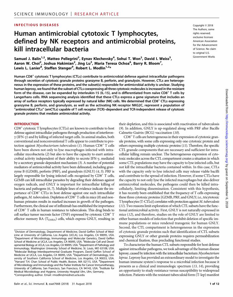
SC I ENCE IMMUNOLOGY | R E S EARCH ART I C L E
I N FECT IOUS D I S EASES
1Division of Dermatology, Department of Medicine, David Geffen School of Med-icine at University of California, Los Angeles (UCLA), Los Angeles, CA 90095, USA.2Department of Microbiology, Immunology and Molecular Genetics, David GeffenSchool of Medicine at UCLA, Los Angeles, CA 90095, USA. 3Molecular Cell and Devel-opmental Biology at UCLA, Los Angeles, CA 90095, USA. 4Department of Pathology andImmunology, Washington University School of Medicine, St. Louis, MO 63108, USA.5Molecular Biology Interdepartmental Graduate Program, David Geffen School ofMedicine at UCLA, Los Angeles, CA 90095, USA. 6Department of Dermatology, Uni-versity of Southern California School of Medicine, Los Angeles, CA 90033, USA.7Harvard T.H. Chan School of Public Health, Boston, MA 02115, USA. 8Departmentof Microbiology and Immunology and the Parker Institute for Cancer Immunotherapy,University of California, San Francisco, San Francisco, CA 94143, USA. 9Institute forMedical Microbiology and Hygiene, University Hospital Ulm, Ulm, Germany.*Corresponding author. Email: [email protected]
Balin et al., Sci. Immunol. 3, eaat7668 (2018) 31 August 2018
Copyright © 2018
The Authors, some
rights reserved;
exclusive licensee
American Association
for the Advancement
of Science. No claim
to original U.S.
Government Works
htD
ownloaded from
Human antimicrobial cytotoxic T lymphocytes,defined by NK receptors and antimicrobial proteins,kill intracellular bacteriaSamuel J. Balin1,2, Matteo Pellegrini3, Eynav Klechevsky4, Sohui T. Won2, David I. Weiss5,Aaron W. Choi2, Joshua Hakimian2, Jing Lu3, Maria Teresa Ochoa6, Barry R. Bloom7,Lewis L. Lanier8, Steffen Stenger9, Robert L. Modlin1,2*
Human CD8+ cytotoxic T lymphocytes (CTLs) contribute to antimicrobial defense against intracellular pathogensthrough secretion of cytotoxic granule proteins granzyme B, perforin, and granulysin. However, CTLs are heteroge-neous in the expression of these proteins, and the subset(s) responsible for antimicrobial activity is unclear. Studyinghuman leprosy,we found that the subset of CTLs coexpressing all three cytotoxicmolecules is increased in the resistantform of the disease, can be expanded by interleukin-15 (IL-15), and is differentiated from naïve CD8+ T cells byLangerhans cells. RNA sequencing analysis identified that these CTLs express a gene signature that includes anarray of surface receptors typically expressed by natural killer (NK) cells. We determined that CD8+ CTLs expressinggranzyme B, perforin, and granulysin, as well as the activating NK receptor NKG2C, represent a population of“antimicrobial CTLs” (amCTLs) capable of T cell receptor (TCR)–dependent and TCR-independent release of cytotoxicgranule proteins that mediate antimicrobial activity.
tp://
by guest on September 6, 2018
imm
unology.sciencemag.org/
INTRODUCTIONCD8+ cytotoxic T lymphocytes (CTLs) are known to contribute to hostdefense against intracellular pathogens through production of interferon-g (IFN-g) and by killing of infected target cells. In animal studies, bothconventional and nonconventional T cells appear to contribute to pro-tection againstMycobacterium tuberculosis (1). Human CD8+ T cellshave been shown not only to lyse macrophages infected with intra-cellular mycobacteria (2) but also to have the capacity to exert antimi-crobial activity independent of their ability to secrete IFN-g, mediatedby a secretory granule-dependent mechanism (3). A number of potentialmediators of antimicrobial activity have been delineated, including gran-zyme B (GZMB), perforin (PRF), and granulysin (GNLY) (4, 5). PRF islargely responsible for lysing infected cells recognized by CD8+ T cells,GZMB can kill intracellular parasites by degrading their defenses againstoxygen radicals, and GNLY is important for intracellular killing ofbacteria and pathogens (6, 7). Multiple lines of evidence indicate the im-portance of CD8+ CTLs in host defense against one such intracellularpathogen,M. tuberculosis. Depletion ofCD8+T cells inmice (8) andnon-human primates results in marked increases in growth of the pathogen.Furthermore, the clinical use of infliximab has established the importanceof CD8+ T cells in human resistance to tuberculosis. This drug binds tocell surface tumor necrosis factor (TNF) expressed by cytotoxic CD8+ Teffector memory RA (TEMRA) cells, which express GNLY, resulting in
their depletion, and this is associated with reactivation of tuberculosis(9). In addition, GNLY is up-regulated along with PRF after BacilleCalmette-Guérin (BCG) vaccination (10).
CD8+T cells are heterogeneous in their expression of cytotoxic gran-ule proteins, with some cells expressing only one cytotoxic protein andothers expressing multiple cytotoxic proteins (11). Therefore, the specificCTL granule components that are necessary and sufficient for intra-cellular killing remain unclear. The heterogeneous expression of cyto-toxicmolecules across the CTL compartment creates a situation inwhichsome CTL populationsmay have the capacity to lyse infected cells, butnot kill the intracellular bacteria contained within. In this case, CTLswith the capacity only to lyse infected cells may release viable bacilliand contribute to the spread of infection. However, if some CTLs havethe capacity to not only lyse the infected macrophages but also deliverantimicrobial molecules, the pathogens could then be killed intra-cellularly, limiting dissemination. Consistent with this hypothesis,it has recently been established that the frequency of T cells expressingthese three cytotoxic proteins [GZMB, PRF, andGNLY; i.e., tricytotoxicT lymphocytes (T-CTLs)] correlateswithprotectionagainstM. tuberculosis(11). Two reasons limit exploration ofwhichCTL subsets have the func-tional antimicrobial activity. First, GNLY is not naturally expressed inmice (12), and therefore, studies on the role of GNLY are limited toeither humanmodels of infection that prohibit deletion of specific im-mune populations or mice rendered transgenic for human GNLY.Second, the CTL compartment is heterogeneous in the expressionof cytotoxic granule proteins such that identification of CTL subsetsexpressing GNLY or other granule proteins requires permeabilizationand chemical fixation, thus precluding functional studies.
To characterize the humanCTL subsets responsible for host defenseagainst intracellular pathogens, we took advantage of the humandiseaseleprosy, causedby infectionwith the intracellular bacteriumMycobacteriumleprae. Leprosy has provided an extraordinarymodel to investigate thehuman immune system’s response to a microbial infection because itpresents as a clinical and immunologic spectrum (13, 14), providingan opportunity to study resistance versus susceptibility to widespreadinfection. Patientswith the resistant tuberculoid form (T-lep)manifest
1 of 12
SC I ENCE IMMUNOLOGY | R E S EARCH ART I C L E
strong cell-mediated immunity (CMI) to the pathogen, skin lesions arefew, and bacilli are rare. However, CMI is absent or diminished in theprogressive lepromatous form (L-lep) (14, 15), skin lesions are numerous,and growth of the pathogen in macrophages is unabated. Clinical pre-sentations of leprosy correlate with the innate and adaptive cytokineprofiles at the site of disease (15, 16). Of relevance, expression of theantimicrobial protein GNLY in leprosy patient lesions has been shownto correlatewith host defense againstM. leprae (5).Here, we addressedwhether distinct CTL subsets differentially contribute to the host an-timicrobial responses against human intracellular pathogens, includ-ing M. leprae.
by guest on Septem
ber 6, 2018http://im
munology.sciencem
ag.org/D
ownloaded from
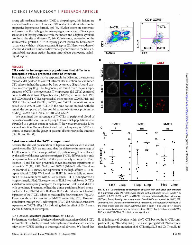
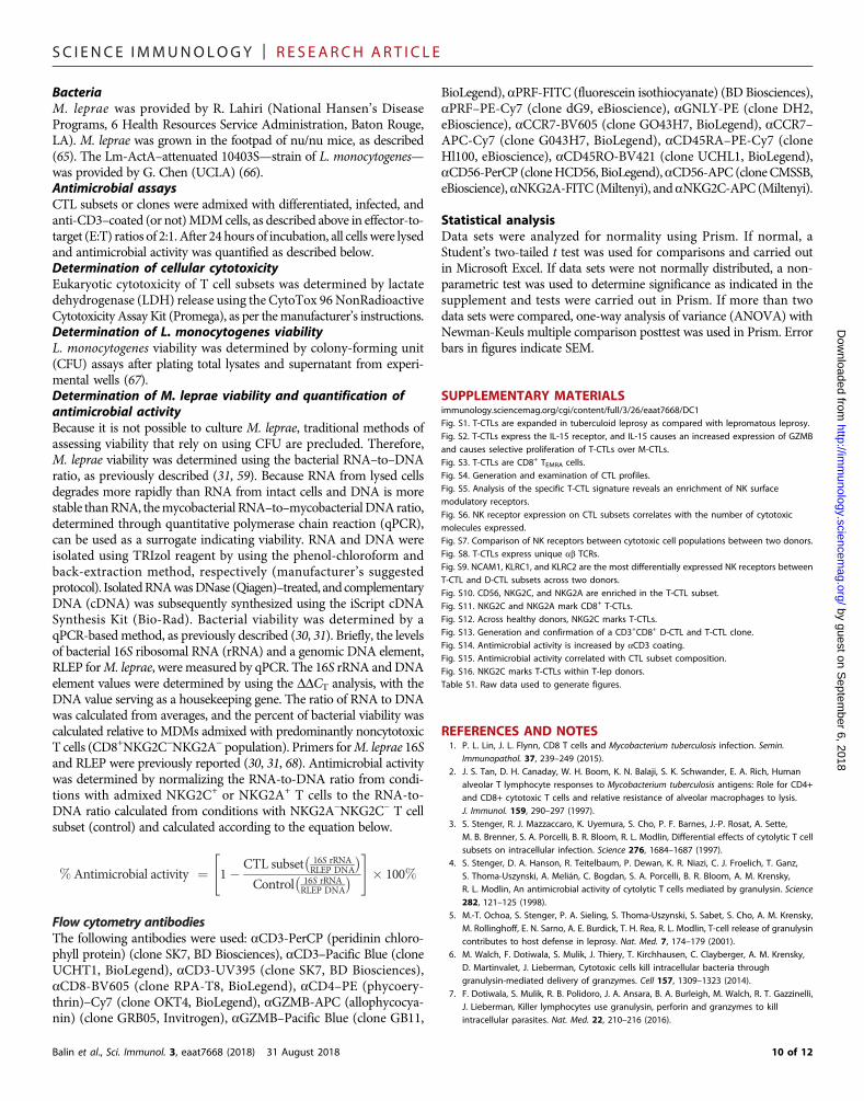
RESULTSCTLs exist in heterogeneous populations that differ in asusceptible versus protected state of infectionTo elucidate which cells may be responsible for delivering the necessarymicrobicidal payload to control intracellular infection, we examinedCTL subsets in healthy donors by flow cytometry (Fig. 1A) and con-focal microscopy (Fig. 1B). In general, we found three major subpo-pulations of CTLs: monocytotoxic T lymphocytes (M-CTLs) expressedonly GZMB; dicytotoxic T lymphocytes (D-CTLs) expressed both PRFand GZMB; and T-CTLs expressed all three proteins GZMB, PRF, andGNLY. The defined M-CTL, D-CTL, and T-CTL populations com-prised 95 to 99% of CD8+ CTLs in the nine donors studied, with theremainder composed of other combinations of cytotoxic proteins in-cluding GZMB and GNLY, or PRF and GNLY.
We examined the percentage of T-CTLs in peripheral blood ofpatients across the spectrum of leprosy to learn which populations wereexpanded to a greater extent in resistant T-lep versus progressive L-lepstates of infection. Our results indicated that the frequency of T-CTLs inleprosy is greatest in the group of patients able to restrict the infection(Fig. 1C and fig. S1).
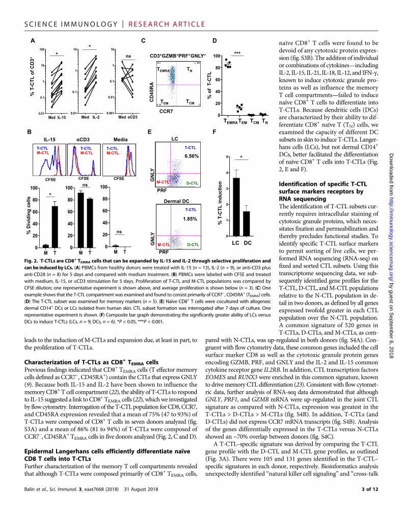
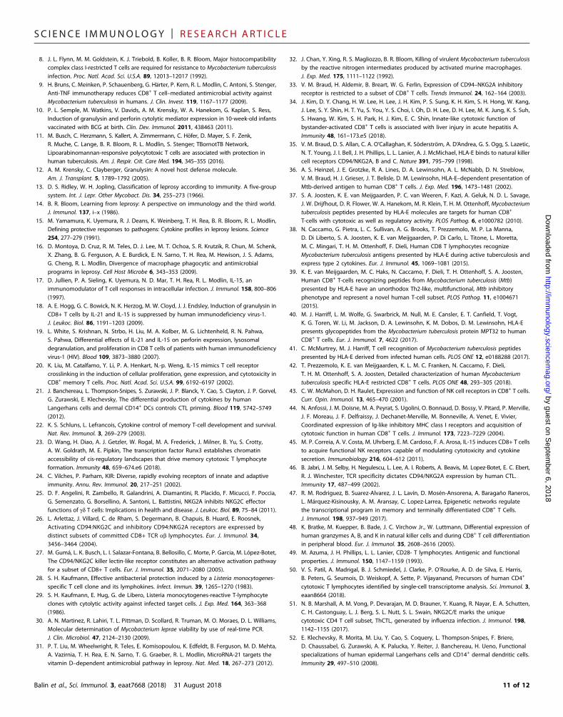
Cytokines control the T-CTL compartmentBecause the clinical presentation of leprosy correlates with distinctcytokine profiles (15), we reasoned that the difference in percentage ofT-CTLs found inT-lep, as opposed toL-lep, patientsmight be explainedby the ability of distinct cytokines to trigger T-CTL differentiation and/or expansion. Interleukin-15 (IL-15) is preferentially expressed in T-leplesions (17) and has been previously shown in separate experiments toinduce GNLY (18), PRF (19–21), and GZMB (20) in T cells. Therefore,we examined CTL subsets for expression of the high-affinity IL-15 re-ceptor subunit IL2Rb. We found that IL2Rb is preferentially expressedon T-CTLs, as compared withM-CTLs andN-CTLs (noncytotoxic Tlymphocytes; fig. S2A). The expression of IL2Rbwas variable onD-CTLssuch thatwe subsequently compared the response of T-CTLs andM-CTLswith cytokines. Treatment of healthy donor peripheral blood mono-nuclear cells (PBMCs) with IL-15 or IL-2 induced an about fivefoldexpansion of the T-CTL subset, asmeasured by flow cytometry (Fig. 2A).There was also an increase in the M-CTL subset (fig. S2B). However,stimulation through the T cell receptor (TCR) did not cause consistentexpansion of T-CTLs (Fig. 2A), indicating that the effect of IL-15 was aspecific function of its receptor.
IL-15 causes selective proliferation of T-CTLsTodeterminewhether IL-15 triggers the specific expansionof theM-CTLand/or T-CTL subsets, we used carboxyfluorescein diacetate succini-midyl ester (CFSE) labeling to interrogate cell division. We found that
Balin et al., Sci. Immunol. 3, eaat7668 (2018) 31 August 2018
IL-15 induced cell division within the T-CTL but not the M-CTL com-partment (Fig. 2B and fig. S2C). IL-15 also up-regulated GZMB expres-sion, leading to the induction ofM-CTLs (fig. S2, B and C). Thus, IL-15
0
1
2
3
4
5
68
10
GNLY PRF GZMB CD3
T-C
TL
D
-CT
L
M-C
TL
GZMB
CD
3
N-CTL
A
B
(%
of
CD
3+)
C
T-lep L-lep T-lep L-lep
0
1
2
3
4
5
6
20
40
0
2
4
6
8
20
40
T-lep L-lep
D-CTL M-CTL
* T-CTL
ns ns
PRF
GN
LY
CD3+ GZMB+
T-CTL
D-CTL M-CTL
Fig. 1. T-CTLs are defined by expression of GZMB, PRF, and GNLY and enrichedin T-lep versus L-lep. (A) PBMCs were stained with antibodies to CD3, GZMB, PRF,and GNLY. T-CTL, D-CTL, M-CTL, and N-CTL cells were delineated by flow cytometry.(B) T cells from a healthy donor were sorted from PBMCs and stained for GNLY, PRF,andGZMB. Cells were examinedby confocalmicroscopy, and representative images ofthe types of cells seen are shown. (C) PBMCs from T-lep (n = 8) or L-lep (n = 7) donorswere examined and compared for thepercentageof CD3+ T cells that coexpressGZMB,PRF, and GNLY (T-CTLs). *P < 0.05. ns, not significant.
2 of 12
SC I ENCE IMMUNOLOGY | R E S EARCH ART I C L E
by guest on Septem
ber 6, 2018http://im
munology.sciencem
ag.org/D
ownloaded from
leads to the induction of M-CTLs and expansion due, at least in part, tothe proliferation of T-CTLs.
Characterization of T-CTLs as CD8+ TEMRA cellsPrevious findings indicated that CD8+ TEMRA cells (T effector memorycells defined as CCR7−, CD45RA+) contain the CTLs that express GNLY(9). Because both IL-15 and IL-2 have been shown to influence thememoryCD8+T cell compartment (22), the ability of T-CTLs to respondto IL-15 suggested a link toCD8+TEMRA cells (22), whichwe investigatedby flow cytometry. Interrogation of the T-CTLpopulation for CD8, CCR7,and CD45RA expression revealed that a mean of 75% (47 to 93%) ofT-CTLs were composed of CD8+ T cells in seven donors analyzed (fig.S3A) and a mean of 86% (81 to 94%) of T-CTLs were composed ofCCR7−, CD45RA+ TEMRA cells in five donors analyzed (Fig. 2, C andD).
Epidermal Langerhans cells efficiently differentiate naïveCD8 T cells into T-CTLsFurther characterization of the memory T cell compartments revealedthat although T-CTLs were composed primarily of CD8+ TEMRA cells,
Balin et al., Sci. Immunol. 3, eaat7668 (2018) 31 August 2018
naïve CD8+ T cells were found to bedevoid of any cytotoxic protein expres-sion (fig. S3B). The addition of individualor combinations of cytokines—includingIL-2, IL-15, IL-21, IL-18, IL-12, and IFN-g,known to induce cytotoxic granule pro-teins as well as influence the memoryT cell compartments—failed to inducenaïve CD8+ T cells to differentiate intoT-CTLs. Because dendritic cells (DCs)are characterized by their ability to dif-ferentiate CD8+ naïve T (TN) cells, weexamined the capacity of different DCsubsets in skin to induce T-CTLs. Langer-hans cells (LCs), but not dermal CD14+
DCs, better facilitated the differentiationof naïve CD8+ T cells into T-CTLs (Fig.2, E and F).
Identification of specific T-CTLsurface markers receptors byRNA sequencingThe identification of T-CTL subsets cur-rently requires intracellular staining ofcytotoxic granule proteins, which neces-sitates fixation and permeabilization andthereby precludes functional studies. Toidentify specific T-CTL surface markersto permit sorting of live cells, we per-formed RNA sequencing (RNA-seq) onfixed and sorted CTL subsets. Using thistranscriptome sequencing data, we sub-sequently identified gene profiles for theT-CTL,D-CTL, andM-CTLpopulationsrelative to the N-CTL population in de-tail in two donors, as defined by all genesexpressed twofold greater in each CTLpopulation over the N-CTL population.A common signature of 520 genes inT-CTLs, D-CTLs, and M-CTLs, as com-
pared with N-CTLs, was up-regulated in both donors (fig. S4A). Con-gruent with flow cytometry data, these common genes included the cellsurface marker CD8 as well as the cytotoxic granule protein genesencoding GZMB, PRF, and GNLY and the IL-2 and IL-15 commoncytokine receptor gene IL2RB. In addition, CTL transcription factorsEOMES and RUNX3 were enriched in this common signature, knownto drivememory CTL differentiation (23). Consistent with flow cytomet-ric data, further analysis of RNA-seq data demonstrated that althoughGNLY, PRF1, and GZMB mRNA were up-regulated in the joint CTLsignature as compared with N-CTLs, expression was greatest in theT-CTLs > D-CTLs > M-CTLs (fig. S4B). In addition, T-CTLs (andD-CTLs) did not express CCR7 mRNA transcripts (fig. S4B). Analysisof the genes differentially expressed in the T-CTLs versus N-CTLsshowed an ~70% overlap between donors (fig. S4C).
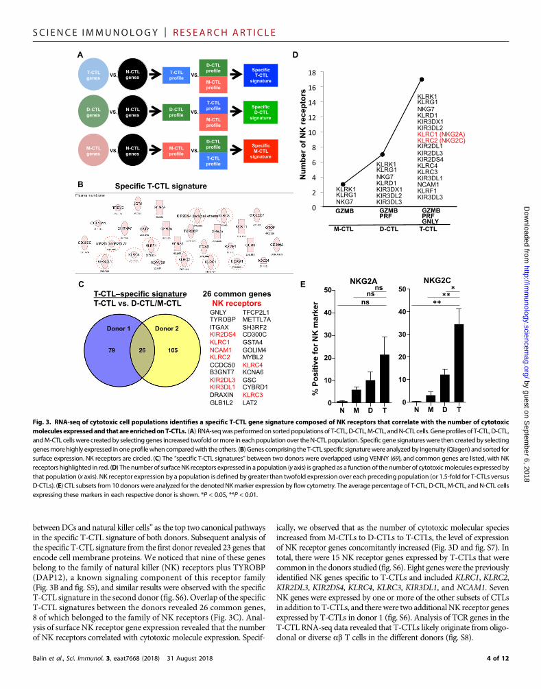
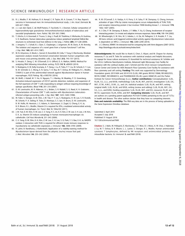
A T-CTL–specific signature was derived by comparing the T-CTLgene profile with the D-CTL and M-CTL gene profiles, as outlined(Fig. 3A). There were 105 and 131 genes identified in the T-CTL–specific signatures in each donor, respectively. Bioinformatics analysisunexpectedly identified “natural killer cell signaling” and “cross-talk
Med IL-150.01
0.1
1
10
100
Med IL-20.01
0.1
1
10
Med CD30.001
0.01
0.1
1
10
0
20
40
60
80
100
M T0
20
40
60
80
100
M T0
20
40
60
80
100
M T0
20
40
60
80
100
CFSE
T-CTL
M-CTL
CFSE
T-CTL
M-CTL
CFSE
T-CTL
M-CTL
A
B
*
% D
ivid
ing
ce
lls
IL-15 CD3 Media
ns
ns
*
*
ns
% T
-CT
L o
f C
D3
+
LC
PRF
GN
LY
6.56%
T-CTL
D-CTL M-CTL
Dermal DC
PRF
GN
LY
1.85%
T-CTL
D-CTL M-CTL
TEMRA TEM TCM TN
% o
f T
-CT
L
***
D C
CCR7
TEMRA TN
TEM TCM
CD3+GZMB+PRF+ GNLY+
CD
45
RA
E F
0
1
2
3
4
5 *
% T
-CT
L i
nd
uc
tio
n
LC DC
Fig. 2. T-CTLs are CD8+ TEMRA cells that can be expanded by IL-15 and IL-2 through selective proliferation andcan be inducedby LCs. (A) PBMCs from healthy donors were treated with IL-15 (n = 13), IL-2 (n = 9), or anti-CD3 plusanti-CD28 (n = 8) for 5 days and compared with medium treatment. (B) PBMCs were labeled with CFSE and treatedwith medium, IL-15, or aCD3 stimulation for 5 days. Proliferation of T-CTL and M-CTL populations was compared byCFSE dilution; one representative experiment is shown above, and average proliferation is shown below (n = 3). (C) Oneexample shows that the T-CTL compartment was examined and found to consist primarily of CCR7−, CD45RA+ (TEMRA) cells.(D) The T-CTL subset was examined for memory markers (n = 5). (E) Naïve CD8+ T cells were cocultured with allogeneicdermal CD14+ DCs or LCs isolated from human skin. CTL subset formation was interrogated after 7 days of culture. Onerepresentative experiment is shown. (F) Composite bar graph demonstrating the significantly greater ability of LCs versusDCs to induce T-CTLs (LCs, n = 9; DCs, n = 6). *P < 0.05, ***P < 0.001.
3 of 12
SC I ENCE IMMUNOLOGY | R E S EARCH ART I C L E
by guest on Septem
ber 6, 2018http://im
munology.sciencem
ag.org/D
ownloaded from
betweenDCs and natural killer cells” as the top two canonical pathwaysin the specific T-CTL signature of both donors. Subsequent analysis ofthe specific T-CTL signature from the first donor revealed 23 genes thatencode cell membrane proteins. We noticed that nine of these genesbelong to the family of natural killer (NK) receptors plus TYROBP(DAP12), a known signaling component of this receptor family(Fig. 3B and fig. S5), and similar results were observed with the specificT-CTL signature in the second donor (fig. S6). Overlap of the specificT-CTL signatures between the donors revealed 26 common genes,8 of which belonged to the family of NK receptors (Fig. 3C). Anal-ysis of surface NK receptor gene expression revealed that the numberof NK receptors correlated with cytotoxic molecule expression. Specif-
Balin et al., Sci. Immunol. 3, eaat7668 (2018) 31 August 2018
ically, we observed that as the number of cytotoxic molecular speciesincreased from M-CTLs to D-CTLs to T-CTLs, the level of expressionof NK receptor genes concomitantly increased (Fig. 3D and fig. S7). Intotal, there were 15 NK receptor genes expressed by T-CTLs that werecommon in the donors studied (fig. S6). Eight genes were the previouslyidentified NK genes specific to T-CTLs and included KLRC1, KLRC2,KIR2DL3, KIR2DS4, KLRC4, KLRC3, KIR3DL1, and NCAM1. SevenNK genes were expressed by one or more of the other subsets of CTLsin addition toT-CTLs, and therewere two additionalNK receptor genesexpressed by T-CTLs in donor 1 (fig. S6). Analysis of TCR genes in theT-CTL RNA-seq data revealed that T-CTLs likely originate from oligo-clonal or diverse ab T cells in the different donors (fig. S8).
N M D T0
10
20
30
40
50NKG2A
ns
A
N M D T0
10
20
30
40
50NKG2C C
T-CTL genes
N-CTL genes VS. VS.
D-CTL profile
M-CTL profile
Specific T-CTL
signature
T-CTL profile
M-CTL genes
M-CTL profile
N-CTL genes
VS. VS.
D-CTL profile
T-CTL profile
Specific M-CTL
signature
D-CTL genes
D-CTL profile
N-CTL genes
VS. VS.
T-CTL profile
M-CTL profile
Specific D-CTL
signature
Specific T-CTL signature B
Donor 1 Donor 2
T-CTL–specific signature T-CTL vs. D-CTL/M-CTL
26 common genes NK receptors
GNLY TYROBP ITGAX KIR2DS4 KLRC1 NCAM1 KLRC2 CCDC50 B3GNT7 KIR2DL3 KIR3DL1 DRAXIN GLB1L2
TFCP2L1 METTL7A SH3RF2 CD300C GSTA4 GOLIM4 MYBL2 KLRC4 KCNA6 GSC CYBRD1 KLRC3 LAT2
GZMB GZMB PRF
Nu
mb
er o
f N
K r
ecep
tors
KLRK1 KLRG1 NKG7 KLRD1 KIR3DX1 KIR3DL2 KLRC1 (NKG2A) KLRC2 (NKG2C) KIR2DL1 KIR2DL3 KIR2DS4 KLRC4 KLRC3 KIR3DL1 NCAM1 KLRF1 KIR3DL3
KLRK1 KLRG1 NKG7 KLRD1 KIR3DX1 KIR3DL2 KIR3DL3
KLRK1 KLRG1 NKG7
M-CTL D-CTL T-CTL
D
E
GZMB PRF GNLY
% P
osi
tive
fo
r N
K m
arke
r
ns ns
Fig. 3. RNA-seq of cytotoxic cell populations identifies a specific T-CTL gene signature composed of NK receptors that correlate with the number of cytotoxicmolecules expressedand that are enrichedonT-CTLs. (A) RNA-seqwas performedon sortedpopulations of T-CTL, D-CTL,M-CTL, andN-CTL cells. Geneprofiles of T-CTL, D-CTL,andM-CTL cellswere createdby selectinggenes increased twofold ormore in eachpopulationover theN-CTL population. Specific gene signatureswere then createdby selectinggenesmore highly expressed in oneprofilewhen comparedwith the others. (B) Genes comprising the T-CTL specific signaturewere analyzedby Ingenuity (Qiagen) and sorted forsurface expression. NK receptors are circled. (C) The “specific T-CTL signatures” between two donors were overlapped using VENNY (69), and common genes are listed, with NKreceptors highlighted in red. (D) The number of surfaceNK receptors expressed in a population (y axis) is graphed as a function of the number of cytotoxicmolecules expressed bythat population (x axis). NK receptor expression by a population is defined by greater than twofold expression over each preceding population (or 1.5-fold for T-CTLs versusD-CTLs). (E) CTL subsets from 10 donors were analyzed for the denoted NKmarker expression by flow cytometry. The average percentage of T-CTL, D-CTL, M-CTL, and N-CTL cellsexpressing these markers in each respective donor is shown. *P < 0.05, **P < 0.01.
4 of 12
SC I ENCE IMMUNOLOGY | R E S EARCH ART I C L E
by guest on Shttp://im
munology.sciencem
ag.org/D
ownloaded from
NK cell surface markers are expressed on T-CTLs and can beused to purify viable T-CTLsRNA-seq identified eight-candidate NK surface markers specific toT-CTLs: KLRC1, KLRC2, KIR2DL3, KIR2DS4, KLRC4, KLRC3,KIR3DL1, and NCAM1 (Fig. 3, C and D). To select genes for furtherstudy, we interrogated these surface markers to determine which hadthe highest differential expression on T-CTLs versus the next most clo-sely related subset, D-CTLs. From the RNA-seq data, three markers—KLRC2 (NKG2C), KLRC1 (NKG2A), and NCAM1 (CD56)—bestdistinguished T-CTLs from D-CTLs (fig. S9). Therefore, we measuredexpressionofNKG2C,NKG2A, andCD56by flow cytometry onT-CTLsfrom healthy donors.
We found that T-CTLs expressed the greatest percentage ofNKG2C+,NKG2A+, and CD56+ cells, as compared with the D-CTL, M-CTL,and N-CTL populations (Fig. 3E and fig. S10), validating the RNA-seqdata. Across 10 donors, ~30% of all T-CTLs were NKG2C+, albeitwith donor-to-donor variability congruent with the known genetic var-iation in NK receptor usage (24). We confirmed that the NK receptor–expressing T-CTLs were in the CD8+ compartment (fig. S11A).Furthermore, we found that NKG2C selected for T-CTLs such that,generally, NKG2C+ cells were more highly enriched for T-CTLs thanNKG2A+ cells and there were few T-CTLs in the NKG2A−NKG2C−
population (figs. S11B and S12, A and B). Hence, NKG2C and NKG2Acan be used as molecular markers to isolate enriched populations ofT-CTLs and investigate their functional capacity, which was previ-ously precluded by the need for intracellular flow cytometry to iden-tify these cells.
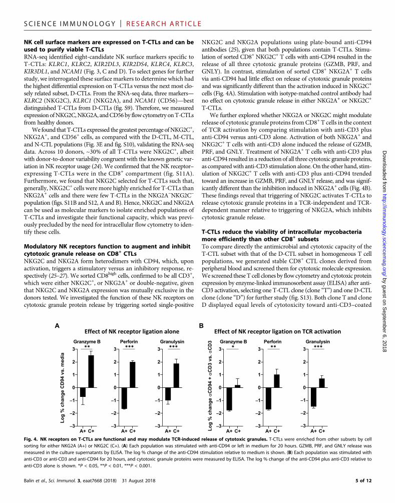
Modulatory NK receptors function to augment and inhibitcytotoxic granule release on CD8+ CTLsNKG2C and NKG2A form heterodimers with CD94, which, uponactivation, triggers a stimulatory versus an inhibitory response, re-spectively (25–27). We sorted CD8high cells, confirmed to be all CD3+,which were either NKG2C+, or NKG2A+ or double-negative, giventhat NKG2C and NKG2A expression was mutually exclusive in thedonors tested. We investigated the function of these NK receptors oncytotoxic granule protein release by triggering sorted single-positive
Balin et al., Sci. Immunol. 3, eaat7668 (2018) 31 August 2018
NKG2C and NKG2A populations using plate-bound anti-CD94antibodies (25), given that both populations contain T-CTLs. Stimu-lation of sorted CD8+ NKG2C+ T cells with anti-CD94 resulted in therelease of all three cytotoxic granule proteins (GZMB, PRF, andGNLY). In contrast, stimulation of sorted CD8+ NKG2A+ T cellsvia anti-CD94 had little effect on release of cytotoxic granule proteinsand was significantly different than the activation induced in NKG2C+
cells (Fig. 4A). Stimulation with isotype-matched control antibody hadno effect on cytotoxic granule release in either NKG2A+ or NKG2C+
T-CTLs.We further explored whether NKG2A or NKG2C might modulate
release of cytotoxic granule proteins fromCD8+ T cells in the contextof TCR activation by comparing stimulation with anti-CD3 plusanti-CD94 versus anti-CD3 alone. Activation of both NKG2A+ andNKG2C+ T cells with anti-CD3 alone induced the release of GZMB,PRF, and GNLY. Treatment of NKG2A+ T cells with anti-CD3 plusanti-CD94 resulted in a reduction of all three cytotoxic granule proteins,as comparedwith anti-CD3 stimulation alone.On the other hand, stim-ulation of NKG2C+ T cells with anti-CD3 plus anti-CD94 trendedtoward an increase in GZMB, PRF, and GNLY release, and was signif-icantly different than the inhibition induced in NKG2A+ cells (Fig. 4B).These findings reveal that triggering of NKG2C activates T-CTLs torelease cytotoxic granule proteins in a TCR-independent and TCR-dependent manner relative to triggering of NKG2A, which inhibitscytotoxic granule release.
T-CTLs reduce the viability of intracellular mycobacteriamore efficiently than other CD8+ subsetsTo compare directly the antimicrobial and cytotoxic capacity of theT-CTL subset with that of the D-CTL subset in homogeneous T cellpopulations, we generated stable CD8+ CTL clones derived fromperipheral blood and screened them for cytotoxic molecule expression.We screened these T cell clones by flow cytometry and cytotoxic proteinexpression by enzyme-linked immunosorbent assay (ELISA) after anti-CD3 activation, selecting one T-CTL clone (clone “T”) and one D-CTLclone (clone “D”) for further study (fig. S13). Both clone T and cloneD displayed equal levels of cytotoxicity toward anti-CD3–coated
eptember 6, 2018
A+ C+–3
–2
–1
0
1
2
3
Granzyme B
A+ C+
0
1
2
3
Perforin
A+ C+–3
–2
–1
0
1
2
3
Granulysin
A+ C+–3
–2
–1
0
1
2
3
Granzyme B
A+ C+–3
–2
–1
0
1
2
3
Perforin
A+ C+–3
–2
–1
0
1
2
3
Granulysin
Lo
g %
ch
an
ge
αC
D94 +
αC
D3
vs
. α
CD
3
Lo
g %
ch
an
ge C
D94 v
s. m
ed
ia
A B
–3
–2
–1
Fig. 4. NK receptors on T-CTLs are functional and may modulate TCR-induced release of cytotoxic granules. T-CTLs were enriched from other subsets by cellsorting for either NKG2A (A+) or NKG2C (C+). (A) Each population was stimulated with anti-CD94 or left in medium for 20 hours. GZMB, PRF, and GNLY release wasmeasured in the culture supernatants by ELISA. The log % change of the anti-CD94 stimulation relative to medium is shown. (B) Each population was stimulated withanti-CD3 or anti-CD3 and anti-CD94 for 20 hours, and cytotoxic granule proteins were measured by ELISA. The log % change of the anti-CD94 plus anti-CD3 relative toanti-CD3 alone is shown. *P < 0.05, **P < 0.01, ***P < 0.001.
5 of 12
SC I ENCE IMMUNOLOGY | R E S EARCH ART I C L E
by guest on Septem
ber 6, 2018http://im
munology.sciencem
ag.org/D
ownloaded from
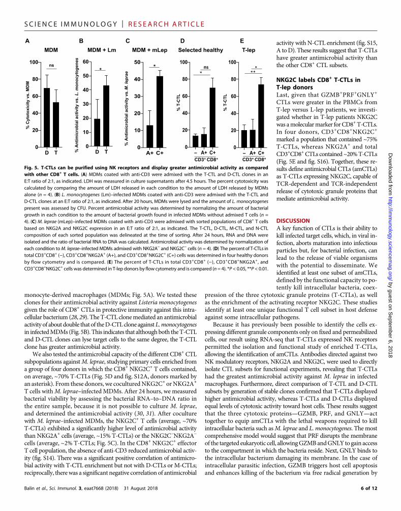
monocyte-derived macrophages (MDMs; Fig. 5A). We tested theseclones for their antimicrobial activity against Listeria monocytogenesgiven the role of CD8+ CTLs in protective immunity against this intra-cellular bacterium (28, 29). The T-CTL clonemediated an antimicrobialactivity of about double that of theD-CTLclone againstL.monocytogenesin infectedMDMs (Fig. 5B). This indicates that although both theT-CTLand D-CTL clones can lyse target cells to the same degree, the T-CTLclone has greater antimicrobial activity.
We also tested the antimicrobial capacity of the different CD8+ CTLsubpopulations againstM. leprae, studying primary cells enriched froma group of four donors in which the CD8+ NKG2C+ T cells contained,on average, ~70% T-CTLs (Fig. 5D and fig. S12A, donors marked byan asterisk). From these donors, we coculturedNKG2C+ orNKG2A+
T cells withM. leprae–infected MDMs. After 24 hours, we measuredbacterial viability by assessing the bacterial RNA–to–DNA ratio inthe entire sample, because it is not possible to culture M. leprae,and determined the antimicrobial activity (30, 31). After coculturewith M. leprae–infected MDMs, the NKG2C+ T cells (average, ~70%T-CTLs) exhibited a significantly higher level of antimicrobial activitythan NKG2A+ cells (average, ~15% T-CTLs) or the NKG2C−NKG2A−
cells (average, ~2% T-CTLs; Fig. 5C). In the CD8+ NKG2C+ effectorT cell population, the absence of anti-CD3 reduced antimicrobial activ-ity (fig. S14). There was a significant positive correlation of antimicro-bial activity with T-CTL enrichment but not with D-CTLs or M-CTLs;reciprocally, there was a significant negative correlation of antimicrobial
Balin et al., Sci. Immunol. 3, eaat7668 (2018) 31 August 2018
activity with N-CTL enrichment (fig. S15,A toD). These results suggest that T-CTLshave greater antimicrobial activity thanthe other CD8+ CTL subsets.
NKG2C labels CD8+ T-CTLs inT-lep donorsLast, given that GZMB+PRF+GNLY+
CTLs were greater in the PBMCs fromT-lep versus L-lep patients, we investi-gated whether in T-lep patients NKG2Cwas amolecularmarker forCD8+T-CTLs.In four donors, CD3+CD8+NKG2C+
marked a population that contained ~75%T-CTLs, whereas NKG2A+ and totalCD3+CD8+CTLs contained ~20%T-CTLs(Fig. 5E and fig. S16). Together, these re-sults define antimicrobial CTLs (amCTLs)as T-CTLs expressing NKG2C, capable ofTCR-dependent and TCR-independentrelease of cytotoxic granule proteins thatmediate antimicrobial activity.
DISCUSSIONA key function of CTLs is their ability tokill infected target cells, which, in viral in-fection, aborts maturation into infectiousparticles but, for bacterial infection, canlead to the release of viable organismswith the potential to disseminate. Weidentified at least one subset of amCTLs,defined by the functional capacity to po-tently kill intracellular bacteria, coex-
pression of the three cytotoxic granule proteins (T-CTLs), as wellas the enrichment of the activating receptor NKG2C. These studiesidentify at least one unique functional T cell subset in host defenseagainst some intracellular pathogens.
Because it has previously been possible to identify the cells ex-pressing different granule components only on fixed and permeabilizedcells, our result using RNA-seq that T-CTLs expressed NK receptorspermitted the isolation and functional study of enriched T-CTLs,allowing the identification of amCTLs. Antibodies directed against twoNK modulatory receptors, NKG2A and NKG2C, were used to directlyisolate CTL subsets for functional experiments, revealing that T-CTLshad the greatest antimicrobial activity against M. leprae in infectedmacrophages. Furthermore, direct comparison of T-CTL and D-CTLsubsets by generation of stable clones confirmed that T-CTLs displayedhigher antimicrobial activity, whereas T-CTLs and D-CTLs displayedequal levels of cytotoxic activity toward host cells. These results suggestthat the three cytotoxic proteins—GZMB, PRF, and GNLY—acttogether to equip amCTLs with the lethal weapons required to killintracellular bacteria such asM. leprae and L. monocytogenes. Themostcomprehensive model would suggest that PRF disrupts the membraneof the targeted eukaryotic cell, allowingGZMBandGNLY to gain accessto the compartment in which the bacteria reside. Next, GNLY binds tothe intracellular bacterium damaging its membrane. In the case ofintracellular parasitic infection, GZMB triggers host cell apoptosisand enhances killing of the bacterium via free radical generation by
CD3+ CD8+
D T0
10
20
30
40
50
60
MDM + Lm
A+ C+0
10
20
30
40
50
MDM + mLep
C
% A
nti
mic
rob
ial a
ctiv
ity
vs. M
. lep
rae
B
D T0
20
40
60
80
100
MDM
ns
% C
yto
toxi
city
vs.
MD
M
A
% A
nti
mic
rob
ial a
ctiv
ity
vs.
L. m
onoc
ytog
enes
– A+ C+0
20
40
60
80
T-lep
E
– A+ C+0
20
40
60
80
100
CD3+ CD8+
Selected healthy
D
% T
-CT
L
% T
-CT
L
100
Fig. 5. T-CTLs can be purified using NK receptors and display greater antimicrobial activity as comparedwith other CD8+ T cells. (A) MDMs coated with anti-CD3 were admixed with the T-CTL and D-CTL clones in anE:T ratio of 2:1, as indicated. LDH was measured in culture supernatants after 4.5 hours. The percent cytotoxicity wascalculated by comparing the amount of LDH released in each condition to the amount of LDH released by MDMsalone (n = 4). (B) L. monocytogenes (Lm)–infected MDMs coated with anti-CD3 were admixed with the T-CTL andD-CTL clones at an E:T ratio of 2:1, as indicated. After 20 hours, MDMs were lysed and the amount of L. monocytogenespresent was assessed by CFU. Percent antimicrobial activity was determined by normalizing the amount of bacterialgrowth in each condition to the amount of bacterial growth found in infected MDMs without admixed T cells (n =4). (C) M. leprae (mLep)–infected MDMs coated with anti-CD3 were admixed with sorted populations of CD8+ T cellsbased on NKG2A and NKG2C expression in an E:T ratio of 2:1, as indicated. The T-CTL, D-CTL, M-CTL, and N-CTLcomposition of each sorted population was delineated at the time of sorting. After 24 hours, RNA and DNA wereisolated and the ratio of bacterial RNA to DNA was calculated. Antimicrobial activity was determined by normalization ofeach condition toM. leprae–infected MDMs admixed with NKG2A− and NKG2C− cells (n = 4). (D) The percent of T-CTLs intotal CD3+CD8+ (−), CD3+CD8+NKG2A+ (A+), and CD3+CD8+NKG2C+ (C+) cells was determined in four healthy donorsby flow cytometry and is compared. (E) The percent of T-CTLs in total CD3+CD8+ (−), CD3+CD8+NKG2A+, andCD3+CD8+NKG2C+ cells was determined in T-lep donors by flow cytometry and is compared (n = 4). *P < 0.05, **P < 0.01.
6 of 12
SC I ENCE IMMUNOLOGY | R E S EARCH ART I C L E
by guest on Septem
ber 6, 2018http://im
munology.sciencem
ag.org/D
ownloaded from
GZMB cleavage of oxidative stress defense proteins (6, 7). Oxidativeradicals, which are known to be microbicidal for many pathogens,may not be sufficient in the amounts produced to kill mycobacteriabecause they are highly resistant to killing by reactive oxygen species(32). Conversely, several mycobacterial proteins that have importantroles in metabolic and biosynthetic pathways have been shown to beGZMB substrates (6). Thus, GNLY may play a more direct role inantimicrobial activity directly through damage to the bacterial cellwall (3, 4) and/or indirectly through permeabilization of the bacterialcell wall, allowing GZMB to enter and cleave bacterial substrates (6).
Through examination of the gene expression signature of CTL sub-sets, we unexpectedly found that the number of cytotoxic granule pro-teins expressed by the CTL subsets correlated with the frequency ofNK receptors, with the most NK receptors found in T-CTLs, followedby D-CTLs, M-CTLs, and N-CTLs, respectively. Previous studies haveimplicated that several surfaceNK receptors havemodulatory functions(activating or inhibitory) of cytolytic activity (25–27, 33). Of the NKreceptors we identified on T-CTLs, five are known to be activating,including NKG2C, and four have been defined as inhibitory, includingNKG2A. Here, we show that these two receptors both marked the T-CTL subset but were, in general, mutually exclusively expressed atthe individual cell level. We found that NKG2C and NKG2A hadopposing effects on CTL function as defined by cytotoxic granule re-lease. Stimulation through NKG2C alone is sufficient to trigger cyto-toxic granule release and had little effect on TCR function, whereasstimulation through NKG2A does not induce cytotoxic granule releaseand inhibits TCR function. Therefore, the expression of NKG2A onsome T-CTLs may serve to negatively regulate their function. In addi-tion, we show that the frequency of NK receptors increased as CTLsexpressed more granule proteins. Together, our data suggest thatCTL antimicrobial activity may be controlled by both TCR ligationand engagement of activating and inhibitory NK receptors such thatthese receptors may act as checkpoints or costimulators to modulateCTL function. The identification of other activating receptors expressedon terminally differentiatedT-CTLs, includingKLRC3,KLRC4,KIR2DS4,and KLRF1, raises the possibility that there may be a spectrum ofother subsets of amCTLs yet to be defined.
The result that NKG2C activation of CD8+ T-CTLs induced cyto-toxic granule release in the absence of anti-CD3 stimulation revealed aTCR- and hence antigen-independentmechanism of T-CTL activation.This finding elucidates an innate-like pathway for activation of this an-timicrobial T cell subpopulation. Activation of CD8+ T cells viaNKG2D, independent of TCR ligation, is thought to contribute to liverinjury in hepatitis A infection (34). NKG2A and NKG2C, when com-plexed with CD94, both bind human leukocyte antigen E (HLA-E)loaded with major histocompatibility complex (MHC) class I leaderpeptides (35).HLA-E–restrictedCD8+T cells that recognizeM. tuberculosishave been reported to vary in their cytotoxic versus immunoregulatorycapacity (36–42) such that examination of the antimicrobial capacity ofthese T cells is now warranted.
In mouse models of bacterial and viral infection, CD8+ T cellsbearing NK receptors have been observed and are thought to representclones of antigen-specific T cells (33). In humans, NK receptor–bearingCD8+ ab T cells have been found to comprise up to 5% of circulatingCD8+ T cells (43) and are typically thought to represent subsets of term-inally differentiatedCD8+memory or effector T cells (44). Furthermore,NK receptor–expressing CD8+ T cells have been shown to have cyto-toxic activity (45, 46). Our result that amCTLs are a subset of TEMRA
cells is consistent with the reported finding that some TEMRA cells also
Balin et al., Sci. Immunol. 3, eaat7668 (2018) 31 August 2018
express NK receptors (47), consistent with the ordered expression ofcytotoxic granule proteins during CD8 T cell differentiation (9, 48, 49).In addition, NK receptors are expressed on a subpopulation of CD4+
T cells (50), which are present at sites of viral infection (51).Several lines of translational evidence point to a key role for
amCTLs in human host defense against intracellular mycobacteria.We reported previously that PRF and GNLY were more stronglyexpressed in T-lep versus L-lep lesions (5). Here, we found that thefrequency of T-CTLs is greater in PBMCs from patients with the self-limited T-lep form of leprosy comparedwith the progressive L-lep formand theT-CTLs inT-lep express the amCTLmarkerNKG2C. Similarly,the frequency of T-CTLs in peripheral blood correlates with protectiveimmunity in tuberculosis (11). Finally, reports showing that the anti–TNF-a biologic infliximab causes reactivation of latent tuberculosis anddepletes CD8+ TEMRA cells, a cell population that we show containsthe T-CTL subset, in vitro and in vivo (9), are compelling examplesof the importance of these cells in resistance to intracellular patho-gens in humans.
Identification of the signals that govern induction and expansion ofthe amCTL compartment is pivotal to the development of strategies tomodulate this response in the prevention and/or treatment of clinicalinfection caused by intracellular pathogens. The finding that T-CTLsconstitutively express high levels of IL2Rb led to the realization thatthese cells are a subset of effector memory T cells that can be expandedby either IL-15 or IL-2. In addition, we found that epidermal LCs, butnot dermal CD14+ DCs, induce naïve CD8+ T cells to differentiate intoT-CTLs, consistent with the known ability of LC to facilitate the differ-entiation of naïve CD8+ T cells into CTLs (52). Together, these resultsreveal a distinct functional capacity for skin LCs and suggest that thetissue microenvironment in which the immune system in the skinencounters pathogens dictates differential immune responses. Thestronger expression of IL-15 in T-lep versus L-lep patientsmay contrib-ute to the induction and expansion of T-CTLs in T-lep patients.
Our characterization of amCTLs, a subset of CD8+ CTLs that canexert antimicrobial activity, as well as establishing the methods to inducethem with LCs and expand them with IL-15 or IL-2, offers newpossibilities for translational application and potential therapy. Itappears that production of IFN-g by CD4+ T cells within granulomasis not sufficient to assure protection against tuberculosis. CD8+ T cellsare localized to the periphery of granulomas in both leprosy (53) andtuberculosis (54, 55). One possible function of these cells, we wouldsuggest, may be to serve as guardians of the granuloma, killing macro-phages and their intracellular pathogens in the process of escaping thegranuloma, thus reducing dissemination of the infection.
Unlike T-CTLs, D-CTLs do not express GNLY, indicating that theantimicrobial protein GNLY is essential for human host defense againstintracellular pathogens. GNLY is not present in mice (12, 56) such thatthe critical function of this antimicrobial protein by CTLs in humanhost defense cannot be directly studied in standard mouse models ofinfection. Consistent with our data, transgenic mice engineered toexpress human GNLY indicated a synergy of these granule proteinsagainst Escherichia coli and intracellular parasites (6, 7). However, itremains difficult to draw conclusions about the differentiation andfunction of transgenic CTL subsets and how they may compare withthe true function of a human immune response. In addition, the ef-fect of NK modulatory receptor function on antigen-specific TCRactivation remains to be fully determined because we were obligedto rely upon anti-CD3 redirected TCR activation given that antigen-specificCD8+CTLTcell clones from leprosy patients have beendifficult
7 of 12
SC I ENCE IMMUNOLOGY | R E S EARCH ART I C L E
Dow
nloaded
to isolate and maintain. Last, our results included two intracellularbacteria (M. leprae and L. monocytogenes) such that it remains to bedetermined the extent to which the present findings will extend to otherbacterial species or intracellular parasites.
In terms of translational application, the association of T-CTLs withhost defense in leprosy and tuberculosis (11) suggests that a combina-tion of the identified T-CTL cell surface receptors described here couldbe used as biomarkers to monitor the role of CD8+ T cells in protectiveimmunity in ongoing vaccine trials. In addition, regulating amCTLfunction by small molecules or cytokines may enhance immuno-therapy. Last, the finding that CTLs contain activating and inhibitorysurfaceNKreceptors suggests that the license to kill a targetmay dependnot only on cognate TCR-MHC recognition but also on the matrix ofinhibitory or activating receptors and their ligands expressed by theCTLs and the target cell. In summary, the present studies have definedand characterized a functional subset of human CD8+ T cells that hasantimicrobial activity against intracellular pathogens, the conditions fortheir differentiation fromTN cells, and a complex framework thatmod-ulates their activity.
by guest on Septem
ber 6, 2018http://im
munology.sciencem
ag.org/ from
MATERIALS AND METHODSStudy designThis study was aimed at identifying the human CD8+ cytotoxic T cellsubsets that may be important for controlling intracellular bacterial in-fection.Humanperipheral bloodwas collected at two locations, either atUniversity of California, LosAngeles (UCLA) or through collaborationswith University of Southern California, as outlined below. Researcherswere not blinded as to the source (healthy donors versus leprosy pa-tients) of the blood samples. All blood collections were carried out withinformed consent according to institutional review board protocols.Patients and healthy donorsLeprosy blood specimens were obtained through collaborations withM.T.O. at the Los Angeles County/University of Southern CaliforniaMedical Center. The diagnosis of leprosy was established by means ofclinical criteria according to Ridley and Jopling (13). Healthy donorsserved as controls and were used for baseline examination. We wereblinded as to the race of the leprosy patients, but on the basis of ep-idemiology of the leprosy patients in Los Angeles, most patients areof Hispanic or Asian descent; a large proportion of healthy donorblood comes from donors in these ethnic/gender categories to bestmatch the population of leprosy donors.Isolation and expansion of CTL subsets from PBMCs andhuman T cells using cytokinesPBMCs were isolated from the peripheral blood of healthy donors, orpatients with a diagnosis of leprosy, using Ficoll-Paque gradients(Amersham Biosciences). T cells were negatively selected by subjectingPBMCs to magnetic bead separation using immunomagnetic negativeselection (EasySep, STEMCELL Technologies) and then cultured inRPMI 1640 with 10% fetal calf serum (FCS; HyClone) with or withoutcytokines for 5, 7, or 12 days. The following cytokines were used: IL-15(15 ng/ml; R&D Systems and a gift from T. Waldman at the NationalInstitutes of Health), 50 nM IL-2 (Chiron), and anti-CD3/CD28microbeads (Dynabeads, Gibco).Confocal microscopyCD3+ T cells were adhered to poly-L-lysine–coated slides (1 mg/ml;Sigma). The cells were surface-stained with anti-human CD3–PacificBlue (clone UCHT1, BioLegend) or isotype-matched control antibody.After fixation and permeabilization (BD Cytofix/Cytoperm, BD Bio-
Balin et al., Sci. Immunol. 3, eaat7668 (2018) 31 August 2018
sciences), the cells were stained intracellularlywith anti-humanGNLY(clone DH4), anti-human PRF (clone dG9, BD Biosciences), and anti-human GZMB (clone GB11, Thermo Fisher Scientific) or isotype-matched control antibodies. Isotype-specific fluorochrome-conjugated(Alexa Fluor 488, 568, and 647) secondary antibodies were incubatedwith the cells, and slides were mounted with ProLong gold antifadereagent and a coverslip. Immunofluorescence imageswere taken using aLeica SP5 microscope at the Advanced Light Microscopy/SpectroscopyCore Facility Laboratory, California NanoSystems Institute, UCLA.Calculating the percentage of CTL subsetsT-CTLs were defined as CD3+ cells coexpressing GZMB, PRF, andGNLY. The %T-CTL of CD3+ T cells was calculated using multicolorflow cytometry by dividing the number of T-CTL events by the totalnumber of CD3+ events. FlowJo software (FlowJo LLC, Ashland, OR)was used to analyze flow cytometry data.Analysis of memory subpopulationsFluorescence-activated cell sorting (FACS) was used to analyze mem-ory subpopulations of T-CTLs. Cells were labeled with combina-tions of CD3, CD8, CCR7, and CD45RA to distinguish between TN,TCM (central memory T cells), TEM (effector memory T cells), andTEMRA cells.T cell proliferationPBMCs were isolated from donors as described above. Cells werelabeled ex vivo with 0.5 mMCFSE (CellTrace, Invitrogen) and culturedwith IL-15 (15 ng/ml), medium, oraCD3/aCD28microbeads. Flow cy-tometrywas usedwithCD3,CD8, CD4,GZMB, PRF,GNLY, andCFSEstaining to interrogate proliferation within the T-CTL, D-CTL,M-CTL,and N-CTL compartments by examining CFSE dilution. FlowJosoftware (FlowJo LLC) was used to analyze flow cytometry data.Priming CTL subsets using skin DCsLCs or dermal CD14+ DCs were isolated from cells that migrated fromskin specimens, as described (52, 57). DCs (live Lin−HLA-DR+) weresorted on the basis of CD1a, CD1c, and CD14 expression. Naïve T cellswere enriched using the Pan-Naïve T Cell Isolation Kit (STEMCELLTechnologies) according to the manufacturer’s protocol and sorted asCCR7+CD45RA+CD8+ cells. T cells were then CFSE-labeled andcultured with allogeneic sorted skin DCs at a DC:T cell ratio of 1:40for 7 days in the presence of IL-7 (10 ng/ml) and IL-2 (10 IU/ml; addedonday 2). CD40L (100ng/ml)was used to activate theDCs, andGZMB,PRF, and GNLY expression was assessed by flow cytometry.Cell sorting of CTL populations and RNA isolation from fixedand sorted cellsRNAwas isolated from fixed sorted cells based on theMARIS (methodfor analyzing RNA following intracellular sorting) protocol, as de-scribed by Hrvatin et al. (58). Briefly, we used FACS to obtain highlypurified populations of T-CTLs, D-CTLs, M-CTLs, and N-CTLsfrom donors based on staining with CD3, GZMB, PRF, and GNLY,as described above. Before sorting, cells were fixed in 2% electronmicroscopy–grade paraformaldehyde (Electron Microscopy Sciences)and permeabilized with 0.5% deoxyribonuclease (DNase)/ribonuclease(RNase)–free saponin (Sigma) to permit intracellular staining. Allstaining and sorting took place inDNase/RNase-free phosphate-bufferedsaline (PBS) supplemented with microbiology-grade bovine serumalbumin (Gemini Bio-Products) in the constant presence of RNasinplus RNase inhibitor (Promega) 1:25 to 1:100 (1:100 for washes and1:25 for staining and sorting). After sorting, RNA was isolated usingthe RecoverAll Total Nucleic Acid Isolation Kit (Ambion), as per themanufacturer’s instructions, with the same modification to theprotocol used as described by Hrvatin et al. (58).
8 of 12
SC I ENCE IMMUNOLOGY | R E S EARCH ART I C L E
by guest on Septem
ber 6, 2018http://im
munology.sciencem
ag.org/D
ownloaded from
RNA-seq of cytotoxic cell populationsSequencing libraries were constructed from RNA using IlluminaTruSeq Stranded Total RNA Sample Prep and sequenced at theClinical Microarray Core at UCLA by single-end sequencing onan Illumina HiSeq3000.Analysis of RNA-seq dataRNA-seq analysis was performed as described (59). Briefly, sequencedreads were demultiplexed and aligned to the human reference genomehg19 (University of California, Santa Cruz) using TopHat (version2.0.6). The HTSeq package was then used to assign uniquely mappedreads to exons and genes using the gene annotation file for buildhg19 from Ensembl to generate raw count data. Data normalizationand differential expression analysis using a negative binomial modelwas performed in the R statistical programming environment usingthe DESeq2 Bioconductor package.Analysis of genes expressed across CTL populations andgeneration of the CTL-specific signaturesTodetermine theM-CTL,D-CTL, andT-CTLgene profiles, we selectedgenes that were expressed twofold or greater in each subset comparedwith N-CTLs. To determine theM-CTL–, D-CTL–, and T-CTL–specificsignatures, we performed three-way comparisons of the gene profiles.For example, to generate the “T-CTL–specific signature,”we comparedthe “T-CTL gene profile” with the “D-CTL gene profile” and “M-CTLgene profile,” selecting genes that were more strongly expressed in theT-CTL profile versus both the D-CTL and M-CTL profiles. For thecomparisons of M-CTLs with either T-CTLs or D-CTLs, we used atwofold cutoff. For comparison of T-CTLs with D-CTLs, a cutoff of1.5-fold was used because of the similarities of these signatures. Thisspecific signaturewas then analyzed by Ingenuity (Qiagen) to sort genesexpressed on the cell membrane as candidate T-CTLmarkers to test forvalidation.Validation of T-CTL markers by flow cytometryOnce identified (as outlined above), surface markers were validated.PBMCs or T cells isolated as described above were interrogated byflow cytometry for expression of CD56, NKG2A, andNKG2C acrossT-CTL, D-CTL, M-CTL, and N-CTL populations.Assessment of NK receptor functionHigh-expressing CD8+ cells exclusively expressing either NKG2C orNKG2A were sorted from PBMCs. Concomitant portions of PBMCswere stained with CD3, CD8, NKG2C, NKG2A, GZMB, PRF, andGNLY, allowing confirmation that the CD8high population was entirelyCD3+ and allowing us to delineate the percentage of T-CTLs, D-CTLs,M-CTLs, andN-CTLs in each sorted population. SortedCD8+NKG2C+
orCD8+NKG2A+ cells were incubated overnight in RPMI 1640 (Gibco)supplemented with 10% AB serum (Sigma), penicillin, streptomycin, andL-glutamine (Gibco), sodium pyruvate (Gibco), 55 mM2-mercaptoethanol(Gibco), 12.5mMHepes (Gibco), 0.5× nonessential amino acids (Gibco),0.25× essential amino acids (Gibco), gentamicin (10 mg/ml; Gibco), 1 nMIL-2 (Chiron), IL-7 (5 ng/ml; BioLegend), and IL-15 (5 ng/ml; R&DSystems). After overnight incubation, cells were stimulated withmediumalone, plate-bound anti-CD94 (100 mg/ml), plate-bound anti-CD20isotype-matched control (100 mg/ml), anti-CD3 microbeads, or anti-CD3 microbeads in the presence of plate-bound anti-CD94 (100 mg/ml).For these experiments, we used a suboptimal dose of anti-CD3 sothat it would be possible to augment and inhibit responses—10%of the manufacturer’s recommended concentration was used asdetermined by titration experiments. After 24 hours of stimulation,GZMB (Abcam), PRF (Abcam), and GNLY (R&D Systems) releasewas measured by ELISA as per the manufacturer’s instructions. The
Balin et al., Sci. Immunol. 3, eaat7668 (2018) 31 August 2018
effect of anti-CD94 versus mediumwas calculated as log {[(aCD94 −medium)/medium] × 100}. The effect of anti-CD94 plus anti-CD3versus anti-CD3 was calculated as log {[(aCD94&aCD3 − aCD3)/medium] × 100}.Generation of clonesT cell clones were generated as previously described (60, 61). Briefly,PBMCs were isolated fromwhole blood of T-lep donors, and DCs weregenerated by CD14+ negative selection without CD16 depletion(STEMCELL Technologies), followed by culture in RPMI 1640 with10% FCS, granulocyte-macrophage colony-stimulating factor (GM-CSF; 800 IU/ml), and IL-4 (1000 IU/ml). On day 5, DCswere harvestedandculturedwithRPMI1640mediumsupplementedwith12.5mMHepes,4 mM L-glutamine, penicillin (100 U/ml), streptomycin (100 mg/ml),50 mM b-mercaptoethanol, and 10% AB human serum and pulsedwithML0049 peptide pool (10 mg/ml) for 2 hours. These cells were thenmixed with CD8+ T cells isolated by negative selection (STEMCELLTechnologies) from the same donor in 96-well U-bottom plates tocreate CD8+ T cell lines. T cell lines were refed or split 1 to 2 every3 to 4 days depending on confluence, using the same medium withIL-7 (5 ng/ml; BioLegend), IL-15 (5 ng/ml; R&D Systems), and 1 nMIL-2 (Chiron). After 2 weeks, all cells were collected and positivelyselected CD8+ T cells were then cloned by limiting dilution in 96-wellU-bottomplates in the presence of 200,000 irradiated (75Gy) allogeneicPBMCs, 40,000 lymphoblastoid cell line (provided by D. Lewinsohn),anti-CD3 (10 ng/ml; cloneOKT3, eBioscience), IL-7 (5 ng/ml), IL-15(5 ng/ml), and 1 nM IL-2. Clones were either split 1 to 2 or refed withfreshmedium and cytokines every 3 to 4 days. After 2 to 4 weeks, clonalpopulations were tested for expression of GZMB, PRF, and GNLY byflow cytometry.Generation of target cellsTo assess the antimicrobial activity directed toward M. leprae– orL. monocytogenes–infected cells, we used infected MDMs as targets.MDMs are known to have a high capacity to engulf and becomeinfected by mycobacteria but a low intrinsic ability to kill withoutthe presence of IFN-g (62), making them ideal targets for our assays.Briefly, 5 to 7 days before infection with bacteria, monocytes were pur-ified from peripheral blood using Ficoll-Paque gradients (AmershamBiosciences) followed by negative selection of CD14+ CD16+ cells usingthe EasySepHumanMonocyte Enrichment Kit without CD16Depletion(STEMCELL Technologies). Negatively selected cells were cultured for5 to 7 days in RPMI 1640 with 10% superlow immunoglobulin FCSwithout antibiotics in the presence of M-CSF to differentiate intoMDMs, as previously described (62–64). After 5 to 7 days, MDMs wereinfected withM. leprae at amultiplicity of infection (MOI) of 10 or withL. monocytogenes at an MOI of 1.Coating MDMs with aCD3Because isolated effector CTL subsets were of varying TCR specificities,we coated MDM target cells with aCD3 (20 ng/ml) by incubating withthis antibody for 15 min before admixing effector cells, as previouslydescribed (7, 26). Briefly, 24 hours after infection with bacteria, MDMswere washed and then admixedwith aCD3 (20 ng/ml) in completeme-dium. Cells were subsequently washed and were then ready to bemixedwith effector cells.Cell sorting of viable cytotoxic cell populationsFACSwas used to purifyCD8+T-CTLs fromother populations of CTLsby labeling cells with aCD3, aCD8, and combinations of the identifiedsurface markers that were previously validated. To sort, staining wasperformed in sterile PBS with 10% FCS, and sorting was performedin complete medium.
9 of 12
SC I ENCE IMMUNOLOGY | R E S EARCH ART I C L E
by guest on Septem
ber 6, 2018http://im
munology.sciencem
ag.org/D
ownloaded from
BacteriaM. leprae was provided by R. Lahiri (National Hansen’s DiseasePrograms, 6 Health Resources Service Administration, Baton Rouge,LA). M. leprae was grown in the footpad of nu/nu mice, as described(65). The Lm-ActA–attenuated 10403S—strain of L. monocytogenes—was provided by G. Chen (UCLA) (66).Antimicrobial assaysCTL subsets or clones were admixed with differentiated, infected, andanti-CD3–coated (or not)MDMcells, as described above in effector-to-target (E:T) ratios of 2:1. After 24 hours of incubation, all cells were lysedand antimicrobial activity was quantified as described below.Determination of cellular cytotoxicityEukaryotic cytotoxicity of T cell subsets was determined by lactatedehydrogenase (LDH) release using the CytoTox 96NonRadioactiveCytotoxicity Assay Kit (Promega), as per themanufacturer’s instructions.Determination of L. monocytogenes viabilityL. monocytogenes viability was determined by colony-forming unit(CFU) assays after plating total lysates and supernatant from experi-mental wells (67).Determination of M. leprae viability and quantification ofantimicrobial activityBecause it is not possible to culture M. leprae, traditional methods ofassessing viability that rely on using CFU are precluded. Therefore,M. leprae viability was determined using the bacterial RNA–to–DNAratio, as previously described (31, 59). Because RNA from lysed cellsdegrades more rapidly than RNA from intact cells and DNA is morestable thanRNA, themycobacterial RNA–to–mycobacterialDNAratio,determined through quantitative polymerase chain reaction (qPCR),can be used as a surrogate indicating viability. RNA and DNA wereisolated using TRIzol reagent by using the phenol-chloroform andback-extraction method, respectively (manufacturer’s suggestedprotocol). IsolatedRNAwasDNase (Qiagen)–treated, and complementaryDNA (cDNA) was subsequently synthesized using the iScript cDNASynthesis Kit (Bio-Rad). Bacterial viability was determined by aqPCR-basedmethod, as previously described (30, 31). Briefly, the levelsof bacterial 16S ribosomal RNA (rRNA) and a genomic DNA element,RLEP forM. leprae, weremeasured by qPCR. The 16S rRNA andDNAelement values were determined by using the DDCT analysis, with theDNA value serving as a housekeeping gene. The ratio of RNA to DNAwas calculated from averages, and the percent of bacterial viability wascalculated relative to MDMs admixed with predominantly noncytotoxicT cells (CD8+NKG2C−NKG2A− population). Primers forM. leprae 16Sand RLEP were previously reported (30, 31, 68). Antimicrobial activitywas determined by normalizing the RNA-to-DNA ratio from condi-tions with admixed NKG2C+ or NKG2A+ T cells to the RNA-to-DNA ratio calculated from conditions with NKG2A−NKG2C− T cellsubset (control) and calculated according to the equation below.
%Antimicrobial activity ¼ 1� CTL subset 16S rRNARLEP DNA
� �Control 16S rRNA
RLEP DNA
� �" #
� 100%
Flow cytometry antibodiesThe following antibodies were used: aCD3-PerCP (peridinin chloro-phyll protein) (clone SK7, BD Biosciences), aCD3–Pacific Blue (cloneUCHT1, BioLegend), aCD3-UV395 (clone SK7, BD Biosciences),aCD8-BV605 (clone RPA-T8, BioLegend), aCD4–PE (phycoery-thrin)–Cy7 (clone OKT4, BioLegend), aGZMB-APC (allophycocya-nin) (clone GRB05, Invitrogen), aGZMB–Pacific Blue (clone GB11,
Balin et al., Sci. Immunol. 3, eaat7668 (2018) 31 August 2018
BioLegend), aPRF-FITC (fluorescein isothiocyanate) (BD Biosciences),aPRF–PE-Cy7 (clone dG9, eBioscience), aGNLY-PE (clone DH2,eBioscience), aCCR7-BV605 (clone GO43H7, BioLegend), aCCR7–APC-Cy7 (clone G043H7, BioLegend), aCD45RA–PE-Cy7 (cloneHl100, eBioscience), aCD45RO-BV421 (clone UCHL1, BioLegend),aCD56-PerCP (cloneHCD56, BioLegend),aCD56-APC (cloneCMSSB,eBioscience),aNKG2A-FITC (Miltenyi), andaNKG2C-APC (Miltenyi).
Statistical analysisData sets were analyzed for normality using Prism. If normal, aStudent’s two-tailed t test was used for comparisons and carried outin Microsoft Excel. If data sets were not normally distributed, a non-parametric test was used to determine significance as indicated in thesupplement and tests were carried out in Prism. If more than twodata sets were compared, one-way analysis of variance (ANOVA) withNewman-Keuls multiple comparison posttest was used in Prism. Errorbars in figures indicate SEM.
SUPPLEMENTARY MATERIALSimmunology.sciencemag.org/cgi/content/full/3/26/eaat7668/DC1Fig. S1. T-CTLs are expanded in tuberculoid leprosy as compared with lepromatous leprosy.Fig. S2. T-CTLs express the IL-15 receptor, and IL-15 causes an increased expression of GZMBand causes selective proliferation of T-CTLs over M-CTLs.Fig. S3. T-CTLs are CD8+ TEMRA cells.Fig. S4. Generation and examination of CTL profiles.Fig. S5. Analysis of the specific T-CTL signature reveals an enrichment of NK surfacemodulatory receptors.Fig. S6. NK receptor expression on CTL subsets correlates with the number of cytotoxicmolecules expressed.Fig. S7. Comparison of NK receptors between cytotoxic cell populations between two donors.Fig. S8. T-CTLs express unique ab TCRs.Fig. S9. NCAM1, KLRC1, and KLRC2 are the most differentially expressed NK receptors betweenT-CTL and D-CTL subsets across two donors.Fig. S10. CD56, NKG2C, and NKG2A are enriched in the T-CTL subset.Fig. S11. NKG2C and NKG2A mark CD8+ T-CTLs.Fig. S12. Across healthy donors, NKG2C marks T-CTLs.Fig. S13. Generation and confirmation of a CD3+CD8+ D-CTL and T-CTL clone.Fig. S14. Antimicrobial activity is increased by aCD3 coating.Fig. S15. Antimicrobial activity correlated with CTL subset composition.Fig. S16. NKG2C marks T-CTLs within T-lep donors.Table S1. Raw data used to generate figures.
REFERENCES AND NOTES1. P. L. Lin, J. L. Flynn, CD8 T cells and Mycobacterium tuberculosis infection. Semin.
Immunopathol. 37, 239–249 (2015).2. J. S. Tan, D. H. Canaday, W. H. Boom, K. N. Balaji, S. K. Schwander, E. A. Rich, Human
alveolar T lymphocyte responses to Mycobacterium tuberculosis antigens: Role for CD4+and CD8+ cytotoxic T cells and relative resistance of alveolar macrophages to lysis.J. Immunol. 159, 290–297 (1997).
3. S. Stenger, R. J. Mazzaccaro, K. Uyemura, S. Cho, P. F. Barnes, J.-P. Rosat, A. Sette,M. B. Brenner, S. A. Porcelli, B. R. Bloom, R. L. Modlin, Differential effects of cytolytic T cellsubsets on intracellular infection. Science 276, 1684–1687 (1997).
4. S. Stenger, D. A. Hanson, R. Teitelbaum, P. Dewan, K. R. Niazi, C. J. Froelich, T. Ganz,S. Thoma-Uszynski, A. Melián, C. Bogdan, S. A. Porcelli, B. R. Bloom, A. M. Krensky,R. L. Modlin, An antimicrobial activity of cytolytic T cells mediated by granulysin. Science282, 121–125 (1998).
5. M.-T. Ochoa, S. Stenger, P. A. Sieling, S. Thoma-Uszynski, S. Sabet, S. Cho, A. M. Krensky,M. Rollinghoff, E. N. Sarno, A. E. Burdick, T. H. Rea, R. L. Modlin, T-cell release of granulysincontributes to host defense in leprosy. Nat. Med. 7, 174–179 (2001).
6. M. Walch, F. Dotiwala, S. Mulik, J. Thiery, T. Kirchhausen, C. Clayberger, A. M. Krensky,D. Martinvalet, J. Lieberman, Cytotoxic cells kill intracellular bacteria throughgranulysin-mediated delivery of granzymes. Cell 157, 1309–1323 (2014).
7. F. Dotiwala, S. Mulik, R. B. Polidoro, J. A. Ansara, B. A. Burleigh, M. Walch, R. T. Gazzinelli,J. Lieberman, Killer lymphocytes use granulysin, perforin and granzymes to killintracellular parasites. Nat. Med. 22, 210–216 (2016).
10 of 12
SC I ENCE IMMUNOLOGY | R E S EARCH ART I C L E
by guest on Septem
ber 6, 2018http://im
munology.sciencem
ag.org/D
ownloaded from
8. J. L. Flynn, M. M. Goldstein, K. J. Triebold, B. Koller, B. R. Bloom, Major histocompatibilitycomplex class I-restricted T cells are required for resistance to Mycobacterium tuberculosisinfection. Proc. Natl. Acad. Sci. U.S.A. 89, 12013–12017 (1992).
9. H. Bruns, C. Meinken, P. Schauenberg, G. Härter, P. Kern, R. L. Modlin, C. Antoni, S. Stenger,Anti-TNF immunotherapy reduces CD8+ T cell–mediated antimicrobial activity againstMycobacterium tuberculosis in humans. J. Clin. Invest. 119, 1167–1177 (2009).
10. P. L. Semple, M. Watkins, V. Davids, A. M. Krensky, W. A. Hanekom, G. Kaplan, S. Ress,Induction of granulysin and perforin cytolytic mediator expression in 10-week-old infantsvaccinated with BCG at birth. Clin. Dev. Immunol. 2011, 438463 (2011).
11. M. Busch, C. Herzmann, S. Kallert, A. Zimmermann, C. Höfer, D. Mayer, S. F. Zenk,R. Muche, C. Lange, B. R. Bloom, R. L. Modlin, S. Stenger; TBornotTB Network,Lipoarabinomannan-responsive polycytotoxic T cells are associated with protection inhuman tuberculosis. Am. J. Respir. Crit. Care Med. 194, 345–355 (2016).
12. A. M. Krensky, C. Clayberger, Granulysin: A novel host defense molecule.Am. J. Transplant. 5, 1789–1792 (2005).
13. D. S. Ridley, W. H. Jopling, Classification of leprosy according to immunity. A five-groupsystem. Int. J. Lepr. Other Mycobact. Dis. 34, 255–273 (1966).
14. B. R. Bloom, Learning from leprosy: A perspective on immunology and the third world.J. Immunol. 137, i–x (1986).
15. M. Yamamura, K. Uyemura, R. J. Deans, K. Weinberg, T. H. Rea, B. R. Bloom, R. L. Modlin,Defining protective responses to pathogens: Cytokine profiles in leprosy lesions. Science254, 277–279 (1991).
16. D. Montoya, D. Cruz, R. M. Teles, D. J. Lee, M. T. Ochoa, S. R. Krutzik, R. Chun, M. Schenk,X. Zhang, B. G. Ferguson, A. E. Burdick, E. N. Sarno, T. H. Rea, M. Hewison, J. S. Adams,G. Cheng, R. L. Modlin, Divergence of macrophage phagocytic and antimicrobialprograms in leprosy. Cell Host Microbe 6, 343–353 (2009).
17. D. Jullien, P. A. Sieling, K. Uyemura, N. D. Mar, T. H. Rea, R. L. Modlin, IL-15, animmunomodulator of T cell responses in intracellular infection. J. Immunol. 158, 800–806(1997).
18. A. E. Hogg, G. C. Bowick, N. K. Herzog, M. W. Cloyd, J. J. Endsley, Induction of granulysin inCD8+ T cells by IL-21 and IL-15 is suppressed by human immunodeficiency virus-1.J. Leukoc. Biol. 86, 1191–1203 (2009).
19. L. White, S. Krishnan, N. Strbo, H. Liu, M. A. Kolber, M. G. Lichtenheld, R. N. Pahwa,S. Pahwa, Differential effects of IL-21 and IL-15 on perforin expression, lysosomaldegranulation, and proliferation in CD8 T cells of patients with human immunodeficiencyvirus-1 (HIV). Blood 109, 3873–3880 (2007).
20. K. Liu, M. Catalfamo, Y. Li, P. A. Henkart, N.-p. Weng, IL-15 mimics T cell receptorcrosslinking in the induction of cellular proliferation, gene expression, and cytotoxicity inCD8+ memory T cells. Proc. Natl. Acad. Sci. U.S.A. 99, 6192–6197 (2002).
21. J. Banchereau, L. Thompson-Snipes, S. Zurawski, J. P. Blanck, Y. Cao, S. Clayton, J. P. Gorvel,G. Zurawski, E. Klechevsky, The differential production of cytokines by humanLangerhans cells and dermal CD14+ DCs controls CTL priming. Blood 119, 5742–5749(2012).
22. K. S. Schluns, L. Lefrancois, Cytokine control of memory T-cell development and survival.Nat. Rev. Immunol. 3, 269–279 (2003).
23. D. Wang, H. Diao, A. J. Getzler, W. Rogal, M. A. Frederick, J. Milner, B. Yu, S. Crotty,A. W. Goldrath, M. E. Pipkin, The transcription factor Runx3 establishes chromatinaccessibility of cis-regulatory landscapes that drive memory cytotoxic T lymphocyteformation. Immunity 48, 659–674.e6 (2018).
24. C. Vilches, P. Parham, KIR: Diverse, rapidly evolving receptors of innate and adaptiveimmunity. Annu. Rev. Immunol. 20, 217–251 (2002).
25. D. F. Angelini, R. Zambello, R. Galandrini, A. Diamantini, R. Placido, F. Micucci, F. Poccia,G. Semenzato, G. Borsellino, A. Santoni, L. Battistini, NKG2A inhibits NKG2C effectorfunctions of gd T cells: Implications in health and disease. J. Leukoc. Biol. 89, 75–84 (2011).
26. L. Arlettaz, J. Villard, C. de Rham, S. Degermann, B. Chapuis, B. Huard, E. Roosnek,Activating CD94:NKG2C and inhibitory CD94:NKG2A receptors are expressed bydistinct subsets of committed CD8+ TCR ab lymphocytes. Eur. J. Immunol. 34,3456–3464 (2004).
27. M. Gumá, L. K. Busch, L. I. Salazar-Fontana, B. Bellosillo, C. Morte, P. Garcia, M. López-Botet,The CD94/NKG2C killer lectin-like receptor constitutes an alternative activation pathwayfor a subset of CD8+ T cells. Eur. J. Immunol. 35, 2071–2080 (2005).
28. S. H. Kaufmann, Effective antibacterial protection induced by a Listeria monocytogenes-specific T cell clone and its lymphokines. Infect. Immun. 39, 1265–1270 (1983).
29. S. H. Kaufmann, E. Hug, G. de Libero, Listeria monocytogenes-reactive T-lymphocyteclones with cytolytic activity against infected target cells. J. Exp. Med. 164, 363–368(1986).
30. A. N. Martinez, R. Lahiri, T. L. Pittman, D. Scollard, R. Truman, M. O. Moraes, D. L. Williams,Molecular determination of Mycobacterium leprae viability by use of real-time PCR.J. Clin. Microbiol. 47, 2124–2130 (2009).
31. P. T. Liu, M. Wheelwright, R. Teles, E. Komisopoulou, K. Edfeldt, B. Ferguson, M. D. Mehta,A. Vazirnia, T. H. Rea, E. N. Sarno, T. G. Graeber, R. L. Modlin, MicroRNA-21 targets thevitamin D–dependent antimicrobial pathway in leprosy. Nat. Med. 18, 267–273 (2012).
Balin et al., Sci. Immunol. 3, eaat7668 (2018) 31 August 2018
32. J. Chan, Y. Xing, R. S. Magliozzo, B. R. Bloom, Killing of virulent Mycobacterium tuberculosisby the reactive nitrogen intermediates produced by activated murine macrophages.J. Exp. Med. 175, 1111–1122 (1992).
33. V. M. Braud, H. Aldemir, B. Breart, W. G. Ferlin, Expression of CD94–NKG2A inhibitoryreceptor is restricted to a subset of CD8+ T cells. Trends Immunol. 24, 162–164 (2003).
34. J. Kim, D. Y. Chang, H. W. Lee, H. Lee, J. H. Kim, P. S. Sung, K. H. Kim, S. H. Hong, W. Kang,J. Lee, S. Y. Shin, H. T. Yu, S. You, Y. S. Choi, I. Oh, D. H. Lee, D. H. Lee, M. K. Jung, K. S. Suh,S. Hwang, W. Kim, S. H. Park, H. J. Kim, E. C. Shin, Innate-like cytotoxic function ofbystander-activated CD8+ T cells is associated with liver injury in acute hepatitis A.Immunity 48, 161–173.e5 (2018).
35. V. M. Braud, D. S. Allan, C. A. O’Callaghan, K. Söderström, A. D’Andrea, G. S. Ogg, S. Lazetic,N. T. Young, J. I. Bell, J. H. Phillips, L. L. Lanier, A. J. McMichael, HLA-E binds to natural killercell receptors CD94/NKG2A, B and C. Nature 391, 795–799 (1998).
36. A. S. Heinzel, J. E. Grotzke, R. A. Lines, D. A. Lewinsohn, A. L. McNabb, D. N. Streblow,V. M. Braud, H. J. Grieser, J. T. Belisle, D. M. Lewinsohn, HLA-E–dependent presentation ofMtb-derived antigen to human CD8+ T cells. J. Exp. Med. 196, 1473–1481 (2002).
37. S. A. Joosten, K. E. van Meijgaarden, P. C. van Weeren, F. Kazi, A. Geluk, N. D. L. Savage,J. W. Drijfhout, D. R. Flower, W. A. Hanekom, M. R. Klein, T. H. M. Ottenhoff, Mycobacteriumtuberculosis peptides presented by HLA-E molecules are targets for human CD8+
T-cells with cytotoxic as well as regulatory activity. PLOS Pathog. 6, e1000782 (2010).38. N. Caccamo, G. Pietra, L. C. Sullivan, A. G. Brooks, T. Prezzemolo, M. P. La Manna,
D. Di Liberto, S. A. Joosten, K. E. van Meijgaarden, P. Di Carlo, L. Titone, L. Moretta,M. C. Mingari, T. H. M. Ottenhoff, F. Dieli, Human CD8 T lymphocytes recognizeMycobacterium tuberculosis antigens presented by HLA-E during active tuberculosis andexpress type 2 cytokines. Eur. J. Immunol. 45, 1069–1081 (2015).
39. K. E. van Meijgaarden, M. C. Haks, N. Caccamo, F. Dieli, T. H. Ottenhoff, S. A. Joosten,Human CD8+ T-cells recognizing peptides from Mycobacterium tuberculosis (Mtb)presented by HLA-E have an unorthodox Th2-like, multifunctional, Mtb inhibitoryphenotype and represent a novel human T-cell subset. PLOS Pathog. 11, e1004671(2015).
40. M. J. Harriff, L. M. Wolfe, G. Swarbrick, M. Null, M. E. Cansler, E. T. Canfield, T. Vogt,K. G. Toren, W. Li, M. Jackson, D. A. Lewinsohn, K. M. Dobos, D. M. Lewinsohn, HLA-Epresents glycopeptides from the Mycobacterium tuberculosis protein MPT32 to humanCD8+ T cells. Eur. J. Immunol. 7, 4622 (2017).
41. C. McMurtrey, M. J. Harriff, T cell recognition of Mycobacterium tuberculosis peptidespresented by HLA-E derived from infected human cells. PLOS ONE 12, e0188288 (2017).
42. T. Prezzemolo, K. E. van Meijgaarden, K. L. M. C. Franken, N. Caccamo, F. Dieli,T. H. M. Ottenhoff, S. A. Joosten, Detailed characterization of human Mycobacteriumtuberculosis specific HLA-E restricted CD8+ T cells. PLOS ONE 48, 293–305 (2018).
43. C. W. McMahon, D. H. Raulet, Expression and function of NK cell receptors in CD8+ T cells.Curr. Opin. Immunol. 13, 465–470 (2001).
44. N. Anfossi, J. M. Doisne, M. A. Peyrat, S. Ugolini, O. Bonnaud, D. Bossy, V. Pitard, P. Merville,J. F. Moreau, J. F. Delfraissy, J. Dechanet-Merville, M. Bonneville, A. Venet, E. Vivier,Coordinated expression of Ig-like inhibitory MHC class I receptors and acquisition ofcytotoxic function in human CD8+ T cells. J. Immunol. 173, 7223–7229 (2004).
45. M. P. Correia, A. V. Costa, M. Uhrberg, E. M. Cardoso, F. A. Arosa, IL-15 induces CD8+ T cellsto acquire functional NK receptors capable of modulating cytotoxicity and cytokinesecretion. Immunobiology 216, 604–612 (2011).
46. B. Jabri, J. M. Selby, H. Negulescu, L. Lee, A. I. Roberts, A. Beavis, M. Lopez-Botet, E. C. Ebert,R. J. Winchester, TCR specificity dictates CD94/NKG2A expression by human CTL.Immunity 17, 487–499 (2002).
47. R. M. Rodriguez, B. Suarez-Alvarez, J. L. Lavín, D. Mosén-Ansorena, A. Baragaño Raneros,L. Márquez-Kisinousky, A. M. Aransay, C. Lopez-Larrea, Epigenetic networks regulatethe transcriptional program in memory and terminally differentiated CD8+ T Cells.J. Immunol. 198, 937–949 (2017).
48. K. Bratke, M. Kuepper, B. Bade, J. C. Virchow Jr., W. Luttmann, Differential expression ofhuman granzymes A, B, and K in natural killer cells and during CD8+ T cell differentiationin peripheral blood. Eur. J. Immunol. 35, 2608–2616 (2005).
49. M. Azuma, J. H. Phillips, L. L. Lanier, CD28- T lymphocytes. Antigenic and functionalproperties. J. Immunol. 150, 1147–1159 (1993).
50. V. S. Patil, A. Madrigal, B. J. Schmiedel, J. Clarke, P. O’Rourke, A. D. de Silva, E. Harris,B. Peters, G. Seumois, D. Weiskopf, A. Sette, P. Vijayanand, Precursors of human CD4+
cytotoxic T lymphocytes identified by single-cell transcriptome analysis. Sci. Immunol. 3,eaan8664 (2018).
51. N. B. Marshall, A. M. Vong, P. Devarajan, M. D. Brauner, Y. Kuang, R. Nayar, E. A. Schutten,C. H. Castonguay, L. J. Berg, S. L. Nutt, S. L. Swain, NKG2C/E marks the uniquecytotoxic CD4 T cell subset, ThCTL, generated by influenza infection. J. Immunol. 198,1142–1155 (2017).
52. E. Klechevsky, R. Morita, M. Liu, Y. Cao, S. Coquery, L. Thompson-Snipes, F. Briere,D. Chaussabel, G. Zurawski, A. K. Palucka, Y. Reiter, J. Banchereau, H. Ueno, Functionalspecializations of human epidermal Langerhans cells and CD14+ dermal dendritic cells.Immunity 29, 497–510 (2008).
11 of 12
SC I ENCE IMMUNOLOGY | R E S EARCH ART I C L E
by guhttp://im
munology.sciencem
ag.org/D
ownloaded from
53. R. L. Modlin, F. M. Hofman, R. A. Kempf, C. R. Taylor, M. A. Conant, T. H. Rea, Kaposi’ssarcoma in homosexual men: An immunohistochemical study. J. Am. Acad. Dermatol. 8,620–627 (1983).
54. J. J. van den Oprd, C. de Wolf-Peeters, F. Facchetti, V. J. Desmet, Cellular composition ofhypersensitivity-type granulomas: Immunohistochemical analysis of tuberculous andsarcoidal lymphadenitis. Hum. Pathol. 15, 559–565 (1984).
55. T. Ulrichs, G. A. Kosmiadi, V. Trusov, S. Jörg, L. Pradl, M. Titukhina, V. Mishenko, N. Gushina,S. H. Kaufmann, Human tuberculous granulomas induce peripheral lymphoid follicle-likestructures to orchestrate local host defence in the lung. J. Pathol. 204, 217–228 (2004).
56. J. Jongstra, T. J. Schall, B. J. Dyer, C. Clayberger, J. Jorgensen, M. M. Davis, A. M. Krensky,The isolation and sequence of a novel gene from a human functional T cell line.J. Exp. Med. 165, 601–614 (1987).
57. M. N. Artyomov, A. Munk, L. Gorvel, D. Korenfeld, M. Cella, T. Tung, E. Klechevsky, Modularexpression analysis reveals functional conservation between human Langerhans cellsand mouse cross-priming dendritic cells. J. Exp. Med. 212, 743–757 (2015).
58. S. Hrvatin, F. Deng, C. W. O’Donnell, D. K. Gifford, D. A. Melton, MARIS: Method foranalyzing RNA following intracellular sorting. PLOS ONE 9, e89459 (2014).
59. S. Realegeno, K. M. Kelly-Scumpia, A. T. Dang, J. Lu, R. Teles, P. T. Liu, M. Schenk, E. Y. Lee,N. W. Schmidt, G. C. Wong, E. N. Sarno, T. H. Rea, M. T. Ochoa, M. Pellegrini, R. L. Modlin,S100A12 is part of the antimicrobial network against Mycobacterium leprae in humanmacrophages. PLOS Pathog. 12, e1005705 (2016).
60. M. Wolfl, J. Kuball, W. Y. Ho, H. Nguyen, T. J. Manley, M. Bleakley, P. D. Greenberg,Activation-induced expression of CD137 permits detection, isolation, and expansion ofthe full repertoire of CD8+ T cells responding to antigen without requiring knowledge ofepitope specificities. Blood 110, 201–210 (2007).
61. D. M. Lewinsohn, M. R. Alderson, A. L. Briden, S. R. Riddell, S. G. Reed, K. H. Grabstein,Characterization of human CD8+ T cells reactive with Mycobacterium tuberculosis–infected antigen-presenting cells. J. Exp. Med. 187, 1633–1640 (1998).
62. M. Fabri, S. Stenger, D.-M. Shin, J.-M. Yuk, P. T. Liu, S. Realegeno, H. M. Lee, S. R. Krutzik,M. Schenk, P. A. Sieling, R. Teles, D. Montoya, S. S. Iyer, H. Bruns, D. M. Lewinsohn,B. W. Hollis, M. Hewison, J. S. Adams, A. Steinmeyer, U. Zugel, G. Cheng, E. K. Jo,B. R. Bloom, R. L. Modlin, Vitamin D is required for IFN-g–mediated antimicrobial activityof human macrophages. Sci. Transl. Med. 3, 104ra102 (2011).
63. J. M. Yuk, D. M. Shin, H. M. Lee, C. S. Yang, H. S. Jin, K. K. Kim, Z. W. Lee, S. H. Lee, J. M. Kim,E. K. Jo, Vitamin D3 induces autophagy in human monocytes/macrophages viacathelicidin. Cell Host Microbe 6, 231–243 (2009).
64. C.-S. Yang, D.-M. Shin, K.-H. Kim, Z.-W. Lee, C.-H. Lee, S. G. Park, Y. S. Bae, E.-K. Jo, NADPHoxidase 2 interaction with TLR2 is required for efficient innate immune responses tomycobacteria via cathelicidin expression. J. Immunol. 182, 3696–3705 (2009).
65. R. Lahiri, B. Randhawa, J. Krahenbuhl, Application of a viability-staining method forMycobacterium leprae derived from the athymic (nu/nu) mouse foot pad.J. Med. Microbiol. 54, 235–242 (2005).
Balin et al., Sci. Immunol. 3, eaat7668 (2018) 31 August 2018
66. R. M. O’Connell, S. A. Vaidya, A. K. Perry, S. K. Saha, P. W. Dempsey, G. Cheng, Immuneactivation of type I IFNs by Listeria monocytogenes occurs independently of TLR4, TLR2,and receptor interacting protein 2 but involves TANK-binding kinase 1. J. Immunol. 174,1602–1607 (2005).
67. A. I. Chin, P. W. Dempsey, K. Bruhn, J. F. Miller, Y. Xu, G. Cheng, Involvement of receptor-interacting protein 2 in innate and adaptive immune responses. Nature 416, 190–194 (2002).
68. M. Wheelwright, E. W. Kim, M. S. Inkeles, L. A. De, M. Pellegrini, S. R. Krutzik, P. T. Liu,All-trans retinoic acid-triggered antimicrobial activity against Mycobacterium tuberculosisis dependent on NPC2. J. Immunol. 192, 2280–2290 (2014).
69. J. C. Oliveros, VENNY. An interactive tool for comparing lists with Venn diagrams (2007–2015);http://bioinfogp.cnb.csic.es/tools/venny/index.html.
Acknowledgments: We would like to thank G. Chen, S. Aliyari, and M. Chapon for sharingresources; P. Liu and R. Teles for assistance with statistical analysis and helpful discussion;A. Legaspi for tissue culture assistance; D. Korenfield for technical assistance; M. Schibler andthe UCLA, California NanoSystems Institute, Advanced Light Microscopy Core Facility forassistance with the confocal studies; and I. Schmid and the UCLA Jonsson ComprehensiveCancer Center and Center for AIDS Research Flow Cytometry Core Facility for assistance withflow cytometry and cell sorting. Funding: This work was supported by DermatologyFoundation grants 20151604 and 20141510 (S.J.B.); NIH grants R01HL119068, R01AI022553,R01HL129887, R01AR040312, and P50AR063020 (R.L.M.); grant AI068129; and the ParkerInstitute for Cancer Immunotherapy (L.L.L.). Author contributions: Conceptualization: S.J.B.,R.L.M., S.S., L.L.L., and B.R.B.; methodology: S.J.B., R.L.M., M.P., and E.K.; investigation: S.J.B., E.K.,M.P., S.T.W., A.W.C., D.I.W., J.L., and J.H.; statistical analysis: S.J.B., R.L.M., and M.P.; writing(original draft): S.J.B., R.L.M., and B.R.B.; writing (review and editing): S.J.B., R.L.M., M.P., E.K.,S.S., L.L.L., and B.R.B.; funding acquisition: S.J.B., R.L.M., M.P., and E.K.; resources: R.L.M. andM.T.O.; supervision: R.L.M., B.R.B., and M.P. Competing interests: S.J.B., R.L.M., and M.P.are authors on a pending patent application PCT/US17/25842 pertaining to the use of T-CTLsin health and disease. The other authors declare that they have no competing interests.Data and materials availability: The RNA-seq data are in the process of being uploaded tothe Gene Expression Omnibus database.
Submitted 2 April 2018Accepted 3 July 2018Published 31 August 201810.1126/sciimmunol.aat7668
Citation: S. J. Balin, M. Pellegrini, E. Klechevsky, S. T. Won, D. I. Weiss, A. W. Choi, J. Hakimian,J. Lu, M. T. Ochoa, B. R. Bloom, L. L. Lanier, S. Stenger, R. L. Modlin, Human antimicrobialcytotoxic T lymphocytes, defined by NK receptors and antimicrobial proteins, killintracellular bacteria. Sci. Immunol. 3, eaat7668 (2018).
est
12 of 12
on Septem
ber 6, 2018
proteins, kill intracellular bacteriaHuman antimicrobial cytotoxic T lymphocytes, defined by NK receptors and antimicrobial
Lu, Maria Teresa Ochoa, Barry R. Bloom, Lewis L. Lanier, Steffen Stenger and Robert L. ModlinSamuel J. Balin, Matteo Pellegrini, Eynav Klechevsky, Sohui T. Won, David I. Weiss, Aaron W. Choi, Joshua Hakimian, Jing
DOI: 10.1126/sciimmunol.aat7668, eaat7668.3Sci. Immunol.
immune defense against human leprosy. T cells. The results of this study define a specific lymphocyte subset needed for effective+potent antimicrobial CD8
identified the NK-activating receptor NKG2C as a valuable surface marker for identification and enrichment of these T cell subsets+expressed three granule proteins: granzyme B, perforin, and granulysin. Transcriptional profiling of CD8
T cell subset with the highest efficiency of mycobacterial killing simultaneously+. The CD8Mycobacterium leprae T cells expressing different combinations of granule proteins to kill macrophages infected with +subsets of human CD8
. studied the ability ofet alpathogens by assisting in the killing of both infected cells and intracellular bacteria. Balin T cells contribute to host immunity to intracellular bacterial+Cytotoxic granule proteins secreted by CD8
Killer trifecta
ARTICLE TOOLS http://immunology.sciencemag.org/content/3/26/eaat7668
MATERIALSSUPPLEMENTARY http://immunology.sciencemag.org/content/suppl/2018/08/27/3.26.eaat7668.DC1
REFERENCES
http://immunology.sciencemag.org/content/3/26/eaat7668#BIBLThis article cites 68 articles, 27 of which you can access for free
Terms of ServiceUse of this article is subject to the
a registered trademark of AAAS. isScience ImmunologyAssociation for the Advancement of Science. No claim to original U.S. Government Works. The title
New York Avenue NW, Washington, DC 20005. 2017 © The Authors, some rights reserved; exclusive licensee American (ISSN 2470-9468) is published by the American Association for the Advancement of Science, 1200Science Immunology
by guest on Septem
ber 6, 2018http://im
munology.sciencem
ag.org/D
ownloaded from
![Enforcing Consistency Constraints in Uncalibrated Multiple ...wojtek/papers/multiHomogr.pdf · Enforcing Consistency Constraints in Uncalibrated Multiple ... tection [25, 46] or enhanced](https://img.pdfslide.net/doc/110x75/5f0d905a7e708231d43afb29/enforcing-consistency-constraints-in-uncalibrated-multiple-wojtekpapersmultihomogrpdf.jpg)