Embed Size (px)
Citation preview
HUMAN BEHAVIOR, LEARNING,AND THE DEVELOPING BRAIN

Human Behavior,Learning, andthe Developing Brain
ATYPICAL DEVELOPMENT
E D I T E D B Y
Donna CochGeraldine Dawson
Kurt W. Fischer
THE GUILFORD PRESSNew York London

© 2007 The Guilford PressA Division of Guilford Publications, Inc.72 Spring Street, New York, NY 10012www.guilford.com
All rights reserved
No part of this book may be reproduced, translated, stored in a retrieval system,or transmitted, in any form or by any means, electronic, mechanical,photocopying, microfilming, recording, or otherwise, without written permissionfrom the Publisher.
Printed in the United States of America
This book is printed on acid-free paper.
Last digit is print number: 9 8 7 6 5 4 3 2 1
Library of Congress Cataloging-in-Publication Data
Human behavior, learning, and the developing brain : atypical development /edited by Donna Coch, Geraldine Dawson, Kurt W. Fischer.
p. cm.Includes bibliographical references and index.ISBN-10: 1-59385-137-5 ISBN-13: 978-1-59385-137-8 (hardcover: alk. paper)1. Developmental disabilities. 2. Developmental psychobiology.
3. Cognitive neuroscience. I. Coch, Donna. II. Dawson, Geraldine.III. Fischer, Kurt W.
[DNLM: 1. Developmental Disabilities. 2. Child Development Disorders,Pervasive. 3. Learning Disorders. 4. NeurobehavioralManifestations. 5. Brain—growth & development. 6. Child. 7. Infant.WS 350.6 H9178 2007]
RJ506.D47H86 2007618.92′8—dc22
2006029878

About the Editors
Donna Coch, EdD, is Assistant Professor in the Department of Educationat Dartmouth College. She earned a doctoral degree from the Harvard Uni-versity Graduate School of Education and conducted postdoctoral researchat the University of Oregon. Using a noninvasive brain wave recordingtechnique, Dr. Coch’s research focuses on what happens in the brain aschildren learn how to read, particularly in terms of phonological andorthographic processing. A goal of both her research and teaching is tomake meaningful connections between the fields of developmental cogni-tive neuroscience and education.
Geraldine Dawson, PhD, is Professor of Psychology at the University ofWashington, where she is also Director of the Autism Center. She has hadan active career as a scientist and clinician specializing in autism and theeffects of experience on early brain development, and is internationally rec-ognized for her pioneering research on early diagnosis and brain functionin autism and early biological risk factors for psychopathology. Dr.Dawson has published over 125 scientific articles and chapters on thesetopics, and edited or authored a number of books, including Autism:Nature, Diagnosis, and Treatment (1989), Human Behavior and theDeveloping Brain (1994), and A Parent’s Guide to Asperger Syndrome andHigh-Functioning Autism: How to Meet the Challenges and Help YourChild Thrive (2002), all published by The Guilford Press. She has been therecipient of continuous research funding from the National Institutes ofHealth for her studies on autism and child psychopathology.
v
Kurt W. Fischer, PhD, is Charles Bigelow Professor of Education andHuman Development at the Harvard University Graduate School of Educa-tion and founder and director of the program in Mind, Brain, and Educa-tion. He studies cognitive and emotional development from birth throughadulthood, combining analysis of the commonalities across people with thediversity of pathways of learning and development. Dr. Fischer’s studiesconcern students’ learning and problem solving, brain development, con-cepts of self in relationships, cultural contributions to social-cognitivedevelopment, reading skills, emotions, and child abuse. One product of Dr.Fischer’s research is a single scale for measuring learning, teaching, andcurriculum across domains, which is being used to assess and coordinatekey aspects of pedagogy and assessment in schools. He is the author of sev-eral books as well as over 200 scientific articles. Leading an internationalmovement to connect biology and cognitive science to education, Dr.Fischer is founding president of the International Mind, Brain, and Educa-tion Society and the new journal Mind, Brain, and Education.
vi About the Editors
Contributors
Emma K. Adam, PhD, School of Education and Social Policy, Program inHuman Development and Social Policy, Northwestern University, Evanston,Illinois. Her research focuses on how everyday life factors such as work,school, and family relationships influence levels of stress, health, and well-being in parents and their children, and how stress affects children’sbehavioral, cognitive, and emotional development.
Catherine C. Ayoub, EdD, Risk and Prevention Program, Harvard GraduateSchool of Education, Cambridge, Massachusetts, and Department ofPsychiatry, Harvard Medical School, Boston, Massachusetts. She is acounseling psychologist and nurse with interests in the impact of childhoodtrauma and the development and evaluation of prevention and interventionsystems in the educational, mental health, and legal arenas.
Theodore P. Beauchaine, PhD, Department of Psychology, University ofWashington, Seattle, Washington. His research interests include theautonomic and central nervous system substrates of psychopathology;biobehavioral models of inhibition, disinhibition, and emotiondysregulation; advanced research methods; and taxometrics.
Francine M. Benes, MD, PhD, Program in Structural and MolecularNeuroscience, McLean Hospital, Belmont, Massachusetts, and Program inNeuroscience and Department of Psychiatry, Harvard Medical School,Boston, Massachusetts. Her research program uses a combination ofneuroanatomical, electrophysiological, molecular, and cellular approaches toinvestigate limbic lobe circuitry, particularly the GABA and glutamatesystems, in relation to normal and abnormal development.
vii
Raphael Bernier, MA, Child Clinical Psychology Program, University ofWashington, Seattle, Washington. His current research interests concern theneuroscience of social impairments in autism.
Elizabeth J. Carter, MA, Department of Psychological and Brain Sciences, DukeUniversity, Durham, North Carolina. She is interested in usingneuroimaging techniques to detect changes throughout development in theneural substrates underlying social perception abilities, particularly inindividuals with autism.
Geraldine Dawson, PhD (see “About the Editors”).
Stanislas Dehaene, PhD, INSERM, Cognitive Neuroimaging Unit, ServiceHospitalier Frédéric Joliot of the Atomic Energy Commission, Orsay,France. His research investigates the neural bases of human cognitivefunctions such as reading, calculation, and language, with a particularinterest in the differences between conscious and nonconscious processing.
Lisa M. Gatzke-Kopp, PhD, Department of Psychology, University ofWashington, Seattle, Washington. Her research focuses on theneurobiological substrates of externalizing disorders and influences oncentral nervous system development leading to psychopathology.
Usha Goswami, PhD, Centre for Neuroscience in Education, Faculty ofEducation, University of Cambridge, Cambridge, England. Her researchinterests include the relation between phonology and reading acrosslanguages, with special reference to auditory temporal processing, rhymeand analogy in reading acquisition, and rhyme processing in dyslexic anddeaf children’s reading.
Elena L. Grigorenko, PhD, Child Study Center and PACE Center, YaleUniversity, New Haven, Connecticut, and Department of Psychology,Moscow State University, Moscow, Russia. Her primary research interest isin understanding the contributions of genetic and environmental risk factorsto developmental and learning disabilities in children, with specific interestin the risk factors for language and mathematical disabilities, autism, andviolent criminal behaviors in young children.
Megan R. Gunnar, PhD, College of Education and Human Development,Institute of Child Development, University of Minnesota, Minneapolis,Minnesota. Her research focuses on the emotional and social processes thatregulate physiological responses to stressful events in early childhood.
Bonnie Klimes-Dougan, PhD, Department of Psychiatry, University ofMinnesota, Minneapolis, Minnesota. Her research with youth at risk fordepression and those experiencing depressive and suicidal symptoms focuseson understanding how disruptions in the stress reactivity system may placeindividuals at risk for depression.
Frederique Liegeois, PhD, Developmental Cognitive Neuroscience Unit, Instituteof Child Health, University College London, and Great Ormond StreetHospital for Children, London, England. Her research and clinical work are
viii Contributors
focused on the diagnosis and neurorehabilitation of language and motor–speech disorders in young people with acute brain injury or disease.
Dennis L. Molfese, PhD, Psychological and Brain Sciences, University ofLouisville, Louisville, Kentucky. His research interests includedevelopmental changes in brain, language, and cognitive processes;predicting cognitive and linguistic skills from infancy; electrophysiologicalmeasures of learning and intervention strategies in infancy and earlychildhood; cognitive functions in adults with head injuries; and factorsunderlying lateralization of language and cognitive functions.
Peter J. Molfese, BS, Psychological and Brain Sciences, University of Louisville,Louisville, Kentucky. His research interests include language, learning,human–computer interactions, neuroanalysis, and source localization.
Victoria J. Molfese, PhD, Center for Research in Early Childhood, University ofLouisville, Louisville, Kentucky. Her research interests include factorsaffecting intelligence and achievement test performance in preschool- andschool-age children, prediction of developmental delay, and theidentification and prediction of early reading skills in preschool children.
Angela Morgan, PhD, Developmental Cognitive Neuroscience Unit, Institute ofChild Health, University College London, and Great Ormond StreetHospital for Children, London, England. Her research and clinical workfocus on the use of objective techniques to improve the diagnosis of motor–speech impairment in pediatric populations with developmental andacquired impairments.
Charles A. Nelson, PhD, Developmental Medicine Center, Laboratory ofCognitive Neuroscience, Harvard Medical School and Boston Children’sHospital, Boston, Massachusetts. His research interests are broadlyconcerned with the effects of early experience on the brain and behavioraldevelopment; his specific interests are concerned with memory developmentand the development of face and object recognition.
Kevin A. Pelphrey, PhD, Department of Psychological and Brain Sciences, DukeUniversity, Durham, North Carolina. His research focuses on thedevelopment and neural basis of social, cognitive, and affective informationprocessing in children with and without autism, with a specific interest inthe development of the functional organization of the human brain forsocial perception.
Gabrielle Rappolt-Schlichtmann, EdM, Human Development and Psychology,Harvard University Graduate School of Education, Cambridge,Massachusetts. Her research focuses on the relationships among stress,brain development, and social systems.
Daniela Plesa Skwerer, PhD, Lab of Developmental Cognitive Neuroscience,Department of Anatomy and Neurobiology, Boston University School ofMedicine, Boston, Massachusetts. Her work focuses on social understandingin people with developmental disorders.
Contributors ix
Helen Tager-Flusberg, PhD, Lab of Developmental Cognitive Neuroscience,Department of Anatomy and Neurobiology, Boston University School ofMedicine, Boston, Massachusetts. Her research focuses on children withdevelopmental disorders, including Williams syndrome, specific languageimpairment, Down syndrome, and autism, and the connections betweengenes, brain pathology, and cognitive and language impairments in thesepopulations.
Faraneh Vargha-Khadem, PhD, Developmental Cognitive Neuroscience Unit,Institute of Child Health, University College London, and Great OrmondStreet Hospital for Children, London, England. Her research and clinicalwork are focused on understanding the cognitive and behavioral deficits ofchildren with brain injuries in relation to the underlying neuropathology,with the goal of developing new knowledge about the ontogeny of specificneural systems.
Anna J. Wilson, PhD, INSERM, U562, Orsay, France. Her research interestsinclude dyscalculia, numerical cognition, and educational applications ofresearch in cognitive psychology; she is currently working on a project toimplement and test rehabilitation software for dyscalculia.
x Contributors
Preface
The relatively new fields of developmental cognitive neuroscience anddevelopmental psychopathology are located at the junction of brain sci-ence, cognitive science, and behavioral science in human development.These interdisciplinary fields are focused on the scientific investigation ofbrain–behavior relations in typical and atypical populations. In turn, thisinvestigation is characterized by multiple levels of analysis, ranging frommolecular and genetic bases to neural structure and function to behavioralmanifestation to sociocultural influences and context. By necessity, then,these investigations are also marked by the use of a wide range of researchmethods and tools, from genetic markers to brain imaging, such as func-tional magnetic resonance imaging (fMRI) scans, to performance on class-room evaluations.
Each of the chapters in this volume (see also the companion volume,Human Behavior, Learning, and the Developing Brain: Typical Develop-ment) deftly illustrates this transdisciplinary approach to the study of atypi-cal development. The themes of multiple levels of analysis and multipletools for analysis are revisited in each chapter, emphasizing the intellectualimportance and scientific benefit of a converging evidence approach tounderstanding atypical development. Summarizing across the chapters, it isabundantly clear that specific pathways of atypical development will onlycome to be fully understood, in terms of both strengths and weaknesses, byusing this sort of multifaceted approach.
Across the chapters, the reader will also gain a sense of the dynamicnature of atypical human development, as well as an understanding of the
xi
complex etiologies of atypical developmental pathways that necessitateinterdisciplinary approaches to diagnosis and remediation. This sort ofunderstanding based on multiple perspectives has not only scientific impli-cations but also real-world educational implications. With greater knowl-edge comes the prospect of prediction, accompanied by the possibility ofearly identification and more appropriately targeted interventions. For chil-dren and teachers in the context of the classroom, greater knowledge leadsto the potential for both improved teaching and better learning.
Another theme touched upon in many chapters is the dynamic natureof our scientific and educational understanding of atypical development:Although further research is required to elucidate the intricacies of many ofthe atypical pathways presented in these chapters, current data and theoryreveal an impressive, rich knowledge of development in various popula-tions. Each of the chapters, introduced below, illustrates the scientific, clini-cal, educational, and theoretical promise of using findings from atypicallydeveloping populations to inform an understanding of human developmentat multiple, interactive levels.
THE CHAPTERS: A PREVIEW
Charles A. Nelson opens with a model for a developmental cognitive neu-roscience approach to researching and understanding atypical develop-ment. Using infants of mothers with diabetes as an illustrative study, hedemonstrates the uses of cross-sectional and longitudinal data; compari-sons of typically and atypically developing populations on multiple, tar-geted measures; integration of biological and behavioral approaches; andthe critical importance of converging evidence and interdisciplinarily in-formed, theory- and data-driven investigations for shedding light on themechanisms involved in atypical development. Nelson reviews ongoingstudies of memory development in infants and children of mothers withdiabetes, reporting event-related potential (ERP) data that suggest that chil-dren of diabetic mothers show deficits in recognition and delayed recallmemory (for voices, faces, objects, and event sequences) that are likelyrelated to prenatal hippocampal development. Interestingly, standardizedbehavioral testing revealed no differences between infants of mothers withdiabetes and control infants, implying that neural measures may access sub-tle differences to which behavioral tests are not sensitive. The research pro-gram described clearly illustrates the promise of using neuroimaging tech-niques to investigate the development of circuitry related to specificcognitive functions and behaviors.
Following, a pair of chapters address perhaps one of the most complexdevelopmental disorders: autism. Geraldine Dawson and Raphael Bernier
xii Preface
provide a developmental perspective on the social impairments that charac-terize this disorder. They review and connect data within five domains ofsocial functioning often deficient in young children with autism: social ori-enting, attention to emotional cues, joint attention, imitation, and face pro-cessing. They then discuss how normal development of social brain net-works might be compromised in autism, according to the social motivationhypothesis. Central to this hypothesis are the roles of oxytocin andvasopressin within the dopamine reward system, particularly with regardto the emotional tagging of social stimuli and interactions. This hypothesisleads to an important discussion about research-based early interventionstrategies. The authors close with a review of evidence for a genetic basisfor autism, once again linking the evidence to early intervention strategies.
The second chapter of the pair explores deeply the brain bases of thesocial deficits observed in autism. Kevin A. Pelphrey and Elizabeth J. Cartersummarize exciting recent research that is beginning to provide clues aboutthe functioning of the neural systems that underlie the pervasive social defi-cits characteristic of autism spectrum disorders. Integrating findings acrossstudies with animals, typically developing children and adults, and personswith autism, the authors identify the amygdala, the superior temporalsulcus region, and the fusiform gyrus as the structures most implicated inthe social deficits observed in autism. Given the interconnectivity of thesebrain regions and the heterogeneity of autistic symptoms, it seems likelythat the marked social deficits in autism spectrum disorders have a complexetiology that might best be differentiated by a combined behavioral andfunctional neuroimaging approach that takes into account developmentalchange over time.
Helen Tager-Flusberg and Daniela Plesa Skwerer next offer a chapterthat reviews experimental, developmental, and brain imaging findings re-lated to visual–spatial cognition and social cognition in Williams syndrome, arare, genetically based neurodevelopmental disorder. Despite early claims ofextreme impairment in visual–spatial cognition and relative sparing in socialcognition in Williams syndrome, the authors report a much more complexpattern of strengths and weaknesses that rests on clever experimental designand more accurate conceptualization of the neurocognitive systems involved.Indeed, they claim that the clearly defined genetic basis of Williams syndromemakes it “a model syndrome for exploring a fine-grained analysis of thedevelopment and organization of neurocognitive architecture.” This chapterclearly illustrates the promise of using findings from atypically developingpopulations to inform a scientific understanding of human development atmultiple levels.
The chapter that follows begins a series of four chapters exploring dif-ferent aspects of atypical language and reading development. In this out-standing chapter, Elena L. Grigorenko offers a clear definition of develop-
Preface xiii
mental dyslexia and an extensive discussion about both the genetic bases ofdevelopmental dyslexia and the environmental factors that interact withbiological factors leading to reading disability. Four models presented inthe chapter capture the dynamic complexity and interplay of genetic defi-ciencies, brain networks, and cognitive processes in developmental dys-lexia—and these are just a few of many possible models. The chapter closeswith a thoughtful discussion about the educational implications of thegenetic research on dyslexia; because of the interactive nature of biologicaland environmental factors, it is concluded that the protective environmen-tal factor of good teaching is currently the best intervention for atypicalreading development.
Usha Goswami further expands the discussion about language andreading development in a chapter on developmental dyslexia across cul-tures. She reviews evidence indicating that poor phonological awareness—an insensitivity to phonological structure or the sounds of language—isassociated with developmental dyslexia across orthographies. According tothe psycholinguistic grain-size framework, phonological development isessentially universal but the ways in which orthographic units are mappedto sounds are language specific. In inconsistent orthographies, such as Eng-lish, developmental dyslexia can be diagnosed on the basis of reduced accu-racy on phonological awareness tasks, whereas in languages with moreconsistent orthographies, such as Italian, developmental dyslexia is usuallydiagnosed on the basis of reduced speed and poor spelling. Goswamiargues that, given the universal phonological deficit behavioral marker ofdevelopmental dyslexia, it follows that “biological unity” could be ex-pected in terms of the brain bases of this disorder, motivating the need forappropriate cross-cultural data.
Frederique Liegeois, Angela Morgan, and Faraneh Vargha-Khademcontinue the discussion about language and reading development in a chapterthat focuses on members of the KE family who have a severe speech and lan-guage disorder that is characterized as chronic developmental verbal andorofacial dyspraxia (DVOFD) and encoded by the FOXP2 gene. Many of thebehaviors of the affected KE family members are similar to behaviors ob-served in children with developmental verbal dyspraxia (DVD) or specificlanguage impairment; indeed, the authors conclude that current evidence isinsufficient to provide a differential diagnosis between DVOFD and DVD. Inthe second part of the chapter, the authors attempt to connect both the behav-ioral phenotype and the genotype observed in affected members of the KEfamily to neural processes. They review the results of functional neuroimag-ing studies that suggest atypical processing in both motor and languageregions. Liegeois and colleagues conclude that fluent, intelligible speech is theproduct of multiple sensory and cognitive systems in complex and dynamic
xiv Preface
interaction, and that meaningfully teasing apart both the contributing com-ponents and the interactions is an area ripe for future research.
In the final chapter on atypical language and reading, Dennis L.Molfese, Victoria J. Molfese, and Peter J. Molfese look more specifically atearly language development and review persuasive evidence indicating thatrecordings of ERPs in infancy and early childhood can be used to predictlater language and reading outcomes. They provide a historical review oftheir own pioneering work as well as others’ longitudinal studies showingthat ERP responses to speech sounds in infants are related to standardizedbehavioral measures of language and reading in the same children whenthey have reached preschool and school age. These results highlight thecritical role of speech perception abilities in language and reading develop-ment and offer the real possibility of combined brain–behavior approachesto early identification, intervention, and remediation for language-relateddisorders and disabilities. In their conclusion, the authors speculate thatsuch a combined approach might be powerful enough not only to mitigatelater emerging cognitive disabilities, but possibly to eliminate them.
Moving on from discussions of language and reading, Anna J. Wilsonand Stanislas Dehaene review behavioral and biological evidence related todevelopmental dyscalculia. They convincingly build their arguments for acore deficit in number sense and the possibility of subtypes of developmen-tal dyscalculia by connecting evidence about the localization of numericalcognition functions in typical adults, the causes of acalculia, and the typicaldevelopment of numerical cognition to what is known about developmen-tal dyscalculia. Emphasizing brain–behavior relations and the preliminarynature of much of the data, they encourage further research in this area anddemonstrate with an example of their own remediation software the impor-tance of an interdisciplinary scientific and educational understanding ofmathematical difficulties.
Once again shifting focus, in the next chapter Lisa M. Gatzke-Koppand Theodore P. Beauchaine review the evidence for a frontostriatal,dopaminergic deficit in developmental disorders of impulsivity. In particu-lar, they review findings indicating nigrostriatal, mesocortical (cognitive),and mesolimbic (motivational) system deficits in attention-deficit/hyperac-tivity disorder (ADHD) and conduct disorder (CD). Although the precisemechanism—in terms of hyperactivity or hypoactivity of the dopaminergicsystem—remains unknown, it appears that each of these functionally inte-grated systems may contribute to different symptoms observed in theADHD syndrome. The authors suggest that this complex etiology mayresult in different patterns of symptoms across individuals diagnosable withADHD and may necessitate a combined behavioral and neurobiologicalapproach to diagnosis.
Preface xv
In the chapter that follows, Emma K. Adam, Bonnie Klimes-Dougan,and Megan R. Gunnar provide a broad review of studies investigatingsocial effects on the regulation of the hypothalamic–pituitary–adrenal(HPA) system involved in the human response to stress. They present devel-opmental findings indicating regulation of the HPA axis in infants, chil-dren, and adolescents by varied social experiences, including institutionalrearing, maternal depression, abuse and neglect, and everyday interper-sonal interactions and relationships. Furthermore, they highlight a convinc-ing connection among exposure to social stress, HPA axis functioning, andthe development of both internalizing and externalizing psychopathologies.In closing, they contextualize these links among brain, behavior, and envi-ronment by thoughtfully considering the implications of the research theyhave reviewed for education.
In the next chapter, Catherine C. Ayoub and Gabrielle Rappolt-Schlichtmann provide a review of findings relating brain and behaviordevelopment in the case of child maltreatment. The authors argue that thebehaviors of maltreated children are highly complex and adaptive in con-text, but can be maladaptive outside the context of the mistreatment; mal-treated children are not simply delayed in development but grow along analternative, trauma-induced developmental pathway. The authors contendthat understanding the plasticity of neurobiological systems affected byabuse and neglect can clarify the immediate and long-term vulnerabilities ofmaltreated children and guide the design of intervention plans. They con-clude that both behavior and biology are both adaptive and maladaptive incases of child maltreatment and that only by investigating the relationsamong different components of the developing system in context can anunderstanding of the whole child be established.
To close the volume and reiterate many of the themes introduced inprevious chapters, Francine M. Benes provides a fascinating tour of thephylogenetic and ontogenetic development of brain systems involved in theintegration of emotion and thought processes: the corticolimbic system.Focusing on structures within the hippocampal formation and the neocor-tex, this chapter provides integrated reviews of evolution and maturationthat allow for an exploration of the bases of the complex emotional andcognitive behaviors found in humans. This extensive developmental frame-work provides the background for a discussion of the hypothesis thatmaturational changes in adolescence may serve as a “trigger” for theappearance of a mental illness phenotype; schizophrenia is used as an illus-trative example. Interestingly, it seems that maturational changes in thecorticolimbic system during the teenage years can be related to either typi-cal or atypical behavioral development, demarcating a fine line betweennormal and abnormal maturation.
xvi Preface
Contents
1. A Developmental Cognitive Neuroscience Approachto the Study of Atypical Development: A Model SystemInvolving Infants of Diabetic Mothers
1
Charles A. Nelson
2. Development of Social Brain Circuitry in Autism 28
Geraldine Dawson and Raphael Bernier
3. Brain Mechanisms Underlying Social Perception Deficitsin Autism
56
Kevin A. Pelphrey and Elizabeth J. Carter
4. Williams Syndrome: A Model Developmental Syndromefor Exploring Brain–Behavior Relationships
87
Helen Tager-Flusberg and Daniela Plesa Skwerer
5. Triangulating Developmental Dyslexia:Behavior, Brain, and Genes
117
Elena L. Grigorenko
6. Typical Reading Development and Developmental Dyslexiaacross Languages
145
Usha Goswami
xvii
7. Neurocognitive Correlates of Developmental Verbaland Orofacial Dyspraxia
168
Frederique Liegeois, Angela Morgan, and Faraneh Vargha-Khadem
8. Relation between Early Measures of Brain Responsesto Language Stimuli and Childhood Performanceon Language and Language-Related Tasks
191
Dennis L. Molfese, Victoria J. Molfese, and Peter J. Molfese
9. Number Sense and Developmental Dyscalculia 212
Anna J. Wilson and Stanislas Dehaene
10. Central Nervous System Substrates of Impulsivity:Implications for the Development of Attention-Deficit/Hyperactivity Disorder and Conduct Disorder
239
Lisa M. Gatzke-Kopp and Theodore P. Beauchaine
11. Social Regulation of the Adrenocortical Response to Stressin Infants, Children, and Adolescents:Implications for Psychopathology and Education
264
Emma K. Adam, Bonnie Klimes-Dougan,and Megan R. Gunnar
12. Child Maltreatment and the Development of AlternatePathways in Biology and Behavior
305
Catherine C. Ayoub and Gabrielle Rappolt-Schlichtmann
13. Corticolimbic Circuitry and Psychopathology:Development of the Corticolimbic System
331
Francine M. Benes
Index 363
xviii Contents
C H A P T E R 1
A Developmental CognitiveNeuroscience Approach to the Studyof Atypical DevelopmentA MODEL SYSTEM INVOLVING INFANTSOF DIABETIC MOTHERS
Charles A. Nelson
In an earlier publication (Dawson & Fischer, 1994) I focused on the utilityof recording the brain’s electrical activity—specifically, the recording ofevent-related potentials (ERPs)—in the context of studying the neural cor-relates of memory development. Over the ensuing decade my colleaguesand I have continued this work with typically developing infants and chil-dren, and have expanded our research focus to include infants and childrenat risk for developing disorders of memory. We have focused our efforts onsuch populations for two reasons. First, the study of atypical developmentcan inform the study of typical development. In this context, our hope wasto shed light on the neural systems involved in typical memory develop-ment by studying infants who had experienced perturbations in the devel-opment of such systems. Second, we felt that our work had matured to thepoint at which it was appropriate to extend our armamentarium of tools tovarious clinical populations. The value of doing so is hopefully obvious:Improved diagnostic imaging with greater neural specificity should lead toearlier and better diagnosis, which in turn should lead to better treatment.
1
This chapter takes the following form. I begin with a brief tutorial onERPs in general and on the recording of ERPs in developmental popula-tions. I then talk about the virtues and limitations of studying atypical pop-ulations and draw an analogy to the classic lesion approach used for over100 years in neuropsychology. In the last section of the chapter I review aresearch program, in which my colleagues and I have been engaged fornearly a decade, that focuses on one specific population of children at riskfor developing a memory disorder: infants born to mothers with diabetes.
WHAT IS AN ERP?
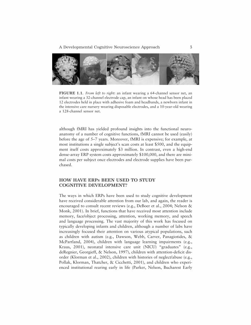
Because our group has extensively discussed the utility of ERPs in the studyof cognitive development (see DeBoer, Scott, & Nelson, 2004; Nelson &Monk, 2001), my review of the ERP here is relatively brief. ERPs representthe summation of electrical activity generated by large populations of neu-rons that volume conduct to the scalp surface. This activity can then berecorded by means of electrodes placed on the scalp surface. Figure 1.1illustrates several of the ways in which we have recorded ERPs over theyears.
It is generally believed that ERPs represent the activity of pyramidalcell neurons whose orientation is configured in such a way so as to permittheir recording at the scalp surface (if oriented in the “wrong” direction,the currents generated by such neurons would not be detectable). Theimplications of this constraint are worth mentioning. First, in theory,neural structures or circuits that do not contain pyramidal cells (e.g.,amygdala) should not give rise to electrical currents that take the form ofERPs. Second, because the electrical currents that give rise to ERPs are cre-ated by the simultaneous activity of large populations of neurons that vol-ume conduct to the surface of the scalp, the spatial resolution of ERPs islimited; even with dense-array sampling (see far right image of Figure 1.1),it has been hypothesized that the spatial resolution is approximately 1 cen-timeter (cm; far less than is obtained with fMRI). Of course, with low-density array sampling, the resolution is likely less than 1 cm.
Despite the relatively poor spatial resolution of ERPs, their manyadvantages over other neuroimaging tools are noteworthy. First, ERPs haveexquisite temporal resolution, on the order of milliseconds, comparable tomagnetoencephalography (MEG) and far superior to fMRI (which reflectsneural activity on the order of seconds). Second, ERPs are entirely non-invasive, there are no safety issues that can concern institutional reviewboards (IRBs), they do not depend on motor or verbal responses, and theycan be used across the entire lifespan without a change in methods. Finally,
2 Nelson
although fMRI has yielded profound insights into the functional neuro-anatomy of a number of cognitive functions, fMRI cannot be used (easily)before the age of 5–7 years. Moreover, fMRI is expensive; for example, atmost institutions a single subject’s scan costs at least $500, and the equip-ment itself costs approximately $3 million. In contrast, even a high-enddense-array ERP system costs approximately $100,000, and there are mini-mal costs per subject once electrodes and electrode supplies have been pur-chased.
HOW HAVE ERPS BEEN USED TO STUDYCOGNITIVE DEVELOPMENT?
The ways in which ERPs have been used to study cognitive developmenthave received considerable attention from our lab, and again, the reader isencouraged to consult recent reviews (e.g., DeBoer et al., 2004; Nelson &Monk, 2001). In brief, functions that have received most attention includememory, face/object processing, attention, working memory, and speechand language processing. The vast majority of this work has focused ontypically developing infants and children, although a number of labs haveincreasingly focused their attention on various atypical populations, suchas children with autism (e.g., Dawson, Webb, Carver, Panagiotides, &McPartland, 2004), children with language learning impairments (e.g.,Kraus, 2001), neonatal intensive care unit (NICU) “graduates” (e.g.,deRegnier, Georgieff, & Nelson, 1997), children with attention-deficit dis-order (Klorman et al., 2002), children with histories of neglect/abuse (e.g.,Pollak, Klorman, Thatcher, & Cicchetti, 2001), and children who experi-enced institutional rearing early in life (Parker, Nelson, Bucharest Early
A Developmental Cognitive Neuroscience Approach 3
FIGURE 1.1. From left to right: an infant wearing a 64-channel sensor net, aninfant wearing a 32-channel electrode cap, an infant on whose head has been placed12 electrodes held in place with adhesive foam and headbands, a newborn infant inthe intensive care nursery wearing disposable electrodes, and a 10-year-old wearinga 128-channel sensor net.
Intervention Project Core Group, 2005a, 2005b). Virtually all of thesestudies have been performed with infants or school-age children; very fewhave been done with adolescent-age children, although such work is nowunder way in several laboratories (e.g., Davies, Segalowitz, & Gavin,2004).
In a typical cognitive paradigm, participants are presented with punc-tuated trains of visual or auditory stimuli while ERPs are recorded. Abehavioral measure (e.g., reaction time) may or may not be recorded simul-taneously. Depending on the age of the participant, as few as 40–50 trialsto several hundred trials might be presented. A typical experimental sessiongenerally lasts 5–15 minutes. If data have been collected continuously (e.g.,to record EEG), they are then down-sampled to create epochs or trials; inmany cases, however, data are not recorded continuously and instead trialsare constructed at the outset. After rigorously rejecting and/or correctingartifactual data (e.g., eye movements), individual trials are typically aver-aged and these averages are then averaged across participants in order toexamine components that exist in the full group of participants.
Conventional data analysis tools historically have been confined toexamining the amplitude, latency, and scalp topography of individual com-ponents. With higher-density arrays of electrodes, more sophisticated anal-yses can be run, such as independent components analysis and source anal-ysis (see Johnson et al., 2001, for discussion in the context of development).
WHAT ARE THE VIRTUES AND LIMITATIONSOF STUDYING ATYPICAL POPULATIONS?
As I elaborate below, there are two broad reasons for considering theadvantages and disadvantages of studying atypical developmental popula-tions: to provide insight into clinical phenomena, per se, and to use thestudy of atypical development to inform the study of typical development.
The Study of Clinical Populationsfor Their Own Sake
The primary driving force in the study of clinical populations is to use ERPsto shed light on the pathophysiology of atypical development. A case inpoint might be work by Dawson and colleagues in the field of autism. Forexample, this group has reported that ERPs elicited by face stimuli in chil-dren with autism differ considerably from ERPs in response to the samestimuli in control children, whereas the ERPs to objects are remarkablysimilar (Dawson et al., 2002). This observation provides critical informa-
4 Nelson
tion as to the neural correlates of face-processing deficits, something thathas not been possible by studying overt behavior alone. Similarly, Pollakand colleagues have reported that the ERPs generated in response to dis-crete facial expressions (e.g., anger) differ in children with histories ofneglect/abuse from those who are reared in typical environments (e.g.,Pollak et al., 2001). And, we (Parker et al., 2005a, 2005b) have reportedthat the ERPs elicited by facial emotion differ in toddlers with a history ofinstitutionalization relative to a community sample. In all cases the motiva-tion underlying this work was to illuminate the neuropathophysiology of aspecific cognitive function.
The Study of Clinical Populations as a Meansof Shedding Light on Typical Development
The work I present below in some detail is a case in point. Specifically,because of my interest in the neural architecture that underlies typicalmemory development, I have been drawn to the study of populations ofchildren with damage to the structures/circuits of the brain that underliememory. In so doing I provide a form of converging operations on thestudy of memory. Thus, for example, if I hypothesize that the hippocampusis critically involved in some forms of recognition memory, and that a par-ticular component of the ERP reflects such memory, then children with hip-pocampal damage should show perturbations in that particular compo-nent. In the following section I focus my attention on one particularpopulation, the study of which is designed to shed light on typically devel-oping children while, at the same time, adding to what is known about theneuropathophysiology of this particular condition.
A MODEL SYSTEM:THE INFANT OF THE DIABETIC MOTHER
Here I begin by laying out the rationale for studying infants of diabeticmothers (IDMs). I then proceed to review our ERP findings to date.
Background
The fetal metabolic milieu of the diabetic pregnancy is characterized by sig-nificant risk factors to the developing brain, including chronic hypoxia andiron deficiency, with intermittent metabolic acidosis and hyper/hypo-glycemia (Georgieff et al., 1990; Petry et al., 1992; Widness, 1989). All ofthese risk factors are a function of lack of maternal glycemic control during
A Developmental Cognitive Neuroscience Approach 5
the third trimester of pregnancy (Georgieff et al., 1990). Maternal hyper-glycemia (i.e., too much sugar in the blood) results in fetal hypergly-cemia and reactive fetal hyperinsulinemia (i.e., too much insulin in thefetus’s blood). The latter two factors independently (Milley, Papacostas, &Tabata, 1986; Stonestreet, Golstein, Oh, & Widness, 1989) and collec-tively (Widness et al., 1981) increase the fetal rate of oxygen consumptionbeyond the placental capacity to transport oxygen. The resultant fetalhypoxemia (i.e., lack of oxygen—see Milley et al., 1986; Stonestreet et al.,1989; Widness et al., 1981) is pervasive and chronic, as evidenced by anelevated serum erythropoietin concentration and a compensatory rise infetal hemoglobin concentration (Georgieff et al., 1990; Widness et al.,1981). Chronic hypoxia constitutes a significant risk factor to the develop-ing brain, with some circuitry (e.g., the hippocampus) more vulnerable thanother circuitry (Nelson & Silverstein, 1994). The differential regional vul-nerability may relate to areas that have high metabolic rates during latefetal and early postnatal life. The theme of regional brain vulnerability(Burdo & Connor, 2002; de Ungria et al., 2000) is consistently seen withmetabolic insults during the perinatal period and is important in our target-ing of hippocampal function in our studies.
Fetal hyperinsulinemia secondary to chronic maternal hyperglyemialeads to stimulation of pancreatic islet cells and increases the fetal risk ofintermittent hypoglycemia. When the glucose flow from the mother vacil-lates abruptly, the fetus is at particular risk. For example, when the motheris hyperglycemic, the fetus will respond to the surge of maternally derivedglucose by secreting an appropriately large amount of insulin. As themother treats her diabetes with exogenous insulin, her blood glucose fallsrapidly, cutting off the supply to the fetus, lowering the fetal glucose con-centration, and leaving the fetus unprotected from its own hyperin-sulinemia. The resultant fetal hypoglycemia constitutes a significant risk tothe developing hippocampus (Barks, Sun, Malinak, & Silverstein, 1995).
Further neurological risk is conferred by a state of chronic brain irondeficiency in the fetus. Early iron deficiency affects multiple develop-ing brain functions, including myelination (Connor & Menzies, 1996;Roncagliolo, Garrdio, Walter, Peirano, & Lozoff, 1998), monoamine neu-rotransmitter metabolism (Nelson, Erikson, Piner, & Beard, 1997), andoxidative metabolism (de Ungria et al., 2000; Rao et al., 1999). The fetalbrain shows regional variability with these effects, with the energy metabo-lism effects being most striking in the hippocampus (de Ungria et al.,2000), whereas dopaminergic effects are found primarily in the striatum(Georgieff, Petry, Mills, McKay, & Wobken, 1997).
The 40% decrease in hippocampal neuronal metabolism results instriking dendritic truncation and alteration in the ontogeny of NMDA sub-
6 Nelson
unit appearance in area hippocampal CA1 in the perinatal rat. These struc-tural changes persist beyond the period of iron repletion. Furthermore,studies using high-field (9.4 Tesla) magnetic resonance (MR) spectroscopyin the developing rat demonstrate significant increases in glutamate concen-trations in the hippocampus in the resting state. More alarming, thesechanges are enhanced following exposure to hypoxia, a condition prevalentin the diabetic fetus. The etiology of iron deficiency in the diabetic fetus isdue to the inability of the diabetic placenta to up-regulate iron transportdue to hyperglycosylation of the transferrin receptor (Georgieff et al.,1997). The inability to meet the increased iron demands for augmentedfetal hemoglobin synthesis results in a shunting of available fetal iron intothe red blood cells at the expense of the developing human brain (Petry etal., 1992). The result is a reduction in hippocampal volume.
Multiple risk factors are associated with the diabetic pregnancy, and aplethora of animal models have been used to elucidate the pathophysiologyassociated with these risk factors. In addition, several studies have demon-strated that neurobehavioral outcomes in human children who were bornto diabetic mothers are correlated with the quality of metabolic regulationduring pregnancy (e.g., Ornoy et al., 2001; Rizzo, Metzger, Burns, &Burns, 1991; Rizzo, Metzger, Dooley, & Cho, 1997; Silverman et al.,1991). However, these investigations have typically examined global cogni-tive development in children well downstream from the proposed insult.Although important from an educational perspective, the disadvantage ofusing global measures is their inability to reveal (1) specific domains of dys-function that may be responsible for the global deficits and (2) the specificneural circuitry that underlies deficits in behavior. Moreover, most mea-sures of global functioning cannot easily (or meaningfully) be performedduring the early infancy period, thereby placing limits on early detectionand prediction of subsequent developmental course. Because of the infant’slimited behavioral repertoire, we have placed a premium in our studies onemploying noninvasive electrophysiological imaging during the first fewyears of life (starting at birth).
The logic of our work is as follows. First, as reviewed in an earlierpublication (Nelson, 1994) as well as in more recent writings (e.g., Nelson& Webb, 2002; Nelson, Thomas, & de Haan, 2006), we know that ERPscan be used to evaluate different forms of memory, including recognitionmemory. Second, we know that the forms of memory in which we are mostinterested are subserved, in large part, by structures that reside in themedial temporal lobe—the hippocampus, in particular. Third, if the ad-verse fetal milieu experienced by IDMs is, in fact, “toxic” to the developinghippocampus, then such infants should show impairments in memory, andERPs should, in turn, provide an index of such impairments.
A Developmental Cognitive Neuroscience Approach 7
Our study is longitudinal in nature; here, however, I focus most on ourcross-sectional findings. We have employed a battery of tasks, includingconventional developmental testing, elicited imitation, neuropsychologicaltesting, and, now that our children are school-age, structural and func-tional MRI. However, for purposes of this chapter I confine myself to a dis-cussion of our ERP findings.
All infants (both IDMs and controls) were screened for various con-founds (e.g., maternal drug use, neurological history) and then enrolledprenatally. At the delivery we obtained cord blood and placental tissue sowe could ascertain the degree of iron deficiency (and attempt to infer infor-mation about hypoxemia). We began our investigation within 2 days ofbirth, then continued to see our study subjects throughout the next fewyears.
Newborn Infants’ Recognition of the Mother’s Voice
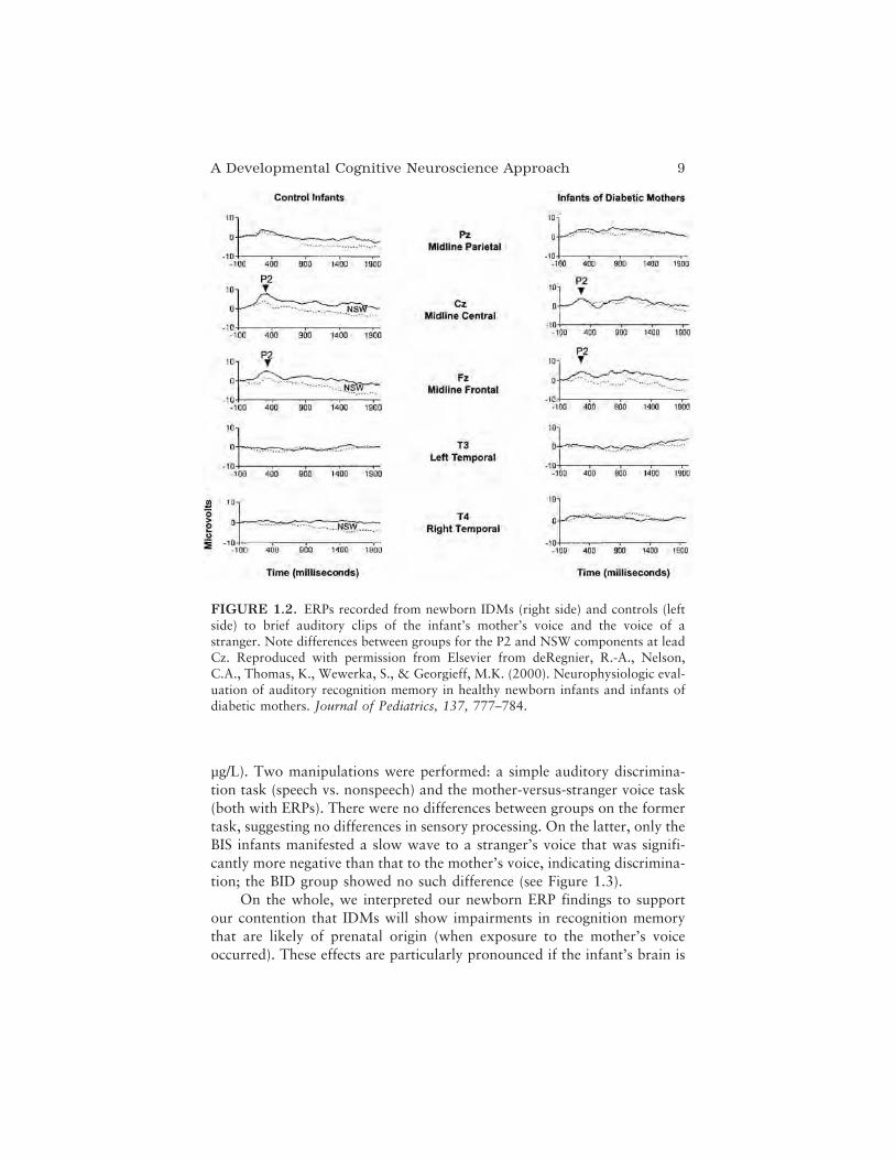
We examined the 1- to 2-day-old infant’s ability to discriminate themother’s voice from a stranger’s voice, based on the assumption that theformer would be familiar to the infant after being exposed to that voiceprenatally. ERPs were recorded as infants were presented with sound clips(via ear insert) of their mother’s voice or a stranger’s voice (random 50%probabilities) pronouncing the word baby. The grand means displayed inFigure 1.2 show electrophysiological responses by group to the mother’svoice (thick line) and stranger’s voice (thin line). Within this sample, weexamined two components of interest, the P2 and the negative slow wave(NSW) that follows the P2. We have associated the P2 with early percep-tual processing of the content of the stimuli, and the NSW with the abilityto detect a novel stimulus set against a background of familiar stimuli. Ourresults reveal P2 amplitudes to be greater to the mother’s versus a stranger’svoice among controls but not the IDM group. In terms of the NSW (ourmetric of memory), our controls display significantly greater NSW area to astranger’s than to the mother’s voice (particularly at leads Fz and T4),whereas no such differences are observed among IDMs (for details, seedeRegnier, Nelson, Thomas, Wewerka, & Georgieff, 2000).
Brain Iron Deficiency versus Sufficiency
We have recently had the opportunity to reclassify some of our newborndata based on the degree of iron deficiency experienced, focusing specifi-cally on those infants we presume to be truly brain iron deficient (BID) (seeSiddappa et al., 2004). Ten BID infants were compared to 22 IDMs whowere presumably brain iron sufficient (BIS) (concentration > 30 µg/L < 60
8 Nelson
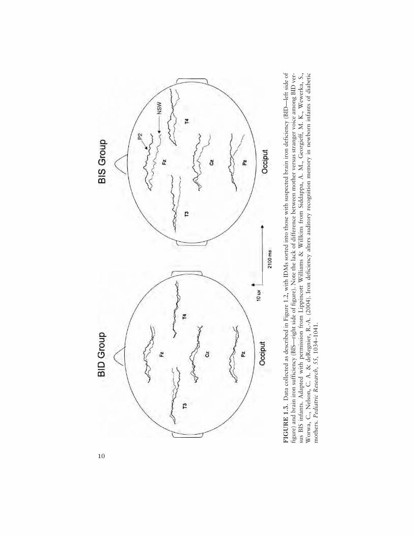
µg/L). Two manipulations were performed: a simple auditory discrimina-tion task (speech vs. nonspeech) and the mother-versus-stranger voice task(both with ERPs). There were no differences between groups on the formertask, suggesting no differences in sensory processing. On the latter, only theBIS infants manifested a slow wave to a stranger’s voice that was signifi-cantly more negative than that to the mother’s voice, indicating discrimina-tion; the BID group showed no such difference (see Figure 1.3).
On the whole, we interpreted our newborn ERP findings to supportour contention that IDMs will show impairments in recognition memorythat are likely of prenatal origin (when exposure to the mother’s voiceoccurred). These effects are particularly pronounced if the infant’s brain is
A Developmental Cognitive Neuroscience Approach 9
FIGURE 1.2. ERPs recorded from newborn IDMs (right side) and controls (leftside) to brief auditory clips of the infant’s mother’s voice and the voice of astranger. Note differences between groups for the P2 and NSW components at leadCz. Reproduced with permission from Elsevier from deRegnier, R.-A., Nelson,C.A., Thomas, K., Wewerka, S., & Georgieff, M.K. (2000). Neurophysiologic eval-uation of auditory recognition memory in healthy newborn infants and infants ofdiabetic mothers. Journal of Pediatrics, 137, 777–784.
10
FIG
UR
E1
.3.
Dat
aco
llect
edas
desc
ribe
din
Figu
re1.
2,w
ith
IDM
sso
rted
into
thos
ew
ith
susp
ecte
dbr
ain
iron
defi
cien
cy(B
ID—
left
side
offi
gure
)an
dbr
ain
iron
suff
icie
ncy
(BIS
—ri
ght
side
offi
gure
).N
ote
the
lack
ofdi
ffer
ence
betw
een
mot
her
vers
usst
rang
ervo
ice
amon
gB
IDve
r-su
sB
ISin
fant
s.A
dapt
edw
ith
perm
issi
onfr
omL
ippi
ncot
tW
illia
ms
&W
illki
nsfr
omSi
ddap
pa,
A.
M.,
Geo
rgie
ff,
M.
K.,
Wew
erka
,S.
,W
orw
a,C
.,N
elso
n,C
.A
.&
deR
egni
er,
R.-
A.
(200
4).
Iron
defi
cien
cyal
ters
audi
tory
reco
gnit
ion
mem
ory
inne
wbo
rnin
fant
sof
diab
etic
mot
hers
.P
edia
tric
Res
earc
h,55
,10
34–1
041.
iron deficient (which is more likely to occur if the mother’s diabetes ispoorly controlled, leading to an even more adverse fetal environment).
Six-Month-Old Infants’ Recognition of Mother’s Face
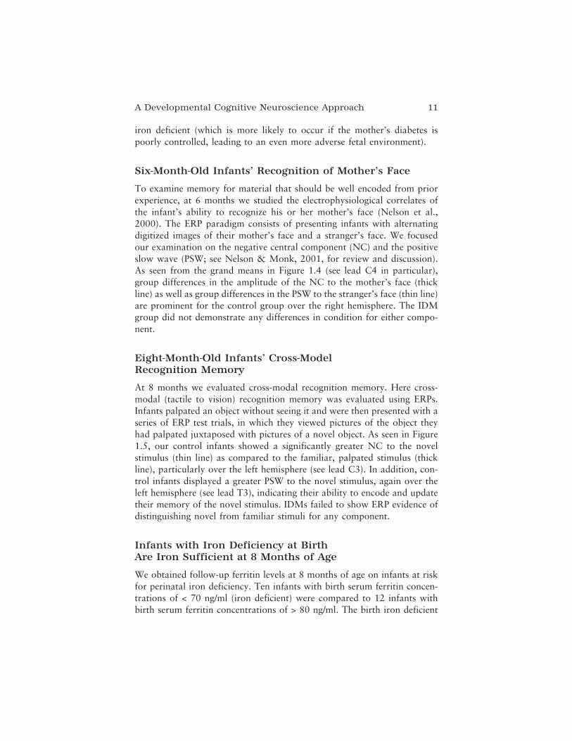
To examine memory for material that should be well encoded from priorexperience, at 6 months we studied the electrophysiological correlates ofthe infant’s ability to recognize his or her mother’s face (Nelson et al.,2000). The ERP paradigm consists of presenting infants with alternatingdigitized images of their mother’s face and a stranger’s face. We focusedour examination on the negative central component (NC) and the positiveslow wave (PSW; see Nelson & Monk, 2001, for review and discussion).As seen from the grand means in Figure 1.4 (see lead C4 in particular),group differences in the amplitude of the NC to the mother’s face (thickline) as well as group differences in the PSW to the stranger’s face (thin line)are prominent for the control group over the right hemisphere. The IDMgroup did not demonstrate any differences in condition for either compo-nent.
Eight-Month-Old Infants’ Cross-ModelRecognition Memory
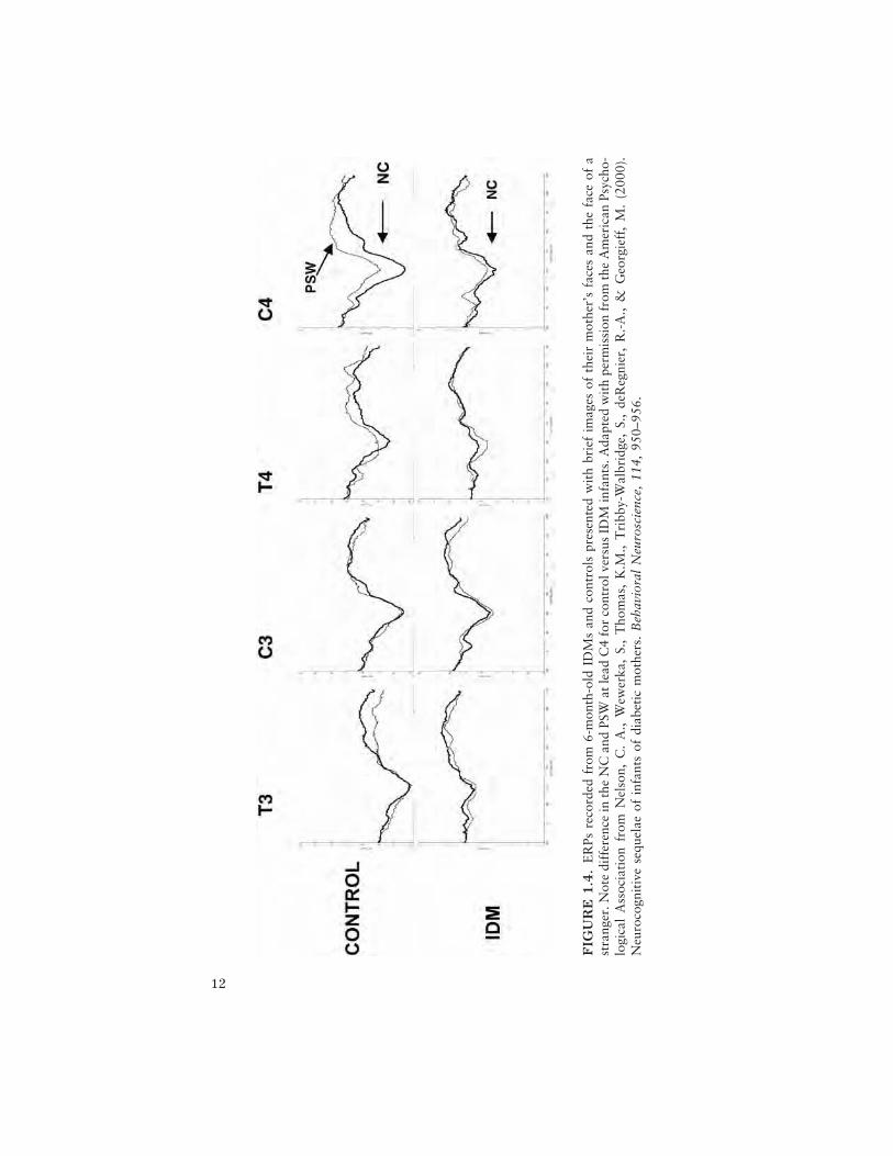
At 8 months we evaluated cross-modal recognition memory. Here cross-modal (tactile to vision) recognition memory was evaluated using ERPs.Infants palpated an object without seeing it and were then presented with aseries of ERP test trials, in which they viewed pictures of the object theyhad palpated juxtaposed with pictures of a novel object. As seen in Figure1.5, our control infants showed a significantly greater NC to the novelstimulus (thin line) as compared to the familiar, palpated stimulus (thickline), particularly over the left hemisphere (see lead C3). In addition, con-trol infants displayed a greater PSW to the novel stimulus, again over theleft hemisphere (see lead T3), indicating their ability to encode and updatetheir memory of the novel stimulus. IDMs failed to show ERP evidence ofdistinguishing novel from familiar stimuli for any component.
Infants with Iron Deficiency at BirthAre Iron Sufficient at 8 Months of Age
We obtained follow-up ferritin levels at 8 months of age on infants at riskfor perinatal iron deficiency. Ten infants with birth serum ferritin concen-trations of < 70 ng/ml (iron deficient) were compared to 12 infants withbirth serum ferritin concentrations of > 80 ng/ml. The birth iron deficient
A Developmental Cognitive Neuroscience Approach 11
12
FIG
UR
E1
.4.
ER
Psre
cord
edfr
om6-
mon
th-o
ldID
Ms
and
cont
rols
pres
ente
dw
ith
brie
fim
ages
ofth
eir
mot
her’
sfa
ces
and
the
face
ofa
stra
nger
.Not
edi
ffer
ence
inth
eN
Can
dPS
Wat
lead
C4
for
cont
rolv
ersu
sID
Min
fant
s.A
dapt
edw
ith
perm
issi
onfr
omth
eA
mer
ican
Psyc
ho-
logi
cal
Ass
ocia
tion
from
Nel
son,
C.
A.,
Wew
erka
,S.
,T
hom
as,
K.M
.,T
ribb
y-W
albr
idge
,S.
,de
Reg
nier
,R
.-A
.,&
Geo
rgie
ff,
M.
(200
0).
Neu
roco
gnit
ive
sequ
elae
ofin
fant
sof
diab
etic
mot
hers
.B
ehav
iora
lN
euro
scie
nce,
114,
950–
956.
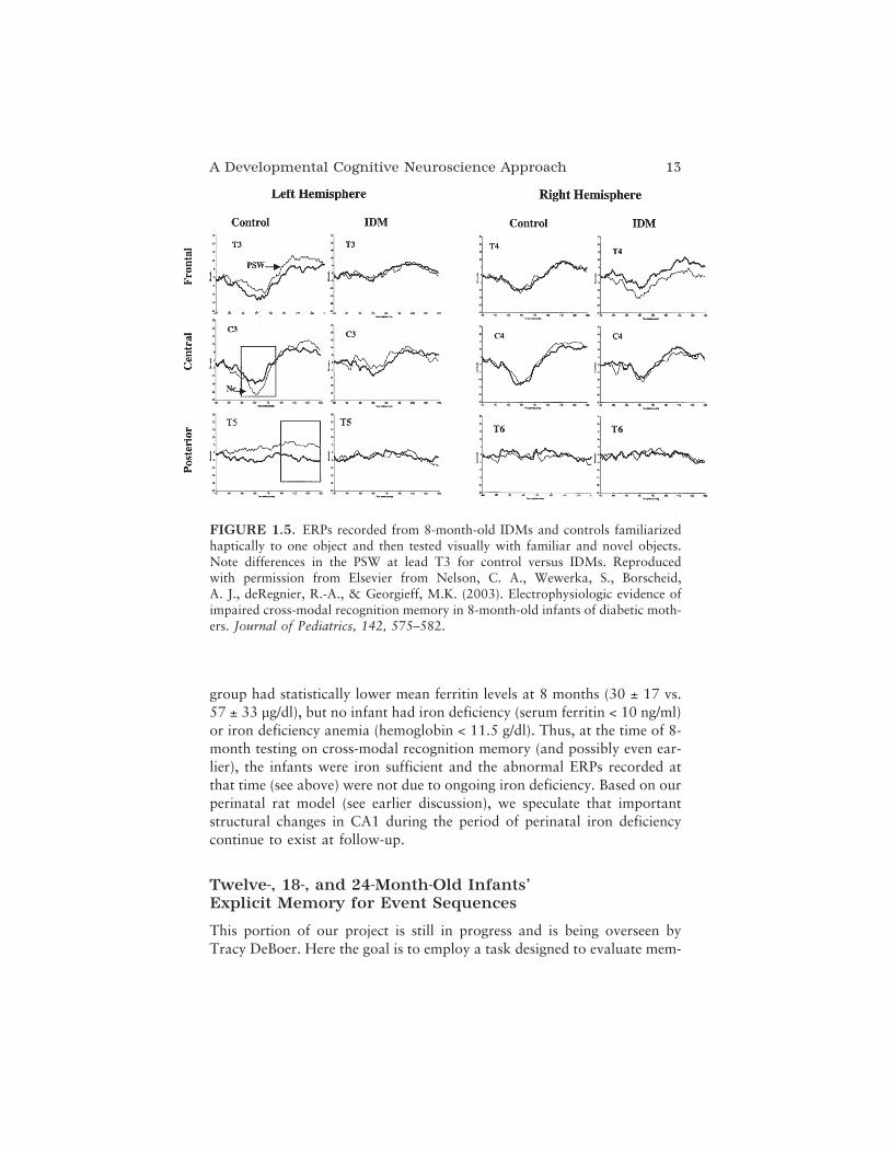
group had statistically lower mean ferritin levels at 8 months (30 ± 17 vs.57 ± 33 µg/dl), but no infant had iron deficiency (serum ferritin < 10 ng/ml)or iron deficiency anemia (hemoglobin < 11.5 g/dl). Thus, at the time of 8-month testing on cross-modal recognition memory (and possibly even ear-lier), the infants were iron sufficient and the abnormal ERPs recorded atthat time (see above) were not due to ongoing iron deficiency. Based on ourperinatal rat model (see earlier discussion), we speculate that importantstructural changes in CA1 during the period of perinatal iron deficiencycontinue to exist at follow-up.
Twelve-, 18-, and 24-Month-Old Infants’Explicit Memory for Event Sequences
This portion of our project is still in progress and is being overseen byTracy DeBoer. Here the goal is to employ a task designed to evaluate mem-
A Developmental Cognitive Neuroscience Approach 13
FIGURE 1.5. ERPs recorded from 8-month-old IDMs and controls familiarizedhaptically to one object and then tested visually with familiar and novel objects.Note differences in the PSW at lead T3 for control versus IDMs. Reproducedwith permission from Elsevier from Nelson, C. A., Wewerka, S., Borscheid,A. J., deRegnier, R.-A., & Georgieff, M.K. (2003). Electrophysiologic evidence ofimpaired cross-modal recognition memory in 8-month-old infants of diabetic moth-ers. Journal of Pediatrics, 142, 575–582.
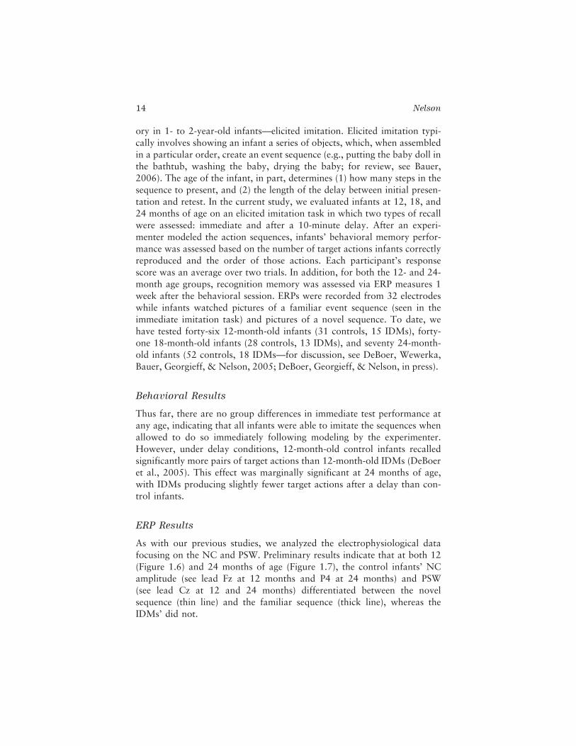
ory in 1- to 2-year-old infants—elicited imitation. Elicited imitation typi-cally involves showing an infant a series of objects, which, when assembledin a particular order, create an event sequence (e.g., putting the baby doll inthe bathtub, washing the baby, drying the baby; for review, see Bauer,2006). The age of the infant, in part, determines (1) how many steps in thesequence to present, and (2) the length of the delay between initial presen-tation and retest. In the current study, we evaluated infants at 12, 18, and24 months of age on an elicited imitation task in which two types of recallwere assessed: immediate and after a 10-minute delay. After an experi-menter modeled the action sequences, infants’ behavioral memory perfor-mance was assessed based on the number of target actions infants correctlyreproduced and the order of those actions. Each participant’s responsescore was an average over two trials. In addition, for both the 12- and 24-month age groups, recognition memory was assessed via ERP measures 1week after the behavioral session. ERPs were recorded from 32 electrodeswhile infants watched pictures of a familiar event sequence (seen in theimmediate imitation task) and pictures of a novel sequence. To date, wehave tested forty-six 12-month-old infants (31 controls, 15 IDMs), forty-one 18-month-old infants (28 controls, 13 IDMs), and seventy 24-month-old infants (52 controls, 18 IDMs—for discussion, see DeBoer, Wewerka,Bauer, Georgieff, & Nelson, 2005; DeBoer, Georgieff, & Nelson, in press).
Behavioral Results
Thus far, there are no group differences in immediate test performance atany age, indicating that all infants were able to imitate the sequences whenallowed to do so immediately following modeling by the experimenter.However, under delay conditions, 12-month-old control infants recalledsignificantly more pairs of target actions than 12-month-old IDMs (DeBoeret al., 2005). This effect was marginally significant at 24 months of age,with IDMs producing slightly fewer target actions after a delay than con-trol infants.
ERP Results
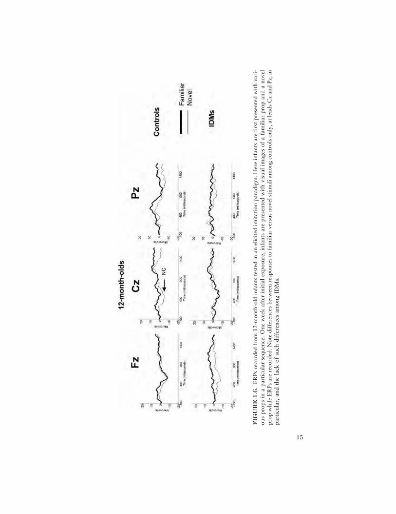
As with our previous studies, we analyzed the electrophysiological datafocusing on the NC and PSW. Preliminary results indicate that at both 12(Figure 1.6) and 24 months of age (Figure 1.7), the control infants’ NCamplitude (see lead Fz at 12 months and P4 at 24 months) and PSW(see lead Cz at 12 and 24 months) differentiated between the novelsequence (thin line) and the familiar sequence (thick line), whereas theIDMs’ did not.
14 Nelson
15
FIG
UR
E1
.6.
ER
Psre
cord
edfr
om12
-mon
th-o
ldin
fant
ste
sted
inan
elic
ited
imit
atio
npa
radi
gm.H
ere
infa
nts
are
firs
tpr
esen
ted
wit
hva
ri-
ous
prop
sin
apa
rtic
ular
sequ
ence
.O
new
eek
afte
rin
itia
lex
posu
re,
infa
nts
are
pres
ente
dw
ith
visu
alim
ages
ofa
fam
iliar
prop
and
ano
vel
prop
whi
leE
RPs
are
reco
rded
.Not
edi
ffer
ence
sbe
twee
nre
spon
ses
tofa
mili
arve
rsus
nove
lsti
mul
iam
ong
cont
rols
only
,atl
eads
Cz
and
Pz,i
npa
rtic
ular
,an
dth
ela
ckof
such
diff
eren
ces
amon
gID
Ms.
16
FIG
UR
E1
.7.
ER
Psre
cord
edfr
om24
-mon
th-o
ldin
fant
ste
sted
inan
elic
ited
imit
atio
npa
radi
gm(s
eeFi
gure
1.6
for
deta
ils).
Ana
lysi
sof
the
data
indi
cate
dth
atth
ePS
Ww
asgr
eate
rto
nove
lver
sus
fam
iliar
stim
ulia
tle
adC
zfo
rco
ntro
linf
ants
only
.(A
ltho
ugh
ther
eap
pear
sto
bea
diff
eren
cefo
rbo
thth
eN
Can
dPS
Wam
ong
IDM
s,th
ese
diff
eren
ces
wer
e,in
fact
,no
tst
atis
tica
llysi
gnif
ican
t.)
Relations between Behavioral Recall and ERPs
At both 12 and 24 months, immediate imitation of target actions also cor-related with PSW activity to the familiar stimulus at Cz after a 1-weekdelay. Specifically, the number of target actions produced at Session 1 wasassociated with greater positive slow wave activity at Session 2. Because weinterpret the PSW to reflect memory, we were not surprised to see that theamplitude of this component correlated with the number of target actionsproduced.
Summary
The observation that the 12-month-old control infants recalled more thanIDMs on the delayed imitation task but not the immediate imitation task(and the trend in this same direction at 24 months of age) suggests thatIDMs show evidence of forgetting over delays as short as 10 minutes.Furthermore, at 12 months, newborn ferritin levels were marginallyrelated to performance on the delayed recall task (DeBoer et al., 2005).The electrophysiological data, collected after a 1-week delay for both the12- and 24-month-old infants, also suggest that there are group differ-ences in long-term memory storage. Specifically, although the infantsdid not show differences in behavioral recall of the sequences immedi-ately, they did show differential brain activity to pictures of these samesequences after a delay, and correlations between immediate recall perfor-mance and positive slow wave activity indicate that ERPs may allow fordetection of subtler differences in the processing of the informationrelated to recall performance.
A Follow-Up on Explicit Memory for EventSequences in 36- and 48-Month-Old Children
In order to characterize more accurately differences in behavioral recallbetween controls and IDMs, we recently added a follow-up investigation at36 and 48 months. Specifically, at these follow-up assessments children aregiven three nine-step sequences of different degrees of difficulty (one easy,one medium, and one difficult, with difficulty level determined by the num-ber of causal relations in the sequence) and tested for recall of targetactions and the order of those actions both immediately and after a 1-weekdelay. As at 12 and 24 months, an electrophysiological assessment isobtained after the 1-week delay to allow for direct comparison with previ-ous ages; in addition, a 1-year delay ERP to familiar and novel stimuli from36 months of age is also collected at 48 months. To date we have tested
A Developmental Cognitive Neuroscience Approach 17
twenty-two 36-month-olds (14 controls, 8 IDMs) and nineteen 48-month-olds (13 controls, 6 IDMs).
Behavioral Results
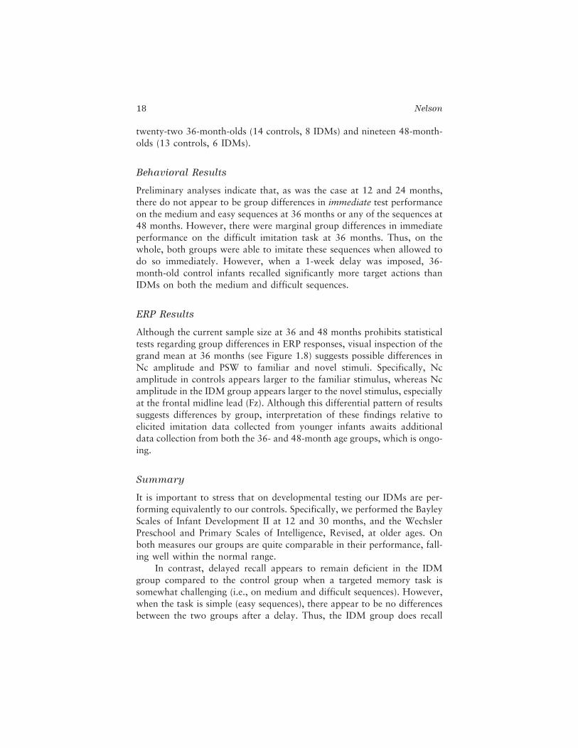
Preliminary analyses indicate that, as was the case at 12 and 24 months,there do not appear to be group differences in immediate test performanceon the medium and easy sequences at 36 months or any of the sequences at48 months. However, there were marginal group differences in immediateperformance on the difficult imitation task at 36 months. Thus, on thewhole, both groups were able to imitate these sequences when allowed todo so immediately. However, when a 1-week delay was imposed, 36-month-old control infants recalled significantly more target actions thanIDMs on both the medium and difficult sequences.
ERP Results
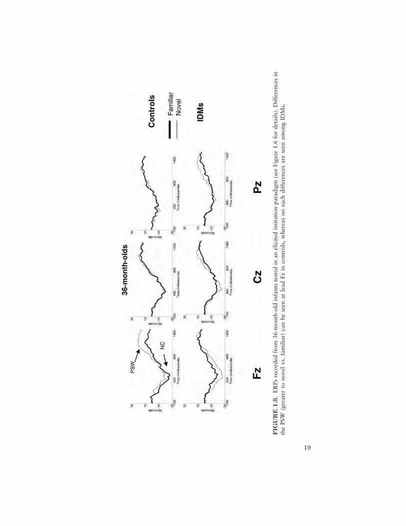
Although the current sample size at 36 and 48 months prohibits statisticaltests regarding group differences in ERP responses, visual inspection of thegrand mean at 36 months (see Figure 1.8) suggests possible differences inNc amplitude and PSW to familiar and novel stimuli. Specifically, Ncamplitude in controls appears larger to the familiar stimulus, whereas Ncamplitude in the IDM group appears larger to the novel stimulus, especiallyat the frontal midline lead (Fz). Although this differential pattern of resultssuggests differences by group, interpretation of these findings relative toelicited imitation data collected from younger infants awaits additionaldata collection from both the 36- and 48-month age groups, which is ongo-ing.
Summary
It is important to stress that on developmental testing our IDMs are per-forming equivalently to our controls. Specifically, we performed the BayleyScales of Infant Development II at 12 and 30 months, and the WechslerPreschool and Primary Scales of Intelligence, Revised, at older ages. Onboth measures our groups are quite comparable in their performance, fall-ing well within the normal range.
In contrast, delayed recall appears to remain deficient in the IDMgroup compared to the control group when a targeted memory task issomewhat challenging (i.e., on medium and difficult sequences). However,when the task is simple (easy sequences), there appear to be no differencesbetween the two groups after a delay. Thus, the IDM group does recall
18 Nelson
19
FIG
UR
E1
.8.
ER
Psre
cord
edfr
om36
-mon
th-o
ldin
fant
ste
sted
inan
elic
ited
imit
atio
npa
radi
gm(s
eeFi
gure
1.6
for
deta
ils).
Dif
fere
nces
inth
ePS
W(g
reat
erto
nove
lvs
.fa
mili
ar)
can
bese
enat
lead
Fzin
cont
rols
,w
here
asno
such
diff
eren
ces
are
seen
amon
gID
Ms.
some information over a delay, especially when the to-be-rememberedmaterials support such recall. However, when the task is more challenging,deficits in recall remain apparent. Whether this difference decreases withage remains to be seen, as data collection with these older age groups con-tinues.
Electrophysiological Measuresof Continuous Recognition Memory
Many in our sample are now approaching 7 years of age, and we haveembarked on an ambitious follow-up program that involves high-densityERP recordings and fMRI (being overseen by Kathleen Thomas). Below Ireport our preliminary ERP data with control children and adults only;thus far only three IDMs have been tested.
Eighteen adults (M = 24 years; nine males and nine females) and 12children (M = 8 years, 3 months; five males and seven females) are partici-pating in electrophysiological investigation of recognition memory, using a128-channel high-density electrode array (see far right image of Figure 1.1).Stimuli consist of color photos or three-dimensional drawings of everydayobjects (concrete) and unnamable objects (abstract). Stimuli are presentedon a black background for a duration of 500 milliseconds (ms), followedby a 3,500-ms interstimulus interval (ISI). These parameters were selectedspecifically to duplicate those necessary for the fMRI paradigm.
On each trial, participants indicate whether the presented image is new(never seen previously) or old (seen previously), using the index and middlefingers of the right hand, respectively. Behavioral and electrophysiologicaldata are collected in three runs of 72 trials. Electrophysiological and behav-ioral responses are compared for the first occurrence of a stimulus com-pared to a repeat occurrence following either 2 or 5 intervening trials. Newstimuli are presented in each run, resulting in a total of 120 new trials, 42trials of lag 2, and 42 trials of lag 5. Foil trials, which consist of stimuli thatnever repeated or stimuli repeating after a nonstandard lag (1 or 3 interven-ing trials), are interspersed to prevent anticipatory recognition responses.Factors for analysis included lag condition (new, lag 2, or lag 5 trials) andstimulus category (concrete or abstract).
As we have now reported at several conferences (see Hunt, Couperus,Nelson, & Thomas, 2004a, 2004b; Hunt, Couperus, et al., 2005a; Hunt,Townsend, et al., 2005b; Thomas et al., 2004a, 2004b), behavioral dataindicate that adults perform the task more accurately and more quicklythan children (93% vs. 88%, 746 ms vs. 1,325 ms). Response accuracydecreased with memory load for adults (94%, 93%, 92%) but not for chil-
20 Nelson
dren. Both adults and children performed more poorly for abstract itemsthan for concrete items. For adults, this effect did not differ by memory lag;however, for children, this effect was not apparent for long memory delays(lag 5). Reaction time differences were observed by age group but not bycondition.
Electrophysiologically, effects of memory lag were observed for theparietal P3b component for adults (~500 ms). The P3b to old items wasfaster than that to new items, with items at lag 2 peaking prior to items atlag 5. Both adults and children showed evidence of an old/new effect forthe P3a (~350 ms), with greater amplitude for old items compared to newitems. This P3 difference was accompanied by a slower return to baselinefor old items than for new items in both adults and children (negative com-ponent at 1,000 ms). Adults evidenced a very prominent difference in P3amplitude for concrete compared to abstract objects, despite comparablebehavioral performance across stimulus types. These P300 data suggestpeak latency differences for old compared to new concrete objects, whereasno such difference is apparent for abstract objects. Children did not dem-onstrate significant P300 differences by stimulus type, although some leadsshowed a trend in this direction.
Source analysis techniques were used to estimate the number and loca-tion of neural generators of the recorded scalp activity. Eye-movement arti-facts were modeled first using regional dipoles with three orthogonal direc-tions. Scalp data were then modeled with single dipoles based on a principalcomponents analysis (PCA) of the scalp-recorded data. The location and ori-entation of the dipoles were unconstrained. Data were modeled separately foradults and children. Residual variance of each model was less than 5%. Bothmodels produced dipole generators in temporal, parietal, and cingulate corti-ces, as well as sources in medial temporal lobe regions near the hippocampusor parahippocampal gyrus. Both models showed good fit with the oppositeage group (<5% residual variance), suggesting that the two groups showedessentially the same dipole generators. Current source density (CSD) plots(not shown) in adults showed differential activity for old and new items attemporal lobe dipoles around 960 ms (old > new). A similar effect wasobserved for children around 1,040 ms (lag 5 > lag 2 > new).
The current data support the feasibility of using these methods, bothhigh-density ERPs and the continuous recognition memory task, in this agerange. Seven- to 8-year-old children as well as adults showed differentialscalp-recorded brain activity as a function of memory load and, in somecases, by stimulus type. Dipole source modeling techniques suggest that atleast some of these memory effects stem from dipole sources in medial tem-poral lobe brain regions hypothesized to be affected in IDMs.
A Developmental Cognitive Neuroscience Approach 21
CONCLUSIONS
Data drawn from our longitudinal study of IDMs reveal a reliable patternof electrophysiological findings consistent with our hypothesis that the dia-betic pregnancy places the fetus at risk for developing memory problemspostnatally, due to underlying perturbations in hippocampal development.Over and above the clinical implications of such work (e.g., to alert pedia-tricians who care for such infants to monitor the children later in life forlearning and memory problems), these data also serve as a model systemfor how we can use ERPs to examine the neural circuitry involved in thedevelopment of a specific cognitive function—in our case, various types ofexplicit memory. Thus, we should think of this research as an example ofthe value of converging operations: Specifically, studying this particularpopulation has permitted us to examine more directly our hypothesis thatthe hippocampus is involved in recognition memory.
Although our source modeling efforts are providing invaluable infor-mation about the neural sources responsible for various ERP components,the spatial resolution of ERPs will always be limited. For this reason webelieve that coregistering ERP data with MRI and fMRI data will proveextremely useful. Specifically, the superior spatial resolution of MRI-basedmeasures, coupled with the superior temporal resolution of ERP measures,will provide investigators with temporal–spatial information that cannot beprovided by either measure alone. Unfortunately, MRI-based measures lackfeasibility for children younger than approximately 6 years. For this reasonit is important for those recording from high-density electrode arrays tocontinue developing source modeling algorithms that have increased preci-sion, thereby affording investigators a tool that permits reasonable infer-ences to be drawn about the neural sources that underlie ERP components.This effort will be aided by conducting research with atypical populationsin whom there is a suspicion of discrete neural injury (as we have done inour IDM population). For example, if we demonstrate among typicallydeveloping children that a particular ERP component is localized to thehippocampus, then when the same experimental manipulation is performedwith children who have experienced hippocampal damage, source model-ing should reveal the lack of (normal) hippocampal activation. This methodof converging operations will go a long way toward providing a fullerunderstanding of the development of function–structure relations amongboth typically developing and atypically developing children.
One additional area worthy of new investigation unique to early devel-opment concerns the ability to time-lock overt behavior with ERPs. Cur-rently most ERP investigations do not record behavior coincident with
22 Nelson
ERPs, for the very simple reason that most ERP designs do not permit thesimultaneous recording of behavior (e.g., when stimuli are presented for500 ms, there is no infant behavior that can be measured). We and othershave devoted considerable attention to this problem over the past fewyears, thus far with little to show for our effort. Still, we are optimistic thatnew eye-tracking technology may permit us to record some visual behav-iors simultaneously with ERPs, furthering our efforts to investigate brain–behavior links.
In conclusion, the field of developmental cognitive neuroscience hasmade tremendous advances in the last decade. This is particularly true inthe domain of ERPs, where the number of laboratories employing thismethod has increased exponentially. It is my hope that when in anotherdecade hence, the field will have advanced even further.
ACKNOWLEDGMENTS
The writing of this chapter was made possible, in part, by support from theNational Institutes of Health (Grant No. NS32755). I gratefully acknowledge themany contributions of the developmental cognitive neuroscience laboratory and mycollaborators Drs. Michael Georgieff and Raye-ann deRegnier.
REFERENCES
Barks, J. D., Sun, R., Malinak, C., & Silverstein, F. S. (1995). Gp120, an HIV-1protein, increases susceptibility to hypoglycemic and ischemic brain injury inperinatal rats. Experimental Neurology, 132, 123–133.
Bauer, P. J. (2006). Event memory. In W. Damon, R. Lerner, D. Kuhn, & R. Siegler(Eds.), Handbook of child psychology (6th ed.): Vol. 2. Cognition, perceptionand language (pp. 373–425). Hoboken, NJ: Wiley.
Burdo, J. R., & Connor, J. R. (2002). Iron transport in the central nervous system.In D. Templeton (Ed.), Molecular and cellular iron transport (pp. 487–505).New York: Dekker.
Connor, J. R., & Menzies, S. L. (1996). Relationship of iron to oligodendrocytesand myelination. Glia, 17, 83–93.
Davies, P. L., Segalowitz, S. J., & Gavin, W. J. (2004). Development of error-monitoring event-related potentials in adolescents. Annals of the New YorkAcademy of Sciences, 1021, 324–328.
Dawson, G., Carver, L., Meltzoff, A. N., Panagiotides, H., McPartland, J., &Webb, S. J. (2002). Neural correlates of face and object recognition in youngchildren with autism spectrum disorder, developmental delay, and typicaldevelopment. Child Development, 73, 700–717.
A Developmental Cognitive Neuroscience Approach 23
Dawson, G., & Fischer, K. W. (Eds.). (1994). Human behavior and the developingbrain. New York: Guilford Press.
Dawson, G., Webb, S. J., Carver, L., Panagiotides, H., & McPartland, J. (2004).Young children with autism show atypical brain responses to fearful ver-sus neutral facial expressions of emotion. Developmental Science, 7, 340–359.
DeBoer, T., Georgieff, M. K., & Nelson, C. A. (in press). Declarative memory per-formance in infants of diabetic mothers. In P. J. Bauer (Ed.), Varieties of earlyexperience: Influences on declarative memory development. Mahwah, NJ:Erlbaum.
DeBoer, T., Scott, L., & Nelson, C. A. (2004). Event-related potentials in develop-mental populations. In T. Handy (Ed.), Event-related potentials: A methodshandbook (pp. 263–297). Cambridge, MA: MIT Press.
DeBoer, T., Wewerka, S., Bauer, P. J., Georgieff, M. K., & Nelson, C. A. (2005).Explicit memory performance in infants of diabetic mothers at 1 year of age.Developmental Medicine and Child Neurology, 47, 525–531.
deRegnier, R.-A., Georgieff, M. K., & Nelson, C. A. (1997). Visual event-relatedbrain potentials in 4–month-old infants at risk for neurodevelopmental im-pairments. Developmental Psychobiology, 30, 11–28.
deRegnier, R.-A., Nelson, C. A., Thomas, K., Wewerka, S., & Georgieff, M. K.(2000). Neurophysiologic evaluation of auditory recognition memory inhealthy newborn infants and infants of diabetic mothers. Journal of Pediatrics,137, 777–784.
De Ungria, M., Rao, R., Wobken, J. D., Luciana, M., Nelson, C. A., & Georgieff,M. K. (2000). Perinatal iron deficiency decreases cytochromecoxidase (CytOx)activity in selective regions of neonatal rat brain. Pediatric Research, 48, 169–176.
Georgieff, M. K., Landon, M. B., Mills, M. M., Hedlund, B. E., Faassen, A. E.,Schmidt, R. L., et al. (1990). Abnormal iron distribution in infants of diabeticmothers: Spectrum and maternal antecedents. Journal of Pediatrics, 117, 455–461.
Georgieff, M. K., Petry, C. D., Mills, M. M., McKay, H., & Wobken, J. D. (1997).Increased N-glycosylation and reduced transferrin-binding capacity of trans-ferrin receptor isolated from placentae of diabetic women. Placenta, 18, 563–568.
Hunt, R. H., Couperus, J. W., Nelson, C. A., & Thomas, K. M. (2004a, June).Continuous recognition memory in adults and children: An ERP study ofparametric memory load manipulation. Poster presented at the annual meetingof the Organization for Human Brain Mapping, Budapest, Hungary.
Hunt, R. H., Couperus, J. W., Nelson, C. A., & Thomas, K. M. (2004b, April).Electophysiological correlates of continuous recognition memory in adults andchildren: A parametric manipulation of memory load. Poster presented at theannual meeting of the Cognitive Neuroscience Society, San Francisco.
Hunt, R. H., Couperus, J. W., Townsend, E. L., Nelson, C. A., & Thomas, K. M.(2005, April). Developmental differences in continuous recognition memory:An fMRI and ERP study of parametrically manipulated memory load. Paper
24 Nelson
presented at the annual meeting of the Society for Research in Child Develop-ment, Atlanta, GA.
Hunt, R. H., Townsend, E. L., Couperus, J. W., Nelson, C. A., & Thomas, K. M.(2005, April). A developmental fMRI study of medial temporal lobe function:Differences in continuous recognition memory. Poster presented at the annualmeeting of the Society for Research in Child Development, Atlanta, GA.
Johnson, M. H., de Haan, M., Oliver, A., Smith, W., Hatzakis, H., Tucker, L. A., etal. (2001). Recording and analyzing high-density event-related potentials withinfants: Using the Geodesic sensor net. Developmental Neuropsychology, 19,295–323.
Klorman, R., Thatcher, J. E., Shaywitz, S. E., Fletcher, J. M., Marchione, K. E.,Holahan, J. M., et al. (2002). Effects of event probability and sequence onchildren with attention-deficit/hyperactivity, reading, and math disorder. Bio-logical Psychiatry, 52, 795–804.
Kraus, N. (2001). Auditory pathway encoding and neural plasticity in children withlearning problems. Audiology Neurootolaryngology, 6, 221–227.
Milley, J. R., Papacostas, J. S., & Tabata, B. K. (1986). Effect of insulin on uptakeof metabolic substrates by the fetus. American Journal of Physiology—Endocrinology and Metabolism, 251, E349–E359.
Nelson, C. A. (1994). Neural correlates of recognition memory in the first postnatalyear of life. In G. Dawson & K. W. Fischer (Eds.), Human behavior and thedeveloping brain (pp. 269–313). New York: Guilford Press.
Nelson, C., Erikson, K., Pinero, D. J., & Beard, J. L. (1997). In vivo DA metabo-lism is altered in iron deficient anemic rats. Journal of Nutrition. 127,2282–2288.
Nelson, C. A., & Monk, C. (2001). The use of event-related potentials in the studyof cognitive development. In C. A. Nelson & M. Luciana (Eds.), Handbook ofdevelopmental cognitive neuroscience (pp. 125–136). Cambridge, MA: MITPress.
Nelson, C., & Silverstein, F. S. (1994). Acute disruption of cytochrome oxidaseactivity in brain in a perinatal rat stroke model. Pediatric Research, 36, 12–19.
Nelson, C. A., Thomas, K. M., & de Haan, M. (2006). Neural bases of cognitivedevelopment. In W. Damon, R. Lerner, D. Kuhn, & R. Siegler (Eds.), Hand-book of child psychology (6th ed.): Vol. 2. Cognition, perception and language(pp. 3–57). Hoboken, NJ: Wiley.
Nelson, C. A., & Webb, S. J. (2002). A cognitive neuroscience perspective on earlymemory development. In M. de Haan & M. H. Johnson (Eds.), The cognitiveneuroscience of development (pp. 99–125). London: Psychology Press.
Nelson, C. A., Wewerka, S., Borscheid, A. J., deRegnier, R.-A., & Georgieff,M. K. (2003). Electrophysiologic evidence of impaired cross-modal recogni-tion memory in 8–month-old infants of diabetic mothers. Journal of Pediat-rics, 142, 575–582.
Nelson, C. A., Wewerka, S., Thomas, K. M., Tribby-Walbridge, S., deRegnier, R.-A., & Georgieff, M. (2000). Neurocognitive sequelae of infants of diabeticmothers. Behavioral Neuroscience, 114, 950–956.
Ornoy, A., Ratson, N., Greenbaum, C., Wolf, A., & Dulitzky, M. (2001). School-
A Developmental Cognitive Neuroscience Approach 25
age children born to diabetic mothers and to mothers with gestational diabetesexhibit a high rate of inattention and fine and gross motor impairment. Jour-nal of Pediatric Endocrinology and Metabolism, 14, 681–689.
Parker, S. W., Nelson, C. A., & Bucharest Early Intervention Project Core Group.(2005a). An event-related potential study of the impact of institutional rearingon face recognition. Development and Psychopathology, 17, 621–639.
Parker, S. W., Nelson, C. A., & Bucharest Early Intervention Project Core Group.(2005b). The impact of deprivation on the ability to discriminate facial expres-sions of emotion: An event-related potential study. Child Development, 76,54–72.
Petry, C. D., Eaton, M. D., Wobken, J. D., Mills, M. M., Johnson, D. E., &Georgieff, M. K. (1992). Iron deficiency of liver, heart and brain in infants ofdiabetic mothers. Journal of Pediatrics, 121, 109–114.
Pollak, S. D., Klorman, R., Thatcher, J. E., & Cicchetti, D. (2001). P3b reflects mal-treated children’s reactions to facial displays of emotion. Psychophysiology,38, 267–274.
Rao, R., De Ungria, M., Sullivan, D., Wu, P., Wobken, J. D., Nelson, C. A., et al.(1999). Perinatal iron deficiency increases the vulnerability of rat hippocam-pus to hypoxic ischemic insult. Journal of Nutrition, 129, 199–206.
Rizzo, T., Metzger, B. E., Burns, W. J., & Burns, K. (1991). Correlations betweenantepartum maternal metabolism and intelligence of offspring. New EnglandJournal of Medicine, 325, 911–916.
Rizzo, T. A., Metzger, B. E., Dooley, S. L., & Cho, N. H. (1997). Early malnutri-tion and child neurobehavioral development: Insights from the study of chil-dren of diabetic mothers. Child Development, 68, 26–38.
Roncagliolo, M., Garrdio, M., Walter, T., Peirano, P., & Lozoff, B. (1998). Evi-dence of altered central nervous system development in infants with iron defi-ciency anemia at 6 months: Delayed maturation of auditory brainstemresponses. American Journal of Clinical Nutrition 68, 683–690.
Siddappa, A. M., Georgieff, M. K., Wewerka, S., Worwa, C., Nelson, C. A., &deRegnier, R.-A. (2004). Iron deficiency alters auditory recognition memory innewborn infants of diabetic mothers. Pediatric Research, 55, 1034–1041.
Silverman, B. L., Rizzo, T., Green, O. C., Cho, N. H., Winter, R. J., Ogata, E. S., etal. (1991). Long-term prospective evaluation of offspring of diabetic mothers.Diabetes, 40,121–125.
Stonestreet, B. S., Golstein, M., Oh, W., & Widness, J. A. (1989). Effect of pro-longed hyperinsulinemia on erythropoiesis in fetal sheep. American Journal ofPhysiology, 257, R1199–R1204.
Thomas, K. M., Hunt, R. H., Townsend, E. L., Couperus, J. W., Nelson, C. A., &Mueller, B. A. (2004a, June). Developmental differences in continuous recog-nition memory: An fMRI study of medial temporal lobe function. Poster pre-sented at the annual meeting of the Organization for Human Brain Mapping,Budapest, Hungary.
Thomas, K. M., Hunt, R. H., Townsend, E. L., Couperus, J. W., Nelson, C. A., &Mueller, B. A. (2004b, April). Developmental differences in hippocampal
26 Nelson
function: An fMRI study of continuous recognition memory. Poster presentedat the annual meeting of the Cognitive Neuroscience Society, San Francisco.
Widness, J. A. (1989). Fetal risks and neonatal complications of diabetes mellitus.In S. A. Brody & K. Ueland (Eds.), Endocrine disorders in pregnancy(pp. 273–297). Norwalk, CT: Appleton & Lange.
Widness, J. A., Susa, J. B., Garcia, J. F., Singer, O. B., Sehgal, O., Oh, W., et al.(1981). Increased erythropoiesis and elevated erythropoietin in infants born todiabetic mothers and in hyperinsulinemic rhesus fetuses. Journal of ClinicalInvestigation, 67, 637–642.
A Developmental Cognitive Neuroscience Approach 27
C H A P T E R 2
Development of Social Brain Circuitryin Autism
Geraldine DawsonRaphael Bernier
The past two decades have witnessed a tremendous expansion in ourknowledge of the neural basis of social behavior. Improved neuroimagingtechniques have allowed us to study the brain in action in young children,offering insights into the brain basis of early social behavior. This knowl-edge has provided new understanding of brain-based disorders that affectsocial development. In turn, studies of such disorders have provided uniqueperspectives on the normal development of the social brain.
Autism, most fundamentally, is a disorder of social communication.Young children with autism fail to show early preferences for the socialenvironment; higher-order social behaviors, such as shared attention andtheory of mind, are core impairments found in the disorder. As we learnmore about the neural basis of autism, we come to understand the biologi-cal underpinnings of fundamental aspects of social behavior. Which neuralsubstrates are critical to an ability to imitate others, to share emotionalstates, and to engage in coordinated, reciprocal interactions with others?When do such neural substrates come on line and how do they influencethe child’s ability to engage with the social environment, thereby providingthe basis and mechanism for further development? These are the types ofquestions that the disorder of autism poses.
28
In this chapter, we begin by describing some of the early impairmentsin social behavior found in autism. Next, a developmental model for thenormal emergence of social brain circuitry during early infancy is de-scribed, along with a theory of how this development might be disrupted inautism. Finally, we discuss research on the genetic basis of aspects of socialbehavior and the potential role of this research in understanding the etiol-ogy of autism.
EARLY SOCIAL IMPAIRMENTS IN AUTISM
Five domains of social behavior that typically emerge during the first yearof life have been found to be affected in autism. As reviewed below, thesedomains are social orienting, joint attention, attention to others’ emotions,motor imitation, and face processing.
Social Orienting
Dawson and her coworkers coined the term “social orienting impair-ment” to refer to the failure of young children with autism to spontane-ously orient to naturally occurring social stimuli in their environment(Dawson, Meltzoff, Osterling, Rinaldi, & Brown, 1998). Mundy andNeal (2001) proposed that the developmental pathway of young childrenwith autism is altered by this social orienting impairment because thechildren are deprived of appropriate social stimulation. Very early in life,typical infants show remarkable sensitivity to social stimuli (Rochat,1999). Neonates are naturally attracted to people, including humansounds, human movements, and features of the human face (Maurer &Salapatek, 1976; Morton & Johnson, 1991). For example, infants asyoung as 5 months observe even very small deviations in eye gaze duringsocial interactions with adults and stop smiling and look away when theadult partner’s eyes are averted (Symons, Hains, & Muir, 1998). Thisearly emerging sensitivity and attention to the social world is reflexiverather than voluntary. Very likely, the acquisition of subsequent socialbehaviors depends on this very early propensity to devote particularattention to people (Rochat & Striano, 1999). Active volitional orientingto a social stimulus, such as head turning when one’s name is called, typ-ically emerges by 5–7 months of age. Around this age early joint at-tention skills also may begin to develop: Typically developing infantsbetween the ages of 6 and 12 months have been shown to match thedirection of mother’s head turn toward a target (Brooks & Meltzoff,2002; Morales, Mundy, & Rojas, 1998).
Social Brain Circuitry in Autism 29
One of the earliest and most basic social impairments in autism is afailure to orient to social stimuli, and this failure almost certainly con-tributes to the later social and communicative impairments observed inthe disorder (Dawson, Meltzoff, Osterling, & Rinaldi, 1998; Mundy &Neal, 2001). Retrospective studies of home videotapes of first birthdayshave shown that, in comparison to typically developing 12-month-olds,1-year-old infants later diagnosed with autism fail to orient to theirnames, attend less to people, and show impairments in joint attention(Osterling & Dawson, 1994; Osterling, Dawson, & Munson, 2002). Ahome videotape study examining behaviors in even younger infants dem-onstrated that 8- to 10-month-old infants later diagnosed with autismwere much less likely to orient when their names were called, compared totypically developing infants of the same age (Werner, Dawson, Osterling,& Dinno, 2000). These behaviors are also seen in toddlers with autism.Swettenham and colleagues observed attentional patterns in 20-month-old toddlers with autism, typical development, and developmental delayand found that the toddlers with autism spent less time overall looking atpeople, looked more briefly at people, and looked longer at objects(Swettenham et al., 1998). Dawson and colleagues demonstrated in twoexperimental studies that, compared to children with mental retardationwithout autism and typically developing children, children with autismmore frequently failed to orient to both social and nonsocial stimuli, butthis failure was much more extreme for social stimuli (Dawson, Meltzoff,Osterling, Rinaldi, & Brown, 1998; Dawson, Toth, et al., 2004). Thesestudies also indicated that children with autism were more impaired intheir joint attention ability; furthermore, severity of their joint attentionability was strongly correlated with social orienting ability but not withnonsocial orienting ability.
Joint Attention
Joint attention is the ability to coordinate attention between interactivesocial partners with respect to objects or events in order to share an aware-ness of the objects or events (Mundy, Sigman, Ungerer, & Sherman, 1986).There is a range of joint attention behaviors that includes sharing attention(e.g., through the use of alternating eye gaze), following the attention ofanother (e.g., following eye gaze or a point), and directing the attention ofanother. Typically developing infants generally demonstrate all of theseskills by 12 months of age (Carpenter, Nagell, & Tomasello, 1998), butsome infants as young as 6 months display aspects of joint attention (e.g.,matching direction of mother’s head turn to a visible target; Morales et al.,1998).
30 Dawson and Bernier
The absence of joint attention ability has been unequivocally establishedas an early emerging and fundamental impairment in autism, present by 1year of age in children wiht early-onset autism and incorporated into thediagnostic criteria for the disorder (American Psychiatric Association, 1994;Mundy et al., 1986). Through numerous studies, joint attention ability hasbeen shown to distinguish preschool-age children with autism from thosewith developmental delays and typical development (Bacon, Fein, Morris,Waterhouse, & Allen, 1998; Charman et al., 1998; Dawson, Meltzoff,Osterling, & Rinaldi, 1998; Mundy et al., 1986; Sigman, Kasari, Kwon, &Yirmiya, 1992). Joint attention skills have also been found to be a good pre-dictor of both concurrent and future language skills in children with autism.In a longitudinal study of social competence and language skills in childrenwith autism and Down syndrome, Sigman and Ruskin (1999) found thatjoint attention skills were concurrently associated with language ability forboth groups and for the children with autism were predictive of long-termgains in expressive language skills. Using path analysis in a sample of 72young children with autism, Dawson and colleagues found that social orient-ing ability was indirectly related to language ability through its contributionto joint attention skills. The authors hypothesized that a child’s ability toattend to social information contributes critically to the acquisition of jointattention skills because such skills require the child to attend actively to socialcues, particularly those expressed on the face (e.g., direction of eye gaze;Dawson, Meltzoff, Osterling, Rinaldi, & Brown, 1998). Other perspectivesposit that it is a lack of a “shared attention mechanism” that is fundamentallyresponsible for the joint attention impairments seen in autism, rather than animpairment in attention to social stimuli (Baron-Cohen, 1995).
Attention to Others’ Emotions
Another early emerging social behavior is noticing and responding to oth-ers’ emotions. Infants within the first 6 months of life show great sensitivityto the emotions displayed by others (Trevarthen, 1979) and differentiallyrespond to faces showing different emotions (e.g., neutral, happy, sad).Infants will attend longer and smile more frequently to a happy face ascompared to a neutral or sad face (Rochat & Striano, 1999; Tronick, Als,Adamson, Wise, & Brazelton, 1978). Social referencing, whereby childrenseek emotional information from an adult’s face when presented with astimulus of uncertain valence, is established by 9–12 months of age(Feinman, 1982; Moore & Corkum, 1994). By 2 years of age, childrenbegin to respond to another person’s distress affectively and prosocially byhelping, comforting, and sharing (Rheingold, Hay, & West, 1976; Zahn-Waxler & Radke-Yarrow, 1990).
Social Brain Circuitry in Autism 31
Many, but not all, children with autism demonstrate a lack of sensitiv-ity to the emotional states of others. Studies have shown that, when adultsdisplayed facial expressions of distress, children with autism looked less atthe adult and showed less concern compared to children with mental retar-dation and typical development (Bacon et al., 1998; Charman et al., 1998;Dawson, Meltzoff, Osterling, & Rinaldi, 1998; Dawson, Toth, et al., 2004;Sigman et al., 1992). Further examination of these behaviors has shownthat when a neutral affect condition was included, children with autismcould distinguish between negative and neutral affect displays. Childrenwith autism looked more at the examiner’s face and showed more concernwhen the examiner showed a distressed expression than a neutral expres-sion. However, they looked for shorter durations and showed less interestand concern in both conditions than did children with mental retardation(Corona, Dissanayake, Arbelle, Wellington, & Sigman, 1998).
Dawson and colleagues used event-related electrical brain potentials(ERPs) to examine whether young children with autism responded differen-tially to distinct emotional expressions. Differential ERPs to different facialexpressions of emotion have been shown in adults (Eimer & Holmes,2002), in typically developing children (de Haan, Nelson, Gunnar, & Tout,1998), and even in infants as young as 7 months (Nelson & de Haan,1996). To assess emotion perception skills in young children with autism,Dawson and colleagues showed 3- to 4-year-old children pictures of twofacial expressions. In one picture the model’s face depicted a neutralexpression; in the other her face depicted a prototypic expression of fear(Dawson, Webb, et al., 2004). Compared to typically developing children,the children with autism exhibited significantly slower early (N300) brainresponses to the facial expression of fear. Children with autism also failedto show a larger amplitude negative slow-wave response to the fearful facethat characterized the ERPs of typically developing children. Moreover, thechildren with autism displayed aberrant ERP scalp topography in responseto the fearful face. The delayed response to the fearful face suggests thatinformation-processing speed is compromised, and the abnormal topogra-phy suggests a failure of cortical specialization or atypical recruitment ofcortical areas in autism. Additionally, individual differences in N300latency to the fearful face were associated with performance on behavioraltasks requiring social attention (tasks that were administered on a differentday from ERP testing). The children with better joint attention skills, fewersocial orienting errors, and who spent more time looking at an experi-menter expressing distress displayed a faster N300 latency to the fear-ful face. In contrast, there was no association between N300 latencyand performance on nonsocial tasks. These findings suggest that slowerinformation-processing speed for emotional face stimuli is associated withmore severe social attention impairments in children with autism.
32 Dawson and Bernier
Motor Imitation
The ability to imitate is a very early emerging and pivotal aspect of socialdevelopment. Even newborns are capable of imitating facial expressions(Meltzoff & Moore, 1977, 1979, 1983), and imitation ability develops rap-idly such that, by 1 year of age, infants are able to imitate actions onobjects and gestures, such as waving. The imitation of observed actionslater in new contexts, termed deferred imitation, emerges between the firstand second year, although some researchers suggest that it develops muchearlier (Meltzoff & Moore, 1994).
The importance of imitation in social development has long been rec-ognized. Imitation has been proposed to serve as a basis for social connect-edness with others as well as a basis for the child’s ability to differentiateself from others (Eckerman, Davis, & Didow, 1989; Meltzoff & Gopnick,1993; Nadel, Guerini, Peze, & Rivet, 1999; Trevarthen, Kokkinaki, &Fiamenghi, 1999; U�giris, 1981). Imitation also promotes learning andunderstanding about the intentions and goals of others (Kugiumutzakis,1999; U�giris, 1999) and likely serves as a precursor for the development ofa theory of mind (Meltzoff & Gopnick, 1993; Rogers & Pennington,1991). Imitation also plays a role in symbolic play (Piaget, 1962), peerrelationships (Trevarthen al., 1999), language (Avikainen, Wohlschlager,Liuhanen, Hanninen, & Hari, 2003; Charman et al., 2003), and emotionalsharing (Hatfield, Cacioppo, & Rapson, 1994).
A failure to spontaneously imitate others, especially in social play con-texts, appears to be a core early impairment in autism (Dawson & Adams,1984; Dawson & Lewy, 1989; Rogers, Bennetto, McEvoy, & Penning-ton, 1996; Rogers & Pennington, 1991; Williams, Whiten, Suddendorf,& Perrett, 2001). Imitation ability discriminates toddlers with autismfrom those with mental retardation or a communication disorder (Stone,Lemanek, Fishel, Fernandez, & Altemeier, 1990; Stone, Ousley, & Little-ford, 1997). Numerous studies have demonstrated that individuals withautism perform poorly in virtually all aspects of imitation (Rogers, Hep-burn, Stackhouse, & Wehner, 2003), including imitating motor movements(Hertzig, Snow, & Sherman, 1989), facial expressions (Loveland et al.,1994), the style of tasks (Hobson & Lee, 1999), actions involving imagi-nary objects (Rogers et al., 1996), and vocalizations (Dawson & Adams,1984).
Williams and colleagues (2001) hypothesize that the imitation deficitsobserved in autism are the result of a deficit in self–other mapping. That is,imitation deficits reflect an impairment in the ability to map the complexactions of others onto a reference for the self. Meltzoff and colleagues(Meltzoff & Decety, 2003; Meltzoff & Gopnick, 1993) have proposed asimilar concept in the “Like Me” hypothesis.
Social Brain Circuitry in Autism 33
Face Processing
Faces have special significance and provide nonverbal information impor-tant for communication for typically developing infants (Darwin, 1872/1965). Face recognition ability is present very early in life. Indeed, at birth,neonates show the capacity for very rapid face recognition (Walton &Bower, 1993) and a visual preference for faces (Goren, Sarty, & Wu,1975). By 4 months, infants recognize upright faces better than upsidedown faces (Fagan, 1972) and by 6 months, infants show differential ERPsto familiar versus unfamiliar faces (de Haan & Nelson, 1997, 1999). By theend of the first year of life, infants are capable of differentiating facial ges-tures, determining the direction of eye gaze, and attending to expressionsof emotion. These early developing abilities, particularly attention andresponse to the face and gaze, are essential to successful joint attention andsocial orienting interactions. Face processing has also been suggested to beimportant in the development of social relationships and theory of mind(Baron-Cohen, 1995; Brothers, Ring, & Kling, 1990; Perrett, Harries,Mistlin, & Hietanen, 1990; Perrett, Hietanen, Oram, & Benson, 1992;Williams et al., 2001).
Face processing abilities in individuals with autism have consistentlybeen shown to be impaired. In a retrospective study using videotapesof first birthday parties, the failure to look at others was the singlebest discriminator between infants who were later diagnosed with anautism spectrum disorder and those with typical development (Osterling &Dawson, 1994; see also Adrien et al., 1991). In a case study published byDawson and colleagues (Dawson, Osterling, Meltzoff, & Kuhl, 2000), ayoung infant who was diagnosed with autism at 1 year of age andrediagnosed at 2 years of age was reported to show atypical eye contact. Inearly medical records chronicling his first 6 months, he reportedly demon-strated “generally good eye contact, although at times he averted his eyes”and smiled responsively. However, on four different evaluations from 9 to13 months, his eye contact was reported as “a transfixed stare,” “poor,”and “within normal limits.” Thus, the infant’s use of eye gaze appeared todevelop normally at first, becoming variable and typical only during thesecond half of the first year of postnatal life. Reportedly, during this sameperiod, the infant showed reduced social responsiveness, and social interac-tions were described as “aversive” to the infant.
Using ERPs Dawson, Carver, and colleagues (2002) examined facerecognition abilities in 3- to 4-year-old children with autism, developmen-tal delay, and typical development. In this study, high-density ERPs wererecorded from the children while they watched images of familiar (mother)and unfamiliar (another female) faces and familiar (favorite toy) and unfa-
34 Dawson and Bernier
miliar (novel toy) objects. The typical children demonstrated increasedamplitude to the novel faces and objects for two ERP components. Thechildren with autism showed the same differential ERP response for theobjects, but did not show the differential response for the faces. These find-ings indicate that children with autism demonstrate selective face recogni-tion impairments as early as 3 years of age.
Studies of face memory in autism have shown that by middle child-hood, children with autism perform worse than mental-age- and chronological-age-matched peers on a number of face processing tasks. These tasksinclude tests of both face recognition (Boucher & Lewis, 1992; Boucher,Lewis, & Collis, 1998; Gepner, de Gelder, & de Schonen, 1996; Klin et al.,1999) and face discrimination (Tantam, Monoghan, Nicholson, & Stirling,1989). Whereas typically developing children show better memory perfor-mance for faces than non-face visual stimuli, children with autism performcomparably on face and non-face tasks (Serra et al., 2003) or show betterperformance on non-face tasks (e.g., memory for buildings) than on facetasks (Boucher & Lewis, 1992). Studies also suggest that individuals withautism process faces using abnormal strategies. By middle childhood, typi-cally developing children (1) are better at recognizing parts of a face whenthe parts are presented in the context of a whole face, (2) perform betterwhen recognition involves the eyes versus the mouth (Joseph & Tanaka,2003), (3) show a greater decrement in memory for inverted versus uprightfaces as compared with non-face visual stimuli, and (4) attend to uprightfaces for longer lengths of time than inverted faces (van der Geest, Kemner,Verbatem, & van Engeland, 2002). In contrast, children with autism arebetter at recognizing isolated facial features and partially obscured facesthan typical children (Hobson, Ouston, & Lee, 1988; Tantam et al., 1989)and show better memory performance for the lower half of the face thanthe upper half during childhood (Langdell, 1978). Other studies of visualattention to faces indicate that individuals with autism exhibit reducedattention to the core features of the face, such as the eyes and nose, relativeto typical individuals (Klin, Jones, Schultz, Volkmar, & Cohen, 2002;Pelphrey et al., 2002; Trepagnier, Sebrechts, & Peterson, 2002).
HYPOTHESES REGARDING THE NEURAL BASISOF EARLY SOCIAL IMPAIRMENTS IN AUTISM
As reviewed above, autism is associated with a wide range of early impair-ments in social behavior. We now address the question of what theneurodevelopmental basis of such impairments in autism might be. Wedescribe a general model of the emergence of social brain circuitry in the
Social Brain Circuitry in Autism 35
first year of life and discuss how the trajectory of normal development ofsocial brain circuitry is altered in autism.
At least two alternatives can be offered to explain the early socialimpairments found in autism. The first is that there might exist basicperceptual–cognitive impairments. For example, these might be deficits ingeneral abilities that are important for face processing, such as the ability toperceptually bind features of a stimulus (Dawson, Webb, et al., 2002) or toform prototypes (Klinger & Dawson, 2001), or deficits in specific neuralmechanisms that are specialized for processing social information, such asthe fusiform gyrus for faces (Haxby et al., 1994, 1999; Hoffman & Haxby,2000; Kanwisher, McDermott, & Chun, 1997) or the superior temporalsulcus for eye movements (Perrett et al., 1985, 1992; see also Pelphrey &Carter, Chapter 3, this volume). A primary perceptual deficit would impactother aspects of social brain circuitry, especially those aspects that rely onsocial perception, such as joint attention, interpretation of emotionalexpression, and even speech perception.
The second hypothesis, referred to as the social motivation hypothesis,posits a primary impairment in social motivation—that is, the affective tag-ging of socially relevant stimuli (Dawson, Webb, & McPartland, 2005;Dawson, Carver, et al., 2002; Grelotti, Gauthier, & Schultz, 2002; Water-house, Fein, & Modahl, 1996). The evidence for a social motivation impair-ment in autism comes from both clinical observations and research findings.Clinically, diagnostic criteria for autism include “a lack of spontaneous seek-ing to share enjoyment, interests, or achievements with other people” and“lack of social or emotional reciprocity” (American Psychiatric Association,1994). Dawson, Hill, Galpert, Spencer, and Watson (1990) found thatpreschool-age children with autism were less likely to smile when looking attheir mothers during social interaction, and young children with autism havebeen found to be less likely to express positive emotion during joint attentionepisodes (Kasari, Sigman, Mundy, & Yirmiya, 1990).
According to this hypothesis, reduced social motivation results in lesstime spent paying attention to faces as well as to all other social stimuli,such as the human voice, hand gestures, and so forth. Previously, Dawsonhypothesized that social motivational impairments in autism are related toa difficulty in forming representations of the reward value of social stimuli(Dawson, Carver, et al., 2002). One of the primary neural systems involvedin processing reward information is the dopamine system (Schultz, 1998).Dopaminergic projections to the striatum and frontal cortex, particularlythe orbitofrontal cortex, are critical in mediating the effects of reward onapproach behavior. The orbitofrontal cortex, which is dependent on inputfrom the basolateral amygdala, is implicated in the formation of represen-tations of reward value (Schoenbaum, Setlow, Saddoris, & Gallagher,
36 Dawson and Bernier
2003). The dopamine reward system is activated in response to socialrewards, including eye contact (Kampe, Frith, Dolan, & Frith, 2001).Gingrich, Liu, Cascio, Wang, and Insel (2000) showed that dopamine D2receptors in the nucleus accumbens are important for social attachment involes. Dawson, Munson, and colleagues (2002) reported that performanceon neurocognitive tasks that tap the medial temporal lobe–orbitofrontalcircuit (e.g., object discrimination reversal) is strongly correlated with theseverity of joint attention impairments in young children with autism. Wehypothesize that dysfunction of the dopamine reward system, especially itsfunctioning in social contexts, might account for impairments in socialmotivation found in autism.
Oxytocin and Its Relationto the Dopamine Reward System
Waterhouse and colleagues (1996) hypothesized that impaired functioningof the oxytocin system in autism reduces social bonding and affiliation.Insel (1997) has discussed the role of peptides, specifically oxytocin andvasopressin, in the modulation of the dopamine reward circuit in socialcontexts. These peptides play an important role in linking social input tothe reinforcement system (Pedersen et al., 1994). Several animal studieshave shown that vasopressin and oxytocin are critical in facilitating “socialmemory.” For example, oxytocin knockout mice show a profound and spe-cific deficit in social memory (Ferguson, Young, Hearn, Insel, & Winslow,2000; Ferguson, Young, & Insel, 2002; Nishimori et al., 1996). Theseknockout mice studies provide support for the notion that social memoryhas a neural basis distinct from other forms of memory. Interestingly, dur-ing the initial exposure to a familiar conspecific, oxytocin acts in the medialamygdala to facilitate social recognition. Indeed, both oxytocin and vaso-pressin appear to play a role in a variety of social behaviors, includ-ing social affiliation (Witt, Winslow, & Insel, 1992), maternal behavior(Pedersen et al., 1994), and social attachment (Insel & Hulihan, 1995;Winslow, Hastings, Carter, Harbaugh, & Insel, 1993). Insel and Fernald(2004) suggest that these peptides may operate on social behavior throughtheir influence on the mesocorticolimbic dopamine circuit that links theanterior hypothalamus to the ventral tegmental area and the nucleusaccumbens. This circuit may be especially important for mediating sensitiv-ity to social reward in the context of social interaction.
We hypothesize that reduced reward value (“emotional tagging”) ofsocial stimuli may result in the profound social impairments found in indi-viduals in autism as well as contribute to the language processing impair-ments characteristic of the disorder. Like Insel, O’Brien, and Leckman
Social Brain Circuitry in Autism 37
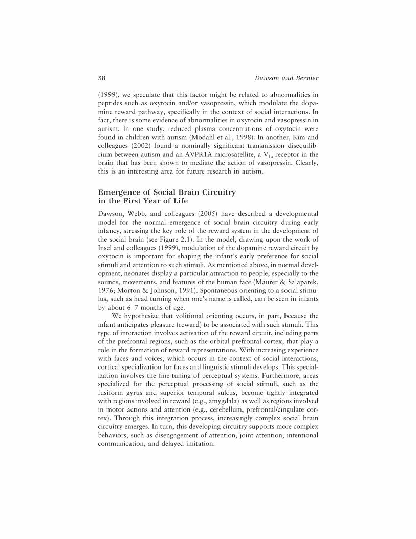
(1999), we speculate that this factor might be related to abnormalities inpeptides such as oxytocin and/or vasopressin, which modulate the dopa-mine reward pathway, specifically in the context of social interactions. Infact, there is some evidence of abnormalities in oxytocin and vasopressin inautism. In one study, reduced plasma concentrations of oxytocin werefound in children with autism (Modahl et al., 1998). In another, Kim andcolleagues (2002) found a nominally significant transmission disequilib-rium between autism and an AVPR1A microsatellite, a V1a receptor in thebrain that has been shown to mediate the action of vasopressin. Clearly,this is an interesting area for future research in autism.
Emergence of Social Brain Circuitryin the First Year of Life
Dawson, Webb, and colleagues (2005) have described a developmentalmodel for the normal emergence of social brain circuitry during earlyinfancy, stressing the key role of the reward system in the development ofthe social brain (see Figure 2.1). In the model, drawing upon the work ofInsel and colleagues (1999), modulation of the dopamine reward circuit byoxytocin is important for shaping the infant’s early preference for socialstimuli and attention to such stimuli. As mentioned above, in normal devel-opment, neonates display a particular attraction to people, especially to thesounds, movements, and features of the human face (Maurer & Salapatek,1976; Morton & Johnson, 1991). Spontaneous orienting to a social stimu-lus, such as head turning when one’s name is called, can be seen in infantsby about 6–7 months of age.
We hypothesize that volitional orienting occurs, in part, because theinfant anticipates pleasure (reward) to be associated with such stimuli. Thistype of interaction involves activation of the reward circuit, including partsof the prefrontal regions, such as the orbital prefrontal cortex, that play arole in the formation of reward representations. With increasing experiencewith faces and voices, which occurs in the context of social interactions,cortical specialization for faces and linguistic stimuli develops. This special-ization involves the fine-tuning of perceptual systems. Furthermore, areasspecialized for the perceptual processing of social stimuli, such as thefusiform gyrus and superior temporal sulcus, become tightly integratedwith regions involved in reward (e.g., amygdala) as well as regions involvedin motor actions and attention (e.g., cerebellum, prefrontal/cingulate cor-tex). Through this integration process, increasingly complex social braincircuitry emerges. In turn, this developing circuitry supports more complexbehaviors, such as disengagement of attention, joint attention, intentionalcommunication, and delayed imitation.
38 Dawson and Bernier
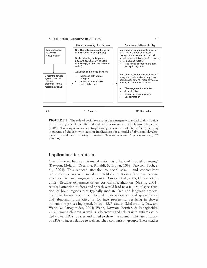
Implications for Autism
One of the earliest symptoms of autism is a lack of “social orienting”(Dawson, Meltzoff, Osterling, Rinaldi, & Brown, 1998; Dawson, Toth, etal., 2004). This reduced attention to social stimuli and concomitantreduced experience with social stimuli likely results in a failure to becomean expert face and language processor (Dawson et al., 2005; Grelotti et al.,2002). Because experience drives cortical specialization (Nelson, 2001),reduced attention to faces and speech would lead to a failure of specializa-tion of brain regions that typically mediate face and language process-ing. This failure would be reflected in decreased cortical specializationand abnormal brain circuitry for face processing, resulting in slowerinformation-processing speed. In two ERP studies (McPartland, Dawson,Webb, & Panagiotides, 2004; Webb, Dawson, Bernier, & Panagiotides,2006), young children as well as adolescents and adults with autism exhib-ited slower ERPs to faces and failed to show the normal right lateralizationof ERPs to faces relative to well-matched comparison groups. These studies
Social Brain Circuitry in Autism 39
FIGURE 2.1. The role of social reward in the emergence of social brain circuitryin the first years of life. Reproduced with permission from Dawson, G., et al.(2005). Neurocognitive and electrophysiological evidence of altered face processingin parents of children with autism: Implications for a model of abnormal develop-ment of social brain circuitry in autism. Development and Psychopathology, 17,679–697.
suggest that autism is associated with both slower information-processingspeed and atypical cortical specialization for face processing.
The abnormal trajectory for brain development in autism is not causedby a simple lack of exposure to human faces and voices. Infants withautism, like typically developing infants, are held, talked to, and fed bytheir parents during face-to-face interactions. However, if the infant withautism does not find such interactions inherently interesting or rewarding,then the infant might not actively attend to the face and voice or perceivethe face within a larger social–affective context. Recent research suggeststhat simple exposure to language does not necessarily facilitate the develop-ment of brain circuitry specialized for language (Kuhl, Tsao, & Liu, 2003).Rather, for speech perception to develop, language needs to be experiencedby the infant within a socially interactive context. Very early in life, infantsare capable of discerning differences among the phonetic units of all lan-guages, including both native and foreign languages. However, between 6and 12 months of age, as the brain becomes proficient at speech perception,the ability to discriminate foreign language phonetic units declines (Kuhl etal., 1997). Kuhl and colleagues (2003) investigated the possibility of pre-venting this decline in foreign language phonetic perception by exposingAmerican infants to native Mandarin Chinese speakers. They found thatthe decline of foreign language phonetic perception was preventable, butonly if exposure to speech occurred in the context of interpersonal interac-tion. That is, an experimental condition in which infants were exposed tothe same speech stimuli via audiotapes without social interaction did notavoid the narrowing of speech perception for the non-native language.
In the case of autism, if the child is not actively attending to faces andspeech sounds as part of the social context, his or her early exposure to thesesocial stimuli might not facilitate face and speech perception. The results of arecent study are consistent with this notion. Kuhl, Coffey-Corina, Padden,and Dawson (2004) found that listening preferences in 3- to 4-year-old chil-dren with autism differed dramatically from those of typically develop-ing children. The children with autism preferred listening to mechanical-sounding auditory signals (computer-based signals acoustically matched tospeech) rather than speech (motherese). The preference for the mechanical-sounding stimuli was associated with lower language ability, more severeautistic symptoms, and abnormal ERPs to speech sounds. The children withautism who did prefer motherese were more likely to show differentialERPs to different phonemes, whereas those who preferred the mechanical-sounding signal showed no such ERP waveform differences. We hypothesizethat a failure to affectively tag social and linguistic stimuli as relevant andrewarding, and the resultant failure to attend to such stimuli, impedes the cor-tical specialization for brain regions typically associated with face and lan-guage processing. Therefore, perceptual fine-tuning of such social stimuli and
40 Dawson and Bernier
the formation of representations of these stimuli are hampered. As a result,more complex behaviors that require the integration of social stimuli withcoordinated, intentional movements and volitional attention, such as disen-gagement of attention and joint attention, then fail to emerge.
Potential Impact of Early Intervention
If the social motivation hypothesis is correct, it should be possible to alterchildren’s attention to, and experience with, faces and speech through earlyintervention aimed at making social interactions more rewarding andmeaningful. The impact of such an intervention on the development of faceprocessing could then be assessed by examining the brain’s responses tofaces by using ERPs (Dawson & Zanolli, 2003). Through intervention,children can increase their use of eye contact, their use of affectiveexchanges, and their joint attention skills. An increase in these behavioralskills may be related to improvements such as increasing specialization inthe neural face processing system. Indeed, interventions based on appliedbehavior analysis are designed to enhance the reward value of social stimulithrough learning principles (see Figure 2.2). For example, during most earlyintervention programs, the therapist’s face (a previously neutral stimulus) isdeliberately paired with a nonsocial reinforcer (usually access to food or atoy). Via classical conditioning, the face then acquires reinforcer value.Early intervention could facilitate the development of the face processingsystem in two ways: first, by helping the child engage in meaningful socialinteractions that might lead to active attention to faces, and second, byaltering the child’s motivational preferences for faces so that engaging inface-to-face interaction becomes more rewarding and therefore more fre-quent (Dawson & Zanolli, 2003).
The timing of intervention might also have important consequences inrelation to the plasticity of the face processing system. Increases in socialmotivation and active attention to faces might have differing results in chil-dren as compared to adults. For example, adults with autism might benefitfrom being trained to attend to faces and being taught explicit face process-ing strategies. However, although these interventions might result in betterbehavioral performance, it is unclear if they would result in alterations inpatterns of neural activation. That is, interventions might support compen-satory processes but fail to activate or develop typical processing mecha-nisms. Conversely, given the plasticity of the developing brain, young chil-dren who receive early intervention might demonstrate both improvedbehavioral performance (e.g., increases in eye contact, joint attention, andface recognition) as well as normalized brain functioning. This normalizedbrain functioning could be observed in differential responses to familiarversus unfamiliar faces, different phonemes, speed of neural responses, and
Social Brain Circuitry in Autism 41
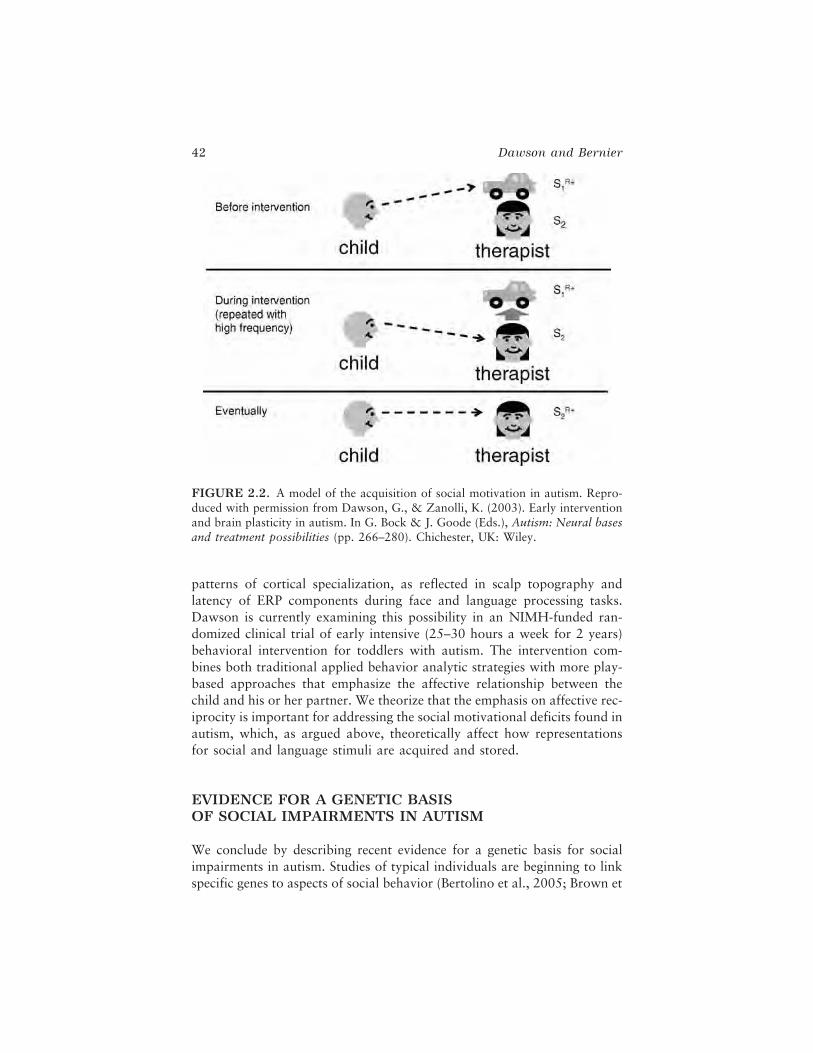
patterns of cortical specialization, as reflected in scalp topography andlatency of ERP components during face and language processing tasks.Dawson is currently examining this possibility in an NIMH-funded ran-domized clinical trial of early intensive (25–30 hours a week for 2 years)behavioral intervention for toddlers with autism. The intervention com-bines both traditional applied behavior analytic strategies with more play-based approaches that emphasize the affective relationship between thechild and his or her partner. We theorize that the emphasis on affective rec-iprocity is important for addressing the social motivational deficits found inautism, which, as argued above, theoretically affect how representationsfor social and language stimuli are acquired and stored.
EVIDENCE FOR A GENETIC BASISOF SOCIAL IMPAIRMENTS IN AUTISM
We conclude by describing recent evidence for a genetic basis for socialimpairments in autism. Studies of typical individuals are beginning to linkspecific genes to aspects of social behavior (Bertolino et al., 2005; Brown et
42 Dawson and Bernier
FIGURE 2.2. A model of the acquisition of social motivation in autism. Repro-duced with permission from Dawson, G., & Zanolli, K. (2003). Early interventionand brain plasticity in autism. In G. Bock & J. Goode (Eds.), Autism: Neural basesand treatment possibilities (pp. 266–280). Chichester, UK: Wiley.
al., 2005; Hariri et al., 2005), but autism risk genes have yet to be identi-fied. However, evidence for a genetic basis of autism does exist. There isstrong evidence for genetic influence in autism, with estimates of herit-ability ranging from 91–93% (Bailey et al., 1995). Several studies haveshown that identical twins are 60–95% concordant for autism (Bailey etal., 1995; Folstein & Rutter, 1977; Ritvo, Freeman, Mason-Brothers, Mo,& Ritvo, 1985; Steffenburg et al., 1989), whereas fraternal twins and sib-lings have a much lower concordance rate, with estimates ranging from 3–7% (August, Stewart, & Tsai, 1981; Bailey et al., 1995; Bolton et al., 1994;Smalley, Asarnow, & Spence, 1988). This rapid decrease in risk rates fromidentical twins to siblings and differential risk rates for male versus femalesiblings suggest epistatic effects involving interactions among as many as10 or more genes (Delong & Dwyer, 1988; Jorde, Mason-Brothers,Waldmann, & Ritvo, 1990; Pickles et al., 1995; Risch et al., 1999; Smalleyet al., 1988). Several linkage studies have reported moderate positive sig-nals on several chromosomes; however, findings have not been stronglyconsistent across these studies.
One challenging issue for genetic studies is the complex phenotype thatcomprises the autism syndrome. The disorder involves at least three differ-ent symptom domains (social, communication, and restrictive behaviors/flexibility) and its presentation is extremely heterogeneous. Furthermore,the autism phenotype appears to extend beyond classic autism to “lesservariant” phenotypes, referred to as the “broader autism phenotype”(Rutter, Bailey, Bolton, & Le Couteur, 1993). Numerous studies haveshown that relatives of individuals with autism, including parents and sib-lings, exhibit higher than normal rates of autism-related impairments(Bailey et al., 1995; Bailey, Phillips, & Rutter, 1996; Baker, Piven,Schwartz, & Patil, 1994; Bolton et al., 1994; Landa, Folstein, & Isaacs,1991; Landa et al., 1992; Narayan, Moyes, & Wolff, 1990; Wolff,Narayan, & Moyes, 1988). For example, Piven and colleagues (Piven,Palmer, Jacobi, Childress, & Arndt, 1997; Piven, Palmer, Landa, et al.,1997) found that parents of two or more children with autism showed ele-vated rates of social and communication impairments and stereotypedbehaviors, and Bolton and colleagues (1994) reported that 10–20% of sib-lings of individuals with autism exhibit symptoms related to the disorder,including language, learning, communication, and social impairments.
To date, most linkage studies have characterized the autism phenotypein terms of qualitative discrete diagnoses. However, it is likely that autismsusceptibility genes increase the chance of developing one or more compo-nents of the syndrome rather than causing autism, per se. In theory, itmight be the case that multiple genetically related traits accumulate to crossa threshold into the full-blown syndrome autism. If so, in order to identifythe genes related to autism it would be essential to define these genetically
Social Brain Circuitry in Autism 43
related traits, or endophenotypes, and determine their association with spe-cific genes (Dawson, Webb, et al., 2002; Holden, 2003). Such biological orbehavioral markers of latent vulnerability to autism are likely not discrete,all-or-nothing characteristics, but rather continuously distributed traits.
There have been few genetic studies that have attempted to measureautism-related traits along a continuum. Constantino and colleagues (Con-stantino, Przybeck, Friesen, & Todd, 2000; Constantino, Davis, et al., 2003)conducted one such study by developing a questionnaire that captured autismas one continuous trait. They found evidence for a genetic basis of this trait intwin studies (Constantino, Hudziak, & Todd, 2003; Constantino & Todd,2000, 2003). More recently, Dawson and colleagues (2006) have developeda quantitative measure of the autism broader phenotype that separatelyassesses several distinct domains of autism symptoms (social motivation,social expressiveness, conversation skills, and restrictive behaviors/flexibil-ity) as well as age of language onset. The Broader Phenotype Autism Symp-tom Scale (BPASS) assesses autism-related traits in both parents and siblingsof children with autism through interview and direct observation of behav-iors. Parents are interviewed about their own functioning or the functioningof their children and observations of both parent and child are made by theinterviewer through direct interactions. Nonverbal behaviors, such as eyecontact, are assessed via direct observation, whereas behaviors related torestricted activities and routines are assessed via interview. A genetic investi-gation of these quantitative traits was conducted using BPASS data collectedon a sample of 201 autism multiplex families that included 694 individuals(Sung et al., 2005). Participants included parents, probands, and nonaffectedsiblings from nuclear families that had at least two children on the autismspectrum. Multivariate polygenic models with ascertainment adjustment toestimate heritabilities and genetic and environmental correlations betweenthe traits were used. Among the traits analyzed, social motivation andrestricted activities/flexibility showed the highest heritability (0.19 and 0.16,respectively), indicating that these traits may be promising for gene mapping.These two traits also showed strong genetic correlation (0.92), suggesting ashared genetic basis.
Interestingly, in studies of face processing, not only children withautism but also parents of children with autism show decrements in thisskill (Dawson, Webb, et al., 2005). Viewing faces typically is associatedwith a faster and larger negative ERP component over the right temporalregion at about 170 milliseconds (ms) poststimulus presentation (this face-sensitive ERP is referred to as the N170; Bentin, Allison, Puce, Perez, &McCarthy, 1996; Kanwisher et al., 1997). As mentioned previously, it wasfound that the N170 was atypical in individuals with autism: They did notexhibit the expected N170 latency advantage for faces as compared to non-face stimuli, and they showed bilateral rather than right lateralized ERP
44 Dawson and Bernier
responses to faces (McPartland et al., 2004). Dawson, Webb, and col-leagues (2005) examined performance of parents of children with autismon standardized Wechsler cognitive tasks assessing verbal (Vocabulary,Verbal Comprehension), visual spatial (Block Design, Object Assembly),and face recognition (Immediate Memory for Faces) abilities. It was foundthat the parents of children with autism showed a significant deficit on theface recognition task relative to their performance on the visual spatial andverbal tasks; in fact, 29% of the sample had face recognition scores thatwere 1 standard deviation (SD) lower (> 3 points) than the other cognitivetasks. High-density ERPs to faces and chairs were recorded from a subsetof the parents of children with autism and control adults with no familialhistory of autism. Control adults demonstrated the expected larger right-than-left hemisphere N170 to faces, whereas parents of children withautism demonstrated reduced right hemisphere N170 amplitude to faces,resulting in bilaterally distributed ERPs to faces. Furthermore, controladults exhibited the expected pattern of a faster N170 to upright faces thanupright chairs, whereas parents of children with autism showed no signifi-cant difference in N170 latency to upright faces versus upright chairs.Based on these results, it can be hypothesized that face processing might bea functional neural trait marker of genetic susceptibility to autism.
Although these initial studies suggest that quantitative analysis ofautism symptom-related traits is a promising approach for genetic studies,ultimately, a more refined measure of functional neural trait markers—onethat is informed by contemporary affective and social neuroscience—willlikely yield greater precision and validity. Through the discovery of autismsusceptibility genes, it may someday be possible to identify newborn infantsat risk for autism, allowing for very early intervention. By providing appro-priate stimulation during the early years when social brain circuitry is firstdeveloping, prevention or at least meaningful amelioration of symptoms ofautism might eventually be possible, especially for children without signifi-cant comorbid mental retardation. As our understanding of the develop-ment of social brain circuitry evolves, our interventions can become moretargeted and focused on those aspects of social behavior that are consideredfundamental and pivotal for the acquisition of more complex social andcommunication skills over time.
ACKNOWLEDGMENTS
The writing of this chapter was supported by Grant No. U19HD34565 from theNational Institute of Child Health and Human Development (NICHD), as part ofthe NICHD Collaborative Program of Excellence in Autism, and Center Grant No.U54MH066399 from the National Institute of Mental Health, as part of theNational Institutes of Health STAART Centers Program.
Social Brain Circuitry in Autism 45
REFERENCES
Adrien, J., Faure, M., Perrot, A., Hameury, L., Garreau, B., Barthelemy, C., et al.(1991). Autism and family home movies: Preliminary findings. Journal ofAutism and Developmental Disorders, 21, 43–49.
American Psychiatric Association. (1994). Diagnostic and statistical manual ofmental disorders (4th ed.). Washington, DC: Author.
August, G. J., Stewart, M., & Tsai, L. (1981). The incidence of cognitive disabilitiesin the siblings of autistic children. British Journal of Psychiatry, 138, 416–422.
Avikainen, S., Wohlschlager, A., Liuhanen, S., Hanninen, R., & Hari, R. (2003).Impaired mirror-image imitation in Asperger and high-functioning autisticsubjects. Current Biology, 13(4), 339–341.
Bacon, A. L., Fein, D., Morris, R., Waterhouse, L., & Allen, D. (1998). Theresponses of autistic children to the distress of others. Journal of Autism andDevelopmental Disorders, 28(2), 129–142.
Bailey, A., Le Couteur, A., Gottesman, I., Bolton, P., Simonoff, E., Yuzda, E., et al.(1995). Autism as a strongly genetic disorder: Evidence from a British twinstudy. Psychological Medicine, 25, 63–77.
Bailey, A., Phillips, W., & Rutter, M. (1996). Autism: Toward an integration ofclinical, genetic, neuropsychological, and neurobiological perspectives. Journalof Child Psychology and Psychiatry and Allied Disciplines, 37, 89–126.
Baker, P., Piven, J., Schwartz, L., & Patil, S. (1994). Brief report: Duplication ofchromosome 15q11–13 in two individuals with autistic disorder. Journal ofAutism and Developmental Disorders, 24(4), 529–535.
Baron-Cohen, S. (1995). Mindblindness: An essay on autism and theory of mind.Cambridge, MA: Bradford/MIT Press.
Bentin, S., Allison, T., Puce, A., Perez, E., & McCarthy, G. (1996). Electro-physiological studies of face perception in humans. Journal of Cognitive Neu-roscience, 8, 551–565.
Bertolino, A., Arciero, G., Rubino, V., Latorre, V., De Candia, M., Mazzola, V., etal. (2005). Variation of human amygdala response during threatening stimulias a function of 5’httlpr genotype and personality style. Biological Psychiatry,57(12), 1517–1525.
Bolton, P., MacDonald, H., Pickles, A., Pios, P., Goode, S., Crowson, M., et al.(1994). A case-control family history of autism. Journal of Child Psychologyand Psychiatry, 35, 877–900.
Boucher, J., & Lewis, V. (1992). Unfamiliar face recognition in relatively able autis-tic children. Journal of Child Psychology and Psychiatry, 33, 843–859.
Boucher, J., Lewis, V., & Collis, G. (1998). Familiar face and voice matching andrecognition in children with autism. Journal of Child Psychology and Psychia-try, 39, 171–181.
Brooks, R., & Meltzoff, A. N. (2002). The importance of eyes: How infants inter-pret adult looking behavior. Developmental Psychology, 38(6), 958–966.
Brothers, L., Ring, B., & Kling, A. (1990). Response of temporal lobe neurons tosocial stimuli in maraca arctoides. Society of Neuroscience Abstract, 16, 184.
Brown, S. M., Peet, E., Manuck, S. B., Williamson, D. E., Dahl, R. E., Ferrell, R. E.,
46 Dawson and Bernier
et al. (2005). A regulatory variant of the human tryptophan hydroxylase-2gene biases amygdala reactivity. Molecular Psychiatry, 10(9), 884–888.
Carpenter, M., Nagell, K., & Tomasello, M. (1998). Social cognition, joint atten-tion, and communicative competence from 9 to 15 months of age. Monographof the Society for Research in Child Development, 63(4), 1–143.
Charman, T., Baron-Cohen, S., Swettenham, J., Baird, G., Drew A., & Cox, A.(2003). Predicting language outcome in infants with autism and pervasivedevelopmental disorder. International Journal of Language and Communica-tion Disorders, 38(3), 265–285.
Charman, T., Swettenham, J., Baron-Cohen, S., Cox, A., Baird, G., & Drew, A.(1998). An experimental investigation of social-cognitive abilities in infants withautism: Clinical implications. Infant Mental Health Journal, 19, 260–275.
Constantino, J., Davis, S. A., Todd, R. D., Schindler, M. K., Gross, M. M., Brophy,S. L., et al. (2003). Validation of a brief quantitative measure of autistic traits:Comparison of the social responsiveness scale with the Autism DiagnosticInterview—Revised. Journal of Autism and Developmental Disorders, 33,427–433.
Constantino, J., Hudziak, J. J., & Todd, R. D. (2003). Deficits in reciprocal socialbehavior in male twins: Evidence for a genetically independent domain of psy-chopathology. Journal of the American Academy of Child and Adolescent Psy-chiatry, 42, 458–467.
Constantino, J., Przybeck, T., Friesen, D., & Todd, R. D. (2000). Reciprocal socialbehavior in children with and without pervasive developmental disorders.Journal of Developmental and Behavioral Pediatrics, 21(1), 2–11.
Constantino, J., & Todd, R. D. (2000). Genetic structure of reciprocal social behav-ior. American Journal of Psychiatry, 157, 2043–2045.
Constantino, J., & Todd, R. D. (2003). Autistic traits in the general population: Atwin study. Archives of General Psychiatry, 60(5), 524–530.
Corona, R., Dissanayake, C., Arbelle, S., Wellington, P., & Sigman, M. (1998).Is affect aversive to young children with autism? Behavioral and cardiacresponses to experimenter distress. Child Development, 69, 1494–1502.
Darwin, C. (1965). The expression of the emotions in man and animals. London:Murray. (Original work published 1872)
Dawson, G., & Adams, A. (1984). Imitation and social responsiveness in autisticchildren. Journal of Abnormal Child Psychology, 12, 209–225.
Dawson, G., Carver, L., Meltzoff, A., Panagiotides, H., McPartland, J., & Webb, S.(2002). Neural correlates of face and object recognition in young children withautism spectrum disorder, developmental delay, and typical development.Child Development, 3(3), 700–717.
Dawson, G., Estes, A., Munson, J., Schellenberg, G., Bernier, R., & Abbott, R.(2006). Quantitative Assessment of Autism Symptom–related traits in pro-bands and parents: Broader Phenotype Autism Symptom Scale. Journal ofAutism and Developmental Disorders.
Dawson, G., Hill, D., Galpert, L., Spencer, A., & Watson, L. (1990). Affectiveexchanges between young autistic children and their mothers. Journal ofAbnormal Child Psychology, 18, 335–345.
Social Brain Circuitry in Autism 47
Dawson, G., & Lewy, A. (1989). Arousal, attention, and the socioemotionalimpairments of individuals with autism. In G. Dawson (Ed.), Autism: Nature,diagnosis, and treatment (pp. 49–74). New York: Guilford Press.
Dawson, G., Meltzoff, A. N., Osterling, J., & Rinaldi, J. (1998). Neuropsychologi-cal correlates of early symptoms of autism. Child Development, 69, 1276–1285.
Dawson, G., Meltzoff, A. N., Osterling, J., Rinaldi, J., & Brown, E. (1998).Children with autism fail to orient to naturally occurring social stimuli. Jour-nal of Autism and Developmental Disorders, 28, 479–485.
Dawson, G., Munson, J., Estes, A., Osterling, J., McPartland, J., Toth, K., et al.(2002). Neurocognitive function and joint attention ability in young childrenwith autism spectrum disorder versus developmental delay. Child Develop-ment, 73(2), 345–358.
Dawson, G., Osterling, J., Meltzoff, A., & Kuhl, P. (2000). Case study of the devel-opment of an infant with autism from birth to two years of age. Journal ofApplied Developmental Psychology, 21(3), 299–313.
Dawson, G., Toth, K., Abbott, R., Osterling, J., Munson, J., Estes, A., et al. (2004).Early social attention impairments in young children with autism: Social ori-enting, joint attention, and attention to distress. Developmental Psychology,40, 271–283.
Dawson, G., Webb, S., Carver, L., Panagiotides, H., & McPartland, J. (2004).Young children with autism show atypical brain responses to fearful versusneutral facial expressions. Developmental Science, 7, 340–359.
Dawson, G., Webb, S., & McPartland, J. (2005).Understanding the nature of faceprocessing impairment in autism: Insights from behavioral and electrophysio-logical studies. Developmental Neuropsychology, 27(3), 403–424.
Dawson, G., Webb, S., Schellenberg, G., Aylward, E., Richards, T., Dager, S., et al.(2002). Defining the broader phenotype of autism: Genetic, brain, and behav-ioral perspectives. Development and Psychopathology, 14, 581–611.
Dawson, G., Webb, S., Wijsman, E., Schellenberg, G., Estes, A., Munson, J., et al.(2005). Neurocognitive and electrophysiological evidence of altered face pro-cessing in parents of children with autism: Implications for a model of abnor-mal development of social brain circuitry in autism. Development and Psycho-pathology, 17, 679–697.
Dawson, G., & Zanolli, K. (2003). Early intervention and brain plasticity inautism. In G. Bock & J. Goode (Eds.), Autism: Neural bases and treatmentpossibilities (pp. 226–280). Chichester, UK: Wiley.
de Haan, M., & Nelson, C. (1997). Recognition of the mother’s face by 6-month-old infants: A neurobehavioral study. Child Development, 68, 187–210.
de Haan, M., & Nelson, C. (1999). Electrocortical correlates of face and object rec-ognition by 6-month-old infants. Developmental Psychology, 35, 1113–1121.
de Haan, M., Nelson, C. A., Gunnar, M. R., & Tout, K. A. (1998). Hemisphericdifferences in brain activity related to the recognition of emotional expres-sions by 5-year-old children. Developmental Neuropsychology, 14(4), 495–518.
Delong, G. R., & Dwyer, J. H. (1988). Correlation of family history with specific
48 Dawson and Bernier
autistic subgroups: Aspergers and bipolar affective disease. Journal of Autismand Developmental Disorders, 18, 593–600.
Eckerman, C., Davis, C., & Didow, S. (1989). Toddlers’ emerging ways of achiev-ing social coordinations with a peer. Child Development, 60(2), 440–453.
Eimer, M., & Holmes, A. (2002). An ERP study on the time course of emotionalface processing. NeuroReport, 13(4), 427–431.
Fagan, J. (1972). Infants’ recognition memory for face. Journal of ExperimentalChild Psychology, 14, 453–476.
Feinman, S. (1982). Social referencing in infancy. Merrill–Palmer Quarterly, 28,445–470.
Ferguson, J., Young, H., Hearn, E., Insel, T., & Winslow, J. (2000). Social amnesiain mice lacking the oxytocin gene. Nature Genetics, 25, 284–288.
Ferguson, J., Young, H., & Insel, T. R. (2002). The neuroendocrine basis of socialrecognition. Frontiers in Neuroendocrinology, 23, 200–224.
Folstein, S., & Rutter, M. (1977). Infantile autism: A genetic study of 21 twin pairs.Journal of Child Psychology and Psychiatry, 18, 297–321.
Gepner, B., de Gelder, B., & de Schonen, S. (1996). Face processing in autistics:Evidence for a generalized deficit? Child Neuropsychology, 2(2), 123–139.
Gingrich, B., Liu, Y., Cascio, C., Wang, Z., & Insel, T. R. (2000). Dopamine D2receptors in the nucleus accumbens are important for social attachment infemale prairie voles. Behavioral Neuroscience, 114, 173–183.
Goren, C., Sarty, M., & Wu, P. (1975). Visual following and pattern discriminationof face-like stimuli by newborn infants. Pediatrics, 56, 544–549.
Grelotti, D., Gauthier, I., & Schultz, R. (2002). Social interest and the developmentof cortical face specialization: What autism teaches us about face processing.Developmental Psychobiology, 40, 213–225.
Hariri, A. R., Drabant, E. M., Munoz, K. E., Kolachana, B. S., Mattay, V. S., Egan,M. F., et al. (2005). A susceptibility gene for affective disorders and theresponse of the human amygdala. Archives of General Psychiatry, 62(2), 146–152.
Hatfield, E., Cacioppo, H., & Rapson, R. (1994). Emotional contagion. New York:Cambridge University Press.
Haxby, J., Horwitz, B., Ungerleider, L., Maisog, J., Pietrini, P., & Grady, C. (1994).The functional organization of human extrastriate cortex: A PET–rCBF studyof selective attention to faces and locations. Journal of Neuroscience, 14,6336–6353.
Haxby, J., Ungerleider, L., Clark, A., Schouten, J., Hoffman, E., & Martin, A.(1999). The effect of face inversion on activity in human neural systems forface and object perception. Neuron, 22, 189–199.
Hertzig, M., Snow, M., & Sherman, T. (1989). Affect and cognition in autism.Journal of the American Academy of Child and Adolescent Psychiatry, 28,195–199.
Hobson, P., Ouston, J., & Lee, A. (1988). Emotion recognition in autism: Coordi-nating faces and voices. Psychological Medicine, 18, 911–923.
Hobson, R., & Lee, A. (1999). Imitation and identification in autism. Journal ofChild Psychology and Psychiatry, 40(4), 649–659.
Social Brain Circuitry in Autism 49
Hoffman, E., & Haxby, J. (2000). Distinct representations of eye gaze and identityin the distributed human neural system for face perception. Nature Neurosci-ence, 3, 80–84.
Holden, C. (2003). Deconstructing schizophrenia. Science, 299, 333–335.Insel, T. R. (1997). A neurobiological basis of social attachment. American Journal
of Psychiatry, 154(6), 726–735.Insel, T. R., & Fernald, R. D. (2004). How the brain processes social information:
Searching for the social brain. Annual Review of Neuroscience, 27, 697–722.Insel, T. R., & Hulihan, T. J. (1995). A gender-specific mechanism for pair bond-
ing: Oxytocin and partner preference formation in monogamous voles. Behav-ioral Neuroscience, 109, 782–789.
Insel, T. R., O’Brien, D. J., & Leckman, J. F. (1999). Oxytocin, vasopressin, andautism: Is there a connection? Biological Psychiatry, 45, 145–157.
Jorde, L. B., Mason-Brothers, A., Waldmann, R., & Ritvo, E. (1990). The UCLA–University of Utah epidemiology survey of autism: Genealogical analysis offamilial aggregation. American Journal of Medical Genetics, 36, 85–88.
Joseph, R., & Tanaka, J. (2003). Holistic and part-based face recognition in chil-dren with autism. Journal of Child Psychology and Psychiatry, 44(4), 529–542.
Kampe, K., Frith, C., Dolan, R., & Frith, U. (2001). Attraction and gaze—thereward of social stimuli. Nature, 413, 589.
Kanwisher, N., McDermott, J., & Chun, M. (1997). The fusiform face area: Amodule in human extrastriate cortex specialized for face perception. Journal ofNeuroscience, 17, 4302–4311.
Kasari, C., Sigman, M., Mundy, P., & Yirmiya, N. (1990). Affective sharing in thecontext of joint attention interactions of normal, autistic, and mentally retardedchildren. Journal of Autism and Developmental Disorders, 20, 87–100.
Kim, S. J., Young, L. J., Gonen, D., Veenstra-VanderWeele, J., Courchesne, R.,Courchesne, E., et al. (2002). Transmission disequilibrium testing of argininevasopressin receptor 1A (AVPR1A) polymorphisms in autism. Molecular Psy-chiatry, 7(5), 503–507.
Klin, A., Jones, W., Schultz, R., Volkmar, F., & Cohen, D. (2002). Visual fixationpatterns during viewing of naturalistic social situations as predictors of socialcompetence in individuals with autism. Archives of General Psychiatry, 59,809–816.
Klin, A., Sparrow, S., deBildt, A., Cicchetti, D., Cohen, D., & Volkmar, F. (1999).A normed study of face recognition in autism and related disorders. Journal ofAutism and Developmental Disorders, 29, 499–508.
Klinger, L., & Dawson, G. (2001). Prototype formation in children with autism andDown syndrome. Development and Psychopathology, 13, 111–124.
Kugiumutzakis, G. (1999). Genesis and development of early infant mimesis tofacial and vocal models. In J. Nadel & G. Butterworth (Eds.), Imitation ininfancy (pp. 36–59). Cambridge, UK: Cambridge University Press.
Kuhl, P., Andruski, J., Chistovich, I. A., Chistovich, L. A., Kozhevnikova, E.,Ryskina, V., et al. (1997). Cross-language analysis of phonetic units in lan-guage addressed to infants. Science, 277, 684–686.
50 Dawson and Bernier
Kuhl, P., Coffey-Corina, S., Padden, D., & Dawson, G. (2004). Links betweensocial and linguistic processing of speech in preschool children with autism:Behavioral and electrophysiological measures. Developmental Science, 7, 19–30.
Kuhl, P., Tsao, F., & Liu, H. (2003). Foreign-language experience in infancy:Effects of short-term exposure and social interaction on phonetic learning.Proceedings of the National Academy of Sciences USA, 100(15), 9096–9101.
Landa, R., Folstein, S., & Isaacs, C. (1991). Spontaneous narrative discourse per-formance of parents of autistic individuals. Journal of Speech and HearingResearch, 34, 1339–1345.
Landa, R., Piven, J., Wzorek, M., Gayle, J., Chase, G., & Folstein, S. (1992). Sociallanguage use in parents of autistic individuals. Psychological Medicine, 22,245–254.
Langdell, T. (1978). Recognition of faces: An approach to the study of autism.Journal of Child Psychology and Psychiatry, 19, 255–268.
Loveland, K., Tunali-Kotoski, B., Pearson, D., Bresford, K., Ortegon, J., & Chen,C. (1994). Imitation and expression of facial affect in autism. Developmentand Psychopathology, 6, 433–444.
Maurer, D., & Salapatek, P. (1976). Developmental changes in the scanning offaces by young infants. Child Development, 47(2), 523–527.
McPartland, J., Dawson, G., Webb, S., & Panagiotides, H. (2004). Event-relatedbrain potentials reveal anomalies in temporal processing of faces in autism.Journal of Child Psychiatry and Psychology, 45(7), 1235.
Meltzoff, A., & Decety, J. (2003). What imitation tells us about social cognition: Arapprochement between developmental psychology and cognitive neurosci-ence. Philosophical Transactions of the Royal Society: Biological Sciences,358, 491–500.
Meltzoff, A., & Gopnick, A. (1993). The role of imitation in understanding per-sons and developing a theory of mind. In S. Baron-Cohen, H. Tager-Flusberg,& D. J. Cohen (Eds.), Understanding other minds: Perspectives from autism(pp. 335–336). Oxford, UK: Oxford University Press.
Meltzoff, A., & Moore, M. (1977). Imitation of facial and manual gestures byhuman neonates. Science, 198, 75–78.
Meltzoff, A., & Moore, M. (1979). Interpreting “imitative” responses in earlyinfancy. Science, 205, 217–219.
Meltzoff, A., & Moore, M. (1983). Newborn infants imitate adult facial gestures.Child and Development, 54(3), 702–709.
Meltzoff, A., & Moore, M. (1994). Imitation, memory, and the representation ofpersons. Infant Behavior and Development, 17, 83–99.
Modahl, C., Green, L., Fein, D., Morris, M., Waterhouse, L., Feinstein, C., et al.(1998). Plasma oxytocin levels in autistic children. Biological Psychiatry, 43,270–277.
Moore, C., & Corkum, V. (1994). Social understanding at the end of the first yearof life. Developmental Review, 14, 349–372.
Morales, M., Mundy, P., & Rojas, J. (1998). Brief report: Following the direction
Social Brain Circuitry in Autism 51
of gaze and language development in 6-month-olds. Infant Behavior andDevelopment, 21, 373–377.
Morton, J., & Johnson, M. H. (1991). CONSPEC and CONLERN: A two-processtheory of infant face recognition. Psychological Review, 2, 164–181.
Mundy, P., & Neal, R. (2001). Neural plasticity, joint attention, and a transaction-al social-orienting model of autism. San Diego, CA: Academic Press.
Mundy, P., Sigman, M., Ungerer, J., & Sherman, T. (1986). Defining the social def-icits of autism: The contribution of nonverbal communication measure. Jour-nal of Child Psychology and Psychiatry, 27, 657–669.
Nadel, J., Guerini, C., Peze, A., & Rivet, C. (1999). The evolving nature of imita-tion as a format for communication. In J. Nadel & G. Butterworth (Eds.), Imi-tation in infancy (pp. 209–234). Cambridge, UK: Cambridge University Press.
Narayan, S., Moyes, B., & Wolff, S. (1990). Family characteristics of autistic chil-dren: A further report. Journal of Autism and Developmental Disorders,20(4), 523–535.
Nelson, C. A. (2001). The development and neural bases of face recognition. Infantand Child Development, 10(1–2), 3–18.
Nelson, C. A., & de Haan, M. (1996). Neural correlates of infants’ visual respon-siveness to facial expressions of emotion. Developmental Psychobiology, 29,577–595.
Nishimori, K., Young, L., Guo, Q., Wang, Z., Insel, T. R., & Matzuk, M. M.(1996). Oxytocin is required for nursing but is not essential for parturition orreproductive behavior. Proceedings of the National Academy of Science USA,93, 11699–11704.
Osterling, J., & Dawson, G. (1994). Early recognition of children with autism: Astudy of first birthday home videotapes. Journal of Autism and DevelopmentalDisorders, 24, 247–257.
Osterling, J., Dawson, G., & Munson, J. (2002). Early recognition of one-year-oldinfants with autism spectrum disorder versus mental retardation: A study offirst birthday party home videotapes. Development and Psychopathology, 14,239–251.
Pedersen, C., Caldwell, J., Walker, C., Ayers, G., & Mason, G. (1994). Oxytocinactivates the postpartum onset of rat maternal behavior in the ventraltegmental and medial preoptic areas. Behavioral Neuroscience, 108, 1163–1171.
Pelphrey, K., Sasson, N., Reznick, J., Paul, G., Goldman, B., & Piven, J. (2002).Visual scanning of faces in autism. Journal of Autism and Developmental Dis-orders, 32(4), 249–261.
Perrett, D., Harries, M., Mistlin, A., & Hietanen, J. (1990). Social signals analyzedat the single cell level: Someone is looking at me, something touched me, some-thing moved. International Journal of Comparative Psychology, 4, 25–55.
Perrett, D., Hietanen, J., Oram, M., & Benson, P. (1992). Organization and func-tions of cells responsive to faces in the temporal cortex. In V. Bruce & A.Cowey (Eds.), Processing the facial image (pp. 23–30). New York: ClarendonPress.
Perrett, D., Smith, P., Mistlin, A., Chitty, A., Head, A., Potter, D., et al. (1985).
52 Dawson and Bernier
Visual analysis of body movements by neurons in the temporal cortex of themacaque monkey: A preliminary report. Behavioral Brain Research, 16, 153–170.
Piaget, J. (1962). Play, dreams and imitation in childhood. New York: Norton.Pickles, A., Bolton, P., MacDonald, H., Bailey, A., Le Couteur, A., Sim, C. H., et al.
(1995). Latent-class analysis of recurrence risks for complex phenotypes withselection and measurement error: A twin and family history study of autism.American Journal of Human Genetics, 57, 717–726.
Piven, J., Palmer, P., Jacobi, D., Childress, D., & Arndt, S. (1997). Broader autismphenotype: Evidence from a family history study of multiple-incidence autismfamilies. American Journal of Psychiatry, 154, 185–190.
Piven, J., Palmer, P., Landa, R., Santangelo, S., Jacobi, D., & Childress, D. (1997).Personality and language characteristics in parents from multiple-incidenceautism families. American Journal of Medical Genetics, 74, 398–411.
Rheingold, H. L., Hay, D. F., & West, M. J. (1976). Sharing in the second year oflife. Child Development, 47, 1148–1158.
Risch, N., Spiker, D., Loptspeich, L., Nouri, N., Hinds, D., Hallmayer, J., et al.(1999). A genomic screen of autism: Evidence for a multilocus etiology. Amer-ican Journal of Human Genetics, 65(2), 493–507.
Ritvo, E., Freeman, B., Mason-Brothers, A., Mo, A., & Ritvo, A. (1985). Concor-dance for the syndrome of autism in 40 pairs of afflicted twins. AmericanJournal of Psychiatry, 142, 74–77.
Rochat, P. (1999). Early social cognition: Understanding others in the first monthsof life. Mahwah, NJ: Erlbaum.
Rochat, P., & Striano, T. (1999). Emerging self-exploration by 2-month-olds.Developmental Science, 2, 206–218.
Rogers, S., Bennetto, L., McEvoy, R., & Pennington, B. (1996). Imitation and pan-tomime in high-functioning adolescents with autism spectrum disorders. ChildDevelopment, 67(5), 2060–2073.
Rogers, S., Hepburn, S., Stackhouse, T., & Wehner, E. (2003). Imitation perfor-mance in toddlers with autism and those with other developmental disorders.Journal of Child Psychology and Psychiatry, 44(5), 763–781.
Rogers, S., & Pennington, B. (1991). A theoretical approach to the deficits in infan-tile autism. Development and Psychopathology, 3, 137–162.
Rutter, M., Bailey, A., Bolton, P., & Le Couteur, A. (1993). Autism: Syndrome defi-nition and possible genetic mechanisms. In R. Plomin & G. E. McClearn(Eds.), Nature, nurture, and psychology (pp. 269–284). Washington, DC:American Psychological Association.
Schoenbaum, G., Setlow, B., Saddoris, M., & Gallagher, M. (2003). Encoding pre-dicted outcome and acquired value in orbitofrontal cortex during cue sam-pling depends upon input from basolateral amygdala. Neuron, 39(5), 731–733.
Schultz, W. (1998). Predictive reward signal of dopamine neurons. Journal ofNeurophysiology, 80, 1–27.
Serra, M., Althaus, M., de Sonneville, L. M., Stant, A.D., Jackson, A. E., &Minderaa, R. B. (2003). Face recognition in children with a pervasive develop-
Social Brain Circuitry in Autism 53
mental disorder not otherwise specified. Journal of Autism and DevelopmentalDisorders, 33(3), 303–317.
Sigman, M., Kasari, C., Kwon, J., & Yirmiya, N. (1992). Responses to the negativeemotions of others by autistic, mentally retarded, and normal children. ChildDevelopment, 63, 796–807.
Sigman, M., & Ruskin, E. (1999). Continuity and change in the social competenceof children with autism, Down syndrome, and developmental delays. Mono-graphs of the Society for Research in Child Development, 64(1), Serial No.256.
Smalley, S. L., Asarnow, R. F., & Spence, A. (1988). Autism and genetics: A decadeof research. Archives of General Psychiatry, 45, 953–961.
Steffenburg, S., Gillberg, C., Hellgren, L., Andresson, L., Gillberg, I., Jakobsson, G.,et al. (1989). A twin study of autism in Denmark, Finland, Iceland, Norway,and Sweden. Journal of Child Psychology and Psychiatry, 30, 405–416.
Stone, W., Lemanek, K., Fishel, P., Fernandez, M., & Altemeier, W. (1990). Playand imitation skills in the diagnosis of autism in young children. Pediatrics,86, 267–272.
Stone, W., Ousley, O., & Littleford, C. (1997). Motor imitation in young childrenwith autism: What’s the object? Journal of Abnormal Child Psychology, 25,475–485.
Sung, J. U., Dawson, G., Munson, J., Estes, A., Schellenberg, J., & Wijsman, E. M.(2005). Genetic investigation of quantitative traits related to autism: Use ofmultivariate polygenic models with ascertainment adjustment. American Jour-nal of Human Genetics, 76, 68–81.
Swettenham, J., Baron-Cohen, S., Charman, T., Cox, A., Baird, G., Drew, et al.(1998). The frequency and distribution of spontaneous attention shifts be-tween social and nonsocial stimuli in autistic, typically developing, and non-autistic developmentally delayed infants. Journal of Child Psychology and Psy-chiatry, 39(5), 747–753.
Symons, L., Hains, S., & Muir, S. (1998). Look at me: 5-month-old infants’ sensi-tivity to very small deviations in eye-gaze during social interactions. InfantBehavior and Development, 21, 531–536.
Tantam, D., Monaghan, L., Nicholson, J., & Stirling, J. (1989). Autistic children’sability to interpret faces: A research note. Journal of Child Psychology andPsychiatry, 30, 623–630.
Trepagnier, C., Sebrechts, M., & Peterson, R. (2002). Atypical face gaze in autism.Cyberpsychological Behavior, 5(3), 213–217.
Trevarthen, C. (1979). Communication and cooperation in early infancy: Adescription of primary intersubjectivity. In M. Bullowa (Ed.), Before speech:The beginnings of interpersonal communication (pp. 321–347). Cambridge,UK: Cambridge University Press.
Trevarthen, C., Kokkinaki, T., & Fiamenghi, G., Jr. (1999). What infants’ imita-tions communicate: With mothers, with fathers, and with peers. In J. Nadel &G. Butterworth (Eds.), Imitation in infancy (pp. 127–185). Cambridge, UK:Cambridge University Press.
Tronick, E., Als, H., Adamson, L., Wise, S., & Brazelton, T. B. (1978). The infant’s
54 Dawson and Bernier
response to entrapment between contradictory messages in face-to-face inter-action. Journal of the American Academy of Child Psychiatry, 17, 1–13.
U�giris, E. (1981). Probing immune reactions by laser light scattering spectroscopy.Methods in Enzymology, 74, 177–198.
U�giris, E. (1999). Imitation as activity: Its developmental aspect. In J. Nadel & G.Butterworth (Eds.), Imitation in infancy (pp. 209–234) Cambridge, UK: Cam-bridge University Press.
van der Geest, J., Kemner, C., Verbatem, M., & van Engeland, H. (2002). Gazebehavior of children with pervasive developmental disorder toward humanfaces: A fixation time study. Journal of Child Psychiatry and Psychology, 443,669–678.
Walton, G., & Bower, T. (1993). Amodal representations of speech in infants.Infant Behavior and Development, 16(2), 233–243.
Waterhouse, L., Fein, D., & Modahl, C. (1996). Neurofunctional mechanisms inautism. Psychological Review, 103, 457–489.
Webb, S. J., Dawson, G., Bernier, R., & Panagiotides, H. (2006). ERP evidence ofatypical face processing in young children with autism. Journal of Autism andDevelopmental Disorders, 36, 881–890.
Werner, E., Dawson, G., Osterling, J., & Dinno, J. (2000). Recognition of autismbefore 1 year of age: A retrospective study based on home videotapes. Journalof Autism and Developmental Disorders, 30, 157–162.
Williams, J., Whiten, A., Suddendorf, T., & Perrett, D. (2001). Imitation, mirrorneurons and autism. Neuroscience and Biobehavioral Review, 25(4), 287–295.
Winslow, J., Hastings, N., Carter, C. S., Harbaugh, C. R., & Insel, T. R. (1993). Arole for central vasopressin in pair bonding in monogamous prairie voles.Nature, 365, 545–548.
Witt, D. M., Winslow, J. T., & Insel, T. R. (1992). Enhanced social interactions inrats following chronic, centrally infused oxytocin. Pharmacology, Biochemis-try, and Behavior, 43(3), 855–861.
Wolff, S., Narayan, S., & Moyes, B. (1988). Personality characteristics of parents ofautistic children: A controlled study. Journal of Child Psychology and Psychia-try, 29, 143–153.
Zahn-Waxler, C., & Radke-Yarrow, M. (1990). The origins of empathic concern.Motivation and Emotion, 14, 107–130.
Social Brain Circuitry in Autism 55
C H A P T E R 3
Brain Mechanisms UnderlyingSocial Perception Deficits in Autism
Kevin A. PelphreyElizabeth J. Carter
Autism is a severe and pervasive neurodevelopmental disorder character-ized by the presence and distinct developmental course of a triad of deficits:(1) social reciprocity and engagement deficits, (2) communication and lan-guage skills deficits, and (3) stereotyped, repetitive behaviors and limitedinterests (American Psychiatric Association, 1994; see also Dawson &Bernier, Chapter 2, this volume). Within these three domains, there is agreat deal of variability in the extent of deficits. For example, social domainsymptoms can range from a nearly complete absence of interest in interper-sonal interaction to subtler difficulties in managing complex social interac-tions, understanding social contexts, and detecting the intentions of others.Language deficits can vary from mutism to mild pragmatic language defi-cits. The restricted, repetitive, and stereotyped behaviors and interests canbe anything from a preference for sameness and exhibition of simple motorstereotypies to performance of extremely elaborate, complex rituals withsevere distress upon their interruption. Along with the great variability inthese three behavioral domains, there is also a wide range in general intel-lect, with the majority of individuals with autism exhibiting mental retarda-tion, but others possessing intelligence quotients (IQs) that are average orsuperior.
56
Approximately two to three per 1,000 individuals meet the criteriafor a diagnosis of autism (Fombonne, 1999); however, this numberwould be significantly higher if it included other disorders falling on the“autism spectrum,” such as Asperger syndrome and pervasive devel-opmental disorder not otherwise specified (Folstein & Rosen-Sheidley,2001). These other developmental disorders share many of the same fea-tures of autism, such as early social and communication deficits, but donot include the full range of symptoms. The spectrum is further expandedby the inclusion of the broad autism phenotype for describing individualswho do not meet criteria for the other autism spectrum disorders butwho have milder deficits in the three major domains (Folstein & Rutter,1977; see Piven, 2001, for a review). In their twin study of autism, onthe basis of finding a higher concordance rate for a broad autism pheno-type, Folstein and Rutter (1977) proposed the idea that the genetic liabil-ity for autism might be expressed in nonautistic relatives in characteristicsthat were milder but qualitatively similar to those seen in autism. Often,these characteristics are seen in family members of individuals withautism. In parents, this may include subtle difficulties in language andcommunication (Landa et al., 1992; Piven, Palmer, Jacobi, Childress, &Arndt, 1997), social impairments and certain personality characteristics(e.g., aloofness and rigidity; Murphy et al., 2000; Piven et al., 1994;Wolff, Narayan, & Moyes, 1988), and higher rates of depression andsocial phobia (Piven & Palmer, 1997, 1999). Additionally, executivefunctioning deficits have been found in parents (Hughes, Leboyer, &Bouvard, 1997) and siblings (Hughes, Plumet, & Leboyer, 1999) of indi-viduals with autism.
Higher rates of concordance in monozygotic twins and of recurrencewithin families than typically are seen in the general population suggestthat there is a significant genetic component to autism (Cook et al., 1998;see Bailey et al., 1995, for a review), and heritability rates are estimated at60–70% (Veenstra-VanDerWeele, Cook, & Lombroso, 2003). In addition,significantly more males than females meet criteria for the disorder, withestimates of this ratio as high as 4:1 (Wing, 1981; Yeargin-Allsopp et al.,2003). The source of this sex difference could be relatively indirect; forexample, it might emerge through the effects of gonadal steroids on braindevelopment. But it could also be more direct, vis-à-vis expression of pro-teins on the sex chromosomes in neurons. Approximately 10% of individu-als with autism are also affected by a medical condition associated with thedisorder, such as fragile X syndrome, tuberous sclerosis, or Smith–Lemli–Opitz syndrome (Bailey, Hatton, Skinner, & Mesibov, 2001; Baker, Piven,& Sato, 1998; Martin et al., 2001). In those without another medical con-dition (i.e., idiopathic autism), the underlying mechanism is probably the
Social Perception Deficits in Autism 57
result of the interactions of a number of genes (Pickles et al., 1995; Risch etal., 1999).
Although individuals with autism differ in the extent to which theydemonstrate each of these impairments, evidence suggests that social defi-cits are the primary feature of autism (Kanner, 1943; Wing & Gould,1979), and only the social deficits are specific to autism and its related dis-orders. Indeed, early emerging problems with social engagement might ini-tiate a developmental cascade that leads to the commonly seen communica-tion delays and deficits. For instance, social isolation has been shown tocause stereotyped and repetitive behaviors in monkeys that are similar tothose observed in autism (e.g., Cross & Harlow, 1965). Moreover, deficitsin social perception, including visual joint attention, are important earlyindicators of autism (Baron-Cohen, 1995; Dawson, Munson, et al., 2002;Lord et al., 1997; Sigman et al., 1999). Finally, research and theory indevelopmental psychology support the notion that cognitive operations,including executive functions and learning strategies, are coconstructedover ontogeny in the context of social and cultural interactions (e.g.,Fischer & Bidell, 1998; Rogoff, 1990; Valsiner, 1987; Vygotsky, 1978) inwhich children with autism do not readily participate. Here, we review thegrowing knowledge base concerning the neural basis of specific deficits insocial functioning in autism.
Social perception refers to the initial stages of evaluating the intentionsof others by analyzing gaze direction, body movement, and other types ofbiological motion (Allison, Puce, & McCarthy, 2000). This construct fallswithin a domain of cognitive skills referred to by different laboratories associal cognition, social attention, theory of mind, and mentalizing. Theessence of social perception is captured by the notion of an “intentionalstance,” introduced by the philosopher Dennett (1987) to describe theassumptions human beings tend to make when interpreting and predictingthe behavior of others:
Here is how it works: first you decide to treat the object whose behavior is tobe predicted as a rational agent; then you figure out what beliefs that agentought to have, given its place in the world and its purpose. Then you figure outwhat desires it ought to have, on the same considerations, and finally you pre-dict that this rational agent will act to further its goals in the light of its beliefs.A little practical reasoning from the chosen set of beliefs and desires will inmost instances yield a decision about what the agent ought to do; that is whatyou predict the agent will do. (p. 17)
Here Dennett eloquently described a set of processes that occurs in a rapid,automatic fashion during social perception in typically developing individu-als.
58 Pelphrey and Carter
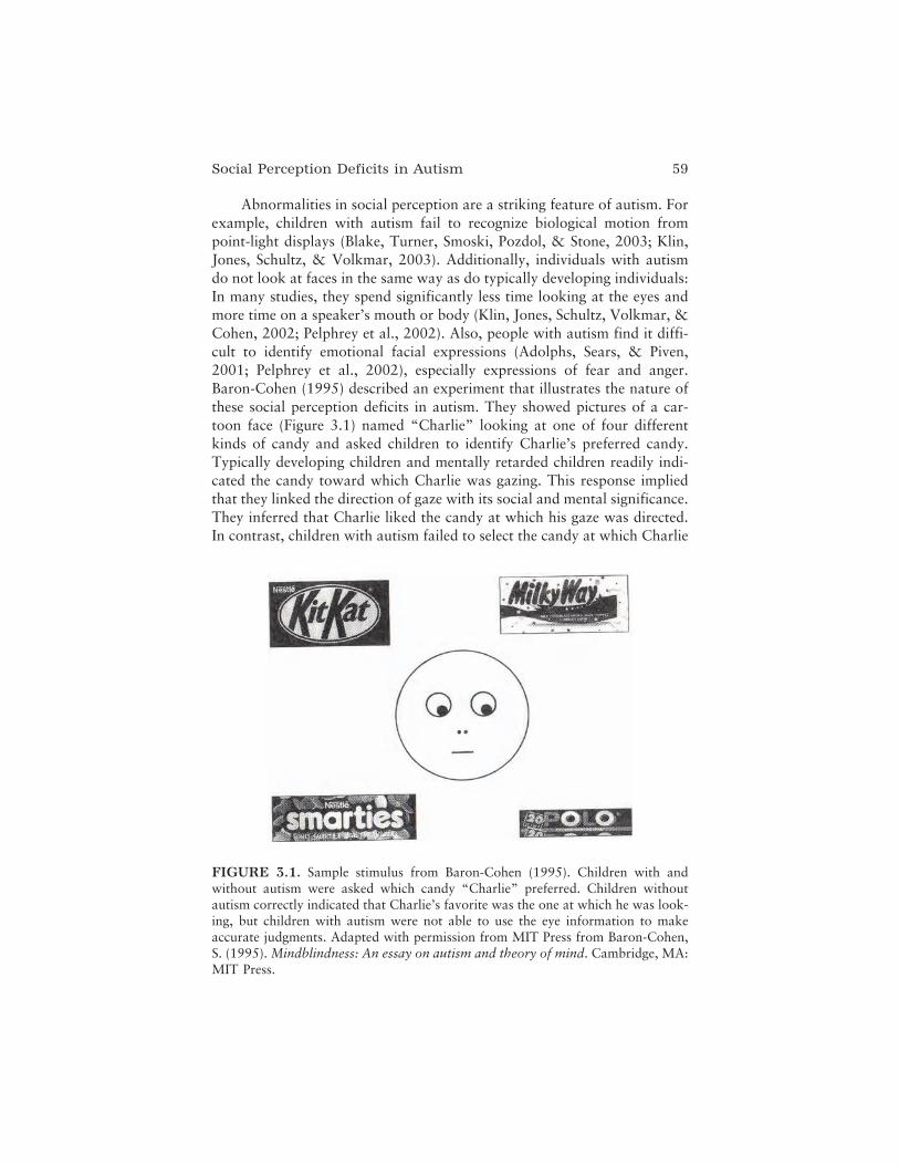
Abnormalities in social perception are a striking feature of autism. Forexample, children with autism fail to recognize biological motion frompoint-light displays (Blake, Turner, Smoski, Pozdol, & Stone, 2003; Klin,Jones, Schultz, & Volkmar, 2003). Additionally, individuals with autismdo not look at faces in the same way as do typically developing individuals:In many studies, they spend significantly less time looking at the eyes andmore time on a speaker’s mouth or body (Klin, Jones, Schultz, Volkmar, &Cohen, 2002; Pelphrey et al., 2002). Also, people with autism find it diffi-cult to identify emotional facial expressions (Adolphs, Sears, & Piven,2001; Pelphrey et al., 2002), especially expressions of fear and anger.Baron-Cohen (1995) described an experiment that illustrates the nature ofthese social perception deficits in autism. They showed pictures of a car-toon face (Figure 3.1) named “Charlie” looking at one of four differentkinds of candy and asked children to identify Charlie’s preferred candy.Typically developing children and mentally retarded children readily indi-cated the candy toward which Charlie was gazing. This response impliedthat they linked the direction of gaze with its social and mental significance.They inferred that Charlie liked the candy at which his gaze was directed.In contrast, children with autism failed to select the candy at which Charlie
Social Perception Deficits in Autism 59
FIGURE 3.1. Sample stimulus from Baron-Cohen (1995). Children with andwithout autism were asked which candy “Charlie” preferred. Children withoutautism correctly indicated that Charlie’s favorite was the one at which he was look-ing, but children with autism were not able to use the eye information to makeaccurate judgments. Adapted with permission from MIT Press from Baron-Cohen,S. (1995). Mindblindness: An essay on autism and theory of mind. Cambridge, MA:MIT Press.
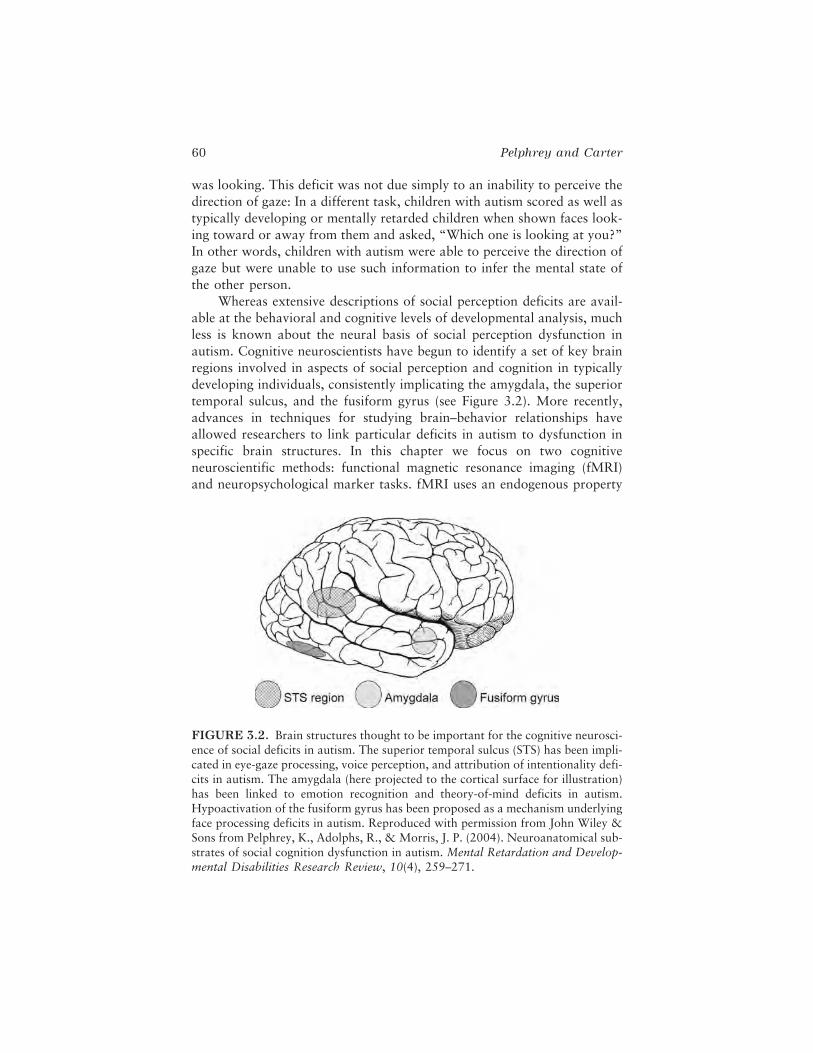
was looking. This deficit was not due simply to an inability to perceive thedirection of gaze: In a different task, children with autism scored as well astypically developing or mentally retarded children when shown faces look-ing toward or away from them and asked, “Which one is looking at you?”In other words, children with autism were able to perceive the direction ofgaze but were unable to use such information to infer the mental state ofthe other person.
Whereas extensive descriptions of social perception deficits are avail-able at the behavioral and cognitive levels of developmental analysis, muchless is known about the neural basis of social perception dysfunction inautism. Cognitive neuroscientists have begun to identify a set of key brainregions involved in aspects of social perception and cognition in typicallydeveloping individuals, consistently implicating the amygdala, the superiortemporal sulcus, and the fusiform gyrus (see Figure 3.2). More recently,advances in techniques for studying brain–behavior relationships haveallowed researchers to link particular deficits in autism to dysfunction inspecific brain structures. In this chapter we focus on two cognitiveneuroscientific methods: functional magnetic resonance imaging (fMRI)and neuropsychological marker tasks. fMRI uses an endogenous property
60 Pelphrey and Carter
FIGURE 3.2. Brain structures thought to be important for the cognitive neurosci-ence of social deficits in autism. The superior temporal sulcus (STS) has been impli-cated in eye-gaze processing, voice perception, and attribution of intentionality defi-cits in autism. The amygdala (here projected to the cortical surface for illustration)has been linked to emotion recognition and theory-of-mind deficits in autism.Hypoactivation of the fusiform gyrus has been proposed as a mechanism underlyingface processing deficits in autism. Reproduced with permission from John Wiley &Sons from Pelphrey, K., Adolphs, R., & Morris, J. P. (2004). Neuroanatomical sub-strates of social cognition dysfunction in autism. Mental Retardation and Develop-mental Disabilities Research Review, 10(4), 259–271.
of the brain called blood oxygenation level dependent (BOLD) contrast. Bytracking relative changes in the BOLD signal in different areas of the brain,fMRI provides an indirect measure of regional brain activity. This in vivoimaging method can be used repeatedly in relatively large numbers of indi-viduals because it is noninvasive and does not rely on the use of ionizingradiation. This technique can be used with marker tasks, which are devel-oped by comparing neurologically normal individuals with individuals whohave known brain lesions in order to explore the function of the lost neuralstructures. Researchers can combine these methods in order to furtherknowledge of the neural structures that appear to be dysfunctional inautism spectrum disorders. Using this information, neurophysiologicalmodels of autism are being developed along with candidate mechanisms forthe expression of individual differences in behavioral and treatment out-comes.
In this chapter, we describe progress toward understanding the brainmechanisms underlying social perception and social perception deficits inautism. We begin with an analytic approach, dissecting the individual con-tributions of the three aforementioned social brain structures. We presentbasic research aimed at identifying the functional significance of each brainregion in social perception. With this fundamental neuroscientific work asa backdrop, we then present translational studies that have implicated eachstructure in autism, comparing findings, when possible, from nonhumanprimates, neurologically normal humans, and individuals with lesions toresearch on people with autism. After examining the contributions of eachstructure in isolation, we adopt a more integrative posture and discussresearch that has added to our understanding of how the social brain oper-ates at the level of neural circuitry (i.e., characterizations of normal andabnormal functional interactions among the individual structures). In pre-senting this work, we emphasize the potential benefits of a developmentalperspective and propose several future research directions. We concludewith a discussion of the potential for the cognitive neuroscience of autismto inform educational interventions and treatment efforts.
AMYGDALA
The amygdala is a complex structure that is comprised of at least 13 differ-ent nuclei and is highly interconnected with other brain structures, includ-ing the thalamus, hippocampus, brainstem, basal forebrain, and claustrum(Amaral & Insausti, 1992; Amaral, Veazey, & Cowan, 1982). Brothers(1990) proposed that the amygdala is a key component of a neural circuitthat forms the neurobiological basis of social perception and social cogni-
Social Perception Deficits in Autism 61
tion, along with the superior temporal sulcus and gyrus and the orbito-frontal cortices. Some of the most compelling data for the function of theamygdala in a variety of socially relevant behaviors come from animalresearch. For example, in studies of mice, LeDoux (1996) discovered thatthe amygdala is critical for fear conditioning, which is the acquisition offear-related responses to a stimulus via its association with a different,strongly aversive stimulus. Additionally, in nonhuman primates, neurons inthe amygdalae of monkeys respond both to the basic motivational signifi-cance of stimuli and their complex social significance (Brothers, Ring, &Kling, 1990; Rolls, 1992). Studies in macaques with bilateral lesions of theamygdala illuminate the importance of a developmental perspective. Spe-cifically, adult macaques with lesions of the amygdala and surroundingmedial temporal structures have been described as tamer and as possessingabnormal food preferences and sexual behaviors (Brown & Schafer, 1888;Kluver & Bucy, 1938, 1939). Macaques with amygdala lesions at 2 weeksof age demonstrate an absence of fear in response to typically fear-inducingstimuli, but more screams and fear grimaces during conspecific social inter-actions (Prather et al., 2001). Macaques with neonatal lesions of bilateralmedial temporal lobes, including the amygdala, appear more severelyaffected, displaying autistic-like behaviors such as decreased eye contact,locomotor stereotypies, and increased social avoidance (Bachevalier, 1994,1996; Bachevalier & Mishkin, 1994).
The abovementioned work was limited by a lack of precision regard-ing the extent and specificity of the lesions. A more precise tool forlesioning brain regions is the use of ibotenic acid, which destroys the cellbodies in the area but leaves the axons passing through it intact. Re-searchers used this technique to create bilateral amygdala lesions in adultrhesus monkeys, which were later observed to make more approaches andbe less inhibited during dyadic social interactions with unlesioned con-trol conspecifics and also to lack normal fear behaviors to novel, fear-provoking stimuli (Emery et al., 2001; Prather et al., 2001). Such findingsled to the proposal that the primary role of the amygdala is the assessmentof threat and danger in the environment (Emery et al., 2001).
In humans, lesion and functional neuroimaging studies have alsopointed to the amygdala’s role in social perception and social behavior. Forexample, humans with lesions of the amygdala show social information-processing deficits, including problems in recognizing and judging emo-tions, particularly fear (Adolphs, Tranel, Damasio, & Damasio, 1994;Anderson & Phelps, 2000; Broks et al., 1998; Sprengelmeyer et al., 1999).In neuroimaging studies, amygdala activation is consistently detected inresponse to viewing facial expressions of fear, even when participants arenot asked to judge the emotions portrayed in the stimuli (Breiter et al.,
62 Pelphrey and Carter
1996; Morris et al., 1996). Whalen and others (1998) found that whenfearful faces were briefly presented and then backward-masked so as to besubliminal, amygdala activation was still seen, suggesting that rapid, auto-matic processing takes place in this structure. A recent study by Whalenand colleagues (2004) demonstrated that the amygdala responds to fearfuleye sclera presented alone, suggesting that emotional eye information aloneis sufficient to evoke activity in the amygdala.
The performance of patients with amygdala lesions is also compro-mised in social judgments. When asked to determine certain characteristicsof photographed people, these individuals consider the people in the imagesto be more trustworthy and approachable than do nonlesioned controls(Adolphs, Tranel, & Damasio, 1998). Neurologically normal individualsshow activation in the amygdala, superior temporal sulcus, orbitofrontalcortex, and insular cortex during this task (Winston, Strange, O’Doherty,& Dolan, 2002). This finding further supports the hypothesis derived fromanimal research that the amygdala is involved in danger and threat as-sessment (Adolphs et al., 1999; Adolphs, Damasio, Tranel, Cooper, &Damasio, 2000).
The role of the amygdala in autism has been explored in several func-tional neuroimaging studies (see summary in Table 3.1). The first, per-formed by Baron-Cohen and colleagues (1999), explored the ability of theparticipants to judge the mental or emotional state of a person in a photo-graph that only showed the eyes. Previously, high-functioning individualswith autism had been found to perform poorly in a behavioral version ofthis task outside of the scanner (Baron-Cohen, Jolliffe, Mortimore, & Rob-ertson, 1997). In typically developing subjects, the left amygdala, superiortemporal gyri, and insula were activated. However, in the high-functioningsubjects with autism, there was less frontal cortical and amygdala activa-tion.
Critchley and others (2000) compared neurologically normal individu-als to those with autism on two versions of a task with emotional faces, onerequiring judgments of the expressions (an explicit emotion-processingtask) and the other requiring gender identification (an implicit task). Unlikethe other participants, those with autism showed no fusiform gyrus activa-tion in the explicit task and no left amygdala activation in the implicit task.A later study explored the modulation of amygdala activation in childrenand adolescents with and without autism when viewing faces in two differ-ent tasks: labeling the emotions and matching the expressions (Wang,Dapretto, Hariri, Sigman, & Bookheimer, 2004). There were no significantgroup differences in the labeling task, but the subjects with autism showedlower fusiform gyrus and higher precuneus activation in the matching task.In the control subjects, the amygdala was modulated by task demands;
Social Perception Deficits in Autism 63
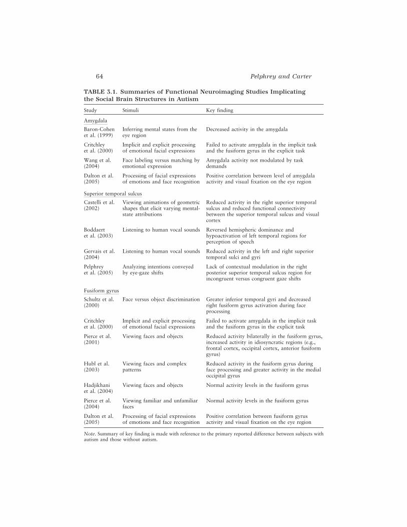
64 Pelphrey and Carter
TABLE 3.1. Summaries of Functional Neuroimaging Studies Implicatingthe Social Brain Structures in Autism
Study Stimuli Key finding
Amygdala
Baron-Cohenet al. (1999)
Inferring mental states from theeye region
Decreased activity in the amygdala
Critchleyet al. (2000)
Implicit and explicit processingof emotional facial expressions
Failed to activate amygdala in the implicit taskand the fusiform gyrus in the explicit task
Wang et al.(2004)
Face labeling versus matching byemotional expression
Amygdala activity not modulated by taskdemands
Dalton et al.(2005)
Processing of facial expressionsof emotions and face recognition
Positive correlation between level of amygdalaactivity and visual fixation on the eye region
Superior temporal sulcus
Castelli et al.(2002)
Viewing animations of geometricshapes that elicit varying mental-state attributions
Reduced activity in the right superior temporalsulcus and reduced functional connectivitybetween the superior temporal sulcus and visualcortex
Boddaertet al. (2003)
Listening to human vocal sounds Reversed hemispheric dominance andhypoactivation of left temporal regions forperception of speech
Gervais et al.(2004)
Listening to human vocal sounds Reduced activity in the left and right superiortemporal sulci and gyri
Pelphreyet al. (2005)
Analyzing intentions conveyedby eye-gaze shifts
Lack of contextual modulation in the rightposterior superior temporal sulcus region forincongruent versus congruent gaze shifts
Fusiform gyrus
Schultz et al.(2000)
Face versus object discrimination Greater inferior temporal gyri and decreasedright fusiform gyrus activation during faceprocessing
Critchleyet al. (2000)
Implicit and explicit processingof emotional facial expressions
Failed to activate amygdala in the implicit taskand the fusiform gyrus in the explicit task
Pierce et al.(2001)
Viewing faces and objects Reduced activity bilaterally in the fusiform gyrus,increased activity in idiosyncratic regions (e.g.,frontal cortex, occipital cortex, anterior fusiformgyrus)
Hubl et al.(2003)
Viewing faces and complexpatterns
Reduced activity in the fusiform gyrus duringface processing and greater activity in the medialoccipital gyrus
Hadjikhaniet al. (2004)
Viewing faces and objects Normal activity levels in the fusiform gyrus
Pierce et al.(2004)
Viewing familiar and unfamiliarfaces
Normal activity levels in the fusiform gyrus
Dalton et al.(2005)
Processing of facial expressionsof emotions and face recognition
Positive correlation between fusiform gyrusactivity and visual fixation on the eye region
Note. Summary of key finding is made with reference to the primary reported difference between subjects withautism and those without autism.
however, this modulation did not occur for those with autism. Takentogether, these studies suggest that the functioning of the amygdala isabnormal in people with autism during social perception, both in terms oflevels of activation (most studies have found amygdala hypoactivation inautism) and with regard to modulation by the context established by vary-ing task demands.
Neuropsychological marker tasks have been used to compare individu-als with autism to those with amygdala lesions and have provided tantaliz-ing, albeit indirect, evidence concerning the role of the amygdala in autism.Adolphs and colleagues (2001) compared high-functioning participantswith autism to a woman with bilateral focal amygdala lesions (patient SM).The individuals with autism and patient SM were remarkably similar inexhibiting abnormal social judgments regarding the trustworthiness andapproachability of others seen in photographs, overattributing these char-acteristics relative to judgments made by neurologically normal individuals.Also, judgments of facial expressions displaying negative affect were simi-larly poor, with particularly low performance on identification of fear andanger. The results of a later study by Adolphs and colleagues (2005) ofpatient SM’s visual scanpaths, combined with data concerning abnormalscanpaths for faces in autism (Klin et al., 2002; Pelphrey et al., 2002), sug-gest a potential mechanism underlying these shared social perceptionabnormalities. The eye-tracking data demonstrated that patient SM failedto fixate on the eyes when viewing facial expressions of emotion (see theirFigure 2). Similarly, Pelphrey and colleagues (2002) observed that individu-als with autism did not make use of the eyes when judging facial expres-sions of emotion (see Plate 3.1). Notably, when Adolphs and colleaguesinstructed patient SM to look at the eyes, her deficit in recognizing feartemporarily disappeared, but it returned after the instruction was forgot-ten. It remains to be determined whether such an experimental manipula-tion would affect the emotion judgments of individuals with autism, but astudy by Dalton and colleagues (2005) combined eye tracking and fMRIand found a strong positive correlation between the level of amygdalaactivity and gaze fixation upon the eye region in individuals with and with-out autism. Adolphs and colleagues interpreted their results as indicatingthat the amygdala is normally involved in directing the visual system toseek out and attend to the eyes as a socially important stimulus. Whetherthe amygdala fails to serve this purpose in autism remains to be tested.
SUPERIOR TEMPORAL SULCUS REGION
The role of the superior temporal sulcus region in social cognition andsocial perception has become the subject of intense investigation (see
Social Perception Deficits in Autism 65
Allison et al., 2000, for a review). As with the amygdala, work on thisbrain region has been conducted in both human and nonhuman primates.In monkeys, gaze direction can indicate dominance or submission and isintegral to facial expressions (Emery, Lorincz, Perrett, Oram, & Baker,1997). Campbell, Heywood, Cowey, Regard, and Landis (1990) found thatmonkeys were able to discriminate small shifts in gaze direction accurately,but monkeys with lesions of the superior temporal sulcus could not do so.Similarly, they found that human patients with prosopagnosia showed defi-cits on the task. Single neuron recordings performed by Perrett and col-leagues have shown that portions of the superior temporal sulcus in themacaque respond to head and gaze direction (Perrett et al., 1985, 1989),hand movement (Perrett et al., 1989), head movement (Hasselmo, Rolls,Baylis, & Nalwa, 1989), and whole body motion (Oram & Perrett, 1996).This work also demonstrates that the superior temporal sulcus in monkeysis important for social attention because it processes where others’ aware-ness is directed and the context within which their actions occur (Perrett,Hietanen, Oram, & Benson, 1992). Additional work in macaques has iden-tified a region of the superior temporal sulcus—the superior temporalpolysensory region—that integrates audio and visual components of com-plex social stimuli (Cusick, 1997).
In humans, the superior temporal sulcus region is a term used todescribe the superior temporal sulcus proper, portions of the superior andmiddle temporal gyri, and areas of the angular gyrus near the ascendinglimb of the superior temporal sulcus (Allison et al., 2000). Functional neu-roimaging studies in humans have implicated this area, particularly the pos-terior portion, in biological motion perception (for reviews see Allison etal., 2000; Decety & Grèzes, 1999). Below, we present work from our labo-ratory that has focused on identifying the role of the superior temporalsulcus region in the visual analysis of the actions and intentions of otherhumans as conveyed by different types of biological motion, including eye-gaze shifts (Pelphrey, Singerman, Allison, & McCarthy, 2003; Pelphrey,Viola, & McCarthy, 2004), walking (Pelphrey, Mitchell, et al., 2003),reaching (Pelphrey, Morris, & McCarthy, 2004), and audiovisual speech(Wright, Pelphrey, Allison, McKeown, & McCarthy, 2003).
In the first study of this series (Pelphrey, Mitchell, et al., 2003), wesought to determine whether the superior temporal sulcus region respondedto biological motion more than to other types of complex coherent motionor random motion. As illustrated in the top panel of Plate 3.2, subjectsviewed four types of animated stimuli: a walking human, a walking robot,a grandfather clock, and a disjointed mechanical figure. We establishedthat the superior temporal sulcus region responded more strongly to bio-logical motion (as conveyed by the walking robot or walking human)
66 Pelphrey and Carter
than to nonmeaningful but complex nonbiological motion (the disjointedmechanical figure) or complex and meaningful nonbiological motion (themovements of the grandfather clock). Importantly, not every brain regionshowed this pattern of effects. We observed a functional dissociationbetween the superior temporal sulcus region and an area corresponding tothe functionally defined motion-sensitive visual area MT/V5 (McCarthy etal., 1995; Zeki et al., 1991). Whereas the superior temporal sulcus regionpreferred biological motion, MT/V5 responded equally to all four types ofmotion. This study allowed us to conclude that the superior temporalsulcus region is involved in social perception by representing perceivedactions, but we could not yet determine whether this region was involved indeciphering the intentions, mental states, and goals conveyed by thoseactions. It remained possible that the posterior superior temporal sulcusregion served as a simple biological motion detector and was not sensitiveto the goals and intentions conveyed by the observed movements of actors.
Next, we conducted a study to determine whether the superior tempo-ral sulcus region was sensitive to the intentionality or goal-directedness ofobserved gaze shifts (Pelphrey, Singerman, et al., 2003). During fMRI scan-ning, neurologically normal adults watched as a small checkerboardappeared and flickered in an animated character’s visual field (see left panelof Plate 3.3). On congruent (goal-directed) trials, the character shifted hergaze toward the checkerboard, confirming the subject’s expectations.On incongruent (non-goal-directed) trials, the character shifted her gazetoward empty space, violating the subject’s expectations. We hypothesizedthat, if the superior temporal sulcus region is sensitive to the intentions con-veyed by actions in this simple virtual social setting, then activity in thesuperior temporal sulcus region evoked by observation of gaze shifts woulddifferentiate between congruent and incongruent trials. This differentia-tion, we reasoned, would reflect the ability of an adult to link the percep-tion of the gaze shift with its mentalistic significance. We observed moreactivity in the posterior superior temporal sulcus region for incongruentthan for congruent gaze, suggesting that additional processing was requiredof the superior temporal sulcus region when the character violated the sub-ject’s expectation. These initial findings have since been replicated in a sam-ple of neurologically normal adults observing congruent and incongruentreaching-to-grasp movements (Pelphrey, Morris, & McCarthy, 2004) andusing the same eye-gaze paradigm in 7- to 10-year-old typically developingchildren (Mosconi, Mack, McCarthy, & Pelphrey, 2005).
In a subsequent study, we examined whether the superior temporalsulcus region participated in the visual analysis of social information con-veyed by gaze shifts in an overtly social and more complex virtual encoun-ter: a stranger passing by the subject in a hallway (Pelphrey, Viola, &
Social Perception Deficits in Autism 67
McCarthy, 2004). Neurologically normal adults viewed an animated char-acter who walked toward them and shifted his neutral gaze either toward(mutual gaze) or away (averted gaze) from them. We reasoned that, if gaze-related activity in the superior temporal sulcus region reflected the opera-tion of a simple eye-movement detector (or more generally, a biologicalmotion detector), the region should not respond differentially to mutualand averted gaze. The motion of the man walking toward the subject andthe gaze shift evoked robust activity in the superior temporal sulcus andfusiform gyrus. Mutual gaze evoked greater activity in the superior tempo-ral sulcus than did averted gaze (see bottom panel of Plate 3.2). In contrast,the fusiform gyrus responded equally to mutual and averted gaze, demon-strating a functional dissociation. This study extended our understandingof the role of the superior temporal sulcus region in social perception bydemonstrating that it is highly sensitive to the social context in which a spe-cific biological motion occurs, in this case, approach versus avoidance.Overall, the results of our studies of eye-gaze processing, together with thefindings from the processing of intentions conveyed by reaching-to-graspactions (Pelphrey, Morris, & McCarthy, 2004), strongly suggest that theposterior superior temporal sulcus region is involved in the visual analysisof the intentions underlying other people’s actions.
Eye-gaze processing deficits, including failures to coordinate visualattention with others (i.e., initiating and responding to joint attention) anddifficulties comprehending the mental states and social intentions of otherpeople as conveyed by the eyes (Baron-Cohen, 1995; Dawson, Meltzoff,Osterling, Rinaldi, & Brown, 1998; Leekam, Hunnisett, & Moore, 1998;Leekam, Lopez, & Moore, 2000; Loveland & Landry, 1986; Mundy,Sigman, Ungerer, & Sherman, 1986), are key features of autism (Baron-Cohen, Wheelwright, Hill, Raste, & Plumb, 2001; Frith & Frith, 1999),and these impairments are identifiable early in the ontogeny of childrenwith autism (Dawson et al., 1998; Mundy et al., 1986). Many affected chil-dren eventually learn to use joint attention in social interactions, but evenhigher-functioning adults exhibit impairments on tasks requiring subtle dis-tinctions regarding intentions and mental states from viewing the eyes(Baron-Cohen et al., 2001). Eye-gaze processing deficits in autism are notthe result of abnormal gaze discrimination but rather represent an inabilityto use gaze spontaneously to understand and predict other people’s mentalstates and behaviors (Baron-Cohen, 1995; Baron-Cohen et al., 1999).
The nature of gaze processing deficits in autism combined withour prior neuroimaging findings led us to hypothesize that superiortemporal sulcus dysfunction might be involved (Pelphrey, Morris, &McCarthy, 2005). To test this question, we employed our congruent-versus-incongruent eye-gaze paradigm (left panel of Plate 3.3). We pre-
68 Pelphrey and Carter
dicted that, in autism, the superior temporal sulcus region would not besensitive to intentions conveyed by the character’s gaze shifts. As shown inthe right panel of Plate 3.3, in neurologically normal control participants,“errors” (incongruent gaze shifts) again evoked more activity in the supe-rior temporal sulcus and other regions involved in social perception, indi-cating a strong effect of intention (see Plate 3.3b). The same brain regionswere activated during observation of gaze shifts in individuals with autism(see Plate 3.3c) but did not differentiate between congruent and incongru-ent trials (see Plate 3.3d), indicating that activity in these regions was notmodulated by the context of the perceived gaze shift. This finding suggeststhat lack of modulation in activation of the superior temporal sulcus regionis a potential mechanism underlying the eye-gaze processing deficits associ-ated with autism. These findings are consistent with those of a priorpositron emission tomography (PET) study conducted by Castelli, Frith,Happé, and Frith (2002) that found hypoactivation of the posterior supe-rior temporal sulcus and reduced functional connectivity between the supe-rior temporal sulcus and portions of the inferior occipital gyrus (visual areaV3) in autism during tasks involving attribution of intentions to movinggeometric figures.
Three other functional neuroimaging studies have explored the role ofthe superior temporal sulcus region in autism (see Table 3.1), focusing onthe neural basis of speech perception deficits. A PET study reported abnor-mal laterality of responses and hypoactivation of the left superior temporalsulcus region (Boddaert et al., 2003) during speech perception. Similarly,an fMRI study reported abnormal responses to human voices in the supe-rior temporal sulcus region (Gervais et al., 2004). A region of the superiortemporal sulcus that had previously been shown to respond selectively tovocal sounds (Belin, Zatorre, Lafaille, Ahad, & Pike, 2000) failed to beactivated by this stimulus category in individuals with autism (Gervais etal., 2004). These subjects showed normal auditory cortex activation tononvocal sounds. Similarly, by comparing responses to various auditoryand visual speech stimuli, we have explored the role of the superior tempo-ral sulcus region in audiovisual speech perception in a small sample of indi-viduals with autism and neurologically normal controls (Smith, Morris, &Pelphrey, 2005). The superior temporal sulcus region was activated bilater-ally during audiovisual speech perception in both groups of subjects. Forcontrol subjects, responses from this region to matching audiovisual speechwere greater than responses to the audio and visual components presentedalone, and the mismatching audiovisual speech evoked a depressed re-sponse consistent with the inhibition that is generally observed in para-digms of this type (see Calvert, 2001, for a review). For the subjects withautism, there was overall hypoactivation in the superior temporal sulcus
Social Perception Deficits in Autism 69
region, and this area responded equally to the matching and mismatchingaudiovisual stimuli, suggesting abnormal integration of the two sources ofinformation.
FUSIFORM GYRUS
The fusiform gyrus is found in the ventral occipitotemporal cortex ofhumans (see Figure 3.2). When a neurologically normal individual is pre-sented with faces versus non-face objects or scrambled images, face-evokedactivity in the lateral portions of the fusiform gyrus is observed bilaterally,but typically more in the right than the left hemisphere (Kanwisher,McDermott, & Chun, 1997; McCarthy, Puce, Gore, & Allison, 1997;Puce, Allison, Asgari, Gore & McCarthy, 1996); thus, portions of this cor-tical region are specialized for processing faces (Kanwisher et al., 1997;McCarthy et al., 1997). Some of the clearest evidence for face specificity inthe fusiform gyrus comes from studies during which electrodes inserted inthe ventral occipitotemporal cortical surface during brain surgery displayedface-specific evoked activity within 200 ms of the presentation of a facestimulus (Allison, Puce, Spencer, & McCarthy, 1999; McCarthy, Puce,Belger, & Allison, 1999; Puce, Allison, & McCarthy, 1999), and electricalstimulation of this area led to transient prosopagnosia (Allison, McCarthy,Nobre, Puce, & Belger, 1994; see McCarthy, 1999, for a review). Modelsof the human face processing system emphasize the role of the fusiformgyrus in detecting faces and processing their static aspects, which are usedfor face identification (Haxby, Hoffman, & Gobbini, 2000; McCarthy,1999).
In autism, behavioral studies have consistently demonstrated face pro-cessing deficits in affected individuals (e.g., Hobson, Ouston, & Lee, 1988;Loveland et al., 1997), although the specific mechanisms underlying thesedeficits remain elusive. Schultz and colleagues (2000) performed the firstfMRI study of the fusiform gyrus response to faces and non-face objects inadolescents and adults with high-functioning autism and Asperger syn-drome. They found that there was less face-evoked activation in this regionand that areas typically recruited for non-face object perception were usedfor face perception in these individuals. Since this original study, threeother fMRI studies have replicated the finding of fusiform gyrus hypo-activation in autism (Critchley et al., 2000; Hubl et al., 2003; Pierce,Mueller, Ambrose, Allen, & Courchesne, 2001). Schultz and colleagues(2000) and Dawson, Webb, and colleagues (2002; Dawson, Webb, &McPartland, 2005; see also Dawson & Bernier, Chapter 2, this volume)emphasize the importance of a developmental perspective and suggest a
70 Pelphrey and Carter
possible link between amygdala dysfunction and the lack of normal devel-opment of face-specific fusiform gyrus responsivity and, consequently, nor-mal face perception (Schultz, 2005). In fleshing out the neurobiologicalbasis for this developmental hypothesis, both Dawson, Webb, and col-leagues (2002, 2005) and Schultz (2005) have argued that early amygdaladisruption in autism leads to a lack of interest in faces. The correspondinglack of attention to faces throughout development leads to abnormalities inthe functional pathways between the amygdala and the fusiform gyrus;these functional pathways are dependent upon ongoing input from theamygdala to develop fusiform gyrus face specificity. These hypotheses aresupported by indirect evidence, such as the previously described findings byAdolphs and colleagues (2005) regarding the role of the amygdala in direct-ing visual attention to the eyes (see Plate 3.1, bottom panel), but would betested best in a longitudinal study of the development of face-specificresponses in the fusiform gyrus and connectivity among the amygdala andother social brain structures.
It is important to note that the original findings by Schultz and col-leagues (2000) have been contended. Some eye-tracking studies have foundthat individuals with autism do not look at faces in the same manner as dotypically developing individuals, with this research reporting that affectedindividuals spend less time scanning the core features of the face (eyes,nose, and mouth—Dalton et al., 2005; Klin et al., 2002; Pelphrey et al.,2002; but see van der Geest, Kemner, Verbaten, & van Engeland, 2002, forcontrasting results). These abnormal scanpaths could actually be the causeof the observed hypoactivation. For example, in a study by McCarthy andothers (1999), the N200 response of the ERP waveform had the greatestamplitude for whole faces, followed by eyes, contours, lips, and noses, sug-gesting that attention to the eyes in face images could drive the fusiformgyrus response. To address the issue of visual attention to faces, Hadjikhaniand colleagues (2004) added fixation points to the center of images of facesand objects and determined that people with autism exhibited enhancedfusiform gyrus activation under that condition. However, it is difficult todraw definitive conclusions from this study because the requirement to fix-ate on the crosshair would have altered scanpaths in both groups.
Dalton and colleagues (2005) directly explored the relationship be-tween gaze fixation on the eyes and brain activation patterns in thefusiform gyrus and the amygdala in participants with and without autism.The participants with autism had significantly less gaze fixation on the eyesand showed greater activation than controls in the left amygdala and theleft orbitofrontal gyrus when viewing emotional and neutral faces. In a sec-ond study, the participants viewed familiar and unfamiliar people. The neu-rologically normal group showed greater activation in the right occipital
Social Perception Deficits in Autism 71
gyrus and fusiform gyrus for the familiar faces relative to the unfamiliarfaces, and more activation for the familiar faces than did the group withautism. Again the participants with autism spent less time looking at theeyes and had less activation in the right and left fusiform gyri, but moreactivity in the right amygdala overall. Across both studies, the researchersfound a strong positive correlation between the number and length of gazefixations on the eyes of faces and the fusiform gyrus response in individualswith and without autism. Like Schultz and Dawson, they also implicatedfunctional interactions between the amygdala and fusiform gyrus. How-ever, unlike Schultz and Dawson, these authors interpreted this relationshipfrom the perspective of social anxiety, suggesting that people with autismactively avoid eye gaze because of an autonomic hyperreactivity caused bydysregulation of affective processes in the amygdala in response to salientsocial stimuli. Notably, the amount of amygdala activation had a strongpositive correlation with the amount of fixation on the eye region amongthe participants with autism.
An alternative explanation for the findings by Dalton and colleagues(2005) is that individuals with autism could be failing to marshal a socialresponse to environmental cues even in the absence of anxiety. Rather thanavoiding social situations in an active manner, as Schultz and Dawsonargue, they instead might not appreciate the significance of the faces intheir environments. In an effort to increase the probability that individualswith autism would find faces to be socially meaningful, Pierce, Haist,Sedaghat, and Courschesne (2004) showed participants with and withoutautism photographs of both very familiar people (friends and family mem-bers) and strangers. In the fusiform gyrus, both groups of subjects showedsimilar activation for the familiar faces. In response to the unfamiliar faces,the individuals with autism showed a significant, but much lower, level ofactivation. However, we note that this result and the findings from Daltonand colleagues regarding responses to familiar faces in subjects with andwithout autism are inconsistent. Further work is needed to explore poten-tial reasons for these incompatible findings.
INTEGRATING THE SOCIAL BRAIN:FUTURE DIRECTIONSAND OUTSTANDING QUESTIONS
We have reviewed functional neuroimaging studies of individuals with andwithout autism that focused on identifying dysfunction in specific struc-tures linked to particular aspects of social perception. We emphasized the
72 Pelphrey and Carter
unique contributions of three structures: the amygdala, the superior tempo-ral sulcus region, and the fusiform gyrus. This analytic perspective washelpful in providing a framework for organizing our emerging understand-ing of social brain dysfunction in autism, but this approach does not fullyreflect the complexity of interactions among these and other brain regionsduring social perception. These structures undoubtedly function in paralleland are better understood as components in a network of regions subserv-ing social perception. For example, when encountering a socially ambigu-ous situation, such as when a person is approaching in a hallway, theamygdala will provide a rapid and automatic assessment of the potentiallythreatening aspects of the situation and, through its interconnections withthe other structures, allocate processing resources accordingly. The fusi-form gyrus will provide a perceptual representation of the face and will aidin identification of the person. The posterior superior temporal sulcus willconduct a visual analysis of the person’s gait and other socially and com-municatively important actions, including movements of facial features andshifts in eye gaze, while other sectors of this region incorporate auditoryand visual components of speech. The rapid integration of the functionsperformed by each structure would guide social perception and the subse-quent behavior of the observer. To date, disruption in each of the compo-nents of this system has been studied in autism, but their integration hasgenerally been neglected, probably owing to the relative lack of techniques,until recently, for this type of analysis. A challenge for the future will be tooffer a more precise account of the interplay between all of these differentprocesses as a function of the detailed specification of the performancedemands required by a given experimental task or by a given situation inreal life.
A few studies have begun to examine functional interactions betweenbrain regions and potential disruptions in functional connectivity in autism.For example, Just, Cherkassky, Keller, and Minshew (2004) reported lowerlevels of functional connectivity (defined as the correlation, over time, ofactivity in two brain regions) between Wernicke’s and Broca’s areas duringsentence comprehension in individuals with autism relative to neurologi-cally normal controls. In an earlier PET study, Castelli and colleagues(2002) reported reduced functional connectivity between the posteriorsuperior temporal sulcus and the inferior occipital gyrus in individuals withautism during a task involving attribution of intentions to moving geomet-ric figures. We conducted analyses of functional connectivity in our ownresearch on the neural basis of eye-gaze processing deficits in autism(Pelphrey et al., 2005). Specifically, we reasoned that the lack of differentialactivity for incongruent versus congruent gaze in subjects with autism (see
Social Perception Deficits in Autism 73
Plate 3.3, right panel) could be caused by a functional disruption in thesuperior temporal sulcus region itself. On the other hand, the abnormalpatterns of superior temporal sulcus activation could be the result of atypi-cal connectivity between this and other brain regions involved in social per-ception. According to this hypothesis, the superior temporal sulcus couldbe activated during the initial perception of an action performed byanother, creating a perceptual representation of the socially relevant infor-mation. These data are then sent to higher systems (possibly includingprefrontal regions and/or the amygdala) for analysis of the intentions orgoal orientations displayed, and these areas might maintain superior tem-poral sulcus activation in circumstances during which additional processingis required, such as violations of expectations. In autism, this connectioncould be faulty, leaving these higher areas incapable of sustaining activa-tion in the superior temporal sulcus when needed. We explored this alter-native by examining the temporal profile of the correlation between activityin the right posterior superior temporal sulcus and the right posterior mid-dle temporal gyrus. In subjects without autism, there was a strong positivecorrelation between these regions, but this correlation was greatly reducedin the subjects with autism, suggesting abnormal functional connectivityduring this social perception task.
Such analyses of fMRI data can provide indirect assessments of func-tional connectivity as revealed by correlations in temporal profiles of activ-ity between two or more brain regions, but these correlation analyses donot provide direct information regarding connectivity in the living brain.The combined use of diffusion tensor imaging (DTI) and fMRI is promisingfor studying the role of abnormal connectivity in autism. DTI has emergedas a powerful method for the investigation of white matter architecture. Bymeasuring the tensor associated with self-diffusion (Brownian motion) ofendogenous water molecules in brain tissue—which diffuse less readilyacross, as compared to along, membranes—DTI permits investigators tomeasure aspects of connectivity and to trace the extent and direction ofwhite matter tracts in the living brain. In future work, the use of DTI inautism to study connections between functional regions of interest will addto our understanding of dysfunction in the social brain in this disorder.
Another important future direction is the continued development ofstimuli and tasks with more ecological validity. Progress has been limitedby the lack of ecologically valid social situations that can be manipulatedand presented with conditions adequately controlled within the constraintsof an MRI scanner. Most functional imaging studies have used simple,static images; therefore, questions remain unanswered about the effects ofcontext and motion on the social brain. After all, the perceived participa-
74 Pelphrey and Carter
tion of the subject is an important element for successfully imaging socialperception. Subtle but important social deficits are often most evident inhigh-functioning individuals with autism during unstructured social set-tings. As future studies begin to shift focus from single regions to networksinvolved in social perception, continued developments in imaging tech-niques will drive experimental innovation. We will see researchers morefrequently combining data from complementary imaging techniques (i.e.,multimodal imaging) to examine structure, function, and connectivity.
Autism is a neurodevelopmental disorder that has its onset in the earli-est years of childhood, and its symptoms change over ontogeny. Althoughthe core deficits persist throughout the lifespan, the specific manifestationsshift across development. It will be very important to expand the body ofresearch to include functional neuroimaging studies of young children withautism. Neuroimaging studies of autism that take into account psychologi-cal and behavioral continuities and discontinuities from childhood to adult-hood would better inform us of the neurobiological mechanisms in autismthan would studies that provide only a static picture in adults. We can thusincorporate a developmental perspective (i.e., a perspective involving earlyand longitudinal study of brain and behavioral development) into our anal-yses of the social brain.
A developmental perspective is important for understanding howhigher-order deficits in aspects of social interactions might originate in pri-mary impairments in joint attention, action understanding and imitation,and early language functioning. This stance could reveal that autism arisesfrom subtle and more diffuse early neuropathology and ultimately affectsmultiple neural systems, directly and through compensatory experience-dependent reorganization, rather than being a disorder of a specific neuro-anatomical structure. Neuroimaging studies focusing on changing brain–behavior relationships in the developing child may also provide clues tocandidate genes in autism, as developmentally important genes begin to belinked to particular patterns of brain development, gene expression, andphenotypes.
Longitudinal studies of the social brain in neurologically normal chil-dren will be as important as developmental studies of children with autism.Currently, very little is known about the neural correlates of social percep-tion in children or about the changes in brain function that underlie norma-tive development in this domain. Fundamental scientific questions concern-ing the maturation of the brain and its relationship to changes in socialperception in children without autism thus remain unanswered. This basicknowledge is essential to efforts aimed at understanding the neural basis ofsocial perception deficits in autism.
Social Perception Deficits in Autism 75
UNDERSTANDING SOCIALBRAIN DYSFUNCTION IN AUTISM:EDUCATION AND TREATMENT IMPLICATIONS
Techniques for earlier diagnosis of autism are still being improved. Unlikesome other neurodevelopmental disorders (e.g., fragile X syndrome), we donot have a genetic test for autism, and the complexity of the interactiveroles played by genes and environment makes it unlikely that such a testwill be feasible. However, the analysis of functional brain correlates andnetworks could prove to be a useful tool in unambiguously identifying thisdisorder at an early age. Reliable and specific structural and/or functionaldifferences in the brains of people with autism might serve as valuable neu-robiological “markers” for the diagnosis of autism. In our own work,activity in the superior temporal sulcus offers a possible example of a neu-robiological marker for the social perception deficits in autism. Currently,the Autism Diagnostic Observation Schedule—General (ADOS-G; Lord etal., 2000) and the Autism Diagnostic Interview—Revised (ADI-R; Lord,Rutter, & Le Couteur, 1994) are the most widely accepted tools for diag-nosing autism. In a set of exploratory correlation analyses, we integratedinformation from the ADI-R with patterns of neural activity from the supe-rior temporal sulcus region during an eye-gaze processing task (Pelphrey etal., 2005). As discussed previously, activity in the superior temporal sulcusregion in the group of participants with autism did not differ significantlyfor incongruent and congruent gaze shifts, but individual differences in thepatterns of activation were apparent. We wondered whether these individ-ual differences might be related to the phenotypic variability in real-worldsocial functioning seen in this disorder. To explore this question further,we correlated scores on several algorithmic domains of the ADI-R with themagnitude of differentiation in the right superior temporal sulcus regionbetween stimulus conditions, predicting that the individuals with less severebehavioral deficits would show activity more similar to the participantswithout autism. We found a strong correlation between superior temporalsulcus activation patterns and the severity of impairments in the ReciprocalSocial Interaction Domain, but correlations did not extend to the do-mains of Communication and Restricted–Repetitive–Stereotyped Behav-iors. Notably, the patterns of brain activity obtained during the brief fMRIscan accounted for 61% of the variance in ADI-Reciprocal Social Interac-tion Domain scores. These findings suggest that the degree of neuro-functional impairment in the right superior temporal sulcus region isrelated to the severity of specific and relevant features of the autism pheno-type. Future research using larger samples could elucidate the validity ofthis difference as a marker for autism severity.
76 Pelphrey and Carter
It is generally accepted that earlier behavioral interventions are moreeffective for treating children with autism. Therefore, early diagnosis is piv-otal to individual treatment outcomes for these children. A neurobiologicalmarker, then, would be important not solely for improving early diagnosisbut also for offering advantages in terms of earlier interventions. Moreover,based on research such as that described above, it could be that the markerwould relate to the severity of specific deficits and might help us to under-stand better the heterogeneity in the core features of the autism phenotype.With this information, more targeted treatments could be developed on achild-by-child basis and implemented early in ontogeny, which might thenguarantee the most effective course of intervention possible. In addition,the assessment of individual treatment needs could be far more efficientand standardized. Furthermore, functional neuroimaging techniques mightactually provide a means to better, and more quantitatively, assess theeffectiveness of treatment and reveal whether behavioral improvementscorrespond to compensatory changes in brain function or normalization ofdevelopmental pathways.
By defining functional brain phenotypes based on neurofunctional/behavioral developmental pathways and altered activation patterns, fMRIstudies of children and adults with autism have the potential to dissect theheterogeneity present in autism as a behaviorally defined syndrome. Func-tional neuroimaging studies could reveal different brain phenotypes in thecircuitry involved in social perception. This approach may allow us to par-tition this complex disorder, which is thought to be etiologically heteroge-neous, into more homogenous subgroups. Thus a goal for future work is toidentify developmental brain “endophenotypes” linking functioning in thesocial brain to clinically observed social perception and social cognitiondeficits. This link would be valuable because progress in understanding andtreating autism has been hindered by its heterogeneity, which reduces thestatistical power to (1) identify genes through linkage studies, (2) identifyeffective pharmacological treatments, and (3) describe the pathophysiologyof autism. Early and longitudinal study will be critical in defining brainphenotypes in autism because the shape of developmental trajectories ofbrain functioning in specific circuits will provide more detail on the natureof the abnormalities in autism than will analysis of brain phenotypes inadults. These phenotypes might relate to behavioral outcomes and couldsuggest novel and more targeted intervention and treatment strategies.Knowledge of the development of the social brain could help geneticistssearch for abnormal genes or assist other researchers in the detection ofpossible environmental factors related to autism. A better understanding ofabnormal and normal brain systems mapping onto behavioral strengthsand weaknesses in the autism phenotype could help scientists and clinicians
Social Perception Deficits in Autism 77
to develop strategies for assisting individuals with autism to utilize nor-mally developing regions and circuits to compensate for abnormal func-tion, and to create therapies targeted at training and reinforcing the ab-normally functioning regions and circuits as early as possible in theontogeny of affected individuals.
ACKNOWLEDGMENTS
Kevin A. Pelphrey is supported by a Career Development Award from theNational Institutes of Health, National Institute of Mental Health Grant No. K01-MH071284. We gratefully acknowledge our collaborators Drs. Gregory McCarthy,James Morris, Truett Allison, Kevin LaBar, Joseph Piven, and Ralph Adolphs.
REFERENCES
Adolphs, R., Damasio, H., Tranel, D., Cooper, G., & Damasio, A. R. (2000). Arole for somatosensory cortices in the visual recognition of emotion asrevealed by three-dimensional lesion mapping. Journal of Neuroscience, 20(7),2683–2690.
Adolphs, R., Gosselin, F., Buchanan, T. W., Tranel, D., Schyns, P., & Damasio, A.R. (2005). A mechanism for impaired fear recognition after amygdala damage.Nature, 433, 68–72.
Adolphs, R., Sears, L., & Piven, J. (2001). Abnormal processing of social informa-tion from faces in autism. Journal of Cognitive Neuroscience, 13(2), 232–240.
Adolphs, R., Tranel, D., & Damasio, A. R. (1998). The human amygdala in socialjudgment. Nature, 393, 470–474.
Adolphs, R., Tranel, D., Damasio, H., & Damasio, A. (1994). Impaired recognitionof emotion in facial expressions following bilateral damage to the humanamygdala. Nature, 372, 669–672.
Adolphs, R., Tranel, D., Hamann, S., Young, A. W., Calder, A. J., Phelps, E. A., etal. (1999). Recognition of facial emotion in nine individuals with bilateralamygdala damage. Neuropsychologia, 37(10), 1111–1117.
Allison, T., McCarthy, G., Nobre, A., Puce, A., & Belger, A. (1994). Humanextrastriate visual cortex and the perception of faces, words, numbers, andcolors. Cerebral Cortex, 4(5), 544–554.
Allison, T., Puce, A., & McCarthy, G. (2000). Social perception from visual cues:Role of the STS region. Trends in Cognitive Science, 4(7), 267–278.
Allison, T., Puce, A., Spencer, D. D., & McCarthy, G. (1999). Electrophysiologicalstudies of human face perception I: Potentials generated in occipitotemporalcortex by face and non-face stimuli. Cerebral Cortex, 9(5), 415–430.
Amaral, D. G., & Insausti, R. (1992). Retrograde transport of d-[3h]-aspartateinjected into the monkey amygdaloid complex. Experimental Brain Research,88(2), 375–388.
78 Pelphrey and Carter
Amaral, D. G., Veazey, R. B., & Cowan, W. M. (1982). Some observations onhypothalamo-amygdaloid connections in the monkey. Brain Research, 252(1),13–27.
American Psychiatric Association. (1994). Diagnostic and statistical manual ofmental disorders (4th ed., text rev.).Washington, DC: Author.
Anderson, A. K., & Phelps, E. A. (2000). Expression without recognition: Contri-butions of the human amygdala to emotional communication. PsychologicalScience, 11(2), 106–111.
Bachevalier, J. (1994). Medial temporal lobe structures and autism: A review ofclinical and experimental findings. Neuropsychologia, 32(6), 627–648.
Bachevalier, J. (1996). Medial temporal lobe and autism: A putative animalmodel in primates. Journal of Autism and Developmental Disorders, 26(2),217–220.
Bachevalier, J., & Mishkin, M. (1994). Effects of selective neonatal temporal lobelesions on visual recognition memory in rhesus monkeys. Journal of Neurosci-ence, 14(4), 2128–2139.
Bailey, A., Le Couteur, A., Gottesman, I., Bolton, P., Simonoff, E., Yuzda, E., et al.(1995). Autism as a strongly genetic disorder: Evidence from a British twinstudy. Psychological Medicine, 25(1), 63–77.
Bailey, D. B., Jr., Hatton, D. D., Skinner, M., & Mesibov, G. (2001). Autisticbehavior, fMR1 protein, and developmental trajectories in young males withfragile X syndrome. Journal of Autism and Developmental Disorders, 31(2),165–174.
Baker, P., Piven, J., & Sato, Y. (1998). Autism and tuberous sclerosis complex:Prevalence and clinical features. Journal of Autism and Developmental Disor-ders, 28(4), 279–285.
Baron-Cohen, S. (1995). Mindblindness: An essay on autism and theory of mind.Cambridge, MA: MIT Press.
Baron-Cohen, S., Jolliffe, T., Mortimore, C., & Robertson, M. (1997). Anotheradvanced test of theory of mind: Evidence from very high functioning adultswith autism or Asperger syndrome. Journal of Child Psychology and Psychia-try, 38(7), 813–822.
Baron-Cohen, S., Ring, H. A., Wheelwright, S., Bullmore, E. T., Brammer, M. J.,Simmons, A., et al. (1999). Social intelligence in the normal and autistic brain:An fMRI study. European Journal of Neuroscience, 11(6), 1891–1898.
Baron-Cohen, S., Wheelwright, S., Hill, J., Raste, Y., & Plumb, I. (2001). The“reading the mind in the eyes” test, revised version: A study with normaladults and adults with Asperger syndrome or high-functioning autism. Journalof Child Psychology and Psychiatry, 42(2), 241–251.
Belin, P., Zatorre, R. J., Lafaille, P., Ahad, P., & Pike, B. (2000). Voice-selectiveareas in human auditory cortex. Nature, 403, 309–312.
Blake, R., Turner, L. M., Smoski, M. J., Pozdol, S. L., & Stone, W. L. (2003).Visual recognition of biological motion is impaired in children with autism.Psychological Science, 14, 51–57.
Boddaert, N., Belin, P., Chabane, N., Poline, J. B., Barthélémy, C., Mouren-Simeoni, M. C., et al. (2003). Perception of complex sounds: Abnormal pat-
Social Perception Deficits in Autism 79
tern of cortical activation in autism. American Journal of Psychiatry, 160(11),2057–2060.
Breiter, H. C., Etcoff, N. L., Whalen, P. J., Kennedy, W. A., Rauch, S. L., Buckner,R. L., et al. (1996). Response and habituation of the human amygdala duringvisual processing of facial expression. Neuron, 17(5), 875–887.
Broks, P., Young, A. W., Maratos, E. J., Coffey, P. J., Calder, A. J., Isaac, C. L., etal. (1998). Face processing impairments after encephalitis: Amygdala damageand recognition of fear. Neuropsychologia, 36(1), 59–70.
Brothers, L. (1990). The social brain: A project for integrating primate behaviorand neurophysiology in a new domain. Concepts in Neuroscience, 1, 27–51.
Brothers, L., Ring, B., & Kling, A. (1990). Response of neurons in the macaqueamygdala to complex social stimuli. Behavior and Brain Research, 41, 199–213.
Brown, S., & Schafer, E. A. (1888). An investigation into the functions of theoccipital and temporal lobes of the monkey’s brain. Philosophical Transac-tions of the Royal Society of London (Series B: Biological Sciences), 179,303–327.
Calvert, G. A. (2001). Crossmodal processing in the human brain: Insights fromfunctional neuroimaging studies. Cerebral Cortex, 11(12), 1110–1123.
Campbell, R., Heywood, C. A., Cowey, A., Regard, M., & Landis, T. (1990). Sensi-tivity to eye gaze in prosopagnosic patients and monkeys with superior tempo-ral sulcus ablation. Neuropsychologia, 28(11), 1123–1142.
Castelli, F., Frith, C., Happé, F., & Frith, U. (2002). Autism, Asperger syndromeand brain mechanisms for the attribution of mental states to animated shapes.Brain, 125, 1839–1849.
Cook, E. H., Jr., Courchesne, R. Y., Cox, N. J., Lord, C., Gonen, D., Guter, S. J., etal. (1998). Linkage-disequilibrium mapping of autistic disorder, with 15q11–13 markers. American Journal of Human Genetics, 62(5), 1077–1083.
Critchley, H. D., Daly, E. M., Bullmore, E. T., Williams, S. C., Van Amelsvoort, T.,Robertson, D. M., et al. (2000). The functional neuroanatomy of social behav-iour: Changes in cerebral blood flow when people with autistic disorder pro-cess facial expressions. Brain, 123(11), 2203–2212.
Cross, H. A., & Harlow, H. F. (1965). Prolonged and progressive effects of partialisolation on the behavior of macaque monkeys. Journal of ExperimentalResearch in Personality, 1, 39–49.
Cusick, C. G. (1997). The superior temporal polysensory region in monkeys. In K.S. Rockland, J. H. Kaas, & A. Peters (Eds.), Cerebral cortex. Vol. 12:Extrastriate cortex in primates (pp. 435–468). New York: Plenum Press.
Dalton, K. M., Nacewicz, B. M., Johnstone, T., Schaefer, H. S., Gernsbacher, M.A., Goldsmith, H. H., et al. (2005). Gaze fixation and the neural circuitry offace processing in autism. Nature Neuroscience, 8(4), 519–526.
Dawson, G., Meltzoff, A. N., Osterling, J., Rinaldi, J., & Brown, E. (1998).Children with autism fail to orient to naturally occurring social stimuli. Jour-nal of Autism and Developmental Disorders, 28(6), 479–485.
Dawson, G., Munson, J., Estes, A., Osterling, J., McPartland, J., Toth, K., et al.(2002). Neurocognitive function and joint attention ability in young children
80 Pelphrey and Carter
with autism spectrum disorder versus developmental delay. Child Develop-ment, 73(2), 345–358.
Dawson, G., Webb, S. J., & McPartland, J. (2005). Understanding the nature offace processing impairment in autism: Insights from behavioral and electro-physiological studies. Developmental Neuropsychology, 27(3), 403–424.
Dawson, G., Webb, S., Schellenberg, G. D., Dager, S., Friedman, S., Aylward, E.,et al. (2002). Defining the broader phenotype of autism: Genetic, brain,and behavioral perspectives. Developmental Psychopathology, 14(3), 581–611.
Decety, J., & Grezès, J. (1999). Neural mechanisms subserving the perception ofhuman actions. Trends in Cognitive Science, 3(5), 172–178.
Dennett, D. C. (1987). The intentional stance. Cambridge, MA: MIT Press.Emery, N. J., Capitanio, J. P., Mason, W. A., Machado, C. J., Mendoza, S. P., &
Amaral, D. G. (2001). The effects of bilateral lesions of the amygdala ondyadic social interactions in rhesus monkeys (macaca mulatta). BehavioralNeuroscience, 115(3), 515–544.
Emery, N. J., Lorincz, E. N., Perrett, D. I., Oram, M. W., & Baker, C. I. (1997).Gaze following and joint attention in rhesus monkeys (macaca mulatta). Jour-nal of Computational Psychology, 111(3), 286–293.
Fischer, K. W., & Bidell, T. R. (1998). Dynamic development of psychologicalstructures in action and thought. In R. M. Lerner (Ed.), Handbook of childpsychology. Vol. 1: Theoretical models of human development (5th ed., pp.467–561). New York: Wiley.
Folstein, S. E., & Rosen-Sheidley, B. (2001). Genetics of autism: Complex aetiologyfor a heterogeneous disorder. Nature Reviews Genetics, 2(12), 943–955.
Folstein, S., & Rutter, M. (1977). Genetic influences and infantile autism. Nature,265(5596), 726–728.
Fombonne, E. (1999). The epidemiology of autism: A review. Psychological Medi-cine, 29(4), 769–786.
Frith, C. D., & Frith, U. (1999) Interacting minds—biological basis. Science, 286,1692–1695.
Gervais, H., Belin, P., Boddaert, N., Leboyer, M., Coez, A., Sfaello, I., et al. (2004).Abnormal cortical voice processing in autism. Nature Neuroscience, 7(8),801–802.
Hadjikhani, N., Joseph, R. M., Snyder, J., Chabris, C. F., Clark, J., Steele, S., et al.(2004). Activation of the fusiform gyrus when individuals with autism spec-trum disorder view faces. NeuroImage, 22(3), 1141–1150.
Hasselmo, M. E., Rolls, E. T., Baylis, G. C., & Nalwa, V. (1989). Object-centeredencoding by face-selective neurons in the cortex in the superior temporalsulcus of the monkey. Experimental Brain Research, 75(2), 417–429.
Haxby, J. V., Hoffman, E. A., & Gobbini, M. I. (2000). The distributed neural sys-tem for face perception. Trends in Cognitive Science, 4(6),223–233.
Haxby, J. V., Horwitz, B., Ungerleider, L. G., Maisog, J. M., Pietrini, P., & Grady,C. L. (1994). The functional organization of human extrastriate cortex: APET–RCBF study of selective attention to faces and locations. Journal of Neu-roscience, 14(11), 6336–6353.
Social Perception Deficits in Autism 81
Hobson, R. P., Ouston, J., & Lee, A. (1988). What’s in a face? The case of autism.British Journal of Psychology, 79(4), 441–453.
Hubl, D., Bèolte, S., Feineis-Matthews, S., Lanfermann, H., Federspiel, A., Strik,W., et al. (2003). Functional imbalance of visual pathways indicates alterna-tive face processing strategies in autism. Neurology, 61(9), 1232–1237.
Hughes, C., Leboyer, M., & Bouvard, M. (1997). Executive function in parents ofchildren with autism. Psychological Medicine, 27(1), 209–220.
Hughes, C., Plumet, M. H., & Leboyer, M. (1999). Towards a cognitive phenotypefor autism: Increased prevalence of executive dysfunction and superior spatialspan amongst siblings of children with autism. Journal of Child Psychologyand Psychiatry, 40(5), 705–718.
Just, M. A., Cherkassky, V. L., Keller, T. A., & Minshew, N. J. (2004). Corti-cal activation and synchronization during sentence comprehension in high-functioning autism: Evidence of underconnectivity. Brain, 127(8), 1811–1821.
Kanner, L. (1943). Autistic disturbances of affective contact. Nervous Child, 2,217–250.
Kanwisher, N., McDermott, J., & Chun, M. M. (1997). The fusiform face area: Amodule in human extrastriate cortex specialized for face perception. Journal ofNeuroscience, 17(11), 4302–4311.
Klin, A., Jones, W., Schultz, R., & Volkmar, F. (2003). The enactive mind—fromactions to cognition: Lessons from autism. Philosophical Transactions of theRoyal Society of London (Series B: Biological Sciences), 358, 345–360.
Klin, A., Jones, W., Schultz, R., Volkmar, F., & Cohen, D. (2002). Visual fixationpatterns during viewing of naturalistic social situations as predictors of socialcompetence in individuals with autism. Archives of General Psychiatry, 59(9),809–816.
Kluver, H., & Bucy, P. (1938). An analysis of certain effects of bilateral temporallobectomy in the rhesus monkey, with special reference to “psychic blind-ness.” Journal of Psychology, 5, 33–54.
Kluver, H., & Bucy, P. (1939). Preliminary analyses of the functions of temporallobes in monkeys. Archives of Neurology and Psychiatry, 42, 979–1000.
Landa, R., Piven, J., Wzorek, M. M., Gayle, J. O., Chase, G. A., & Folstein, S. E.(1992). Social language use in parents of autistic individuals. PsychologicalMedicine, 22(1), 245–254.
LeDoux, J. (1996). Emotional networks and motor control: A fearful view. Prog-ress in Brain Research, 107, 437–446.
Leekam, S. R., Hunnisett, E., & Moore, C. (1998). Targets and cues: Gaze-following in children with autism. Journal of Child Psychology and Psychiatryand Allied Disciplines, 39(7), 951–962.
Leekam, S. R., Lopez, B., & Moore, C. (2000). Attention and joint attention in pre-school children with autism. Developmental Psychology, 36, 261-273.
Lord, C., Pickles, A., McLennan, J., Rutter, M., Bregman, J., Folstein, S., et al.(1997). Diagnosing autism: Analyses of data from the Autism DiagnosticInterview. Journal of Autism and Developmental Disorders, 27(5), 501–517.
Lord, C., Risi, S., Lambrecht, L., Cook, E. H., Leventhal, B. L., DiLavore, P. C., etal. (2000). The Autism Diagnostic Observation Schedule—Generic: A stan-
82 Pelphrey and Carter
dard measure of social and communication deficits associated with the spec-trum of autism. Journal of Autism and Developmental Disorders, 30(3), 205–223.
Lord, C., Rutter, M., & Le Couteur, A. (1994). Autism Diagnostic Interview—Revised: A revised version of a diagnostic interview for caregivers of individu-als with possible pervasive developmental disorders. Journal of Autism andDevelopmental Disorders, 24, 659–685.
Loveland, K. A., & Landry, S. H. (1986). Joint attention and language in autismand developmental language delay. Journal of Autism and Developmental Dis-orders, 16(3), 335–349.
Loveland, K. A., Tunali-Kotoski, B., Chen, Y. R., Ortegon, J., Pearson, D. A.,Brelsford, K. A., et al. (1997). Emotion recognition in autism: Verbal and non-verbal information. Developmental Psychopathology, 9(3), 579–593.
Martin, A., Koenig, K., Scahill, L., Tierney, E., Porter, F. D., & Nwokoro, N. A.(2001). Smith–Lemli–Opitz syndrome. Journal of the American Academy ofChild and Adolescent Psychiatry, 40(5), 506–507.
McCarthy, G. (1999). Physiological studies of face processing in humans. In M. S.Gazzaniga (Ed.), The new cognitive neurosciences (2nd ed., pp. 393–410).Cambridge, MA: MIT Press.
McCarthy, G., Puce, A., Belger, A., & Allison, T. (1999). Electrophysiological stud-ies of human face perception II: Response properties of face-specific potentialsgenerated in occipitotemporal cortex. Cerebral Cortex, 9(5), 431–444.
McCarthy, G., Puce, A., Gore, J. C., & Allison, T. (1997). Face-specific processingin the human fusiform gyrus. Journal of Cognitive Neuroscience, 9(5), 605–610.
McCarthy, G., Spicer, M., Adrignolo, A., Luby, M., Gore, J., & Allison, T. (1995).Brain activation associated with visual motion studied by functional magneticresonance imaging in humans. Human Brain Mapping, 2, 234–243.
Morris, J. S., Frith, C. D., Perrett, D. I., Rowland, D., Young, A. W., Calder, A. J.,et al. (1996). A differential neural response in the human amygdala to fearfuland happy facial expressions. Nature, 383, 812–815.
Mosconi, M. W., Mack, P. B., McCarthy, G., & Pelphrey, K. A. (2005). Taking an“Intentional Stance” on eye-gaze shifts: A functional neuroimaging study ofsocial perception in children. NeuroImage, 27(1), 247–252.
Mundy, P., Sigman, M., Ungerer, J., & Sherman, T. (1986). Defining the social def-icits of autism: The contribution of non-verbal communication measures.Journal of Child Psychology and Psychiatry, 27(5), 657–669.
Murphy, M., Bolton, P. F., Pickles, A., Fombonne, E., Piven, J., & Rutter, M.(2000). Personality traits of the relatives of autistic probands. PsychologicalMedicine, 30(6), 1411–1424.
Oram, M. W., & Perrett, D. I. (1996). Integration of form and motion in the ante-rior superior temporal polysensory area (STPA) of the macaque monkey. Jour-nal of Neurophysiology, 76(1), 109–129.
Pelphrey, K., Adolphs, R., & Morris, J. P. (2004). Neuroanatomical substrates ofsocial cognition dysfunction in autism. Mental Retardation and Developmen-tal Disabilities Research Review, 10(4), 259–271.
Social Perception Deficits in Autism 83
Pelphrey, K. A., Mitchell, T. V., McKeown, M. J., Goldstein, J., Allison, T., &McCarthy, G. (2003). Brain activity evoked by the perception of human walk-ing: Controlling for meaningful coherent motion. Journal of Neuroscience,23(17), 6819–6825.
Pelphrey, K. A., Morris, J., & McCarthy, G. (2004). Grasping the intentions of oth-ers: The perceived intention of an action influences activity in the superiortemporal sulcus during social perception. Journal of Cognitive Neuroscience,16, 1706–1716.
Pelphrey, K. A., Morris, J., & McCarthy, G. (2005). Neural basis of eye gaze pro-cessing deficits in autism. Brain, 128(5), 1038–1048.
Pelphrey, K. A., Sasson, N. J., Reznick, J. S., Paul, G., Goldman, B. D., & Piven, J.(2002). Visual scanning of faces in autism. Journal of Autism and Develop-mental Disorders, 32(4), 249–261.
Pelphrey, K. A., Singerman, J. D., Allison, T., & McCarthy, G. (2003). Brain acti-vation evoked by perception of gaze shifts: The influence of context. Neuro-psychologia, 41(2), 156–170.
Pelphrey, K. A., Viola, R. J., & McCarthy, G. (2004). When strangers pass: Pro-cessing of mutual and averted social gaze in the superior temporal sulcus. Psy-chological Science, 15(9), 598–603.
Perrett, D. I., Harries, M. H., Bevan, R., Thomas, S., Benson, P. J., Mistlin, A. J., etal. (1989). Frameworks of analysis for the neural representation of animateobjects and actions. Journal of Experimental Biology, 146, 87–113.
Perrett, D. I., Hietanen, J. K., Oram, M. W., & Benson, P. J. (1992). Organizationand functions of cells responsive to faces in the temporal cortex. PhilosophicalTransactions of the Royal Society of London (Series B: Biological Sciences),335, 23–30.
Perrett, D. I., Smith, P. A., Potter, D. D., Mistlin, A. J., Head, A. S., Milner, A. D.,et al. (1985). Visual cells in the temporal cortex sensitive to face view and gazedirection. Proceedings of the Royal Society of London (Series B: Biological Sci-ences), 223, 293–317.
Pickles, A., Bolton, P., Macdonald, H., Bailey, A., Le Couteur, A., Sim, C. H., et al.(1995). Latent-class analysis of recurrence risks for complex phenotypes withselection and measurement error: A twin and family history study of autism.American Journal of Human Genetics, 57, 717–726.
Pierce, K., Haist, F., Sedaghat, F., & Courchesne, E. (2004). The brain response topersonally familiar faces in autism: Findings of fusiform activity and beyond.Brain, 127(12), 2703–2716.
Pierce, K., Mueller, R. A., Ambrose, J., Allen, G., & Courchesne, E. (2001). Faceprocessing occurs outside the fusiform “face area” in autism: Evidence fromfunctional MRI. Brain, 124, 2059–2073.
Piven, J. (2001). The broad autism phenotype: A complementary strategy formolecular genetic studies of autism. American Journal of Medical Genetics,105(1), 34–35.
Piven, J., & Palmer, P. (1997). Cognitive deficits in parents from multiple-incidenceautism families. Journal of Child Psychology and Psychiatry, 38(8), 1011–1021.
84 Pelphrey and Carter
Piven, J., & Palmer, P. (1999). Psychiatric disorder and the broad autism pheno-type: Evidence from a family study of multiple-incidence autism families.American Journal of Psychiatry, 156(4), 557–563.
Piven, J., Palmer, P., Jacobi, D., Childress, D., & Arndt, S. (1997). Broader autismphenotype: Evidence from a family history study of multiple-incidence autismfamilies. American Journal of Psychiatry, 154(2), 185–190.
Piven, J., Wzorek, M., Landa, R., Lainhart, J., Bolton, P., Chase, G. A., et al.(1994). Personality characteristics of the parents of autistic individuals. Psy-chological Medicine, 24(3), 783–795.
Prather, M. D., Lavenex, P., Mauldin-Jourdain, M. L., Mason, W. A., Capitanio, J.P., Mendoza, S. P., et al. (2001). Increased social fear and decreased fear ofobjects in monkeys with neonatal amygdala lesions. Neuroscience, 106(4),653–658.
Puce, A., Allison, T., Asgari, M., Gore, J. C., & McCarthy, G. (1996). Differentialsensitivity of human visual cortex to faces, letterstrings, and textures: A func-tional magnetic resonance imaging study. Journal of Neuroscience, 16(16),5205–5215.
Puce, A., Allison, T., & McCarthy, G. (1999). Electrophysiological studies ofhuman face perception III. Effects of top-down processing on face-specificpotentials. Cerebral Cortex, 9, 445–448.
Risch, N., Spiker, D., Lotspeich, L., Nouri, N., Hinds, D., Hallmayer, J., et al.(1999). A genomic screen of autism: Evidence for a multilocus etiology. Amer-ican Journal of Human Genetics, 65(2), 493–507.
Rogoff, B. (1990). Apprenticeship in thinking: Cognitive development in social con-text. New York: Oxford University Press.
Rolls, E. T. (1992). Neurophysiological mechanisms underlying face processingwithin and beyond the temporal cortical visual areas. Philosophical Trans-actions of the Royal Society of London (Series B: Biological Sciences), 335,11–20.
Schultz, R. T. (2005). Developmental deficits in social perception in autism: Therole of the amygdala and fusiform face area. International Journal of Develop-mental Neuroscience, 23(2–3), 125–141.
Schultz, R. T., Gauthier, I., Klin, A., Fulbright, R. K., Anderson, A. W., Volkmar,F., et al. (2000). Abnormal ventral temporal cortical activity during face dis-crimination among individuals with autism and Asperger syndrome. Archivesof General Psychiatry, 57(4), 331–340.
Sigman, M., Ruskin, E., Arbeile, S., Corona, R., Dissanayake, C., Espinosa, M., etal. (1999). Continuity and change in the social competence of children withautism, Down syndrome, and developmental delays. Monographs of the Soci-ety for Research in Child Development, 64(1), 1–114.
Smith, E. G., Morris, J. P., & Pelphrey, K. A. (2005, May). Functional neuroimag-ing of audiovisual speech perception in autism. Poster presented at the Interna-tional Meeting for Autism Research, Atlanta, GA.
Sprengelmeyer, R., Young, A. W., Schroeder, U., Grossenbacher, P. G., Federlein,J., Buttner, T., et al. (1999). Knowing no fear. Proceedings of the Royal Soci-ety of London (Series B: Biological Sciences), 266, 2451–2456.
Social Perception Deficits in Autism 85
Valsiner, J. (1987). Culture and the development of children’s action. Chichester,UK: Wiley.
van der Geest, J. N., Kemner, C., Verbaten, M. N., & van Engeland, H. (2002).Gaze behavior of children with pervasive developmental disorder towardhuman faces: A fixation time study. Journal of Child Psychology and Psychia-try, 43(5), 669–678.
Veenstra-Vanderweele, J., Cook, E., Jr., & Lombroso, P. J. (2003). Genetics ofchildhood disorders: XLVI. Autism, Part 5: Genetics of autism. Journal of theAmerican Academy of Child and Adolescent Psychiatry, 42(1), 116–118.
Vygotsky, L. S. (1978). Mind and society: The development of higher mental pro-cesses. Cambridge, MA: Harvard University Press.
Wang, A. T., Dapretto, M., Hariri, A. R., Sigman, M., & Bookheimer, S. Y. (2004).Neural correlates of facial affect processing in children and adolescents withautism spectrum disorder. Journal of the American Academy of Child andAdolescent Psychiatry, 43(4), 481–490.
Whalen, P. J., Kagan, J., Cook, R. G., Davis, F. C., Kim, H., Polis, S., et al.(2004). Human amygdala responsivity to masked fearful eye whites. Science,306, 2061.
Whalen, P. J., Rauch, S. L., Etcoff, N. L., McInerney, S. C., Lee, M. B., & Jenike,M. A. (1998). Masked presentations of emotional facial expressions modulateamygdala activity without explicit knowledge. Journal of Neuroscience, 18(1),411–418.
Wing, L. (1981). Sex ratios in early childhood autism and related conditions. Psy-chiatry Research, 5, 129–137.
Wing, L., & Gould, J. (1979). Severe impairments of social interaction and associ-ated abnormalities in children: Epidemiology and classification. Journal ofAutism and Developmental Disorders, 9(1), 11–29.
Winston, J. S., Strange, B. A., O’Doherty, J., & Dolan, R. J. (2002). Automatic andintentional brain responses during evaluation of trustworthiness of faces.Nature Neuroscience, 5(3), 277–283.
Wolff, S., Narayan, S., & Moyes, B. (1988). Personality characteristics of parents ofautistic children: A controlled study. Journal of Child Psychology and Psychia-try, 29(2), 143–153.
Wright, T. M., Pelphrey, K. A., Allison, T., McKeown, M. J., & McCarthy, G.(2003). Polysensory interactions along lateral temporal regions evoked byaudiovisual speech. Cerebral Cortex, 13(10), 1034–1043.
Yeargin-Allsopp, M., Rice, C., Karapurkar, T., Doernberg, N., Boyle, C., &Murphy, C. (2003). Prevalence of autism in a U.S. metropolitan area. Journalof the American Medical Association, 289, 49–55.
Zeki, S., Watson, J. D., Lueck, C. J., Friston, K. J., Kennard, C., & Frackowiak, R.S. (1991). A direct demonstration of functional specialization in human visualcortex. Journal of Neuroscience, 11(3), 641–649.
86 Pelphrey and Carter
C H A P T E R 4
Williams SyndromeA MODEL DEVELOPMENTAL SYNDROMEFOR EXPLORING BRAIN–BEHAVIOR RELATIONSHIPS
Helen Tager-FlusbergDaniela Plesa Skwerer
Williams syndrome (WS) is a relatively rare genetically based neuro-developmental disorder that was not described until the early 1960s (Wil-liams, Barratt-Boyes, & Lowe, 1961; Beuren, 1972). Initial descriptions ofWS focused on the medical and craniofacial characteristics. These clinicallydefining features include a specific heart defect (narrowing of the mainaorta), gastrointestinal problems, hypercalcemia, hoarse voice, stellate iris,and “elfin” facies. Most people with WS have IQ scores in the mild to mod-erate range of mental retardation, or they have learning disabilities. Wenow know that WS is caused by a microdeletion of about 20 genes on onecopy of the long arm of chromosome 7 (7q11.23), and current moleculargenetic research is rapidly identifying the specific genes in this region (e.g.,Korenberg et al., 2000; Stock et al., 2003; Tipney et al., 2004).
Ursula Bellugi was the first to introduce WS to the field of cognitivepsychology (Bellugi, Sabo, & Vaid, 1988). Her early publications sparkedgreat interest because children and adolescents with WS exhibited strik-ingly uneven performance across a range of cognitive and linguistic tests
87
(Bellugi, Marks, Bihrle, & Sabo, 1988; Bellugi, Sabo, & Vaid, 1988). Theseinitial studies led to strong claims about the dissociation in WS betweenintact language and severely impaired cognition, and this profile was usedto garner support for the independence of a language module (e.g.,Jackendoff, 1994; Pinker, 1994). As research progressed it became clearthat, in fact, language was not intact in either children or adults with WS,but was generally commensurate with their lower mental age level (e.g.,Volterra, Capirci, Pezzini, Sabbadini, & Vicari, 1996). Moreover, somestudies demonstrated that children with WS who acquire languages thathave complex morphology show specific problems in acquiring certainmorphological features (e.g., Karmiloff-Smith et al., 1997), and young chil-dren with WS have semantic and pragmatic impairments as compared totypically developing children at the same level of grammatical development(Levy & Hermon, 2003). Based on these findings, global dissociationsbetween “language” and “cognition” in WS have been replaced withincreasingly focused sets of hypotheses that address more precisely defineddeficits and strengths within these domains (for discussion, see Landau etal., 2006; Mervis, 2003). The prevalent new research approach, advancedby carefully designed and controlled experimental studies, is consistentwith the view that even cognitive systems that appeared plausible candi-dates for specialization involve multiple mechanisms and develop in inter-action with other systems, at multiple levels, and possibly with differentrates of development (Thomas & Karmiloff-Smith, 2005).
In our view, WS is a model syndrome for exploring a fine-grainedanalysis of the development and organization of neurocognitive architec-ture and for mapping brain–gene relations. Unlike complex disorders suchas autism or specific language impairment (see Dawson & Bernier, Chapter2; Pelphrey & Carter, Chapter 3; Liegeois, Morgan, & Vargha-Khadem,Chapter 7, this volume), WS has a clearly defined genetic cause shared byalmost all individuals who exhibit clinical features of the syndrome. Thisgenetic consistency is reflected in specific cognitive and personality pro-files that can be captured on standardized assessments (Klein-Tasman &Mervis, 2003; Mervis et al., 2000). Significant progress has been made overthe past decade in mapping the functions of specific genes in the deletedregion of WS in both human studies (e.g., Bellugi & St. George, 2000;Danoff, Taylor, Blackshaw, & Desiderio, 2004; Frangiskakis et al., 1996)and research using animal models (Meng et al., 2002; Zhao et al., 2005). Incontrast to other genetic syndromes, such as Down syndrome or fragile Xsyndrome, many children and adults with WS have only mild mental retar-dation or learning deficits, which allow researchers to explore more directlythe specific rather than global effects of genetic variation on brain and cog-nitive development. For these reasons, WS has fulfilled the expectations,
88 Tager-Flusberg and Plesa Skwerer
initially promised by Bellugi (Bellugi & St. George, 2000; Bellugi, Wang, &Jernigan, 1994), that research on this unique syndrome would have a sig-nificant impact on the burgeoning field of developmental cognitive neuro-science (Mervis, 2003; Tager-Flusberg, 2004).
In this chapter, we focus on two domains that have been extensivelyinvestigated in WS: visual–spatial and social cognition. At first blush, itappears that visual–spatial cognition is extremely impaired on standardizedtesting in WS, whereas the socially engaging and superficially sophisticatedsocial skills of children and adults with WS suggest spared social cognitivecapacities. However, studies conducted over the past decade lead to a morecomplex and nuanced perspective on the relative strengths and limitationsof visual–spatial and social information neural processing systems in WS.These studies of WS complement advances made in our understanding ofthe neurocognitive systems that underlie these domains, based on differentpopulations and methodologies within cognitive neuroscience research, aswe illustrate in our review of the literature on WS.
VISUAL–SPATIAL COGNITION IN WS
The most striking weakness in the cognitive profile of people with WS hasbeen found consistently in the area of visual–spatial cognition (Bellugi etal., 1988; Bellugi, Bihrle, Neville, Jernigan, & Doherty, 1992; Farran &Jarrold, 2003; Morris & Mervis, 1999). Poor performance on psychomet-ric measures such as the block design test (Wechsler, 1974) or the equiva-lent pattern construction test (Elliott, 1990) is considered a hallmark of thespatial deficit associated with WS. At the same time, many researchers havefound that people with WS show relative strength in face recognition(Bellugi et al., 1988; Bellugi, Wang, & Jernigan, 1994; Tager-Flusberg,Plesa Skwerer, Faja, & Joseph, 2003; Wang, Doherty, Rourke, & Bellugi,1995), obtaining scores within age norms or significantly higher thanmental-age-matched controls on standardized measures such as the BentonFacial Recognition test (Benton, Hamsher, Varney, & Spreen, 1983). Profi-ciency in object recognition, assessed by the ability to name familiar objectsdisplayed in noncanonical or unusual views, as well as sparing in otheraspects of complex visual perception (e.g., biological motion) have alsobeen reported in several studies (Hoffman & Landau, 2000; Jordan, Reiss,Hoffman, & Landau, 2002; Wang et al., 1995). Thus, performance on avariety of visual–spatial tasks is not uniformly impaired in WS but reflectsthe coexistence of strengths and extreme weaknesses within the visual per-ception domain, raising interesting questions about possibly genetically tar-geted specialization, selective preservation of cognitive structure, and the
Brain–Behavior Relationships in Williams Syndrome 89
nature of the developmental processes involved in defining the profile ofperformances in this domain.
Clarifying the nature of the visual–spatial deficit in WS has proven tobe a case in point for the need to understand the complex organization andthe “normal” specialization of spatial cognitive systems, including the neu-ral basis of visual–spatial processing and its potential relations to candidategenes. In our view, only the convergence of different methodologies, includ-ing the decomposition and analysis of behavioral performance, eye track-ing, electrophysiological, and neuroimaging studies, can help reconcile andclarify apparently contradictory findings of extreme impairment in sometasks (e.g., pattern construction, copying, drawing) and relative proficiencyin others (e.g., face and object recognition) found in people with WS.
Experimental Research
Cognitive research on the visual–spatial abilities of people with WS hasrevolved around three main hypotheses for explaining profiles of perfor-mance: (1) the local processing bias hypothesis (Bellugi et al., 1992, 1994),which suggests that the visual–spatial deficit stems from a deviant process-ing style characteristic of individuals with WS, specifically their focus onthe parts of an image rather than its whole and their preferential processingof local features over global configurations; (2) the dorsal stream deficithypothesis (Atkinson et al., 1997), which relates the visual–spatial deficit toa deficit in the parietal lobe, specifically to abnormalities in the functioningof the dorsal cortical stream of visual information, primarily involved inprocessing spatial relationships and the visual control of spatially directedactions; and (3) the hypothesis of visual short-term memory deficits (Lan-dau et al., 2006), which suggests that impairments in visual short-termmemory impact the acquisition and internal manipulation of spatial infor-mation, leading to selective deficits in the ability to use spatial representa-tions appropriately in visual–spatial construction tasks.
Initial descriptions of the difficulties shown by people with WS onvisual–spatial tasks emphasized the qualitative differences in the types oferrors produced in block construction (Bellugi, Sabo, & Vaid, 1988) orcopying tasks (Bellugi et al., 1994; Bihrle, Bellugi, Delis, & Marks, 1989)as compared to matched individuals with Down syndrome (DS): Childrenwith WS tended to choose the correct parts on the block construction taskbut placed them incorrectly, producing broken configurations, whereaschildren with DS tended to capture the overall organization while makingmore errors in the internal details of their designs. Similarly, on other testsof spatial organization, such as the Developmental Test of Visuo–MotorIntegration (Beery & Buktenica, 1967), which involves copying a series of
90 Tager-Flusberg and Plesa Skwerer
figures of varying complexity, participants with WS often drew details ofthe figure quite accurately but failed to capture their global arrangement.These results have been interpreted by Bellugi and colleagues as reflecting aglobal representation deficit in which details appear preserved but the over-all configuration is disrupted.
Several types of evidence argue against a pervasive local processingbias hypothesis in WS. First, people with WS show normal or near nor-mal performance on some visual–spatial tasks that involve reliance onglobal or configural perceptual processes, such as face and object recogni-tion (Atkinson et al., 1997; Hoffman & Landau, 2000; Mervis, Morris,Bertrand, & Robinson, 1999). Second, the use of configural processing byindividuals with WS has been shown on tasks designed to tap perceptualgrouping and on tests of visual closure (e.g., Mooney faces test, 1957),which require the ability to use fragmentary information to achieve aglobal percept. We also note that in typical development faster processingof information at the global compared to the local level becomes apparentonly by late childhood (Farran, Jarrold, & Gatherole, 2003).
Using a visual search task that probed sensitivity to global configura-tion effects, Pani, Mervis, and Robinson (1999) found that visual groupingmechanisms facilitated search in adults with WS in the same way as in thetypical control group. Both groups were influenced strongly by gestaltgrouping more than by display size. Recently, Deruelle, Rondan, Mancini,and Livet (2006) tested children and adolescents with WS on a battery offour tasks tapping configural abilities and found that individuals with WSperformed as well as mental-age-matched typical controls on all tasks andat the same level and in the same manner as age-matched typical controlson three of the tasks.
Close inspection of performance on tasks that probe sensitivity to bothglobal and local organization of stimuli indicates that people with WS canprocess visual information at both global and local levels when primed todo so, and that processing biases are dependent on the type of task admin-istered (Farran et al., 2003). This lack of evidence for either a local or aglobal processing preference suggests that the local processing bias hypoth-esis likely cannot explain the profound deficits on complex visual construc-tion tasks, such as block design, which require the processing and storing ofspatial representations, and the ability to use spatial information to guidemotor actions. Indeed, as Farran and Jarrold (2003) point out, a local pro-cessing bias would enhance the ability to analyze the component parts ofthe block design stimulus and lead to improved, rather than impaired, per-formance.
Given that visual construction tasks involve spatial working memory,attentional, planning, and executive functions, it becomes clear that only a
Brain–Behavior Relationships in Williams Syndrome 91

detailed componential analysis of performance can help disentangle thepossible sources of difficulty shown by people with WS. Such an analysiswas carried out by Hoffman, Landau, and Pagani (2003), who examinedeye fixation and error patterns in solving simple and complex visual–spatialtasks using a computerized block construction task. On simple puzzles, inwhich block designs contained a single color, children with WS and mental-age-matched controls were comparable in performance and showed similareye-fixation patterns, indicating that basic executive processes and someaspects of perceptual organization were intact in WS. However, on morecomplex block designs, the performance of the WS children droppedabruptly because they fixated models and checked partial solutions lessoften than the controls and attempted fewer repairs when detecting errors.The authors concluded that in WS a persistent deficit in representing spatialrelationships influences how intact executive processes are deployed, pro-ducing increasingly impoverished performance as the complexity of a taskincreases. This explanation is an example of “cascading” processes (e.g.,Thelen & Smith, 1994), in which one aspect of a breakdown can lead tochanges in other processes within a task as well as over the course of devel-opment (Hoffman et al., 2003). The implication is that only understandingthe exact points of breakdown can clarify the nature of the visual–spatialdeficit in WS and possibly advance our understanding of the organizationof spatial cognitive systems in typical development.
Atkinson and her colleagues have related performance profiles onvisual–spatial tasks in WS to a possible deficit of the cortical dorsal streamfunction, which encodes information about spatial relations and the visualcontrol of action (Atkinson et al., 1997). In this view, the dissociations inrelatively spared performance on tasks of object and face recognition, incontrast to impairments on tasks of visual construction, map onto the func-tional distinction between a preserved ventral stream (occipitotemporallobes), principally involved in processing object properties, and an im-paired dorsal stream (occipitoparietal lobes) involved in spatial processessuch as object localization and movement detection (Ungerleider & Mishkin,1982). Anatomical findings reported by Galaburda and Bellugi (2000),based on autopsied brains from four children and adults with WS, appearto be consistent with this hypothesis: Across all brains they found a curtail-ment in the posterior parietal and occipital regions, and a short centralsulcus, bringing attention to a possible developmental anomaly affectingthe dorsal part of the hemispheres.
Targeting the dissociation between dorsal and ventral cortical streamfunction, Atkinson and colleagues (1997, 2003) tested children with WS onglobal motion coherence thresholds, compared to an analogous form-coherence test, and on a visual manual task of posting a card through a
92 Tager-Flusberg and Plesa Skwerer
slot, compared with a task of perceptually matching the slot orientation. Inaddition, a set of inhibition tasks tapping frontal lobe functions was admin-istered to the children. Patterns of performance differed among the childrenwith WS, with a small group showing essentially normal performance onmost tasks, and other subgroups showing a range of difficulties, especiallywith tasks involving frontoparietal circuits (e.g., frontal control processesassociated with spatially directed responses). These findings were inter-preted as consistent with “a persisting immaturity of global visual process-ing, which shows a similar balance of dorsal and ventral function to thatseen in other impaired or immature systems” (Atkinson et al., 2003,p. 166). It appears from the lack of consistency in the results on these tasksthat generalized dorsal-stream impairment cannot account for the extremeweakness in visual–spatial constructive cognition in WS, and that furtherspecification of the neural basis of this deficit is needed. Moreover, Reiss,Hoffman, and Landau (2003) showed that individuals with WS performedat normal levels on both biological and motion coherence tasks—two tasksmediated by a key dorsal area (V5)—suggesting that any dorsal-streamdamage must be selective.
Given the evidence of impoverished spatial representations as onepotential source of difficulty with block construction tasks (Hoffman et al.,2003), several authors have suggested that deficits in visual–spatial mem-ory are involved in selective deficits in the ability to acquire, maintain, andmanipulate spatial information in WS (Jarrold, Baddeley, & Hewes, 1999;Vicari, Bellucci, & Carlesimo, 2003). Dilks, Landau, Hoffman, and Oberg(2003) administered a similar set of “posting” (action/dorsal stream) versus“matching slot orientation” (perception/ventral stream) tasks as those usedby Atkinson and her colleagues, with and without a delay between viewingthe slot and providing a response. Participants with WS performed thesame as controls in the matching condition when there was no delay butsignificantly worse than controls in both conditions when there was adelay, providing support for the view that visual short-term memory playsa significant role in the visual–spatial deficits in WS.
In summary, findings from research on the visual–spatial skills ofindividuals with WS provide a complex picture of variability in perfor-mance as a function of specific task demands (e.g., perceptual or con-structional) and suggest several different interpretations for the sourceand nature of the severe difficulties shown by people with WS in selectiveaspects of visual–spatial cognition. Further investigations of the develop-mental path of visual–spatial skills and of the neural mechanisms in-volved in spatial cognition are needed to better understand the unusualpattern of sparing and impairment within the visual–spatial domain inWS.
Brain–Behavior Relationships in Williams Syndrome 93
Developmental Research
Fewer studies have explored developmental aspects of visual–spatial cogni-tion in WS, and conflicting claims of atypical developmental trajectoriesversus typical but delayed or arrested developments in this domain remainlargely unresolved. Early studies contrasting performance by children withWS and children with DS were taken as evidence for deviant rather thandelayed development (Bellugi, Sabo, & Vaid, 1988; Bellugi et al., 1994).However, subsequent research using different contrast groups and includ-ing analyses of performance profiles in typically developing children hasdemonstrated that developmental models are more appropriate for charac-terizing the deficits evidenced by children with WS on a range of visualconstruction tasks. For example, on pattern construction tasks, young,typically developing children tend to produce solutions where the configu-rations are broken, as seen in people with WS (Akshoomof & Stiles, 1996),and children with WS show significant improvements in performance asthey get older, with the majority of adults producing no broken configura-tions (Mervis et al., 1999). Similar clear improvements with age were foundin longitudinal analyses of drawing abilities in people with WS (Bertrand &Mervis, 1996).
A recent investigation of copying abilities (Georgeopoulos, Georgeopoulos,Kurz, & Landau, 2004) compared children with WS to mental-age-matched, typically developing children. Similar to reports of the develop-ment of drawing ability in WS (Bertrand, Mervis, & Eisenberg, 1997), thefigure copies of the children with WS resembled those of typically develop-ing children at a much earlier developmental point, suggesting that the defi-cit in WS reflected arrested development rather than a deviant copyingstyle.
Nevertheless, claims of aberrant or atypical development of abilities inWS persist in the literature, despite a paucity of longitudinal studies orstudies involving infants and very young children with WS. One studyinvolving toddlers with WS (23–37 months) examined saccade planningbased on a “double-step saccade paradigm” used to probe the spatial repre-sentations underlying simple visually guided actions (Brown et al., 2003).The control groups of mental-age-matched toddlers with DS and typicallydeveloping children executed saccades within body-centered spatial coordi-nates. The toddlers with WS were not able to combine extraretinal infor-mation with retinal information to the same extent as the other groups inorienting to a target, and displayed deficits in saccade planning indicativeof greater reliance on subcortical processing mechanisms than controls.Although these differences in the spatial representations underlying visuallyguided actions early in life may have the potential to impact the develop-
94 Tager-Flusberg and Plesa Skwerer
mental trajectories of visual–spatial abilities, more research, especiallyusing longitudinal designs, is needed to demonstrate whether such abilitiesdevelop in aberrant or normative ways in WS.
So far, the majority of experimental evidence suggests that, in WS, spa-tial abilities develop normally but at a slower rate than verbal abilities(Atkinson et al., 2001; Jarrold, Baddeley, & Hewes, 1998), and in someareas, they do not develop beyond the level achieved by a typical 6-year-old. Thus, it may be the case that WS leads to selective sparing and break-down of spatial systems along the lines of normal cognitive architecture(Landau et al., 2006), suggesting that neurocognitive development ofvisual–spatial cognition is a highly constrained process.
Brain Imaging Research
As revealed in our review of behavioral research, the nature of the visual–spatial deficit associated with WS remains elusive in light of persisting con-tradictory findings or interpretations of experimental results. Recently, agroup of researchers (Meyer-Lindenberg et al., 2004) investigated the neu-ral concomitants and underpinnings of the visual–spatial construction defi-cit in WS, using multiple converging methods: (1) multimodal neuroimag-ing to identify functional activation patterns, (2) regional volumetricmeasures to identify underlying morphological abnormalities, as well as (3)an analysis of the pathways connecting the hierarchical levels in the dorsalstream of the visual system. Simple but elegantly designed functional mag-netic resonance imaging (fMRI) tasks were used to delineate the ventraland dorsal visual streams. One task required comparison (match condition)or assembly into a square (construction condition) of pairs of shapes with aboundary of elementary line segments (see Plate 4.1A and 4.1C), whereasanother task involved directed attention to either the location (i.e., whetherthe pictures on the left and right were at the same vertical position relativeto a fixation cross) or the content (i.e., house, face) of pairs of imagesshown sequentially (see Plate 4.1E). A group of adults with WS but withinnormal-range intelligence scores was matched with typical controls on age,sex, handedness, and IQ, thus assuring that any group differences foundwould be directly attributable to WS and not to general cognitive function-ing.
On the first fMRI task, in the construction minus match comparison,participants with WS showed significant hypoactivation in dorsal streamareas, whereas normal controls activated the dorsal stream bilaterally(Plate 4.1D); however, both groups activated the ventral stream equallywell. On the second fMRI task, there were no group differences for eitherfaces or houses in the content (ventral) condition; however, in the location
Brain–Behavior Relationships in Williams Syndrome 95
condition, the WS group showed significant hypoactivation in the parietalportion of the dorsal stream (Plate 4.1F). Voxel-based morphometric anal-yses revealed reduced gray matter volume in the immediately adjacentparietal–occipital–intraparietal sulcus in WS, and path analysis showedthat the functional abnormalities in the dorsal stream could be attributed toimpaired input from this structurally altered region. This study demon-strates effects of a highly localized abnormality on visual information pro-cessing in the dorsal visual stream and provides a systems-level phenotypefor potentially mapping the genetic determinants of visual–spatial construc-tion.
The same research team recently used multimodal neuroimaging tocharacterize hippocampal structure, function, and metabolic integrity in thesame WS adults and controls (Meyer-Lindenberg, Mervis, et al., 2005). Thehippocampal formation (HF) is of great interest because of its role in bothdeclarative memory and processing of spatial navigational information.Recent studies of murine models, in which key genes in the WS regionlinked to the visual–spatial deficits in WS were eliminated (LIMK1 andCYLN2), showed significant functional and metabolic abnormalities butgrossly normal structure in the HF of the knockout mice. Consistent withthe findings from the murine models in the above-referenced study, Meyer-Lindenberg, Mervis, and colleagues (2005) found that positron emissiontomography (PET) and fMRI studies on adults with WS revealed significantmetabolic and functional abnormalities of the anterior portion of the HF(profound reduction in resting blood flow and absent differential responseto visual stimuli in the anterior HF) compared to controls. The convergingevidence from this study for depressed hippocampal energy metabolismand synaptic activity in WS provides a neurobiological correlate for the dif-ficulties in long-term memory and spatial navigation prominent in WS andpoints to the involvement of specific genes deleted in WS that are importantin human hippocampal function.
Summary of Researchon Visual–Spatial Cognition in WS
This review of research that has been conducted on visual–spatial cognitionin WS reveals that visual–spatial abilities in WS are not impaired across theboard, as once thought. Moreover, the more fine-grained within-domaindeficits that have been found are associated with specialized structures thatare potentially identifiable at the neural systems level. Studies currentlyunderway, involving individuals with atypical genetic deletions or duplica-tions, as well as murine models, target the potential genetic determination
96 Tager-Flusberg and Plesa Skwerer
of the abnormalities found in the neural basis of certain behavioral pheno-types, including the visual–spatial construction deficit in WS.
SOCIAL COGNITION IN WS
One of the most striking and endearing aspects of children and adults withWS is their social profile. Children with WS have a strong interest in otherpeople, including strangers. They show empathic concern and are highlysociable, as documented in parental anecdotes, clinical reports, and system-atic investigations (Jones et al., 2000; Mervis & Klein-Tasman, 2000).Indeed, these characteristics in conjunction with relatively spared languageabilities, highly expressive verbal communication (Reilly, Klima, & Bellugi,1990), and an outgoing, affectionate personality (Gosch & Pankau, 1994),led some to propose that, in contrast to autism, WS represented a syndromein which social-cognitive capacities were intact (Karmiloff-Smith, Klima,Bellugi, Grant, & Baron-Cohen, 1995; Tager-Flusberg, Boshart, & Baron-Cohen, 1998). However, despite their sociable disposition, children withWS have problems making friends and the majority of adults with WSexperience difficulties in social interactions, with many developing high lev-els of anxiety and social isolation (Davies, Udwin, & Howlin, 1998;Dykens & Rosner, 1999; Udwin & Yule, 1991). Thus, as we saw in thevisual–spatial domain, the social profile of WS presents a more complexpicture of positive and negative features. Research on social cognition inWS has focused on delineating the unusual social phenotype to determinethe neurocognitive mechanisms that underlie the social strengths andimpairments that characterize this population.
Experimental Research
Initial studies on social-cognitive abilities in WS focused on theory-of-mindskills: the ability to predict and explain other people’s actions based oninferences about their mental states, such as intentions, knowledge, orbeliefs. One early study by Karmiloff-Smith and her colleagues (Karmiloff-Smith et al., 1995) reported that the majority of the participants with WSin their study passed standard first-order and higher-order belief tasks,leading them to conclude that theory of mind may be an “islet of preservedability” in WS (Karmiloff-Smith et al., 1995, p. 202). Later studies, how-ever, demonstrated that theory-of-mind skills are not spared in WS. Tager-Flusberg and Sullivan (2000) investigated performance on false belief andother theory-of-mind tasks in younger children with WS who were matched
Brain–Behavior Relationships in Williams Syndrome 97
on age, IQ, and standardized language measures to two comparisongroups, children with Prader–Willi syndrome, and children with nonspeci-fic mental retardation. Prader–Willi syndrome is a rare genetic disordercaused by the hemizygous loss of paternally donated genes in the q11–13region of chromosome 15. The IQ distribution among individuals withPrader–Willi syndrome is very similar to that found in WS, but the typicalcognitive profile of people with Prader–Willi syndrome does not include theunusual “peaks and valleys” in verbal and nonverbal abilities characteristicof people with WS. Thus, individuals with Prader–Willi syndrome providea good match for the participants with WS on general cognitive measuresbut, at the same time, allow researchers to capture the distinctive socialphenotype of people with WS.
On three different first-order theory-of-mind tasks the children withWS performed no better than the matched comparison groups (PlesaSkwerer & Tager-Flusberg, 2006; Tager-Flusberg & Sullivan, 2000). Inanother series of studies, higher-order theory-of-mind tasks were adminis-tered to adolescents with WS and matched groups of adolescents withPrader–Willi syndrome and mental retardation. Again, no differences werefound among these groups in second-order belief reasoning (Sullivan &Tager-Flusberg, 1999), distinguishing between lies and jokes (Sullivan,Winner, & Tager-Flusberg, 2003), or in using trait information to at-tribute intentionality (Plesa Skwerer & Tager-Flusberg, 2006). Across thesehigher-order tasks, most of the participants with WS, as well as those in thecomparison groups, had difficulty passing test questions, especially in justi-fying their answers by correctly referring to mental states. Thus, studies ontheory of mind in WS provide no evidence of relative sparing in thisdomain for either children or adolescents with WS. On the contrary, chil-dren and adolescents with WS exhibit problems in social-cognitive abilitiesthat involve making inferences from narratives to interpret mental stateinformation.
More direct information about other people may be inferred from per-ceiving cues in social stimuli, including faces, voices, and body movements.Research on these social-perceptual skills in WS has produced a mixed pic-ture of preserved and impaired abilities. As noted earlier, children andadults with WS generally perform within the normal range and better thanmental-age-matched controls on standardized tests of face recognition(Bellugi, Marks, et al., 1988; Bellugi et al., 1994; Karmiloff-Smith, 1997;Tager-Flusberg et al., 2003; Udwin & Yule, 1991). Some researchers haveargued that despite their good performance on standardized measures, peo-ple with WS recognize faces using atypical piecemeal strategies, instead ofprocessing faces holistically (Deruelle, Mancini, Livet, Cassé-Perrot, & deSchonen, 1999; Elgar & Campbell, 2001; Gagliardi et al., 2003; Karmiloff-
98 Tager-Flusberg and Plesa Skwerer
Smith, 1997; Karmiloff-Smith, Scerif, & Thomas, 2002). Tager-Flusbergand her colleagues (Tager-Flusberg et al., 2003) tested this hypothesis usingthe whole–part method introduced by Tanaka and Farah (1993), whichcompares recognition of face features presented in the context of wholefaces or in isolation for both upright and inverted faces. Adolescents andadults with WS showed the same patterns of performance across the differ-ent conditions, performing significantly better in the whole-face conditionfor upright faces, but not for inverted faces, as did age-matched controls,although their overall performance was lower. The whole-face advantageonly in the upright condition provides strong evidence that people with WSencode and recognize faces holistically, in the same way as unimpaired con-trols, and confirms the relative preservation of face-processing skills in thispopulation. Similar findings have been obtained in other experiments usingdifferent paradigms to test holistic face processing in WS (Schofield,Verbalis, Plesa Skwerer, Faja, & Tager-Flusberg, 2004).
Several experimental studies have examined emotion recognition inpeople with WS. Tager-Flusberg and colleagues (1998) reported that adultswith WS were significantly better than a matched group of adults withPrader–Willi syndrome on Baron-Cohen’s eyes task (Baron-Cohen, Jolliffe,Mortimore, & Robertson, 1997), which tests the ability to recognize subtleand fairly complex mental states from photographs taken only of the eyeregion of faces. However, using a more rigorous, revised version of the task(Baron-Cohen, Wheelwright, Hill, Raste, & Plumb, 2001), performance bya different, substantially larger group of adults with WS was significantlyworse than that of the age-matched typical controls and no different fromthe age- and IQ-matched participants with mental retardation or learningdisabilities (Plesa Skwerer, Verbalis, Schofield, Faja, & Tager-Flusberg,2006).
Other studies have also found that children and adults with WS are nobetter than well-matched controls at discriminating, matching, or labelingfacial expressions of basic emotions. Tager-Flusberg and Sullivan (2000)administered an emotion-matching task, developed by Hobson, Ouston,and Lee (1988), to children with WS, children with Prader–Willi syndrome,and children with nonspecific mental retardation, matched on age, IQ, andlanguage. The children with WS were as proficient, but no better than, thecontrol groups in discriminating and matching facial expressions of emo-tion. Similar results were reported by Gagliardi and colleagues (2003), whoused an animated facial expression comprehension test.
Plesa Skwerer, Faja, Schofield, Verbalis, and Tager-Flusberg (2006)compared adolescents and adults with WS to matched groups of individu-als with typical development and individuals with mental retardation/learn-ing disabilities (LD) on a standardized measure of emotion recognition: the
Brain–Behavior Relationships in Williams Syndrome 99
Diagnostic Analysis of Nonverbal Accuracy test (Nowicki & Duke, 1994).This computerized task requires the participant to select the appropriateemotion label (happy, sad, angry, fearful) for each face or voice presented.The participants with WS were less accurate than age-matched normal con-trol participants, but were similar to the age- and IQ-matched LD compari-son group on both the faces and voices subtests. All three groups showedthe same pattern of performance, across both modalities and all emotions,suggesting that the same mechanisms underlie the ability to perceive emo-tions across populations. At the same time, the WS group in this studyshowed spared face recognition skills but impaired facial expression recog-nition skills, supporting other research in the cognitive neuroscience litera-ture on the separable mechanisms that subserve the identification of facesand emotions (e.g., Haxby, Hoffman, & Gobbini, 2002). According to themodel developed by Haxby and his colleagues, face perception is mediatedby a distributed neural system that involves a distinction between the repre-sentation of invariant aspects of faces, which is crucial for face identifica-tion, and the representation of changeable aspects, such as facial expres-sion, which underlies the perception of information that facilitates socialcommunication.
A different social-perceptual task that has been used with adults withWS taps the ability to judge the approachability and trustworthiness ofstrangers from facial cues. Bellugi, Adolphs, Cassady, and Chiles (1999)administered such a task, developed by Adolphs (Adolphs, Tranel, &Damasio, 1998), to adults with WS, who were compared to and age- andgender-matched normal controls. The adults with WS gave abnormallypositive ratings overall, judging the stimulus people as more approachableand more trustworthy than did matched controls. These findings have beeninterpreted as evidence for specific deficits in WS in processing the socialand psychological significance of facial displays of social cues; these deficitshave been attributed to amygdala dysfunction (Adolphs, Sears, & Piven,2001). More recently, Frigerio and colleagues (2006) administered a simi-lar task, using a series of faces taken from the Ekman and Friesen (1976)set, including faces with positive and negative emotional expressions. Onthis task, the group with WS only rated the happy faces as more approach-able than the matched controls. Faces expressing negative emotions (anger,disgust, fear, sadness) were rated as less approachable by the WS groupcompared to the controls. Frigerio and colleagues argue that people withWS can discriminate people’s faces on the basis of approachability, butthey cannot inhibit their compulsion to interact with strangers, whether ornot they appear approachable. One problem with this study, however, isthat the WS ratings may have been based on different facial cues than theratings obtained by Adolphs and colleagues (1998). Specifically, the WS
100 Tager-Flusberg and Plesa Skwerer
participants may simply have rated their reactions to the emotionalexpressions rather than making more complex social judgments of ap-proachability and trustworthiness, which depend on subtle cues that are in-dependent of emotional expression.
Taken together, the studies reviewed here suggest that people with WSare better than IQ-matched controls (i.e., other individuals with mentalretardation) at recognizing faces, are equally impaired in their ability torecognize emotional expressions or understand and interpret other mentalstates, but may show atypical social judgments that relate to their uniqueinterest in, and attention to, people. These experimental studies comple-ment findings from studies that have used parent-report measures, whichdocument the focused attention of children with WS toward people, theirintense social engagement and empathy coupled with difficulties withfriendship and inappropriate behavior, and their indiscriminate friendlinessto strangers (e.g., Davies et al., 1998; Dykens & Rosner, 1999; Klein-Tasman & Mervis, 2003).
Developmental Research
Studies of infants and young children with WS have explored the origins ofthe unusual attention to people, sociability, and empathic responsivenessthat define the personality profile of WS (Klein-Tasman & Mervis, 2003;Tager-Flusberg & Plesa Skwerer, in press). Mervis and colleagues (2003)reported on two observational studies examining the attention behavior ofinfants and toddlers with WS. The first study compared the looking behav-ior of one toddler with WS to typically developing infants in free-play inter-action with the mother and with unfamiliar partners. The toddler with WSdiffered from the controls in the greater amount of time spent looking atthe play partner’s face and in the unusual intensity of her gaze towardfaces. The second study compared a group of infants and toddlers with WSobserved during a medical evaluation to a large sample of age-matchedchildren assessed in a similar setting. The children with WS in all agegroups looked primarily at the physician throughout the session, and themajority of them, especially those younger than 30 months, evidencedintense looking behavior, whereas none of the control participants everlooked intensely at the physician. Mervis and colleagues explain these find-ings in terms of highly increased arousal during social interaction in theinfants and toddlers with WS.
Experimental studies have also found atypical patterns of social atten-tion in toddlers with WS. Laing and her colleagues (Laing et al., 2002)administered the Early Social Communication Scales (ESCS; Mundy &Hogan, 1996) to a group of toddlers with WS and to a group of normal
Brain–Behavior Relationships in Williams Syndrome 101
toddlers matched on developmental age. The toddlers with WS showedfewer object-related behaviors (pointing, reaching, requesting toys) andmore social-interactive behaviors (requests for tickling, turn-taking behav-iors, eye contact not related to objects) than the control group. Overall, theWS group scored higher on dyadic social-interactive behaviors, whereas thecontrol group scored higher on all joint-attention-related behaviors, espe-cially initiating requesting and producing more triadic eye contact. Thefindings from this study confirmed earlier research by Mervis and Bertrand(1997) showing that, in contrast to normally developing children, inchildren with WS there is no predictive relationship between social–communication behaviors such as joint attention and measures of languageacquisition.
Studies of temperament in toddlers and young children with WS usingparent-report measures found that many fall into the category of “difficult”children, although unlike other difficult children, they show more ap-proach, rather than withdrawal, behavior (e.g., Tomc, Williamson, &Pauli, 1990). Jones, Anderson, Reilly, and Bellugi (1998) conducted anexperimental study of temperament in children with WS and comparisongroups of typically developing controls using a subset of the LaboratoryTemperament Assessment Battery to code emotional responses during aparental separation task. The children with WS showed less negative facialand vocal affect than controls and a lower intensity of distress, whereaschildren in control groups showed clear signs of frustration or distress inthe parent’s absence. The children with WS also recovered more quicklyand needed less consoling than the controls. It is difficult to conclude fromthis study whether young children with WS experience negative emotionsduring parental separation but manage to regulate their expression in antic-ipation of resuming social interaction upon the parent’s return, or whetherthey show an abnormal pattern of attachment, possibly related to theirindiscriminate approach behavior toward strangers.
The capacity of children with WS to empathize with a person in dis-tress has been tested experimentally in two investigations using a simulateddistress paradigm. Tager-Flusberg and Sullivan (1999) coded the responsesof children with WS and a matched comparison group of children withPrader–Willi syndrome as they observed an experimenter feigning distressafter banging her knee. Although both groups spent most of the time dur-ing the distress scene looking at the experimenter, the children with WSshowed significantly greater empathy, as evidenced by their comfortingbehavior, expressions of sympathy, and overall concern. Similar findingswere reported by Rowe, Beccera, and Mervis (2002), who used the sameprocedure with preschoolers with WS in comparison to matched groups ofnormal children and children with developmental delays. The young chil-
102 Tager-Flusberg and Plesa Skwerer
dren with WS showed higher levels of empathic behavior relative to bothcomparison groups, suggesting that this distinctive personality characteris-tic is evident by age 4 and is manifest to a greater degree than in typicallydeveloping children of similar age. Taken together, these studies of infantsand toddlers with WS demonstrate that their unusual attention and respon-siveness toward other people is evident from the earliest stages of develop-ment.
Brain Imaging Research
The neural bases for both the intact and atypical components of the socialphenotype of WS have been studied using several different methodologies.As noted earlier, in the behavioral literature there is controversy overwhether preserved face recognition skills are related to normal or aberrantcognitive mechanisms. Mills and colleagues (2000) used electrophysio-logical recordings of event-related potentials (ERPs) to investigate the tim-ing of the neural response to face identity in adults with WS and age-matched controls. Participants judged whether two faces, presented ineither upright or inverted orientations, were the same or different. Theadults with WS showed an unusually small N100 component but largeN200 peaks to all the face stimuli. Mills and her colleagues suggested thatthe larger N200 peak reflected the increased attention that people with WSpay to faces. On the later N320 component, which is linked to face recog-nition processes, the ERP patterns found among the adults with WS weresimilar to those found among younger typically developing adolescents, butwere somewhat larger and delayed relative to the age-matched adults. Theauthors concluded that “in WS, the brain systems that mediate face recog-nition might be normally organized but developmentally delayed” (Mills etal., 2000, p. 59).
Research using fMRI has demonstrated that there are specialized brainmechanisms that support face processing, primarily the so-called fusiformface area (FFA) located on the underside of the temporal lobes along thefusiform gyrus (Damasio, Tranel, & Damasio, 1990; Haxby et al., 1994;Kanwisher, Tong, & Nakayama, 1998). In a preliminary study, Schultzand his colleagues (Schultz, Grelotti, & Pober, 2001) investigated FFA acti-vation to faces in adults with WS. They found that these adults had normalactivation patterns in both location and intensity of activation in the rightand left hemisphere FFA, and that activity in the FFA was selective to facesin adults with WS.
A more recent study by Mobbs and colleagues (2004) compared adultswith WS to age- and gender-matched controls in a paradigm that requiredparticipants to monitor eye-gaze direction in faces presented at different
Brain–Behavior Relationships in Williams Syndrome 103

orientations. Mobbs and colleagues reported numerous significant differ-ences in brain activation patterns between the WS and control groups;however, these differences are not easy to interpret, given the complexnature of the task and stimuli used in their study. Nevertheless, the studyhighlighted three main regions of interest selected on the basis of theirknown connection to face and gaze processing: fusiform gyrus, superiortemporal sulcus, and the amygdala. There were no significant differencesbetween the groups in activation patterns in any of these regions. Thus,these findings replicate those reported by Schultz and colleagues (2001) andconfirm that in WS, the same areas of the brain are used to process faces asin controls. These functional imaging studies support the conclusionsdrawn earlier from the cognitive studies: In WS, the same cognitive andneurobiological mechanisms are involved in processing and recognizingfaces as in normally developing individuals.
Studies using high-resolution structural magnetic resonance imaging(MRI) have focused on the size of specific neural areas, relative to overallbrain size, in adults and children with WS as compared to controls. Tworegions that have been studied in relation to the unusual social phenotypeof WS are the cerebellar vermis and the superior temporal gyrus (STG). Fol-lowing up on earlier small-scale reports in the literature (Jernigan &Bellugi, 1990, 1994), Schmitt, Eliez, Warsofksy, Bellugi, and Reiss (2001)compared the neuroanatomical structure of the cerebellar vermis andrelated neocerebellar structures in adults with WS and age- and gender-matched controls. They found that the neocerebellum in the WS group wasrelatively enlarged. They speculated that the neocerebellum sparing mayrelate to the relative sparing of language (cf. Schmahmann, 1991) and espe-cially social–emotional behavior in WS, because in autism, a neuro-developmental disorder characterized by significant impairments in social–emotional behavior, the neocerebellum is reduced in size. Jones andcolleagues (2002) found similar evidence of an enlarged cerebellum in MRIscans from infants and toddlers with WS. These findings suggest that theatypical cerebellar enlargement is present from a very early age; however,there is no clear evidence for a relationship between cerebellar size andsocial functioning.
Another area that has been identified as relatively preserved in size inWS is the STG, which has been associated with perception of biologicalmotion and with auditory, language, and music processing (Reiss et al.,2000). Reiss and his colleagues compared adults with WS to age- andgender-matched controls. They found that when they controlled for overallcerebral gray matter, the volumes for STG were proportionally larger in thegroup with WS (see also Hickok et al., 1995). Reiss and his colleagues sug-gest that the preservation of STG may relate to the strong language abilities
104 Tager-Flusberg and Plesa Skwerer
and emotional responsiveness to music that characterize people with WS,and are closely linked to the social–affective responsiveness toward otherpeople that defines the social phenotype of WS.
Current interest in the neurobiological substrate for the atypical socialresponsiveness of people with WS is focused on the role of the amygdala.Bellugi and her colleagues (Bellugi et al., 1999) highlight the similarity ofthe atypical performance of the adults with WS on the approachability/trustworthiness task to patients with bilateral amygdala damage (Adolphset al., 1998, 2001). These individuals share the same behavior patterns aspeople with WS—approaching strangers and acting in an overly friendlymanner—and they also have intact face recognition skills but impaired rec-ognition of negative facial emotional expressions. However, structural MRIstudies have yielded inconsistent findings on the relative size of theamygdala in WS. Earlier studies, which compared WS to DS, found that thesize of the amygdala was preserved, relative to total brain size (e.g.,Jernigan & Bellugi, 1994). This finding was replicated in a recent studyconducted in Australia (Martens, Wilson, & Reuters, n.d.), which found nodifferences between children and adults with WS and matched controls inthe absolute volumes of either the left or right amygdala. Furthermore, thisunpublished study reported a significant correlation between the volume ofthe right amygdala and the participants’ approachability ratings on a modi-fied version of the Adolph’s task.
In contrast to these structural MRI studies, Galaburda and Bellugi(2000) investigated amygdala size in a WS autopsy specimen and foundthat the overall volume of the amygdala was significantly smaller comparedto matched controls. The dorsal portion of the lateral nucleus was espe-cially small and abnormal in shape compared to the control amygdalae.Galaburda and Bellugi speculate that since this nucleus of the amygdalareceives connections from the visual cortex, perhaps in WS a reduction inconnectivity between these brain regions means that sensory experiencesfail to acquire the appropriate emotional valence, including the dangerassociated with unfamiliar people. This neuroanatomical difference couldaccount for the unusual approach and overfriendliness toward strangersdemonstrated by people with WS. It is not clear how to interpret the con-tradictory findings on amygdala volumes in WS; however, comprehensiveMRI studies of typically developing children have highlighted significantsex differences and age-related changes in amygdala size, which need to betaken into consideration in designing future MRI studies of WS (Giedd,2004; Schumann et al., 2004).
More direct evidence for the role of the amygdala in the atypicalsocial responsiveness of people with WS comes from a recent functionalimaging study. Meyer-Lindenberg, Hariri, and colleagues (2005) used
Brain–Behavior Relationships in Williams Syndrome 105
fMRI to investigate amygdala function in response to threatening socialand nonsocial stimuli. They compared activation patterns in nonretardedadults with WS to controls who were asked to match photographs ofangry and fearful faces or threatening scenes. The adults with WSshowed significantly lower amygdala activation to the faces but signifi-cantly higher amygdala activation to the scenes. The authors also founddifferences in activation between the groups in the prefrontal regions,which are densely connected to the amygdala. In contrast to the controls,the WS group did not show activation of the orbitofrontal cortex to theface stimuli; instead, these participants showed equivalent activation tothe social and nonsocial stimuli in both medial and dorsolateral pre-frontal regions, suggesting that the neurobiological substrate for the atyp-ical social phenotype in WS may lie in the abnormal regulation or mod-ulation of amygdala function from the orbitofrontal cortex (Meyer-Lindenberg, Hariri, et al., 2005).
Summary of Research on Social Cognition in WS
Beginning in infancy, individuals with WS are unusually interested in otherpeople, attending and responding to people in an exuberant and indiscrimi-nate way. Children and adults with WS appear very sociable and engagingand although they are good at recognizing people, they are not spared intheir ability to read social–affective cues or in theory-of-mind skills. Studiesusing neuroimaging methods, especially the exciting findings reported byMeyer-Lindenberg, Hariri, and colleagues (2005), highlight a possible neu-ral basis for the social-cognitive phenotype of WS that eventually will leadto the discovery of specific genes associated with this component of humancognition.
CLINICAL AND EDUCATIONAL IMPLICATIONS
Children and adults with WS are not like other people with mental retarda-tion. In everyday interactions, they seem quite able, linguistically fluent,and socially engaging. This appearance masks both general intellectual dis-ability as well as more significant deficits in many areas of cognition. In thischapter, we have focused on visual–spatial and social cognition, identifyingthe neurocognitive abnormalities that underlie the deficits that requirebehavioral interventions beginning early in childhood, when these deficitsare already apparent. The possibility of developing efficient interventionand educational strategies to improve certain skills in people with WSdepends on the quality of the research that aims to explain the nature of the
106 Tager-Flusberg and Plesa Skwerer
deficits and sparing in specific cognitive domains in WS. Thus, distinguish-ing the different aspects of visual–spatial cognition that are severelyimpaired from those that are relatively spared could lead to finding strate-gies for helping people with WS compensate for their impairments bybuilding on their relatively spared abilities. In the domain of social cogni-tion, the strong interest in other people exhibited in people with WS at allages could be used to motivate remediation of their limitations in socialperception and social cognition.
Weaknesses in visual–spatial abilities in WS are related to daily diffi-culties that may be manifested at the level of large-scale motor move-ments, such as walking on uneven surfaces or down stairs; in terms ofspatial navigation; and in terms of fine-motor skills, such as learninghandwriting, holding and using a pencil, or tying shoelaces. Semel andRosner (2003) describe several forms of intervention that may be suitedfor improving perceptual and motor skills in children with WS. The sameprinciples can also be used in the design of interventions for the socialdeficits in WS.
One approach is to design task-specific interventions that involve tar-geted training and guided learning experiences in a particular area of weak-ness. This type of intervention might build on the plasticity of brainconnections, potentially leading to a gradual improvement in those braincircuits deficient in WS (cf. Meyer-Lindenberg et al., 2004; Meyer-Lindenberg,Hariri, et al., 2005). Another approach involves the guided use of compen-satory strategies, especially verbal mediation, thus building on the relativestrength in verbal abilities and the spontaneous tendency of children withWS to “talk themselves through activities” (Mervis et al., 1999). Teachingindividuals with WS to use verbal self-direction and words to rememberplaces or the succession of steps in completing activities that involve spatialand temporal aspects can help them find alternative ways to overcome dif-ficulties in areas of weakness, such as motor tasks and spatial memory.Similarly, verbal labels for emotional expressions or other types of subtlesocial cues could be taught and then used to compensate for the deficits insocial cognition in children and adults with WS, an approach that is oftentaken in the treatment of children with autism spectrum disorders. Futureresearch should systematically investigate and compare the efficacy of theseand other types of interventions in the treatment of visual–spatial andsocial-cognitive impairments in WS, using both behavioral and neuroimag-ing outcome measures. Moreover, the development of novel interventionsthat could be implemented during infancy for these domains of impairmentcould be especially important, given what is known about the greater effi-cacy of very early intensive intervention and the opportunities for identifi-cation of WS at birth using genetic testing.
Brain–Behavior Relationships in Williams Syndrome 107
CONCLUSIONS
Although the study of people with WS may not ultimately resolve contro-versial theoretical debates about the architecture of the mind, it will con-tinue to focus interest on the kind of complex questions, amenable toempirical research, that can provide insights into the organization of themind and brain in connection to our genetic endowment: Do impairmentsin a cognitive domain reflect genetically targeted specialization, and if so, atwhat level of organization are these effects found? The specific questions tobe asked then become whether the genetic deletion impacts the cortex dif-fusely or focally (Reiss et al., 2004) and whether performance reflects qual-itatively different architecture that has evolved from an altered geneticpotential (Karmiloff-Smith, 1998) or reflects normal cognitive structure“which evolves from an architecture that is so highly constrained that itsurvives despite altered genetic potential” (Landau et al., 2006, p. 3). Howcan we determine the links between behavior, neural processes, and, ulti-mately, specific genes when genetic-to-behavior causation is extremelycomplex and indirect (Karmiloff-Smith et al., 2006)? These are just a few ofthe fascinating questions that may find at least partial answers from contin-uing to explore what such “experiments of nature” as WS have to offer.
ACKNOWLEDGMENTS
Preparation of this chapter was supported by Grant No. R01 HD 33470 from theNational Institute of Child Health and Human Development.
REFERENCES
Adolphs, R., Sears, L., & Piven, J. (2001). Abnormal processing of social informa-tion from faces in autism. Journal of Cognitive Neuroscience, 13, 232–240.
Adolphs, R., Tranel, D., & Damasio, A. R. (1998). The human amygdala in socialjudgment. Nature, 393, 470–475.
Akshoomoff, N., & Stiles, J. (1996). The influence of pattern type on children’sblock design performance. Journal of the International NeuropsychologicalSociety, 2, 392–402.
Atkinson, J., Anker S., Braddick, O., Nokes, L., Mason, A., & Braddick, F. (2001).Visual and visuospatial development in young children with Williams syn-drome. Developmental Medicine and Child Neurology, 43(5), 330–337.
Atkinson, J., Braddick, O., Anker, S., Curran, W., Andrew, R., Wattam-Bell, J., etal. (2003). Neurobiological models of visuospatial cognition in children withWilliams syndrome: Measures of dorsal stream and frontal function. Develop-mental Neuropsychology, 23, 139–172.
108 Tager-Flusberg and Plesa Skwerer
Atkinson, J., King, J., Braddick, O., Nokes, L., Anker, S., & Braddick, F. (1997). Aspecific deficit of dorsal stream function in Williams syndrome. NeuroReport,8, 1919–1922.
Baron-Cohen, S., Jolliffe, T., Mortimore, C., & Robertson, M. (1997). Anotheradvanced test of theory of mind: Evidence from very high-functioning adultswith autism or Asperger syndrome. Journal of Child Psychology and Psychia-try, 38, 813–822.
Baron-Cohen, S., Wheelwright, S., Hill, J., Raste, Y., & Plumb, I. (2001). The“Reading the Mind in the Eyes” Test revised version: A study with normaladults, and adults with Asperger syndrome or high-functioning autism. Jour-nal of Child Psychology and Psychiatry, 42, 241–251.
Beery, K. E., & Buktenica, N. A. (1967). Developmental test of visual–motor inte-gration. Cleveland, OH: Modern Curriculum Press.
Bellugi, U., Adolphs, R., Cassady, C., & Chiles M. (1999). Towards the neuralbasis for hypersociability in a genetic syndrome. NeuroReport, 10(8), 1653–1657.
Bellugi, U., Bihrle, A., Neville, H., Jernigan, T., & Doherty, S. (1992). Language,cognition, and brain organization in a neurodevelopmental disorder. In M.Gunnar & C. Nelson (Eds.), Developmental behavioral neuroscience: TheMinnesota symposium (pp. 201–232). Hillsdale, NJ: Erlbaum.
Bellugi, U., Marks, S., Bihrle, A., & Sabo, H. (1988). Dissociation betweenlanguage and cognitive functions in Williams syndrome. In D. Bishop &K. Mogford (Eds.), Language development in exceptional circumstances(pp. 177–189). London: Churchill Livingstone.
Bellugi, U., Sabo, H., & Vaid, J. (1988). Spatial deficits in children with Williamssyndrome. In J. Stiles-Davis, M. Kritchevsky, & U. Bellugi (Eds.), Spatial cog-nition: Brain bases and development (pp. 273–298). Hillsdale, NJ: Erlbaum.
Bellugi, U., & St. George, M. (Eds.). (2000). Linking cognitive neuroscience andmolecular genetics: New perspectives from Williams syndrome. Journal ofCognitive Neuroscience, 12(1), 1–107.
Bellugi, U., Wang, P., & Jernigan, T. L. (1994). Williams syndrome: An unusualneuropsychological profile. In S. Broman & J. Grafman (Eds.), Atypical cogni-tive deficits in developmental disorders: Implications for brain function(pp. 23–56). Hillsdale, NJ: Erlbaum.
Benton, A. L., Hamsher, K. de S., Varney, N. R., & Spreen, O. (1983). Contribu-tions to neuropsychological assessment. New York: Oxford University Press.
Bertrand, J., & Mervis, C. B. (1996). Longitudinal analysis of drawings by childrenwith Williams syndrome: Preliminary results. Visual Arts Research, 22, 19–34.
Bertrand, J., Mervis, C. B., & Eisenberg, J. D. (1997). Drawing by children withWilliams syndrome: A developmental perspective. Developmental Neuropsy-chology, 13, 41–67.
Beuren, A. J. (1972). Supravalvular aortic stenosis: A complex syndrome with andwithout mental retardation. Birth Defects, 8, 45–46.
Bihrle, A. M., Bellugi, U., Delis, D., & Marks, S. (1989). Seeing either the forest orthe trees: Dissociation in visuospatial processing. Brain and Cognition, 11,37–49.
Brain–Behavior Relationships in Williams Syndrome 109
Brown, J., Johnson, M. H., Paterson, S., Gilmore, R. O., Gsödl, M., Longhi, E., etal. (2003). Spatial representation and attention in toddlers with Williams syn-drome and Down syndrome. Neuropsychologia, 41(8), 1037–1046.
Damasio, A. R., Tranel, D., & Damasio, H. (1990). Face agnosia and the neuralsubstrates of memory. Annual Review of Neuroscience, 13, 89–109.
Danoff, S. K., Taylor, H. E., Blackshaw, S., & Desiderio, S. (2004). TFII-I, a candi-date gene for Williams syndrome cognitive profile: Parallels between regionalexpression in mouse brain and human phenotype. Neuroscience, 123, 931–938.
Davies, M., Udwin, O., & Howlin, P. (1998). Adults with Williams syndrome. Brit-ish Journal of Psychiatry, 172, 273–274.
Deruelle, C., Mancini, J., Livet, M., Cassé-Perrot, C., & de Schonen, S. (1999).Configural and local processing of faces in children with Williams syndrome.Brain and Cognition, 41, 276–298.
Deruelle, C., Rondan, C., Mancini, J., & Livet, M. O. (2006). Do children withWilliams syndrome fail to process visual configural information? Research inDevelopmental Disabilities, 27, 243–253.
Dilks, D., Landau, B., Hoffman, J., & Oberg, P. (2003, March). Vision for actionvs. perception in Williams syndrome: Evidence for developmental delay in thedorsal stream. Poster presented at the annual meeting of the Cognitive Neuro-science Society, New York.
Dykens, E. M., & Rosner, B. (1999). Refining behavioral phenotypes: Personality–motivation in Williams and Prader–Willi syndromes. American Journal onMental Retardation, 104(2), 158–169.
Ekman, P., & Friesen, W. V. (1976). Pictures of facial affect. Palo Alto, CA: Con-sulting Psychological Press.
Elgar, K., & Campbell, R. (2001). Annotation: The cognitive neuroscience of facerecognition: Implications for developmental disorders. Journal of Child Psy-chology and Psychiatry 42(6), 705–717.
Elliott, C. D. (1990). Differential ability scales: Introductory and technical hand-book. New York: Psychological Corporation.
Farran, E. K., & Jarrold, C. (2003). Visuospatial cognition in Williams syndrome:Reviewing and accounting for the strengths and weaknesses in performance.Developmental Neuropsychology, 23(1–2), 173–200.
Farran, E. K., Jarrold, C., & Gatherole, S. (2003). Divided attention, selectiveattention and drawing: Processing preferences in Williams syndrome are de-pendent on the task administered. Neuropsychologia, 41(6), 676–687.
Frangiskakis, J. M., Ewart, A., Morris, C. A., Mervis, C. B., Bertrand, J., Robinson,B. F., et al. (1996). LIM-kinase 1 hemizygosity implicated in impairedvisuospatial constructive cognition. Cell, 86, 59–69.
Frigerio, E., Gagliardi, C., Burt, D. M., Cazzaniga, I., Perrett, D., & Borgatti, R. (inpress). Is everybody always my friend? Perception of approachability in Wil-liams syndrome. Neuropsychologia, 44, 254–259.
Gagliardi, C., Frigerio, E., Burt, D. M., Cazzaniga, I., Perrett, D., & Borgatti, R.(2003). Facial expression recognition in Williams syndrome. Neuropsycho-logia, 41, 733–738.
110 Tager-Flusberg and Plesa Skwerer
Galaburda, A. M., & Bellugi, U. (2000). Multi-level analysis of corticalneuroanatomy in Williams syndrome. Journal of Cognitive Neuroscience,12(Suppl.), 74–88.
Galaburda, A. M., Schmitt, E., Atlas, S. W., Eliez, S., Bellugi, U., & Reiss, A. L.(2001). Dorsal forebrain anomaly in Williams syndrome. Archives of Neurol-ogy, 58, 1865–1869.
Georgeopoulos, M. A., Georgeopoulos, A. P., Kurz, N., & Landau, B. (2004). Fig-ure copying in Williams syndrome and normal subjects. Experimental BrainResearch, 157(2), 137–146.
Giedd, J. N. (2004). Structural magnetic resonance imaging of the adolescent brain.Annals of the New York Academy of Sciences, 1021, 77–85.
Gosch A., & Pankau, R. (1994). Social–emotional and behavioral adjustmentin children with Williams–Beuren syndrome. American Journal of MedicalGenetics, 52, 291–296.
Haxby, J. V., Hoffman, E. A., & Gobbini, M. I. (2002). Human neural systems forface recognition and social communication. Biological Psychiatry, 51(1), 59–67.
Haxby, J. V., Horwitz, B., Ungerleider, L. G., Maisog, J. M., Pietrini, P., & Grady,C. L. (1994). The functional organization of human extrastriate cortex: APET–rCBF study of selective attention to faces and locations. Journal of Neu-roscience, 14, 6336–6353.
Hickok, G., Neville, H., Mills, D., Jones, W., Rossen, M., & Bellugi, U. (1995).Electrophysiological and quantitative MR analysis of the cortical auditory sys-tem in Williams syndrome. Cognitive Neuroscience Society Abstracts, 2, 66.
Hobson, R. P., Ouston, J., & Lee, A. (1988). What’s in a face?: The case of autism.British Journal of Psychiatry, 79, 441–453.
Hoffman, J. E., & Landau, B. (2000). Spared object recognition with profoundspatial deficits: Evidence from children with Williams Syndrome. Poster pre-sented at the annual meeting of the Cognitive Neuroscience Society, SanFrancisco.
Hoffman, J. E., Landau, B., & Pagani, B. (2003). Spatial breakdown in spatial con-struction: Evidence from eye fixations in children with Williams syndrome.Cognitive Psychology, 46(3), 260–301.
Jarrold, C., Baddeley, A., & Hewes, A. K. (1998). Verbal and nonverbal abilities inthe Williams syndrome phenotype: Evidence for diverging developmental tra-jectories. Journal of Child Psychology and Psychiatry, 39, 511–523.
Jarrold, C., Baddeley, A. D., & Hewes, A. K. (1999). Genetically dissociated com-ponents of working memory: Evidence from Down’s and Williams syndrome.Neuropsychologia, 37, 637–651.
Jackendoff, R. (1994). Patterns in the mind: Language and human nature. NewYork: Basic Books.
Jernigan, T. L., & Bellugi, U. (1990). Anomalous brain morphology on magneticresonance images in Williams syndrome and Down syndrome. Archives ofNeurology, 47, 529–533.
Jernigan, T. L., & Bellugi, U. (1994). Neuroanatomical distinctions between Wil-liams and Down syndromes. In S. Broman & J. Grafman (Eds.), Atypical cog-
Brain–Behavior Relationships in Williams Syndrome 111
nitive deficits in developmental disorders: Implications for brain function(pp. 57–66). Hillsdale, NJ: Erlbaum.
Jones, W., Anderson, D., Reilly, J., & Bellugi, U. (1998). Emotional expression ininfants and children with Williams syndrome: A relationship between temper-ament and genetics? Journal of the International Neuropsychological Society,4, 56.
Jones, W., Bellugi, U., Lai, Z., Chiles, M., Reilly, J., Lincoln, A., et al. (2000).Hypersociability in Williams syndrome. Journal of Cognitive Neuroscience,12(Suppl.), 30–46.
Jones, W., Hesselink, J., Courchesne, E., Duncan, T., Matsuda, K., & Bellugi, U.(2002). Cerebellar abnormalities in infants and toddlers with Williams syn-drome. Developmental Medicine and Child Neurology, 44, 688–694.
Jordan, H., Reiss, J. E., Hoffman, J. E., & Landau, B. (2002). Intact perception ofbiological motion in the face of profound spatial deficits: Williams syndrome.Psychological Science, 13, 162–167.
Kanwisher, N., Tong, F., & Nakayama, K. (1998). The effect of face inversion onthe human fusiform face area. Cognition, 68, B1–B11.
Karmiloff-Smith, A. (1997). Crucial differences between developmental cognitiveneuroscience and adult neuropsychology. Developmental Neuropsychology,13, 513–524.
Karmiloff-Smith, A. (1998). Development itself is the key to understanding devel-opmental disorders. Trends in Cognitive Sciences, 2, 389–398.
Karmiloff-Smith, A., Ansari, D., Campbell, L., Scerif, G., & Thomas. M. (2006).Theoretical implications of studying cognitive development in genetic disor-ders: The case of Williams–Beuren syndrome. In C. Morris, H. Lenhoff, & P.Wang (Eds.), Williams–Beuren syndrome: Research and clinical perspectives(pp. 254–273). Baltimore: Johns Hopkins University Press.
Karmiloff-Smith, A., Grant, J., Berthoud, I., Davies, M., Howlin, P., & Udwin, O.(1997). Language and Williams syndrome: How intact is “intact”? ChildDevelopment, 68, 246–262.
Karmiloff-Smith, A., Klima, E., Bellugi, U., Grant, J., & Baron-Cohen, S. (1995). Isthere a social module? Language, face processing and theory of mind in indi-viduals with Williams syndrome. Journal of Cognitive Neuroscience, 7, 196–208.
Karmiloff-Smith, A., Scerif, G., & Thomas, M. S. C. (2002). Different approachesto relating genotype to phenotype in developmental disorders. DevelopmentalPsychobiology, 40, 311–322.
Klein-Tasman, B. P., & Mervis, C. B. (2003). Distinctive personality characteristicsof 8-, 9-, and 10-year-olds with Williams syndrome. Developmental Neuro-psychology, 23(1–2), 269–290.
Korenberg, J. R., Chen, X.-N., Hirota, H., Lai, Z., Bellugi, U., Burian, D., et al.(2000). Genome structure and cognitive map of Williams syndrome. Journalof Cognitive Neuroscience, 12(Suppl.), 89–107.
Laing, E., Butterworth, G., Ansari, D., Gsodl, M., Longhi, E., Panagiotaki, G., et al.(2002). Atypical development of language and social communication in tod-dlers with Williams syndrome. Developmental Science, 5(2), 233–246.
112 Tager-Flusberg and Plesa Skwerer
Landau, B., Hoffman, J. E., Reiss, J. E., Dilks, D., Lakusta, L., & Chunyo, G.(2006). Specialization, breakdown, and sparing in spatial cognition: Lessonsfrom Williams syndrome. In C. Morris, H. Lenhoff, & P. Wang (Eds.), Wil-liams–Beuren syndrome: Research and clinical perspectives (pp. 207–236).Baltimore: Johns Hopkins University Press.
Levy, Y., & Hermon, S. (2003). Morphological abilities of Hebrew-speaking ado-lescents with Williams syndrome. Developmental Neuropsychology, 23(1–2),59–83.
Martens, M., Wilson, S., & Reutens, D. (n.d.). The amygdala and the developmentof sociability: Insights from Williams syndrome. Unpublished manuscript,Monash University, Victoria, Australia.
Meng, Y., Zhang, Y., Tregoubov, V., Janus, C., Cruz, L., Jackson, M., et al. (2002).Abnormal spine morphology and enhanced LTP in LIMK-1 knockout mice.Neuron, 35(1), 121–133.
Mervis, C. B. (2003). Williams syndrome: 15 years of psychological research.Developmental Neuropsychology, 23(1–2), 1–12.
Mervis, C. B., & Bertrand, J. (1997) Developmental relations between cognitionand language: Evidence from Williams syndrome. In L. B. Adamson & M. A.Romski (Eds.), Research on communication and language disorders: Contri-butions to theories of language development (pp. 75–106). New York:Brookes.
Mervis, C. B., & Klein-Tasman, B. P. (2000). Williams syndrome: Cognition, per-sonality, and adaptive behavior. Mental Retardation and Developmental Dis-abilities Research Reviews, 6(2), 148–158.
Mervis, C. B., Morris, C. A., Bertrand, J., & Robinson, B. F. (1999). Williams syn-drome: findings from an integrated program of research. In H. Tager-Flusberg(Ed.), Neurodevelopmental disorders (pp. 65–110). Cambridge, MA: MITPress.
Mervis, C., Morris, C. A., Klein-Tasman, B. P., Bertrand, J., Kwitny, S.,Appelbaum, L. G., et al. (2003). Attentional characteristics of infants and tod-dlers with Williams syndrome during triadic interactions. Developmental Neu-ropsychology, 23(1–2), 243–268.
Mervis, C. B., Robinson, B. F., Bertrand, J., Morris C. A., Klein-Tasman, B. P., &Armstrong, S. C. (2000). The Williams syndrome cognitive profile. Brain andCognition, 44, 604–628.
Meyer-Lindenberg, A., Hariri, A. R., Munoz, K. E., Mervis, C. B., Mattay, V. S.,Morris, C. A., et al. (2005). Neural correlates of genetically abnormal socialcognition in Williams syndrome. Nature Neuroscience, 8(8), 991–993.
Meyer-Lindenberg, A., Kohn, P., Mervis, C. B., Kippenhan, J. S., Olsen, R. K.,Morris, C. A., et al. (2004). Neural basis of genetically determined visuo-spatial construction deficit in Williams syndrome. Neuron, 43, 623–631.
Meyer-Lindenberg, A., Mervis, C. B., Sarpal, D., Koch, P., Steele, S., Kohn, P., et al.(2005). Functional, structural, and metabolic abnormalities of the hippocam-pal formation in Williams syndrome. Journal of Clinical Investigation, 115(7),1888–1895.
Mills, D. L., Alvarez, T. D., St. George, M., Appelbaum, L. G., Bellugi, U., &
Brain–Behavior Relationships in Williams Syndrome 113
Neville, H. (2000). Electrophysiological studies of face processing in Williamssyndrome. Journal of Cognitive Neuroscience, 12(Suppl.), 47–64.
Mobbs, D., Garrett, A. S., Menon, V., Rose, F. E., Bellugi, U., & Reiss, A. L.(2004). Anomalous brain activation during face and gaze processing in Wil-liams syndrome. Neurology, 62, 2070–2076.
Mooney, C. M. (1957). Age in the development of closure ability in children. Cana-dian Journal of Psychology, 11, 219–310.
Morris, C. A., & Mervis, C. B. (1999). Williams syndrome. In S. Goldstein & C. R.Reynolds (Eds), Handbook of neurodevelopmental and genetic disorders inchildren (pp. 555–590). New York: Guilford Press.
Mundy, P., & Hogan, A. (1996). A preliminary manual for the abridged EarlySocial Communication Scales (ESCS). Coral Gables, FL: University of Miami.
Nowicki, S., Jr., & Duke, M. P. (1994). Individual differences in the nonverbalcommunication of affect: The Diagnostic Analysis of Nonverbal AccuracyScale. Journal of Nonverbal Behavior, 18(1), 9–35.
Pani, J., Mervis, C. B., & Robinson, B. F. (1999). Global spatial organization byindividuals with Williams syndrome. Psychological Science, 10, 453–458.
Pinker, S. (1994). The language instinct. London: Penguin.Plesa Skwerer, D., Faja, S., Schofield, C., Verbalis, A., & Tager-Flusberg, H.
(2006). Perceiving facial and vocal expression of emotion in Williams syn-drome. American Journal on Mental Retardation, 111(1), 15–26.
Plesa Skwerer, D., & Tager-Flusberg, H. (2006). Social cognition in Williams–Beuren syndrome. In C. A. Morris, H. M. Lenhoff, & P. Wang (Eds.), Wil-liams–Beuren syndrome: Research and clinical perspectives (pp. 237–253).Baltimore: Johns Hopkins University Press.
Plesa Skwerer, D., Verbalis, A., Schofield, C., Faja, S., & Tager-Flusberg, H.(2006). Social-perceptual abilities in adolescents and adults with Williams syn-drome. Cognitive Neuropsychology, 23, 338–348.
Reilly, J., Klima, E., & Bellugi, U. (1990). Once more with feeling: Affect and lan-guage in atypical populations. Development and Psychopathology, 2, 367–391.
Reiss, A. L., Eckert, M. A., Rose, F. E., Karchemisky, A., Kesler, S., Chang, M., etal. (2004). An experiment of nature: Brain anatomy parallels cognition andbehavior in Williams syndrome. Journal of Neuroscience, 24, 5009–5015.
Reiss, A., Eliez, S., Schmitt, J. E., Strous, E., Lai, Z., Jones, W., et al. (2000).Neuroanatomy of Williams syndrome: A high-resolution MRI study. Journalof Cognitive Neuroscience, 12(Suppl.), 67–73.
Reiss, J. E., Hoffman, J. E., & Landau, B. (2003). Motion processing in Williamssyndrome: Evidence against a general dorsal stream deficit [Abstract]. Journalof Vision, 3(9), 288a. Retrieved from http://journalofvision.org/3/9/288.
Rowe, M., Beccera, A., & Mervis, C. B. (2002, July). The development of empathyin 4-year-old children with Williams syndrome. Paper presented at the 9thInternational Professional Conference on Williams Syndrome, Long Beach,CA.
Schmahmann, J. D. (1991). An emerging concept: The cerebellar contribution ofhigher function. Archives of Neurology, 48, 1178–1187.
114 Tager-Flusberg and Plesa Skwerer
Schmitt, J. E., Eliez, S., Warsofksy, I., Bellugi, U., & Reiss, A. L. (2001). Enlargedcerebellar vermis in Williams syndrome. Journal of Psychiatric Research, 35,225–229.
Schofield, C., Verbalis, A., Plesa Skwerer, D., Faja, S., & Tager-Flusberg, H. (2004,July). Perceptual processes in face and object recognition in Williams syn-drome. The 10th International Professional Conference on Williams Syn-drome, Grand Rapids, MI.
Schultz, R. T., Grelotti, D. J., & Pober, B. A. (2001). Genetics of childhood disor-ders: XXVI. Williams syndrome and brain–behavior relationships. Journal ofthe American Academy of Child and Adolescent Psychiatry, 40(5), 606–609.
Schumann, C. M., Harustra, J., Goodlin-Jones, B. L., Lotspeich, L. J., Kwon, H.,Buonocore, M. H., et al. (2004). The amygdala is enlarged in children but notadolescents with autism; the hippocampus is enlarged at all ages. Journal ofNeuroscience, 24, 6392–6401.
Semel, E., & Rosner, S. (2003). Understanding Williams syndrome. Hillsdale, NJ:Erlbaum.
Stock, D. A., Spallone, P. A., Dennis, T. R., Netski, D., Morris, C. A., Mervis, C.B., et al. (2003). Heat shock protein 27 gene: Chromosomal and molecularlocation and relationship to Williams syndrome. American Journal of MedicalGenetics, 120A, 320–325.
Sullivan, K., & Tager-Flusberg, H. (1999). Second-order belief attribution in Wil-liams syndrome: Intact or impaired? American Journal on Mental Retardation,104, 523–532.
Sullivan, K., Winner, E., & Tager-Flusberg, H. (2003). Can adolescents with Wil-liams syndrome tell the difference between lies and jokes? DevelopmentalNeuropsychology, 23, 87–105.
Tager-Flusberg, H. (2004). Fulfilling the promise of the cognitive neurosciences.Neuron, 43, 595–596.
Tager-Flusberg, H., Boshart, J., & Baron-Cohen, S. (1998). Reading the windowsto the soul: Evidence of domain-specific sparing in Williams syndrome. Jour-nal of Cognitive Neuroscience, 10(5), 631–639.
Tager-Flusberg, H., & Plesa Skwerer, D. (in press). Social engagement in Williamssyndrome. In P. J. Marshall & N. A. Fox (Eds.), The development of socialengagement: Neurobiological perspectives. New York: Oxford UniversityPress.
Tager-Flusberg, H., Plesa Skwerer, D., Faja, S., & Joseph, R. M. (2003). Peoplewith Williams syndrome process faces holistically. Cognition, 89, 11–24.
Tager-Flusberg, H., & Sullivan, K. (1999, April). Are children with Williams syn-drome spared in theory of mind? Paper presented at the biennial meeting ofthe Society for Research in Child Development, Albuquerque, NM.
Tager-Flusberg, H., & Sullivan, K. (2000). A componential view of theory of mind:Evidence from Williams syndrome. Cognition, 76, 59–89.
Tanaka, J. W., & Farah, M. J. (1993). Parts and wholes in face recognition. Quar-terly Journal of Experimental Psychology, 46A, 225–245.
Thelen, E., & Smith, L. B. (1994). A dynamic systems approach to the developmentof cognition and action. Cambridge, MA: MIT Press.
Brain–Behavior Relationships in Williams Syndrome 115
Thomas, M. S. C., & Karmiloff-Smith, A. (2005). Can developmental disor-ders reveal the component parts of the human language faculty? LanguageLearning and Development, 1(1), 65–92.
Tipney, H. J., Hinsley, T. A., Brass, A., Metcalfe, K., Donnai, D., & Tassabenji, M.(2004). Isolation and characterisation of GTF2IRD2, a novel fusion gene andmember of the TFII-I family of transcription factors, deleted in Williams–Beuren syndrome. European Journal of Human Genetics, 12(7), 551–560.
Tomc, S. A., Williamson, N. K., & Pauli, R. M. (1990). Temperament in Williamssyndrome. American Journal of Medical Genetics, 36, 345–352.
Udwin, O., & Yule, W. (1991). A cognitive and behavioural phenotype in Williamssyndrome. Journal of Clinical and Experimental Neuropsychology, 13, 232–244.
Ungerleider, L. D., & Mishkin, M. (1982). Two cortical visual systems. In D. Ingle,M. Goodale, & R. Mansfield (Eds.), Analysis of visual behavior (pp. 549–586). Cambridge, MA: MIT Press.
Vicari, S., Bellucci, S., & Carlesimo, G. A. (2003). Visual and spatial working mem-ory dissociation: Evidence from Williams syndrome. Developmental Medicineand Child Neurology, 45(4), 269–273.
Volterra, V., Capirci, O., Pezzini, G., Sabbadini, L., & Vicari, S. (1996). Linguisticabilities in Italian children with Williams syndrome. Cortex, 32, 663–677.
Wang, P. P., Doherty, S., Rourke, S. B., & Bellugi, U. (1995). Unique profile ofvisuo-perceptual skills in a genetic syndrome. Brain and Cognition, 29, 54–65.
Wechsler, D. (1974). Wechsler Intelligence Scale for Children—Revised. New York:Psychological Corporation.
Williams, J. C., Barratt-Boyes, B. G., & Lowe, J. B. (1961). Supravalvular aorticstenosis. Circulation, 23, 1311–1318.
Zhao, C., Aviles, C., Abel, R. A., Almli, C. R., McQuillen, P., & Pleasure, S. J.(2005). Hippocampal and visuospatial learning defects in mice with a deletionof frizzled 9, a gene in the Williams syndrome deletion interval. Development,132(12), 2917–2927.
116 Tager-Flusberg and Plesa Skwerer
C H A P T E R 5
Triangulating Developmental DyslexiaBEHAVIOR, BRAIN, AND GENES
Elena L. Grigorenko
Many volumes of work have been published on the topic of developmentaldyslexia, and the scope of the field is truly breathtaking. A simple searchfor the keyword dyslexia—limited only to the last decade—returns 1,702titles in PsycLIT and 1,663 titles in Medline. The complexity of the field isunderscored by its varied names: dyslexia, developmental dyslexia (347records in PsycLIT and 217 records in Medline), and reading disability(413 records in PsycLIT and 199 records in Medline). Some authors use allthree concepts interchangeably; others preserve the specificity of each term.But whichever term is used, even a simple list of these publications exceedsthe page limit for this chapter! Correspondingly, this review is inevitablyselective. I structure the chapter around the following discussion topics: (1)the converging definition of developmental dyslexia; (2) definition-basedhypotheses of the etiological bases of developmental dyslexia and, in moredetail, evidence supporting the hypotheses of its genetic bases; (3) varioustheoretical models that have attempted to account for the observed patternof genetic influences; and (4) our current understanding of the impact ofthe main effects and interactions of etiological forces contributing to themanifestation of developmental dyslexia.
117
TERMINOLOGY AND DEFINITION:WHAT IS DEVELOPMENTAL DYSLEXIA?
It is important to note that in this chapter the terms dyslexia, developmen-tal dyslexia (DD), reading disability/difficulty (RD), and specific readingdisability/difficulty (SRD) are used interchangeably. The only distinctionamong the types of dyslexia important in the context of this chapter isbetween the terms above and the term acquired dyslexia (or acquiredreading disability/difficulty, a type of dyslexia caused by extensive left-hemispheric brain damage resulting from brain trauma, stroke, or someother traumatic event); acquired dyslexia will not be discussed here,although research on that topic has made fundamental theoretical andempirical contributions to the field of DD.
With that said, what is meant by DD, RD, or SRD (hereafter, DD)?Nowadays, the definition of the phenomenon underlying these concepts istypically six-faceted.
1. The first facet assumes the experience of unexpected difficulty inmastering reading (e.g., Shaywitz & Shaywitz, 2005). This facet of the defi-nition embodies a number of assumptions. Specifically, it is assumed that(1) development in other domains of cognitive functioning progresses nor-mally within expected developmental norms (i.e., a horizontal developmen-tal comparison with other skills is made), and (2) this difficulty becomesnoticeable when the child begins to learn how to read at an appropriatedevelopmental stage (i.e., a vertical comparison with the rate of acquisitionof other skills is made). In short, DD can be established only as specific toreading, that is, with all other skills preserved, and only when a child startsmastering reading, not earlier in development.
2. The second facet of the definition points to the basis of DD that isassumed to be linguistic (phonological) in origin (e.g., Lyon, Shaywitz, &Shaywitz, 2003; see also Goswami, Chapter 6, this volume). Althoughthere are other theories of DD that collectively view the disorder as a gen-eral sensorimotor syndrome (for a review, see Ramus, 2003), the phonolog-ical theory of DD has predominated for the last 25 years or so, and thereis no evidence of a decline in its power, credibility, or popularity. Yetthere is a substantial body of literature suggesting the importance ofother points of view on DD; other researchers have defined the disorderthrough lenses of specific deficiencies in auditory processing (e.g., Tallal,1980; Tallal, Miller, & Fitch, 1993), visual processing (e.g., Livingstone,Rosen, Drislane, & Galaburda, 1991; Lovegrove, Bowling, Badcock, &Blackwood, 1980), cerebellar function (e.g., Nicolson & Fawcett, 1990;
118 Grigorenko
Nicolson, Fawcett, & Dean, 2001), and common dysfunction of magno-cells in sensory pathways (Stein, 2001).
3. The third facet of the definition assumes that DD is a lifetime con-dition (Felton, Naylor, & Wood, 1990; Gottesman, Bennett, Nathan, &Kelly, 1996) diagnosable only when the individual is confronted withprinted letters (as per the first facet of the definition). However, both pro-spective (e.g., Gallagher, Frith, & Snowling, 2000; van Alphen et al., 2004)and retrospective (e.g., Catts, Fey, & Proctor-Williams, 2000; Feldman etal., 1993; Snowling, Bishop, & Stothard, 2000) longitudinal studies pointto pre-reading-age precursors of DD (e.g., Lyytinen, Poikkeus, Laakso,Eklund, & Lyytinen, 2001; Scarborough, 1991; see also Molfese, Molfese,& Molfese, Chapter 8, this volume).
4. The fourth facet defines DD as a complex common condition, withan estimated prevalence of 12–17% (http://www.interdys.org, Fact Sheet#62-05/00). This commonly observed disorder appears to be best explainedby a componential model of complex traits, similar to those successfullyemployed with a number of medical conditions (e.g., cardiovascular condi-tions and diabetes). There are two relevant assumptions for this facet: (1)DD is multidimensional, that is, it arises from a combination of traits (seealso facet 2 of the definition); and (2) DD appears to be continuously dis-tributed in the population, so that deficiencies in multiple related processescan give rise to a holistic manifestation of DD, and complex combinationsof these deficiencies might be related to the severity or degree of affected-ness by the disorder.
5. The fifth facet of the definition is most critical to this chapter; thisfacet attests to the etiology of DD. It is commonly recognized that DD is adisorder that has biological bases (e.g., Definition of Dyslexia adopted bythe Board of Directors, November 12, 2002, http://www.interdys.org;Lyon et al., 2003). An explicit discussion of this facet can be found later inthis chapter.
6. Finally, the sixth facet of the definition is exclusionary rather thaninclusive. Specifically, as stated in “facts on dyslexia” presented by theInternational Dyslexia Association, “15–20% of the population has a read-ing disability.1 Of those, 85% has [sic] dyslexia” (www.interdys.org, FactSheet #62-05/00). In other words, about 15% of reading problems2 can beattributed to other causes, such as a developmental disability in whichreading problems are either part of the diagnosis (e.g., mental retardation)or can be attributed to poor schooling (e.g., impoverished schooling in eco-nomically disadvantaged developing countries or poor teaching in devel-oped countries) or the unavailability of schooling (e.g., as for Sudanesechildren in Darfur, a region fraught with current ethnic and political con-
Triangulating Developmental Dyslexia 119
flict). Thus, not everyone who has reading difficulties has DD: Pheno-typically similar reading-related processes and attributes can originate fordifferent etiological reasons.
It is important to note the division of opinions about every singlefacet of the hexagonal definition of DD discussed above. Yet consensus isa powerful force in science and, driven by an agreement among themajority, the definition of DD used in this chapter has emerged as fol-lows: Dyslexia refers to a complex, componential, common, biologicallygrounded, lifetime condition that is marked by early signs of atypicalityin linguistic development, is fully manifested in serious unexpected diffi-culty in mastery and automatization of the skill of reading, is present inthe absence of other developmental disorders and in spite of adequate in-struction, and is often characterized by comorbid developmental condi-tions and negative developmental outcomes.3 With this definition inplace, I now proceed to the discussion of definition-based hypothesesregarding the etiological bases of DD and the research-based evidencesupporting these hypotheses.
ETIOLOGICAL BASES OF DD:A CAPSULE REVIEW OF MAIN EFFECTS
The reason for providing a definition of DD was to preface the discussionof its etiological factors. Specifically, the discussion in this section is basedon facets 2, 4, 5, and 6 of the hexagonal definition of DD. To reiterate,“true” DD is viewed as a multivariate phenomenon (facets 2 and 4 of thedefinition) with an unknown biological basis that is detectable through thedeficient functioning of brain networks (facets 5 and 6). In an attempt tounderstand the texture of the biological bases of DD, many researchers inthe field study genes. It is no leap of the imagination to assume that genesinvolved with brain development and maturation, especially in those neuralareas that appear to be related to reading, form the biological bases forDD. Yet, currently, no single theory accounts for the full construction ofDD. These days, there is significant appreciation of the complexity of thelinks between the genome, the brain, behavior, and the crucial role of theenvironment that contextualize the development of a single cell into ahuman being. To steer this discussion toward the presentation of somemodels that might underlie these links in dyslexia, I briefly review mainpoints of the literature on brain, genetic, and environmental findings withregard to DD.
120 Grigorenko
Reading-Related Processes
So far, there have been only generic references to the disruption of both theacquisition and mastery of reading skills that constitute the texture of DD.When this generic reference is closely considered, another massive body ofliterature materializes: (1) cognitive psychology literature on the types ofrepresentation of information involved in reading (i.e., reading involves thetranslation of meaningful symbolic visual codes [orthographical representa-tion] into pronounceable and distinguishable sounds of language [phono-logical representation] so that a meaning [semantic representation] arises;e.g., Harm & Seidenberg, 2004); (2) developmental psychology literatureon when these representations develop and what might cause the develop-ment of a dysfunctional representational system (e.g., Karmiloff-Smith,1998); and (3) educational psychology literature on how the formation offunctional representations can be aided or corrected when at risk for mal-function (e.g., Blachman et al., 2004).
Because of space limitations I comment here only on four points in thisliterature that are relevant to the discussion of the etiological factors of DD.Today, given the predominance of the phonology-based connectionistaccount of DD, the phenotype (i.e., behavioral manifestation) of DD is cap-tured through a collection of highly correlated traits. Although differentresearchers use different terms for specific traits, these traits can be looselystructured into groups aimed at capturing different types of informationrepresentation, for example: (1) performance on orthographic choice orhomonym choice judgment tasks for quantifying parameters of ortho-graphical representation; (2) phonemic awareness, phonological decoding,and phonological memory for quantifying phonological representation;and (3) vocabulary and indices of comprehension at different levels of lin-guistic processing for quantifying semantic representation. Correspond-ingly, in studies of the etiology, development, and educational malleabilityof DD, the quantification of the disorder is carried out through thesevarious phenotypes (often referred to as componential phenotypes orendophenotypes of DD). Thus, many studies attempt to subdivide DD intoits components and explore their etiological bases, developmental trajecto-ries, and susceptibility to pedagogical interventions separately as well asjointly.
DD Is Biologically Grounded
The definition of DD, as stated earlier, acknowledges the biological foun-dations of the disorder. More specifically, it states that the very presence of
Triangulating Developmental Dyslexia 121
this foundation defines DD—if there is no detectable biological foundation,there is no disorder (facet 6 of the definition). Thus, not all reading difficul-ties can be called DD, only those that have a biological foundation. So,what is meant when it is said that DD is biologically grounded? Two linesof evidence for the biological foundation of DD are typically cited.
The first line goes back to early skull and brain damage and trepana-tion surgeries carried out in ancient Greece (as evidenced in the writings ofHippocrates, c. 460–355 B.C.). Even those first documented observationsimplied the link between damage to the brain and loss of function of read-ing, and subsequent centuries of clinical cases and psychological ex-periments established the unequivocal brain–reading skill link (Simos,Billingsley-Marshall, Sarkari, & Papanicolaou, in press).
Multiple methodological techniques (e.g., EEG, ERP, fMRI, MEG,PET, and TMS,4 to name a few) have been used to investigate brain–reading relationships. When data from multiple sources are combined, itappears that a developed, automatized skill of reading engages a wide-spread, bilateral (but predominantly left-hemispheric) network of brainareas, passing activation from the occipitotemporal regions through tempo-ral (posterior) areas and toward the frontal (precentral and inferior frontalgyri) lobes (e.g., Fiez & Petersen, 1998; Mechelli, Gorno-Tempini, & Price,2003; Petersen, Fox, Posner, Mintun, & Raichle, 1988; Price & Mechelli,2005; Price, Wise, & Frackowiak, 1996; Pugh et al., 2001; Snyder,Abdullaev, Posner, & Raichle, 1995; Turkeltaub, Gareau, Flowers, Zeffiro,& Eden, 2003). Clearly, the process of reading is multifaceted and in-volves evocation of orthographic, phonological, and semantic (Fiez, 1997;Poldrack et al., 1999; Pugh et al., 1996; Tagamets, Novick, Chalmers, &Friedman, 2000) representations that, in turn, call for the activation ofbrain networks participating in visual, auditory, and conceptual processing(for a review, see Turkeltaub, Eden, Jones, & Zeffiro, 2002). Correspond-ingly, it is expected that the areas of activation observed in reading studiesserve as anatomic substrates supporting all these types of representationsand processing.5
However, possibly somewhat surprisingly, per recent reviews (e.g.,Price & Mechelli, 2005; Shaywitz & Shaywitz, 2005; Simos et al., in press),there appear to be only four areas of the brain of particular, specific inter-est with regard to reading. These areas are the fusiform gyrus (i.e., theoccipitotemporal cortex in the ventral portion of Brodmann’s area [BA]37), the posterior portion of the middle temporal gyrus (roughly BA 21, butpossibly more specifically, the ventral border with BA 37 and the dorsalborder of the superior temporal sulcus), the angular gyrus (BA 39), and theposterior portion of the superior temporal gyrus (BA 22). It is also impor-tant to note that developmental changes in patterns of brain functioning
122 Grigorenko
occur with increased mastery of reading skill: progressive, behaviorallymodulated development and increased engagement of left-hemispheric“versions” of these areas and progressive disengagement of homologousright-hemispheric areas (Eden et al., 2004; Gaillard, Balsamo, Ibrahim,Sachs, & Xu, 2003; Pugh et al., 2001; Turkeltaub et al., 2003).
The second line of evidence pertains to a now well-established fact:DD is a heritable condition. This statement originates from years ofresearch into the familiality of DD (i.e., similarity on the skill of readingamong relatives of different degree), characterized by studies that haveengaged multiple genetic methodologies, specifically, twin (e.g., Byrne etal., 2005; Cardon et al., 1994, 1995), family (e.g., Cope et al., 2005;Grigorenko, Wood, Meyer, & Pauls, 2000; Wolff & Melngailis, 1994),and sib-pair (e.g., Francks et al., 2004; Ziegler et al., 2005) designs.Although each of these methodologies has its own resolution power withregard to explaining similarities among relatives by means of referring togenes and environments as sources of these similarities and obtaining corre-sponding estimates of relative contributions of genes and environments, allmethodologies have produced data that unanimously point to genetic simi-larities as the main source of the familiality of DD.
There are multiple comprehensive reviews of the literature on thegenetic bases of DD (e.g., Barr & Couto, in press; Fisher & DeFries, 2002;Grigorenko, 2005). Though delivered in different words, voices, and oftenunder different assumptions, three main conclusions can be deduced fromthis literature.
The first is that, even though the genetic basis of DD is extremelyimportant, genes, at best, explain only a portion, although substantial, ofthe relevant variance (e.g., Grigorenko, 2004). Merely to provide an illus-tration, let me cite a few specific figures here. For example, the heritabilityestimates for word reading range from .19 (Stevenson, Graham, Fredman,& McLoughlin, 1987) to .55–.59 (Harlaar, Spinath, Dale, & Plomin,2005). The estimates for phonemic awareness also leave a lot of roomfor environmental influences: .52 (Hohnen & Stevenson, 1999) and .83(Gayan & Olson, 2003).
In other words, even if we fully understand the genetic machinerybehind DD, we will be only halfway there. We will still need to identifywhich factors account for the rest of the variance. Needless to say,researchers in the field have a great many guesses as to what these factorsmight be. Years of research into individual differences in reading skill pointto the importance of three environmental (outside-the-child) factors: achild’s (1) socioeconomic strata at large (e.g., a child’s country, community,and school, and his or her economic well-being); (2) teachers (e.g., globalpedagogical approach of teachers and teacher-specific pedagogical tech-
Triangulating Developmental Dyslexia 123
niques); and (3) home environment (e.g., availability of relevant materialsto practice the skill and motivate its further development).
The second conclusion refers to the realization that multiple genesappear to form the genetic bases for DD. Three lines of research are rele-vant here:
a. The first line originates from quantitative genetics (Falconer &Mackay, 1996) and suggests that, to reach the appearance of uninter-rupted, continuous distributions of a behavioral trait, many genes, each ofwhich is responsible for a definable, discrete contribution, should form thegenetic basis of this trait. This tradition has been very successful in contrib-uting to the understanding of complex continuous phenotypes such as cho-lesterol concentration (Reed, Nanthakumar, North, Bell, & Price, 2001)and bone density (Karasik et al., 2002), and appears to be highly relevantto the literature on DD.
The field has certainly enjoyed the quantitative nature of all reading-related traits, at least at some developmental periods (i.e., many phonemicawareness tasks trigger significant individual variability among preschool-ers and show a ceiling effect after primary school), and no researcher willargue that reading-related traits are continuously distributed in the generalpopulation, although not everyone will agree on the nature of these distri-butions with regard to the number of modes and other characteristics ofdistributional moments. Thus, looking at which behavioral traits the fieldof DD works with, it is only natural to assume that many genes areinvolved in forming these traits.
b. The second line of research is found in the literature that attemptsto fit different genetic models to behavioral data in an effort to estimate thenumber of genes contributing to the manifestation of different reading-related skills (e.g., Marlow et al., 2003; Wijsman et al., 2000). The pointof interest here is to model the observed familiality of reading-relatedcomponential processes by assuming various models of inheritance (e.g.,dominant, recessive, additive), the number of genes involved, and the con-stellations of genes with regard to common and specific genes contributingto specific reading-related traits. Although there are disagreements aboutwhether the genes contribute to all versus some reading-related processes,there is consensus that there are many genes, and likely genes of fairly smalleffects, contributing to DD-related processes.
c. Finally, the third line of research is traceable to the 1983 paper bySmith and colleagues that triggered the development of the molecular–genetic field of DD (Smith, Kimberling, Pennington, & Lubs, 1983). In thatpaper, the field got its first candidate region for DD, a region somewherearound the centromere on chromosome 15. The precision of genetic map-
124 Grigorenko
ping was so low at that time that the boundaries of the region were huge,by genetic standards, and subsequent attempts to work with chromosome15 resulted in both replications and nonreplications simply because re-searchers looked at various subregions of this initially flagged piece of thechromosome. Little did we know that it was only the beginning! The cur-rent state of affairs is quite remarkable: The field has nine candidateregions to entertain (Grigorenko, 2005)! These regions are recognized asDD candidate regions; they are abbreviated as DYX1-9 and refer to theregions on chromosomes 15q, 6p, 2p, 6q, 3cen, 18p, 11p, 1p, and Xq,respectively. Each of these regions harbors dozens of genes, so, clearly, thefield offers empirical validation that multiple genes contribute to the mani-festation of DD.
The third conclusion refers to the putative function of the contributinggenes. A number of different research groups work on these loci in anattempt to identify plausible candidate genes. Four successful attempts havebeen announced in the literature: one for the 15q region—the candidategene known as DYX1C1 (Taipale et al., 2003); two for the 6p region—thecandidate gene known as KIAA0319 (Cope et al., 2005) and the candidategene DCDC2 (Meng et al., 2005); and one for the 3cen region, ROBO1(Hannula-Jouppi et al., 2005). Yet after the first presentation of theDYX1C1 gene, somewhat controversial evidence followed that challengedthe association between DYX1C1 and dyslexia (Cope et al., 2004; Scerri etal., 2004). The association between KIAA0319 also awaits further confir-mation, because there is at least one nonreplication (Barr, 2005), but thereis some promising supporting evidence for DCDC2 (Schumacher et al.,2006). To my knowledge, no replications of ROBO1 have yet beenattempted. Although the field has not yet converged on “firm” candidates,it is remarkable and of great scientific interest that all three current candi-date genes for DD are involved with biological functions of neuronalmigration and axonal development. It is possible that these genes arerelated to the establishment and dynamic development of the connectivitynetworks forming the brain bases for typical and atypical reading.
It is also important to note in the context of this discussion that thereis a growing body of literature specifically attesting to the universality ver-sus specificity of biological mechanisms underlying reading failure indifferent populations and across different languages (e.g., Grigorenko,Ngorosho, Jukes, & Bundy, 2006; Paulesu et al., 2001; see also Goswami,Chapter 6, this volume). Although relatively few in number, these studiessuggest that (a) the current dominant cognitive model of different, differen-tially damaged types of representations involved in reading (especially withregard to phonological representation) appears to hold across languages
Triangulating Developmental Dyslexia 125
with very different characteristics (e.g., Goulandris, 2003); and (b) theserepresentations appear to have universal bases in both the brain (e.g.,Paulesu et al., 2001) and the genome (e.g., Grigorenko et al., 1997; Marinoet al., 2004; Morris et al., 2000; Schulte-Körne et al., 1998). In summary,the biological risks predisposing a child to the manifestation of DD appearto be influential and powerful regardless of the child’s natal linguistic sys-tem. Depending on the stress (degree of challenge) imposed by a particularlinguistic system on the maturing brain, these risk factors might result indifferent manifestations of DD.6
DD Is Environmentally Sensitive
Throughout the chapter, there have been references to reading difficultiesoutside the scope of DD (i.e., reading difficulties that appear to manifestwithout biological bases). In the still very prevalent, but far from valid,tradition of separating biological and environmental bases of humantraits, I briefly reference another substantial body of literature that mustbe acknowledged here. This literature covers research on environmentalrisk and protective factors relevant to academic learning, in general, andmastery of reading, in particular. Multiple comprehensive reviews coverthe latest frontiers of this research (e.g., Vellutino, Fletcher, Snowling, &Scanlon, 2004). In general, this research finds that the odds of preventingand remediating the manifestation of DD are higher for (1) children fromfamilies of higher levels of socioeconomic status (SES; e.g., Nicholson,1997); (2) individuals who have had more schooling (e.g., D’Angiulli,Siegel, & Hertzman, 2004); (3) children who are taught to read bydirected methods of teaching reading and who are trained in meta-linguistic skills, especially phonemic awareness (e.g., Snowling, 1996); (4)children whose early home environments are enriched by literacy-relevantmaterials (e.g., Kamhi & Laing, 2001); and (5) children with diets thatare adequately nutritious rather than nutrient and microelement impover-ished (e.g., Ames, 2004). This is by no means a complete list of environ-mental factors that may prevent or remediate the manifestation of thebiological risk factors for DD. The point I would like to make here isthat none of these factors is deterministic. Even when all of these riskfactors are present, the manifestation of reading difficulties is still linkedonly to odds, not outcomes. And none of these factors, when viewed inisolation (as if there were such a thing!) from biological factors, canexplain DD. Again, in the context of the definition of DD used here (fac-ets 5 and 6), these environmental factors can only moderate, mediate, orhave some other type of interactive influence on the impact of the biolog-ical bases (hereafter, genes) of DD.
126 Grigorenko
THEORETICAL MODELS OF GENETIC INFLUENCES
Having defined the phenomenon and briefly discussed the special charac-teristics of etiological studies of DD, I now turn to the third section of thechapter, which offers a speculative discussion of possible underlying mech-anisms of DD. In this section, the biological bases of DD are equated withgenetic bases of DD. This is not to say that I do not allow for the identifica-tion of other, nongenetic, biological factors influencing the manifestationof DD. I make the distinction simply to limit the scope of the discussion inthis chapter.
A number of thinkers have highlighted characteristic signatures of theinfluences imposed by genetic factors in attempts to develop models of etio-logical mechanisms of common complex disorders (e.g., Merikangas &Risch, 2003; Sing & Reilly, 1993; Thompson, 1994). Collectively, thesemodels incorporate the following considerations: (1) the continuous andquantitative nature of a liability distribution for a given disorder, with a dis-order status categorization based on a somewhat arbitrary threshold; (2) dif-ficulty in identifying, measuring, and characterizing holistic phenotypes fordisorders and therefore the need to rely on endophenotypes (Gottesman& Gould, 2003)—phenotypic traits that may capture intermediate statesbetween the pattern of expression of underlying genes and the disorder; (3) anabundance of evidence concerning the importance of environmental contri-butions to complex genetically based diseases, resulting in a hypothesis thatenvironmental exposure is often a prerequisite to a disorder (Merikangas &Risch, 2003); (4) the determination of a particular manifestation and severityof a disorder by a complex interaction of genetic and environmental risk andprotective factors; (5) the pleiotropic foundation of genetic and environmen-tal risk factors prohibiting exclusive gene–disorder connections and estab-lishing “many genes–many disorders” connections that, in turn, form thebases of comorbidity among multiple disorders; and (6) the knowledge thatrisk factors are typically shared among a number of disorders and, therefore,comorbidity of common disorders is the rule rather than exception.
Following other researchers (Morton & Frith, 1995; Pennington,2002), in presenting the models below, I distinguish between (1) observedbehavioral phenotypes (i.e., a constellation of symptoms specifying a disor-der), (2) cognitive or other processes underlying these phenotypes (i.e.,endophenotypes), (3) neurological processes associated with both cognitiveprocesses and behavioral phenotypes, and (4) etiological mechanisms ofthese neurological processes. Similar and relevant ideas have been discussedin a variety of writings on complex phenotypes related to developmen-tal disorders (Pennington, 2002; Pennington & Lefly, 2001; Snowling,Gallagher, & Frith, 2003).
Triangulating Developmental Dyslexia 127
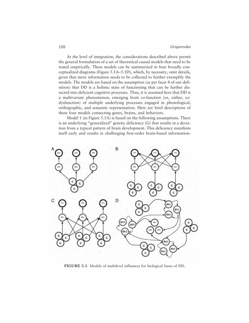
At the level of integration, the considerations described above permitthe general formulation of a set of theoretical causal models that need to betested empirically. These models can be summarized in four broadly con-ceptualized diagrams (Figure 5.1A–5.1D), which, by necessity, omit details,given that more information needs to be collected to further exemplify themodels. The models are based on the assumption (as per facet 4 of our defi-nition) that DD is a holistic state of functioning that can be further dis-sected into deficient cognitive processes. Thus, it is assumed here that DD isa multivariate phenomenon, emerging from co-function (or, rather, co-dysfunction) of multiple underlying processes engaged in phonological,orthographic, and semantic representation. Here are brief descriptions ofthese four models connecting genes, brains, and behaviors.
Model 1 (in Figure 5.1A) is based on the following assumptions. Thereis an underlying “generalized” genetic deficiency (G) that results in a devia-tion from a typical pattern of brain development. This deficiency manifestsitself early and results in challenging first-order brain-based information-
128 Grigorenko
FIGURE 5.1. Models of multilevel influences for biological bases of DD.
processing neuronal network deficiencies (BN for brain networks, BN1–BN3) underlying the neurological and cognitive foundation for atypicaldevelopment of cognitive processes (CP for cognitive processes, CP1–CP3).Note that it is assumed here that the genetic bases are common for all dis-rupted neuronal networks, but each network provides an independentcause for a disordered cognitive process. For example, a disrupted neuronalmigration (which can be caused by genetic malfunction) might result inbrain miswiring, which, in turn, might lead (as a secondary cause) to theformation of disordered (dysfunctional) cognitive functions, reflective ofdysfunctional neuronal networks. The impact of this genetic deficiency fac-tor undermines the whole domain of functioning of the developing individ-ual. A proximal analogy here is Spearman’s g-factor model of cognitivefunctioning (Spearman, 1927). A distal analogy of this impact is the crackin the foundation of a building, where the structural damage in the founda-tion results in problems in the whole building, but the specific nature ofthese problems is not predictable and is characterized by the presence ofrandom factors, for example, environmental influences. Within this model,the observed correlations of deficient cognitive processes characteristic ofDD (i.e., the processes descriptive of phonological, orthographic, andsemantic representations) are secondary and reflect the causal links be-tween the brain networks and deficient information processing at thephenotypic, not genotypic, levels. This model assumes a search for a com-mon g-factor governed by the generalist gene(s) (Plomin & Kovas, 2005).
The following models (2 and 3, in Figures 5.1B and 5.1C, respectively)are quite similar and assume the presence of a set of genes influencing spe-cific brain networks, which in turn contribute to the manifestation of thechallenged cognitive phenotypes in unique or overlapping fashion. To illus-trate these models, we can think of a “latent variable” model, often used inthe DD literature (Wagner, Torgesen, & Rashotte, 1994). A proximal anal-ogy of models 2 and 3 is the Thurstone model of cognitive functioning7
(Thurstone, 1938). A distal analogy is multiple operations malfunctioningin our hypothetical building (electricity, heating, and water systems), eachfor specific overlapping or unique reasons. The difference between models2 and 3 occurs at the level of genetic etiology and brain bases of cognitiveprocessing: Model 2 specifies unique sets of genes influencing specific brainnetworks, but overlapping brain networks contributing to different cogni-tive phenotypes, whereas model 3 specifies both network-specific andnetwork-shared genes influencing more than one process, but each cogni-tive process is governed by a specific network only.
Specifically, in model 2 (in Figure 5.1B) it is assumed that a set of dis-tinct genes affects the development or functioning of a particular neuronalnetwork that, in turn, forms the foundation for a cognitive process. Thus,
Triangulating Developmental Dyslexia 129
the formation and functioning of a particular brain network is governed bya set of specific genes (G), and each network (BN1–BN3) contributes to themanifestation of multiple dysfunctional cognitive processes (CP1–CP3).The observed phenotypic correlation between cognitive processes CP1–CP3is not due to shared genetic etiology, but rather, because of overlappingcontributions of the brain’s neuronal networks, reflects the engagement ofoverlapping networks with more than one cognitive process (e.g., differenttypes of attention; Fan & Posner, 2004), or arises as a property of a com-plex system in which many independent elements are involved in a com-mon function.
Model 3 (Figure 5.1C) is a modification of model 2 in that it assumesthat (1) the development and functioning of particular brain networks areinfluenced by sets of partially overlapping genes—that is, there are generaland specific genes (G) for each brain network (BN); and (2) each networkcontributes uniquely to a particular dysfunctional cognitive process (CP).In this model, the phenotypic correlation between cognitive processes is dueto shared genetic factors mediated by a particular neuronal network.
Finally, model 4 (in Figure 5.1D) assumes that multiple unrelatedgenetic factors influence the many neuronal networks recruited into the sys-tems of complex architecture that form the foundation for cognitive func-tioning. In this model, no shared or general factors are assumed, and theappearance of commonalities is attributed to epiphenomenological mani-festations. A proximal analogy of this model is Thompson’s model of cog-nitive functioning8 (Thompson, 1939). A distal analogy is the behav-ior of a complex system constructed of independent “particles” (as inCrichton’s Prey [2002]), where the common factor is the characteristic ofthe system, not of its separate elements, which can arise only when a criti-cal mass of elements is present. At the phenotypic level, model 4 calls forthe use of multivariate models, and at the genetic level, calls not only forthe analysis of the mutations in single genes (i.e., linkage and associationanalyses), but also for the analysis of the context (the genome as a whole)in which these genes are expressed.
Clearly, these four models are only a sample of possible models. Theyare intended to capture the importance of considering multiple levels ofcausal influences and the lack of one-to-one and the abundance of one-to-many, many-to-one, and many-to-many correspondences. Thus, by varyingthe number of genes influencing the emerging neuronal networks, the num-ber of neuronal networks influencing cognitive processes, and the numberof shared and unique connections between these levels, a fairly large num-ber of models can be created. To appreciate the number, just reflect for amoment on the thought that, of the ~30,000 genes in the human genome,~5,000 are supposedly expressed in the brain. If we follow the estimates
130 Grigorenko
obtained in segregation analyses of DD and related traits (e.g., Wijsman etal., 2000) and assume that, on average, 4 genes contribute to a reading-related trait, we face 26,010,428,123,7509 possible unique combinations.Thus, to understate the case, there are many possibilities, and we are talk-ing about the lowest level of hierarchy considered in these causal models.
It must be noted, of course, that I did not include the impact of envi-ronmental factors in the models discussed above. That is because these fac-tors do not operate within the models and do not form a separate layer ofinfluences; they act across all four levels of the models considered above.To illustrate, consider the impact of SES. SES has been shown to influencedietary choices (Monteiro, Moura, Conde, & Popkin, 2004), which, inturn, have been shown to result in altered gene expression (Li et al., 2005).Different levels of SES are also associated with differences in brain net-working and functioning (Noble, McCandliss, & Farah, in press). Thereare robust correlations linking SES and variability in cognitive functioning(Bornstein & Bradley, 2003). Finally, the manifestation and remediation ofDD itself is likely to be linked to SES: the higher the level of SES, in general,the more remediation and support services the family can afford, and thebetter the outcome (Kelman & Lester, 1998). In fact, in considering theserelationships, it becomes clear that environment is inseparable from “bio-logical” influences. Environmental influences have an impact on what isconsidered biological at the next level of hierarchy in the model (e.g., pro-teins formed under the influence of different diets [i.e., environmentallymodified proteins] are still viewed as biological factors in processes utiliz-ing these proteins [e.g., neuronal signal transduction]). With this remark, Imove to the last section of the chapter, a discussion that attempts to sum-marize the current state of the field with regard to our understanding of theimpact of main effects and interactions of etiological forces contributing tothe manifestation of DD.
The Etiology of Developmental Dyslexia:Main Effects and Interactions
I opened the chapter with a discussion of the hexagonal definition of DD,progressed through brief descriptions of bodies of literature associated withdifferent facets of this definition, and focused specifically on the brief char-acterization of biological bases of DD. A summary of various points thathave been covered so far is presented in Figure 5.2, illustrating the knowngenetic and environmental risk factors for DD.
In essence, Figure 5.2 represents a two-by-two table, with one dimen-sion of the table separating etiological influences into genetic and environ-mental factors, and another dimension subdividing risk and protective fac-
Triangulating Developmental Dyslexia 131
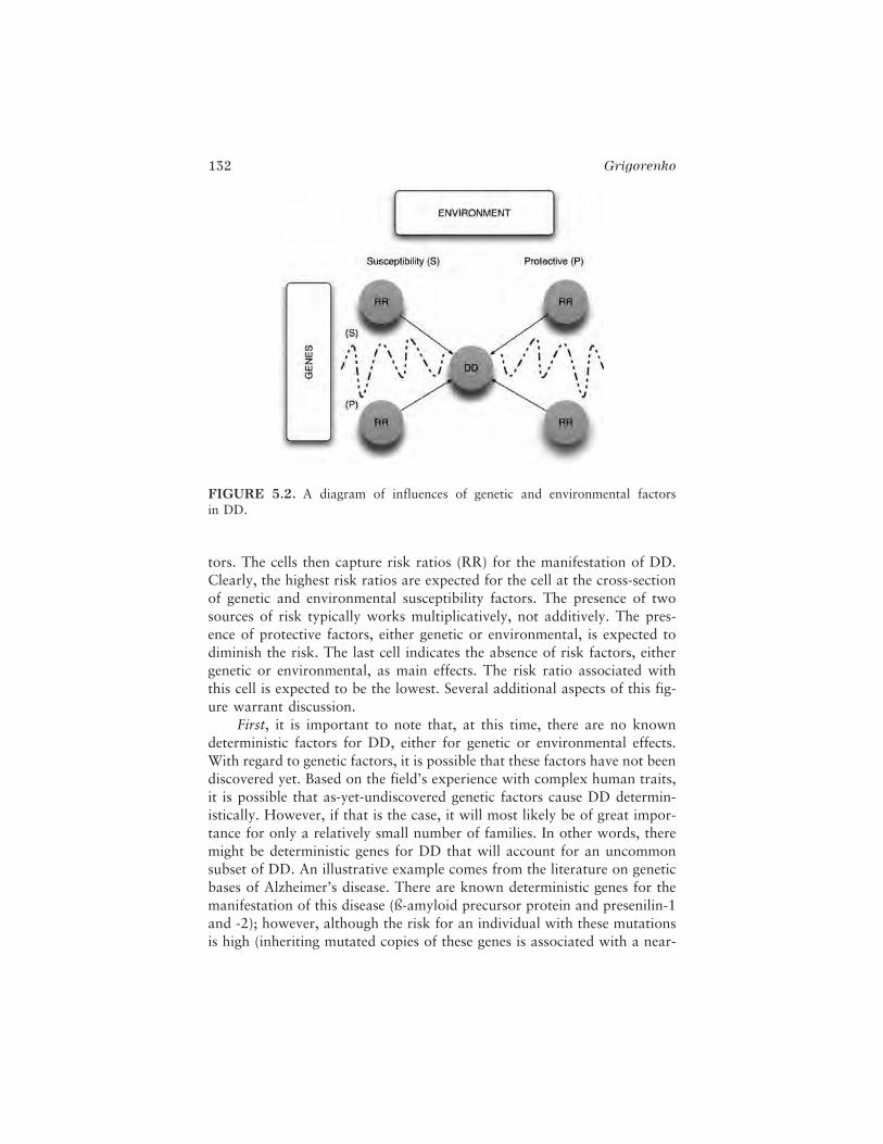
tors. The cells then capture risk ratios (RR) for the manifestation of DD.Clearly, the highest risk ratios are expected for the cell at the cross-sectionof genetic and environmental susceptibility factors. The presence of twosources of risk typically works multiplicatively, not additively. The pres-ence of protective factors, either genetic or environmental, is expected todiminish the risk. The last cell indicates the absence of risk factors, eithergenetic or environmental, as main effects. The risk ratio associated withthis cell is expected to be the lowest. Several additional aspects of this fig-ure warrant discussion.
First, it is important to note that, at this time, there are no knowndeterministic factors for DD, either for genetic or environmental effects.With regard to genetic factors, it is possible that these factors have not beendiscovered yet. Based on the field’s experience with complex human traits,it is possible that as-yet-undiscovered genetic factors cause DD determin-istically. However, if that is the case, it will most likely be of great impor-tance for only a relatively small number of families. In other words, theremight be deterministic genes for DD that will account for an uncommonsubset of DD. An illustrative example comes from the literature on geneticbases of Alzheimer’s disease. There are known deterministic genes for themanifestation of this disease (ß-amyloid precursor protein and presenilin-1and -2); however, although the risk for an individual with these mutationsis high (inheriting mutated copies of these genes is associated with a near-
132 Grigorenko
FIGURE 5.2. A diagram of influences of genetic and environmental factorsin DD.
100% likelihood of developing the disease), these mutations are very rareand thus the population risk is low. There is also a known genetic suscepti-bility factor, the ε4 allele of the APOE gene; it appears that the risk ofdeveloping Alzheimer’s varies with the number of copies of the ε4 allele.Specifically, it has been reported that one copy of the ε4 allele is associatedwith 2.5–3.2 odds (i.e., the proportion of having the disease versus not hav-ing the disease) and two copies of the ε4 allele are associated with 14.9odds of the manifestation of the disease (Farrer et al., 1997). The frequencyof this allele is relatively high in the general population (although it variessubstantially for different ethnic groups), and, although the associated indi-vidual risks are probabilistic rather than deterministic, the ε4 allele has avery high population attributable risk. For DD, there are currently noknown deterministic genes, but a number of genes are currently being eval-uated as susceptibility genes (as noted, DCDC2, DYX1C1, KIAA0319,ROBO1).
Listing the currently investigated candidate genes for DD is relevant tothe second point of this discussion. Although the genes listed above areconsidered to be susceptibility factors for DD, it is possible that somealleles of these genes can act as risk factors and others as protective factors.To illustrate, let us consider the APOE gene again. As discussed earlier, theε4 allele of this gene has a high population attributable risk. However, twoother alleles of this gene, ε2 and ε3, when forming the ε2/ε3 genotype,appear to act as a protective factor (Farrer et al., 1997). Although the can-didate genes for DD have not yet been confirmed as susceptibility factors,there is an interesting inconsistency in the literature with regard to thealleles of the DYX1C1 gene. The first presentation of this gene as a suscep-tibility gene named the A allele of the 3G-to-A change in the promoterregion of the DYX1C1 gene as a risk factor (Taipale et al., 2003). The firstattempt to replicate this finding suggested that an alternative allele (G) wasassociated with DD (Wigg et al., 2004); thus, it is possible that the G alleleis a protective factor. These intriguing results are yet to be sorted out inlarge, phenotypically well-characterized, preferably population-based sam-ples. To my knowledge, there are currently no such samples available.Thus, one of the field’s many tasks on the road to understanding geneticpopulation risks for DD will be to build such samples.
The third important point is that, from what we know from the devel-opmental literature at large, there are virtually no factors among envi-ronmental influences that are typically considered by developmentalistsas truly deterministic. However, there are countless environmental fac-tors that can be named as susceptibility (e.g., poverty, printed-material-impoverished home environment, low level of parental education) and pro-tective (e.g., amount of schooling, high-quality teaching, presence of
Triangulating Developmental Dyslexia 133
mentors and role models, positive peer networking) factors for the manifes-tation of DD. The impact of all these environmental factors is probabilisticby nature. Yet, as is true for development in general and maladaptivebehavior in particular (e.g., Sameroff & Mackenzie, 2003), an accumula-tion of risk factors changes the odds of the manifestation of DD, mostlikely in a bidirectional and nonlinear manner.
The fourth point brings us back to the definition of DD (facet 6).How, in this diagram, is the risk ratio conceived when genetic protectivefactors and environmental susceptibility factors are considered? Facet 6 ofthe hexagonal definition of DD assumes that only the presence of biologicalbases qualifies reading difficulties to be referred to as DD. Does the pres-ence of protective alleles in a gene whose alleles are also risk alleles etiologi-cally connect DD and high reading performance? The answer is yes—thereis a significant possibility of allele heterogeneity with regard to DD, andunderstanding this heterogeneity will be a major task of molecular–geneticresearch on DD even after candidate genes for DD are identified and con-firmed. The protective gene/environmental susceptibility cell in Figure 5.2calls for a discussion of an interesting paradox: If the same gene, throughits differential alleles, forms differential genetic backgrounds (disadvanta-geous and advantageous) for DD, how might this consideration influencethe field’s sampling strategies? Does this consideration, among others, sug-gest the necessity of recruiting individuals with high and very high perfor-mance on reading and reading-related tasks? Although the detailed consid-eration of the issue of sampling in genetic studies of DD is a very interestingtopic in its own right (e.g., Grigorenko et al., 2006; Ziegler et al., 2005),here I simply want to stress the importance of considering all kinds of sam-ples (e.g., large rare families with high incidence of DD, small nuclear fami-lies with a DD proband, sib-pairs ascertained through a single probandwith a reading deficit, as well as through sib-pairs [i.e., double probandselection] concordant for deficit or high performance and highly discor-dant), because each of these sampling strategies will contribute to ourunderstanding of the intricacies of gene–environment co-action in DD.
The fifth point of discussion involves an exploration of risk and pro-tective genetic and environmental factors. As noted earlier, the current stateof affairs suggests a multilevel causality model of biological (i.e., gene–brain-based) predisposition for DD that is alterable by environmental fac-tors at all levels. There are no convergent data just yet that are powerfulenough to establish a preference for one of the models suggested above,connecting genes and the brain in the etiological pathway so as to result inthe manifestation of DD. Yet the emerging evidence suggests that, mostlikely, the model will engage multiple genes that will contribute probab-ilistically in unique and shared fashions to the formation of neuronal net-
134 Grigorenko
works, which, in turn, will probabilistically contribute to the establishmentof cognitive processes involved in forming different types of informationrepresentation. It appears that the manifestation of DD, mostly likely, is aproduct of complex probabilistic machinery that has no single deterministicfactor, but rather many factors involved in many possible interactive ways.
Now, to conclude, where does this discussion leave us with regard topublic health policies and considerations?
The link between biomedical research and public health is in formingpolicies that ultimately result in the reduction of morbidity and mortalitythrough prevention and treatment. The link between biomedical researchand education is in providing informed summaries to educational authori-ties, whose outreach to children is unprecedented in terms of its potentialeffectiveness. What other civic institution but school has the power toestablish policies that reach every boy and girl between the ages of 5 and17? There is none. So, what might this summary be? It appears that itmight be inclusive of the following components.
First, at this point, the genomic priorities for DD are related to findingthe genes and genetic pathways underlying the biological bases of DD. Ascompared with research in other developmental disorders, it is quite possi-ble that researchers will discover both rare variants with high level ofpenetrance (i.e., a high, possibly deterministic, correlation between geneticpredisposition and the disorder) and probabilistic genetic risk factors, eachof which, in isolation, will be associated with lower individual risk butmight be associated with high population risk. Identification of both rare(high individual risk/lower population risk) and common (low individualrisk/high population risk) genetic risk factors is very important for two rea-sons. With regard to rare families in which DD accumulates at high ratesand is transmitted from generation to generation, the identification ofcausal genetic factors segregating in these families will result in the optionof informed genetic counseling; the development of drug therapies; and thedevelopment of specialized, intensive remediation programs for childrenwho carry these rare genetic variants and for whom this intense educationwill be delivered, under federal laws and regulation, upon proper diagnosesor screening. With regard to the identification of common genetic risk fac-tors, it is important to understand the extent of their “riskiness” and todetermine the environmental forces that are able to diminish the risk. Forexample, it has been established that for carriers of the ε4 allele, one of themajor protective factors against developing Alzheimer’s disease is level ofeducation: The more schooling individuals at genetic risk had, the lowertheir odds ratios were in developing the disease, as compared with riskallele carriers with lower levels of education (Farrer et al., 1997). However,the identification of education as a protective factor became possible only
Triangulating Developmental Dyslexia 135
when the risk allele was identified and its impact studied by methods ofgenetic epidemiology in large, unselected samples of both affected andunaffected individuals. Thus, understanding the genetic machinery underly-ing biological bases of DD is a critical, not optional, step in finding ways toaccommodate children in need of services under the Individuals with Dis-abilities Education Act (IDEA).
Second, this summary should include a comprehensive account ofongoing integration of advances in neuroscience and genetics. Specifically,the majority of neuroscience literature on DD today is focused on discover-ing and discussing correlates rather than presenting causal pathways (e.g.,Price & Mechelli, 2005). Because the brain is an organ whose developmentand maturation is, at least initially, preprogrammed in genes, understand-ing the links between brain neuronal pathways, their patterns of activation,and their malleability is as important, if not more important, than under-standing the connection between the brain and behavior. As of today, inthe field of DD, the ratio of research articles describing studies addressingthe gene–brain link and those addressing the brain–behavior link is 0 tomany. Clearly, the next step forward should be not in changing “many” toinnumerable, but in changing “0” to a positive number.
Finally, and most importantly, it is crucial for educators to understandthat our attempt to decode biological bases of DD is aimed at strengtheningeveryday educational practices. As I hope to have shown throughout thischapter, even under the most liberal estimate, the genetic contributionaccounts only for a portion (most likely, the smallest portion) of individualdifferences in reading. I have also tried to make it clear that environment(described as temperature that changes as part of human emotion, diet thatis a part of cultural belonging, oxygen concentration that is an outcome ofexercise schedule, and so forth) is a part of any biological function, no mat-ter how conservatively preprogrammed that function is. Despite the amaz-ing advances in human genome research that have unfolded within the lastfew years, it is unlikely that any radical transformation of medical practices(Varmus, 2002) for either adults and children, or the upbringing and edu-cation of children, will take place any time soon, if at all. Thus, the lethalweapon against DD is still the protective environmental factor of goodteaching. It is, without a doubt, our most effective public health policy.
ACKNOWLEDGMENTS
Preparation of this chapter was supported by Grant Nos. REC-9979843 from theNational Science Foundation; R206R00001 from the Javits Act Program adminis-tered by the Institute for Educational Sciences, U.S. Department of Education; and
136 Grigorenko
TW006764 from the National Institutes of Health. Grantees undertaking such pro-jects are encouraged to express freely their professional judgment. This chapter,therefore, does not necessarily represent the position or policies of the National Sci-ence Foundation, the Institute for Educational Sciences, or the National Institutes ofHealth, and no official endorsement should be inferred. I express sincere gratitudeto Ms. Robyn Rissman for her editorial assistance and to Mr. Adam Naples for pre-paring the figures.
NOTES
1. Difficulty would be a preferred word here.2. These estimates are imprecise and are used by the IDA generally. The reference
to 15% here is an average of the 12–17% prevalence rate cited above.3. This definition assumes the primacy of DD as an initial diagnosis. This means
that manifested reading difficulties cannot be attributed to other developmentaldisorders (e.g., autism), but there could be secondary comorbid manifestationsof other developmental conditions (e.g., DD is often comanifested with ADHD,dyscalculia, and conduct disorder).
4. EEG: electroencephalogram; ERP: event-related potential; fMRI: functionalmagnetic resonance imaging; MEG: magnetoencephalography; PET: positronemission tomography; TMS: transcranial magnetic stimulation.
5. Though accurate in terms of broad generalization, this statement should beinterpreted differentially with regard to what Price and Mechelli called “the con-spicuous absence of brain areas that are dedicated to orthography” (2005,p. 236).
6. To illustrate, spelling and automatization problems are more indicative of DD inGerman than in English; phonemic awareness deficits are more indicative of DDin English than in German. German and English vary on a number of parame-ters capturing linguistic diversity, two of which are phonological transparency(i.e., English is more phonologically complex) and morphological richness (i.e.,German is more complex morphologically) of language.
7. In opposition to Spearman, Thurstone formulated a model of intelligence cen-tered around “Primary Mental Abilities,” which were independent group factorsof intelligence that different individuals possessed in varying degrees. Thurstonedistinguished seven primary mental abilities: verbal comprehension, verbal flu-ency, inductive reasoning, spatial visualization, number computation, memory,and perceptual speed.
8. Thompson stated that general intelligence could be thought of as including alarge number of independent structural “bonds” (e.g., reflexes, habits, learnedassociations). Correspondingly, a large number of bonds are activated while atask is performed. When overlapping tasks are performed (e.g., those on anintelligence test), many sets of bonds activated for a given task will overlap aswell. This overlap will result in manifested “closeness” captured by a factoranalysis. Thus, although a factor analysis might form an impression of a sin-gle factor, Thompson argues that the communality among tests cannot be
Triangulating Developmental Dyslexia 137
attributed to a unitary source (e.g., mental energy), but should be attributableto a multiplicity of sources where each source is of relatively small magnitude,but many of the same sources contribute to different tasks in an overlappingmanner.
9. http://mathforum.org/dr.math/faq/faq.comb.perm.html.
REFERENCES
Ames, B. N. (2004). A role for supplements in optimizing health: the metabolictune-up. Archives of Biochemistry and Biophysics, 423, 227–234.
Barr, C. L. (2005, June). Linkage studies of reading disabilities and ADHD in thechromosome 6p and 15q regions. Paper presented at the annual meeting of theSociety for Scientific Studies of Reading, Toronto.
Barr, C. L., & Couto, J. M. (in press). Molecular genetics of reading. In E. L.Grigorenko & A. Naples (Eds.), Single-word reading: Cognitive, behavioraland biological perspectives. Mahwah, NJ: Erlbaum.
Blachman, B. A., Schatschneider, C., Fletcher, J. M., Francis, D., Clonan, S. M.,Shaywitz, B. A., et al. (2004). Effects of intensive reading remediation for sec-ond and third graders and a 1–year follow-up. Journal of Educational Psy-chology, 96, 444–461.
Bornstein, M. H., & Bradley, R. H. (2003). Socioeconomic status, parenting, andchild development. Mahwah, NJ: Erlbaum.
Byrne, B., Wadsworth, S., Corley, R., Samuelsson, S., Quain, P., DeFries, J. C., etal. (2005). Longitudinal twin study of early literacy development: Preschooland kindergarten phases. Scientific Studies of Reading, 9, 219–235.
Cardon, L. R., Smith, S. D., Fulker, D. W., Kimberling, W. J., Pennington, B. F., &DeFries, J. C. (1994). Quantitative trait locus for reading disability on chro-mosome 6. Science, 226, 276–279.
Cardon, L. R., Smith, S. D., Fulker, D. W., Kimberling, W. J., Pennington, B. F., &DeFries, J. C. (1995). Quantitative trait locus for reading disability: Correc-tion. Science, 268, 1553.
Catts, H. W., Fey, M. E., & Proctor-Williams, K. (2000). The relationship betweenlanguage and reading. Preliminary results from a longitudinal investigation.Logopedics, Phoniatrics, Vocology, 25, 3–11.
Cope, N., Harold, D., Hill, G., Moskvina, V., Holmans, P., Owen, M. J., et al.(2005). Strong evidence that KIAA0319 on chromosome 6p is a susceptibilitygene for developmental dyslexia. American Journal of Human Genetics, 76,581–591.
Cope, N., Hill, G., van den Bree, M., Harold, D., Moskvina, V., Green, E. K., et al.(2004). No support for association between Dyslexia Susceptibility 1 Candi-date 1 and developmental dyslexia. Molecular Psychiatry, 10, 237–238.
Crichton, M. (2002). Prey. New York: HarperCollins.D’Angiulli, A., Siegel, L. S., & Hertzman, C. (2004). Schooling, socioeconomic con-
text and literacy development. Educational Psychology, 24, 867–883.Eden, G. F., Jones, K. M., Cappell, K., Gareau, L., Wood, F. B., Zeffiro, T. A., et al.
138 Grigorenko
(2004). Neural changes following remediation in adult developmental dys-lexia. Neuron, 44, 411–422.
Falconer, D. S., & Mackay, T. F. C. (1996). Introduction to quantitative genetics(4th ed.). New York: Longman.
Fan, J., & Posner, M. (2004). Human attentional networks. Psychiatrische Praxis,31(Suppl. 2), S210–S214.
Farrer, L. A., Cupples, L. A., Haines, J. L., Hyman, B., Kukull, W. A., Mayeux, R.,et al. (1997). Effects of age, sex, and ethnicity on the association betweenapolipoprotein E genotype and Alzheimer disease: A meta-analysis. APOE andAlzheimer Disease Meta Analysis Consortium. Journal of the American Medi-cal Association, 278, 1349–1356.
Feldman, E., Levin, B. E., Lubs, H., Rabin, M., Lubs, M. L., Jallad, B., et al. (1993).Adult familial dyslexia: A retrospective developmental and psychosocial pro-file. Journal of Neuropsychiatry and Clinical Neurosciences, 5, 195–199.
Felton, R. H., Naylor, C. E., & Wood, F. B. (1990). Neuropsychological profile ofadult dyslexics. Brain and Language, 39, 485–497.
Fiez, J. A. (1997). Phonology, semantics, and the role of the left inferior prefrontalcortex. Human Brain Mapping, 5, 79–83.
Fiez, J. A., & Petersen, S. E. (1998). Neuroimaging studies of word reading. Pro-ceedings of the National Academy of Sciences USA, 95, 914–921.
Fisher, S. E., & DeFries, J. C. (2002). Developmental dyslexia: Genetic dissection ofa complex cognitive trait. Nature Reviews: Neuroscience, 3, 767–780.
Francks, C., Paracchini, S., Smith, S. D., Richardson, A. J., Scerri, T. S., Cardon, L.R., et al. (2004). A 77–kilobase region on chromosome 6p22.2 is associatedwith dyslexia in families from the United Kingdom and from the United States.American Journal of Human Genetics, 75, 1046–1058.
Gaillard, W. D., Balsamo, L. M., Ibrahim, Z., Sachs, B. C., & Xu, B. (2003). fMRIidentifies regional specialization of neural networks for reading in young chil-dren. Neurology, 60, 94–100.
Gallagher, A., Frith, U., & Snowling, M. J. (2000). Precursors of literacy delayamong children at genetic risk of dyslexia. Journal of Child Psychology andPsychiatry and Allied Disciplines, 41, 203–213.
Gayan, J., & Olson, R. K. (2003). Genetic and environmental influences on individ-ual differences in printed word recognition. Journal of Experimental ChildPsychology, 84, 97–123.
Gottesman, I. I., & Gould, T. D. (2003). The endophenotype concept in psychiatry:Etymology and strategic intentions. American Journal of Psychiatry, 160,636–645.
Gottesman, R. L., Bennett, R. E., Nathan, R. G., & Kelly, M. S. (1996). Inner-cityadults with severe reading difficulties: A closer look. Journal of Learning Dis-abilities, 29, 589–597.
Goulandris, N. (Ed.). (2003). Dyslexia in different languages: Cross-linguistic com-parisons. London: Whurr.
Grigorenko, E. L. (2004). Genetic bases of developmental dyslexia: A capsulereview of heritability estimates. Enfance, 3, 273–287.
Grigorenko, E. L. (2005). A conservative meta-analysis of linkage and linkage-
Triangulating Developmental Dyslexia 139
association studies of developmental dyslexia. Scientific Studies of Reading, 9,285–316.
Grigorenko, E. L., Ngorosho, D., Jukes, M., & Bundy, D. (2006). Reading in ableand disabled readers from around the world: Same or different? An illustrationfrom a study of reading-related processes in a Swahili sample of siblings. Jour-nal of Research in Reading, 29, 104–123.
Grigorenko, E. L., Wood, F. B., Meyer, M. S., Hart, L. A., Speed, W. C., Shuster,A., et al. (1997). Susceptibility loci for distinct components of developmentaldyslexia on chromosomes 6 and 15. American Journal of Human Genetics,60, 27–39.
Grigorenko, E. L., Wood, F. B., Meyer, M. S., & Pauls, D. L. (2000). Chromosome6p influences on different dyslexia-related cognitive processes: Further confir-mation. American Journal of Human Genetics, 66, 715–723.
Hannula-Jouppi, K., Kaminen-Ahola, N., Taipale, M., Eklund, R., Nopola-Hemmi,J., Käänäinen, H., et al. (2005). The axon guidance receptor gene ROEO1 is acandidate gene for developmental dyslexia. PLoS Genetics, 1(4), ε50.
Harlaar, N., Spinath, F. M., Dale, P. S., & Plomin, R. (2005). Genetic influences onearly word recognition abilities and disabilities: A study of 7–year-old twins.Journal of Child Psychology and Psychiatry, 46, 373–384.
Harm, M. W., & Seidenberg, M. S. (2004). Computing the meanings of words inreading: Cooperative division of labor between visual and phonological pro-cesses. Psychological Review, 111, 662–720.
Hohnen, B., & Stevenson, J. (1999). The structure of genetic influences on generalcognitive, language, phonological, and reading abilities. Developmental Psy-chology, 35, 590–603.
Kamhi, A. G., & Laing, S. P. (2001). The path to reading success or failure: Achoice for the new millennium. In J. L. Harris, A. G. Kamhi, & K. E. Pollock(Eds.), Literacy in African American communities (pp. 127–145). Mahwah,NJ: Erlbaum.
Karasik, D., Myers, R. H., Cupples, L. A., Hannan, M. T., Gagnon, D. R., Herbert,A., et al. (2002). Genome screen for quantitative trait loci contributing to nor-mal variation in bone mineral density: The Framingham Study. Journal ofBone and Mineral Research, 17, 1718–1727.
Karmiloff-Smith, A. (1998). Development itself is the key to understanding devel-opmental disorders. Trends in Cognitive Sciences, 2, 389–398.
Kelman, M., & Lester, G. (1998). Jumping the queue: An inquiry into the legaltreatment of students with learning disabilities. Cambridge, MA: Harvard Uni-versity Press.
Li, Y., Hou, M. J., Ma, J., Tang, Z. H., Zhu, H. L., & Ling, W. H. (2005). Dietaryfatty acids regulate cholesterol induction of liver CYP7alpha1 expression andbile acid production. Lipids, 40, 455–462.
Livingstone, M. S., Rosen, G. D., Drislane, F. W., & Galaburda, A. M. (1991).Physiological and anatomical evidence for a magnocellular defect in develop-mental dyslexia. Proceedings of the National Academy of Sciences USA, 88,7943–7947.
Lovegrove, W. J., Bowling, A., Badcock, D., & Blackwood, M. (1980). Specific
140 Grigorenko
reading disability: Differences in contrast sensitivity as a function of spatialfrequency. Science, 210, 439–440.
Lyon, G. R., Shaywitz, S. E., & Shaywitz, B. A. (2003). A definition of dyslexia.Annals of Dyslexia, 53, 1–14.
Lyytinen, P., Poikkeus, A. M., Laakso, M. L., Eklund, K., & Lyytinen, H. (2001).Language development and symbolic play in children with and without famil-ial risk for dyslexia. Journal of Speech Language and Hearing Research, 44,873–885.
Marino, C., Giorda, R., Vanzin, L., Nobile, M., Lorusso, M. L., Baschirotto, C., etal. (2004). A locus on 15q15–15qter influences dyslexia: Further support froma transmission/disequilibrium study in an Italian speaking population. Journalof Medical Genetics, 41, 42–48.
Marlow, A. J., Fisher, S. E., Francks, C., MacPhie, I. L., Cherny, S. S., Richardson,A. J., et al. (2003). Use of multivariate linkage analysis for dissection of a com-plex cognitive trait. American Journal of Human Genetics, 72, 561–570.
Mechelli, A., Gorno-Tempini, M. L., & Price, C. J. (2003). Neuroimaging studiesof word and pseudoword reading: Consistencies, inconsistencies, and limita-tions. Journal of Cognitive Neuroscience, 15, 260–271.
Meng, H., Smith, S. D., Hager, K., Held, M., Liu, J., Olson, R. K., et al. (2005).DCDC2 is associated with reading disability and modulates neuronal develop-ment in the brain. Proceedings of the National Academy of Sciences USA, 102,17053–17058.
Merikangas, K. R., & Risch, N. (2003). Genomic priorities and public health. Sci-ence, 302, 599–601.
Monteiro, C. A., Moura, E. C., Conde, W. L., & Popkin, B. M. (2004). Socioeco-nomic status and obesity in adult populations of developing countries: Areview. Bulletin of the World Health Organization, 82, 940–946.
Morris, D. W., Robinson, L., Turic, D., Duke, M., Webb, V., Milham, C., et al.(2000). Family-based association mapping provides evidence for a gene forreading disability on chromosome 15q. Human Molecular Genetics, 9, 843–848.
Morton, J., & Frith, U. (1995). Causal modeling: A structural approach to develop-mental psychopathology. In D. Cicchetti & D. J. Cohen (Eds.), Developmentalpsychopathology (Vol. 2, pp. 357–390). New York: Wiley.
Nicholson, T. (1997). Social class and reading achievement: Sociology meets psy-chology. New Zealand Journal of Educational Studies, 32, 105–108.
Nicolson, R. I., & Fawcett, A. J. (1990). Automaticity: A new framework for dys-lexia research? Cognition, 35, 159–182.
Nicolson, R. I., Fawcett, A. J., & Dean, P. (2001). Developmental dyslexia: The cer-ebellar deficit hypothesis. Trends in Neurosciences, 24, 508–511.
Noble, K. G., McCandliss, B. D., & Farah, M. (in press). Socioeconomic gradientspredict individual differences in neurocognitive abilities. Developmental Sci-ence.
Paulesu, E., Demonet, J. F., Fazio, F., McCrory, E., Chanoine, V., Brunswick, N., etal. (2001). Dyslexia: Cultural diversity and biological unity. Science, 291,2165–2167.
Triangulating Developmental Dyslexia 141
Pennington, B. F. (2002). The development of psychopathology: Nature and nur-ture. New York: Guildford Press.
Pennington, B. F., & Lefly, D. L. (2001). Early reading development in children atfamily risk for dyslexia. Child Development, 72, 816–833.
Petersen, S. E., Fox, P. T., Posner, M. I., Mintun, M., & Raichle, M. E. (1988). Pos-itron emission tomographic studies of the cortical anatomy of single-wordprocessing. Nature, 331, 585–589.
Plomin, R., & Kovas, Y. (2005). Generalist genes and learning disabilities. Psycho-logical Bulletin, 131, 592–617.
Poldrack, R. A., Wagner, A. D., Prull, M. W., Desmond, J. E., Glover, G. H., &Gabrieli, J. D. (1999). Functional specialization for semantic and phonologicalprocessing in the left inferior prefrontal cortex. NeuroImage, 10, 15–35.
Price, C. J., & Mechelli, A. (2005). Reading and reading disturbance. CurrentOpinion in Neurobiology, 15, 231–238.
Price, C. J., Wise, R. J., & Frackowiak, R. S. (1996). Demonstrating the implicitprocessing of visually presented words and pseudowords. Cerebral Cortex, 6,62–70.
Pugh, K. R., Mencl, W. E., Jenner, A. R., Katz, L., Frost, S. J., Lee, J. R., et al.(2001). Neurobiological studies of reading and reading disability. Journal ofCommunication Disorders, 34, 479–492.
Pugh, K. R., Shaywitz, B. A., Shaywitz, S. E., Constable, R. T., Skudlarski, P.,Fulbright, R. K., et al. (1996). Cerebral organization of component processesin reading. Brain, 119, 1221–1238.
Ramus, F. (2003). Developmental dyslexia: Specific phonological deficits or generalsensorimotor dysfunction? Current Opinion in Neurology, 13, 212–218.
Reed, D. R., Nanthakumar, E., North, M., Bell, C., & Price, R. A. (2001). Agenome-wide scan suggests a locus on chromosome 1q21–q23 contributes tonormal variation in plasma cholesterol concentration. Journal of MolecularMedicine, 79, 262–269.
Sameroff, A. J., & Mackenzie, M. J. (2003). Research strategies for capturing trans-actional models of development: The limits of the possible. Development andPsychopathology, 15, 613–640.
Scarborough, H. S. (1991). Antecedents to reading disability: Preschool languagedevelopment and literacy experiences of children from dyslexic families. Read-ing and Writing: An Interdisciplinary Journal, 3, 219–233.
Scerri, T. S., Fisher, S. E., Francks, C., MacPhie, I. L., Paracchini, S., Richardson, A.J., et al. (2004). Putative functional alleles of DYX1C1 are not associated withdyslexia susceptibility in a large sample of sibling pairs from the UK. Journalof Medical Genetics, 41, 853–857.
Schulte-Körne, G., Grimm, T., Nöthen, M. M., Müller-Myhsok, B., Cichon, S.,Vogt, I. R., et al. (1998). Evidence for linkage of spelling disability to chromo-some 15. American Journal of Human Genetics, 63, 279–282.
Schumacher, J., Anthioni, H., Dahdouh, F., Konig, I. R., Hillmer, H. M., Kluck, N.,et al. . (2006). Strong evidence of DCDC2 as a susceptibility gene for dyslexia.American Journal of Human Genetics, 78, 52–62.
142 Grigorenko
Shaywitz, S. E., & Shaywitz, B. A. (2005). Dyslexia (specific reading disability).Biological Psychiatry, 57, 1301–1309.
Simos, P. G., Billingsley-Marshall, B., Sarkari, S., & Papanicolaou, A. C. (in press).Single-word reading: Perspectives from magnetic source imaging. In E. L.Grigorenko & A. Naples (Eds.), Single-word reading. Mahwah, NJ: Erlbaum.
Sing, C. F., & Reilly, S. L. (1993). Genetics of common diseases that aggregate, butdo not segregate in families. In C. L. Harris (Ed.), Genetics of cellular, individ-ual family and population variability (pp. 140–161). New York: Oxford Uni-versity Press.
Smith, S. D., Kimberling, W. J., Pennington, B. F., & Lubs, H. A. (1983). Specificreading disability: Identification of an inherited form through linkage analyses.Science, 219, 1345–1347.
Snowling, M. J. (1996). Contemporary approaches to the teaching of reading. Jour-nal of Child Psychology and Psychiatry, 37, 139–148.
Snowling, M. J., Bishop, D. V., & Stothard, S. E. (2000). Is preschool languageimpairment a risk factor for dyslexia in adolescence? Journal of Child Psychol-ogy and Psychiatry, 41, 587–600.
Snowling, M. J., Gallagher, A., & Frith, U. (2003). Family risk of dyslexia is contin-uous: Individual differences in the precursors of reading skill. Child Develop-ment, 74, 358–373.
Snyder, A. Z., Abdullaev, Y. G., Posner, M. I., & Raichle, M. E. (1995). Scalp elec-trical potentials reflect regional cerebral blood flow responses during process-ing of written words. Proceedings of the National Academy of Sciences USA,92, 1689–1693.
Spearman, C. (1927). The abilities of man. New York: Macmillan.Stein, J. (2001). The magnocellular theory of developmental dyslexia. Dyslexia: The
Journal of the British Dyslexia Association, 7, 12–36.Stevenson, J., Graham, P., Fredman, G., & McLoughlin, V. (1987). A twin study of
genetic influences on reading and spelling ability and disability. Journal ofChild Psychology and Psychiatry, 28, 229–247.
Tagamets, M. A., Novick, J. M., Chalmers, M. L., & Friedman, R. B. (2000). Aparametric approach to orthographic processing in the brain: An fMRI study.Journal of Cognitive Neuroscience, 12, 281–297.
Taipale, M., Kaminen, N., Nopola-Hemmi, J., Haltia, T., Myllyluoma, B., Lyytinen,H., et al. (2003). A candidate gene for developmental dyslexia encodes anuclear tetratricopeptide repeat domain protein dynamically regulated inbrain. Proceedings of the National Academy of Sciences USA, 20, 11553–11558.
Tallal, P. (1980). Auditory temporal perception, phonics, and reading disabilities inchildren. Brain and Language, 9, 182–198.
Tallal, P., Miller, S., & Fitch, R. H. (1993). Neurobiological basis of speech: A casefor the preeminence of temporal processing. Annals of the New York Academyof Science, 682, 27–47.
Thompson, G. (1994). Identifying complex disease genes: Progress and paradigms.Nature Genetics, 8, 108–110.
Triangulating Developmental Dyslexia 143
Thompson, G. H. (1939). The factorial analysis of human ability. London: Univer-sity of London Press.
Thurstone, L. L. (1938). Primary mental abilities. Chicago: University of ChicagoPress.
Turkeltaub, P. E., Eden, G. F., Jones, K. M., & Zeffiro, T. A. (2002). Meta-analysisof the functional neuroanatomy of single-word reading: Method and valida-tion. NeuroImage, 16, 765–780.
Turkeltaub, P. E., Gareau, L., Flowers, D. L., Zeffiro, T. A., & Eden, G. F. (2003).Development of neural mechanisms for reading. Nature Neuroscience, 6, 767–773.
van Alphen, P., de Bree, E., Gerrits, E., de Jong, J., Wilsenach, C., & Wijnen, F.(2004). Early language development in children with a genetic risk of dyslexia.Dyslexia: The Journal of the British Dyslexia Association, 10, 265–288.
Varmus, H. (2002). Getting ready for gene-based medicine. New England Journalof Medicine, 347, 1526–1527.
Vellutino, F. R., Fletcher, J. M., Snowling, M. J., & Scanlon, D. M. (2004). Specificreading disability (dyslexia): What have we learned in the past four decades?Journal of Child Psychology and Psychiatry and Allied Disciplines, 45, 2–40.
Wagner, R. K., Torgesen, J. K., & Rashotte, C. A. (1994). Development of reading-related phonological processing abilities: New evidence of bidirectional causal-ity from a latent variable longitudinal study. Developmental Psychology, 30,73–87.
Wigg, K. G., Couto, J. M., Feng, Y., Anderson, B., Cate-Carter, T. D., Macciardi,F., et al. (2004). Support for EKN1 as the susceptibility locus for dyslexia on15q21. Molecular Psychiatry, 9, 1111–1121.
Wijsman, E. M., Peterson, D., Leutenegger, A. L., Thomson, J. B., Goddard, K. A.B., Hsu, L., et al. (2000). Segregation analysis of phenotypic components oflearning disabilities: I. Nonword memory and digit span. American Journal ofHuman Genetics, 67, 631–646.
Wolff, P. H., & Melngailis, I. (1994). Family patterns of developmental dyslexia.American Journal of Medical Genetics (Neuropsychiatric Genetics), 54, 122–131.
Ziegler, A., Konig, I. R., Deimel, W., Plume, E., Nothen, M. M., Propping, P., et al.(2005). Developmental dyslexia: Recurrence risk estimates from a German bi-center study using the single proband sib pair design. Human Heredity, 59,136–143.
144 Grigorenko
C H A P T E R 6
Typical Reading Developmentand Developmental Dyslexiaacross Languages
Usha Goswami
The best predictor that we have of how well children will learn to read andwrite their language is their ability to detect and manipulate componentsounds in words, called “phonological awareness.” Furthermore, childrenwho have specific problems in detecting and manipulating the speechsounds of language (phonology) are usually at risk for developmental dys-lexia. The component sounds of language can be defined at a number ofdifferent linguistic levels, for example, syllables versus rhymes. Having aspecific problem with phonology appears to be heritable (e.g., Fisher &DeFries, 2002; see also Grigorenko, Chapter 5, this volume) and canbe measured long before reading tuition commences (e.g., Lundberg,Olofsson, & Wall, 1980; Schneider, Roth, & Ennemoser, 2000; see alsoMolfese et al., Chapter 8, this volume). However, there are systematic dif-ferences in the way that the orthographies of the world represent the soundpatterns of the world’s languages. Accordingly, there are systematic differ-ences in the course of reading development and in the manifestation ofdevelopmental dyslexia (see Ziegler & Goswami, 2005). In some of theworld’s languages, developmental dyslexia was believed to be nonexistent(e.g., Makita, 1968, for Chinese). This hope has turned out to be mis-
145
placed. Developmental dyslexia has been found in all of the world’s orthog-raphies in which systematic studies have been done. Just as phonologicalawareness predicts reading acquisition across orthographies, an insensitiv-ity to phonological structure is associated with developmental dyslexiaacross orthographies. This finding is not particularly surprising if it isremembered that the brain evolved for language, not for reading. The act ofreading is the comprehension of speech written down. Hence, broadlyspeaking, biological unity for literacy would be expected across languages.
I argue here that data on literacy acquisition and dyslexia from differ-ent languages can be described theoretically by a “psycholinguistic grain-size” framework (see Goswami, Ziegler, Dalton, & Schneider, 2001, 2003;Ziegler & Goswami, 2005). According to this framework, the sequence ofphonological development is essentially universal across languages. How-ever, the ways in which sounds are mapped to letters (or other ortho-graphic symbols) appears to be language-specific. In particular, solutions tothe “mapping problem” of how sounds are related to symbols differwith orthographic consistency. When orthographies allow 1:1 mappingsbetween symbols and sounds (e.g., as in Italian, a transparent or consistentorthography), children learn to read relatively quickly. When orthographieshave a many:1 mapping between sound and symbol (feedback inconsis-tency, e.g., as in French, consider pain/fin/hein) or between symbol andsound (feedforward inconsistency, e.g., as in English, cough/rough/bough),children learn to read more slowly. French and English are examples ofinconsistent (nontransparent) orthographies. Nevertheless, despite somelanguage-specific differences in the strategies developed for reading, allchildren learn to read by creating mappings between sound and print. Theidea that children can learn to read “visually,” using rote visual memoriza-tion of all the printed words making up a language, has been shown to becharacteristic of no language in the world.
In applying the psycholinguistic grain-size framework to developmen-tal dyslexia, it is important to point out that this developmental disorder isquite different from the acquired dyslexias. This difference means thatthe theoretical frameworks that are useful in adult neuropsychology areunlikely to be of similar value in explaining development (e.g., Bishop,1997; Goswami, 2003; Karmiloff-Smith, 1998). Adults with acquireddyslexias have usually suffered a neural insult, such as a stroke, which hasaffected the developed system. The developed system is the end-state ofdevelopment. Analyses of this end-state may not be able to tell us anythingabout how the system evolved. In fact, most models of the acquireddyslexias are based on theoretical frameworks that assume modularity.Subtyping is popular, with characteristic patterns of deficit being observedfollowing lesions in particular locations (e.g., a relatively pure problem
146 Goswami
with phonological processing, “phonological dyslexia,” vs. a relativelypure problem with orthographic processing, “surface dyslexia”). Develop-mentally, a child cannot choose to learn to read either “phonologically” or“orthographically.” There is no good evidence for subtyping in develop-mental dyslexia if stringent research designs are used (see Ziegler &Goswami, 2005). Rather, all developmental dyslexia appears to be phono-logical in origin. Children with developmental dyslexia have difficultieswith sublexical phonology. They find it difficult to detect and manipulateunits of sound at the subword level.
THE DEVELOPMENTAL TRAJECTORYOF PHONOLOGICAL AWARENESSACROSS LANGUAGES
Phonological awareness is usually measured at three linguistic levels, whichare hierarchically related. Children can become aware that (1) words canbe broken down into syllables (e.g., two syllables in window, three syllablesin popsicle); (2) syllables can be broken down into onset-rime units (todivide a syllable into onset and rime, divide at the vowel, as in t-each andsp-eech); and (3) onsets and rimes can be broken down into sequences ofphonemes. Phonemes can be defined as the smallest speech sounds makingup words, and for many languages phonemes correspond to the soundsmade by alphabetic letters. Linguistically, phonemes are a relatively ab-stract concept and are defined in terms of sound substitutions that changemeaning. For example, pin and pit differ in terms of their final phoneme,and pin and pan differ in terms of their medial phoneme. Children seem tolearn about phonemes by learning about letters. Although letters are usedto symbolize phonemes, the physical sounds corresponding to the same let-ter can be rather different. An example is the p in pit, lap, and spoon. Aschildren learn the conventions of the spelling system of their language, theystop noticing these physical differences. To a prereading child, however, thesounds at the beginning of chair and train are more similar than the soundsat the beginning of train and tip (Read, 1986).
The development of phonological awareness appears to follow a simi-lar trajectory across languages. Studies of preschoolers are particularlyvaluable, because phonological skills can be measured independently of lit-eracy. For example, a 3-year-old can be asked to correct speech errorsmade by a hand puppet (sie for pie), to complete nursery rhymes (“Jackand Jill went up the—[hill]”), or to select the odd word out of a group ofthree rhyming words (oddity detection: cat, pit, fat; see Bradley & Bryant,1983; Bryant, Bradley, Maclean, & Crosland, 1989; Chaney, 1992; Ho &
Reading Development and Developmental Dyslexia 147
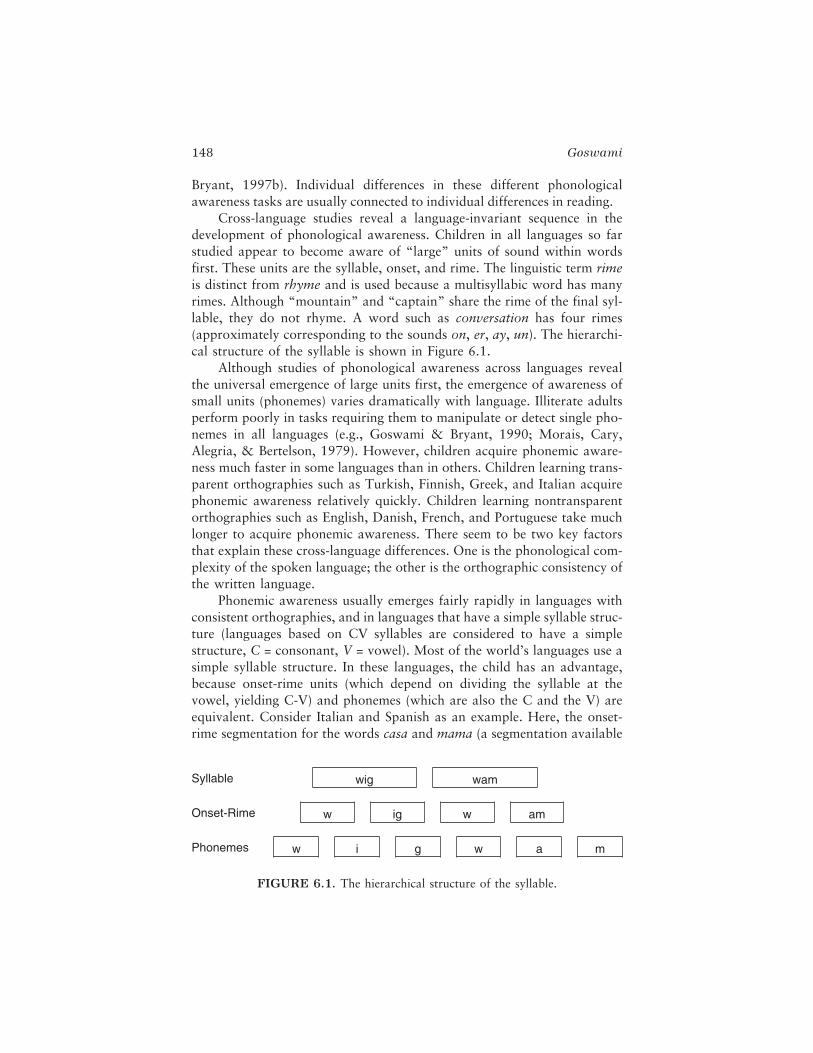
Bryant, 1997b). Individual differences in these different phonologicalawareness tasks are usually connected to individual differences in reading.
Cross-language studies reveal a language-invariant sequence in thedevelopment of phonological awareness. Children in all languages so farstudied appear to become aware of “large” units of sound within wordsfirst. These units are the syllable, onset, and rime. The linguistic term rimeis distinct from rhyme and is used because a multisyllabic word has manyrimes. Although “mountain” and “captain” share the rime of the final syl-lable, they do not rhyme. A word such as conversation has four rimes(approximately corresponding to the sounds on, er, ay, un). The hierarchi-cal structure of the syllable is shown in Figure 6.1.
Although studies of phonological awareness across languages revealthe universal emergence of large units first, the emergence of awareness ofsmall units (phonemes) varies dramatically with language. Illiterate adultsperform poorly in tasks requiring them to manipulate or detect single pho-nemes in all languages (e.g., Goswami & Bryant, 1990; Morais, Cary,Alegria, & Bertelson, 1979). However, children acquire phonemic aware-ness much faster in some languages than in others. Children learning trans-parent orthographies such as Turkish, Finnish, Greek, and Italian acquirephonemic awareness relatively quickly. Children learning nontransparentorthographies such as English, Danish, French, and Portuguese take muchlonger to acquire phonemic awareness. There seem to be two key factorsthat explain these cross-language differences. One is the phonological com-plexity of the spoken language; the other is the orthographic consistency ofthe written language.
Phonemic awareness usually emerges fairly rapidly in languages withconsistent orthographies, and in languages that have a simple syllable struc-ture (languages based on CV syllables are considered to have a simplestructure, C = consonant, V = vowel). Most of the world’s languages use asimple syllable structure. In these languages, the child has an advantage,because onset-rime units (which depend on dividing the syllable at thevowel, yielding C-V) and phonemes (which are also the C and the V) areequivalent. Consider Italian and Spanish as an example. Here, the onset-rime segmentation for the words casa and mama (a segmentation available
148 Goswami
FIGURE 6.1. The hierarchical structure of the syllable.
Syllable wig wam
Onset-Rime w ig w am
Phonemes w i g w a m
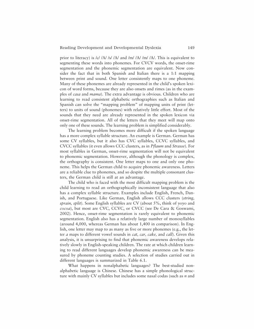
prior to literacy) is /c/ /A/ /s/ /A/ and /m/ /A/ /m/ /A/. This is equivalent tosegmenting these words into phonemes. For CVCV words, the onset-rimesegmentation and the phonemic segmentation are equivalent. Now con-sider the fact that in both Spanish and Italian there is a 1:1 mappingbetween print and sound. One letter consistently maps to one phoneme.Many of these phonemes are already represented in the child’s spoken lexi-con of word forms, because they are also onsets and rimes (as in the exam-ples of casa and mama). The extra advantage is obvious. Children who arelearning to read consistent alphabetic orthographies such as Italian andSpanish can solve the “mapping problem” of mapping units of print (let-ters) to units of sound (phonemes) with relatively little effort. Most of thesounds that they need are already represented in the spoken lexicon viaonset-rime segmentation. All of the letters that they meet will map ontoonly one of these sounds. The learning problem is simplified considerably.
The learning problem becomes more difficult if the spoken languagehas a more complex syllable structure. An example is German. German hassome CV syllables, but it also has CVC syllables, CCVC syllables, andCVCC syllables (it even allows CCC clusters, as in Pflaum and Strasse). Formost syllables in German, onset-rime segmentation will not be equivalentto phonemic segmentation. However, although the phonology is complex,the orthography is consistent. One letter maps to one and only one pho-neme. This helps the German child to acquire phonemic awareness. Lettersare a reliable clue to phonemes, and so despite the multiple consonant clus-ters, the German child is still at an advantage.
The child who is faced with the most difficult mapping problem is thechild learning to read an orthographically inconsistent language that alsohas a complex syllable structure. Examples include English, French, Dan-ish, and Portuguese. Like German, English allows CCC clusters (string,sprain, split). Some English syllables are CV (about 5%, think of yoyo andcocoa), but most are CVC, CCVC, or CVCC (see De Cara & Goswami,2002). Hence, onset-rime segmentation is rarely equivalent to phonemicsegmentation. English also has a relatively large number of monosyllables(around 4,000, whereas German has about 1,400 in comparison). In Eng-lish, one letter may map to as many as five or more phonemes (e.g., the let-ter a maps to different vowel sounds in cat, car, cake, and call). Given thisanalysis, it is unsurprising to find that phonemic awareness develops rela-tively slowly in English-speaking children. The rate at which children learn-ing to read different languages develop phonemic awareness can be mea-sured by phoneme counting studies. A selection of studies carried out indifferent languages is summarized in Table 6.1.
What happens in nonalphabetic languages? The best-studied non-alphabetic language is Chinese. Chinese has a simple phonological struc-ture with mainly CV syllables but includes some nasal codas (such as n and
Reading Development and Developmental Dyslexia 149
ng). The entire syllabary can be described using 22 onsets and 37 rimes (seeSiok & Fletcher, 2001). Studies suggest that Chinese preschoolers developgood onset-rime level skills before entering school. For example, using aChinese version of the oddity task (cat, pit, fat) pioneered by Bradley andBryant (1983), Ho and Bryant (1997b) reported that Chinese 3-year-oldsattained high levels of performance (68% correct). Phonemic awarenesswould seem unlikely to develop in Chinese-speaking children because theyare learning a character-based script. However, this is not the case. Chinesehas two instructional systems for teaching children the sounds of the char-acters, and both of these systems operate at an onset-rime level. They arecalled Pinyin (used in mainland China), and Zhu-Yin-Fu-Hao (used in Tai-wan). Pinyin uses the alphabet to represent the component sounds in sylla-bles, whereas Zhuyin uses simple characters. Because most Chinese sylla-bles have a CV structure, these instructional systems effectively teachchildren correspondences for the phonemes in the syllables. Accordingly,Chinese children growing up in Mainland China and in Taiwan performwell in phonemic awareness tasks (Siok & Fletcher, 2001; Huang &Hanley, 1994). However, Chinese children growing up in Hong Kong, wholearn the Chinese characters by rote and are not taught Pinyin or Zhuyin,show very poor phonemic awareness (Huang & Hanley, 1994).
150 Goswami
TABLE 6.1. Data (% Correct) from StudiesComparing Phoneme Counting in DifferentLanguages in Kindergarten or Early Grade 1
Language% phonemescounted correctly
Greek1 98
Turkish2 94
Italian3 97
Norwegian4 83
German5 81
French6 73
English7 70
English8 71
English9 65
Note. 1 = Harris and Giannoulis (1999); 2 = Durgunoglu andOney (1999); 3 = Cossu, Shankweiler, Liberman, Katz, and Tola(1988); 4 = Hoien, Lundberg, Stanovich, and Bjaalid (1995); 5 =Wimmer, Landerl, Linortner, and Hummer (1991); 6 = Demontand Gombert (1996); 7 = Liberman, Shankweiler, Fischer, andCarter (1974); 8 = Tunmer and Nesdale (1985); 9 = Perfetti, Beck,Bell, and Hughes (1987) and grade 2 children.
THE TRAJECTORY OF READING ACQUISITIONACROSS LANGUAGES
Grapheme–phoneme recoding skills develop in tandem with phonemicawareness. As might be expected, therefore, children learning to read dif-ferent languages also develop grapheme–phoneme recoding skills at differ-ent rates. A comprehensive cross-language comparison of grapheme–phoneme recoding skills during the first year of acquisition was conductedby the European Concerted Action on Learning Disorders as a Barrier toHuman Development. As part of this action, participating scientists from14 European Community (EC) countries developed a matched set of itemsof simple real words and nonwords suitable for first-grade readers. The realand nonword items were then given to children from each country duringtheir first year of reading instruction (see Seymour, Aro, & Erskine, 2003).Because children in different EC countries begin school at different ages,the children varied in age at the time of testing. However, they wereequated for degree of reading instruction across orthography, because theywere all tested at the same time point midway through their first year atschool. The methods of reading instruction used by participating schools inthe different countries could not be equated exactly; however, the schoolswere chosen so that all children were experiencing phoneme-level “pho-nics” teaching (including those children who were learning to read themore inconsistent orthographies). The data from this study for monosylla-bles are shown in Table 6.2.
The table is arranged so that the languages are listed from most toleast orthographic consistency. This makes it easy to see that the childrenwho were acquiring reading in the orthographically consistent EC lan-guages (Greek, Finnish, German, Italian, Spanish) were those performingclose to ceiling. This performance was true for both word and nonwordreading. The children doing less well were those learning to read Danish(71% correct), Portuguese (73% correct), and French (79% correct). How-ever, although grapheme–phoneme recoding skills were less accurate inthese orthographies, the reduced levels of accuracy are in line with thereduced orthographic consistency of these languages. Danish is relativelyinconsistent for reading (Elbro & Pallesen, 2002), whereas Portuguese andFrench are relatively inconsistent for spelling (Defior, Martos, & Cary,2002; Ventura, Morais, Pattamadilok, & Kolinsky, 2004; Ziegler, Jacobs,& Stone, 1996). The children who were performing most poorly werethose learning to read in English (34% correct). These children wereretested a year later, following an extra year of phonics-based literacy in-struction, and they still performed below children reading the other lan-guages. However, this relatively poor performance would be predicted by
Reading Development and Developmental Dyslexia 151
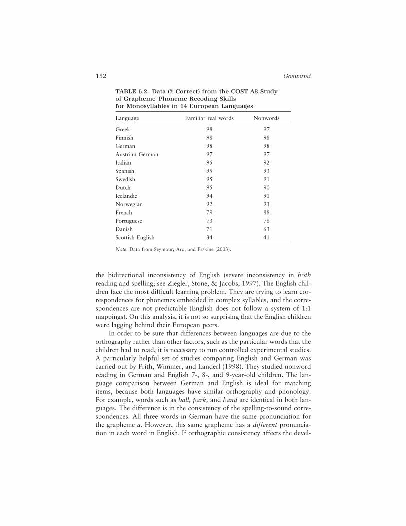
the bidirectional inconsistency of English (severe inconsistency in bothreading and spelling; see Ziegler, Stone, & Jacobs, 1997). The English chil-dren face the most difficult learning problem. They are trying to learn cor-respondences for phonemes embedded in complex syllables, and the corre-spondences are not predictable (English does not follow a system of 1:1mappings). On this analysis, it is not so surprising that the English childrenwere lagging behind their European peers.
In order to be sure that differences between languages are due to theorthography rather than other factors, such as the particular words that thechildren had to read, it is necessary to run controlled experimental studies.A particularly helpful set of studies comparing English and German wascarried out by Frith, Wimmer, and Landerl (1998). They studied nonwordreading in German and English 7-, 8-, and 9-year-old children. The lan-guage comparison between German and English is ideal for matchingitems, because both languages have similar orthography and phonology.For example, words such as ball, park, and hand are identical in both lan-guages. The difference is in the consistency of the spelling-to-sound corre-spondences. All three words in German have the same pronunciation forthe grapheme a. However, this same grapheme has a different pronuncia-tion in each word in English. If orthographic consistency affects the devel-
152 Goswami
TABLE 6.2. Data (% Correct) from the COST A8 Studyof Grapheme–Phoneme Recoding Skillsfor Monosyllables in 14 European Languages
Language Familiar real words Nonwords
Greek 98 97
Finnish 98 98
German 98 98
Austrian German 97 97
Italian 95 92
Spanish 95 93
Swedish 95 91
Dutch 95 90
Icelandic 94 91
Norwegian 92 93
French 79 88
Portuguese 73 76
Danish 71 63
Scottish English 34 41
Note. Data from Seymour, Aro, and Erskine (2003).
opment of grapheme–phoneme recoding strategies, then English children atthe same stage of reading development should be less efficient at recodingnonwords that include graphemes such as a compared to German children.
To test this idea, Frith and colleagues (1998) gave English and Germanchildren identical nonwords to read (such as grall). They found that theGerman children’s nonword reading was close to ceiling after only 1 yearof reading instruction. In contrast, the English children showed much lowerreading accuracy. They did not approach ceiling levels of performance untilthey had been learning to read for at least 3 years. For nonwords such asgrall, the 7-year-old English readers made errors in the region of 55%,compared to 15% for the German readers. In both language groups, per-formance improved with age, but differences between the languages werestill strong at age 9. A significant difference in nonword reading was evenfound when the cross-language comparison was restricted to those Germanand English children whose word reading performance was 100% correctfor analogous items (real words such as ball). The selected German childrenmade only 8% errors with nonwords based on these real words (grall-ball),whereas the selected English-speaking children made 22% errors (for simi-lar results in comparisons of English and German, see also Wimmer &Goswami, 1994; Wimmer & Hummer, 1990).
It might be objected that comparisons between English and Germanchildren cannot rule out cultural or social factors that might differ betweenthe two countries, which in themselves might affect reading acquisition.One way around this objection is to design experimental comparisons ofreading development in Welsh-speaking versus English-speaking childrenliving in Wales. Welsh is a highly consistent orthography. In Wales, thesame school districts include schools that teach in Welsh and schools thatteach in English. The children attending these schools come from the samecatchment areas and follow the same curricula (e.g., Hanley, Masterson,Spencer, & Evans, 2004; Spencer & Hanley, 2003; see also Ellis &Hooper, 2001). The only difference is the language of instruction (it isimportant to note that phonotactic complexity is similar in English andWelsh). Hanley and his colleagues have followed a group of Welsh-speaking children who were learning to read Welsh and a matched group ofEnglish-speaking children living in the same area of Wales who were learn-ing to read English since they were 5 years old. By the age of 11, Hanleyand his colleagues found that the groups of children were comparable intests of single-word reading in their native languages and in nonword read-ing. However, in all previous comparisons the English children had per-formed more poorly than their Welsh peers, at each test point, demonstrat-ing slower reading acquisition. The English children had also been slowerto acquire phonemic awareness.
Reading Development and Developmental Dyslexia 153
Clearly, therefore, grapheme–phoneme recoding skills develop moreslowly in children learning to read English, and in large part this slow rateis due to the inconsistency of the orthography. However, although the Eng-lish orthography is maximally inconsistent at the “small” grain size of thephoneme, it is less inconsistent at the larger grain size of the rime (Treiman,Mullennix, Bijeljac-Babic, & Richmond-Welty, 1995). Treiman and col-leagues (1995) analyzed the spelling–sound correspondences of all themonosyllables of English at both the C-V-C level and also at the largergrain sizes of C-VC and CV-C. For English vowels, Treiman and her col-leagues found that consistency in pronunciation across words was around50%. For example, if a child met the vowel a in the word cat (or the like),the chances that the next word containing an a had the same grapheme–phoneme correspondence was 51%. In contrast, if the child noticedthe spelling–sound correspondence for the entire rime, then consistencyincreased to 77%. Most words that share rime spellings have consistentpronunciations for the rime (e.g., cat, hat, mat), although not all of them(e.g., ball, shall). Average consistency is 77%, which is an improvement onthe 51% yielded by a C-V-C analysis that treats the vowel as a unit inde-pendent of the coda (the final consonant phoneme is called the coda).Attaching the vowel to the onset phoneme did not improve consistency.The consistency of pronunciation for CV units (cat, cab, car) was only52%.
These analyses of orthographic consistency make it likely that Englishchildren need to develop phonological recoding strategies at more than onegrain size in order to become competent readers. They need to develop effi-cient grapheme–phoneme recoding strategies, but they also need to develop“rhyme analogy” strategies to take advantage of spelling–sound consis-tency at the large-unit level of the rime. Children learning to read Englishdo indeed use rhyme analogy strategies (making connections between spell-ing patterns in analogous words such as beak and peak), and this use hasbeen shown by a variety of experimental techniques, including makinganalogies from clue words (Goswami, 1986, 1988), reading pseudo-homophones (Goswami et al., 2001), and comparing the decoding of large-unit versus small-unit nonwords (Goswami et al., 2003). Brown andDeavers (1999) have suggested that children learning to read in Englishadopt “flexible unit size” strategies. Nevertheless, it is important to pointout that there are more orthographic units to learn when the grain size isbig than when the grain size is small. For example, in order to decode themost frequent 3,000 monosyllabic English words at the level of the rime, achild needs to learn mappings between approximately 600 different ortho-graphic patterns and 400 phonological rimes. In addition, English-speakingchildren need to develop whole-word recognition strategies to supplement
154 Goswami
rhyme analogies and grapheme–phoneme recoding. Some spelling patternsin English are unique (e.g., choir, people). These patterns must be learnedholistically, using “look and say” methods.
Finally, the role of visual skills in learning to read logographic orthog-raphies should be mentioned. Logographies such as Chinese and Japaneseusually depend on visually distinctive symbols that are created from thesame basic elements (e.g., horizontal strokes versus vertical strokes). Chi-nese characters are quite visually complex and therefore require fine-grained visual analysis and storage. Surprisingly, perhaps, individual differ-ences in visual processing only make a difference to learning during the firstyear of reading acquisition (e.g., Siok & Fletcher, 2001). It is phonologicalawareness rather than visual processing skills that predicts reading develop-ment in Chinese. This finding has been replicated with a wide range ofvisual tasks. For example, individual differences in the ability to recallabstract designs over short periods of time, to discriminate visual forms, todetect the odd design out of three abstract spatial designs presented in dif-ferent orientations, and to learn visual paired associates are not related toreading acquisition in Chinese (Ho, 1997; Ho & Bryant 1997a; Huang &Hanley, 1994, 1997).
DEVELOPMENTAL DYSLEXIA IN CHILDRENACROSS LANGUAGES
If we now turn to consider dyslexia, we find analogous cross-language dif-ferences in the manifestation (but not in the incidence) of developmentalreading problems. In all languages so far studied, children with develop-mental dyslexia show deficits in phonological awareness. They are poor atperforming tasks such as the oddity task and phoneme counting, they havedifficulties with short-term (phonological) memory, and they are signifi-cantly slower in producing highly familiar words under timed conditions(e.g., in rapidly naming colors, called rapid automatized naming, or RAN).The deficit in phonological awareness can be measured at the linguistic lev-els of syllable, onset-rime, and phoneme, as would be predicted by thepsycholinguistic grain-size framework. However, whereas in inconsistentorthographies such as English, developmental dyslexia can be diagnosed onthe basis of reduced accuracy in phonological awareness tasks, in consistentorthographies developmental dyslexia is usually diagnosed on the basis ofreduced speed. Dyslexic children who learn to read consistent orthogra-phies can use spelling information to help them succeed in phonologicaltasks. For example, they can visualize the spelling pattern of a word andcount its phonemes (because there is a 1:1 correspondence between
Reading Development and Developmental Dyslexia 155
graphemes and phonemes). However, they tend to do this extremelyslowly.
Diagnosis of developmental dyslexia in most languages follows theOECD (Organisation for Economic Co-operation and Development) defi-nition. This definition states that developmental dyslexia is a specific prob-lem with reading and spelling that cannot be accounted for by low intelli-gence, poor educational opportunities, or obvious sensory or neurologicaldamage. In consistent orthographies, this specific problem manifests inextremely effortful decoding and inaccurate spelling. Dyslexic childrenlearning to read languages such as Italian, Spanish, and German can recodeboth words and nonwords accurately, but they do this so slowly that theyare functionally dyslexic compared to their peers. Furthermore, theirreduced phonological memory means that by the time they have decodedall the words in a sentence, they may have forgotten the beginning of thesentence, and so meaning may be lost. However, the hallmark of develop-mental dyslexia in these languages is unexpectedly poor spelling. Becauseorthographies that are consistent for reading are frequently inconsistent forspelling (i.e., there are many possible orthographic patterns that can beused to represent a particular sound), it is spelling rather than reading thatreveals inaccurate performance.
In contrast, developmental dyslexia in inconsistent orthographies isusually obvious because of the inaccurate and effortful reading of thechild. Children who lack phonological sensitivity find it very difficult toacquire grapheme–phoneme recoding skills in these orthographies, becausegraphemes are not a consistent guide to phonemes. Furthermore, it is diffi-cult for these children to use orthographic knowledge to improve the qual-ity of their phonological representations. Children learning to read ininconsistent orthographies cannot use spelling information to succeed onphonological tasks, or at least cannot use spelling information as a consis-tent guide to accurate performance. For example, the words pitch and richcontain the same number of phonemes, but this is not obvious from theirspellings (see Ehri & Wilce, 1980). Hence performance in phonologicalawareness tasks continues to be inaccurate as well as slow. Dyslexic chil-dren in nonalphabetic languages experience similar difficulties in acquiringlogographies and syllabaries. Reduced phonological sensitivity makes it dif-ficult to acquire print–sound relationships even when these are at thewhole-word (i.e., character) level. Studies in Chinese show that dyslexicchildren have the same problems with phonological processing that charac-terize dyslexic children in English. As in consistent orthographies, however,the most striking manifestation of their dyslexia is in writing rather than inreading. Chinese children with developmental dyslexia can recode charac-ters to sound accurately, but when they need to select the correct characterto represent a sound, they have striking difficulties.
156 Goswami
Before giving examples of studies in individual languages conductedwithin the psycholinguistic grain-size framework, the importance of strin-gent subject matching should be mentioned. Obviously, because dyslexiaoccurs in children of wide cognitive ability, cognitive skills should be equiv-alent across the children with dyslexia and comparison children. Becausephonological abilities in dyslexic children are necessarily affected by read-ing experience, however, it is important to compare dyslexic children toyounger children who are reading at the same level (a “reading-level” or“RL-match” design). This control group should be used in addition to chil-dren matched for chronological age (or CA). Children who are the sameage as the dyslexic children will obviously be much better readers. How-ever, if the phonological or reading subskills of the dyslexic children areinferior even to those of the younger children who can read the same num-ber of words, then any deficits found are likely to be fundamental ratherthan a simple consequence of poorer reading experience. In prereaders,only CA match controls can be used, because an RL match is impossible toachieve. A limitation of studying individuals with developmental dyslexiawhen they are adults is that only CA controls can be used here, too.Usually, if adult readers can be found who have reading skills comparableto those with dyslexia (i.e., relatively poor reading skills), these adults alsohave cognitive deficits that preclude an I.Q. match.
THE DEVELOPMENTAL TRAJECTORYOF PHONOLOGICAL AWARENESS IN DYSLEXIA
Studies across languages using stringent experimental designs have shownthat dyslexic children have phonological deficits. A number of studies inEnglish using the RL match design have reported syllable- and onset-rime-level deficits, as well, of course, as the ubiquitous phoneme-level difficulties(e.g., Bowey, Cain, & Ryan, 1992; Bradley & Bryant, 1978; Bruck, 1992;Swan & Goswami, 1997). For example, Bradley and Bryant (1978)reported that English 10-year-olds with dyslexia were significantly poorerin the oddity task than both 10-year-old CA controls and 7-year-old RLcontrols. Using a phoneme version of the oddity task, Bowey and col-leagues (1992) reported a similar result for Australian 9-year-olds. Thechildren with dyslexia were significantly poorer at recognizing shared pho-nemes than both CA and RL controls. In fact, children and adults with dys-lexia in nontransparent languages such as English never seem to developaccurate phoneme awareness skills (e.g., Bruck, 1992). In Bruck’s (1992)study, she asked children with dyslexia ages 8–15 years and RL and CAcontrols to count the phonemes in two- to four-phoneme nonwords suchtisk and leem. The children with dyslexia performed correctly in 47% of
Reading Development and Developmental Dyslexia 157
trials, as compared to 72% of trials for the RL-matched children and 77%of trials for the CA-matched children. When Bruck then compared adultswith dyslexia to third-grade normally progressing readers, she found thatthe children were significantly better than the adults with dyslexia at bothphoneme counting and phoneme deletion. The phoneme deficit in dyslexiain inconsistent orthographies is therefore extremely pervasive.
In contrast, the phoneme deficit in dyslexia in consistent orthographiesis only observable in terms of accuracy at the very beginning of readingacquisition. Porpodas (1999) studied Greek first graders with serious liter-acy difficulties and found significant differences in phoneme awarenessbetween children with dyslexia and control children. The children with dys-lexia scored 88% correct in a phoneme segmentation task, as compared to100% correct for CA controls; and 78% correct in a phoneme deletiontask, as compared to 98% correct for the CA controls (note that the partic-ipating children were beginning readers and hence an RL control groupcould not be generated). The Greek first graders with literacy difficultieshad been selected out of an initial cohort of 564 children and were at leasttwo standard deviations below the controls in spelling accuracy and at least1 standard deviation below the controls in decoding time.
Wimmer (1993) studied 10-year-old German children with dyslexiausing a phoneme substitution task. In this task, the children had to substi-tute a consistent phoneme such as /i/ (e.g., “Mama ist krank” became“Mimi ist krink”). Wimmer found that the children with dyslexia madecorrect substitutions on 86% of trials. Younger RL control children alsomade correct substitutions on 86% of trials. The CA control children madecorrect substitutions on 95% of trials, a significant difference. However, itis important to note that the children with dyslexia had been learning toread a consistent orthography for about 4 years. When Wimmer (1996) ret-rospectively examined the performance of the German children with dys-lexia on a phoneme awareness task in grade 1, before they were diagnosedas dyslexic, he found large differences in comparison to CA controls. Theto-be-dyslexic children scored on average 22% correct in the phonemereversal task used, compared to an average of 69% correct for the controlchildren. Wimmer concluded that German children with dyslexia exhibitthe same difficulties in phonemic segmentation exhibited by older Englishchildren with dyslexia, but only in the earliest phase of learning to read.Greek children with dyslexia appear to be similar.
Longitudinal studies of children who are at risk for reading difficultiesare rare but provide extremely valuable evidence concerning entry-levelphonological skills. Such studies also reveal phonological deficits at therhyme and syllable level in children learning to read consistent orthogra-phies. For example, Schneider, Roth, and Ennemoser (2000) studied a large
158 Goswami
group of 208 at-risk German children when they were in kindergarten.They found that the at-risk children performed significantly more poorlythan control children on tasks of rhyme production, rhyme matching, andsyllable segmentation. The control children were German kindergartners inthe same nurseries who were not thought to be at risk of later reading diffi-culties. In a Dutch longitudinal study, similar results were found. Thosechildren who were later diagnosed with dyslexia showed poorer rhymeawareness than their controls when tested in kindergarten (De Jong & vander Leij, 2003). Longitudinal studies of prereading children at risk for read-ing difficulties in English have also reported poorer syllable and onset-rimeawareness compared to controls (Carroll & Snowling, 2003).
Studies of children with dyslexia who are learning to read non-alphabetic orthographies are rare. Nevertheless, available data suggest thatthese children, too, display phonological deficits at the levels of syllable,rhyme, and phoneme. For example, Ho, Law, and Ng (1998) showed thatChinese children with dyslexia ages 8–9 years were significantly poorerthan both CA and RL controls in rhyme awareness and phonological mem-ory tasks. Kobayashi, Kato, Haynes, Macaruso, and Hook (2003) reportedthat Japanese children with developmental dyslexia displayed difficulties intasks such as syllable deletion, syllable reversal, nonword repetition, andrapid automatized naming, the same type of tasks that distinguish childrenwith dyslexia in other languages. In a study on dyslexia in Korea, 11-year-old children with dyslexia scored only 37% correct in an oddity task inwhich the deviating element was the second phoneme of the first syllable(e.g., mo-ki, bo-ki, ko-ki, sa-ki). This score was in comparison to 83% cor-rect for CA controls (Kim & Davis, 2004). In all world languages so farstudied, therefore, children with developmental dyslexia have difficulties inrecognizing and manipulating phonological units at all linguistic levels.However, learning to read a consistent alphabetic orthography can help toimprove the accuracy of phonological representations in some languages.Nevertheless, children who are dyslexic in these languages still display aphonological deficit in terms of the speed of their phonological processingskills.
THE TRAJECTORY OF READING ACQUISITIONIN DYSLEXIA
As noted earlier, grapheme–phoneme recoding skills develop in tandemwith phonemic awareness, so it might be expected that the development ofphonological recoding skills in children with dyslexia would be impaired.This is indeed the case. As with typical development, however, there are
Reading Development and Developmental Dyslexia 159
important cross-language differences. Although the impaired developmentof phonological recoding skills is a hallmark of developmental dyslexiaacross languages, being most often measured in terms of nonword readingskills, in languages with consistent alphabetic orthographies children caneventually achieve quite accurate performance. The problem is that eventhough children learning to read languages such as Greek, German, andSpanish can recode nonwords to sound accurately, they do so extremelyslowly. This pace makes them functionally dyslexic in comparison to theirpeers.
The nonword reading deficit in consistent alphabetic orthographies ismost marked in younger children with dyslexia. When children are studiedat the beginning of the acquisition process, clear deficits in accuracy as wellas in speed are apparent. For example, when Wimmer (1996) compared thenonword reading accuracy of German children in kindergarten after dis-covering (via a longitudinal study) which of them were destined to be dys-lexic, he found that 7 out of 12 children who later became dyslexic readless than 60% of simple nonwords such as Mana (Mama) and Aufo (Auto)accurately. In comparison, beginning readers who did not subsequentlybecome dyslexic showed an average performance rate of 96% correct withthese simple nonwords. Three years later, when the children with dyslexiawere 10, Wimmer compared them to younger RL-matched children whocould read the same number of real words. The nonword reading deficithad apparently disappeared (Wimmer, 1993). In terms of accuracy, theGerman dyslexic children could recode as many nonwords to sound astheir younger controls. However, they showed a marked phonologicalrecoding deficit in terms of reading speed. It took the German children withdyslexia on average twice as long as the younger children to recode thenonwords to sound. Similar findings have been reported by Porpodas(1999) for Greek and by López and Jiminez González (2000) for Spanish.
In contrast, studies of children with dyslexia who are learning to readEnglish suggest that accuracy deficits in nonword reading are very persis-tent. A review paper of published studies showed that error rates were typi-cally rather high, between 40 and 60% (Rack, Snowling, & Olson, 1992).Most studies find that children with dyslexia continue to be less accuratein nonword reading than younger RL controls. For example, Landerl,Wimmer, and Frith (1997) gave 12-year-old English children with dyslexiaand 8-year-old RL control children nonwords to read that were one, two,or three syllables in length. The one- and two-syllable nonwords were con-structed by changing the onsets of real words (e.g., ball–grall, butter–sutter). The three-syllable nonwords were constructed by syllable substitu-tion (semater, chacustre), and consequently made greater demands ongrapheme–phoneme recoding procedures. Overall the English children with
160 Goswami
dyslexia found the nonword reading task significantly more difficult thanthe RL controls (scoring 48% correct compared to 65% correct). For thethree-syllable nonwords, the English children with dyslexia scored at 30%correct.
Again, if we search for comparable developmental studies in non-alphabetic orthographies, the database is sparse. Kim and Davis (2004)reported that children with dyslexia in Korea read 72% of nonwords cor-rectly compared to 100% for CA controls. In Hebrew, Breznitz (1997)found that third graders with dyslexia were inferior in a nonword readingtask compared to children in first grade (RL controls), who were matchedto the dyslexic group for word recognition accuracy and reading compre-hension. Phonological problems in nonalphabetic scripts are even moremarked in writing. In most world orthographies, sound-to-spelling rela-tions are many-to-one, and this is particularly true for Chinese Kanji (notethat Japanese children also need to learn around 1,000 Kanji). There maybe as many as eight choices of Kanji to represent one sound. This findingmirrors findings in transparent alphabetic orthographies (German, Greek),where children with dyslexia typically become able to decode quite accu-rately (albeit, extremely slowly) but never become fluent writers.
BIOLOGICAL UNITY:THE BRAIN BASIS OF DEVELOPMENTAL DYSLEXIA
According to the cross-language analysis developed above, developmentaldyslexia in all of the world’s languages should have the same underlyingneural causation. Children with developmental dyslexia in all languages sofar studied systematically show the same characteristic phonological deficit.Children with developmental dyslexia have difficulties with phonologicaltasks measuring syllable, rhyme, and phoneme awareness; they have diffi-culties with tasks measuring phonological memory; and they have difficul-ties with “rapid automatized naming” tasks. These are the three hallmarkareas of a phonological difficulty. This analysis predicts that at the neurallevel, similar deficits in brain activity should be found, whether the child isdyslexic in English, French, German, or Chinese. The core difficulty forchildren with dyslexia is a linguistic (phonological) one, and language isrepresented in the same neural areas whether the language is English orChinese.
Work by Turkeltaub and colleagues has shown that three distinct neu-ral areas are involved in these different aspects of phonological processingin children (Turkeltaub, Gareau, Flowers, Zeffiro, & Eden, 2003). Phono-logical working memory appears to depend on neural activity in the left
Reading Development and Developmental Dyslexia 161
intraparietal sulcus (as in adults); phonological awareness, on a network ofareas centered on the left posterior superior temporal sulcus (also the pri-mary area recruited by young children at the beginning of reading develop-ment); and RAN on a different, bilateral network, including right posteriorsuperior temporal gyrus, right middle temporal gyrus, and left ventral infe-rior frontal gyrus. Although studies with children learning to read otherlanguages have yet to be done, in studies of adults with developmental dys-lexia across languages, the most consistent findings concern underac-tivation in left temporoparietal and left occipitotemporal cortices (Eden etal., 2004; Paulesu et al., 2001). This finding has led researchers such asPaulesu to propose that a common neurological network underpins dys-lexia across languages, despite some superficial behavioral differencesacross countries (which can be ascribed to differences in orthographic con-sistency).
Nevertheless, there is currently some debate about whether “biologicalunity” should be expected for literacy, which is a culturally acquired skillrather than a natural adaptation of the brain. For example, in a recentfMRI study of eight Chinese children with developmental dyslexia, it wasclaimed that neural imaging data showed increased activity in brain regionsresponsible for visuospatial analysis rather than phonological analysis(Siok, Perfetti, Jin, & Tan, 2004). The children with developmental dys-lexia in this study showed reduced activity in the left middle frontal gyrus,an area involved in visuospatial analysis, but apparently did not showreduced activity in left temporoparietal regions.
Clearly, accurate visuospatial analysis is mandatory in acquiring acharacter-based system. However, if individual differences in visuospatialmemory do not predict individual differences in typical reading developmentin Chinese (as discussed earlier), then why should a deficit in visuospatialanalysis be the cause of a difficulty in reading acquisition in Chinese? This dif-ference may be a product of the impaired reading development of the childrenbeing studied rather than a clue to the cause of their reading difficulties. Infact, the eight children with dyslexia studied by Siok and colleagues (2004)did have a phonological deficit. This was evident in their poorer performanceon the behavioral task, which was a homophone judgment task. In this task,the participants had to silently read two characters and decide whether theywere pronounced in the same way or not. The children with dyslexia were sig-nificantly worse at recognizing homophones than the controls, and they alsotook significantly longer to make homophone decisions. Furthermore, nocomparisons with RL-matched controls were reported. The atypical imagingprofile reported (i.e., reduced activation in left middle frontal gyrus, an areaassociated with the representation and working memory of visuospatial andverbal information) may have been the profile to be expected for the charac-
162 Goswami
ter reading level achieved by the children. Without longitudinal data andreading level controls, we cannot tell whether or not there is biological unityat the neural level in dyslexia.
CONCLUSION
Across all languages so far studied, there is a fundamental and causal rela-tionship between a child’s phonological awareness and his or her acquisi-tion of reading and spelling skills. There is an apparently universalsequence of phonological development, from awareness of large units (syl-lables, onsets, rimes) to awareness of small units (phonemes). Alongsidethis apparently universal sequence of phonological development, variationsin the phonological complexity of languages and variations in the consis-tency with which phonology is represented in orthography generate cross-language differences in reading acquisition and in the manifestation ofdyslexia. The nature of these cross-language differences can be predicted apriori by considering the availability and consistency of phonological andorthographic units at different grain sizes.
The probable biological unity for literacy at the neural level suggeststhat the teaching of reading should follow a similar approach across cul-tures, beginning with direct instruction in how print is linked to sound, fol-lowed by building fluency through repeated practice of decoding. Similarly,remedial programs for children with dyslexia are likely to need the samecomponents across orthographies, focusing on developing phonologicalskills and on linking phonology to orthography. Given the underactivationof key areas for reading in remediated adults with dyslexia, it seems likelythat the development of fluency will continue to be problematic, even forchildren with dyslexia who are learning to read transparent orthographies.However, the dearth of imaging studies across languages means that sys-tematic cross-language comparisons of neurocognitive trajectories duringliteracy development are needed before strong recommendations for theclassroom can be made.
REFERENCES
Bishop D. V. M. (1997). Cognitive neuropsychology and developmental disorders:Uncomfortable bedfellows. Quarterly Journal of Experimental Psychology,50A, 899–923.
Bowey, J. A., Cain, M. T., & Ryan, S. M. (1992). A reading-level design study ofphonological skills underlying fourth grade children’s word reading difficul-ties. Child Development, 63, 999–1011.
Reading Development and Developmental Dyslexia 163
Bradley, L., & Bryant, P. E. (1978). Difficulties in auditory organization as a possi-ble cause of reading backwardness. Nature, 271, 746–747.
Bradley, L., & Bryant, P. E. (1983). Categorising sounds and learning to read: Acausal connection. Nature, 310, 419–424.
Breznitz, Z. (1997). Enhancing the reading of dyslexic children by reading accelera-tion and auditory masking. Journal of Educational Psychology, 89(1), 103–113.
Brown, G. D. A., & Deavers, R. P. (1999). Units of analysis in nonword reading:Evidence from children and adults. Journal of Experimental Child Psychology,73, 208–242.
Bruck, M. (1992). Persistence of dyslexics’ phonological awareness deficits. Devel-opmental Psychology, 28, 874–886.
Bryant, P., Bradley, L., Maclean, M., & Crosland, J. (1989). Nursery rhymes, pho-nological skills and reading. Journal of Child Language, 16, 407–428.
Carroll, J. M., & Snowling, M. J. (2003). Language and phonological skills in chil-dren at high risk of reading difficulties. Journal of Child Psychology and Psy-chiatry, 45, 631–640.
Chaney, C. (1992). Language development, metalinguistic skills, and print aware-ness in 3–year-old children. Journal of Applied Psycholinguistics, 13, 485–514.
Cossu, G., Shankweiler, D., Liberman, I. Y., Katz, L., & Tola, G. (1988). Aware-ness of phonological segments and reading ability in Italian children. AppliedPsycholinguistics, 9, 1–16.
De Cara, B., & Goswami, U. (2002). Similarity relations among spoken words: Thespecial status of rimes in English. Behavior Research Methods, Instrumentsand Computers, 34(3), 416–423.
Defior, S., Martos, S., & Cary, L. (2002). Differences in reading acquisition devel-opment in two shallow orthographies: Portuguese and Spanish. AppliedPsycholinguistics, 23, 135–148.
De Jong, P. F., & van der Leij, A. (2003). Developmental changes in the manifesta-tion of a phonological deficit in dyslexic children learning to read a regularorthography. Journal of Educational Psychology, 95, 22–40.
Demont, E., & Gombert, J. E. (1996). Phonological awareness as a predictor ofrecoding skills and syntactic awareness as a predictor of comprehension skills.British Journal of Educational Psychology, 66, 315–332.
Durgunoglu, A. Y., & Oney, B. (1999). A cross-linguistic comparison of phonologi-cal awareness and word recognition. Reading and Writing, 11, 281–299.
Eden, G. F., Jones, K. M., Cappell, K., Gareau, L., Wood, F. B., Zeffiro, T. A., et al.(2004). Neural changes following remediation in adult developmental dys-lexia. Neuron, 44, 411–422.
Ehri, L. C., & Wilce, L. S. (1980). The influence of orthography on readers’ concep-tualisation of the phonemic structure of words. Applied Psycholinguistics, 1,371–385.
Elbro, C., & Pallesen, B. R. (2002). The quality of phonological representations: Acausal link? In L. Verhoeven, C. Elbro, & P. Reitsma (Eds.), Precursors offunctional literacy (Vol. 11, pp. 17–32). Amsterdam: Benjamins.
164 Goswami
Ellis, N. C., & Hooper, A. M. (2001). Why learning to read is easier in Welsh thanin English: Orthographic transparency effects evinced with frequency-matchedtests. Applied Psycholinguistics, 22, 571–599.
Fisher, S. E., & DeFries, J. C. (2002). Developmental dyslexia: Genetic dissection ofa complex cognitive trait. Nature Reviews: Neuroscience, 3, 767–780.
Frith, U., Wimmer, H., & Landerl, K. (1998). Differences in phonological recodingin German- and English-speaking children. Scientific Studies of Reading, 2,31–54.
Goswami, U. (1986). Children’s use of analogy in learning to read: A developmen-tal study. Journal of Experimental Child Psychology, 42, 73–83.
Goswami, U. (1988). Orthographic analogies and reading development. QuarterlyJournal of Experimental Psychology, 40A, 239–268.
Goswami, U. (2003). Why theories about developmental dyslexia require develop-mental designs. Trends in Cognitive Sciences, 7, 534–540.
Goswami, U., & Bryant, P. E. (1990). Phonological skills and learning to read.Hillsdale, NJ: Erlbaum.
Goswami, U., Ziegler, J., Dalton, L., & Schneider, W. (2001). Pseudohomophoneeffects and phonological recoding procedures in reading development in Eng-lish and German. Journal of Memory and Language, 45, 648–664.
Goswami, U., Ziegler, J., Dalton, L., & Schneider, W. (2003). Nonword readingacross orthographies: How flexible is the choice of reading units? Journal ofApplied Psycholinguistics, 24, 235–247.
Hanley, J. R., Masterson, J., Spencer, L. H., & Evans, D. (2004). How long do theadvantages of learning a transparent orthography last? An investigation of thereading skills and incidence of dyslexia in Welsh children at 10 years of age.Quarterly Journal of Experimental Psychology, 57(8), 1393–1410.
Harris, M., & Giannoulis, V. (1999). Learning to read and spell in Greek: Theimportance of letter knowledge and morphological awareness. In M. Harris &G. Hatano (Eds.), Learning to read and write: A cross-linguistic perspective(pp. 51–70). Cambridge, UK: Cambridge University Press.
Ho, C. S.-H. (1997). The importance of phonological awareness and verbal short-term memory to children’s success in learning to read Chinese. Psychologia,40(4), 211–219.
Ho, C. S.-H., & Bryant, P. (1997a). Development of phonological awareness ofChinese children in Hong Kong. Journal of Psycholinguistic Research, 26,109–126.
Ho, C. S.-H., & Bryant, P. (1997b). Phonological skills are important in learning toread in Chinese. Developmental Psychology, 33, 946–951.
Ho, C. S.-H., Law, T. P.-S., & Ng, P. M. (1998). The phonological deficithypothesis in Chinese developmental dyslexia. Reading and Writing, 32,276–289.
Hoien, T., Lundberg, L., Stanovich, K. E., & Bjaalid, I. K. (1995). Components ofphonological awareness. Reading and Writing, 7, 171–188.
Huang, H. S., & Hanley, J. R. (1994). Phonological awareness and visual skills inlearning to read Chinese and English. Cognition, 54, 73–98.
Huang, H. S., & Hanley, J. R. (1997). A longitudinal study of phonological aware-
Reading Development and Developmental Dyslexia 165
ness, visual skills, and Chinese reading acquisition among first-graders in Tai-wan. International Journal of Behavioral Development, 20, 249–268.
Karmiloff-Smith, A. (1998). Development itself is the key to understanding devel-opmental disorders. Trends in Cognitive Sciences, 2, 389–398.
Kim, J., & Davis, C. (2004). Characteristics of poor readers of Korean Hangul:Auditory, visual and phonological processing. Reading and Writing, 17(1–2),153–185.
Kobayashi M., Kato J., Haynes, C. W., Macaruso, P., & Hook, P. (2003). Cogni-tive linguistic factors in Japanese children’s reading. Japanese Journal ofLearning Disabilities, 12, 240–247.
Landerl, K., Wimmer, H., & Frith, U. (1997). The impact of orthographic consis-tency on dyslexia: A German–English comparison. Cognition, 63, 315–334.
Liberman, I. Y., Shankweiler, D., Fischer, F. W., & Carter, B. (1974). Explicit sylla-ble and phoneme segmentation in the young child. Journal of ExperimentalChild Psychology, 18, 201–212.
López, M. R., & González, J. E. J. (2000). IQ vs phonological recoding skill inexplaining differences between poor readers and normal readers in word rec-ognition: Evidence from a naming task. Reading and Writing, 12, 129–142.
Lundberg, I., Olofsson A., & Wall, S. (1980). Reading and spelling skills in the firstschool years predicted from phonemic awareness skills in kindergarten. Scan-dinavian Journal of Psychology, 21, 159–173.
Makita, M. (1968). The rarity of reading disability in Japanese children. AmericanJournal of Orthopsychiatry, 38, 599–614.
Morais, J., Cary, L., Alegria, J., & Bertelson, P. (1979). Does awareness of speechas a sequence of phones arise spontaneously? Cognition, 7, 323–331.
Paulesu, E. (2001). Dyslexia: Cultural diversity and biological unity. Science, 291,2163–2167.
Perfetti, C. A., Beck, I., Bell, L., & Hughes, C. (1987). Phonemic knowledge andlearning to read are reciprocal: A longitudinal study of first grade children.Merrill–Palmer Quarterly, 33, 283–319.
Porpodas, C. D. (1999). Patterns of phonological and memory processing in begin-ning readers and spellers of Greek. Journal of Learning Disabilities, 32, 406–416.
Rack, J. P., Snowling, M. J., & Olson, R. (1992). The nonword reading deficit indevelopmental dyslexia: A review. Reading Research Quarterly, 27, 29–53.
Read, C. (1986). Children’s creative spelling. London: Routledge.Schneider W., Roth E., & Ennemoser M. (2000). Training phonological skills and
letter knowledge in children at risk for dyslexia: A comparison of three kinder-garten intervention programs. Journal of Educational Psychology, 92, 284–295.
Seymour, P. H., Aro, M., & Erskine, J. M. (2003). Foundation literacy acquisitionin European orthographies. British Journal of Psychology, 94, 143–174.
Siok, W. T., & Fletcher, P. (2001). The role of phonological awareness and visual–orthographic skills in Chinese reading acquisition. Developmental Psychology,37, 886–899.
166 Goswami
Siok, W. T., Perfetti, C. A., Jin, Z., & Tan, L. H. (2004). Biological abnormality ofimpaired reading is constrained by culture. Nature, 431, 71–76.
Spencer, L. H., & Hanley, J. R. (2003). Effects of orthographic transparency onreading and phoneme awareness in children learning to read in Wales. BritishJournal of Psychology, 94(1), 1–28.
Swan, D., & Goswami, U. (1997). Phonological awareness deficits in developmentdyslexia and the phonological representations hypothesis. Journal of Experi-mental Child Psychology, 66, 18–41.
Treiman, R., Mullennix, J., Bijeljac-Babic, R., & Richmond-Welty, E. D. (1995).The special role of rimes in the description, use, and acquisition of Englishorthography. Journal of Experimental Psychology: General, 124, 107–136.
Tunmer, W. E., & Nesdale, A. R. (1985). Phonemic segmentation skill and begin-ning reading. Journal of Educational Psychology, 77, 417–527.
Turkeltaub, P. E., Gareau, L., Flowers, D. L., Zeffiro, T. A., & Eden, G. F. (2003).Development of neural mechanisms for reading. Nature Neuroscience, 6, 767–773.
Ventura, P., Morais, J., Pattamadilok, C., & Kolinsky, R. (2004). The locus of theorthographic consistency effect in auditory word recognition. Language andCognitive Processes, 19(1), 57–95.
Wimmer, H. (1993). Characteristics of developmental dyslexia in a regular writingsystem. Applied Psycholinguistics, 14, 1–33.
Wimmer, H. (1996). The nonword reading deficit in developmental dyslexia: Evi-dence from children learning to read German. Journal of Experimental ChildPsychology, 61, 80–90.
Wimmer, H., & Goswami, U. (1994). The influence of orthographic consistency onreading development: Word recognition in English and German children. Cog-nition, 51(1), 91–103.
Wimmer, H., & Hummer, P. (1990). How German speaking first graders read andspell: Doubts on the importance of the logographic stage. Applied Psycho-linguistics, 11, 349–368.
Wimmer, H., Landerl, K., Linortner, R., & Hummer, P. (1991). The relationship ofphonemic awareness to reading acquisition: More consequence than precondi-tion but still important. Cognition, 40, 219–249.
Ziegler, J. C., & Goswami, U. (2005). Reading acquisition, developmental dyslexia,and skilled reading across languages: a psycholinguistic grain size theory. Psy-chological Bulletin, 131(1), 3–29.
Ziegler, J. C., Jacobs, A. M., & Stone, G. O. (1996). Statistical analysis of thebidirectional inconsistency of spelling and sound in French. Behavior ResearchMethods, Instruments, and Computers, 28, 504–515.
Ziegler, J. C., Stone, G. O., & Jacobs, A. M. (1997). What’s the pronunciation forOUGH and the spelling for /u/? A database for computing feedforward and
feedback inconsistency in English. Behavior Research Methods, Instruments,and Computers, 29, 600–618.
Reading Development and Developmental Dyslexia 167

C H A P T E R 7
Neurocognitive Correlatesof Developmental Verbaland Orofacial Dyspraxia
Frederique LiegeoisAngela MorganFaraneh Vargha-Khadem
Relatively little is known about the neural mechanisms through which ver-bal communication is achieved. Speech requires input from four main oper-ational levels for every spoken word: (1) choosing and accessing a wordthat is semantically and syntactically correct, (2) retrieving the phonologi-cal or sound structure of the word, (3) encoding or syllabifying the soundsof the word in context, and (4) planning and producing the phonetic orarticulatory gestures (Levelt, 1999). This complicated process, culminatingin fluent and articulate speech, occurs at the rate of two to three words persecond, in the context of a very low error rate. The spontaneity and accu-racy with which speech is produced, even in very young children, has led tothe proposal that this unique human attribute has a strong neurogeneticbasis (Chomsky, 1959; Lenneberg, 1964). In support of this proposal, evi-dence has accumulated over the past two decades indicating that someforms of speech and language disorder are inherited (e.g., Bishop, North,& Donlan, 1996; Hurst, Baraitser, Auger, Graham, & Norell, 1990;Lewis, 1992; Scheffer, 2000; Tallal, Townsend, Curtiss, & Wulfeck, 1991;
168
Vargha-Khadem, Watkins, Alcock, Fletcher, & Passingham, 1995; Vargha-Khadem et al., 1998). One form of speech and language disorder thatshows strong heritability is “verbal dyspraxia” (i.e., a motor–speech dis-order), best exemplified in studies of the unique KE family (Alcock,Passingham, Watkins, & Vargha-Khadem, 2000a, 2000b; Hurst et al.,1990; Vargha-Khadem et al., 1995, 1998; Vargha-Khadem, Gadian, Copp,& Mishkin, 2005; Watkins, Dronkers, & Vargha-Khadem, 2002).
Half of the members of the large three-generational KE family presentwith a severe speech and language disorder, which is transmitted as anautosomal-dominant monogenic trait (Hurst et al., 1990; Pembrey, 1992)and encoded by the FOXP2 gene (Lai, Fisher, Hurst, Vargha-Khadem, &Monaco, 2001). The core deficit in the affected members is a severe andchronic developmental verbal and orofacial dyspraxia (DVOFD) that seri-ously interferes with the rapid selection, sequencing, and execution ofsounds that culminates in the production of articulate and intelligiblespeech. In addition, the affected KE members suffer from a plethora ofexpressive and receptive language problems and have difficulties withaspects of reading, writing, and nonverbal intellectual abilities as well aswith perception and execution of rhythms (Alcock et al., 2000a; Vargha-Khadem et al., 1995, 1998; Watkins, Dronkers, & Vargha-Khadem,2002a). It is not yet clear whether each of these language, cognitive, andrhythm impairments constitute independent secondary deficits or resultdirectly from the core deficits of verbal and orofacial dyspraxia.
The neural correlates of DVOFD in the affected KE members havebeen identified through a series of structural and functional magnetic reso-nance imaging (fMRI) investigations (Belton, Salmond, Watkins, Vargha-Khadem, & Gadian, 2003; Liegeois et al., 2003; Watkins et al., 2002). Thestructural studies have revealed relatively reduced gray matter bilaterally,compared to that in controls, in the cerebellum, neostriatum, and bothdorsomedial premotor and inferior frontal areas, as well as relativelyincreased gray matter bilaterally in the superior temporal and angular gyri,among other regions. The ontogenetic processes giving rise to this patternof abnormality are still unknown, but the anatomical abnormalities docu-mented in adulthood provide a clear neuropathological basis for the behav-ioral phenotype, particularly the speech dyspraxia. The structural evidenceis complemented by functional neuroimaging studies during covert andovert speech, which show relative underactivation, compared to that incontrols, in some of the structurally abnormal motor-related areas, and rel-ative overactivation in some of the structurally abnormal posterior corticalareas.
By reviewing the speech and language, cognitive, and brain imagingfindings on DVOFD resulting from the FOXP2 mutation, the first aim of
Verbal and Orofacial Dyspraxia 169
this chapter is to compare the phenotype of the affected KE members withthat reported for developmental verbal dyspraxia (DVD) and with adult-onset aphasia, which is often comorbid with an acquired verbal dyspraxia(AVD). The second aim is to provide plausible hypotheses about the abnor-mal neural systems and mechanisms that may underlie the phenotype ofDVOFD in the affected KE members.
DVOFD IN THE AFFECTED KE MEMBERSCOMPARED WITH DVD
Morley, Court, and Miller (1954) were the first to describe DVD, noting asimilar pattern of disordered speech characteristics in children compared toadults with aphasia and AVD. Despite the disorder being reported morethan 50 years ago, the diagnosis and identification of the underlying causeshave proven problematic (Shriberg, Aram, & Kwiatkowski, 1997). More-over, it has been difficult to quantify the separate components of verbaldyspraxia because lexical, phonological, and articulation deficits co-occurand are commonly assessed simultaneously during clinical evaluation(Maassen, 2002). However, a consensus has emerged among cliniciansregarding key characteristics of DVD and AVD, including the presence ofarticulatory struggle (groping), particularly at the beginning of words; sub-stitution errors; markedly inconsistent production on repetitions of thesame word; increased phoneme and syllable deletions; increased vowel/diphthong errors; and prosodic disturbance (Austin & Shriberg, 1996; For-rest, 2003).
To date, there have been no reports of a direct comparison betweenthe key diagnostic characteristics of individuals with DVD and KE mem-bers with DVOFD. However, studies of affected KE members have revealedmarkedly similar phenotypic characteristics to those displayed by chil-dren with DVD, including articulatory groping (Morgan, Liegeois, Vogel,Connelly, & Vargha-Khadem, 2005), substitution errors (Fee, 1995),increased phoneme and syllable deletions (Fee, 1995), increased vowel/diphthong errors (Ciocca, van Fletcher, Vargha-Khadem, & Belton, 2004),restricted knowledge of prosodic rules (Piggott & Kessler-Robb, 1999),and token-to-token variability (Fee, 1995; Morgan et al., 2005). In addi-tion, other key features of chronic speech impairment commonly noted in,but not unique to, children with DVD have also been noted in the KE fam-ily, including markedly delayed acquisition of phonemes and inconsistentuse of phonemes already present in their sound repertoire (Fee, 1995).Impairment on the diadochokinesis (DDK) task (e.g., repeating pata asquickly and clearly as possible) has also been advocated as a potential diag-
170 Liegeois, Morgan, and Vargha-Khadem
nostic marker for DVD (Thoonen, Maassen, Gabreels, & Schreuder,1999); affected members of the KE family demonstrate poor skills on DDKtasks with respect to both consistency and accuracy of word or nonwordrepetitions (Belton et al., 2003; Belton, Gadian, & Vargha-Khadem, 2004).
In addition to verbal dyspraxia, the affected KE members also presentwith an orofacial dyspraxia. This is an impairment in the execution ofvoluntary nonspeech oral movements, with automatic or involuntaryoral movements remaining relatively intact. In principle, the diagnosis oforofacial dyspraxia should be made independently of verbal or speechdyspraxia (Ozanne, 1995). In practice, however, this diagnostic distinctionis difficult to make because verbal and orofacial dyspraxia are often comor-bid in children with DVD (Dewey, Roy, Square-Storer, & Hayden, 1988;Milloy, 1991; Stackhouse, 1992). Moreover, it is possible that the limits oforofacial control are best revealed during verbal praxis and not duringnonspeech orofacial movements, which have less precision and range ofmotor demand. Since the comorbidity of verbal and orofacial dyspraxia hasnot been experimentally investigated in children with DVD, it is not clearwhether a dissociation between these two aspects of oromotor functionexists in this population. In the absence of such evidence, it is difficult todetermine whether any differences between the diagnostic characteristics ofDVOFD and DVD are due to the confound of features in the former condi-tion.
DVOFD IN AFFECTED KE MEMBERSCOMPARED WITH AVD
A major difference between developmental and acquired verbal dyspraxiais that the primary diagnosis in the latter condition is usually one of apha-sia, with the verbal dyspraxia presenting as a comorbid feature. Theabsence of aphasia in association with a neurodevelopmental disorder or afrank unilateral lesion sustained early in life is not surprising, given thatduring infancy and childhood enhanced brain plasticity and reorganiza-tional capacity will ensure the preservation of speech and language functionat a basic level (Vargha-Khadem, Isaacs, & Muter, 1994; Vargha-Khademet al., 1997; Vargha-Khadem & Mishkin, 1997). Such is not the case withacquired adult-onset aphasia, in which speech and language function canbe selectively and chronically impaired after a left-sided brain lesion, oftenresulting in aphasic subtypes (e.g., Broca’s aphasia, anomia, conduc-tion aphasia) and comorbid features (e.g., dysarthria, verbal dyspraxia).Whereas with DVD a major underlying problem is presumed to be ineffi-cient movement coordination of the orovocal system, with AVD secondary
Verbal and Orofacial Dyspraxia 171
to an aphasic syndrome there is controversy about whether the verbaldyspraxia arises from a language impairment or from a motor program-ming and execution problem.
Frequently denoted as apraxia of speech, AVD is clinically described asa motor programming disorder resulting from a cerebral lesion, typically toBroca’s area, and primarily manifested as an articulatory impairment(Darley, 1982, p. 10). Disturbed prosody is a common secondary feature ofthe disorder (Murdoch, 1990). Very few cases of “pure” AVD in theabsence of aphasia are reported in the literature (Fox, Kasner, Chatterjee,& Chalela, 2001; Mendez, 2004; Pellat et al., 1991; Rapin & Allen, 1988).Pure AVD is reportedly a disorder that affects speech disproportionately tolanguage (Halpern, Darley, & Brown, 1973). Adult patients with a pureform of the disorder demonstrate speech that is significantly impaired incomparison to the modalities of listening, reading, and writing (Murdoch,1990). In contrast, patients with a primary diagnosis of aphasia with orwithout coexisting apraxia of speech demonstrate a profile wherein audi-tory, visual, and spoken modalities are more equally affected.
Given that the diagnostic criteria for DVD are based on AVD, it is notsurprising to note that the error patterns are almost identical in the two dis-orders. The main features of AVD are visible and audible groping, variableproduction of repeated words, and increased articulatory error with in-creasing complexity or word length of the articulatory exercise (see Croot,2002; Peach, 2004, for a review). As noted previously, affected members ofthe KE family demonstrate all of these features and are therefore compara-ble to patients with AVD with regard to speech production at a clinicallevel.
To date, there are two reports directly comparing the speech and lan-guage and limb and orofacial praxis of the affected KE members with twodifferent groups of adult-onset aphasic patients, some of whom also dem-onstrate AVD. Watkins, Dronkers, and Vargha-Khadem (2002) comparedthe performance of affected KE members (n = 13) to a group of adults withleft-hemispheric stroke and expressive aphasia (n = 11) and unaffected fam-ily members (n = 12) on measures of receptive vocabulary (lexical decision)and receptive grammar, word and nonword repetition, naming to confron-tation, verbal fluency, expressive inflectional and derivational morphology,past tense production, nonword reading and spelling, and nonverbal intelli-gence. Only six of the patients with expressive aphasia presented withcomorbid verbal apraxia, but this was also accompanied by dysarthria and/or anomia, or conduction disorder.
Results indicated that affected KE members and the patients withexpressive aphasia were equally impaired relative to the unaffected KEmembers on receptive grammar, naming accuracy, expressive morphol-
172 Liegeois, Morgan, and Vargha-Khadem
ogy, past tense production, and nonword reading and spelling (Watkins,Dronkers, & Vargha-Khadem, 2002). On the measure of word andnonword repetition, an interesting dissociation emerged between the twospeech- and language-disordered groups. Whereas both the affected mem-bers and the patients with aphasia were equally impaired relative to theunaffected members on the measure of nonword repetition (particularlynonwords with complex articulation patterns and those with increasednumber of syllables), the aphasic patients performed significantly betterthan the affected KE members on word repetition. The advantage of theaphasic group on word repetition is presumably attributable to the normalarticulation patterns of words that had been acquired premorbidly. For theaffected KE members, there was no difference between the accuracy of rep-etitions for words compared to nonwords, suggesting that semantic repre-sentation of real words did not aid repetition accuracy.
In contrast to the disadvantage shown on word repetition, the affectedKE group showed a significant advantage compared to the aphasic groupon all three tests of semantic, phonemic, and written fluency, though theywere impaired on each measure relative to the unaffected members. Thisadvantage is presumably due to the developmental nature of the speech andlanguage disorder, which guards against chronic and persistent aphasicsymptoms—characteristics of adult-onset nonfluent aphasia.
Although DVOFD appears to spare fluency in the affected members, itadversely affects the development of nonverbal intellectual functions. Thus,relative to both the unaffected and aphasic groups, the affected group had asignificantly reduced nonverbal (i.e., Performance) intelligence quotient.This finding suggests that DVOFD extends beyond the speech and languagedomain to influence nonverbal intellectual functions, in contrast to adult-onset expressive aphasia, which spares these functions but affects speechand language functions selectively. As already noted, however, the deficit innonverbal intelligence in DVOFD may be a separate deficit.
As in children diagnosed with DVD, limb and orofacial dyspraxia mayco-occur in AVD (Kimura & Watson, 1989; Mateer, 1978; Mateer &Kimura, 1977; Square, Roy, & Martin, 1997). A comparison of affectedKE members with two groups of patients with adult-onset expressive apha-sia (one with nonfluent aphasia, speech difficulties, and problems withoral movements; Alcock et al., 2000b; Watkins, Dronkers, & Vargha-Khadem, 2002) showed that neither DVOFD nor expressive aphasia inter-feres with limb praxis. Such is not the case with orofacial praxis, which issignificantly impaired in relation to controls in both KE members withDVOFD and patients with adult-onset aphasia. Although both the KE andaphasic groups were able to perform simple single oromotor movements,they experienced difficulties with complex single movements (involving two
Verbal and Orofacial Dyspraxia 173
or more articulators) and sequenced oral movements (Alcock et al., 2000b;Watkins, Dronkers, & Vargha-Khadem, 2002).
These results suggest that the DVOFD in the affected KE members—andpossibly the adult-onset expressive aphasia, which in some cases is also asso-ciated with verbal dyspraxia—may arise, at least in part, from a lower-leveldeficit in orofacial praxis. The integrity of this aspect of oromotor controlappears to be critical for articulate speech (Vargha-Khadem et al., 2005).
THE NEURAL BASIS OF DVOFDIN AFFECTED KE MEMBERS
To date, the neural basis of DVD, and indeed the more general form of lan-guage disorder known as specific language impairment (SLI), has remainedunknown. Investigations using imaging techniques to detect subtle brainpathology (see below) have yet to be reported in these populations.Although family and twin studies of children with DVD (Lewis et al., 2004)or SLI have provided strong evidence in support of a genetic origin (Bishop,North, & Donlan, 1995), the relationship between the phenotype of thesedevelopmental disorders of speech and language and their neural substratesis unclear. Such is not the case with the affected KE members, in whom thegene responsible for the DVOFD has been identified and the relationship ofthis phenotype to an underlying neural network has been studied through aseries of MRI studies.
Neurodevelopmental disorders (e.g., dyslexia, dyscalculia, SLI) with-out comorbid neurological impairments (e.g., frank motor, visual, audi-tory, or other sensory deficits) are not associated with obvious anatomicalbrain abnormalities as indicated by routine MRI studies. In this respect,DVOFD is not an exception. However, systematic examination of MRIdatasets using quantitative techniques (e.g., voxel-based morphometry—VBM [Ashburner & Friston 2000]; volumetric measurements of regions ofinterest [Van Paesschen, Revesz, Duncan, Kling, & Connelly, 1997]; T2relaxometry [Jackson, Connelly, Duncan, Grunewald, & Gadian, 1993];or magnetic resonance spectroscopy—1HMRS [Gadian et al., 1996]) canbe extremely valuable in revealing subtle neurodevelopmentally basedpathology that may not be identifiable using visual inspection alone. Corre-lations can then be examined between any subtle pathology thus found andthe behavioral and/or cognitive indices of the disorder (i.e., the phenotypein question) in order to seek a direct link between brain abnormality andconsequent dysfunction. Such structural MRI techniques have been appliedsuccessfully in patient groups to study brain abnormalities underlying con-
174 Liegeois, Morgan, and Vargha-Khadem
ditions such as dyscalculia (Isaacs, Edmonds, Lucas, & Gadian, 2001),autism and Asperger syndrome (Courshesne, Townsend, & Saitoh, 1994;Salmond et al., 2005), and amnesia (Gadian et al., 2000; Vargha-Khademet al., 2003). Subsequent studies using fMRI can then address specific ques-tions about the sites associated with impaired functions and their relation-ship to both the structural and behavioral abnormalities.
Brain–behavior relationships indicated in this way are particularlyinformative in identifying the neural systems that may be implicated in aspecific neurodevelopmental disorder and in providing hypotheses aboutaberrant neural mechanisms.
BILATERAL PATHOLOGY AFFECTING THE NEURALSUBSTRATES OF SPEECH AND LANGUAGE
Despite the fact that the disorder in the affected KE members is develop-mental in origin, it has failed to resolve throughout childhood and adoles-cence, a time when brain plasticity and reorganizational capacity arehypothesized to be at a maximum. Individual affected members havereceived intensive speech and language therapy throughout their schoolyears and have attended speech and language units to ameliorate theirarticulation and expressive language disorder. Despite these interventions,the disorder has persisted into adulthood. Against this background, themain aim of the brain imaging studies was to identify which brain regions,likely to be part of the circuitry that produces coherent and intelligiblespeech, have failed to develop properly. Before considering the findings, theissues of age at injury and laterality of function need to be considered rela-tive to two hypotheses about the KE phenotype.
It is well established that in adulthood, injury to the left hemispherealone is sufficient to produce a chronic and severe aphasia. In childhood,however, examination of various groups of patients with focal injuries indi-cates that damage to either hemisphere alone is insufficient to produceaphasia (Bates et al., 2001; Teuber, 1975; Vargha-Khadem & Polkey,1992; Vargha-Khadem, Isaacs, Watkins, & Mishkin, 2000; for an illustra-tive case, see Vargha-Khadem et al., 1997). Indeed, it is apparently becausedamage to the perisylvian regions of both hemispheres is so rare that casesof congenital aphasia are also extremely rare.
One such unusual case is the patient Adam (Vargha-Khadem, Watters,& O’Gorman, 1985), who had chronic aphasia after apparently limitedbifrontal lesions that were incurred perinatally. Adam’s lesion on the leftside encompassed Broca’s area entirely. His lesion on the right side was
Verbal and Orofacial Dyspraxia 175
located somewhat more dorsally, yet it was apparently close enough to theregion homologous to Broca’s area to result in virtual mutism. Although hewas not diagnosed until age 6, at which time his lesions were discovered, hedid receive intensive and extended speech and language therapy thereafter.Yet, up to 4 years later, Adam still had not developed intelligible speech.This case revealed that focal brain damage in or near Broca’s area must bebilateral in order to produce a chronic aphasic-like symptom in childrenwho have suffered these pathologies early in life.
TWO HYPOTHESES ABOUT PATHOPHYSIOLOGYIN THE KE PHENOTYPE
Based on the findings with Adam (Vargha-Khadem et al., 1985) and alsofrom a large number of individual cases and patient groups with unilateralfocal lesions, including hemidecortication (Bates et al., 2001; Vargha-Khadem, Isaacs, Papaleloudi, Polkey, & Wilson, 1991; Vargha-Khadem &Polkey, 1992; Vargha-Khadem et al., 1997, 2000), two hypotheses wereproposed about the brain pathology that might have given rise to the KEfamily phenotype. First, in the affected family members, the brain pathol-ogy was predicted to be bilateral because of the neurodevelopmental originof the disorder. Second, the brain regions affected were predicted to includeone or more components of the motor system, because one of the core defi-cits of the phenotype is an orofacial dyspraxia. Thus, the two hypothesescalled for bilaterality and involvement of the motor system.
STRUCTURAL BRAIN ABNORMALITIES
Two of the quantitative MRI techniques referred to above, voxel-basedmorphometry (VBM) and volumetric measurement of specific structures,were used to examine the MRI datasets of the affected KE members in rela-tion to the unaffected family members and normal controls. The first tech-nique, VBM, provides a quantitative measure of gray matter density invoxels across the entire brain and allows for comparison of differencesbetween groups. This technique can also be restricted to detect bilateralabnormalities—that is, those found in homotopic regions of the leftand right hemispheres (Salmond, Ashburner, Vargha-Khadem, Gadian, &Friston, 2000). Through the application of this method, abnormal levels ofgray matter density in the affected members of the KE family, with respectto both unaffected members and controls, were identified (Belton et al.,2003). The second technique involved volumetric measurements of specific
176 Liegeois, Morgan, and Vargha-Khadem
structures that were found to be abnormal in the VBM analyses and/orwere considered important in relation to the two hypotheses outlined above(Watkins et al., 2002).
The results of the VBM analyses showed that both motor- andlanguage-related systems are affected bilaterally by the FOXP2 mutation.Reduced gray matter density was found in the head of the caudate nucleus,the dorsal inferior frontal gyrus, the precentral gyrus (primary motor cor-tex), temporal pole, and the ventral cerebellum (lobule VIIB and VIIIB; foranatomical reference, see Schmahmann et al., 1999), whereas more graymatter was found in the posterior superior temporal gyrus and the angulargyrus. These findings were consistent with gene expression studies in miceand human embryos (see Vargha-Khadem et al., 2005, for a review), whichconfirmed that in all species FOXP2 was expressed in the cerebellum andbasal ganglia. A volumetric analysis also revealed that the caudate nucleuswas significantly reduced in the affected members of the KE family relativeto both controls and unaffected members (Vargha-Khadem et al., 1998;Watkins et al., 2002). Altogether, morphological studies suggest that bothcerebellar- and striatal-cortical networks have been disrupted by theFOXP2 mutation. How this neural phenotype relates to the behavioralphenotype is examined in the next section in conjunction with results fromfunctional imaging studies and from studies of patients with lesions to theseregions.
FUNCTIONAL BRAIN ABNORMALITIES
A series of fMRI studies of the KE family provided further evidence that thestructural abnormalities were indeed associated with functional abnormali-ties in language and motor-related circuits (Liegeois et al., 2003). The firstexperiment examined the pattern of brain activation during covert speechin five affected KE members, five unaffected members, and ten controls.When performing a task involving the silent generation of a verb to a noun(e.g., BREAD-cut), the affected members of the KE family showed under-activation (i.e., significantly lowered blood flow) in the putamen, the poste-rior part of Broca’s area (BA; pars opercularis, BA 44), as well as in theprecentral gyrus (primary motor cortex) and the supramarginal gyrus(Liegeois et al., 2003). Overall, activation in the affected members wasatypical and more posterior and diffuse than in the unaffected and controlgroups, suggesting functional abnormalities even when the execution ofmotor–speech sequences was not required.
In the second experiment, the family members (five unaffected and fiveaffected) were asked to perform an overt (i.e., aloud) verb generation task
Verbal and Orofacial Dyspraxia 177
and a word repetition task to explore functional abnormalities related tointernally generated as compared to externally cued words. Significantunderactivation was found in the putamen and the inferior frontal gyrus(Brodmann’s area 45, anterior part of BA) during the overt verb generationtask, with underactivation in Brodmann’s area 45 being detected duringword repetition as well. Results of this study suggest that the putamen wasunderactive during internally generated mental search for words, whereasthe inferior frontal gyrus was underactive when words were either covertlyor overtly generated. Such a pattern of underactivation is consistent with afunctionally abnormal striatocortical circuit.
NEURAL MECHANISMS DISRUPTED IN DVOFD
The phenotype described in the affected KE members may arise from thedisruption of one or more putative neural circuits subserving motor plan-ning, timing, procedural learning, and/or phonological processing (Vargha-Khadem et al., 2005). Based on the available data, it is not clear whetherdisruption of any one or more of these neural circuits can fully account forthe phenotype of verbal and orofacial dyspraxia in the KE family. Never-theless, it is informative to review the reported findings in patients withneurological disorders resulting in speech and language disturbances akinto those in the affected KE members and the results of neuroimaging stud-ies of normal individuals with a view to understanding the role of each ofthese components.
Disruption of Neural Networks Involvedin Motor Planning
As described earlier, the phenotype of the KE family is characterized as averbal and orofacial dyspraxia. Traditionally, a motor–speech impairmentof this type has been viewed as a disorder of planning and coordination ofsequences of orofacial movements involved in speech. The finding that theaffected KE members show underactivation of the posterior part of BA(pars opercularis), the primary motor cortex, and the putamen duringcovert verb generation is consistent with the disruption of the circuitresponsible for motor planning.
The role of the basal ganglia in the planning of motor sequences haslong been suspected, based on dyspraxic features associated with neo-striatal infarcts in adults. For instance, an adult with bilateral abnormalityof the basal ganglia due to hypoxemia was reported to suffer from speechand language deficits, with particular difficulties sequencing articulatory
178 Liegeois, Morgan, and Vargha-Khadem
gestures (Pickett, Kuniholm, Protopapas, Friedman, & Lieberman, 1998),and another person with infarct to the lentiform nucleus showed transientsigns of speech apraxia (Warren, Smith, Denson, & Waddy, 2000).Involvement of BA in verbal praxis is evident from the study of adults withleft-hemispheric stroke (Watkins, Dronkers, & Vargha-Khadem, 2002) andhas been the subject of several lesion analyses. A recent study of 80 adultswith left-hemispheric stroke (Hillis et al., 2004) indicated that apraxia ofspeech was associated with a lesion in the posterior part of the inferiorfrontal gyrus (BA 44) and not in the anterior insular cortex, as previouslysuggested (Dronkers, 1996), consistent with the hypothesis that the poste-rior part of BA is involved in speech planning. An adult with multiple scle-rosis and verbal dyspraxia showed MRI abnormality in a region subjacentto Brodmann’s area 44/45 (Jaffe, Glabus, Kelley, & Minager, 2003). Inter-estingly, normalization of speech was observed when tissue inflammationin that region was reduced. The strong interconnectivity between the basalganglia and the inferior frontal gyrus is reflected in the finding that sus-tained cortical hypoperfusion (lowered blood flow) in the inferior frontalgyrus, although not visible on structural imaging, may be at the root ofthe speech and language impairments observed after subcortical stroke(Nadeau & Crosson, 1997).
At least two cases of children with chronic and severe speech and artic-ulation difficulties (i.e., apraxia of speech) associated with bilateral damageto BA and its homologue on the right side, and direct or indirect damage tothe basal ganglia, have been described in the literature. The first case,Adam, described earlier (Vargha-Khadem et al., 1985), remained mute as aconsequence of perinatal bilateral prefrontal lesions. The second case wasthat of a young boy with bilateral damage to the head of the caudate nuclei(Tallal, Jernigan, & Trauner, 1994). This patient presented with delayedspeech acquisition, language and articulation deficits, and difficulties pro-ducing verbal as well as nonverbal motor sequences. This case raises thequestion of the role of the basal ganglia in the early acquisition of speech–motor sequences that may be crucial for the subsequent normal develop-ment of intelligible speech.
Although there is evidence of ventral cerebellar abnormality in theaffected KE members on morphometric analyses (Watkins et al., 2002),and gene expression studies in both human embryo and mouse implicatethis structure as an early site of Foxp2 expression, it is not possible todetermine, on the basis of available data, whether any motor planning defi-cits in the KE phenotype are specifically attributable to abnormality in theinferior frontal cerebellar loop (Vargha-Khadem et al., 2005). Similarly,there is no convincing evidence from studies of patients with lesions of thecerebellum to suggest a direct link between pathology in this structure and
Verbal and Orofacial Dyspraxia 179
presence of verbal dyspraxia. For example, cerebellar tumors in chil-dren (e.g., van Mourik, Catsman-Berrevoets, Yousef-Bak, Paquier, & vanDongen, 1998) and adults (Kent, 2000) typically result in ataxic dysarthria,but no reported dyspraxic features. Nevertheless, a recent neuroimagingstudy has concluded that the preparation of movements is carried out by anetwork that includes the left supplementary motor area, the left anteriorinsula, the left dorsolateral prefrontal cortex (including BA), and the rightsuperior cerebellum (Riecker et al., 2005).
Disruption of Neural Networks Involvedin the Timing of Motor Execution
As discussed previously, the results of a study by Alcock and colleagues(2000b) suggested that a deficit in timing might underlie the phenotype ofverbal and orofacial dyspraxia in the affected KE members. Although tim-ing was a component of the task used in this study, there were other fac-tors, such as cross-modal transfer or intramodal perception of the stimuli,that could have interacted with a timing deficit.
Numerous researchers have argued that the cerebellum is involved infine-tuning the timing of movement sequences (see Ivry & Spencer, 2004,for a review). Identification of the role of the cerebellum in timing andmovement execution comes mainly from the observation of patients withlesions to the cerebellum who suffer from hypermetria, in which move-ments are erratic, oscillatory, and hardly reach the targets (see “cerebellarmotor syndrome” in Schmahmann, 2004). Timing is, however, also impli-cated in neurodegenerative diseases affecting the basal ganglia, such as inthe dysarthria associated with Huntington disease (Ackermann & Hertrich,1994; Hertrich & Ackermann, 1994). Some authors have argued thatpatients with Huntington disease or those with Parkinson disease are notimpaired at the level of planning motor sequences but have an underlyingproblem with timing (Ludlow, Connor, & Bassich, 1987). Developmentalstuttering is another speech disorder that originates in early childhood andpersists into adulthood and is marked by timing problems. There is increas-ing evidence that the basal ganglia circuits are implicated in developmentalstuttering, although the nature of the disorder remains controversial (seeAlm, 2004, for a review).
There is now increasing evidence from the neuroimaging literature thatboth cerebellar and striatal networks are involved in the timing of motorexecution (e.g., Rao et al., 1997). In the domain of speech, a study of nor-mal adults, in which activation during covert (i.e., silent) repetition of thesyllable /ta/ at different rates was examined, has reported greater basal gan-glia activation during slow repetition rates, but greater cerebellar activation
180 Liegeois, Morgan, and Vargha-Khadem
during the fastest rate, suggesting different roles played by cerebellarand striatal loops in the control and timing of movement sequences(Wildgruber, Ackermann, & Grodd, 2001). In a recent review, it was simi-larly suggested that both striatal and cerebellar loops contribute to the tim-ing of speech movements (Schirmer, 2004). However, it should be notedthat acquired damage to the cerebellum or the basal ganglia in adults doesnot result in verbal dyspraxia but rather in different subtypes of dysarthria.
Disruption of Neural Networks Involvedin Procedural Learning
A third possible explanation of the KE phenotype is that the disorder is notcaused by a planning or execution deficit, per se, but by a learning deficit. Itis well established, on the basis of research with nonhuman primates, thatan important neural network implicated in the learning of habits and rule-based information is the procedural memory system (Mishkin, Malamut,& Bachavalier, 1984; Squire & Knowlton, 2000; Squire, Knowlton, &Musen, 1993). Procedural learning is implicit, and acquisition of the partic-ular habit or skill builds up gradually during multiple trials. The neuralsubstrate for procedural learning involves the frontal/basal ganglia loop,with the caudate nucleus and the putamen playing critical roles in the learn-ing of new skills.
Recently, it has been proposed that disruption of this network duringdevelopment can underlie some aspects of cognitive, language, and gram-matical deficits that are commonly found in children with SLI (Ullman,2004; Ullman & Pierpont, 2005). A procedural learning deficit of this typecould conceivably affect a wide array of implicit processes that contributeto cognitive learning and retrieval.
Neuroimaging studies using positron emission tomography (PET) andfMRI in healthy adults have revealed that the learning of a new task modi-fies activation in cortical areas, such as the primary motor cortex, but alsoin interconnected regions such as the neostriatum and the cerebellum(Hazeltine, Grafton, & Ivry, 1997; Karni et al., 1995). In a review of theneural basis of learning skilled motor acts, Ungerleider, Doyon, and Karni(2002) concluded that changes in the primary motor cortex occurredslowly and over the course of weeks, whereas changes in the cerebellumand the putamen were more rapid and dynamic over the course of days.
For example, a study involving the learning of sequences of key pressesconcluded that the cerebellum is involved during the process of automatiza-tion, whereas the putamen is involved during both learning and retrieval(Jenkins, Brooks, Nixon, Frackowiak, & Passingham, 1994). Anotherstudy examining implicit sequence learning in healthy adults reported that
Verbal and Orofacial Dyspraxia 181
despite interindividual variability in the side and site (caudate or putamen)of basal ganglia activation, the “best learners” were the ones who showedgreatest putamen activation (Rauch et al., 1997). In addition, there was apositive correlation between activation in the putamen and the gain in reac-tion time associated with learning. Similarly, learning-related changes inthe putamen and in the left motor cortex and supplementary motor areahave been observed in a PET study involving an implicit serial reactiontime task (Hazeltine et al., 1997). Changes in fMRI activation fromcerebellar- to striatal-cortical networks have also been detected withextended practice during a motor sequence learning task (Doyon et al.,2002). Altogether, data from functional imaging studies suggest that thecorticostriatal and corticocerebellar systems are involved at different stagesof skill acquisition (for a review, see Doyon, Penhune, & Ungerleider,2003). The cerebellum appears to be active during the fast learning (Doyonet al., 2002) or automatization phase (Jenkins et al., 1994), but its involve-ment decreases with practice. In contrast, the striatum is more active whenasymptotic performance has been reached relative to the beginning of theacquisition process (Doyon et al., 2002), as well as during retrieval (Jenkinset al., 1994).
To date, direct measurement of procedural learning ability has notbeen reported in the affected KE family members. It is noteworthy, how-ever, that retrieval of previously learned words during repetition is asrestricted as the repetition of nonwords, suggesting that semantic represen-tations associated with known words do not aid retrieval of previ-ously learned utterance patterns. Whether the neostriatal pathology in theaffected KE members interferes with procedural learning, particularly theacquisition of articulation patterns of words, and whether this restrictionleads in turn to deficient retrieval remains to be determined (Vargha-Khadem et al., 2005).
Disruption of the Neural Networks Involvedin Phonological Processing
Functional imaging studies of healthy adults have identified a neural net-work involved in phonological processing. In view of the longstandingproblems with literacy in the affected KE members, particularly the readingand writing of nonwords (Watkins, Dronkers, & Vargha-Khadem, 2002),it is possible that abnormality in this neural network is the underlying causeof the speech and language disorder. In healthy adults, the fMRI paradigminvolving covert and overt generation of verbs has been shown to activatesemantic processing areas of the left hemisphere, in addition to BA and thefrontostriatal articulatory loop (Liegeois et al., 2003). In the affected KE
182 Liegeois, Morgan, and Vargha-Khadem
members, however, abnormal underactivation was found in the tworegions typically associated with phonological processing, namely, thesupramarginal gyrus (Xu et al., 2001) and the inferior frontal gyrus (Siok,Jin, Fletcher, & Tan, 2003). Although the possibility cannot be readily dis-missed that a phonological processing impairment lies at the root of the dis-order in the KE family, a more parsimonious account is that phonologicalimpairment is a by-product of atypical motor learning or sequencing, andthat this ultimately leads to disrupted development of phonological repre-sentations. Even if deficient phonological representation is not at the rootof the disorder in the KE family, the comorbidity of oromotor and literacydeficits in the affected members and in those with DVD and/or SLI cannotbe ignored. It is now well established that the development of adequatephonological representations is a prerequisite for the development of goodliteracy skills (e.g., Muter, Hulme, Snowling, & Stevenson, 2004; see alsoMolfese et al., Chapter 8; Grigorenko, Chapter 5; and Goswami, Chapter6, this volume).
SUMMARY
In the first section of this chapter, the major aim was to determinewhether the phenotype of two neurodevelopmental disorders of speech andlanguage—namely, DVOFD, resulting from the FOXP2 mutation, andDVD of unknown etiology—was distinct. On the basis of the available evi-dence, it was not possible to provide a differential diagnosis between thetwo disorders. The clinical symptoms are very similar, and both quantita-tive measures of motor–speech deficits and classification of speech dys-praxia and/or dysarthria are required before any similarities or differencescan be reliably identified.
A secondary aim of the first section was to compare the two develop-mental disorders of speech and language with acquired verbal dyspraxiaassociated with aphasia in adults. Here the problem was that only someaphasic patients have accompanying speech dyspraxia, and as a result, thecomparison of the phenotype was confounded by the presence of othercomorbid speech- and language-related problems. Nevertheless, directcomparison of the language and cognitive profiles of DVOFD, associatedwith the FOXP2 mutation, with AVD did show striking similarities in thecore deficits characteristic of speech dyspraxia, but also differences acrossmeasures of fluency.
In the second part of the chapter, the neural substrate associated withthe FOXP2 mutation in the KE family was considered. Here the aim was toaccount for neural mechanisms that could have been disrupted as a result
Verbal and Orofacial Dyspraxia 183
of the genetic mutation, thus leading to the phenotype of verbal andorofacial dyspraxia. Overall, it is concluded that fluent and intelligiblespeech is the product of multiple sensory and cognitive processes that aresubserved by similarly complex and interacting neural systems, and thatdysfluent and unintelligible speech may be the product of atypical develop-ment in one or more of these systems. Many of these processes function inparallel, and dissection of the relevant components, both at the behavioraland the neural level, awaits future research.
REFERENCES
Ackermann, H., & Hertrich, I. (1994). Speech rate and rhythm in cerebellardysarthria: An acoustic analysis of syllabic timing. Folia Phoniatrica etLogopaedica, 46, 70–78.
Alcock, K. J., Passingham, R. E., Watkins, K. E., & Vargha-Khadem, F. (2000a).Oral dyspraxia in inherited speech and language impairment and acquireddysphasia. Brain and Language, 75(1), 17–33.
Alcock, K. J., Passingham, R. E., Watkins, K., & Vargha-Khadem, F. (2000b). Pitchand timing abilities in inherited speech and language impairment. Brain andLanguage, 75(1), 34–46.
Alm, P. A. (2004). Stuttering and the basal ganglia circuits: A critical review of pos-sible relations. Journal of Communication Disorders, 37, 325–369.
Ashburner, J., & Friston, K. J. (2000). Voxel-based morphometry: The methods.NeuroImage, 11, 805–821.
Austin, D., & Shriberg, L. D. (1996). Lifespan reference data for ten measures ofarticulation competence using the Speech Disorders Classification System(SDCS) (Tech. Rep. No. 3). Madison: University of Wisconsin, Waisman Cen-ter, Phonology Project.
Bates, E., Reilly, J., Wulfeck, B., Dronkers, N., Opie, M., Fenson, J., et al. (2001).Differential effects of unilateral lesions on language production in children andadults. Brain and Language, 79, 223–265.
Belton, E., Gadian, D. E., & Vargha-Khadem, F. (2004). The KE family: A severemotoric deficit to speech [Abstract]. Washington, DC: Society for Neurosci-ence.
Belton, E., Salmond, C. H., Watkins, K. E., Vargha-Khadem, F., & Gadian, D. G.(2003). Bilateral brain abnormalities associated with dominantly inherited ver-bal and orofacial dyspraxia. Human Brain Mapping, 18(3), 194–200.
Bishop, D. V., North, T., & Donlan, C. (1995). Genetic basis of specific languageimpairment: Evidence from a twin study. Developmental Medicine and ChildNeurology, 37, 56–71.
Bishop, D. V., North, T., & Donlan, C. (1996). Nonword repetition as a behav-ioural marker for inherited language impairment: Evidence from a twin study.Journal of Child Psychology and Psychiatry, 37(4), 391–403.
184 Liegeois, Morgan, and Vargha-Khadem
Chomsky, N. (1959). Review of Verbal Behaviour, by B. F. Skinner. Language, 35,26–58.
Ciocca, V., Yan, T., Fletcher, O., Vargha-Khadem, F., & Belton, E. (2004). Diph-thong and vowel production errors in speakers with genetic speech and lan-guage disorder (Publication No. 93295). Layfayette, LA: ICPLA.
Courshesne, E., Townsend, J., & Saitoh, O. (1994). The brain in infantile autism:Posterior fossa structures are abnormal. Neurology, 44, 214–223.
Croot, K. (2002). Diagnosis of AOS: Definition and criteria. Seminars in Speechand Language, 23(4), 267–280.
Darley, F. L. (1982). Aphasia. Philadelphia: Saunders.Dewey, D., Roy, E. A., Square-Storer, P. A., & Hayden, D. (1988). Limb and oral
praxic abilities of children with verbal sequencing deficits. DevelopmentalMedicine and Child Neurology, 30(6), 743–751.
Doyon, J., Penhune, V., & Ungerleider, L. G. (2003). Distinct contribution of thecortico–striatal and cortico–cerebellar systems to motor skill learning. Neuro-psychologia, 41, 252–262.
Doyon, J., Song, A. W., Karni, A., Lalonde, F., Adams, M. M., & Ungerleider, L.G. (2002). Experience-dependent changes in cerebeller contributions to motorsequence learning. Proceedings of the National Academy of Sciences USA, 99,1017–1022.
Dronkers, N. F. (1996). A new brain region for coordinating speech articulation.Nature, 384, 159–161.
Fee, E. J. (1995). The phonological system of a specifically language-impaired pop-ulation. Clinical Linguistics and Phonetics, 9, 189–209.
Forrest, K. (2003). Diagnostic criteria of developmental apraxia of speech used byclinical speech-language pathologists. American Journal of Speech LanguagePathology, 12(3), 376–380.
Fox, R. J., Kasner, S. E., Chatterjee, A., & Chalela, J. A. (2001). Aphemia: An iso-lated disorder of articulation. Clinical Neurology and Neurosurgery, 103,123–126.
Gadian, D. G., Aicardi, J., Watkins, K. E., Porter, D. A., Mishkin, M., & Vargha-Khadem, F. (2000). Developmental amnesia associated with early hypoxic–ischaemic injury. Brain, 123, 499–507.
Gadian, D. G., Isaacs, E. B., Cross, J. H., Connelly, A., Jackson, G. D., King, M.D.,et al. (1996). Lateralization of brain function in childhood revealed by mag-netic resonance spectroscopy. Neurology, 46, 974–977.
Halpern, H., Darley, F. L., & Brown, J. R. (1973). Differential language and neuro-logic characteristics in cerebral involvement. Journal of Speech and HearingDisorders, 38(2), 162–173.
Hazeltine, E., Grafton, S. T., & Ivry, R. (1997). Attention and stimulus characteris-tics determine the locus of motor-sequence encoding: A PET study. Brain,120(1), 123–140.
Hertrich, I., & Ackermann, H. (1994). Acoustic analysis of speech timing in Hun-tington’s disease. Brain and Language, 47, 182–196.
Hillis, A. E., Work, M., Barker, P. B., Jacobs, M. A., Breese, E. L., & Maurer, K.
Verbal and Orofacial Dyspraxia 185
(2004). Re-examining the brain regions crucial for orchestrating speech articu-lation. Brain, 127, 1479–1487.
Hurst, J. A., Baraitser, M., Auger, E., Graham, F., & Norell, S. (1990). Anextended family with a dominantly inherited speech disorder. DevelopmentalMedicine and Child Neurology, 32, 352–355.
Isaacs, E. B., Edmonds, C. J., Lucas, A., & Gadian, D. G. (2001). Calculation diffi-culties in children of very low birthweight: A neural correlate. Brain, 124(9),1701–1707.
Ivry, R. B., & Spencer, R. M. (2004). The neuronal representation of time. CurrentOpinion in Neurobiology, 14, 225–232.
Jackson, G. D., Connelly, A., Duncan, J. S., Grunewald, R. A., & Gadian, D. G.(1993). Detection of hippocampal pathology in intractable partial epilepsy:Increased sensitivity with quantitative magnetic resonance T2 relaxometry.Neurology, 43, 1793–1799.
Jaffe, S. L., Glabus, M. F., Kelley, R. E., & Minager, A. (2003). Acute verbaldyspraxia, a rare presentation in multiple sclerosis: A case report with MRIlocalization. Multiple Sclerosis, 9, 630–632.
Jenkins, I. H., Brooks, D. J., Nixon, P. D., Frackowiak, R. S., & Passingham, R. E.(1994). Motor sequence learning: A study with positron emission tomography.Journal of Neuroscience, 14, 3775–3790.
Karni, A., Meyer, G., Jezzard, P., Adams, M. M., Turner, R., & Ungerleider, L. G.(1995). Functional MRI evidence for adult motor cortex plasticity duringmotor skill learning. Nature, 377, 155–158.
Kent, R. D. (2000). Research on speech motor control and its disorders: A reviewand prospective. Journal of Communication Disorders, 33, 391–427.
Kimura, D., & Watson, N. (1989). The relation between oral movement controland speech. Brain and Language, 37(4), 565–590.
Lai, C. S., Fisher, S. E, Hurst, J. A., Vargha-Khadem, F., & Monaco, A. P. (2001).A forkhead-domain gene is mutated in a severe speech and language disorder.Nature, 413, 519–523.
Lenneberg, E. H. (1964). Speech as a motor skill with special reference tononaphasic disorders. Monographs of the Society for Research in Child Devel-opment, 29, 115–127.
Lewis, B. A. (1992). Pedigree analysis of children with phonology disorders. Journalof Learning Disabilities, 25(9), 586–597.
Lewis, B. A., Freebairn, L. A., Hansen, A., Gerry-Taylor, H., Iyengar, S., &Shriberg, L. D. (2004). Family pedigrees of children with suspected childhoodapraxia of speech. Journal of Communication Disorders, 37(3), 157–175.
Levelt, W. J. (1999). Models of word production. Trends in Cognitive Sciences,3(6), 223–232.
Liegeois, F., Baldeweg, T., Connelly, A., Gadian, D. G., Mishkin, M., & Vargha-Khadem, F. (2003). Language fMRI abnormalities associated with FOXP2gene mutation. Nature Neuroscience, 6(11), 1230–1237.
Ludlow, C. L., Connor, N. P., & Bassich, C. J. (1987). Speech timing in Parkinson’sand Huntington’s disease. Brain and Language, 32, 195–214.
186 Liegeois, Morgan, and Vargha-Khadem
Maassen, B. (2002). Issues contrasting adult acquired versus developmental apraxiaof speech. Seminars in Speech and Language, 23(4), 257–266.
Mateer, C. (1978). Impairments of nonverbal oral movements after left hemispheredamage: A follow-up analysis of errors. Brain and Language, 6(3), 334–341.
Mateer, C., & Kimura, D. (1977). Impairment of nonverbal oral movements inaphasia. Brain and Language, 4(2), 262–276.
Mendez, M. F. (2004). Aphemia-like syndrome from a right supplementary motorarea lesion. Clinical Neurology and Neurosurgery, 106(4), 337–339.
Milloy, N. R. (1991). Breakdown of speech: Causes and remediation. London:Chapman & Hall.
Mishkin, M., Malamut, B., & Bachevalier, J. (1984). Memories and habits: Twoneural systems. In G. Lynch, J. L. McGaugh, & N. W. Weinburger (Eds.),Neurobiology of human learning and memory (pp. 65–87). New York:Guilford Press.
Morgan, A., Liegeois, F., Vogel, A., Connelly, A., & Vargha-Khadem, F. (2005).Electropalatography findings and functional brain abnormalities associatedwith an inherited speech disorder. Paper presented at the Fourth InternationalElectropalatography symposium, Queen Margaret University College, Edin-burgh, UK.
Morley, M., Court, D., & Miller, H. (1954). Developmental dysarthria. BritishMedical Journal, 2, 8–10.
Murdoch, B. E. (1990). Acquired speech and language disorders: Neuroanatomicaland functional neurological approach. London: Chapman & Hall.
Muter, V., Hulme, C., Snowling, M. J., & Stevenson, J. (2004). Phonemes, rimes,vocabulary, and grammatical skills as foundations of early reading develop-ment: Evidence from a longitudinal study. Developmental Psychology, 40,665–681.
Nadeau, S. E., & Crosson, B. (1997). Subcortical aphasia. Brain and Language, 58,355–402.
Ozanne, A. E. (1995). The search for developmental verbal dyspraxia. In B. Dodd(Ed.), Differential diagnosis of children with speech disorder (pp. 91–109).San Diego, CA: Singular.
Peach, R. K. (2004). Acquired apraxia of speech: Features, accounts, and treatment.Topics in Stroke Rehabilitation, 11(1), 49–58.
Pellat, J., Gentil, M., Lyard, G., Vila, A., Tarel, V., Moreau, O., et al. (1991).Aphemia after a penetrating brain wound: A case study. Brain and Language,40(4), 459–70.
Pembrey, M. (1992). Genetics and language disorders. In P. Fletcher & D. Hall(Eds.), Specific speech and language disorders in children: Correlates, charac-teristics, outcomes (pp. 51–62). London: Whurr.
Pickett, E. R., Kuniholm, E., Protopapas, A., Friedman, J., & Lieberman, P. (1998).Selective speech motor, syntax and cognitive deficits associated with bilateraldamage to the putamen and the head of the caudate nucleus: A case study.Neuropsychologia, 36, 173–188.
Piggott, G. L, & Kessler-Robb, M. (1999). Prosodic features of familial language
Verbal and Orofacial Dyspraxia 187
impairment: Constraints on stress assignment. Folia Phoniatrica et Logo-paedica, 51(1–2), 55–69.
Rao, S. M., Harrington, D. L., Haaland, K. Y., Bobholz, J. A., Cox, R. W., &Binder, J. R. (1997). Distributed neural systems underlying the timing ofmovements. Journal of Neuroscience, 17, 5528–5535.
Rapin, I., & Allen, D. A. (1988). Syndromes in developmental dysphasia and adultaphasia. Research Publications—Association for Research in Nervous andMental Disease, 66, 57–75.
Rauch, S. L., Whalen, P. J., Savage, C. R., Curran, T., Kendrick, A., Brown, H. D.,et al. (1997). Striatal recruitment during an implicit sequence learning task asmeasured by functional magnetic resonance imaging. Human Brain Mapping,5, 124–132.
Riecker, A., Mathiak, K., Wildgruber, D., Erb, M., Hertrich, I., Grodd, W., et al.(2005). fMRI reveals two distinct cerebral networks subserving speech motorcontrol. Neurology, 64, 700–706.
Salmond, C. H., Ashburner, J., Connelly, A., Friston, K. J., Gadian, D. G., &Vargha-Khadem, F. (2005). The role of the medial temporal lobe in autisticspectrum disorders. European Journal of Neuroscience, 22(3), 764–772.
Salmond, C. H., Ashburner, J., Vargha-Khadem, F., Gadian, D. G., & Friston, K. J.(2000). Detecting bilateral abnormalities with voxel-based morphometry.Human Brain Mapping, 11, 223–232.
Scheffer, I. E. (2000). Autosomal dominant rolandic epilepsy with speech dys-praxia. Epileptic Disorders, 2(Suppl. 1), S19–S22.
Schirmer, A. (2004). Timing speech: A review of lesion and neuroimaging findings.Brain Research: Cognitive Brain Research, 21, 269–287.
Schmahmann, J. D. (2004). Disorders of the cerebellum: Ataxia, dysmetria ofthought, and the cerebellar cognitive affective syndrome. Journal of Neuro-psychiatry and Clinical Neuroscience, 16, 367–378.
Schmahmann, J. D., Doyon, J., McDonald, D., Holmes, C., Lavoie, K., Hurwitz, A.S., et al. (1999). Three-dimensional MRI atlas of the human cerebellum in pro-portional stereotaxic space. NeuroImage, 10, 233–260.
Shriberg, L. D., Aram, D. M, & Kwiatkowski, J. (1997). Developmental apraxia ofspeech: I. Descriptive and theoretical perspectives. Journal of Speech, Lan-guage, and Hearing Research, 40(2), 273–285.
Siok, W. T., Jin, Z., Fletcher, P., & Tan, L. H. (2003). Distinct brain regions associ-ated with syllable and phoneme. Human Brain Mapping, 18, 201–207.
Square, P. A, Roy, E. A., & Martin, R. E. (1997). Apraxia of speech: Another formof praxis disruption. In L. A. Gonzales-Rothi & K. M. Heilman (Eds.),Apraxia: The neuropsychology of action (pp. 173–206). Hillsdale, NJ: Erl-baum.
Squire, L. R., & Knowlton, B. J. (2000). The medial temporal lobe, the hippo-campus, and the memory systems of the brain. In M. S. Gazzaniga (Ed.),The new cognitive neurosciences (pp. 765–780). Cambridge, MA: MITPress.
Squire, L. R., Knowlton, B., & Musen, G. (1993). The structure and organizationof memory. Annual Review of Psychology, 44, 453–495.
188 Liegeois, Morgan, and Vargha-Khadem
Stackhouse, J. (1992). Developmental verbal dyspraxia: 1. A review and critique.European Journal of Disorders of Communication, 27(1), 19–34.
Tallal, P., Jernigan, T. L., & Trauner, D. (1994). Developmental bilateral damageto the head of the caudate nuclei: Implications for speech–language pathology.Journal of Medical Speech–Language Pathology, 2, 23–28.
Tallal, P., Townsend, J., Curtiss, S., & Wulfeck B. (1991). Phenotypic profiles oflanguage-impaired children based on genetic/family history. Brain and Lan-guage, 41(1), 81–95.
Teuber, H. L. (1975). Recovery of function after brain injury in man. In R. Porter& D. W. Fitzsimons (Eds.), Outcome of severe damage to the central nervoussystem: CIBA Foundation Symposium 34 (pp. 159–190). Amsterdam: El-sevier.
Thoonen, G., Maassen, B., Gabreels, F., & Schreuder, R. (1999). Validity of maxi-mum performance tasks to diagnose motor speech disorders in children. Clini-cal Linguistics and Phonetics, 13(1), 1–23.
Ungerleider, L. G., Doyon, J., & Karni, A. (2002). Imaging brain plasticity duringmotor skill learning. Neurobiology of Learning and Memory, 78, 553–564.
Ullman, M. T. (2004). Contributions of memory circuits to language: The declara-tive/procedural model. Cognition, 92, 231–270.
Ullman, M. T., & Pierpont, E. I. (2005). Specific language impairment is not spe-cific to language: The procedural deficit hypothesis. Cortex, 41, 399–433.
van Mourik, M., Catsman-Berrevoets, C. E., Yousef-Bak, E., Paquier, P. F., & vanDongen, H. R. (1998). Dysarthria in children with cerebellar or brainstemtumors. Pediatric Neurology, 18, 411–414.
Van Paesschen, W., Revesz, T., Duncan, J. S., Kling, M. D., & Connelly, A. (1997).Quantitative neuropathology and quantitative magnetic resonance imaging ofthe hippocampus in temporal lobe epilepsy. Annals of Neurology, 42, 756–766.
Vargha-Khadem, F., Carr, L. J., Isaacs, E., Brett, E., Adams, C., & Mishkin, M.(1997). Onset of speech after left hemispherectomy in a nine-year-old boy.Brain, 120(1), 159–182.
Vargha-Khadem, F., Gadian, D. G., Copp, A., & Mishkin, M. (2005). FOXP2 andthe neuroanatomy of speech and language. Nature Reviews: Neurosciences, 6,131–138.
Vargha-Khadem, F., Isaacs, E., & Muter, V. (1994). A review of cognitive outcomeafter unilateral lesions sustained during childhood. Journal of Child Neurol-ogy, 9(Suppl. 2), 67–73.
Vargha-Khadem, F., Isaacs, E., Papaleloudi, H., Polkey, C., & Wilson, J. (1991).Development of language in six hemispherectomized patients. Brain, 114,463–495.
Vargha-Khadem, F., Isaacs, E., Watkins, K., & Mishkin, M. (2000). Ontogeneticspecialization of hemispheric function. In J. M. Oxbury, C. E. Polkey, & M.Duchowney (Eds.), Intractable focal epilepsy: Medical and surgical treatment(pp. 405–418). London: Harcourt.
Vargha-Khadem, F., & Mishkin, M. (1997). Speech and language outcome afterhemispherectomy in childhood. In I. Tuxhorn, H. Holthausen, & H. E.
Verbal and Orofacial Dyspraxia 189
Boenigk (Eds.), Paediatric epilepsy syndromes and their surgical treatment(pp. 774–784). London: Libbey.
Vargha-Khadem, F., & Polkey, C. (1992). A review of cognitive outcome afterhemidecortication in humans. In D. Rose & D. Johnson (Eds.), Recovery frombrain damage: Reflections and directions (pp. 32–48). New York: PlenumPress.
Vargha-Khadem, F., Salmond, C. H., Watkins, K. E., Price, C. E., Ashburner, J.,Alcock, K. J., et al. (2003). Developmental amnesia: Effect of age at injury.Proceedings of the National Academy of Sciences USA, 100, 10055–10060.
Vargha-Khadem, F., Watkins, K., Alcock, K., Fletcher, P., & Passingham, R.(1995). Praxic and nonverbal cognitive deficits in a large family with a geneti-cally transmitted speech and language disorder. Proceedings of the NationalAcademy of Sciences USA, 92(3), 930–933.
Vargha-Khadem, F., Watkins, K. E., Price, C. J., Ashburner, J., Alcock, K. J.,Connelly, A., et al. (1998). Neural basis of an inherited speech and languagedisorder. Proceedings of the National Academy of Sciences USA, 95(21),12695–12700.
Vargha-Khadem, F., Watters, G., & O’Gorman, A. (1985). Development of speechand language following bilateral frontal lesions. Brain and Language, 25,167–183.
Warren, J. D., Smith, H. B., Denson, L. A., & Waddy, H. M. (2000). Expressivelanguage disorder after infarction of left lentiform nucleus. Journal of ClinicalNeuroscience, 7, 456–458.
Watkins, K. E., Dronkers, N. F., & Vargha-Khadem, F. (2002). Behavioural analy-sis of an inherited speech and language disorder: Comparison with acquiredaphasia. Brain, 125(3), 452–464.
Watkins, K. E., Vargha-Khadem, F., Ashburner, J., Passingham, R. E., Connelly,A., Friston K. J., et al. (2002). MRI analysis of an inherited speech and lan-guage disorder: Structural brain abnormalities. Brain, 125(3), 465–478.
Wildgruber, D., Ackermann, H., & Grodd, W. (2001). Differential contributions ofmotor cortex, basal ganglia, and cerebellum to speech motor control: Effectsof syllable repetition rate evaluated by fMRI. NeuroImage, 13, 101–109.
Xu, B., Grafman, J., Gaillard, W. D., Ishii, K., Vega-Bermudez, F., Pietrini, P., et al.(2001). Conjoint and extended neural networks for the computation of speechcode: The neural basis of selective impairment in reading words and pseudo-words. Cerebral Cortex, 11, 267–277.
190 Liegeois, Morgan, and Vargha-Khadem
C H A P T E R 8
Relation between Early Measuresof Brain Responses to Language Stimuliand Childhood Performance onLanguage and Language-Related Tasks
Dennis L. MolfeseVictoria J. MolfesePeter J. Molfese
For decades, researchers interested in language development have focusedtheir efforts on investigations of the development of phonology, syntax,semantics, and morphology (e.g., Reese & Lipsitt, 1970). Research interestin language development was ignited in part by Chomsky’s (1957) work onthe syntactic structures of language and research evidence of a biologicalbasis for human language learning (e.g., Lenneberg, 1967). This work chal-lenged the then prevailing view that language development relied heavily onmodeling and reinforcement for word meaning (semantics) and grammar(syntax). Thus began the historical “scientific revolution” in languageresearch (Fraser, Bellugi, & Brown, 1963; Jenkins & Palermo, 1964). Thetheoretical (Chomsky & Halle, 1968) and empirical research on phonologi-cal development (Templin, 1957) and the study of speech perception(Liberman, Cooper, Shankweiler, & Studdert-Kennedy, 1967) attracted theattention of a number of investigators interested in language and phonolog-ical development in infancy (Eimas, Siqueland, Jusczyk, & Vigorito,
191
1971). Interest in phonology has continued into the present time and,indeed, flourished as investigators have begun to expand their research tolanguage-related skills, such as reading, whose development relies on pho-nological skills.
Phonological skills refer to the abilities to perceive and understand thespeech sounds that make up words. The discrimination of speech soundsand the skills needed to identify words from both individual sounds andsound combinations are involved in phonological processing. Phonologicalskills occur at many levels, including phonological awareness (detecting theindividual sounds that comprise words), phonological retrieval (the abilityto access and use the sounds and names of words), and phonological mem-ory (the ability to code phonological information such as a string of wordsor nonwords into memory). Children learn to use phonological skills tolearn and identify words that they hear as well as to sound out or decodewritten words; that is, phonological skills are used to write words as well asfor the oral production of words. For children with language disorders orreading disabilities, detecting phonological information and using phono-logical skills may be difficult and inaccurate (Aram & Kamhi, 1982;Bishop, 1997; Camarata, 1995; Fletcher, Foorman, Shaywitz, & Shaywitz,1999; see also Goswami, Chapter 6, this volume). The renewed interest inphonology as an important aspect of language skill has resulted in greaterinterest in learning about how phonological skills play a role in the typicaland atypical development of language and reading skills.
This chapter reviews research on the perception of speech sounds dur-ing infancy and how phonological skills are linked to the later developmentof language and reading skills. We focus specifically on studies usingelectrophysiological and behavioral assessment techniques to study thebrain’s role in speech perception. The evidence for brain and behavior rela-tions in measures of speech perception and the development of languageand reading skills is discussed from theoretical and empirical perspectives.
PHONOLOGICAL PROCESSINGAND LANGUAGE SKILLS
The belief that there could be an early biologically related influence on theacquisition of language can be traced to a number of research findings.Lenneberg (1967) argued that a biological substrate subserves languageabilities and that evidence for this substrate could be seen at a number oflevels in humans. For example, human beings are physically structured toproduce speech sounds via the anatomical characteristics of the humanvocal tract, and to hear these speech sounds via specific auditory processing
192 Molfese, Molfese, and Molfese
mechanisms, with the pinna of the ear structured to favor acoustic informa-tion in the range of frequencies known to carry much of the speech code.Such biological factors were found to have homologues in nonhumanmammals as well. For example, Kuhl and Miller (1975) found that chin-chillas discriminate some speech cues, such as voicing cues, along dimen-sions that are similar to the discrimination shown by humans. Morse andShowdon (1975) and Kuhl and Padden (1983) reported a somewhat simi-lar effect with rhesus monkeys. Studies by Morse, Molfese, Laughlin,Linnville, and Wetzel (1987) and Molfese and colleagues (1986) alsoreported that the electrophysiological responses of infant rhesus monkeyshad similarities to those reported in studies of humans. Together these find-ings indicated that several different mammalian species discriminate speechsounds in a fashion comparable to humans—findings that support thenotion of a biological substrate for speech perception.
At a neurological level, Lenneberg linked brain organization to thedevelopment of language skills. For Lenneberg, lateralization of brain func-tions was a biological indicator of language acquisition (Lenneberg, 1967).Although research evidence supported his general view that there are spe-cific biological underpinnings that facilitate language development, investi-gators challenged his views that brain lateralization occurred after languageacquisition. Molfese (1972) and Molfese, Freeman, and Palermo (1975)reported the first evidence of brain lateralization in early infancy, beforethe beginning of language. In these and later publications, Molfese reportedthat electrophysiological responses of full-term and preterm newborninfants as well as 2-month-old infants indicate that the left and right hemi-spheres respond differently to specific speech cues (Molfese & Molfese,1983). Other researchers reported that damage to the left hemisphere in thefirst year of life had detrimental effects on the acquisition of syntactic skillsin childhood (Dennis & Whitaker, 1976). Thus this research showed thatbefore language production skills were evident, lateralized regions in thebrains of young children were already differentially responsive to thespeech and language environment.
Whereas early research employed different behavioral paradigms tostudy the discrimination of different auditory stimuli, later research beganto examine more systematically the perception of the auditory dimensionsof speech sounds and to involve measures of brain processing. Thisresearch was greatly facilitated by the use of event-related potential (ERP)techniques that use scalp electrodes to detect electrical activity generated byneurons in the brain. These brain responses, in turn, can be related tobehavioral measures as a way to identify reliable commonalities that wouldbe useful for understanding how brain mechanisms correspond to behav-ior, such as the discrimination of different speech-sound contrasts.
Performance on Language and Language-Related Tasks 193
The ERP is a synchronized portion of the ongoing electroencephalo-gram (EEG) and reflects changes in brain activity over time during stimu-lus processing (Molfese, Molfese, & Pratt, in press; Rockstroh, Elbert,Birbaumer, & Lutzenberger, 1982). Changes in brain activity are reflectedin the amplitude (in microvolts [µV] or height of the wave at different tem-poral points) and latency (time lapsed in milliseconds [ms] measured fromthe onset of the stimulus) of regions of the ERP waveform. Based on brainelectrophysiology and statistical analyses, specific peaks within the ERPwaveform are identified and examined to determine if they are systemati-cally related to independent variables under study, such as voice-onset time(VOT) of an auditory stimulus. The ERP differs from the more traditionalEEG measure because it reflects a portion of the ongoing EEG activity thatis time-locked to the onset of a specific external event, such as the presenta-tion of a speech sound, or the onset of an internal event, such as a cognitiveprocess (e.g., detecting semantic differences between words such as tennisball versus Cinderella’s ball; Molfese, Papanicolaou, Hess, & Molfese,1979). This time-locked feature is the real strength of the ERP measurebecause it enables researchers to track the processing of stimulus eventsacross time. The temporal event-related specificity of the ERP techniquerepresents a major advantage over the traditional EEG measures as wellas other techniques such as fMRI and MRI (Cacioppo, Tassinary, &Berntson, 2000; Papanicolaou, 1998).
ERP responses allow researchers to examine direct correlations be-tween rapid changes in brain responses from different electrode regionsthat may originate from different brain areas and behaviors that reflectprocessing across the very short time periods that are involved. As re-searchers began to examine more specific dimensions of auditory stimuli,they noted variations in ERP components (waveform peak amplitudesand latencies) that occurred in response to changes in stimulus dimen-sions in adults (Molfese, 1978, 1980) as well as in infants and children(Dehaene-Lambertz & Peña, 2001; Dehaene-Lambertz, Peña, Christophe,& Landrieu, 2004; Cheour, Alho, et al., 1998; Cheour, Ceponiene, et al,1998; Cheour et al., 2002; Martynova, Kirjavainen, & Cheour, 2003;Molfese, 1978, 1980, 1989; Molfese & Hess, 1978). As researchers pur-sued these investigations, some sought ways to investigate whether a linkcould be identified between the ability to discriminate auditory stimuli, asevidenced by changes in the ERP, and concurrent or later developing cogni-tive skills, particularly in infants (Molfese & Molfese, 1985). This interestin developmental changes in brain responses and the meaning of thechanges in terms of predicting later outcomes opened what has become arobust area of research. Investigation of the relations between early mea-sures of brain responsiveness and language development has been particu-larly popular.
194 Molfese, Molfese, and Molfese
THE USE OF ERPS TO PREDICT LANGUAGE
Among the earliest studies attempting to relate measures of brain respon-siveness to later behavioral measures are a series of studies using visualERPs with infants and children, recorded in response to a simple flash oflight. An early peak in the brain wave (with the latency of the first negativecomponent occurring at approximately 146 ms after the onset of the lightflash) was found that correlated with several behavioral measures. Forexample, Butler and Engel (1969) reported correlations between the visualERP latencies obtained from neonates and later measures of intelligenceobtained in infancy. In this study, a sample of 433 children was obtainedfrom the Collaborative Project on Cerebral Palsy, Mental Retardation, andOther Neurological and Sensory Disorders of Infancy and Childhood. Per-formance scores on the Bayley Scales of Infant Development at 8 monthswere obtained and used as the criterion measures. Although the correla-tions between the behavioral measures and visual ERP latencies were signif-icant, the effects were moderate (r‘s ranged from .23 to .33) and accountedfor only small amounts of variance. Correlations between gestational ageand Bayley scores were similar in magnitude, although the correlationsbetween birth weight and Bayley scores were somewhat smaller. Althoughphotic latency was found to contribute to the correlations with Bayleydevelopmental scores independently of gestational age and birthweight, theamount of variance accounted for by photic latency alone was little betterthan that accounted for by gestational age alone. In a follow-up to thisstudy, Jensen and Engel (1971) subsequently reported correlations betweenneonatal photic latencies and developmental scores (i.e., age of walking) at1 year of age. In that study visual ERP responses were obtained from 1,074neonates. When the ERP latencies were divided into three regions (short,middle, and long), it was found that infants with short photic latencieswere the first to walk.
Unfortunately, researchers reported less success when attempting toextend prediction beyond the first year of life and into early childhood.Engel and Fay (1972) investigated the relationship between visual ERPsrecorded in 852 neonates and later performance on tests of articulationobtained at 3 years of age and on the Stanford–Binet Intelligence Testobtained at 4 years of age. These researchers reported that infants withfaster visual ERP latencies (less than 146 ms) performed better at 3 years ofage on an initial and final consonant articulation task than did slow reac-tors. Visual ERP latencies, however, were not predictive of differences inperformance on the Stanford–Binet at 4 years. The authors concluded thatalthough visual ERP latency may be predictive of motor development, suchas that involved in articulation, it is not predictive of symbolic/intellectualdevelopment. Engel and Henderson (1973) and Henderson and Engel
Performance on Language and Language-Related Tasks 195
(1974) conducted subsequent studies with somewhat older children. Thesestudies failed to find a relationship between five peaks identified in thevisual ERPs of neonates and scores on the Wechsler Intelligence Scale forChildren—Revised (WISC-R) subtests and the Bender-Gestalt test obtainedwhen the children (n = 122) were 7–8 years of age. The authors concludedthat the latencies of neonatal visual ERP responses in neurologically normalchildren are not related to later intelligence test performance. Hendersonand Engel reached a similar conclusion in their follow-up study, in whichthey assessed whether neonatal visual ERPs could be used to predict totalWISC-R intelligence and subtest scores, sensorimotor, perceptuomotor,and achievement test scores at 7 years of age. The visual ERP latency datafrom the 809 infants participating in the study did not significantly corre-late with any of the performance scores.
Studies conducted during the late 1960s and the 1970s reported someearly links between visual ERP measures and subsequent motor, cognitive–motor, or language-related abilities during the first year of life. However,when longer-term predictions were attempted, researchers failed to findreliable correlations. Although such findings dampened hopes of identify-ing factors in infancy that could be used to identify children’s status ondevelopmental outcome measures, a series of longitudinal studies wasbegun in the late 1970s that offered new evidence that early predictivefactors could be reliably identified from brain and behavior measures(Molfese, 2000; Molfese & Molfese, 1985; Molfese & Searock, 1986). Inretrospect, it appears that the differences in success between the earlier andlater studies were due to changes in research methodology and data analy-ses. For example, Molfese and his associates focused their analyses on theentire evoked potential waveform (a time period of approximately 700 ms),thus expanding the portion of the brain responses that was analyzed. Ana-lyzing a longer portion of the waveform enabled statistical techniques to beapplied to more data in an effort to identify whether brain responsesrecorded in infancy were systematically related to behaviors measured atlater ages. The frequency range of the ERPs analyzed was also expanded toinclude frequencies below 2 Hz because frequencies in this range are char-acteristic of the evoked potentials of young infants. The inclusion of lower-frequency signals through a change in the filtering of the brain responsesenabled an enhanced and more comprehensive brain response to be exam-ined.
Later studies also used language-related stimuli, such as speech sounds,as the evoking stimuli rather than the light flashes used by the researcherscited above. The relation of light flashes to the types of cognitive processingreflected in language skills and in the various cognitive skills tapped byintelligence tests is not known. However, data are available indicating thatspeech perception abilities are specifically related to language skill and that
196 Molfese, Molfese, and Molfese
the verbal skills of young children play an important part in their perfor-mance on intelligence tests (Molfese, Yaple, Helwig, Harris, & Connell,1992). Because predictors of successful performance are generally better ifthey measure factors more directly related to the predicted skills, the inclu-sion of more language-relevant materials as the evoking stimuli in laterstudies increased the likelihood of predicting later language-related skills.These methodological changes yielded more successful results from predic-tive studies designed to link early speech perception skills with later lan-guage and language-related cognitive skills, such as reading.
Molfese and colleagues (1975) were the first to report evidence offunctional laterality to speech sounds in young infants. This early work issignificant because theoreticians previously had speculated that the absenceof hemispheric or lateralized differences in the brain indicated that the childwas at risk for certain cognitive or language disabilities (Travis, 1931).Three decades later, Lenneberg (1967) argued that lateralization is a bio-logical sign of language and that the presence of hemispheric differences,per se, is predictive of language development. In sharp contrast to this view,Molfese and colleagues reported that lateralized brain responding is presentin very early infancy, even before language skills become evident. This find-ing was subsequently replicated and supported by results obtained by otherinvestigators using a variety of different methodologies. A decade later,Molfese and Molfese (1985) argued that lateralization, per se, was not aneffective predictor of later language development. Rather, they argued, andreported evidence to support, the contention that predictions concerninglanguage performance are effective only when specific speech perceptionprocessing capacities are lateralized.
In their study, Molfese and Molfese (1985) recorded ERPs from 16infants, in response to different consonant and vowel sounds and usingelectrodes placed over the left and right temporal scalp areas (traditionallyidentified as T3 and T4 in the 10–20 electrode placement system; Jasper,1958). These electrodes were referenced to linked ears, and ERPs wererecorded with a bandpass set to 0.1–30 Hz. ERPs were recorded at birthand again at 6-month intervals until the child’s third birthday. Informationwas also obtained about prenatal and perinatal events, intelligence, lan-guage, socioeconomic status (SES), and child-centered activities in thehome. In this study, six speech stimuli (/bi/, /bae/, /bau/, /gi/, /gae/, /gau/)and six nonspeech 1-Hz bandwidth sine-wave stimuli, which mimicked thefrequency changes of the speech stimuli, comprised the stimulus set. Analy-ses indicated that ERPs recorded at birth identified children who performedbetter or worse on language tasks by 3 years of age. Two ERP componentsdiscriminated between these two groups of children. The first componentwas an initial large negative peak (N230) over left-hemispheric electrodes,which reliably discriminated children whose scores on the Verbal Index
Performance on Language and Language-Related Tasks 197
from the McCarthy Scales of Children’s Abilities (McCarthy, 1972) wereabove 50 (the high group) from those with lower scores (i.e., the lowgroup). Unlike the high group, the low group displayed no lateralizedN230 component reflecting discrimination for either the speech or thenonspeech sounds. A second component of the ERP, a late negative peak(N664), also discriminated high from low groups This second componentoccurred at 664 ms over both hemispheres and thereforee reflected bilateralactivity. This component discriminated speech from nonspeech stimuliin the high group only and discriminated specific consonant soundsonly when consonants were combined with certain vowels. Thus, bothlateralized and bilateral components in the neonatal ERP responses dis-criminated between the two groups of infants, based on differences be-tween speech and nonspeech sounds. This finding shows that the ERPsreflected a sensitivity to language-related cues rather than brain respon-sivity to any auditory stimulus.
In subsequent analyses of these data, Molfese and Molfese (1986)investigated the generalizability of the ERP components that predicted lan-guage measures. A stepwise multiple regression model used the PeabodyPicture Vocabulary Test (PPVT; Dunn, 1965) and McCarthy Verbal Indexscores obtained at 3 years of age as the dependent variables. The ERPwaveform components obtained at birth that best discriminated the differ-ent speech sounds in previous research served as the independent variables.This model accounted for 78% of the total variance in predicting McCar-thy Verbal Index scores using the brain responses, whereas 69% of thevariance was accounted for in predicting PPVT scores. Clearly, a strong re-lation exists between early ERP discrimination of speech sounds and laterlanguage skills. Interestingly, the inclusion of perinatal measures andBrazelton (1973) scores improved the amount of variance by less than 3%,reinforcing the notion that the brain responses at this early stage of devel-opment were more potent than other neonatal status measures in predictinglater developmental outcomes, at least in the healthy children who partici-pated in the research.
Molfese (1989) replicated these findings using a different sample ofinfants, different electrode placements, and different statistical approaches.In that study, ERPs were recorded at birth from frontal, temporal, andparietal scalp areas over the left and right hemispheres of 30 infants, inresponse to the speech syllables /bi/ and /gi/ and their nonspeech analogues.The infants were brought back at their birthdates for behavioral testingon a variety of cognitive measures, including the McCarthy Scales ofChildren’s Abilities. Discriminant function analyses used the ERP regionsidentified by Molfese and Molfese (1985) to classify each of the 720 aver-aged ERPs obtained from the infants into one of two groups (that resem-bled the low or high language performers of Molfese & Molfese, 1985).
198 Molfese, Molfese, and Molfese
Classification accuracy was significantly above chance, accounting for68.6% of the children in the high group and 69.7% of those in the lowgroup.
In a study with older infants, Molfese and Searock (1986) extendedthe predictive relationship between early ERP activity and emerging lan-guage skills at 3 years of age to include the discrimination of vowel sounds.ERPs that reflected greater discrimination between vowel sounds at 1 yearof age characterized infants who performed better on language tasks at 3years. Thus, ERPs at birth as well as at 1 year successfully predicted lan-guage performance at 3 years. This replication, as well as the other replica-tions reported above, is noteworthy because it indicates the stability of spe-cific regions in the brain waves across different samples of infants measuredat different ages. Overall, these data across studies clearly indicate thatERPs in infancy can be used to accurately predict future language skills.
More recently, Molfese and Molfese (1997) found that the relation-ship between neonatal ERPs and later language performance measures con-tinues into the preschool years. ERPs were recorded from 71 full-terminfants in response to nine consonant–vowel syllables that combined eachof three initial consonants, b, d, g, with one of three following vowels, a, i,u. Electrode sites and recording procedures were identical to Molfese(1989). Children were retested at 5 years of age using the Stanford–BinetIntelligence Test. These 71 children were divided into two groups based ontheir Stanford–Binet verbal scores: 62 children with verbal intelligencescores above 100 (high group) and 9 children with verbal intelligencescores less than 100 (the break point of 100 was based on the standardizednorms for this test, in which the average score is 100). No differences werenoted between groups on prenatal, perinatal, or SES measures. A discrimi-nant function analysis, in which experimental variables were used to differ-entiate participant groups, correctly classified 8 of 9 children belonging tothe low group (88.9%) along with 60 of 62 children (96.8%) belonging tothe high group. Thus, analyses indicated a high level of accuracy in usingneonatal ERPs recorded in response to speech-sound stimuli to classify chil-dren’s language performance, as measured by the standard Stanford–Binetverbal scale, at 5 years of age.
WHY IS SPEECH PERCEPTION PREDICTIVEOF LANGUAGE DEVELOPMENT?
Molfese and Molfese (1997) hypothesized that the findings described abovereflected underlying perceptual mechanisms upon which aspects of laterdeveloping and emerging verbal and cognitive processes are based. Bothbehavioral and electrophysiological studies indicate that some phonological
Performance on Language and Language-Related Tasks 199
skills, important for analyzing patterns in speech sounds, are present at ornear birth (e.g., discriminating differences in speech sounds based on placeof articulation), and other skills (e.g., discriminating differences in speechsounds based on VOT) develop during infancy. Young infants discriminatebetween speech sounds that contain phonetic contrasts characteristic oftheir language environments and also appear to be sensitive to phoneticcontrasts that are characteristic of other languages (Eilers, Wilson, &Moore, 1977; Eimas et al., 1971; Molfese, 2000; Molfese & Molfese,1979a, 1979b). This sensitivity changes in later infancy toward increasedattention to phonetic contrasts unique to the infant’s language environ-ment, a change that appears to facilitate language acquisition.
Further development of phonological skills is evident in the abilities ofpreschool children to segment spoken monosyllabic words into onsetsounds and rimes or rhymes, and thus to play nursery rhyme games(Vellutino & Scanlon, 1987; see also Goswami, Chapter 6, this volume).Children later learn to use phonological skills to segment polysyllabicwords into syllables as they approach kindergarten age and monosyllabicwords into phonemes at around first grade (Liberman et al., 1967). Formost children, fundamental perceptual abilities enable them to use phono-logical skills to discriminate stimuli within the environment in quite simi-lar ways and to use these phonological skills to develop language andlanguage-related abilities.
For other children, however, their perceptual processes may notrespond to environmental elements in the same way. This differentialresponse could indicate future difficulties, given that there is already evi-dence that phonological skills play a role in some types of disorders evidentin language and reading. For example, Kraus and colleagues (1996) tested90 control children and 91 children with learning disabilities between 6and 15 years of age. The latter group was diagnosed clinically with learningdisabilities, attention-deficit disorders, or both. All children had intelligencetest scores greater than 85, but the two groups differed on measures of lis-tening comprehension, visual processing speed, reading, spelling, and audi-tory memory for words. Both groups discriminated small temporal differ-ences (5 ms) between speech stimuli such as /ba/ versus /wa/, but groupdifferences were found for stimuli such as /ba/ versus /ga/. Kraus et al.found that children who differed in language-related skills also generateddifferent ERP responses to these speech sounds. Other researchers havereported comparable results using a behavioral paradigm. Breier and col-leagues (2001) engaged children 7–15 years of age in a behavioral taskinvolving the labeling of speech and nonspeech sounds. Participants werecharacterized by dyslexia, attention-deficit/hyperactivity disorder (ADHD),comorbidity for both dyslexia and ADHD, or absence of any impairment.
200 Molfese, Molfese, and Molfese
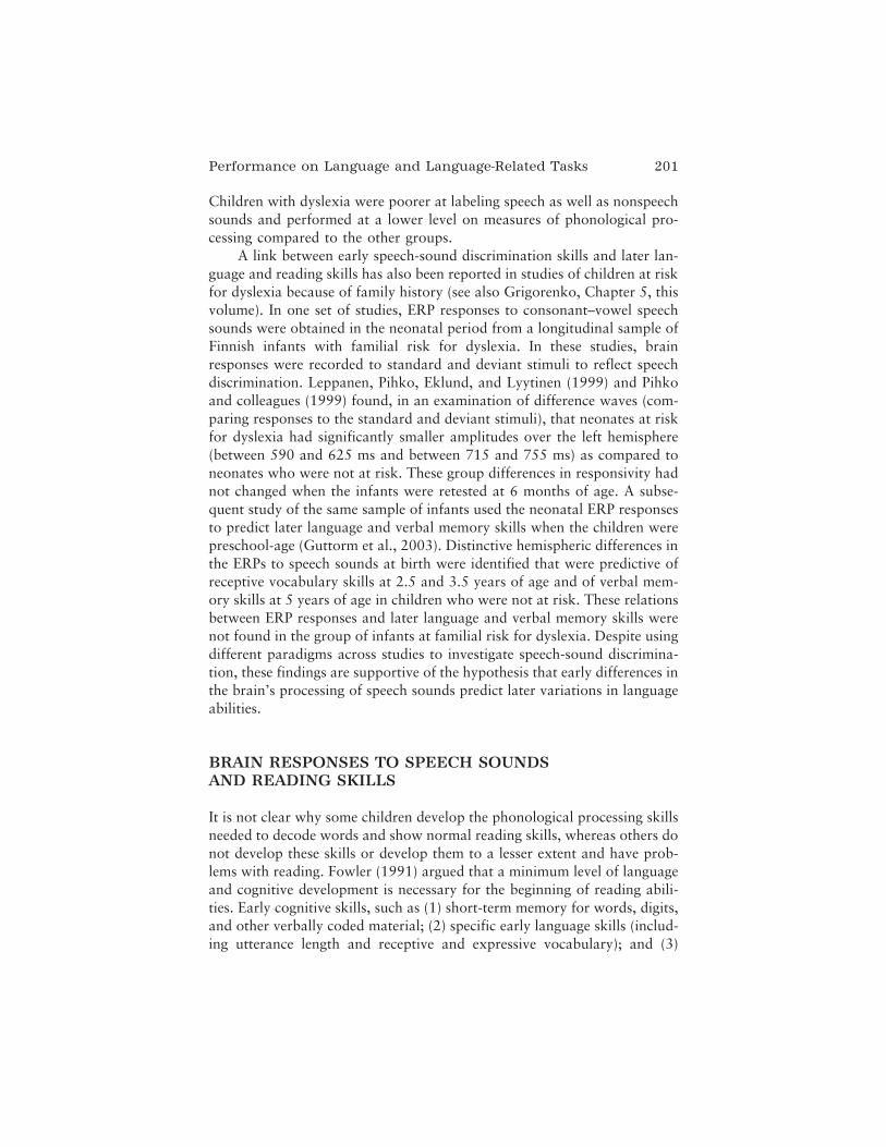
Children with dyslexia were poorer at labeling speech as well as nonspeechsounds and performed at a lower level on measures of phonological pro-cessing compared to the other groups.
A link between early speech-sound discrimination skills and later lan-guage and reading skills has also been reported in studies of children at riskfor dyslexia because of family history (see also Grigorenko, Chapter 5, thisvolume). In one set of studies, ERP responses to consonant–vowel speechsounds were obtained in the neonatal period from a longitudinal sample ofFinnish infants with familial risk for dyslexia. In these studies, brainresponses were recorded to standard and deviant stimuli to reflect speechdiscrimination. Leppanen, Pihko, Eklund, and Lyytinen (1999) and Pihkoand colleagues (1999) found, in an examination of difference waves (com-paring responses to the standard and deviant stimuli), that neonates at riskfor dyslexia had significantly smaller amplitudes over the left hemisphere(between 590 and 625 ms and between 715 and 755 ms) as compared toneonates who were not at risk. These group differences in responsivity hadnot changed when the infants were retested at 6 months of age. A subse-quent study of the same sample of infants used the neonatal ERP responsesto predict later language and verbal memory skills when the children werepreschool-age (Guttorm et al., 2003). Distinctive hemispheric differences inthe ERPs to speech sounds at birth were identified that were predictive ofreceptive vocabulary skills at 2.5 and 3.5 years of age and of verbal mem-ory skills at 5 years of age in children who were not at risk. These relationsbetween ERP responses and later language and verbal memory skills werenot found in the group of infants at familial risk for dyslexia. Despite usingdifferent paradigms across studies to investigate speech-sound discrimina-tion, these findings are supportive of the hypothesis that early differences inthe brain’s processing of speech sounds predict later variations in languageabilities.
BRAIN RESPONSES TO SPEECH SOUNDSAND READING SKILLS
It is not clear why some children develop the phonological processing skillsneeded to decode words and show normal reading skills, whereas others donot develop these skills or develop them to a lesser extent and have prob-lems with reading. Fowler (1991) argued that a minimum level of languageand cognitive development is necessary for the beginning of reading abili-ties. Early cognitive skills, such as (1) short-term memory for words, digits,and other verbally coded material; (2) specific early language skills (includ-ing utterance length and receptive and expressive vocabulary); and (3)
Performance on Language and Language-Related Tasks 201
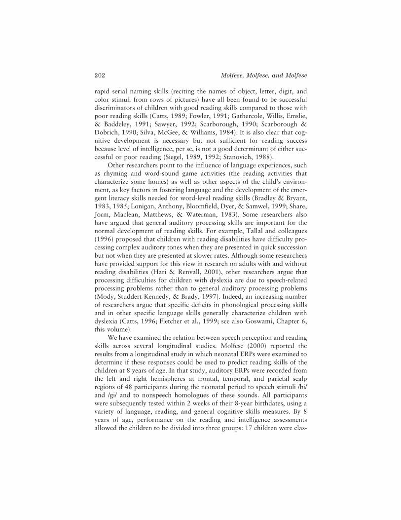
rapid serial naming skills (reciting the names of object, letter, digit, andcolor stimuli from rows of pictures) have all been found to be successfuldiscriminators of children with good reading skills compared to those withpoor reading skills (Catts, 1989; Fowler, 1991; Gathercole, Willis, Emslie,& Baddeley, 1991; Sawyer, 1992; Scarborough, 1990; Scarborough &Dobrich, 1990; Silva, McGee, & Williams, 1984). It is also clear that cog-nitive development is necessary but not sufficient for reading successbecause level of intelligence, per se, is not a good determinant of either suc-cessful or poor reading (Siegel, 1989, 1992; Stanovich, 1988).
Other researchers point to the influence of language experiences, suchas rhyming and word-sound game activities (the reading activities thatcharacterize some homes) as well as other aspects of the child’s environ-ment, as key factors in fostering language and the development of the emer-gent literacy skills needed for word-level reading skills (Bradley & Bryant,1983, 1985; Lonigan, Anthony, Bloomfield, Dyer, & Samwel, 1999; Share,Jorm, Maclean, Matthews, & Waterman, 1983). Some researchers alsohave argued that general auditory processing skills are important for thenormal development of reading skills. For example, Tallal and colleagues(1996) proposed that children with reading disabilities have difficulty pro-cessing complex auditory tones when they are presented in quick successionbut not when they are presented at slower rates. Although some researchershave provided support for this view in research on adults with and withoutreading disabilities (Hari & Renvall, 2001), other researchers argue thatprocessing difficulties for children with dyslexia are due to speech-relatedprocessing problems rather than to general auditory processing problems(Mody, Studdert-Kennedy, & Brady, 1997). Indeed, an increasing numberof researchers argue that specific deficits in phonological processing skillsand in other specific language skills generally characterize children withdyslexia (Catts, 1996; Fletcher et al., 1999; see also Goswami, Chapter 6,this volume).
We have examined the relation between speech perception and readingskills across several longitudinal studies. Molfese (2000) reported theresults from a longitudinal study in which neonatal ERPs were examined todetermine if these responses could be used to predict reading skills of thechildren at 8 years of age. In that study, auditory ERPs were recorded fromthe left and right hemispheres at frontal, temporal, and parietal scalpregions of 48 participants during the neonatal period to speech stimuli /bi/and /gi/ and to nonspeech homologues of these sounds. All participantswere subsequently tested within 2 weeks of their 8-year birthdates, using avariety of language, reading, and general cognitive skills measures. By 8years of age, performance on the reading and intelligence assessmentsallowed the children to be divided into three groups: 17 children were clas-
202 Molfese, Molfese, and Molfese
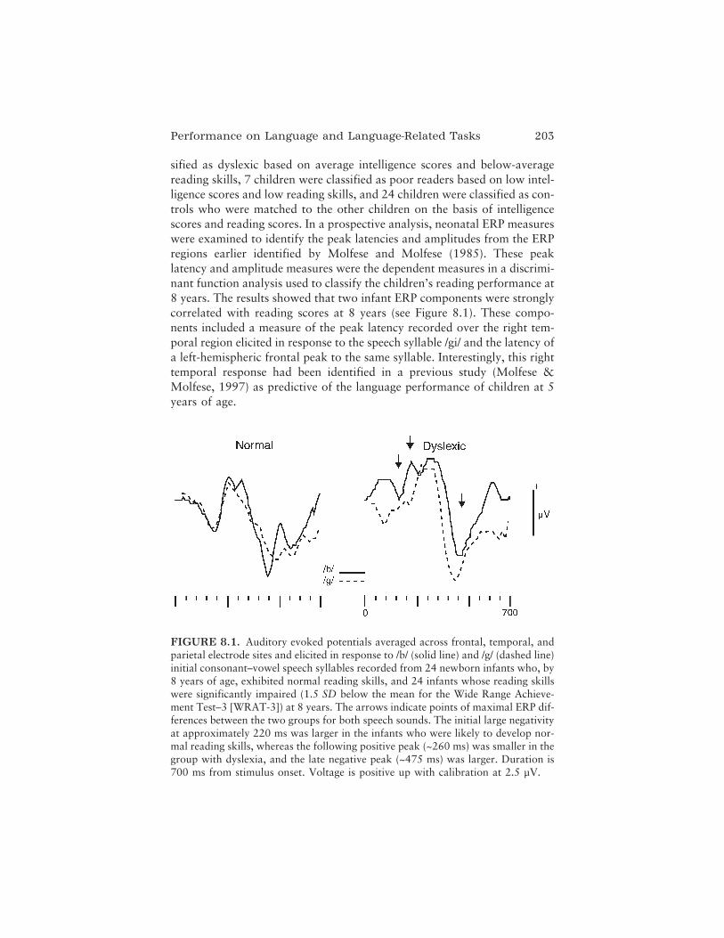
sified as dyslexic based on average intelligence scores and below-averagereading skills, 7 children were classified as poor readers based on low intel-ligence scores and low reading skills, and 24 children were classified as con-trols who were matched to the other children on the basis of intelligencescores and reading scores. In a prospective analysis, neonatal ERP measureswere examined to identify the peak latencies and amplitudes from the ERPregions earlier identified by Molfese and Molfese (1985). These peaklatency and amplitude measures were the dependent measures in a discrimi-nant function analysis used to classify the children’s reading performance at8 years. The results showed that two infant ERP components were stronglycorrelated with reading scores at 8 years (see Figure 8.1). These compo-nents included a measure of the peak latency recorded over the right tem-poral region elicited in response to the speech syllable /gi/ and the latency ofa left-hemispheric frontal peak to the same syllable. Interestingly, this righttemporal response had been identified in a previous study (Molfese &Molfese, 1997) as predictive of the language performance of children at 5years of age.
Performance on Language and Language-Related Tasks 203
FIGURE 8.1. Auditory evoked potentials averaged across frontal, temporal, andparietal electrode sites and elicited in response to /b/ (solid line) and /g/ (dashed line)initial consonant–vowel speech syllables recorded from 24 newborn infants who, by8 years of age, exhibited normal reading skills, and 24 infants whose reading skillswere significantly impaired (1.5 SD below the mean for the Wide Range Achieve-ment Test–3 [WRAT-3]) at 8 years. The arrows indicate points of maximal ERP dif-ferences between the two groups for both speech sounds. The initial large negativityat approximately 220 ms was larger in the infants who were likely to develop nor-mal reading skills, whereas the following positive peak (~260 ms) was smaller in thegroup with dyslexia, and the late negative peak (~475 ms) was larger. Duration is700 ms from stimulus onset. Voltage is positive up with calibration at 2.5 µV.
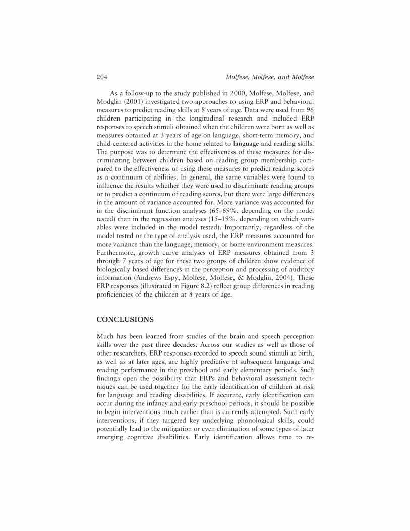
As a follow-up to the study published in 2000, Molfese, Molfese, andModglin (2001) investigated two approaches to using ERP and behavioralmeasures to predict reading skills at 8 years of age. Data were used from 96children participating in the longitudinal research and included ERPresponses to speech stimuli obtained when the children were born as well asmeasures obtained at 3 years of age on language, short-term memory, andchild-centered activities in the home related to language and reading skills.The purpose was to determine the effectiveness of these measures for dis-criminating between children based on reading group membership com-pared to the effectiveness of using these measures to predict reading scoresas a continuum of abilities. In general, the same variables were found toinfluence the results whether they were used to discriminate reading groupsor to predict a continuum of reading scores, but there were large differencesin the amount of variance accounted for. More variance was accounted forin the discriminant function analyses (65–69%, depending on the modeltested) than in the regression analyses (15–19%, depending on which vari-ables were included in the model tested). Importantly, regardless of themodel tested or the type of analysis used, the ERP measures accounted formore variance than the language, memory, or home environment measures.Furthermore, growth curve analyses of ERP measures obtained from 3through 7 years of age for these two groups of children show evidence ofbiologically based differences in the perception and processing of auditoryinformation (Andrews Espy, Molfese, Molfese, & Modglin, 2004). TheseERP responses (illustrated in Figure 8.2) reflect group differences in readingproficiencies of the children at 8 years of age.
CONCLUSIONS
Much has been learned from studies of the brain and speech perceptionskills over the past three decades. Across our studies as well as those ofother researchers, ERP responses recorded to speech sound stimuli at birth,as well as at later ages, are highly predictive of subsequent language andreading performance in the preschool and early elementary periods. Suchfindings open the possibility that ERPs and behavioral assessment tech-niques can be used together for the early identification of children at riskfor language and reading disabilities. If accurate, early identification canoccur during the infancy and early preschool periods, it should be possibleto begin interventions much earlier than is currently attempted. Such earlyinterventions, if they targeted key underlying phonological skills, couldpotentially lead to the mitigation or even elimination of some types of lateremerging cognitive disabilities. Early identification allows time to re-
204 Molfese, Molfese, and Molfese
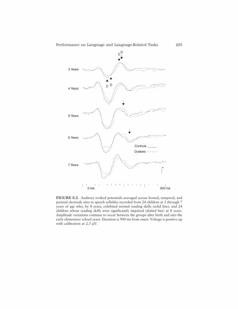
Performance on Language and Language-Related Tasks 205
FIGURE 8.2. Auditory evoked potentials averaged across frontal, temporal, andparietal electrode sites to speech syllables recorded from 24 children at 3 through 7years of age who, by 8 years, exhibited normal reading skills (solid line), and 24children whose reading skills were significantly impaired (dotted line) at 8 years.Amplitude variations continue to occur between the groups after birth and into theearly elementary school years. Duration is 900 ms from onset. Voltage is positive upwith calibration at 2.5 µV.
mediate potential deficits in the development of perceptual skills and, whencoupled with the early plasticity that neurocognitive systems appear to pos-sess, could provide exciting breakthroughs in the treatment of disabilities.At this point it appears that particularly effective interventions are thoseemphasizing phonological skills, such as segmentation and blending ofphonemes, combined with training in alphabetic skills (letter names and let-ter sounds); such approaches have been used with kindergarten childrenand have shown positive effects on reading skills (Ball & Blachman, 1991;Bradley & Bryant 1985; Oudeans, 2003). The development of effectiveinterventions for remediating early language and reading disabilities needsto interface with brain and behavioral assessment techniques previouslyfound to have high levels of accuracy in identifying young children withrisk characteristics. Subsequent studies must then include both early identi-fication and early intervention components to determine if this approach iseffective in improving the language and reading performance of at-risk chil-dren.
ACKNOWLEDGMENTS
This work was supported in part by grants from the National Institute of ChildHealth and Human Development (No. R01-HD17860) and the U.S. Department ofEducation (Nos. R215K000023, R215R990011).
REFERENCES
Andrews Espy, K., Molfese, D., Molfese, V., & Modglin, A. (2004). Developmentof auditory event-related potentials in young children and relations to word-level reading abilities at age 8 years. Annals of Dyslexia, 54, 9–38.
Aram, D., & Kamhi, A. (1982). Perspectives on the relationship between phonolog-ical and language disorders. Seminars in Speech, Language and Hearing, 3, 3–22.
Ball, E. W., & Blachman, B. A. (1991). Does phoneme awareness training in kin-dergarten make a difference in early word recognition and developmentalspelling? Reading Research Quarterly, 26, 49–66.
Bishop, D. (1997). Uncommon understanding: Development and disorders of lan-guage comprehension in children. Hove, UK: Psychology Press.
Bradley, L., & Bryant, P. (1983). Categorizing sounds and learning to read: Acausal connection. Nature, 30, 419–421.
Bradley, L., & Bryant, P. (1985). Rhyme and reason in reading and spelling. AnnArbor: University of Michigan Press.
Brazelton, T. (1973). Neonatal Behavioral Assessment Scale. Clinics in Develop-mental Medicine, 50. London: Heinemann.
206 Molfese, Molfese, and Molfese
Breier, J., Gray, L., Fletcher, J., Diehl, R., Klaas, P., Foorman, B., et al. (2001). Per-ception of voice and tone onset time continua in children with dyslexia withand without attention deficit/hyperactivity disorder. Journal of ExperimentalChild Psychology, 80, 245–270.
Butler, B., & Engel, R. (1969). Mental and motor scores at 8 months in relation toneonatal photic responses. Developmental Medicine and Child Neurology, 11,77–82.
Cacioppo, J., Tassinary, L., & Berntson, G. (Eds.). (2000). Handbook of psycho-physiology. Cambridge, UK: Cambridge University Press.
Camarata, S. (1995). A rationale for naturalistic speech intelligibility intervention.In M. E. Fey, J. Windsor, & S. F. Warren (Eds.), Language intervention: Pre-school through the elementary years (pp. 63–84). Baltimore: Brookes.
Catts, H. W. (1989). Phonological processing deficits and reading disabilities. In A.Kamhi & H. Catts (Eds.), Reading disabilities: A developmental language per-spective (pp. 101–132). Boston: Allyn & Bacon.
Catts, H. W. (1996). Defining dyslexia as a developmental language disorder: Anexpanded view. Topics in Language Disorders, 16(2), 14–29.
Cheour, M., Alho, K., Ceponiene, R., Reinikainen, K., Sainio, ´ K., Pohjavuori, M.,et al. (1998). Maturation of mismatch negativity in infants. International Jour-nal of Psychophysiology, 29, 217–226.
Cheour, M., Ceponiene, R., Lehtokoski, A., Luuk, A., Allik, J., Alho, K., et al.(1998). Development of language-specific phoneme representations in theinfant brain. Nature Neuroscience, 1, 351–353.
Cheour, M., Martynova, O., Naatanen, R., Erkkola, R., Sillanpaa, M., Kero, P., etal. (2002). Speech sounds learned by sleeping newborns. Nature, 415, 599–600.
Chomsky, N. (1957). Syntactic structures. The Hague: Mouton.Chomsky, N., & Halle, M. (1968). The sound pattern of English. New York:
Harper & Row.Dehaene-Lambertz, G., & Peña, M. (2001). Electrophysiological evidence for auto-
matic phonetic processing in neonates. NeuroReport, 12, 3155–3158.Dehaene-Lambertz, G., Peña, M., Christophe, A., & Landrieu, P. (2004). Phoneme
perception in a neonate with a left sylvian infarct. Brain and Language, 88,26–38.
Dennis, M., & Whitaker, H. (1976). Language acquisition following hemide-cortication: Linguistic superiority of the left over the right hemisphere. Brainand Language, 3, 404–433.
Dunn, L. (1965). Peabody Picture Vocabulary Test. Circle Pines, MN: AmericanGuidance Service.
Eilers, R., Wilson, W., & Moore, J. (1977). Developmental changes in speech dis-crimination in infants. Journal of Speech and Hearing Research, 20, 766–780.
Eimas, P. D., Siqueland, E., Jusczyk, P., & Vigorito, J. (1971). Speech perception ininfants. Science, 171, 303–306.
Engel, R., & Fay, W. (1972). Visual evoked responses at birth, verbal scores atthree years, and IQ at four years. Developmental Medicine and Child Neurol-ogy, 14, 283–289.
Performance on Language and Language-Related Tasks 207
Engel, R., & Henderson, N. (1973). Visual evoked responses and IQ scores atschool age. Developmental Medicine and Child Neurology, 15, 136–145.
Fletcher, J. M., Foorman, B. R., Shaywitz, S. E., & Shaywitz, S. A. (1999). Concep-tual and methodological issues in dyslexia research: A lesson for developmen-tal disorders. In H. Tager-Flusberg (Ed.), Neurodevelopmental disorders(pp. 271–305). Hillsdale, NJ: Erlbaum.
Fowler, A. E. (1991). How early phonological development might set the stage forphoneme awareness. In S. A. Brady & D. P. Shankweiler (Eds.), Phonologicalprocesses in literacy: A tribute to Isabelle Y. Liberman (pp. 97–118). Hillsdale,NJ: Erlbaum.
Fraser, C., Bellugi, U., & Brown, R. (1963). Control of grammar in imitation, com-prehension, and production. Journal of Verbal Learning and Verbal Behavior,2, 121–135.
Gathercole, S. E., Willis, C., Emslie, H., & Baddeley, A .D. (1991). The influencesof number of syllables and wordlikeness on children’s repetition of nonwords.Applied Psycholinguistics, 12, 349–367.
Guttorm, T. K., Leppänen, P. H. T., Eklund, K. M., Poikkeus, A.-M., Lyytinen, P.,& Lyytinen, H. (2003). Brain responses to changes in vowel duration mea-sured at birth predict later language skills in children with familial risk for dys-lexia. Manuscript submitted for publication.
Hari, R., & Renvall, H. (2001). Impaired processing of rapid stimulus sequences indyslexia. Trends in Cognitive Science, 5, 525–532.
Henderson, N., & Engel, R. (1974). Neonatal visual evoked potentials as predictorsof psychoeducational testing at age sever. Developmental Psychology, 10,269–276.
Jasper, H. (1958). The ten–twenty system of the International Federation of Soci-eties for Electroencephalography: Appendix to report of the committee onmethods of clinical examination in electroencephalography. Journal of Elec-troencephalography and Clinical Neurophysiology, 10, 371–375.
Jenkins, J., & Palermo, D. (1964). Mediation processes and the acquisition of lin-guistic structure. In U. Bellugi & R. Brown (Eds.), The acquisition of language(pp. 141–169). Monograph of the Society for Research in Child Development,29.
Jensen, D. R., & Engel, R. (1971). Statistical procedures for relating dichotomousresponses to maturation and EEG measurements. Electroencephalography andClinical Neurophysiology, 30, 437–443.
Kraus, N., McGee, T. J., Carrell, T. D., Zecker, S. G., Nicol, T. G., & Koch, D. B.(1996). Auditory neurophysiologic responses and discrimination deficits inchildren with learning problems. Science, 273, 971–973.
Kuhl, P. K., & Miller, J. D. (1975). Speech perception by the chinchilla: Voiced–voiceless distinction in alveolar plosive consonants. Science, 190, 69–72.
Kuhl, P. K., & Padden, D. M. (1983). Enhanced discriminability at the phoneticboundaries for the place feature in macaques. Journal of the Acoustical Societyof America, 73, 1003–1010.
Lenneberg, E. (1967). Biological foundations of language. New York: Wiley.Leppanen, P., Pihko, E., Eklund, K., & Lyytinen, H. (1999). Cortical responses of
208 Molfese, Molfese, and Molfese
infants with and without a genetic risk for dyslexia: II. Group effects.NeuroReport, 10, 969–973.
Liberman, A. M., Cooper, F. S., Shankweiler, D., & Studdert-Kennedy, M. (1967).Perception of the speech code. Psychological Review, 74, 431–461.
Lonigan, C. J., Anthony, J. L., Bloomfield, B. G., Dyer, S. M., & Samwel, C. S.(1999). Effects of two shared-reading interventions on emergent literacy skillsof at-risk preschoolers. Journal of Early Intervention, 22, 306–322.
Martynova, O., Kirjavainen, J., & Cheour, M. (2003). Mismatch negativity andlate discriminative negativity in sleeping human newborns. Neuroscience Let-ters, 340, 75–78.
McCarthy, D. (1972). Manual for the McCarthy Scales of Children’s Abilities. NewYork: Psychological Corporation.
Mody, M., Studdert-Kennedy, M., & Brady, S. (1997). Speech perception deficits inpoor readers: Auditory processing or phonological coding? Journal of Experi-mental Child Psychology, 64, 199–231.
Molfese, D. L. (1972). Cerebral asymmetry in infants, children and adults: Auditoryevoked responses to speech and music stimuli. Unpublished doctoral disserta-tion, Pennsylvania State University, University Park.
Molfese, D. L. (1978). Neuroelectrical correlates of categorical speech perception inadults. Brain and Language, 5, 25–35.
Molfese, D. L. (1980). Hemispheric specialization for temporal information: Impli-cations for the perception of voicing cues during speech perception. Brain andLanguage, 11, 285–299.
Molfese, D. L. (1989). The use of auditory evoked responses recorded from new-borns to predict later language skills. In N. Paul (Ed.), Research in infantassessment (Vol. 25, No. 6, pp. 47–62). White Plains, NY: March of Dimes.
Molfese, D. L. (2000). Predicting dyslexia at 8 years using neonatal brain responses.Brain and Language, 72, 238–245.
Molfese, D. L., Freeman, R., & Palermo, D. (1975). The ontogeny of lateralizationfor speech and nonspeech stimuli. Brain and Language, 2, 356–368.
Molfese, D. L., & Hess, T. M. (1978). Speech perception in nursery school age chil-dren: Sex and hemispheric differences. Journal of Experimental Child Psychol-ogy, 26, 71–84.
Molfese, D. L., Laughlin, N. K., Morse, P. A., Linnville, S., Wetzel, F., & Erwin, R.(1986). Neuroelectrical correlates of categorical perception for place of articu-lation in normal and lead-treated rhesus macaques. Journal of Clinical andExperimental Neuropsychology, 8, 680–696.
Molfese, D. L., & Molfese, V. J. (1979a). Hemisphere and stimulus differences asreflected in the cortical responses of newborn infants to speech stimuli. Devel-opmental Psychology, 15, 505–511.
Molfese, D. L., & Molfese, V. J. (1979b). Infant speech perception: Learned orinnate? In H. Whitaker & H. Whitaker (Eds.), Advances in neurolinguistics(Vol. 4, pp. 225–240). New York: Academic Press.
Molfese, D. L., & Molfese, V. J. (1983). Development of symmetrical and asym-metrical hemisphere responses to speech sounds: Electrophysiological corre-lates. In E. Lasky & J. Katz (Eds.), Central auditory processing disorders:
Performance on Language and Language-Related Tasks 209
Problems of speech, language and learning (pp. 69–89). Baltimore: UniversityPark Press.
Molfese, D. L., & Molfese, V. J. (1985). Electrophysiological indices of auditorydiscrimination in newborn infants: The bases for predicting later languagedevelopment. Infant Behavior and Development, 8, 197–211.
Molfese, D. L., & Molfese, V. J. (1986). Psychophysical indices of early cognitiveprocesses and their relationship to language. In J. E. Obrzut & G. W. Hynd(Eds.), Child neuropsychology: Vol. 1. Theory and research (pp. 95–115).New York: Academic Press.
Molfese, D. L., & Molfese, V. J. (1997). Discrimination of language skills at fiveyears of age using event related potentials recorded at birth. DevelopmentalNeuropsychology, 13, 135–156.
Molfese, D. L., Molfese, V. J., & Pratt, N. L. (in press). The use of event-relatedevoked potentials to predict developmental outcomes. In M. de Haan (Ed.),Infant EEG and event-related potentials. Hove, UK: Psychology Press.
Molfese, D. L., Papanicolaou, A., Hess, T. S., & Molfese, V. (1979). Neuro-electrical correlates of semantic processes. In H. Begleiter (Ed.), Evoked brainpotentials and behavior. New York: Plenum Press.
Molfese, D. L., & Searock, K. (1986). The use of auditory evoked responses at oneyear of age to predict language skills at 3 years. Australian Journal of Commu-nication Disorders, 14, 35–46.
Molfese, V. J., Molfese, D. L., & Modglin, A. (2001). Newborn and preschool pre-dictors of second grade reading scores: An evaluation of categorical and con-tinuous scores. Journal of Learning Disabilities, 34, 545–554.
Molfese, V. J., Yaple, K., Helwig, S., Harris, L., & Connell, S. (1992). Stanford–Binet Intelligence Scale (4th ed.): Factor structure and verbal subscale scoresfor three year olds. Journal of Psychoeducational Assessment, 10, 47–58.
Morse, P. A., Molfese D. L., Laughlin, N. K., Linnville, S., & Wetzel, W. F. (1987).Categorical perception for voicing contrasts in normal and lead-treated rhesusmonkeys: Electrophysiological indices. Brain and Language, 30, 63–80.
Morse, P. A., & Snowdon, C. (1975). An investigation of categorical speech dis-crimination by rhesus monkeys. Perception and Psychophysics 17, 9–16.
Oudeans, M. (2003). Integration of letter–sound correspondences and phonologicalawareness skills of blending and segmenting: A pilot study examining theeffects of instructional sequence on word reading for kindergarten childrenwith low phonological awareness. Learning Disabilities Quarterly, 26, 258–280.
Papanicolaou, A. C. (1998). Fundamentals of functional brain imaging: A guide tothe methods and their applications to psychology and behavioral neuro-sciences. Netherlands: Swets & Zeitlinger.
Pihko, E., Leppanen, P. H., Eklund, K. M., Cheour, M., Guttorm, T. K., &Lyytinen, H. (1999). Cortical responses of infants with and without a geneticrisk for dyslexia: I. Age effects. NeuroReport, 10, 901–905.
Reese, H., & Lipsitt, L. (1970). Experimental child psychology (pp. 425–477). NewYork: Academic Press.
210 Molfese, Molfese, and Molfese
Rockstroh, B., Elbert, T., Birbaumer, N., & Lutzenberger, W. (1982). Slow brainpotentials and behavior. Baltimore: Urban-Schwarzenberg.
Sawyer, D. (1992). Language abilities, reading acquisition, and developmental dys-lexia: A discussion of hypothetical and observed relationships. Journal ofLearning Disabilities, 25, 82–95.
Scarborough, H. (1990). Very early language deficits in dyslexic children. ChildDevelopment, 61, 1728–1743.
Scarborough, H., & Dobrich, W. (1990). Development of children with early lan-guage delay. Journal of Speech and Hearing Research, 33, 70–83.
Share, D., Jorm, A., Maclean, R., Matthews, R., & Waterman, B. (1983). Earlyreading achievement, oral language ability, and a child’s home background.Australian Psychologist, 18, 75–87.
Siegel, L. (1989). IQ is irrelevant to the definition of learning disabilities. Journal ofLearning Disabilities, 22, 469–486.
Siegel, L. (1992). Dyslexia v. poor readers. Is there a difference? Journal ofLearning Disabilities, 25, 618–629.
Silva, P., McGee, R., & Williams, S. (1984). A seven year follow-up study of thecognitive development of children who experienced common perinatal prob-lems. Australian Pediatric Journal, 20, 23–28.
Stanovich, K. (1988). Explaining the differences between dyslexic and the garden-variety poor reader: The phonological core variable difference model. Journalof Learning Disabilities, 21, 590–604.
Tallal, P., Miller, S., Bedi, G., Byma, G., Wang, X., Nagarajan, S., et al. (1996).Language comprehension in language-learning impaired children improvedwith acoustically modified speech. Science, 271, 81–84.
Templin, M. (1957). Certain language skills in children. University of MinnesotaInstitute of Child Welfare Monograph Series 26. Minneapolis: University ofMinnesota Press.
Travis, L. E. (1931). Speech pathology: A dynamic neurological treatment of nor-mal speech and speech deviations. New York: Appleton.
Vellutino, F., & Scanlon, D. (1987). Phonological coding: Phonological awareness,and reading ability: Evidence from a longitudinal and experimental study.Merrill–Palmer Quarterly, 33, 321–363.
Performance on Language and Language-Related Tasks 211
C H A P T E R 9
Number Senseand Developmental Dyscalculia
Anna J. WilsonStanislas Dehaene
Claire is an 8-year-old girl in second grade who participated in aremediation study for dyscalculia that we recently conducted. Despitean average IQ and good motivation and attention, she struggles inschool, especially in math. This does not seem to be due to her readingabilities; her reading speed is average, and although she has some trou-ble with comprehension, she already receives special education servicesfor this difficulty, with no seeming improvement in her mathematicalperformance. Our tests confirm that her basic numerical skills are farbehind those of her peers: She shows a developmental lag in counting,understanding of place value, and in addition and subtraction of one-digit numbers. The latter are only around 80 and 40% accurate,respectively, and both are carried out in a painstaking fashion usingfinger counting.
Claire is a fairly typical case of developmental dyscalculia, which isgenerally defined as a disorder in mathematical abilities presumed to be dueto a specific impairment in brain function (Kosc, 1974; Shalev & Gross-Tsur, 1993, 2001). This definition is highly similar to that of “mathematicsdisorder” in the Diagnostic and Statistical Manual of Mental Disorders—
212
Fourth Edition (DSM-IV; American Psychiatric Association, 1994) andthat of “mathematical learning disabilities” (Geary, 1993, 2004). Becauseof this similarity, and following Butterworth (2005a, 2005b), we take theseconstructs to be one and the same.1
Despite its professional and practical consequences (e.g., Rivera-Batiz,1992) and a similar population prevalence of around 3–6% (Badian, 1983;Gross-Tsur, Manor, & Shalev, 1996; Kosc, 1974; Lewis, Hitch, & Walker,1994), developmental dyscalculia is much less recognized, researched, andtreated than its cousin developmental dyslexia. This neglect has partiallybeen a consequence of the later development of our knowledge about theneural bases of numerical cognition, as opposed to reading. As research inthe numerical cognition field has started to increase in volume, so has cog-nitive neuroscience research on dyscalculia, with several research teamsconducting studies on its associated cognitive profile, brain bases, andgenetics.
THE CORE DEFICIT HYPOTHESIS
An advantage provided by the 20-or-so-year lag between dyscalculia anddyslexia research is an opportunity to speed up research on the former bylooking for analogies that may be made between the two disorders. Oneinteresting and important analogy that has been made, for which we reviewevidence in this chapter, is that of a “core deficit.” The core phonologicaldeficit hypothesis is now accepted by many in the dyslexia field (Goswami,2003; see also Goswami, Chapter 6, and Grigorenko, Chapter 5, this vol-ume) and has resulted in many advances in prevention and remediation(Eden, 2002; Tallal et al., 1996; Temple et al., 2003). Importantly, thenature of the core deficit can be somewhat counterintuitive—who wouldhave thought that training children in distinction of sounds would improvereading? However, this idea is now confirmed by our knowledge of thebrain circuits involved in reading, especially the continuing role of phono-logical areas in reading in even practiced readers.
Could there be a similar core deficit in dyscalculia? Although ourknowledge of its behavioral manifestations is incomplete, our knowledge ofthe adult circuits involved in numerical cognition is by now fairly ad-vanced. It has been argued that the core aspect of numerical cognition is“number sense,” which is a shorthand term for our ability to quicklyunderstand, approximate, and manipulate numerical quantities (Dehaene,1997, 2001). We now have a plausible candidate for a neural substrate ofnumber sense: a specific region of the parietal cortex, the horizontalintraparietal sulcus (HIPS), which, based on neuroimaging results, is
Number Sense and Developmental Dyscalculia 213
hypothesized to contain a nonverbal representation of numerical quantityanalogous to a spatial map or “number line” (Dehaene, Piazza, Pinel, &Cohen, 2003).
Our knowledge of the intact adult system allows us, therefore, to makea prediction about the impaired system in the child. The behavioral hypoth-esis that a deficit in number sense is the cause of at least some types of dys-calculia has, in fact, been previously proposed by authors in the spe-cial education field (Gersten & Chard, 1999; Robinson, Menchetti, &Torgesen, 2002) and in the numerical cognition field (Butterworth, 1999).Here we propose a neural specification of this hypothesis: that at least sometypes of dyscalculia may be due to an impairment of functioning and/orstructure in the HIPS and/or in its connections to other numerical cognitionregions. In this chapter we examine how much evidence there is to supportthis hypothesis.
A COMPLEMENTARY HYPOTHESIS:MULTIPLE SUBTYPES OF DYSCALCULIA
As well as presenting and discussing the hypothesis of a core deficit, wepresent and discuss the possibility of multiple causes of dyscalculia.Although a majority of children might suffer from a core impairment innumber sense, other sources of dyscalculia are likely to exist. Hypothesesabout their nature may be generated from several sources, such as the adultneuroimaging literature, the developmental literature, and educationalresearch on subtypes of dyscalculia. To take one example, in the adult neu-rological literature it is possible to find a “number sense” acalculia, inwhich the patient has lost all sense of the meaning of numbers, but it is alsopossible to find a “number fact retrieval” acalculia, in which the patientstill understands the meaning of numbers but is unable to retrieve frommemory basic multiplication or addition facts, and is thus forced to labori-ously recalculate these facts each time by counting, or worse, retrieves thewrong result without noticing (Dehaene & Cohen, 1997). We also discusswhether there is evidence for a subtype of developmental dyscalculia simi-lar to this latter type of patient.
OUTLINE OF THE CHAPTER
We begin by describing cases of adult-acquired dyscalculia and discussingwhat we know about the underlying numerical cognition systems in theadult. We then overview the developmental numerical cognition literature.
214 Wilson and Dehaene
The combination of these two literatures allows us to elaborate some pre-dictions about possible types and causes of developmental dyscalculia.Next we discuss in depth developmental dyscalculia: how it is identified, itscharacteristics and common comorbid disorders, and what we have learnedfrom subtype research. We then discuss the extent to which this literaturesupports our hypotheses, in particular the behavioral and neural evidencefor a deficit in number sense. Finally, we discuss future directions to betaken, the implications for education, and the construction of number senseremediation software in our laboratory.
ADULT NUMERICAL COGNITION
There are two important areas of adult numerical cognition research thatcan be used to shed light on the possible causes of developmental dys-calculia. The first is neuropsychological research on acquired dyscalculia(referred to here as acalculia), which may have similar causes and symp-toms to developmental dyscalculia (referred to here as dyscalculia). Thesecond is our knowledge of the functioning of the intact numerical cogni-tion circuits in the adult brain, which gives us a clue as to what kind of defi-cits we might expect if developmental dyscalculia is caused by abnormali-ties in these circuits.
Acquired Dyscalculia or “Acalculia”: Lesion Evidence
The adult neuropsychological literature supports a causative role of inferiorparietal lesions in acalculia (see Cohen, Wilson, Izard, & Dehaene, in press,for a detailed review of this literature). Acalculia is often associated withGerstmann syndrome, in which patients also present left–right disorienta-tion, agraphia, and finger agnosia (Gerstmann, 1940). This syndrome isusually due to lesions around the region of the left angular gyrus (Jackson& Warrington, 1986; Rosselli & Ardila, 1989). However, the existence ofa coherent syndrome has been questioned because it appears that the fourdeficits can be dissociated (Benton, 1992).
Dehaene and Cohen (1997) discuss two cases of acalculia (patientsMAR and BOO) who together provide a double dissociation between a“number sense” type acalculia and a “verbal memory” type acalculia.Patient MAR, a left-hander with a right inferior parietal lesion and a pureGerstmann syndrome, showed difficulties with tasks requiring quantitymanipulation, such as choosing the larger of two digits, bisecting numberlines, and subtraction. Patient BOO, a right-hander with a left subcorticallesion, showed a very different pattern with few difficulties in the former
Number Sense and Developmental Dyscalculia 215
tasks but severe difficulties with more verbal or memory-related numericaltasks, such as multiplication. Other similar cases have also been reportedmore recently by Lemer, Dehaene, Spelke, and Cohen (2003), Cappeletti,Butterworth, and Kopelman (2001), and Delazer, Karner, Zamarian,Donnemiller, and Benke (2006).
To the extent that acquired and developmental dyscalculia are similarin their causes and manifestations, the acalculia literature suggests twoimportant hypotheses for developmental dyscalculia: (1) that we shouldexpect to find more than one type of dyscalculia, in particular, a numbersense dyscalculia and a verbal memory dyscalculia; and (2) that we shouldexpect to find a link between number sense dyscalculia and the inferiorparietal lobes, particularly the angular gyrus. In the next section we showthat the adult imaging literature supports these two suggestions and addsfurther precision to our hypothesis about anatomical location.
Imaging Evidence: Number Senseand the Horizontal Intraparietal Sulcus
Early PET imaging studies consistently showed activation in the parietalcortex during numerical tasks (Dehaene et al., 1996; Pesenti, Thioux,Seron, & De Volder, 2000; Zago et al., 2001), although only some of theseearly results (e.g., Dehaene et al., 1996) supported a dissociation betweenareas activated by quantity manipulation versus rote memory tasks, aswould be expected from the neuropsychological literature.
Later fMRI studies provided much more precision, and in a recentmeta-analysis of available data from this literature, Dehaene and colleagues(2003) put forward a more precise localization hypothesis, identifying threeparticular areas of the parietal lobe that appear to be differentially involvedin representation and processing of numerical information (see Plate 9.1).The left angular gyrus—the classic Gerstmann lesion center—appears to bemore active in more verbal numerical tasks such as multiplication andexact addition (Chochon, Cohen, van de Moortele, & Dehaene, 1999;Dehaene, Spelke, Pinel, Stanescu, & Tsivkin, 1999; Lee, 2000). The poste-rior superior parietal lobe (PSPL) appears to be activated in numerical tasksthat may require the shifting of spatial attention, such as approximating,subtraction, and number comparison (Dehaene et al., 1999; Lee, 2000;Pinel, Dehaene, Riviere, & Le Bihan, 2001).
In contrast, the nearby HIPS appears to be more active in core quantitymanipulation or number sense tasks, such as comparing the size of num-bers, estimating, subtracting, and approximating (Chochon et al., 1999;Dehaene et al., 1999; Lee, 2000). This role of the HIPS in quantity repre-
216 Wilson and Dehaene
sentation has been further reinforced by subsequent imaging studies (Eger,Sterzer, Russ, Giraud, & Kleinschmidt, 2003; Pinel, Piazza, Le Bihan, &Dehaene, 2004), and by the recent finding by Piazza, Izard, Pinel, Le Bihan,and Dehaene (2004), using an fMRI habituation paradigm, that fMRIadaptation in this area shows tuning curves similar to those found in singleneuron recordings from the analogous area of the monkey brain (Nieder &Miller, 2004).
Overall, the neuroimaging literature leads to the same hypothesesarrived at from the neuropsychological literature: We should expect to finda role of the inferior parietal lobes in dyscalculia, and we should be able tofind different subtypes of dyscalculia. These subtypes should be a numbersense dyscalculia linked to impairment (functional or structural) of theHIPS, a verbal dyscalculia linked to impairment of the angular gyrus, and aspatial attention dyscalculia linked to impairment of the PSPL.
However, one caveat is that these theoretical subtypes might be quitedifficult to dissociate in developmental cases. Firstly, in a developmentalcontext, any deficit in one of the numerical systems is likely to interferewith the normal development of the others, thus leading to an undifferenti-ated dyscalculic pattern, even if the underlying neurological damage is lim-ited. Secondly, the physical proximity of the areas involved makes it likelythat there is a high correlation between impairment in one area and impair-ment in another. As pointed out by Dehaene and colleagues (2003), thisproximity explains why, in the neuropsychological literature, it is hard tofind “pure” cases of verbal or number sense acalculia: areas that are closetogether are likely to be lesioned together. It is possible that this associationof impairment could also occur in developmental dyscalculia; for instance,a gene-influenced growth factor could impact development in a whole sub-section of the cortex, such as the inferior parietal lobule.
NORMAL INFANT AND CHILDNUMERICAL COGNITION
Of course, our ability to make predictions about developmental dyscalculiafrom the adult neuropsychology and imaging literature rests on the as-sumption that numerical cognition systems in the adult and the child aresimilar. However, it is not yet clear whether this is the case, and we havelittle knowledge of how the infant system develops into the child and thenthe adult system (Ansari & Karmiloff-Smith, 2002). In this section webriefly review some of our key knowledge about the infant and child sys-tems.
Number Sense and Developmental Dyscalculia 217
Number Sense: Our Core Magnitude Representation
One obvious difference between the infant and adult numerical cognitionsystem is that the infant is not born with an innate ability to process sym-bolic numerical codes, such as digits and number names. It was initiallythought that all numerical knowledge had to be constructed throughsensorimotor interaction with the environment (Piaget, 1952), but due tothe many studies on infant numerical cognition in the past quarter of a cen-tury, we now know that infants are born with an ability to represent, dis-criminate, and operate on numerosities, although with only a limiteddegree of precision (for a recent review, see Feigenson, Dehaene, & Spelke,2004). For instance, even with continuous visual cues such as luminanceand occupied area controlled, 6-month-old infants can discriminate be-tween groups of 8 and 16 or 16 and 32 dots, but not 16 and 24 dots (Xu &Spelke, 2000; Xu, Spelke, & Goddard, 2005). Recent work by McCrinkand Wynn (2004) has shown that 9-month-olds can approximately addand subtract collections of objects (e.g., 5 + 5 or 10 – 5). These approxi-mate representations of number in infants are constrained by the ratio ofthe two numbers and improve in precision during the first year of life(Lipton & Spelke, 2003).
Although no imaging evidence is available yet on the source of theserepresentations in infants, due to the practical difficulties of conductingsuch studies, these characteristics are similar to the approximate represen-tation present in animals (Gallistel & Gelman, 2000; Nieder, 2005) and inthe HIPS of adult humans (see discussion above). Behavioral studies (andone ERP study) in preschool children also support this conclusion (Berch,Foley, Hill, & Ryan, 1999; Girelli, Lucangeli, & Butterworth, 2000;Huntley-Fenner, 2001; Rubinsten, Henik, Berger, & Shahar-Shalev, 2002;Siegler & Opfer, 2003; Temple & Posner, 1998; see Noël, Rousselle, &Mussolin, 2005, for a review). The fact that this number sense systemmatures in the first year of life and is a core aspect of adult numerical cog-nition makes it a likely candidate for a core deficit in dyscalculia.
A Second Nonsymbolic Core System: Object Files
For some time, opponents of the concept of an innate numerosity represen-tation have argued that infants were able to represent number based ontheir visuospatial abilities (e.g., Simon, 1999). It has now become clear thatto keep track of small quantities of objects, infants are also able to use avisuospatial “object file”–based system that allows them to keep referencesto up to three or four objects and also their continuous properties. Infants,in fact, may sometimes show a preference for using this system over the
218 Wilson and Dehaene
approximate magnitude system (Feigenson, 2005; Feigenson, Carey, &Hauser, 2002; Feigenson, Carey, & Spelke, 2002; Xu, 2003). It has beenargued that adults may still use this system for subitizing (or rapidly identi-fying one to four objects; Trick & Pylyshyn, 1994), although this has notyet been proven to be the case (Feigenson et al., 2004). It is thus not certainwhether, and to what extent, impairments in this second system could beresponsible for developmental dyscalculia.
Development of Symbolic Capabilities
In addition to these two core systems children also develop (in the appro-priate cultural context) the ability to represent numbers in a symbolic fash-ion, first using words and later using Arabic digits. This then allows themto extend their approximate abilities into the realm of exact arithmetic.Few developmental data are available on the neural foundations of devel-oping symbolic representations, particularly on how these might be linkedwith the nonsymbolic core systems discussed above. However, it is clearthat an impairment in symbolic representation or an impairment in the linkbetween the nonsymbolic and the symbolic are both possible causes fordyscalculia.
The first key development that occurs between the ages of 2 and 4years in normal children is the acquisition of counting. In order to makefull use of the counting procedure, children need to learn the sequence ofcount words and to understand and execute their one-to-one mapping to aset of objects. Finally, they need to understand that this procedure gives thecardinality of the set. Some argue that it is only the language and proce-dural execution aspects of the task that need to be acquired and that theconcept of one-to-one correspondence and the comprehension of cardinalsare already present (Fuson, 1988; Wynn, 1990), whereas others claim thatall of these need to be acquired (Cordes & Gelman, 2005; Gelman &Gallistel, 1978). Wynn (1990), in particular, demonstrates that between2½ and 3½ years old, children exhibit a radical change in their understand-ing of counting and in their ability to use counting in simple quantity tasks.
Early work by Siegler and colleagues showed that prior to enteringschool, children spontaneously count on their fingers and thereby computesimple additions (Siegler & Jenkins, 1989; Siegler & Shrager, 1984). Pre-school children also have usually mastered a strategy for exact addition,“counting all,” in which, for example, to add 2 + 4 they would count out 2on their fingers, count out 4 using other fingers, and finally count all thefingers to give 6. Eventually this strategy is replaced by a more advancedone, “counting on,” in which children start at the first number and countup the second number of units, for instance, “2 . . . 3, 4, 5, 6.” Finally, chil-
Number Sense and Developmental Dyscalculia 219
dren learn the “min” strategy of starting at the largest number so that theyhave to count only the smallest distance, for example, “4 . . . 5, 6.” As chil-dren get older, counting (with or without fingers) comes to play a decreas-ing role in addition as memory-based retrieval strategies take over. How-ever, it has been shown that even adults still rely on backup strategies in theevent of a retrieval failure, such as decomposition—for example, 8 + 6 →(8 + 2) + 4 = 10 + 4 = 14—or derivation from related facts—8 + 6 → (9 +6) – 1 = 15 – 1 = 14 (for a review, see LeFevre, Smith-Chant, Hiscock,Daley, & Morris, 2003).
Another possible candidate for dyscalculia is therefore an impairmentin the symbolic system. Although, as mentioned earlier, there is little devel-opmental evidence on the neural bases of this system, in the adult we knowthat at least part of it, especially that governing counting and retrieval ofarithmetic facts, is verbal in nature (Spelke & Tsivkin, 2001). Neuroimag-ing evidence suggests that it is at least partially governed by perisylvian lan-guage areas (Stanescu-Cosson et al., 2000). In the case of strategy execu-tion, there are currently no imaging data available even in the adult;however, we can tentatively say that this ability is likely to be linked to thefrontal lobes. This then generates two hypotheses for dyscalculia: a subtypelinked to verbal impairment and a subtype linked to executive dysfunction.
A further important hypothesis should be mentioned here. From theprevious discussion it is evident that there are many bases for the represen-tation of number, which can be divided into two broad categories:nonsymbolic and symbolic. These representations clearly come from differ-ent sources: Children are born with an approximate nonsymbolic represen-tation of number that is similar to that present in animals, and then theylater learn an exact symbolic representation. Another obvious hypothesisfor dyscalculia is that of a disconnection syndrome: Children with dyscal-culia could have an intact nonsymbolic representation of quantity but failto make the link between this and their newly acquired symbolic represen-tations.
SUMMARY OF HYPOTHESESFOR DEVELOPMENTAL DYSCALCULIA
In summary of the previous sections, we now list the hypotheses put for-ward for developmental dyscalculia based on the neuropsychology, adultnumerical cognition, and developmental numerical cognition literatures.There are two sets of questions to be addressed: first, whether or not thereis a core deficit, and if so, what type of dyscalculia would be associatedwith it, and second, what other subtypes of dyscalculia might be found.
220 Wilson and Dehaene
We propose that if there is a single core deficit that causes dyscalculia,it is likely to be due to one of the following:
1. A deficit in number sense or nonsymbolic representation of num-ber. This deficit would be caused by structural or functional impairment tothe HIPS. Its symptoms would include impaired understanding of themeaning of numbers, deficits in tasks that involve this area (nonsymbolictasks such as comparison and approximate addition of dots, but also thesymbolic tasks of numerical comparison, addition, and subtraction), andreduced automatic activation of quantity from number words and digits.Because these are such basic deficits, they would be likely to cause a devel-opmental delay in all aspects of math, except the highly verbal processes ofcounting and fact retrieval.
2. Impaired connections between symbolic and nonsymbolic represen-tations. In this case, we would expect to see the same pattern described abovebut with one important difference: little impairment on nonsymbolic tasks.
Based on our literature review, we might also expect several other sub-types of developmental dyscalculia:
1. A deficit in verbal symbolic representation, related to impairmentin the angular gyrus, the left inferior frontal and/or temporal languageareas, or the left basal ganglia. This impairment would result in difficultieslearning and retrieving arithmetical facts (particularly for multiplication)and possibly also in learning the counting sequence.
2. A deficit in executive dysfunction, due to frontal dysfunction. Thisimpairment would also be likely to result in difficulties in arithmetical factretrieval, but would furthermore result in difficulties in strategy and proce-dure usage.
3. A deficit in spatial attention, due to posterior superior parietal dys-function. This type of deficit could be linked to the object file tracking sys-tem and might therefore result in difficulties in subitizing (if this is the sys-tem underlying subitizing). It might also result in difficulties in perceptionof nonsymbolic quantity information and in quantity manipulation. How-ever, due to the close intertwining of spatial and numerical representations,this subtype might be difficult to separate from a number sense subtype.
DEVELOPMENTAL DYSCALCULIA
Bearing in mind these hypotheses, we now turn to a review of the develop-mental dyscalculia literature and examine to what extent we find support
Number Sense and Developmental Dyscalculia 221
for them. First we briefly discuss two important issues that should be bornein mind when thinking about this research and which can make it difficultto compare results across different studies.
Identification
In the introductory section we discussed the definition of developmentaldyscalculia: a disorder in mathematical abilities presumed to be due to aspecific impairment in brain function. In the educational field it has beentraditional to identify learning disabilities by using standardized educa-tional tests and defining dyscalculia as a significant lag in performance, tak-ing into account age and IQ (e.g., Geary, Hamson, & Hoard, 2000; Jordan,Hanich, & Kaplan, 2003). Alternatively, some research laboratories study-ing dyscalculia have used their own tests based on neuropsychological bat-teries (e.g., Gross-Tsur et al., 1996). One important issue is the cutoff usedto identify children as dyscalculic, which is essentially arbitrary, as in thecase of dyslexia. This arbitrary cutoff point has led to important differencesin the populations of children studied (see Butterworth, 2005b, for adetailed discussion), making it difficult to be certain of the true symptomsof dyscalculia.
Comorbid Disorders
Two important disorders that appear to be comorbid with dyscalculia aredyslexia and attention-deficit/hyperactivity disorder (ADHD; see Gatzke-Kopp & Beauchaine, Chapter 10; Grigorenko, Chapter 5; and Goswami,Chapter 6, this volume, for further discussion of these disorders). Estima-tions for the comorbidity rate of dyslexia vary wildly, possibly due to dif-ferences in criteria, methodology, and school year. For instance, a longitu-dinal prevalence study by Badian (1999) found that 60% of persistent lowarithmetic achievers were also low reading achievers (using a cutoff of the25th percentile on average achievement over a 7- to 8-year period). Lewisand colleagues (1994) found a comorbidity rate (64%) of a similar order intheir prevalence study, in which they used a cutoff of the 16th percentile forreading and math difficulties. At the other extreme, Gross-Tsur and col-leagues (1996) found a dyslexia–dyscalculia comorbidity of only 17% intheir sample of children with dyscalculia. However, their cutoff for dyslexiawas the 5th percentile on a standardized reading and spelling test, whichwas much more conservative than that of the previous authors. The comor-bidity rate of dyscalculia and ADHD is no less certain, having beenaddressed in only one large prevalence study (Gross-Tsur et al., 1996).These authors found that 26% of their participants with dyscalculiashowed symptoms of ADHD, as measured by Connor’s questionnaire.
222 Wilson and Dehaene
The presence of comorbid disorders in children with dyscalculia isimportant to keep in mind, because studies have not always controlled for,or reported, them and they may in fact be related to the symptomsobserved.
Characteristics
Much work on the characteristics of dyscalculia has already been con-ducted in the educational field, although as we discuss later, this work canbe difficult to compare to the numerical cognition field. Here we review keyfindings in the educational field briefly; for a more in-depth discussion, thereader is referred to excellent reviews by Geary (1993, 2004).
Several key characteristics of dyscalculia have been extensively stud-ied and are generally agreed upon. First, Geary and colleagues (e.g.,Geary, Bow-Thomas, & Yao, 1992; Geary et al., 2000; Geary, Hoard, &Hamson, 1999) have consistently found an early delay in understandingsome aspects of counting (order irrelevance, detection of double counts)among first- and second-grade children with dyscalculia. It is unknownwhether these deficits, or other deficits in counting, continue after this age.Second and probably related, many studies have reported a developmentaldelay in using counting strategies in simple addition; for instance, childrenwith dyscalculia persist in using “counting all” strategies when theirpeers have learned to “count on” (e.g., Geary, 1990; Geary, Brown, &Samaranayake, 1991; Geary et al., 2000; Jordan & Montani, 1997).Finally, a delay and persistent deficit in acquiring and using verbal factshave been well documented: Children with dyscalculia tend to keep usingtime-absorbing finger-counting strategies for simple arithmetic facts thattheir peers have long since memorized (e.g., Ginsburg, 1997; Jordan &Montani, 1997; Kirby & Becker, 1988). A series of excellent longitudinalstudies by Ostad (1997, 1999) suggests that this difference persists up untilat least fifth grade for addition and seventh grade for subtraction.
A more controversial general deficit that has been proposed is a deficitin various components of working memory (Geary, 2004; Koontz & Berch,1996; McLean & Hitch, 1999; Temple & Sherwood, 2002). Several studieshave found impairment on central executive tasks (D’Amico & Guarnera,2005; Gathercole & Pickering, 2000; McLean & Hitch, 1999; Passolunghi& Siegel, 2004), suggesting a possible frontal dysfunction, which fits withthe procedural deficits discussed above. However, only some studies havefound verbal working memory impairments (Wilson & Swanson, 2001),whereas others have not (McLean & Hitch, 1999; Passolunghi & Siegel,2004). This discrepancy in findings may be due to different measures usedfor verbal working memory, particularly whether a digit-span task is used.A recent study by D’Amico and Guarnera (2005) examined children with a
Number Sense and Developmental Dyscalculia 223
thorough battery of tests for all three types of working memory and foundthat children with dyscalculia showed a deficit in digit span, but notpseudo-word span, suggesting that the deficit is in the representation ofnumerical information, rather than the representation or rehearsal of ver-bal information in general. Most earlier studies did not examine spatialworking memory; however, more recent studies have found deficits in thisdomain as well (D’Amico & Guarnera, 2005; Gathercole & Pickering,2000; McLean & Hitch, 1999).
In our view, it is unlikely that working memory deficits in themselvesare the core deficits in dyscalculia, but rather that both are co-occurringsymptoms of other numerical, verbal, or spatial impairments.2 For exam-ple, an impairment in the ability to store numerical information couldresult in dyscalculia, a reduced digit span, and possibly a lower centralexecutive score (but solely for tasks involving numerical information). Animpairment in the ability to shift spatial attention might result in dyscalcu-lia, a reduced spatial span, and possibly a lower central executive score (fortasks involving spatial information). As we have seen, predictions such asthese are starting to be examined, although it may be some time before theissue is clarified.
Dyscalculia Subtypes
It is important to note that not all children with dyscalculia show difficul-ties in all of the areas mentioned above, and that many authors have madethe case for specific subtypes of developmental dyscalculia. Indeed the exis-tence of known subtypes could lead to a clarification of symptoms: Thecurrent long list of characteristics might, in fact, be a mixture of many dif-ferent subtypes. In this section we briefly review major subtype proposalsthat have been subject to, or are based on, several research studies.
Rourke and colleagues conducted much of the early subtyping re-search (see Rourke, 1993; Rourke & Conway, 1997 for reviews) andargued for two subtypes of mathematical disabilities: a verbal type, associ-ated with left-hemispheric impairment, and a spatial type, associated withright-hemispheric impairment. They grouped children with dyscalculiabased on whether they had concurrent reading and spelling deficits (RDSD)or isolated mathematical deficits (MD). Neuropsychological tests on thesegroups revealed a double dissociation: RDSD children performed better onvisuospatial tests and worse on verbal tests, whereas MD children showedthe opposite pattern (Rourke & Finlayson, 1978). Further studies foundthat MD children also showed deficits in psychomotor and perceptuotactiletasks, and on complex nonverbal abstract reasoning tasks (Rourke &Strang, 1978). However, not all independent studies have been able to findevidence for these two subtypes of dyscalculia; for example, Share, Moffitt,
224 Wilson and Dehaene
and Silva (1988) found this pattern only in boys, and Shalev, Manor, andGross-Tsur (1997) failed to find it at all.
More recent studies by Jordan and colleagues (Jordan & Hanich,2000; Jordan et al., 2003; Jordan & Montani, 1997) have also focused ongrouping children into those with mathematical and reading disabilities(MDRD) and those with only MD. However, unlike in previous studies,these authors then measured performance on basic numerical and mathe-matical tasks. In general, the results reveal a single dissociation: MDRDchildren are consistently worse than MD children in exact calculation andsolving story problems. However, there are no tasks at which MD childrenare worse than MDRD children, leaving open the possibility that the differ-ences are simply due to the difficulty of the tasks. Furthermore, as of yet,other researchers, using core numerical cognition tasks, have failed to finda difference between the performance of children with MDRD and thosewith MD (e.g., Landerl, Bevan, & Butterworth, 2004).
A third influential proposal, based on a synthesis of the educationaland neuropsychological literature, has been made by Geary (1993, 2004),who posits three key subtypes of mathematical disabilities. The first is aprocedural subtype, in which children show a delay in acquiring simplearithmetic strategies, and which Geary proposes may be a result of verbalworking memory deficits and perhaps also deficits in conceptual knowl-edge. The second is a semantic memory subtype, in which children showdeficits in retrieval of facts, and which Geary proposes is due to a long-termmemory deficit. As discussed earlier, there is much evidence for proceduraland fact retrieval deficits in children with dyscalculia. The third subtypeproposed by Geary is a visuospatial one, in which children show deficits inthe spatial representation of number. However, there is little evidence forthe existence of this subtype, although this absence may be due to the infre-quency of testing for spatial abilities.
BEHAVIORAL AND NEURAL EVIDENCEFOR A NUMBER SENSE DEFICIT IN DYSCALCULIA
From this overview of the dyscalculia literature, we turn now to a more in-depth evaluation of the evidence for core cognitive deficits in dyscalculia,particularly that of number sense. However, first we note that it is difficultto use research in special education for this purpose because, as pointed outby other authors (e.g., Ansari & Karmiloff-Smith, 2002), many educationalstudies have not used basic measures of numerical cognition but ratherhigher-level tests. These tests are likely to involve many combinations ofcognitive processes and thus may not reveal specific numerical deficits.When low-level tasks have been used, the authors have not always used
Number Sense and Developmental Dyscalculia 225
reaction-time measures that may reveal abnormalities where accuracy doesnot (Butterworth, 2005b; Jordan & Montani, 1997). Thus below we dis-cuss relevant research mostly from clinical neuropsychology.
An important exception to this rule is a recent study by Llanderl andcolleagues (2004), whose results provide preliminary support for the num-ber sense core deficit hypothesis. The authors tested a group of 8- and 9-year-old children with dyscalculia and compared their performance on corenumber processing tasks to that of controls. They found that the childrenwith dyscalculia showed a deficit in speed of number comparison, althoughtheir performance on a nonnumerical comparison task was normal. Theyalso found that children with dyscalculia showed a steeper increase in reac-tion time than controls when enumerating small quantities of dots, suggest-ing an impairment in subitizing, as had been suggested by earlier data(Koontz & Berch, 1996). In the counting range a similar steeper increase inreaction time was found.3
In the context of clinical neuropsychology, there are only a few pub-lished cases of developmental dyscalculia that support the core deficithypothesis. For instance, Butterworth (1999) reports the case of “Charles,”an adult with dyscalculia who, despite normal IQ and reasoning, showed adeficit in numerical comparison (even with a reverse distance effect) and insubitizing. Kaufmann (2002) reports a similar case of a 14-year-old boywho showed no distance effect (although he did show normal subitizing),even though he was perfectly able to complete multidigit calculation proce-dures. Both of these cases also showed a large amount of finger counting asopposed to using mental strategies and retrieval.
Other cases of developmental dyscalculia have been reported in thecontext of a developmental Gerstmann syndrome (Benson & Geschwind,1970; Kinsbourne & Warrington, 1963), in which children show left–rightdisorientation, finger agnosia, agraphia, and dyscalculia, in the context ofnormal intelligence, although not always normal reading performance.Children with this syndrome show little difficulty with fact retrieval butclear difficulties in addition and (particularly) subtraction, which is consis-tent with a number sense deficit. However, as in the adult research, theexistence of the syndrome as a coherent whole has been questioned (Miller& Hynd, 2004; Spellacy & Peter, 1978).
It should be noted that several case studies do support the possibleexistence of different subtypes of developmental dyscalculia. For instance,Kaufmann’s (2002) case appears to have more than just a quantity repre-sentation deficit, because he also showed a large impairment in retrieval ofarithmetical facts that seemed to be due to long-term memory interference,possibly caused by executive dysfunction. Three cases published by Temple(1989, 1991) support the existence of other subtypes of dyscalculia, seem-ingly independent of number sense: a procedural deficit related to frontal
226 Wilson and Dehaene
damage, a fact retrieval deficit in the presence of phonological dyslexia,and a transcoding deficit in the presence of impaired verbal working mem-ory.
Finally, we turn to neural evidence of deficits in dyscalculia, whichalthough in its infancy, is promising for the hypothesis of a number sensedeficit. Several recent studies in specific subpopulations of subjects withdyscalculia have implicated abnormalities in the intraparietal sulcus (IPS),as would be predicted. Isaacs, Edmonds, Lucas, and Gadian (2001) selectedtwo groups of 12 adolescents who had been born preterm (matched forIQ), one group showing impairment in arithmetic and the other, not. Theauthors compared the density of gray matter between the two groups ofadolescents and found that only the left IPS showed reduced gray matter inthe arithmetically impaired group, at the precise coordinates of the HIPS(see Plate 9.2, left panel).
Likewise, Molko and colleagues (Bruandet, Molko, Cohen, & Dehaene,2004; Molko et al., 2003) studied women with Turner syndrome (Xmonosomy), for whom mathematical learning difficulties have been consis-tently reported (e.g., Mazzocco & McCloskey, 2005; Rovet, Szekely, &Hockenberry, 1994; Temple & Marriott, 1998). Bruandet and colleagues(2004) used a testing battery of symbolic and nonsymbolic tasks, andfound that subjects with Turner syndrome showed deficits in number sensetasks, such as cognitive estimation, subitizing, addition, and subtraction. Ina further imaging study, Molko and colleagues (2003) observed a disorga-nization of the right IPS, which was of abnormal depth. Furthermore, fMRIrevealed reduced activation in the right IPS as a function of number sizeduring exact calculation (see Plate 9.2, right panel).
A similar reduction in normal activation levels, extending to a broaderparietoprefrontal network, has been observed in at least one other geneticcondition associated with dyscalculia; fragile X syndrome (Rivera, Menon,White, Glaser, & Reiss, 2002). Other genetic conditions, such as velo-cardiofacial syndrome, may show similar impairments (Eliez et al., 2001;Simon et al., 2002, 2005).
SUMMARY
Although much research remains to be done, the preliminary evidence sup-porting the role of a number sense deficit in dyscalculia is promising.Behavioral evidence suggests that individuals with dyscalculia show impair-ments in numerical comparison and subitizing, which would be expectedfrom an impairment in number sense. Neural evidence, although as of yetonly from special populations, points to the role of the HIPS, which isbelieved to represent quantity. Furthermore, children with dyscalculia have
Number Sense and Developmental Dyscalculia 227
been reported as showing persistent difficulties in learning simple additionand subtraction strategies—difficulties that would fit with a reduced under-standing of the meaning of numbers or ability to manipulate them.
However, whether an impairment of number sense is a core deficitresponsible for dyscalculia is not clear. Nor are we currently able to distin-guish between a deficit in number sense itself or in its connections to sym-bolic representations of numerosity. The possibility of multiple types ofdyscalculia remains an important one, with supporting information fromspecial education research and clinical case studies. In particular, there ismuch evidence for fact retrieval deficits, which have been observed in isola-tion in case studies and which are consistently observed in populations withdyscalculia. Such deficits could be due to either a verbal memory deficit ora deficit in executive function. In the case of the former, the comorbiditybetween dyslexia and dyscalculia may be an important factor, whereas forthe latter, that between ADHD and dyscalculia may be important. The pos-sibility of a spatial attention-deficit subtype of dyscalculia remains animportant one, which should be further investigated.
These possibilities should be borne in mind by researchers in the dys-calculia and mathematical disabilities fields. We suggest that future studiesin these areas test children for dyslexia, ADHD, executive function, andspatial attention in order to allow for an analysis of results by possible sub-types.
IMPLICATIONS FOR EDUCATIONAND INTERVENTION
In this final section we discuss recommendations for the field of education.We highlight the importance of the cognitive neuroscience and educationalfields working more closely together in order to try and achieve three keyaims: (1) the development of a “neurocognitive” description of dyscalculia;(2) the development, norming, and educational use of core tests based onnumerical cognition research; and (3) the development and testing of neweducational remediation methods.
Toward a “Neurocognitive” Descriptionof Dyscalculia
By a neurocognitive description of dyscalculia, we mean a description thatis based on, and ideally measured by, behavioral and neuroimaging para-digms previously studied in normal subjects, and with a theoretical basis innumerical cognition. Such a description would specify, at the neural level,
228 Wilson and Dehaene
the brain systems implicated in dyscalculia, and at the behavioral level, spe-cific cognitive deficits that would be expected as a result of neural impair-ments. We argue that this focus would allow for better identification, bettertreatment, and the possibility of prevention. Better identification might oneday mean being able to scan children for brain function and immediatelyidentifying the subtype of dyscalculia present. Better treatment could meandesigning a custom-built targeted remediation that would give each childthe greatest chance of success. The possibility of prevention depends onearly identification and on the plasticity of the brain circuits involved.Given that early identification may be possible for dyslexia (Lyytinen et al.,2005), this might also be the case for dyscalculia.
Core Ability Tests
To the extent that dyscalculia is caused by, or correlated with, deficits incore numerical cognition processes, it is important to test for it with batter-ies of basic numerical cognition tasks. These batteries should measure reac-tion time as well as accuracy and include symbolic and well as nonsymbolictasks. The presence of personal computers in most Western classroomsnow makes measures such as these feasible, and indeed, some countriesare already starting to move to this system for testing for dyscalculia;for instance, in the United Kingdom, Brian Butterworth’s “DyscalculiaScreener” (Butterworth, 2003) is now being used by some schools to iden-tify children with dyscalculia on the basis of dot enumeration and numbercomparison. Which tests should be used in such instruments is still an issuethat is under discussion and should be informed by ongoing research. Thusfar, subitizing and number comparison are good candidates.
New Remediation Methods
Identifying neurocognitive deficits and subtypes of dyscalculia should allowfor new remediation methods to be developed, as has already been the casein the field of dyslexia (as discussed in the introductory section). Based onwhat we know about dyscalculia, we hypothesize that remediation tech-niques based on number sense training should be effective. As of yet it isdifficult to say whether techniques based on verbal memory training,visuospatial attention, or executive attention training would also be effec-tive, but this is a possibility.
In our laboratory, we have developed and tested an adaptive computergame remediation, similar in concept to those used in dyslexia. This soft-ware, “The Number Race,” is based on the number sense core deficithypothesis and is designed to provide intensive training on the key number
Number Sense and Developmental Dyscalculia 229
sense task of numerical comparison and to reinforce links between non-symbolic and symbolic representations of number (see Plate 9.3). The soft-ware adapts to children’s performance by increasing the difficulty of thenumerical comparison, by imposing a variable speed limit, and by increas-ing the ratio of symbolic to nonsymbolic stimuli according to performance.An early “open-trial” pilot study with this software shows promisingresults, with children showing significant improvements in subitizing, sub-traction, and numerical comparison. Whether this tool will ultimatelyprove useful for all children with developmental dyscalculia is a matter forfurther research.
CONCLUSION
Developmental dyscalculia is a disorder in mathematical abilities presumedto be due to impaired brain function. It appears to have a similar preva-lence to its equivalent in reading (dyslexia) but is vastly understudied incomparison. Its basic behavioral symptoms and neurological bases are onlyjust starting to be investigated. In this chapter we proposed possible causesof dyscalculia from reviewing the literature on the neurological bases ofadult numerical cognition and on development of numerical cognition inchildren. We identified two possible causes of a core deficit: (1) a deficit innumber sense, or nonsymbolic representation of number, related to animpairment in the horizontal intraparietal sulcus area, and (2) a failure tobuild adequate connections between nonsymbolic and symbolic representa-tions of number. We also identified three other possible causes of differentsubtypes of dyscalculia: deficits in verbal symbolic representation, execu-tive dysfunction, or spatial attention.
We then reviewed what is currently known about dyscalculia to exam-ine which of these hypotheses are supported by current data. Research con-ducted in the education field has identified several key deficits indyscalculic children’s acquisition of counting, counting-based strategies,and verbal facts. Some researchers in this field have argued that dyscalculiacan be divided into verbal and nonverbal subtypes; however, researchresults have not always supported this proposal. One of the problems isthat education research has typically used higher-level tasks composed ofmany component processes. We emphasize the need for future studies ofdyscalculia symptoms to use a wide variety of tasks, including low-levelnumerical cognition tasks and nonsymbolic as well as symbolic tasks.
The evidence that is available from research using low-level numericalcognition tasks provides preliminary support for the number sense core def-icit hypothesis. Children with dyscalculia show impairments in numeri-
230 Wilson and Dehaene
cal comparison and subitizing, and research in special populations sug-gests that these impairments may be linked to an underfunctioning of theHIPS, which is known to represent quantity. However, this research is in itsinfancy, and much more investigation is needed for the issue to be resolved.
Finally, we discussed implications for education and intervention andemphasized three key aims. The first is the development of a neurocognitivedescription of dyscalculia, which would allow for better identification,treatment, and possibly prevention. The second is the development of coreability tests that would be based on the neurocognitive description devel-oped. The third is the development of new remediation methods that targetchildren’s core deficits.
ACKNOWLEDGMENTS
We kindly acknowledge financial support from the Fyssen Foundation (to Anna J.Wilson), Institut National de la Santé et de la Recherche Médicale (INSERM), and aMcDonnell Foundation centennial fellowship (to Stanislas Dehaene).
NOTES
1. See Geary and Hoard for a discussion of the similarities between the dyscalculiaand mathematical learning disabilities literature.
2. Alternatively, an association could be purely circumstantial, due to the proxim-ity of brain areas involved in working memory and numerical representation.
3. These results must be taken with caution, because the first was not significant,and the second only marginal, possibly due to a lack of statistical power.
REFERENCES
American Psychiatric Association. (1994). Diagnostic and statistical manual ofmental disorders (4th ed.). Washington, DC: Author.
Ansari, D., & Karmiloff-Smith, A. (2002). Atypical trajectories of number develop-ment: A neuroconstructivist perspective. Trends in Cognitive Sciences, 6(12),511–516.
Badian, N. A. (1983). Dyscalculia and nonverbal disorders of learning. In H. R.Myklebust (Ed.), Progress in learning disabilities (Vol. 5, pp. 235–264). NewYork: Stratton.
Badian, N. A. (1999). Persistent arithmetic, reading, or arithmetic and reading dis-ability. Annals of Dyslexia, 49, 45–70.
Benson, D. F., & Geschwind, N. (1970). Developmental Gerstmann syndrome.Neurology, 20(3), 293–298.
Number Sense and Developmental Dyscalculia 231
Benton, A. L. (1992). Gerstmann’s syndrome. Archives of Neurology, 49(5), 445–447.
Berch, D. B., Foley, E. J., Hill, R. J., & Ryan, P. M. (1999). Extracting parity andmagnitude from Arabic numerals: Developmental changes in number process-ing and mental representation. Journal of Experimental Child Psychology,74(4), 286.
Bruandet, M., Molko, N., Cohen, L., & Dehaene, S. (2004). A cognitive characteri-zation of dyscalculia in Turner syndrome. Neuropsychologia, 42(3), 288–298.
Butterworth, B. (1999). The mathematical brain. London: Macmillan.Butterworth, B. (2003). Dyscalculia screener. London: nferNelson.Butterworth, B. (2005a). The development of arithmetical abilities. Journal of Child
Psychology and Psychiatry, 46(1), 3–18.Butterworth, B. (2005b). Developmental dyscalculia. In J. D. Campbell (Ed.),
Handbook of mathematical cognition (pp. 455–467). New York: PsychologyPress.
Cappelletti, M., Butterworth, B., & Kopelman, M. (2001). Spared numerical abili-ties in a case of semantic dementia. Neuropsychologia, 39(11), 1224.
Chochon, F., Cohen, L., van de Moortele, P. F., & Dehaene, S. (1999). Differentialcontributions of the left and right inferior parietal lobules to number process-ing. Journal of Cognitive Neuroscience, 11(6), 617–630.
Cohen, L., Wilson, A. J., Izard, V., & Dehaene, S. (in press). Acalculia andGerstmann’s syndrome. In O. Godefroy & J. Bogousslavsky (Eds.), Cognitiveand behavioral neurology of stroke. Cambridge, UK: Cambridge UniversityPress.
Cordes, S., & Gelman, R. (2005). The young numerical mind. In J. D. Campbell(Ed.), Handbook of mathematical cognition (pp. 127–142). New York: Psy-chology Press.
D’Amico, A., & Guarnera, M. (2005). Exploring working memory in children withlow arithmetical achievement. Learning and Individual Differences, 15(3),189–202.
Dehaene, S. (1997). The number sense: How the mind creates mathematics.Oxford, UK: Oxford University Press.
Dehaene, S. (2001). Précis of the number sense. Mind and Language, 16, 16–36.Dehaene, S., & Cohen, L. (1997). Cerebral pathways for calculation: Double disso-
ciation between rote verbal and quantitative knowledge of arithmetic. Cortex,33(2), 219–250.
Dehaene, S., Molko, N., Cohen, L., & Wilson, A. J. (2004). Arithmetic and thebrain. Current Opinion in Neurobiology, 14(2), 218–224.
Dehaene, S., Piazza, M., Pinel, P., & Cohen, L. (2003). Three parietal circuits fornumber processing. Cognitive Neuropsychology, 20, 487–506.
Dehaene, S., Spelke, E., Pinel, P., Stanescu, R., & Tsivkin, S. (1999). Sourcesof mathematical thinking: Behavioral and brain-imaging evidence. Science,284, 970–974.
Dehaene, S., Tzourio, N., Frak, V., Raynaud, L., Cohen, L., Mehler, J., et al.(1996). Cerebral activations during number multiplication and comparison: APET study. Neuropsychologia, 34(11), 1097–1106.
232 Wilson and Dehaene
Delazer, M., Karner, E., Zamarian, L., Donnemiller, E., & Benke, T. (2006). Num-ber processing in posterior cortical atrophy: A neuropsycholgical case study.Neuropsychologia, 44(1), 36–51.
Eden, G. F. (2002). The role of neuroscience in the remediation of students withdyslexia. Nature Neuroscience, 5(Suppl.), 1080–1084.
Eger, E., Sterzer, P., Russ, M. O., Giraud, A.-L., & Kleinschmidt, A. (2003). Asupramodal number representation in human intraparietal cortex. Neuron,37(4), 719.
Eliez, S., Blasey, C. M., Menon, V., White, C. D., Schmitt, J. E., & Reiss, A. L.(2001). Functional brain imaging study of mathematical reasoning abilities invelocardiofacial syndrome (del22q11.2). Genetics in Medicine, 3(1), 49–55.
Feigenson, L. (2005). A double-dissociation in infants’ representations of objectarrays. Cognition, 95(3), B37.
Feigenson, L., Carey, S., & Hauser, M. (2002). The representations underlyinginfants’ choice of more: Object files versus analog magnitudes. PsychologicalScience, 13(2), 150–156.
Feigenson, L., Carey, S., & Spelke, E. (2002). Infants’ discrimination of number vs.continuous extent. Cognitive Psychology, 44(1), 33–66.
Feigenson, L., Dehaene, S., & Spelke, E. (2004). Core systems of number. Trends inCognitive Sciences, 8(7), 307–314.
Fuson, K. C. (1988). Children’s counting and concepts of number. New York:Springer-Verlag.
Gallistel, C. R., & Gelman, R. (2000). Non-verbal numerical cognition: From realsto integers. Trends in Cognitive Sciences, 4(2), 59–65.
Gathercole, S. E., & Pickering, S. J. (2000). Working memory deficits in childrenwith low achievements in the national curriculum at 7 years of age. BritishJournal of Educational Psychology, 70(2), 177–194.
Geary, D. C. (1990). A componential analysis of an early learning deficit in mathe-matics. Journal of Experimental Child Psychology, 49(3), 363–383.
Geary, D. C. (1993). Mathematical disabilities: Cognitive, neuropsychological andgenetic components. Psychological Bulletin, 114(2), 345–362.
Geary, D. C. (2004). Mathematics and learning disabilities. Journal of LearningDisabilities, 37(1), 4–15.
Geary, D. C., Bow-Thomas, C. C., & Yao, Y. (1992). Counting knowledge andskill in cognitive addition: A comparison of normal and mathematicallydisabled children. Journal of Experimental Child Psychology, 54(3), 372–391.
Geary, D. C., Brown, S. C., & Samaranayake, V. A. (1991). Cognitive addition: Ashort longitudinal study of strategy choice and speed-of-processing differencesin normal and mathematically disabled children. Developmental Psychology,27(5), 787–797.
Geary, D. C., Hamson, C. O., & Hoard, M. K. (2000). Numerical and arithmeticalcognition: A longitudinal study of process and concept deficits in children withlearning disability. Journal of Experimental Child Psychology, 77(3), 236–263.
Geary, D. C., Hoard, M. K., & Hamson, C. O. (1999). Numerical and arithmetical
Number Sense and Developmental Dyscalculia 233
cognition: Patterns of functions and deficits in children at risk for a mathemat-ical disability. Journal of Experimental Child Psychology, 74(3), 213–239.
Gelman, R., & Gallistel, C. R. (1978). The child’s understanding of number. Cam-bridge, MA: Harvard University Press.
Gersten, R., & Chard, D. (1999). Number sense: Rethinking arithmetic instructionfor students with mathematical disabilities. Journal of Special Education,33(1), 18–28.
Gerstmann, J. (1940). Syndrome of finger agnosia disorientation for right and leftagraphia and acalculia. Archives of Neurology and Psychiatry, 44, 398–408.
Ginsburg, H. P. (1997). Mathematics learning disabilities: A view from develop-mental psychology. Journal of Learning Disabilities, 30(1), 20–33.
Girelli, L., Lucangeli, D., & Butterworth, B. (2000). The development of auto-maticity of accessing number magnitude. Journal of Experimental Child Psy-chology, 76, 104–122.
Goswami, U. (2003). Why theories about developmental dyslexia require develop-mental designs. Trends in Cognitive Sciences, 7(12), 534.
Gross-Tsur, V., Manor, O., & Shalev, R. S. (1996). Developmental dyscalculia:Prevalence and demographic features. Developmental Medicine and ChildNeurology, 38(1), 25–33.
Huntley-Fenner, G. (2001). Children’s understanding of number is similar toadults’ and rats’: Numerical estimation by 5–7-year-olds. Cognition, 78(3),B27–B40.
Isaacs, E. B., Edmonds, C. J., Lucas, A., & Gadian, D. G. (2001). Calculation diffi-culties in children of very low birthweight: A neural correlate. Brain, 124(9),1701–1707.
Jackson, M., & Warrington, E. K. (1986). Arithmetic skills in patients with unilat-eral cerebral lesions. Cortex, 22(4), 611–620.
Jordan, N. C., & Hanich, L. B. (2000). Mathematical thinking in second-grade chil-dren with different forms of LD. Journal of Learning Disabilities, 33(6), 567–578.
Jordan, N. C., Hanich, L. B., & Kaplan, D. (2003). A longitudinal study of mathe-matical competencies in children with specific mathematics difficulties versuschildren with comorbid mathematics and reading difficulties. Child Develop-ment, 74(3), 834–850.
Jordan, N. C., & Montani, T. O. (1997). Cognitive arithmetic and problem solving:A comparison and children with specific and general mathematics difficulties.Journal of Learning Disabilities, 30(6), 624–634.
Kaufmann, L. (2002). More evidence for the role of the central executive in retriev-ing arithmetic facts: A case study of severe developmental dyscalculia. Journalof Clinical and Experimental Neuropsychology, 24(3), 302–310.
Kinsbourne, M., & Warrington, E. K. (1963). The developmental Gerstmann syn-drome. Archives of Neurology, 8, 490–501.
Kirby, J. R., & Becker, L. D. (1988). Cognitive components of learning problems inarithmetic. Remedial and Special Education, 9(5), 7–16.
Koontz, K. L., & Berch, D. B. (1996). Identifying simple numerical stimuli: Pro-
234 Wilson and Dehaene
cessing inefficiencies exhibited by arithmetic learning disabled children. Math-ematical Cognition, 2(1), 1–23.
Kosc, L. (1974). Developmental dyscalculia. Journal of Learning Disabilities, 7(3),164–177.
Landerl, K., Bevan, A., & Butterworth, B. (2004). Developmental dyscalculia andbasic numerical capacities: A study of 8–9-year-old students. Cognition, 93(2),99–125.
Lee, K. M. (2000). Cortical areas differentially involved in multiplication and sub-traction: A functional magnetic resonance imaging study and correlation witha case of selective acalculia. Annals of Neurology, 48(4), 657–661.
LeFevre, J.-A., Smith-Chant, B. L., Hiscock, K., Daley, K. E., & Morris, J. (2003).Young adults’ strategic choices in simple arithmetic: Implications for the devel-opment of mathematical representations. In A. J. Baroody & A. Dowker(Eds.), The development of arithmetic concepts and skills: Constructive adap-tive expertise (pp. 203–228). Mahwah, NJ: Erlbaum.
Lemer, C., Dehaene, S., Spelke, E., & Cohen, L. (2003). Approximate quantitiesand exact number words: Dissociable systems. Neuropsychologia, 41(14),1942–1958.
Lewis, C., Hitch, G. J., & Walker, P. (1994). The prevalence of specific arithmeticdifficulties and specific reading difficulties in 9- to 10-year old boys and girls.Journal of Child Psychology and Psychiatry and Allied Disciplines, 35(2),283–292.
Lipton, J., & Spelke, E. (2003). Origins of number sense: Large-number discrimina-tion in human infants. Psychological Science, 14(5), 396–401.
Lyytinen, H., Guttorm, T. K., Huttunen, T., Hamalainen, J., Leppanen, P. H. T., &Vesterinen, M. (2005). Psychophysiology of developmental dyslexia: A reviewof findings including studies of children at risk for dyslexia. Journal ofNeurolinguistics, 18(2), 167–195.
Mazzocco, M. M. M., & McCloskey, M. (2005). Math performance in girls withTurner or fragile X syndrome. In J. D. Campbell (Ed.), Handbook of mathe-matical cognition (pp. 269–298). New York: Psychology Press.
McCrink, K., & Wynn, K. (2004). Large-number addition and subtraction by 9-month-old infants. Psychological Science, 15(11), 776–781.
McLean, J. F., & Hitch, G. J. (1999). Working memory impairments in childrenwith specific arithmetic learning difficulties. Journal of Experimental ChildPsychology, 74(3), 240–260.
Miller, C. J., & Hynd, G. W. (2004). What ever happened to developmentalGerstmann’s syndrome? Links to other pediatric, genetic, and neurodevelop-mental syndromes. Journal of Child Neurology, 19(4), 282–289.
Molko, N., Cachia, A., Riviere, D., Mangin, J. F., Bruandet, M., Le Bihan, D., et al.(2003). Functional and structural alterations of the intraparietal sulcus in adevelopmental dyscalculia of genetic origin. Neuron, 40(4), 847–858.
Nieder, A. (2005). Counting on neurons: The neurobiology of numerical compe-tence. Nature Reviews: Neuroscience, 6(3), 177–190.
Nieder, A., & Miller, E. K. (2004). A parieto–frontal network for visual numerical
Number Sense and Developmental Dyscalculia 235
information in the monkey. Proceedings of the National Academy of SciencesUSA, 101(19), 7457–7462.
Noël, M.-P., Rousselle, L., & Mussolin, C. (2005). Magnitude representation inchildren: Its development and dysfunction. In J. D. Campbell (Ed.), Handbookof mathematical cognition (pp. 179–196).New York: Psychology Press.
Ostad, S. A. (1997). Developmental differences in addition strategies: A comparisonof mathematically disabled and mathematically normal children. British Jour-nal of Educational Psychology, 67, 345–357.
Ostad, S. A. (1999). Developmental progression of subtraction strategies: A com-parison of mathematically normal and mathematically disabled children.European Journal of Special Needs Education, 14(1), 21–36.
Passolunghi, M. C., & Siegel, L. S. (2004). Working memory and access to numeri-cal information in children with disability in mathematics. Journal of Experi-mental Child Psychology, 88(4), 348.
Pesenti, M., Thioux, M., Seron, X., & De Volder, A. (2000). Neuroanatomical sub-strates of Arabic number processing, numerical comparison, and simple addi-tion: A PET study. Journal of Cognitive Neuroscience, 12(3), 461–479.
Piaget, J. (1952). The child’s conception of number. New York: Norton.Piazza, M., Izard, V., Pinel, P., Le Bihan, D., & Dehaene, S. (2004). Tuning curves
for approximate numerosity in the human intraparietal sulcus. Neuron, 44,547–555.
Pinel, P., Dehaene, S., Riviere, D., & Le Bihan, D. (2001). Modulation of parietalactivation by semantic distance in a number comparison task. NeuroImage,14(5), 1013.
Pinel, P., Piazza, M., Le Bihan, D., & Dehaene, S. (2004). Distributed and overlap-ping cerebral representations of number, size, and luminance during compara-tive judgments. Neuron, 41(6), 983.
Rivera, S. M., Menon, V., White, C. D., Glaser, B., & Reiss, A. L. (2002). Func-tional brain activation during arithmetic processing in females with fragile Xsyndrome is related to fmr1 protein expression. Human Brain Mapping, 16,206–218.
Rivera-Batiz, F. L. (1992). Quantitative literacy and the likelihood of employmentamong young adults in the United States. Journal of Human Resources, 27(2),313–328.
Robinson, C. S., Menchetti, B. M., & Torgesen, J. K. (2002). Toward a two-factortheory of one type of mathematics disabilities. Learning Disabilities Researchand Practice, 17(2), 81–89.
Rosselli, M., & Ardila, A. (1989). Calculation deficits in patients with right and lefthemisphere damage. Neuropsychologia, 27(5), 607–617.
Rourke, B. P. (1993). Arithmetic disabilities, specific and otherwise: A neuropsy-chological perspective. Journal of Learning Disabilities, 26(4), 214–226.
Rourke, B. P., & Conway, J. A. (1997). Disabilities of arithmetic and mathematicalreasoning: Perspectives from neurology and neuropsychology. Journal ofLearning Disabilities, 30(1), 34–46.
Rourke, B. P., & Finlayson, M. A. (1978). Neuropsychological significance of vari-
236 Wilson and Dehaene
ations in patterns of academic performance: Verbal and visual–spatial abilities.Journal of Abnormal Child Psychology, 6(1), 121–133.
Rourke, B. P., & Strang, J. D. (1978). Neuropsychological significance of variationsin patterns of academic performance: Motor, psychomotor, and tactile–perceptual abilities. Journal of Pediatric Psychology, 3, 62–66.
Rovet, J., Szekely, C., & Hockenberry, M.-N. (1994). Specific arithmetic calcula-tion deficits in children with Turner syndrome. Journal of Clinical and Experi-mental Neuropsychology, 16(6), 820–839.
Rubinsten, O., Henik, A., Berger, A., & Shahar-Shalev, S. (2002). The developmentof internal representations of magnitude and their association with Arabicnumerals. Journal of Experimental Child Psychology, 81(1), 74–92.
Shalev, R. S., & Gross-Tsur, V. (1993). Developmental dyscalculia and medicalassessment. Journal of Learning Disabilities, 26(2), 134–137.
Shalev, R. S., & Gross-Tsur, V. (2001). Developmental dyscalculia. Pediatric Neu-rology, 24(5), 337–342.
Shalev, R. S., Manor, O., & Gross-Tsur, V. (1997). Neuropsychological aspects ofdevelopmental dyscalculia. Mathematical Cognition, 3(2), 105–120.
Share, D. L., Moffitt, T. E., & Silva, P. A. (1988). Factors associated witharithmetic-and-reading disability and specific arithmetic disability. Journal ofLearning Disabilities, 21(5), 313–320.
Siegler, R. S., & Jenkins, E. A. (1989). How children discover new strategies.Hillsdale, NJ: Erlbaum.
Siegler, R. S., & Opfer, J. E. (2003). The development of numerical estimation: Evi-dence for multiple representations of numerical quantity. Psychological Sci-ence, 14(3), 237–243.
Siegler, R. S., & Shrager, J. (1984). Strategy choices in addition and subtraction:How do children know what to do? In C. Sophian (Ed.), Origins of cognitiveskill (pp. 241–312). Hillsdale, NJ: Erlbaum.
Simon, T. J. (1999). The foundations of numerical thinking in a brain without num-bers. Trends in Cognitive Sciences, 3(10), 363.
Simon, T. J., Bearden, C. E., Moss, E. M., McDonald-McGinn, D., Zackai, E., &Wang, P. P. (2002). Cognitive development in VCFS. Progress in PediatricCardiology, 15(2), 109.
Simon, T. J., Ding, L., Bish, J. P., McDonald-McGinn, D. M., Zackai, E. H., &Gee, J. (2005). Volumetric, connective, and morphologic changes in the brainsof children with chromosome 22q11.2 deletion syndrome: An integrativestudy. NeuroImage, 25(1), 169.
Spelke, E. S., & Tsivkin, S. (2001). Language and number: A bilingual trainingstudy. Cognition, 78(1), 45–88.
Spellacy, F., & Peter, B. (1978). Dyscalculia and elements of the developmentalGerstmann syndrome in school children. Cortex, 14(2), 197–206.
Stanescu-Cosson, R., Pinel, P., van de Moortele, P.-F., Le Bihan, D., Cohen, L., &Dehaene, S. (2000). Understanding dissociations in dyscalculia: A brain imag-ing study of the impact of number size on the cerebral networks for exact andapproximate calculation. Brain, 123(11), 2240–2255.
Number Sense and Developmental Dyscalculia 237
Tallal, P., Miller, S. L., Bedi, G., Byma, G., Wang, X., Nagarajan, S. S., et al.(1996). Language comprehension in language-learning impaired children im-proved with acoustically modified speech. Science, 271, 81–84.
Temple, C. M. (1989). Digit dyslexia: A category-specific disorder in developmentdyscalculia. Cognitive Neuropsychology, 6(1), 93–116.
Temple, C. M. (1991). Procedural dyscalculia and number fact dyscalculia: Doubledissociation in developmental dyscalculia. Cognitive Neuropsychology, 8(2),155–176.
Temple, C. M., & Marriott, A. J. (1998). Arithmetical ability and disability inTurner’s syndrome: A cognitive neuropsychological analysis. DevelopmentalNeuropsychology, 14(1), 47–67.
Temple, C. M., & Sherwood, S. (2002). Representation and retrieval of arithmeti-cal facts: Developmental difficulties. Quarterly Journal of Experimental Psy-chology A, 55A(3), 733–752.
Temple, E., Deutsch, G. K., Poldrack, R. A., Miller, S. L., Tallal, P., Merzenich, M.M., et al. (2003). Neural deficits in children with dyslexia ameliorated bybehavioral remediation: Evidence from functional MRI. Proceedings of theNational Academy of Sciences USA, 100(5), 2860–2865.
Temple, E., & Posner, M. I. (1998). Brain mechanisms of quantity are similar in 5-year-old children and adults. Proceedings of the National Academy of SciencesUSA, 95, 7836–7841.
Trick, L. M., & Pylyshyn, Z. W. (1994). Why are small and large numbers enumer-ated differently?: A limited-capacity preattentive stage in vision. PsychologicalReview, 101(1), 80–102.
Wilson, K. M., & Swanson, H. L. (2001). Are mathematics disabilities due to adomain-general or a domain-specific working memory deficit? Journal ofLearning Disabilities, 34(3), 237–248.
Wynn, K. (1990). Children’s understanding of counting. Cognition, 36, 155–193.Xu, F. (2003). Numerosity discrimination in infants: Evidence for two systems of
representations. Cognition, 89(1), B15–B25.Xu, F., & Spelke, E. S. (2000). Large number discrimination in 6-month-old
infants. Cognition, 74(1), B1–B11.Xu, F., Spelke, E., & Goddard, S. (2005). Number sense in human infants. Devel-
opmental Science, 8(1), 88–101.Zago, L., Pesenti, M., Mellet, E., Crivello, F., Mazoyer, B., & Tzourio-Mazoyer, N.
(2001). Neural correlates of simple and complex mental calculation. Neuro-Image, 13(2), 314–327.
238 Wilson and Dehaene
C H A P T E R 1 0
Central Nervous System Substratesof ImpulsivityIMPLICATIONS FOR THE DEVELOPMENTOF ATTENTION-DEFICIT/HYPERACTIVITY DISORDERAND CONDUCT DISORDER
Lisa M. Gatzke-KoppTheodore P. Beauchaine
Attention-deficit/hyperactivity disorder (ADHD) is among the most com-mon childhood psychiatric conditions, affecting 3–7% of school-age chil-dren (American Psychiatric Association, 2000). Disruptions in behaviorrelated to ADHD can lead to a host of difficulties, including academicunderachievement, disturbed family relations, and peer rejection. Further-more, disinhibition, the core behavioral trait underlying both the hyperac-tive/impulsive and combined subtypes of ADHD (see Barkley, 1997), con-fers risk for a host of adverse sequelae in adolescence and adulthood, suchas occupational underachievement, substance abuses and dependencies,and disrupted interpersonal relationships (e.g., Johnston, 2002). Althoughmedication is useful in alleviating hyperactive and impulsive symptoms formany of those afflicted with ADHD, a complete understanding of the neu-robiological substrates of the disorder still eludes researchers.
Recently, several theories have been advanced toward delineating thespecific neurological deficits underlying ADHD (e.g., Barkeley, 1997;
239
Sagvolden, Johansen, Aase, & Russell, 2005), yet none of these theories iscanonical. In part, disagreements regarding the neurobiological substratesof ADHD are likely the result of etiological heterogeneity among thoseafflicted with the behavioral syndrome. Different genetic polymorphismsand neurological deficits may result in similar behavioral profiles, and mul-tiple combinations of risk may produce apparently similar diagnostic syn-dromes (Beauchaine & Marsh, 2006). Scientific advances in our under-standing of complex disorders such as ADHD are therefore more likely tobe realized by focusing on tightly defined traits and their central ner-vous system (CNS) correlates (endophenotypes) rather than exclusivelyon behavioral symptoms (phenotypes), which are imprecise by nature(Castellanos & Tannock, 2002; see also Grigorenko, Chapter 5, this vol-ume). Following from this assumption, we provide an overview of brainstructures that are likely implicated in the expression of impulsivity.
Before proceeding, it is important to note that the DSM-IV-TR (Amer-ican Psychiatric Association, 2000) includes three subtypes of ADHDreflecting different combinations of hyperactivity/impulsivity and inatten-tion. These subtypes are referred to as primarily inattentive, primarilyhyperactive, and combined. Although debate exists regarding the degree towhich these subtypes identify discrete behavioral syndromes, many ADHDresearchers now believe that the primarily inattentive subtype is likely to beetiologically distinct from the hyperactive/impulsive and combined sub-types (see Milich, Balentine, & Lynam, 2001). Proponents of this view sug-gest that the inattention observed in combined cases is a behavioral mani-festation of deficits that are not directly related to attentional allocationand processing. Rather, inattention in such cases is more likely a conse-quence of impulsivity (Barkley, 1997; Milich et al., 2001). Although spacelimitations preclude us from exploring these issues in detail, our assump-tion in writing this chapter is that the purely inattentive subtype is etiologi-cally distinct. Given this assumption, our intent in the sections to follow isto discuss neurobiological models of the hyperactive/impulsive and com-bined subtypes of ADHD. Although models of the inattentive subtypecould be discussed, this form of ADHD affects far fewer children and suchmodels have been omitted due to space constraints.
In addition to subtype considerations, it should also be noted that chil-dren with ADHD experience high rates of comorbidity with other ex-ternalizing disorders, including oppositional defiant disorder (ODD) andconduct disorder (CD). This comorbidity presents additional challengeswhen attempting to identify unique etiological substrates of ADHD.Although ADHD clearly exists without ODD or CD behaviors, early-onsetCD rarely, if ever, manifests without both prior and current ADHD symp-toms. Furthermore, comorbidity of ADHD with ODD and CD has notbeen carefully controlled in most studies. Thus, if differences in CNS func-
240 Gatzke-Kopp and Beauchaine
tioning exist among these conditions, such differences have not been iso-lated and may contribute to inconsistencies and complexities in findingsreported for studies of ADHD that have not excluded participants withcomorbid ODD and/or CD.
On the other hand, recent behavioral genetics studies have demon-strated that the spectrum of externalizing behaviors, including impulsivity,conduct problems, antisocial personality, and substance abuse, share acommon genetic vulnerability conferred by a latent impulsivity trait, thebehavioral expression of which is dictated by environmental opportunities(Krueger et al., 2002). To the extent that the theories discussed below relateto the neurobiological substrates of impulsivity, they are likely to extend tocommon genetic risks for disorders beyond ADHD. Such an interpretationis consistent with biosocial developmental models of CD, in which inher-ited impulsivity results in pure ADHD in protective familial environmentsbut progresses to more serious conduct problems and delinquency in high-risk environments that include coercive and labile parenting practices,exposure to delinquent peers, and neighborhoods with high rates of vio-lence and crime (see, e.g., Beauchaine, Gatzke-Kopp, & Mead, in press;Lynam et al., 2000; Patterson, DeGarmo, & Knutson, 2000). With thesecaveats in mind, our objective in writing this chapter is to discuss the neu-robiological substrates of impulsivity and to advance a theory of dopa-minergic deficiency that results in motivational deficits leading to thehyperactive/impulsive and combined forms of ADHD.
DOPAMINE NEUROBIOLOGY
Dopamine (DA) is widely considered to be directly involved in the patho-physiology of ADHD. One of the most compelling sources of evidence forthis involvement is the effectiveness of methylphenidate (Ritalin) and otherdopamine agonists in treating ADHD symptoms. Many animal models ofADHD also implicate central dopaminergic systems (Davids, Kehong,Tarazi, & Baldessarini, 2003; Sagvolden, 2001; Sullivan & Brake, 2003),and molecular genetics studies of ADHD have focused almost exclusivelyon genes involved in DA functioning (DiMaio, Frizenko, & Joober, 2003;Swanson & Castellanos, 2002). However, despite convergent evidence thatDA is fundamentally involved in the expression of ADHD, the exact DAsubsystem and the nature of the proposed dysfunction vary widely acrosscompeting theories.
Dopaminergic projections in the CNS are both extensive and complexand are usually grouped into three main subsystems: the mesocortical,mesolimbic, and nigrostriatal. These systems include neuroanatomical cir-cuits that function in concert. Broadly speaking, the mesocortical system is
Central Nervous System Substrates of Impulsivity 241
associated with executive functioning, including maintenance of behaviorin reference to specified goals or rule structures; the mesolimbic system isassociated with motivated responding to reward; and the nigrostriatal sys-tem is associated with motoric responding (Kandel, 2000). Although thesesystems are often conceptualized as functionally distinct, interconnectionsamong each are extensive, and functional integration of each is criticalfor appropriate execution of goal-directed behavior (Tisch, Silberstein,Limousin-Dowsey, & Jahanshahi, 2004). Moreover, each of these systemshas been implicated in theories of ADHD. Nevertheless, it remains unclearwhether or not there is widespread DA dysfunction across subsystemsamong ADHD probands, implicating all three systems, or if individual sys-tems are compromised, with others remaining intact. A more detailed pre-sentation of the neuroanatomical circuits comprising each system is pre-sented below, along with evidence linking each to the pathophysiology ofADHD. This discussion is followed by a description of additional brainregions that have been implicated in the expression of ADHD, includingthe anterior cingulate cortex and the cerebellar vermis.
Nigrostriatal System
The nigrostriatal system consists of dopaminergic neurons originating inthe substantia nigra pars compacta, which is located near the ventraltegmental area (VTA) in the midbrain (Saper, 2000). These neurons projectto the dorsal striatum, consisting of the caudate nucleus and the putamen(Swartz, 1999), components of the basal ganglia. The nigrostriatal systemhas long been implicated in motor functions and is known to be dysfunc-tional in movement-related disorders such as Parkinson disease and Hun-tington’s chorea (DeLong, 2000). However, the involvement of the basalganglia in conjunction with motivational processes has also been suggestedin research indicating that these structures may facilitate planned or goal-oriented movement by suppressing extraneous and unrelated activity (Mar-sden & Obeso, 1994). This possibility suggests a modulatory, but not pri-mary, role in the execution of movement (Tisch et al., 2004), which mayoccur through integration of the multiple functions of the dopaminergicpathways. For instance, substantia nigra neurons appear to alter their firingpatterns in response to predicted reward (Ljungberg, Apicella, & Schultz,1992), a role typically governed by the nucleus accumbens. These neuronsmay not be responding to reward salience. Rather, they may provide inputto the nigrostriatal system indicating that behavior needs to be modulatedin reference to the cued reward. This conceptualization suggests that a DAdeficiency in the nigrostriatal system could lead to an increase in indiscrimi-nate movements that are unassociated with specific goals—behavior that is
242 Gatzke-Kopp and Beauchaine
consistent with the observation that children with ADHD exhibit excessmotor responses that interfere with goal achievement.
Neuroanatomical evidence also suggests the presence of dorsal striataldeficits in ADHD. Neuroimaging research has repeatedly demonstrateddysfunction in these regions, leaving little room for doubt regarding theirinvolvement in ADHD symptoms. Specifically, structural neuroimagingstudies have shown either volumetric abnormalities or abnormalities inasymmetry in the caudate nucleus in children with ADHD (Castellanos etal., 1996, 2003; Filipek et al., 1997; Hynd et al., 1993). Functional neuro-imaging studies provide additional support for caudate dysfunction. Posi-tron emission tomography (PET) studies completed at rest have demon-strated reductions in cerebral blood flow in dorsal striatal regions ofparticipants with ADHD (Lou, Henriksen, Bruhn, Borner, & Nielsen,1989). In addition, single photon emission computed tomography (SPECT)studies of ADHD indicate increased DA transporter density in the dorsalstriatum (Krause, Dresel, Krause, la Fougere, & Ackenheil, 2003), whichcould result in increased DA uptake and a consequent reduction in receptoractivation. This finding suggests that structural deficits may reflect reduc-tions in DA function in this region. Functional magnetic resonance imaging(fMRI) studies are especially beneficial in that they are able to associatecompromises in function directly with behavioral performance on selectedtasks. Most fMRI studies have included tasks of motor inhibition, such asthe stop-signal task or the go/no-go task, wherein participants are expectedto inhibit a dominant motor response. Such tasks are designed to recruitdorsal striatal regions, and studies using these tasks routinely show func-tional deficiencies in nigrostriatal structures among ADHD samples (Rubiaet al., 1999).
Although support for the involvement of the nigrostriatal system, pri-marily the dorsal striatum, in the pathology of ADHD has been fairly con-sistent, whether such deficiencies are primary to the expression of ADHDsymptoms is uncertain. Rarely are the nigrostriatal system and its compo-nent structures implicated in isolation. Many theories of ADHD suggestthat dysfunction within the nigrostriatal system may be a functional conse-quence of deficient feedback from other compromised DA systems and thusnot necessarily the etiological source of the disorder. Most research sup-ports an additional role for either the mesocortical or mesolimbic systemsin ADHD pathology and their relationship to the dorsal striatum.
Mesocortical System
The mesocortical system is comprised of dopaminergic projections originat-ing in the VTA and projecting to the dorsolateral prefrontal cortex, the
Central Nervous System Substrates of Impulsivity 243
temporal cortex, and the anterior cingulate cortex (Swartz, 1999). Thedorsolateral prefrontal cortex is implicated in higher-level cognitive func-tions. Circuits within this system include connections with the head of thecaudate nucleus, which is implicated in planning, organizing, and monitor-ing long-term goal-directed behaviors—actions that comprise “executivefunctioning” (Tisch et al., 2004). These higher-level tasks allow humans tomaintain objectives and regulate behavior over long intervals, an abilitythat is often considered deficient in children with ADHD. This inability hasled researchers to consider frontal dysfunction to be the primary deficit inthe ontogenesis of ADHD (Barkley, 1997; Nigg, 2001). Barkley (1997)argues that the frontal lobe is responsible for governing behavior withrespect to long-term goals and engaging self-imposed rules/structure in situ-ations where immediate reinforcement is lacking and long-term objectivesmust be reached. Dysfunction in this system is offered as an explanation forthe observation that ADHD children do not perform well when rewardsare inconsistent and/or delayed. However, the absence of behavior providesonly weak support for the neural structures involved; studies of neurologi-cal function are needed before firm conclusions can be drawn.
Neuroanatomically, this is a plausible theory. Frontal impairmentcould return control over goal-directed behavior to phylogenetically oldermotivational structures in the mesolimbic DA system, described below. Intheory, this shift would result in a focus on immediate reinforcement, withno ability to modulate behavior over longer intervals. Sullivan and Brake(2003) describe a rodent model of ADHD that supports this supposition.This model proposes that the frontal cortex is the primary source of dys-function leading to ADHD. A deficit in dopaminergic transmission in thisarea, the authors argue, results in hyperdopaminergic function in the ven-tral striatum, consisting of the nucleus accumbens, the primary structureresponsible for reward-seeking behavior (see below). According to thismodel, tonic control over the ventral striatum, normally provided by thefrontal cortex, is absent in ADHD, allowing the nucleus accumbens to firein response to an exceedingly large variety of stimuli. Uncontrolled phasicfiring of DA neurons in the accumbens is theorized to lead to increasedbehavioral responding and an overactive association of reward contingen-cies to irrelevant stimuli (Grace, 2001).
Evidence for frontal involvement in ADHD is also found in the neuro-imaging literature. Structural neuroimaging studies have uncovered re-duced prefrontal volume in children with ADHD (Castellanos et al., 1996;Filipek et al., 1997; Kates et al., 2002; Mostofsky, Cooper, Kates, Denckla,& Kaufmann, 2002). However, many of these studies focused exclusivelyon frontal and striatal systems and did not examine other cerebral areas(Durston, 2003). Recently, Castellanos and colleagues (2002) reported cor-
244 Gatzke-Kopp and Beauchaine

tical volumetric reductions in ADHD participants in all four cerebral lobes,suggesting that, from a structural standpoint, the deficiencies are wide-spread. It is not entirely clear what the functional implications of suchstructural deficits are. Functional studies conducted at rest have demon-strated reduced activity in the prefrontal cortex among children withADHD (Kim, Lee, Shin, Cho, & Lee, 2002). However, although structuralstudies continually demonstrate reduced prefrontal volumes, functionalstudies frequently show an increase in prefrontal activity during inhibition-related tasks (Schulz, Newcorn, Fan, Tangy, & Halperin, 2005; Schweitzeret al., 2004). The relative increase could suggest inefficiency or compensa-tion, but a clear mechanistic explanation for this phenomenon is lacking.
According to mesocortical models of ADHD, DA agonists such asmethylphenidate could function in one of two ways. Because methyl-phenidate increases synaptic DA levels by inhibiting reuptake (Patrick &Markowitz, 1997), we might assume that the site of action is in the regionthat is DA deficient—in this model, the frontal cortex. However, SPECTand PET studies demonstrate clearly that the primary site of methylpheni-date action is on DA activity in the striatum (Vles et al., 2003; Volkow,Fowler, Wang, Ding, & Gatley, 2002). This finding argues against thefrontal cortex as the site of therapeutic action. Moreover, given theassumption of this model that DA activity in the ventral striatum is exces-sive, the application of DA agonists should make symptoms worse, notbetter.
In order to render the therapeutic effect of methylphenidate consistentwith mesocortical models of ADHD, it is necessary to describe DA func-tioning within the striatum in greater detail. According to Grace (2001),striatal DA has both tonic and phasic properties. Tonic activity refers to theregulatory control that frontal structures exert on the ventral striatum,which inhibits striatal cells from firing. When a rewarding stimulus isstrong enough, the striatum fires phasically, activating dopaminergic post-synaptic receptors. This phasic activity is in direct response to a specificstimulus. Thus, tonic control is inhibitory, modulating excitatory phasicfiring. These two functions are differentiated by alternative DA receptorsubtypes. There are five dopamine receptors that have been identified todate, which are generally grouped as D1-like (excitatory) or D2-like (inhib-itory) in nature (Laakso & Caron, 2004). The D1 subtypes are post-synaptic receptors that are activated by DA that is released from thepresynaptic neuron into the synaptic cleft. Once activated, they potentiateneural activity. This is a phasic response. In contrast, D2 receptors arepresynaptic, activated by extracellular DA that escapes the synapse. Thisaction serves as an inhibitory feedback mechanism as DA concentrationsreach levels exceeding reuptake capacity. Thus, when DA escapes the syn-
Central Nervous System Substrates of Impulsivity 245
aptic cleft, presynaptic receptors signal that DA release should cease. Undernormal conditions, the striatum is thought to be suffused with extracelluarDA, which tonically inhibits the release of more DA unless a powerfulaction potential overrides this input (Grace, 2001). At low doses, DAagonists may inhibit phasic DA release by increasing inhibitory tonic con-trol (Cooper, Bloom, & Roth, 2003).
Although this frontal dysfunction hypothesis has appeal, neuroimag-ing research has provided only modest support for it. Moreover, an inter-esting functional imaging experiment with children who had ADHD, whichincluded conditions both on and off methylphenidate, calls into questionthe primary role of the frontal cortex. In this fMRI study, children withADHD exhibited higher frontal activation and lower striatal activationwhen off medication, in direct contrast with the mesocortical deficiencytheory (Vaidya et al., 1998). Methylphenidate administration resulted inincreased frontal perfusion for both children with ADHD and normal con-trols, but only ADHD participants showed improved striatal perfusion.This finding questions whether deficits observed in the frontal lobe in neu-roimaging studies indicate a core dysfunction in the frontal cortex, orwhether input from other DA systems is deficient. Some measure of frontaldysfunction is implicated in nearly every psychological disorder, which islikely a reflection of the role that the frontal lobes play in monitoring andregulating most lower brain structures. It is possible that etiological deficitsspecific to ADHD reside in still another location.
Mesolimbic System
As with the mesocortical pathway, the mesolimbic pathway originates in theVTA. These DA neurons project to the ventral striatum (nucleus accumbens),the amygdala, the septum, and the hippocampus (Swartz, 1999). These struc-tures are critical in motivated behavior. The nucleus accumbens is consideredto be a primary structure involved in responding to cues for reward and incor-porating reward contingencies into the behavioral profile of animals. Gray(1987) hypothesized that dopaminergic projections from the VTA to thenucleus accumbens (mesolimbic) are responsible for motivating an organismto respond to incentives, whereas dopaminergic projections from the sub-stantia nigra to the dorsal striatum (nigrostriatal) are involved in executingthe motivated response. Individual differences in the mesolimbic system havebeen implicated in the personality trait of novelty seeking (e.g., Cloninger,1986) and in the predisposition to abuse drugs (Bardo, Donohew, & Har-rington, 1996). Not surprisingly, a role for this system has been suggested in avariety of externalizing disorders, including ADHD. However, the exactnature of the role this system plays remains the topic of much debate.
246 Gatzke-Kopp and Beauchaine
Following from Gray’s theory, behavioral researchers have proposedthat overactivation of the mesolimbic DA system leads to increases inreward-seeking behavior, a hallmark of externalizing disorders (Matthys,van Goozen, de Vries, Cohen-Kettenis, & van Engeland, 1998; Quay,1993). Anatomical support for this supposition derives from the rodentmodel of Sullivan and Brake (2003), who propose excess mesolimbic acti-vation in response to mesocortical deficits. Despite the face validity of sucha theory (an overactive ventral striatum leads to overactive behavior), sev-eral researchers have developed theories of DA hypoactivation in thenucleus accumbens as a direct etiological factor in ADHD (Beauchaine,2002; Sagvolden, 2001; Sagvolden & Sergeant, 1998). The first of thesetheories was developed originally in a rodent model and focuses on the roleof DA in learning (Sagvolden, 2001). Sagvolden and colleagues (2005)speculate that the tonic and phasic coupling of the mesocortical andmesolimbic systems becomes uncoupled in ADHD, and that both the fron-tal and the striatal dopaminergic systems are deficient. The result of thisproposed deficit is a shortened reinforcement delay gradient, outlined ingreater detail below.
Studies in which DA activity has been monitored in primates inresponse to incentives demonstrate that dopaminergic activity is criticallyinvolved in the learning of behavior/reward contingencies (Tremblay,Hollerman, & Schultz, 1998). When an animal encounters a reward, DAcells in the nucleus accumbens fire during the period in which the associa-tive learning takes place. This process enables the animal to bring thereward within its own behavioral control. Sagvolden and colleagues (2005)have argued that this process is deficient in children with ADHD, leading todifficulty associating behavioral contingencies and reward because of adopaminergic deficit in this region. This deficit reduces the amount of timein which a child with ADHD can associate behavioral contingencies withreward. Thus, if a child with ADHD is not rewarded for a behavior imme-diately, he or she will fail to learn the association between the behavior andthe eventual reward, whereas normal children can make this associationover longer intervals. As a result, the child with ADHD is unwilling todelay gratification and will work only for immediate rewards—a commonbehavioral pattern among these children (Sagvolden, Aase, Steiner, &Berger, 1998). The function of DA agonists such as methylphenidate in thismodel is to elevate DA in the nucleus accumbens directly, thereby lengthen-ing the reinforcement gradient, allowing more time for associations be-tween behavior and reward to develop. This point highlights the impor-tance of behavioral treatment in addition to medication. According to thismodel, medication only serves to normalize reinforcement gradients thataccommodate learning.
Central Nervous System Substrates of Impulsivity 247
Mesolimbic DA dysfunction is also supported by a long history ofmotivational research suggesting decreased arousal as an etiological factorin externalizing behaviors (Ellis, 1987). Optimal arousal theory postulatesthat reductions in arousal are experienced as aversive, and that children aremotivated to up-regulate chronically low arousal by engaging in sensation-seeking behaviors. Haenlein and Caul (1987) also speculated that childrenwith ADHD require larger rewards to achieve behavioral performancecomparable to that of normal children. Thus, the unbridled reward-seekingbehavior characteristic of ADHD and other externalizing disorders may bedriven by a need to achieve increases in mesolimbic DA tone. Consistentwith this theory, low central DA activity is a predictor of trait irritability(Laakso et al., 2003). Furthermore, increases in central DA activity havebeen linked with pleasurable affective states and positive emotionality(Ashby et al., 1999). Following from this model, DA agonists should pro-duce a calming effect by increasing mesolimbic DA activity, obviating theneed to achieve the same result behaviorally. This theory is neuroanatomi-cally supported by Sagvolden’s model, but generates a slightly differentexplanatory function.
Additional support for the low mesolimbic DA theory comes frompsychophysiological studies examining sympathetic nervous-system-linkedcardiac reactivity to reward. Both the sympathetic and parasympatheticnervous systems (SNS and PNS, respectively) contribute to chronotropic(rate-related) cardiac activity (Berntson, Cacioppo, Quigley, & Fabro,1994), and a growing body of research indicates that it is important to iso-late the effects of each branch when participants respond to incentives.Studies in which both SNS and PNS activity are measured indicate that SNSreactivity is specific to conditions of reward, whereas PNS reactivity isobserved across a wide range of stimuli (Brenner, Beauchaine, & Sylvers,2005). In such studies, SNS-linked cardiac reactivity is indexed by cardiacpre-ejection period (PEP), a systolic time interval influenced solely by thesympathetic branch of the autonomic nervous system (see Sherwood et al.,1990). As the SNS is mobilized, increasing cardiac output in preparationfor approach behaviors, PEP—the time between left-ventricular depolariza-tion and ejection of blood into the aorta—shortens. Thus, PEP may repre-sent a psychophysiological marker of mesolimbic DA activity in response toincentives. Consistent with this view, infusions of DA agonists into meso-limbic structures result in sympathetically mediated increases in blood pres-sure and cardiac output (van den Buuse, 1998).
If PEP is indeed a valid indicator of mesolimbic activation, using it as aperipheral marker during incentive tasks may help to resolve the debatebetween “overactive” and “underactive” mesolimbic theories of exter-nalizing disorders. We have used PEP in three studies of externalizing psy-
248 Gatzke-Kopp and Beauchaine
chopathology in children spanning preschool to adolescence. In the first,adolescents with ADHD and those with comorbid CD both showed signifi-cant reductions in electrodermal activity (Beauchaine, Katkin, Strassberg,& Snarr, 2001). This finding is consistent with the low-trait anxiety oftenobserved among impulsive male samples. However, only adolescents withADHD and comorbid CD exhibited attenuated PEP responses to reward.This finding may suggest that deficits in mesolimbic reactivity to incentivesare more specific to aggressive externalizing behavior than to pure ADHD.In a later study, preschool children ages 4–6, who were deemed to be athigh risk for developing CD because of early evidence of ADHD and ODD,also exhibited attenuated PEP responses to reward (Crowell et al., 2006).In a third study, similar findings were observed in a middle school sampleof children high on both ADHD and CD symptoms (see Beauchaineet al., in press). Thus, deficiencies in PEP responding can be identified atmultiple developmental time points with similar diagnostic implications.Lengthened PEP, or decreased SNS-linked cardiac reactivity to reward,appears to suggest hypofunctioning of the mesolimbic DA system in thesechildren. Although this mechanism supports the underactive mesolimbichypothesis among externalizing probands, a specific association withADHD in the absence of comorbid disorders has not been established.However, two additional studies examining heart rate reactivity to rewardamong children with ADHD found deficiencies compared with controls(Crone, Jennings, & van der Molen, 2003; Iaboni, Douglas, & Ditto,1997). Although SNS and PNS influences were not separated in these stud-ies, the findings are consistent with the decreased sympathetic influencesfound by Beauchaine and colleagues. Unfortunately, neither study screenedout probands with comorbid CD, so the specificity of the findings toADHD is unknown. If such findings do apply to pure ADHD, combiningmotivational and learning models of mesolimbic dysfunction would suggestthat children with ADHD are especially motivated to seek rewards, but lesscapable of learning how to increase reward probability. These theories con-trast sharply with theories of (1) excess mesolimbic DA activity inducingreward-seeking behaviors and (2) an overattribution of reward salience toirrelevant stimuli.
Anterior Cingulate
Another neurological structure that has been implicated in ADHD is theanterior cingulate cortex (ACC). This structure is being discussed sepa-rately because it does not fall clearly into a mesocortical (cognitive) or amesolimbic (motivational) theory of ADHD. Rather, the ACC appears tohave complex functions that place it at the junction of both systems. The
Central Nervous System Substrates of Impulsivity 249
ACC is a component of Posner’s attention network (Posner & Petersen,1990), giving rise to speculation that it may contribute to attentional symp-toms in ADHD. In support of this conjecture, several neuroimaging studieshave demonstrated reduced activation of the anterior cingulate in ADHDparticipants compared with controls during go/no-go and Stroop tasks(Bush et al., 1999; Durston et al., 2003).
In Posner and Petersen’s original conceptualization, the ACC wasinvolved in target detection (Posner & Petersen, 1990). Subsequent re-search revealed that the ACC is important in the performance of tasks suchas the go-no/go and Stroop, in which participants are required to choosebetween competing responses. This finding led researchers to conclude thatthe ACC is implicated specifically in conflict resolution during behavioraldecision making (Miller & Cohen, 2001). Such conceptualizations place astrong emphasis on the cognitive functions of the ACC. Traditionally, how-ever, the ACC has been associated with motivational functions served bythe mesolimbic system and limbic cortex, as evidenced by the profoundapathy characteristic of ACC damage (Chow & Cummings, 1999).
More recent conceptualizations of the ACC reconcile these differentfunctions by defining it as the “interface between emotion and cognition”(Allman, Hakeem, Erwin, Nimchinsky, & Hof, 2001, p. 107). Accordingly,functional subdivisions within the ACC are now recognized, with the dor-sal region associated with cognitive functions and the ventral region associ-ated with affective processing. Moreover, research has identified importantinterconnectivity between the affective and cognitive subdivisions, yet theseare poorly understood at this time (Bush, Luu, & Posner, 2000). The ACCmay interface these two functions by using information from afferent pro-jections from the limbic system regarding errors in reward prediction tomanage behavioral responding (Holroyd & Coles, 2002). Brown andBraver (2005) have proposed that rather than monitoring actual error com-mission rates, the ACC uses dopaminergic limbic input as a “training sig-nal” to identify situations in which error is more likely to occur, therebyrecruiting cognitive control over behavior to improve performance. Thistheory suggests that a deficit in the mesolimbic system could result in atemporal deficit in learning reward associations, leading to a failure ofinput to the ACC and a resulting decreased recruitment of cognitive controlover vigilance in performance of tasks. In rodent models, lesions of thenucleus accumbens lead to impulsive choices, but lesions of the anteriorcingulate do not (Cardinal, Winstanley, Robbings, & Everitt, 2004). Thisfinding suggests that damage to the ACC in the absence of nucleusaccumbens dysfunction is not likely to lead directly to impulsivity. Accord-ing to this model, deficits in the ACC and the recruitment of executive con-trol are consequences of mesolimbic dysfunction. However, the exact role
250 Gatzke-Kopp and Beauchaine
of the ACC in the network of structures implicated in ADHD remains to beelucidated.
Cerebellum
A final dopaminergic theory of ADHD implicates the cerebellum, whichfunctions to modulate both the timing and coordination of motor behaviorand may be involved in the efficient shifting of attention and synchroniza-tion of neural activity across regions of the cerebral cortex (Ivry, 2003).Until recently, innervation of the cerebellum was assumed to be nor-adrenergic and serotonergic. However, it is now clear that there are exten-sive dopaminergic projections into the cerebellar vermis (Melchitzky &Lewis, 2000), which has received considerable attention as a region ofinterest in the study of ADHD (e.g., Castellanos & Tannock, 2002;Swanson & Castellanos, 2002). Deficiencies among children with ADHDin both motor coordination and temporal processing are well documented,and several studies have revealed volume reductions in the cerebellar vermisin ADHD probands (e.g., Berquin et al., 1998, Castellanos et al., 1996;Durston et al., 2003). These findings, coupled with the coordinative func-tions served by the region, have led some authors to suggest that thecerebellum may modulate mesocortical frontostriatal circuitry (Giedd,Blumenthal, Molloy, & Castellanos, 2001). However, cerebellar theories ofADHD are quite recent, and the neural mechanisms through which suchmodulation might occur have not been articulated. In contrast, theories ofnigrostriatal, mesocortical, and mesolimbic dysfunction in ADHD are wellelaborated, as indicated in previous sections. Because cerebellar theories ofADHD are emergent, and because cerebellar hypoplasia is probably not theneural substrate of impulsivity, the primary focus of this chapter, we do notconsider these theories further.
CONCLUSIONS AND FUTURE DIRECTIONS
Research has led to a number of alternative theories of ADHD, some pri-marily frontal and others primarily limbic. Support for both types of theorysuggests the potential for etiological heterogeneity among children withADHD. ADHD is comprised of a variety of symptoms, including hyperac-tivity, impulsivity, forgetfulness, carelessness, cognitive distractibility, phys-ical restlessness, and an increased need for behavioral activity. Too muchemphasis may have been placed on associating brain function with thediagnostic syndrome, when the compilation of these symptoms into a singlesyndrome may be somewhat arbitrary. A diagnosis of ADHD can be
Central Nervous System Substrates of Impulsivity 251
achieved by a variety of different symptom combinations, even when onlythe hyperactive and combined subtypes are considered. Research to datehas not focused on distinguishing characteristics, if any, among childrenwho exhibit prefrontal deficits, striatal deficits, or both. Rather, theimplicit assumption has been that all children who meet criteria for ADHDrepresent an etiologically homogenous group. Comparisons are then con-ducted with normal controls, and opportunities for examining subgroupdistinctions are lost. Addressing the following questions could alter ourresearch agenda.
Is There Etiological Heterogeneity?
It is possible that no single unifying theory of ADHD can be achieved andthat within the DSM construct lie two or more distinct etiological path-ways leading to similar behavioral manifestations. Considerable evidencesuggests that frontal dysfunction does contribute to the impulsive behaviorsassociated with ADHD. However, functional deficiencies in lower brainstructures that mediate motivational responding may lead to similar behav-iors. If subgroups of children can be identified, through appropriate testing,who have primary deficits in specific regions of interest, researchers canthen explore the behavioral profiles of these groups and examine whetheror not they (1) exhibit distinct symptoms, (2) respond to different types ofbehavioral interventions, (3) respond to different types or different doses ofpharmacological interventions, or (4) require different approaches to pre-vent the development of these symptoms.
Appropriate assessments of frontal lobe dysfunction may begin withneuropsychological test batteries, which have relatively low specificity butmay nevertheless be useful as noninvasive markers to determine candi-dates for further assessment. Follow-up methods should include directassessments of frontal lobe dysfunction using electroencephalography, orideally, functional neuroimaging. Although structural imaging has beenused repeatedly in ADHD, such studies are not as informative as func-tional neuroimaging research. Children with ADHD are at risk for multi-ple psychiatric disorders, particularly when placed in high-risk environ-ments, and structural deficits may reflect underlying comorbidity ratherthan giving rise to information specific to ADHD pathology. Thus, func-tional imaging studies are crucial in highlighting specific structures thatactivate in concert and in response to specific, theoretically meaningfultasks that test hypotheses about dysfunctional systems. Motivational defi-cits appear to be readily indexed by psychophysiological functioning,when appropriately assessed in relevant reward contexts (Beauchaine et
252 Gatzke-Kopp and Beauchaine
al., 2001; Crone et al., 2003; Fowles, 1980; Iaboni et al., 1997), butthese findings await confirmation from neuroimaging assessments of CNSfunction.
Do Deficits in Certain Regions ExplainSpecific Symptoms of the ADHD Syndrome?
A second possibility is that the currently defined ADHD syndrome doesidentify a homogeneous group of impulsive children, but that each of thesystems reviewed in this chapter contributes to different types of deficits.This possibility would suggest that isolated dysfunction in one region oranother may result in subthreshold symptoms but that the combination ofmultiple deficiencies across systems would be required for functionalimpairment. In a recent study focusing specifically on caudate volume,asymmetry scores accounted for unique variance in parent-reported inat-tentive symptoms, but were much poorer at accounting for hyperactive/impulsive symptoms (Schrimsher, Billingsley, Jackson, & Moore, 2002).This finding is consistent with those indicating that the caudate is intri-cately involved in executive functioning and cognition, and it suggests thatmesocortical and nigrostriatal deficiencies may relate more to cognitive fea-tures of ADHD, whereas mesolimbic deficiencies may result in hyperactiveand impulsive symptoms. Some researchers have speculated that hyperac-tive symptoms of ADHD in particular reflect sensation seeking (Antrop,Roeyers, Van Oost, & Buysse, 2000). Thus, hyperactivity symptomsshould correlate with psychophysiological and neurological assessments ofmotivation, but specific comparison of symptoms with these measures hasnot been conducted.
A comment is also in order regarding neuroanatomical specificity inADHD. Much has been written implicating the prefrontal cortex inexplaining psychiatric disorders, which is unsurprising given the fundamen-tal importance of this region in regulating lower brain structures. However,this extremely large region of the cortex consists of several subregionsthat are reliably differentiated in both structure and function (Chow &Cummings, 1999). The ventral and orbitofrontal regions have been impli-cated in reward processing in humans and provide inhibitory feedback tothe ventral striatum (Chow & Cummings, 1999; Elliot, Friston, Dolan,2000; Knutson, Fong, Adams, Varner, & Hommer, 2001; O’Doherty,Kringelbach, Rolls, Hornak, & Andrews, 2001; Tremblay & Schultz,1999). Specifying exact locations of deficits will be critical in identifyingetiologically homogeneous subgroups, because frontal deficiencies mayinclude both executive and motivational components. Furthermore, assess-
Central Nervous System Substrates of Impulsivity 253
ment of orbitofrontal regions may require the use of brain imaging tech-niques, as less invasive strategies are not able to localize function in theseregions.
Do Deficits in These Systems Reliably DifferentiateTypes of Externalizing Disorders?
Nigg (2003) has suggested that cortical deficits predispose to ADHD,whereas motivational deficits are more likely in CD. It is possible thatisolated deficits produce impulsive, ADHD-like symptoms, whereas defi-cits across multiple systems increase the likelihood of developing addi-tional externalizing disorders. Unfortunately, this possibility has not yetbeen examined in developmental paradigms. Psychophysiological researchhas suggested that motivational deficits may be specific to ADHD comor-bid with CD (Beauchaine et al., 2001). Unfortunately, other studies relat-ing motivational deficits to ADHD have not reported the incidence of CDin their samples (Crone et al., 2003; Iaboni et al., 1997). Evidence fromthe Multimodal Treatment Study of Children with ADHD trial alreadyindicates that children with ADHD, with and without comorbid internal-izing disorders, respond differently to pharmacological and behavioralinterventions (Newcorn et al., 2001). Further understanding how co-morbidity with externalizing disorders affects treatment remains to beachieved. Furthermore, no studies have systematically separated and com-pared ADHD and CD and examined them at the CNS level for potentialfunctional differences, particularly in motivational processing. We arecurrently conducting an fMRI study that will address the CNS correlatesof autonomic observations collected during reward responding in groupswith ADHD and CD.
What Sort of Developmental IssuesShould Be Considered?
Theories of ADHD rarely consider the importance of development in symp-tom expression. However, developmental context is important for severalreasons. First, the developmental time point of neurological events can sub-stantially alter behavioral outcomes. Rodent models of dopaminergiclesions demonstrate that the same DA-depleting lesion that produces severemotor impairment in mature rats can result in motor hyperactivity wheninduced in juvenile rats (Davids et al., 2003). Interestingly, adults withADHD frequently show different deficits, both behaviorally and biologi-cally, than younger probands (Ernst, Zametkin, Matochik, Jons, & Cohen,
254 Gatzke-Kopp and Beauchaine
1998), highlighting the importance of taking great care when extrapolatingresearch findings to different developmental periods.
Second, neural activity reflects dynamic processes that may undergosubstantial developmental changes. Plasticity in the CNS often allowscompensation for nonoptimal functioning. For instance, a chronic abun-dance of DA may induce a reduction in receptor sites, increased reuptake,or increased inhibitory presynaptic feedback mechanisms, each of whichmay result in functional corrections for the DA increase. Thus, the stateof dysfunction may not be static, and a better understanding of thesedynamic developmental mechanisms is needed. This is especially truegiven our poor understanding of the long-term structural and functionalchanges incurred in response to prolonged treatment with DA agonists—a confound in many current studies of brain function in children withADHD.
Finally, ADHD pathology itself is rarely static, and the relationshipbetween neurological changes over development and related behavioralchanges is poorly understood. Many cases of childhood ADHD are out-grown by late adolescence (American Psychiatric Association, 2000). Thisphenomenon is potentially informative regarding the role of CNS develop-ment in ADHD, yet it is rarely assessed directly. One study comparing ado-lescents diagnosed in childhood with ADHD but currently in partial remis-sion, with those whose ADHD continued from childhood into adolescence,found that remitters showed significantly less widespread activation ofventrolateral prefrontal cortex than did those whose ADHD persisted. Thisimprovement in ventrolateral prefrontal efficiency paralleled behavioralperformance on a go/no-go task and was intermediate to full diagnosticADHD and normal controls (Schulz et al., 2005).
The observed improvement in frontal lobe function is critical to under-stand, particularly given that the frontal lobe undergoes maturational pro-cesses through adolescence (Sowell, Thompson, Tessner, & Toga, 2001). Itis possible that children who experience slower than average frontal lobematuration will evidence impulsive behaviors and cognitive deficits com-pared with age-matched peers, but that they will catch up over time,thereby outgrowing the disorder. Moreover, children who have mesolimbicmotivational deficits leading to hyperactivity, but who also have strongexecutive functioning, may be buffered from much of the risk associatedwith their impulsivity. Given that motivational deficits are thought to leadto sensation-seeking behaviors, research examining the degree to which thesensation-seeking behavior is outgrown versus channeled into more appro-priate manifestations in children who fall below diagnostic threshold overtime would be quite informative.
Central Nervous System Substrates of Impulsivity 255
RELEVANCE FOR EDUCATION
ADHD represents a major challenge for teachers and school officials andcan severely impair the academic success of afflicted children. As such,efforts to improve the accuracy of assessment and treatment are critical todealing with the disorder. This chapter highlights the fact that multiple andcomplex brain systems may be involved in ADHD pathology. It is also pos-sible that dysfunction in slightly different systems (prefrontal vs. limbic)results in behaviorally similar but neurologically unrelated subtypes ofADHD. As noted in the literature outlined above, the mechanism of actionfor psychostimulant efficacy in the treatment of ADHD has not been fullyidentified. However, according to the theory put forth by Sagvolden andcolleagues (2005), medication may serve to normalize the reinforcementgradient of stimulus–response acquisition, allowing children with ADHDto learn behavioral contingencies over longer reinforcement intervals. Thismechanism implies that medication alone should be insufficient to improvebehavior in environments in which clear contingencies for appropri-ate behaviors are not established. Thus, effective parenting training andteacher training in implementing structured educational environments arenecessary components in the treatment of ADHD, an assertion supportedby evidence from both large-scale treatment–outcome studies and medica-tion trials demonstrating that the most effective approaches to long-termsuccess include behavioral contingency management (Beauchaine, Webster-Stratton, & Reid, 2005; Conners et al., 2001; Hinshaw, Klein, & Abikoff,2002). Previous researchers studying classroom-based behavioral manage-ment have found success using frequent reinforcers in the form of tokens orpoints earned toward obtaining valued rewards (Kotkin, 1998). Optimalbehavioral management of impulsivity among children with ADHD islikely to require both active awareness of positive behavior and responsecosts for negative behavior (e.g., Reid, O’Leary, & Wolff, 1994). Thisapproach may represent a departure from traditional teaching techniquesbut may be especially effective in shaping the behavior of children withADHD, who are high in reward-seeking behavior. Accordingly, effectivemanagement of ADHD will likely require schools to move beyond their tra-ditional role in assessment and referral to include much more active partici-pation, working in conjunction with parents to provide consistent rein-forcement for desired behaviors. Children with ADHD should not beexpected to be “cured” by the administration of stimulant therapy. Byworking with parents to modify inappropriate behaviors that may havebecome entrenched in children’s response repertoires, school personnel aremore likely to contribute to the social and academic success of childrenwith ADHD.
256 Gatzke-Kopp and Beauchaine
REFERENCES
Allman, J. M., Hakeem, A., Erwin, J. M., Nimchinsky, E., & Hof, P. (2001). Theanterior cingulate cortex: The evolution of an interface between emotion andcognition. Annals of the New York Academy of Sciences, 935, 107–117.
American Psychiatric Association. (2000). Diagnostic and statistical manual ofmental disorders (4th ed., text rev.). Washington DC: Author.
Antrop, I., Roeyers, H., Van Oost, P., & Buysse, A. (2000). Stimulation seeking andhyperactivity in children with ADHD. Journal of Child Psychology and Psy-chiatry, 41, 225–231.
Ashby, F. G., Isen, A. M., & Turken, A. U. (1999). A neuropsychological theory ofpositive affect and its influence on cognition. Psychological Review, 106, 529–550.
Bardo, M. T., Donohew, R. L., & Harrington, N. G. (1996). Psychobiology of nov-elty seeking and drug seeking behavior. Behavioural Brain Research, 77, 23–43.
Barkley, R. A. (1997). Behavioral inhibition, sustained attention, and executivefunctions: constructing a unifying theory of ADHD. Psychological Bulletin,121, 65–94.
Beauchaine, T. P. (2002). Autonomic substrates of heart rate reactivity in adoles-cent males with conduct disorder and/or attention-deficit/hyperactivity disor-der. In S. P. Shohov (Ed.), Advances in psychology research (Vol. 18, pp. 83–95). New York: Nova Science.
Beauchaine, T. P., Gatzke-Kopp, L., & Mead, H. (in press). Polyvagal theory anddevelopmental psychopathology: Emotion dysregulation and conduct prob-lems from preschool to adolescence. Biological Psychology.
Beauchaine, T. P., Katkin, E. S., Strassberg, Z., & Snarr, J. (2001). Disinhibitorypsychopathology in male adolescents: Discriminating conduct disorder fromattention-deficit/hyperactivity disorder through concurrent assessment of mul-tiple autonomic states. Journal of Abnormal Psychology, 110, 610–624.
Beauchaine, T. P., Webster-Stratton, C., & Reid, M. J. (2005). Mediators, modera-tors, and predictors of one-year outcomes among children treated for early-onset conduct problems: A latent growth curve analysis. Journal of Consultingand Clinical Psychology, 73, 371–388.
Berntson, G. G., Cacioppo, J. T., Quigley, K. S., & Fabro, V. T. (1994). Autonomicspace and psychophysiological response. Psychophysiology, 31, 44–61.
Berquin, P. C., Giedd, J. N., Jacobsen, L .K., Hamburger, S. D., Krain, A. L.,Rapoport, J. L., et al. (1998). Cerebellum in attention-deficit hyperactivity dis-order: A morphometric MRI study. Neurology, 50, 1087–1093.
Brenner, S. L., Beauchaine, T. P., & Sylvers, P. D. (2005). A comparison ofpsychophysiological and self-report measures of BAS and BIS activation.Psychophysiology, 42, 108–115.
Brown, J. W., & Braver, T. S. (2005). Learned predictions of error likelihood inanterior cingulate cortex. Science, 307, 1118–1121.
Bush, G., Frazier, J. A., Rauch, S. L., Seidman, L. J., Whalen, P. J., Jenike, M. A., et
Central Nervous System Substrates of Impulsivity 257

al. (1999). Anterior cingulated cortex dysfunction in attention-deficit/hyperac-tivity disorder revealed by fMRI and the Counting Stroop. Biological Psychia-try, 45, 1542–1552.
Bush, G., Luu, P., & Posner, M. I. (2000). Cognitive and emotional influences inanterior cingulate cortex. Trends in Cognitive Sciences, 4, 215–222.
Cardinal, R. N., Winstanley, C. A., Robbins, T. W., & Everitt, B. J. (2004). Limbiccorticostriatal systems and delayed reinforcement. Annals of the New YorkAcademy of Sciences, 1021, 33–50.
Castellanos, F. X., Giedd, J. N., Marsh, W. L., Hamburger, S. D., Vaituzis, A. C.,Dickstein, D. P., et al. (1996). Quantitative brain magnetic resonance imagingin attention-deficit hyperactivity disorder. Archives of General Psychiatry, 53,607–616.
Castellanos, F. X., Lee, P. P., Sharp, W., Jeffries, N. O., Greenstein, D. K., Clasen,L. S., et al. (2002). Developmental trajectories of brain volume abnormalitiesin children and adolescents with attention-deficit/hyperactivity disorder. Jour-nal of the American Medical Association, 288, 1740–1748.
Castellanos, F. X., Sharp, W. S., Gottesman, R. F., Greenstein, D. K., Giedd, J. N.,& Rapoport, J. L. (2003). Anatomic brain abnormalities in monozygotic twinsdiscordant for attention deficit hyperactivity disorder. American Journal ofPsychiatry, 160, 1693–1696.
Castellanos, F. X., & Tannock, R. (2002). Neuroscience of attention-deficit/hyper-activity disorder: The search for endophenotypes. Nature Neuroscience Re-views, 3, 617–628.
Chow, T. W., & Cummings, J. L. (1999). Frontal–subcortical circuits. In B. L.Miller & J. L. Cummings (Eds.), The human frontal lobes: Functions and dis-orders (pp. 3–26). New York: Guilford Press.
Cloninger, C. R. (1986). A unified biosocial theory of personality and its role in thedevelopment of anxiety states. Psychiatric Developments, 3, 167–226.
Conners, C. K., Epstein, J. N., March, J. S., Angold, A., Wells, K. C., Klaric, J., etal. (2001). Multimodal treatment of ADHD in the MRA: An alternative out-come analysis. Journal of the American Academy of Child and Adolescent Psy-chiatry, 40, 159–167.
Cooper, J. R., Bloom, F. E., & Roth, R. H. (2003). Dopamine. In J. R. Cooper, F.E. Bloom, & R. H. Roth (Eds.), The biochemical basis of neuropharmacology(8th ed., pp. 225–270). New York: Oxford University Press.
Crone, E. A., Jennings, J. R., & van der Molen, M. W. (2003). Sensitivity to inter-ference and response contingencies in attention-deficit/hyperactivity disorder.Journal of Child Psychology and Psychiatry, 44, 214–226.
Crowell, S., Beauchaine, T. P., Gatzke-Kopp, L., Sylvers, P., Mead, H., &Chipman-Chacon, J. (2006). Autonomic correlates of attention-deficit/hyper-activity disorder and oppositional defiant disorder in preschool children. Jour-nal of Abnormal Psychology, 115, 174–178.
Davids, E., Kehong, Z., Tarazi, F. I., & Baldessarini, R. J. (2003). Animal models ofattention-deficit hyperactivity disorder. Brain Research Reviews, 42, 1–21.
DeLong, M. R. (2000). The basal ganglia. In E. R Kandel, J. H. Schwartz, & T. M.
258 Gatzke-Kopp and Beauchaine
Jessell (Eds.), Principles of neural science (4th ed., pp. 853–867). New York:McGraw-Hill.
DiMaio, S., Frizenko, N., & Joober, R. (2003). Dopamine genes and attention-deficit hyperactivity disorder: A review. Journal of Psychiatry and Neurosci-ence, 28, 27–38.
Durston, S. (2003). A review of the biological bases of ADHD: What have welearned from imaging studies? Mental Retardation and Developmental Dis-abilities Reviews, 9, 184–195.
Durston, S., Tottenham, N. T., Thomas, K. M., Davidson, M. C., Eigsti, I.-M.,Yang, Y., et al. (2003). Differential patterns of striatal activation in youngchildren with and without ADHD. Biological Psychiatry, 53, 871–878.
Elliot, R., Friston, K. J., & Dolan, R. J. (2000). Dissociable neural response inhuman reward systems. Journal of Neuroscience, 20, 6159–6165.
Ellis, L. (1987). Relationships of criminality and psychopathy with eight otherapparent behavioral manifestations of sub-optimal arousal. Personality andIndividual Differences, 8, 905–925.
Ernst, M., Zametkin, A. J., Matochik, J. A., Jons, P. H., & Cohen, R. M. (1998).DOPA decarboxylase activity in attention deficit hyperactivity disorder adults.A [flourine-18]fluourdopa positron emission tomography study. Journal ofNeuroscience, 18, 5901–5907.
Filipek, P. A., Semrud-Clikeman, M., Steingard, R. J., Renshaw, P. F., Kennedy, D.N., & Biederman, J. (1997). Volumetric MRI analysis comparing subjects hav-ing attention-deficit hyperactivity disorder with normal controls. Neurology,48, 589–601.
Fowles, D. C. (1980). The three arousal model: Implications of Gray’s two-factor learning theory for heart rate, electrodermal activity, and psychopathy.Psychophysiology, 17, 87–104.
Giedd, J. N., Blumenthal, J., Molloy, E., & Castellanos, F. X. (2001). Brain imagingof attention deficit/hyperactivity disorder. Annals of the New York Academyof Sciences, 931, 33–49.
Grace, A. A. (2001). Psychostimulant actions on dopamine and limbic system func-tion: Relevance to the pathophysiology and treatment of ADHD. In M. V.Solanto, A. F. T. Arnsten, & F. X. Castellanos (Eds.), Stimulant drugs andADHD: Basic and clinical neuroscience (pp. 134–157). New York: OxfordUniversity Press.
Gray, J. A. (1987). The neuropsychology of emotion and personality. In S. M.Stahl, S. D. Iversen, & E. C. Goodman (Eds.), Cognitive neurochemistry(pp. 171–190). Oxford, UK: Oxford University Press.
Haenlein, M., & Caul, W. F. (1987). Attention deficit disorder with hyperactivity:A specific hypothesis of reward dysfunction. Journal of the American Acad-emy of Child and Adolescent Psychiatry, 26, 356–362.
Hinshaw, S. P., Klein, R. G., & Abikoff, H. B. (2002). Childhood attention-deficithyperactivity disorder: Non-pharmacological treatments and their combina-tion with medication. In P. E. Nathan & J. M. Gorman (Eds.), A guide totreatments that work (2nd ed., pp. 3–23). New York: Oxford University Press.
Central Nervous System Substrates of Impulsivity 259
Holroyd, C. B., & Coles, M. G. H. (2002). The neural basis of human error pro-cessing: Reinforcement learning, dopamine, and the error-related negativity.Psychological Review, 109, 679–709.
Hynd, G. W., Hern, K. L., Novey, E. S., Eliopulos, D., Marshall, R., Gonzalez, J. J.,et al. (1993). Attention deficit-hyperactivity disorder and asymmetry of thecaudate nucleus. Journal of Child Neurology, 8, 339–347.
Iaboni, F., Douglas, V. I., & Ditto, B. (1997). Psychophysiological response ofADHD children to reward and extinction. Psychophysiology, 34, 116–123.
Ivry, R. B. (2003). Cerebellar involvement in clumsiness and other developmentaldisorders. Neural Plasticity, 10, 143–155.
Johnston, C. (2002). The impact of attention deficit hyperactivity disorder on socialand vocational functioning in adults. In P. S. Jenson & J. R. Cooper (Eds.),Attention deficit hyperactivity disorder (pp. 1–21). Kingston, NJ: Civic Re-search Institute.
Kandel, E. R. (2000). Disorders of thought and volition: Schizophrenia. In E. RKandel, J. H. Schwartz, & T. M. Jessell (Eds.), Principles of neural science (4thed., pp. 1188–1208). New York: McGraw-Hill.
Kates, W. R., Frederikse, M., Mostofsky, S. H., Folley, B. S., Cooper, K., Mazur-Hopkins, et al. (2002). MRI parcellation of the frontal lobe in boys with atten-tion deficit hyperactivity disorder or Tourette syndrome. Psychiatry Research:Neuroimaging, 116, 63–81.
Kim, B.-N., Lee, J.-S., Shin, M.-S., Cho, S.-C., & Lee, D.-S. (2002). Regional cere-bral perfusion abnormalities in attention deficit/hyperactivity disorder: Statis-tical parametric mapping analysis. European Archives of Psychiatry and Clini-cal Neuroscience, 252, 219–225.
Knutson, B., Fong, G. W., Adams, C. M., Varner, J. L., & Hommer, D. (2001). Dis-sociation of reward anticipation and outcome with event-related fMRI.NeuroReport, 12, 3683–3687.
Kotkin, R. (1998). The Irvine paraprofessional program: Promising practice forserving students with ADHD. Journal of Learning Disabilities, 31, 556–564.
Krause, K.-H., Dresel, S. H., Krause, J., la Fourgere, C., & Ackenheil, M. (2003).The dopamine transporter and neuroimaging in attention deficit hyperactivitydisorder. Neuroscience and Biobehavioral Reviews, 27, 605–613.
Krueger, R. F., Hicks, B. M., Patrick, C. J., Carlson, S. R., Iacono, W. G., &McGue, M. (2002). Etiologic connections among substance dependence, anti-social behavior, and personality: Modeling the externalizing spectrum. Journalof Abnormal Psychology, 111, 411–424.
Laakso, A., & Caron, M. G. (2004). Dopamine receptors. In D. Robertson, I.Biaggioni, G. Burnstock, & P. A. Low (Eds.), Primer on the autonomic ner-vous system (2nd ed., pp. 39–43). New York: Elsevier.
Laakso, A., Wallius, E., Kajander, J., Bergman, J., Eskola, O. Solin, O., et al.(2003). Personality traits and striatal dopamine synthesis capacity in healthysubjects. American Journal of Psychiatry, 160, 904–910.
Ljungberg, T., Apicella, P., & Schultz, W. (1992). Responses of monkey dopamineneurons during learning of behavioral reactions. Journal of Neurophysiology,67, 145–163.
260 Gatzke-Kopp and Beauchaine
Lou, H. C., Henriksen, L., Bruhn, P., Borner, H., & Nielsen, J. B. (1989). Striataldysfunction in attention deficit and hyperkinetic disorder. Archives of Neurol-ogy, 46, 48–52.
Lynam, D. R., Caspi, A., Moffitt, T. E., Wikström, P. H., Loeber, R., & Novak, S.(2000). The interaction between impulsivity and neighborhood context onoffending: The effects of impulsivity are stronger in poorer neighborhoods.Journal of Abnormal Psychology, 109, 563–574.
Marsden, C. D., & Obeso, J. A. (1994). The functions of the basal ganglia and theparadox of sterotaxic surgery in Parkinson’s disease. Brain, 117, 877–897.
Matthys, W., van Goozen, S. H. M., de Vries, H., Cohen-Kettenis, P. T., & vanEngeland, H. (1998). The dominance of behavioral activation over behavioralinhibition in conduct disordered boys with or without attention deficit hyper-activity disorder. Journal of Child Psychology and Psychiatry, 39, 643–651.
Melchitzky, D. S., & Lewis, D. A. (2000). Tyrosine hydroxylase- and dopaminetransporter-immunoreactive axons in the primate cerebellum: Evidence for alobular+ and laminar-specific dopamine innervation. Neuropsychopharma-cology, 22, 466–472.
Milich, R., Balentine, A. C., & Lynam, D. R. (2001). ADHD combined type andADHD predominantly inattentive type are distinct and unrelated disorders.Clinical Psychology: Science and Practice, 8, 463–488.
Miller, E. K., & Cohen, J. D. (2001). An integrative theory of prefrontal cortexfunction. Annual Review of Neuroscience, 24, 167–202.
Mostofsky, S. H., Cooper, K. L., Kates, W. R., Denckla, M. B., & Kaufmann, W. E.(2002). Smaller prefrontal and premotor volumes in boys with attention-deficit/hyperactivity disorder. Biological Psychiatry, 52, 785–794.
Newcorn, J. H., Halperin, J. M., Jensen, P. S., Abikoff, H. B., Arnold, L. E.,Cantwell, D. P., et al. (2001). Symptom profiles in children with ADHD:Effects of comorbidity and gender. Journal of the American Academy of Childand Adolescent Psychiatry, 40, 137–146.
Nigg, J. T. (2001). Is ADHD a disinhibitory disorder? Psychological Bulletin, 127,571–598.
Nigg, J. T. (2003). Response inhibition and disruptive behaviors: Toward amultiprocess conception of etiological heterogeneity for ADHD combined typeand conduct disorder early-onset type. Annals of the New York Academy Sci-ences, 1008, 170–182.
O’Doherty, J., Kringelbach, M. L., Rolls, E. T., Hornak, J., & Andrews, C. (2001).Abstract reward and punishment representations in the human orbitofrontalcortex. Nature Neuroscience, 4, 95–102.
Patrick, K. S., & Markowitz, J. S. (1997). Pharmacology of methylphenidate,amphetamine enantiomers and pemoline in attention-deficit hyperactivity dis-order. Human Neuropharmacology, 12, 527–546.
Patterson, G. R., DeGarmo, D. S., & Knutson, N. (2000). Hyperactive and antiso-cial behaviors: Comorbid or two points in the same process? Development andPsychopathology, 12, 91–106.
Posner, M. I., & Petersen, S. E. (1990). The attention system of the human brain.Annual Review of Neuroscience, 13, 25–42.
Central Nervous System Substrates of Impulsivity 261
Quay, H. C. (1993). The psychobiology of undersocialized aggressive conduct dis-order: A theoretical perspective. Development and Psychopathology, 5, 165–180.
Reid, M. J., O’Leary, S. G., & Wolff, L. S. (1994). Effects of maternal distractionand reprimands on toddlers’ transgressions and negative affect. Journal ofAbnormal Child Psychology, 22, 237–245.
Rubia, K., Overmeyer, S., Taylor, E., Brammer, M., Williams, S., Simmons, A., etal. (1999). Hypofrontality in attention deficit hyperactivity disorder duringhigher-order motor control: A study with functional MRI. American Journalof Psychiatry, 156, 891–896.
Sagvolden, T. (2001). The spontaneously hypertensive rat as a model of ADHD. InM. V. Solanto, A. F. Arnsten, & F. X Castellanos (Eds.), Stimulant drugs andADHD: Basic and clinical neuroscience (pp. 221–237). London: Oxford Uni-versity Press.
Sagvolden, T., Aase, H., Steiner, P., & Berger, D. (1998). Altered reinforcementmechanisms in attention-deficit/hyperactivity disorder. Behavioural Brain Re-search, 94, 61–71.
Sagvolden, T., Johansen, E. B., Aase, H., & Russell, V. A. (2005). A dynamic devel-opmental theory of attention-deficit/hyperactivity disorder (ADHD) predomi-nantly hyperactive/impulsive and combined subtypes. Behavioral and BrainSciences, 28, 397–468.
Sagvolden, T., & Sergeant, J. A. (1998). Attention deficit/hyperactivity disorder—from brain dysfunction to behavior. Behavioural Brain Research, 94, 1–10.
Saper, C. B. (2000). Brain stem modulation of sensation, movement, and conscious-ness. In E. R Kandel, J. H. Schwartz, & T. M. Jessell (Eds.), Principles of neu-ral science (4th ed., pp. 889–909). New York: McGraw-Hill.
Schrimsher, G. W., Billingsley, R. L., Jackson, E. F., & Moore, B. D. (2002).Caudate nucleus volume asymmetry predicts attention-deficit hyperactivitydisorder (ADHD) symptomotology in children. Journal of Child Neurology,17, 877–884.
Schulz, K. P., Newcorn, J., Fan, J., Tang, C. Y., & Halperin, J. M. (2005). Brainactivation gradients in ventrolateral prefrontal cortex related to persistence ofADHD in adolescent boys. Journal of the American Academy of Child andAdolescent Psychiatry, 44, 47–54.
Schweitzer, J. B., Lee, D. O., Hanford, R. B., Zink, C. F., Ely, T. D., Tagamets, M.A., et al. (2004). Effect of methylphenidate on executive functioning in adultswith attention-deficit/hyperactivity disorder: Normalization of behavior butnot related brain activity. Biological Psychiatry, 56, 597–606.
Sherwood, A., Allen, M. T., Fahrenberg, J., Kelsey, R. M., Lovallo, W. R., & vanDoornen, L. J. P. (1990). Committee report: Methodological guidelines forimpedance cardiography. Psychophysiology, 27, 1–23.
Sowell, E. R., Thompson, P. M., Tessner, K. D., & Toga, A. W. (2001). Mappingcontinued brain growth and gray matter density reduction in dorsal frontalcortex: Inverse relationships during postadolescent brain maturation. Journalof Neuroscience, 21, 8819–8829.
Sullivan, R. M., & Brake, W. G. (2003). What the rodent prefrontal cortex can
262 Gatzke-Kopp and Beauchaine
teach us about attention-deficit/hyperactivity disorder: The critical role ofearly developmental events on prefrontal function. Behavioural Brain Re-search, 146, 43–55.
Swanson, J. M., & Castellanos, F. X. (2002). Biological bases of ADHD—neuroanatomy, genetics, and pathophysiology. In P. S. Jenson & J. R. Cooper(Eds.), Attention deficit hyperactivity disorder (pp. 1–20). Kingston, NJ: CivicResearch Institute.
Swartz, J. R. (1999). Dopamine projections and frontal systems function. In B. L.Miller & J. L. Cummings (Eds.), The human frontal lobes: Functions and dis-orders (pp. 159–173). New York: Guilford Press.
Tisch, S., Silberstein, P., Limousin-Dowsey, P., & Jahanshahi, M. (2004). The basalganglia: Anatomy, physiology, and pharmacology. Psychiatric Clinics ofNorth America, 27, 757–799.
Tremblay, L., Hollerman, J. R., & Schultz, W. (1998). Modifications of rewardexpectation-related neuronal activity during learning in primate striatum.Journal of Neurophysiology, 80, 964–977.
Tremblay, L., & Schultz, W. (1999). Relative reward preference in primateorbitofrontal cortex. Nature, 398, 704–708.
Vaidya, C. J., Austin, G., Kirkorian, G., Ridelhuber, H. W., Desmond, J. E., Glover,G. H., et al. (1998). Selective effects of methylphenidate in attention deficithyperactivity disorder: A functional magnetic resonance study. Proceedings ofthe National Academy of Sciences USA, 95, 14494–14499.
van den Buuse, M. (1998). Role of the mesolymbic dopamine system in cardiovas-cular homeostasis: Stimulation of the ventral tegmental area modulates theeffect of vasopressin in conscious rats. Clinical Experimental Pharmacologyand Physiology, 25, 661–668.
Vles, J. S., Feron, F. J., Hendriksen, J. G., Jolles, J., van Kroonenburgh, M. J., &Weber, W. E. (2003). Methylphenidate down-regulates the dopamine receptorand transporter system in children with attention deficit hyperkinetic disorder.Neuropediatrics, 34, 77–80.
Volkow, N. D., Fowler, J. S., Wang, G., Ding, Y., & Gatley, S. J. (2002). Mecha-nism of action of methylphenidate: Insights from PET imaging studies. Journalof Attention Disorders, 6, S31–S43.
Central Nervous System Substrates of Impulsivity 263
C H A P T E R 1 1
Social Regulation of the AdrenocorticalResponse to Stress in Infants, Children,and AdolescentsIMPLICATIONS FOR PSYCHOPATHOLOGY AND EDUCATION
Emma K. AdamBonnie Klimes-DouganMegan R. Gunnar
In attempting to understand how social experiences impact both typicaland atypical aspects of human social and emotional development, research-ers have looked to the psychobiological stress response as a potential medi-ating and moderating mechanism (Chrousos & Gold, 1992). The neurolog-ical and physiological systems involved in the stress response are acutelysensitive to social events (Dickerson & Kemeny, 2004; Flinn & England,1995). Individual differences in stress physiology have been related tosocial and emotional functioning within the normal range (e.g., tempera-mental variations; Gunnar, Sebanc, Tout, Donzella, & van Dulman, 2003)and have also been associated with abnormal functioning (psychopatholo-gy) in children (Kaufman, Martin, King, & Charney, 2001), adolescents(Klimes-Dougan, Hastings, Granger, Usher, & Zahn-Waxler, 2001), andadults (Chrousos & Gold, 1992).
Inclusion in social groups and successful negotiation of intimate per-sonal relationships are vital features of successful child, adolescent, and
264
adult functioning. It is therefore not surprising that social and interpersonalthreats are among the most highly aversive of human experiences, servingto activate both negative emotion and stress-responsive physiological sys-tems, including the sympathetic adrenal medullary (SAM) system and thehypothalamic–pituitary–adrenal (HPA) axis (Dickerson & Kemeny, 2004;Flinn & England, 1995). That social experiences may also have long-termeffects on the organization and functioning of these physiological stress sys-tems is perhaps less obvious. A growing body of evidence suggests thatsocial experience may have important short- and long-term influences onthe HPA axis, in particular, especially in the developing organism (Gunnar& Vazquez, 2006; Vazquez, 1998).
After a brief description of the historical foundations of HPA-axisresearch, we review current evidence regarding the role of social experi-ences in the regulation of HPA-axis physiology. We describe research onhow social variables influence HPA-axis functioning in normal samples ofinfants, children, and adolescents, and we also describe variations in HPA-axis functioning observed in children exposed to atypical social experiencessuch as institutional rearing, maternal depression, and abuse and neglect.Data on the associations between exposure to social stress, HPA-axis func-tioning, and the development of psychopathology are then reviewed. Weend by briefly describing data on the associations between HPA-axis activ-ity and cognitive and memory performance and consider the potentialimplications of social regulation of the HPA axis for understanding educa-tional performance.
SOCIAL INFLUENCES ON STRESS PHYSIOLOGY:HISTORICAL FOUNDATIONS
Stress research traces its history to Hans Selye’s 1936 publication in theBritish journal Nature, in which he proposed that the body passes throughthree stages as it confronts and attempts to adapt to what he then termednoxious agents but later called stressors. Although many details of Seyle’stheory have been challenged and revised, the idea that a wide variety ofstressors can produce illness as a result of the body’s physiological stressresponse forged a new field focused on biological stress and its effects. Ayoung psychologist working in the newly minted field of neuroendo-crinology forged the first links between stress biology and development anddirected attention to the modulation of HPA-axis functioning and organi-zation by social factors. In 1957, Seymour Levine demonstrated in Sciencethat simply removing rat pups from their mother for a few moments daily(termed handling) permanently modified activity of what, then, was under-
Social Regulation of the Adrenocortical Response 265
stood to be the core neuroendocrine system of the stress response, the HPAsystem. In the decades that followed, it became apparent that the brain wasthe primary site for the long-term alterations in HPA-axis functioning as aresult of social experience; this discovery was due, in part, to McEwen’slandmark work demonstrating that glucocorticoids (GCs; cortisol in hu-mans and nonhuman primates, corticosterone in rats), the hormonal prod-ucts of the HPA system, sustain normal brain function and paradoxicallyendanger nerve cells (McEwen, Weiss, & Schwartz, 1968). More recently,the link between early experience, brain development, and both normal anddisordered functioning has become increasingly evident and better under-stood, due largely to evidence that early experience (especially deprivationexperiences) reduces neural plasticity to stress experienced later in life (e.g.,Mirescu, Peters, & Gould, 2004) and even permanently silences genes criti-cal to the regulation of the stress response (e.g., Weaver et al., 2004). Truly,there is no way that this remarkable field and its implications for ourunderstanding of healthy and disturbed neurodevelopment can be reviewedin a short chapter. Therefore, we focus our review primarily on the humandata regarding social influences on the regulation of the HPA axis duringdevelopment; this human literature primarily relies on measurement ofCORT (cortisol in humans, corticosterone in many rodents) rather than thecentral nervous system (CNS) elements of HPA-axis regulation. We refer torodent and nonhuman primate literatures when necessary to help interpretthe human literature, particularly with regard to causation.
OVERVIEW OF HPA-AXIS PHYSIOLOGY
The two effector arms of the mammalian stress system are composed of theSAM system and the HPA axis (Gunnar & Vazquez, 2006). Generallyspeaking, the SAM system supports rapid mobilization and response, oftendescribed as “fight/flight” reactions. In contrast, the HPA system, throughmodulation of its basal activity, supports the efficacy of the SAM systemand related CNS fight/flight responses. In addition, through elevation inresponse to stressors, the HPA axis counteracts or suppresses acute stresseffects, including its own activation (Sapolsky, Romero, & Munck, 2000).The hormone produced by the HPA system, CORT, serves as a gene tran-scription factor that permits sculpting of the neural systems involved inlearning, memory, and emotion (De Kloet, 2003). One function of CORTin stress is to alter the way the organism responds to a stressor the nexttime it is encountered (Sapolsky et al., 2000).
The HPA system is depicted in Figure 11.1. Briefly, the cascadeof events resulting in elevations in CORT begins with the release of
266 Adam, Klimes-Dougan, and Gunnar
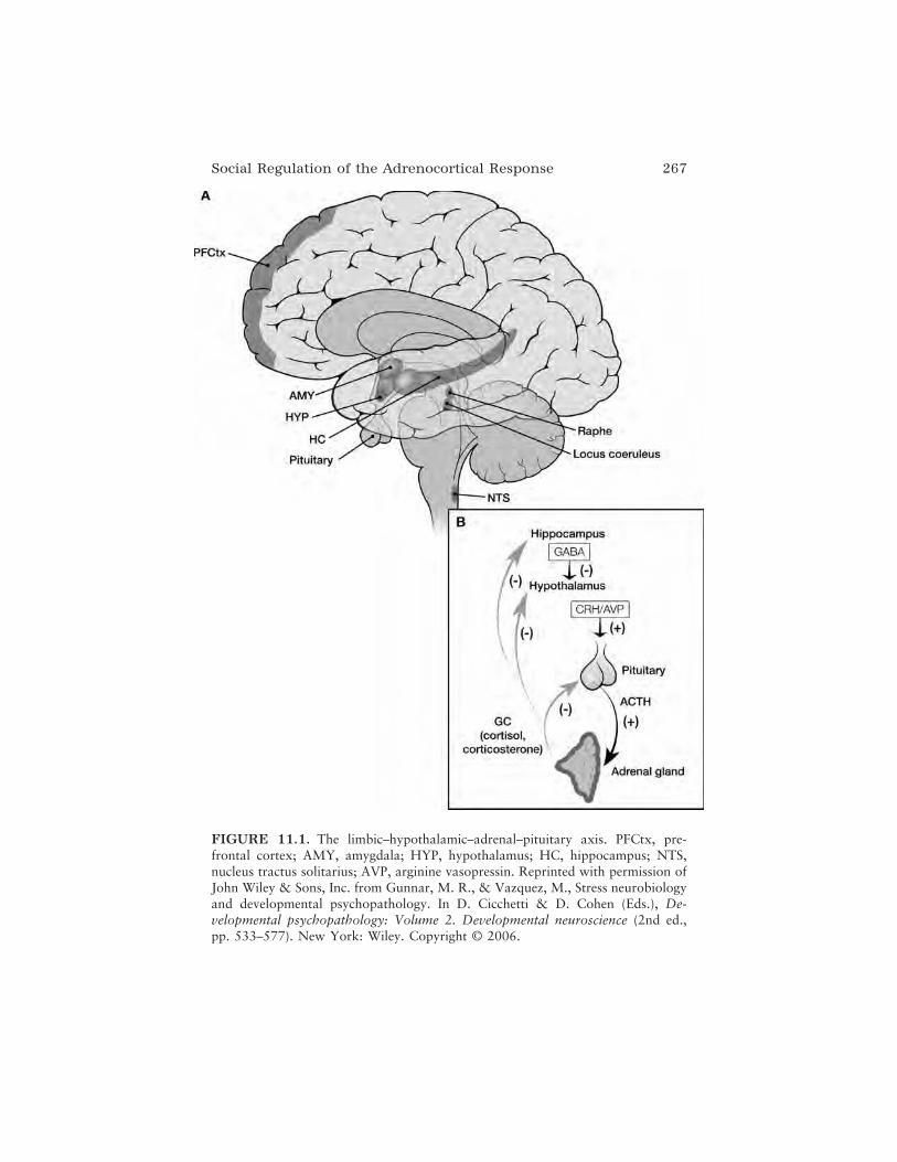
Social Regulation of the Adrenocortical Response 267
FIGURE 11.1. The limbic–hypothalamic–adrenal–pituitary axis. PFCtx, pre-frontal cortex; AMY, amygdala; HYP, hypothalamus; HC, hippocampus; NTS,nucleus tractus solitarius; AVP, arginine vasopressin. Reprinted with permission ofJohn Wiley & Sons, Inc. from Gunnar, M. R., & Vazquez, M., Stress neurobiologyand developmental psychopathology. In D. Cicchetti & D. Cohen (Eds.), De-velopmental psychopathology: Volume 2. Developmental neuroscience (2nd ed.,pp. 533–577). New York: Wiley. Copyright © 2006.
corticotropin-releasing hormone (CRH) and vasopressin (VP) from theparaventricular nuclei (PVN) in the hypothalamus. CRH and VP travelthrough small blood vesicles to the anterior pituitary (AP), where they stim-ulate the production and release of adrenocorticotropic hormone (ACTH).Of the two releasing hormones, CRH is the more potent, but VP appears toenhance the capacity of CRH to affect increases in ACTH production andrelease. Released into general circulation, ACTH interacts with receptorson the cortex of the adrenal glands to stimulate the production and releaseof CORT.
Multiple pathways are involved in the activation and inhibition ofthe HPA axis through regulating gamma-aminobutyric acid (GABA)-inhi-bition of CRH- and VP-producing cells in the PVN (for review, seeHerman & Cullinan, 1997). With regard to activation, physical stressorsthat pose immediate threats to health and viability (e.g., cold stress, heatstress, blood loss) activate the HPA axis largely through norepineph-rine (NE) neurons ascending from the brainstem. Psychological stressors(sometimes called processive stressors) activate the HPA axis throughcorticolimbic pathways, including pathways from the central nucleus ofthe amygdala (CEA). The HPA axis is under negative feedback control,meaning that increases in CORT result in inhibition of PVN, CRH, andVP production. This inhibition is effected by CORT-responsive cells inthe hypothalamus, hippocampus, and, it now appears, medial regions ofthe prefrontal cortex (Herman & Cullinan, 1997; Sullivan & Gratton,1999). None of these pathways is direct (i.e., they are multi-synaptic);thus, HPA regulation is multifactorial.
CORT acts by interacting with two classes of receptors: mineralo-corticoid and glucocorticoid receptors (MRs and GRs, respectively; notethat both receptors are responsive to CORT in the brain; de Kloet,Vreugdenhil, Oitzl, & Joels, 1998). MRs are high- and GRs are low-affinity CORT receptors. This means that when CORT is at low levels,MRs are activated, whereas GRs are only activated when CORT levels arehigh, as at the peak of the circadian cycle or during stress. MRs tend tomediate what have been termed “permissive” effects of CORT, whichinclude maintaining the responsiveness of neurons to their neurotrans-mitters, maintaining the HPA circadian rhythm, and maintaining bloodpressure. GRs mediate most of the effects of stress elevations in CORT.Presumably related to MR and GR activation, both chronically low andchronically high levels of CORT are associated with nonoptimal function-ing. In contrast, moderate basal levels, a clear circadian rhythm in CORT,and rapid increases and then rapid reestablishment of basal levels inresponse to stressors tend to be associated with optimal physical andbehavioral health.
268 Adam, Klimes-Dougan, and Gunnar
MEASUREMENT, ANALYSIS, AND INTERPRETATIONOF CORT ACTIVITY IN HUMAN POPULATIONS
Assessment of the HPA axis in humans has been accomplished in many ways,but it is important to distinguish between measures obtained under non-stimulated or basal conditions and measures obtained in response to specificstressors. In most human HPA-axis research, CORT levels are assessednoninvasively in small samples of saliva (Kirschbaum & Hellhammer, 1989).
Basal Activity
Researchers examining basal CORT activity are interested in identifyingthe typical, unstimulated level of CORT for individuals at a particular timeof day or their typical pattern of CORT across the whole day. Although ithas been common to use pretest measures of CORT in the laboratory as abasal or baseline measure (e.g., Vanyukov et al., 1993), recent evi-dence suggests that pretest measures are often significantly different frommeasures obtained in familiar (e.g., home) settings (Larson, Gunnar, &Hertsgaard, 1991; van Eck, Nicolson, Berkhof, & Sulon, 1996; Walker,Walder, & Reynolds, 2001). Increasing numbers of researchers are nowtaking advantage of the ease of having subjects collect home samples toexamine basal CORT activity. Individual differences in CORT levels atparticular times of the day, the pattern of CORT production across thewaking day or across a 24-hour period (e.g., amplitude or slope of the diur-nal rhythm), and the amount of within-person variability noted across mul-tiple days are typical methods of assessing natural variation in basalcortisol activity. In some protocols, CORT is assessed at the same clocktime across all subjects in an effort to control for the strong diurnal vari-ability inherent in this neuroendocrine system. In other protocols, CORT isassessed at fixed periods following awakening in an effort to track activityof the HPA system in relation to the subject’s own day–night behavior.Cortisol levels in humans are typically high in the morning upon waking,increase 50–60% in the first 30–45 minutes post-wakening (known as thecortisol awakening response), drop rapidly over the first few hours afterwaking, and then more slowly across the day to reach a low point aroundmidnight (Kirschbaum & Hellhammer, 1989; Pruessner et al., 1997). Ininfants and young children, this pattern is not quite as clear due to contin-ued maturation of sleep-wake processes and the influence of daytime naps(Gunnar & Donzella, 2002). There is some evidence that CORT levelsobtained early in the morning or in response to awakening reflect relativelystable traits of the HPA axis with high heritability quotients (e.g., Bartels,de Geus, Kirschbaum, Sluyter, & Boomsma, 2003), although the awaken-
Social Regulation of the Adrenocortical Response 269
ing response is also influenced by recent stressful experiences, includ-ing experiences occurring the prior day (Adam, Hawkley, Cacioppo, &Kudielka, 2006). Conversely, there is some evidence that levels assessed inthe afternoon and evening are more influenced by state contributions to thesystem as it reacts to the cumulative challenges of the day. Nevertheless, thedemarcation between state and trait contributions to CORT levels acrossthe day is not distinct (e.g., Shirtcliff, Zahn-Waxler, & Klimes-Dougan,2005). Other factors such as sleep, food, exercise, medication use, andmenstrual timing also influence CORT levels and need to be measured andcontrolled in analyses (Kirschbaum & Hellhammer, 1989). Recent statisti-cal approaches (reviewed below) allow the isolation of variance associatedwith assay error, control factors, diurnal rhythms, and trait and state con-tributions to CORT activity. In general, researchers now recognize that sin-gle samples of CORT do not provide sufficient information to interpretCORT activity; multiple samples across the day and across multiple daysprovide the best characterization of basal activity, and measurement ofCORT responses to stressors in addition to basal CORT provides a morecomplete picture of CORT functioning.
Cortisol Reactivity to Stressors
Assessment of the HPA axis may also involve examining reactivity to phar-macological probes and/or environmental stressors; however, pharmaco-logical probes are rarely used in studies of children. Developmentalresearchers tend to rely on laboratory stressor tasks or naturally occurringstressors such as the first day of school. Regardless, the optimal samplingprotocol involves some home basal assessments, measures prior to thestressor, and multiple measures (often at 20- to 30-minute intervals) afterthe onset of the stressor to examine both reactivity and recovery. The clas-sic CORT stress response profile consists of moderate CORT levels at pre-test, high CORT levels in response to a stressor, and a subsequent return tonear-pretest levels within 40–60 minutes after stressor termination. Becauseelevations in CORT may represent normative stress responses, the chal-lenges of defining disruptions in the stress reactivity cycle are consider-able. Typically, researchers consider individual differences in elevations ofCORT in anticipation of a stressor (anticipatory stress reaction), the degreeof elevation in response to a stressor (reactivity), and the degree of delay inreturning to pretest levels following a stressor (recovery). Exaggerated lev-els of anticipatory stress or stress reactivity, a longer delay in returning tobaseline, and a failure to show an HPA reaction in response to knownstressors have all been considered disruptions of the expected HPA-axisresponse to stress. Recently, researchers have begun to note a potentially
270 Adam, Klimes-Dougan, and Gunnar
dysregulated pattern termed declivity in which rather than elevating,CORT decreases below not only pretest levels (which might be elevated inanticipation of testing) but also below home basal levels at the same time ofday (Shirtcliff, Zahn-Waxler, & Klimes-Dougan, 2005; Van Goozen et al.,1998). At this point there is too little study of declivity to know whether itis associated with disordered functioning or is a normative pattern ofresponse to laboratory stressors of a particular type.
Advanced Statistical Models
Advanced statistical approaches have recently been developed to help iso-late and model separate aspects of HPA-axis activity, including trait varia-tion in basal CORT levels and diurnal CORT patterning and day-to-dayand moment-to-moment state variations in CORT in large samples. Theseapproaches include latent state–trait modeling, which uses a latent variableapproach to isolate and predict variance in CORT that is stable within indi-viduals across time and is thus presumed to represent “trait” variance inCORT (Kirschbaum et al., 1990; Shirtcliff, Granger, Booth, & Johnson,2005). Another approach, multilevel growth curve modeling, allows re-searchers to derive a latent estimate of each individual’s basal pattern ofCORT production across the day (presumably, trait cortisol variation), andto predict individual differences in these basal CORT patterns from traitvariables of interest (Hruschka, Kohrt, & Worthman, 2005). It also allowsresearchers to model the effects of time-varying (state) factors such asmood and activity level on within-person changes in CORT (Adam, 2006;Adam & Gunnar, 2001; van Eck, Berkhof, Nicolson, & Sulon, 1996a), andconversely, to examine the effects of within-person changes in CORT onchanges in subjective experience (Adam, Hawkley, et al., 2006). Usingthese approaches, associations between CORT and other parameters ofinterest, such as psychopathology, are much more robust than when traitand state variance in CORT are not isolated as effectively.
Now that the basic physiology, typical activity, and primary methodsof examining HPA-axis activity have been described, research on theimpact of social factors on HPA-axis functioning during development isreviewed.
SOCIAL REGULATION OF THE HPA AXISIN INFANCY AND EARLY CHILDHOOD
Studies in rats and monkeys indicate that the HPA axis is under powerfulsocial regulation early in development (for review, see Sanchez, Ladd, &
Social Regulation of the Adrenocortical Response 271
Plotsky, 2001). In rats, shortly after birth the axis enters a period describedas the “stress hyporesponsive period” or SHRP. This period lasts untilabout postnatal day 14. During this time it is difficult to elevate CORT inresponse to many stressors, and it has been suggested that this period servesto protect the developing brain from adverse impacts of chronic CORT ele-vations. Removing the mother for 12–24 hours produces elevated CORTlevels, but mimicking her stimulation of the pups (licking) by stroking themwith a wet paintbrush and providing milk through a canula will keepCORT levels at baseline during maternal separation (Suchecki, Rosenfeld,& Levine, 1993). Thus it is social stimulation that maintains the axis in anonstress state during development in the rat. Importantly, maternal lick-ing also seems to regulate methlyation of the GR gene in the brain, suchthat pups who receive high levels of maternal licking have more GR recep-tors for CORT, as compared to those who receive low levels of licking andgrooming (Weaver et al., 2004). More CORT receptors confer better nega-tive feedback control of the HPA axis and less stress vulnerability.
Rats are less mature than primates at birth; therefore, it is not clearwhether the mechanisms that mediate early experience effects in monkeysand humans are similar to those that operate in rats. What is clear, how-ever, is that not long after birth in monkey infants, maternal availabilityand responsiveness serve as powerful CORT buffers. Capturing motherand infant from their social group produces behavioral expressions of dis-tress from both parties, but if the infant can maintain contact with themother, it exhibits only small increases in CORT (Levine & Wiener, 1988).Separation of mother and infant results in large increases in CORT andfear-related behaviors that may be regulated, at least in part, by CRHproduction in the amygdala and other extrahypothalamic areas (Kalin,Shelton, & Barksdale, 1989). Thus, in some ways similar to rats, nonhu-man primate infants maintain basal CORT levels throughout most of earlydevelopment through contact and proximity to attachment figures. The pri-mate work, however, makes it clear that in some species alloparents canserve similar functions. That is, if the infant is separated from the motherbut is then “mothered” by another female during separation, this interven-tion attenuates elevations in CORT and reduces behavioral distress (Levine& Wiener, 1988).
The human HPA axis also comes under strong social regulation orbuffering during early development. In the earliest months of life, however,being held and soothed by the mother or other adults does little to reduceCORT responses to stressors such as being physically manipulated (e.g.,during a doctor’s exam) or experiencing a mildly painful stressor such as aninoculation, even though these caregiving actions do reduce crying (Gun-
272 Adam, Klimes-Dougan, and Gunnar
nar, Marvinney, Isensee, & Fisch, 1988). As these studies demonstrate,although crying and CORT elevations are sometimes correlated in infancy,actions that reduce crying do not necessarily reduce the CORT response,and vice versa. Nonetheless, over the course of the first year it becomesincreasingly difficult to elevate CORT to stressors such as brief separations(of a few minutes), wariness-eliciting stimuli, and inoculations (for review,see Gunnar & Donzella, 2002). The uncoupling of CORT responses andbehavioral distress, which is often focused on eliciting contact with attach-ment figures, is particularly marked under acute threat conditions amongtoddlers who are in secure attachment relationships with the person who isavailable for immediate succor (Spangler & Schieche, 1998). Indeed, onlyin secure attachment relationships does the caregiver appear to be able tocompletely buffer CORT increases in distressed toddlers, as observed inlaboratory (Nachmias, Gunnar, Mangelsdorf, Parritz, & Buss, 1996) andnaturalistic contexts (Ahnert, Gunnar, Lamb, & Barthel, 2004). Insecurelyattached toddlers show CORT responses to stressful events even with theirattachment figure present, and toddlers who exhibit disorganized/disori-ented attachment patterns may be particularly vulnerable to elevations inthis hormone (Hertsgaard, Gunnar, Erickson, & Nachmias, 1995). Thus,over the course of the first year of life, the HPA axis comes under strongsocial regulation in human children, and the social buffer is more potent ifthe child is securely attached to his or her caregiver(s).
As in nonhuman primates, alloparents do appear to be capable of buff-ering CORT elevations in young children. This finding has been noted instudies of babysitters and child care providers (for review, see Gunnar &Donzella, 2002). Babysitters were instructed to provide sensitive, respon-sive care or more distant, neglectful care for brief (i.e., 30-minute) separa-tions. Sensitive and responsive care by a babysitter, even if she were astranger to the child, buffered or prevented elevations in CORT at least forbrief periods. When young children are in group care settings, the quality ofcare provided by the alloparent appears to determine whether CORT levelsrise or fall over the day. Typically, for young children, CORT levels aver-age about the same in the midafternoon and midmorning, in contrast to themature rhythm, which exhibits a decline over this period. The more matureadult rhythm emerges as children begin to outgrow their afternoon nap(Watamura, Donzella, Kertes, & Gunnar, 2004). Nonetheless, rising levelsof CORT over the day suggest loss of adequate regulation of the HPA axis.Daytime increases in CORT have been observed frequently when childrenare in group or congregate care; however, CORT increases less in high-quality than low-quality settings (reviewed in Gunnar & Donzella, 2002).The pattern of CORT response in these settings, which is sometimes lower
Social Regulation of the Adrenocortical Response 273
than home levels in the morning and then higher than home levels in theafternoon, suggests that these responses are not acute separation reactions(Gunnar & Donzella, 2002). Furthermore, CORT levels have been noted todecrease over nap periods at child care, even for children who simply lie ontheir cots and do not appear to fall asleep (Watamura, Sebanc, Donzella, &Gunnar, 2002). Such data suggest that whatever is happening while chil-dren are involved in activity at child care is responsible for stimulating theHPA axis. One reasonable possibility is peer interactions.
Over the toddler and preschool years, children become increasinglymotivated to play with other children; meanwhile, their social skills are justemerging, making peer play challenging (Hartup, 1979). Social competencetends to be negatively correlated with the rise in CORT over the group careday, which is consistent with evidence that age correlates negatively withCORT when preschoolers are at child care but not when they are at home(reviewed in Gunnar & Donzella, 2002). The importance of peer relationsis also demonstrated by evidence that as early as the preschool years, peerrejection is associated with chronic elevations in CORT when children arewith their peers (Gunnar et al., 2003). This finding may explain why exu-berant and undercontrolled as well as fearful and anxious children tend tobe susceptible to producing higher CORT levels and rising CORT patternsover the child care day (Dettling, Gunnar, & Donzella, 1999; Gunnar et al.,2003; Watamura, Donzella, Alwin, & Gunnar, 2003). Thus, whereas tem-perament has been shown to correlate with CORT activity in infancy andearly childhood, its associations may be both mediated by social relation-ships, as in exuberant, undercontrolled temperament influencing peer rejec-tion that is more proximally related to elevated CORT, or moderated bysocial relationships, as when responsive caregiving prevents activation ofthe HPA axis in fearful children.
Although parents can clearly serve as buffers on the effects of socialenvironments on young children’s HPA-axis activity through positive,calming parenting behavior, they can also serve as a profound source ofsocial strain if their behavior is threatening or fails to provide appropriatecomfort. Conflict between two parents may be an especially powerful formof threat, because it combines exposure to anger and even violence withreduced access to parents who may be too preoccupied with their ownproblems to offer attention or comfort to the child. In a group ofkindergarten-age children and their parents, Pendry and Adam (in press)found that young children living in homes characterized by higher levels ofinterparental conflict had significantly higher evening CORT levels andflatter diurnal cortisol slopes. Less effective parenting, defined by low levelsof warmth and involvement, was independently associated with a flatterdiurnal CORT rhythm.
274 Adam, Klimes-Dougan, and Gunnar
SOCIAL REGULATION OF THE HPA AXISIN MIDDLE CHILDHOOD AND ADOLESCENCE
Although less evidence is available on the social regulation of CORT in themiddle childhood and adolescent years, the evidence that does exist suggeststhat family and peer influences continue to play an important role. Beyondthe results for young children mentioned above, several studies have foundconflict in the family environment to be a powerful activator of CORT duringmiddle childhood and adolescence, whereas a positive, stable, supportivehome environment is associated with lower CORT levels (Flinn & England,1995; Pendry & Adam, in press; see also Repetti, Taylor, & Seeman, 2002).In a sample of Caribbean children and adolescents, Flinn and England (1995,1997) noted that high levels of family and peer conflict were among the mostpowerful activators of CORT. In terms of positive social influences, in theCaribbean study, the degree of support available to mothers in the home envi-ronment was also an important determinant of CORT levels in the children ofthis study. Children in single-mother homes without kin support and thoseliving with distant relatives or in stepparent homes with nonrelated siblingshad higher average CORT levels than those in biological parent homes andthose living with biological mothers who had additional kin support.
Very few studies have examined social factors influencing HPA-axisactivity specifically in adolescents—which is surprising, given the impor-tance of changing peer and family relationships, combined with the manyother changes in physical appearance, physiology, sexuality, and socialroles of this age period. The convergence of all these changes in a shortperiod of time is thought to contribute to the experience of high levels ofstress in adolescence. Recent evidence also implicates ongoing brain devel-opment as a factor in the increased emotionality and risk taking that char-acterize the adolescent years (Dahl, 2004). A number of studies now sug-gest that a small increase in basal CORT levels occurs across mid to lateadolescence, perhaps especially in adolescents girls (Elminger, Kuhnel, &Ranke, 2002; Jonetz-Mentzel & Wiedemann, 1993; Kenny, Gancayo,Heald, & Hung, 1966; Kenny, Preeyasombat, & Migeon, 1966; Kiess etal., 1995; Lupien, King, Meaney, & McEwen, 2001; Netherton, Goodyer,Tamplin, & Herbert, 2004 [in girls only]; Tornhage, 2002; Walker,Walder, & Reynolds, 2001). The origin and functional consequences ofincreasing basal CORT levels during adolescence are not yet known.
The combination of increased exposure to stressful circumstances andthe ability of adolescents to report reliably on their own social experiencesmakes adolescence an attractive period during which to study the impact ofsocial events on the HPA axis. Utilizing the experience sampling method(ESM), in which adolescents were signaled to complete diary reports as they
Social Regulation of the Adrenocortical Response 275
went about their daily lives (see Larson, 1989), in conjunction with repeatedsampling of salivary CORT, Adam (2006) found that adolescents’ HPA axeswere responsive to moment-to-moment changes in social and emotionalexperiences in their everyday environments. Controlling for the effects oftime of day, momentary within-person increases in negative emotion wereassociated with significant within-person increases in CORT levels. In addi-tion, being alone, rather than with other people, at the time of sampling wasalso associated with higher momentary CORT levels. Interestingly, beingalone appeared to elevate CORT only for younger and not older adolescents.
In another sample of late adolescents, individuals who had recentlyexperienced high levels of negative life events, those who were experiencingchronic strain in their interpersonal relationships (including family, peer,and romantic) were found to have flatter diurnal CORT curves (Adam,Doane, Mineka, Zinbarg, & Craske, 2006). Thus, as with infants andyoung children, important social relationships in the lives of children andadolescents, both within the immediate family environment and outside thehome, are associated with individual differences in CORT activity.
HPA-AXIS ACTIVITY IN CHILDRENEXPOSED TO ABNORMAL SOCIAL ENVIRONMENTS
Situations in which extreme deviations from the expected social environ-ment occur provide unfortunate illustrations of the importance of the envi-ronment for HPA-axis functioning. Studies of children reared in institu-tions or orphanages have provided important insights. Notably, infants andtoddlers living in such institutions appear to lack the normal diurnalrhythm in CORT production (for review, see Gunnar, 2000). In particular,CORT levels are low early in the morning and fail to decline in the late eve-ning. This atypical pattern has also been observed in rhesus infants raisedon cloth surrogates (Boyce, Champoux, Gunnar, & Suomi, 1995). Lack ofa consistent and supportive caregiver may be the factor that disrupts thenormal diurnal CORT rhythm; indeed, similar disturbances have beennoted in many young children placed in foster care (Fisher, personal com-munication). In contrast, permanently placing children in supportive adop-tive families appears to allow the normal diurnal CORT rhythm to be rees-tablished (Gunnar, 2000). Several studies, however, suggest that not allchildren adopted from severely depriving conditions will reestablish com-pletely normal patterns of HPA-axis regulation. Several studies now showelevated basal CORT levels in postinstitutionalized children many yearsafter adoption (e.g., Gunnar, Morison, Chisholm, & Schuder, 2001). How-ever, there is some evidence that long-term elevations in basal CORT levels
276 Adam, Klimes-Dougan, and Gunnar
may be restricted to children who experienced the most adverse early care,resulting in severe growth failure (Kertes, Gunnar, Madsen, & Long, inpress).
In addition to children reared in institutions, researchers have alsoexamined physically and sexually abused children who have experiencedcatastrophic failure of their caregiving system. There is some evidence ofelevations in basal CORT among these children, particularly if they exhibitchronic posttraumatic stress disorder (PTSD) symptoms (Carrion et al.,2002; De Bellis et al., 1999). However, not all studies have reported alteredHPA-axis activity, raising critical questions about the nature, duration, andseverity of abuse necessary to produce long-term alterations (Cicchetti &Rogosch, 2001). Furthermore, the finding that PTSD pursuant to earlyabuse is associated with elevated basal CORT levels in childhood stands incontrast to the suppressed CORT levels reported for adults with PTSD.Some have suggested that the duration of PTSD symptoms may account forthis discrepancy, with elevated CORT levels characterizing PTSD early inthe disorder and suppressed levels emerging over time as the HPA sys-tem “down-regulates” or adjusts to chronic CORT elevations (Yehuda,Halligan, & Grossman, 2001). Taken together, the data on childrenexposed to severe deprivation early in life and those exposed to severeabuse suggest that such conditions increase the risk for long-term alter-ations in HPA-axis functioning. However, the pattern of effects observedalso raises important questions about developmental changes in stress reac-tivity and regulation and individual difference factors that may determinewhether and how long-term impacts are expressed.
Data presented thus far offer strong evidence for the influence of cur-rent social relationships on current HPA-axis activity and present theintriguing possibility that variations in social experience early in life mayhave organizational effects on HPA-axis activity in humans, supplementingthe already convincing body of evidence on the long-term effects of earlyexperience on the HPA axis in rodents and nonhuman primates. A criticalquestion, however, is whether activity of the HPA axis mediates the impactof social environments on the development of psychopathology.
SOCIAL ENVIRONMENTS, HPA-AXIS FUNCTIONING,AND INTERNALIZING PSYCHOPATHOLOGY
Theoretical Models
Disruptions in the social environments of children are not only implicit totheories of HPA-axis functioning in abusive and neglectful family environ-ments; they are also central to some of the core etiological theories of
Social Regulation of the Adrenocortical Response 277
depression. Chronic exposure to stressful life events has long been consid-ered an important risk factor for the development of depression (Tafet &Bernardini, 2003). Additionally, HPA-axis dysregulation among adultswith major depressive disorder (MDD) is one of the most consistent find-ings in biological psychiatry (Chrousos & Gold, 1992; Pariante, 2003).Models pertaining to childhood depression have been based largely on adownward extension of research with clinically depressed samples ofadults. A more developmental approach, and the approach taken in thecurrent chapter, is to subsume anxiety and depressive symptomatology anddisorders under the more inclusive class of internalizing problems (seeZahn-Waxler, Klimes-Dougan, & Slattery, 2000). This strategy is justified,in part, by (1) evidence that rates of comorbid anxiety and depression arehigh, (2) the fact that they have core negative affect features in common,and (3) the fact that anxiety disorders often precede the development ofdepression. It is even possible that in childhood, disruptions in the HPAaxis are more closely linked to anxiety than depression (e.g., Feder et al.,2004; Granger, Weisz, & Kauneckis, 1996). Indeed, there are many paral-lels in the etiological models of anxiety and depression, and theories ofstress and HPA-axis reactivity may be central to understanding the devel-opment of both.
Developmental models of depression and internalizing symptomatolo-gy focus on the effects of early experience on HPA-axis activity and theimplications of normative variations in anxiety and fear reactions (Gunnar,1992; Gunnar & Vazquez, 2006). The long history of early experience–stress studies in animals provides the basis for attempts to understand howvariations in care early in life may impact individual differences in stressresilience and vulnerability, leading to psychiatric disorders (see Gunnar &Vazquez, 2006, for a recent review). Findings suggest that early experiencesrelated to the quality of maternal care cause changes to extrahypothalamic,corticolimbic circuits that influence activity of the HPA axis. Briefly, thisetiological model suggests that hippocampal alterations produced by pro-longed stress and elevated CORT cause an impairment of the negative feed-back loop and could account for alterations in basal CORT and stress reac-tivity in patients with MDD.
A second developmental approach focuses on individual differences.Pathological anxiety in animals and humans may reflect inhibited tempera-ment, and an exaggeration of normal anticipatory HPA responses (Rosen& Schulkin, 1998) may be most characteristic of this group. In the presenceof elevated GCs and CRH, a cascade of biomolecular events that includesincreased expression of immediate-early genes (Makino, Gold, & Schulkin,1994) increases the sensitivity of central fear circuits, heightens anxiety todistal danger cues, and supports the transition from normal to pathological
278 Adam, Klimes-Dougan, and Gunnar
anxiety. Thus, early social experiences, either on their own or in interactionwith temperamental vulnerability, are thought to contribute to alterationsin HPA-axis functioning associated with depression. What evidence existsfor altered HPA-axis activity in children and adolescents with internalizingproblems and for the contribution of social environments to the emergenceof these differences?
Basal/Diurnal Functioning
In contrast with adults, children with internalizing problems are less likelyto exhibit distortions in basal CORT or the HPA rhythm. Here the conclu-sions are based primarily on reviews by others (e.g., Brooks-Gunn, Auth,Petersen, & Compas, 2001; Dahl & Ryan, 1996; Goodyer, Park, & Her-bert, 2001; Ryan, 1998), although more recent findings are duly noted. Inclinical samples, children and adolescents with clinical levels of anxiety anddepression do not consistently show alterations in their basal CORT. Dis-ruptions are more likely to be noted for diurnal patterns, particularly withregard to sleep onset (Carrion et al., 2002; DeBellis et al., 1999; Feder etal., 2004; Klimes-Dougan et al., 2001; for recent exceptions, see Luby etal., 2003; Martel et al., 1999; Ronsaville et al., 2006). In addition, certaintypes of depression, including melancholic depression and depression withsuicidal symptoms, are more likely to be linked to elevated basal cortisollevels (e.g., Dahl et al., 1991; Klimes-Dougan, Shirtcliff, & Zahn-Waxler,2006; Luby et al., 2004).
Some researchers have considered within-person variability—a sign oferratic basal output—to be an important indicator of HPA-axis dysregula-tion and have examined this dysregulation in relation to the presence ofMDD (Peeters, Nicholson, & Berkhof, 2004; Yehuda, Teicher, Trestman,& Siever, 1996). In one study that examined adolescents, more highly vari-able early-morning basal CORT levels (in particular, a spiking of these lev-els) across days predicted which adolescents would experience their firstbout of clinical depression over the next months (Goodyer, Herbert,Tamplin, & Altham, 2000).
Several other studies suggest that premorbid differences in the axis pre-cede and predict the development of depression in high-risk samples. Essexand colleagues (Essex, Klein, Cho, & Kalin, 2002; Smider et al., 2002)obtained salivary CORT samples in the afternoon when children were 4.5years of age, and these measures predicted internalizing problems in them ayear later, as kindergarteners, and both internalizing and externalizingproblem behavior in first grade. Similar findings have been reported instudies of young adolescents (Granger, Weisz, & McCracken, 1996;Susman, Dorn, & Chrousos, 1991; Susman, Dorn, Inoff-Germain, Nottel-
Social Regulation of the Adrenocortical Response 279
man, & Chrousos, 1997). Higher CORT levels and higher CORT todehydroepiandrosterone (DHEA; an adrenal androgen with effects thatoften counter those of CORT) ratios may also predict longer illness dura-tion (Goodyer, Herbert, & Altham, 1998).
To what extent do early social experiences contribute to elevatedcortisol levels and variability, and hence to risk of developing internaliz-ing symptoms? In the Essex and colleagues (2002) study, the eleva-tions in cortisol at age 4.5 that were found to predict internalizing andexternalizing problems were, in turn, predicted by levels of maternal stress.Notably, however, concurrent maternal stress predicted high child CORTlevels only if the currently stressed mother had also been highly stressedduring the child’s infancy. Furthermore, most of the association withmaternal stress was carried by the effects of maternal depression symptoms.Maternal depression during the child’s early years may seriously compro-mise the mother’s capacity to provide sensitive, responsive, and supportivecare to her child. However, in the Essex and colleagues study, it is not clearwhether children with higher CORT levels had mothers who had been con-sistently depressed over the child’s whole life. Work by Halligan, Herbert,Goodyer, and Murray (2004) suggests the importance of maternal depres-sion in the child’s early years, in particular. They found that adolescentchildren of mothers who were depressed postnatally had higher morningCORT levels and more variability in these levels, even after controlling forcurrent life events and mothers’ current levels of depression.
Stress Reactivity
Like the research on basal functioning, research on stress reactivity suggeststhat MDD in children and adolescents is less associated with endogenousdysregulation of the HPA axis than it is in adults. Nonsuppression ofCORT to dexamethasone, a common finding in adults, is rare in childrenand adolescents with MDD, and ACTH and CORT responses to CRH arealso typically normal, at least in children who have not also been physicallyabused (see Ryan, 1998). In contrast, children with clinical anxiety showelevations in CORT in response to some types of stressors, including a fear-potentiated startle paradigm in which a warning light is sometimes fol-lowed by an unpleasant blast of air (60 psi) to the throat (Ashman,Dawson, Panagiotides, Yamada, & Wilkinson, 2002).
Using experimental paradigms that involve some type of social stressorhas also yielded mixed findings with regard to anticipation of, and reactionto, stress. Granger and his colleagues (e.g., Granger, Stansbury, & Henker,1994; Granger, Weisz, & Kauneckis, 1994) used a conflict paradigm toassess stress reactivity with a clinic-referred sample of children and adoles-
280 Adam, Klimes-Dougan, and Gunnar
cents. They found that increased CORT reactivity was concurrently associ-ated with higher levels of a number of internalizing symptoms, includingsocial withdrawal, social anxiety, task inhibition, and low levels of per-ceived contingency. Using a similar conflict paradigm, Klimes-Dougan andcolleagues (2001) found that moderate levels of CORT reactivity wereassociated with fewer internalizing and externalizing symptoms. A com-monly used paradigm is the Trier Social Stress Test (TSST; Kirschbaum,Pirke, & Hellhammer, 1993) in which the participant gives a speech, thensolves mathematics problems. Various adaptations of this procedure havealso been used with children and adolescents. Although elevations inCORT are typically documented using this procedure, most studies havefailed to find group differences in stress reactivity between children withinternalizing disorders and comparison groups (e.g., Dorn et al., 2003;Klimes-Dougan et al., 2001; Martel et al., 1999). However, when parame-ters of stress reactivity and stress recovery were considered separately, thegreatest increase in CORT reactivity for girls was associated with the high-est levels of internalizing problems (Klimes-Dougan et al., 2001).
In addition to activation, understanding how children at risk for inter-nalizing problems recover from a stressor is critical. In a few studiesrepeated CORT samples after a stressor have been considered (e.g., Klimes-Dougan et al., 2001). Another aspect of recovery—failure of CORTresponses to habituate to familiar stressors—may be associated with inter-nalizing symptomatology (Gunnar & Vazquez, 2006). Increasing CORTlevels to novel stressors may not be unique to shy, inhibited, or anxiouschildren and indeed may initially be associated with assertion and domi-nance, because more assertive children are often the first to approach chal-lenging and uncertain circumstances. However, shy, inhibited, or anxiouschildren may be less likely to habituate CORT responses as the situationbecomes more familiar, and they may be less able to turn off the CORTresponse once they have left the novel situation. Several researchers havenoted higher CORT levels among more anxious, introverted children, butonly when the peer group setting was familiar (e.g., Bruce, Davis, & Gun-nar, 2002; Davis, Donzella, Krueger, & Gunnar, 1999; Granger, Stans-bury, & Henker, 1994; Legendre & Trudel, 1996). Failure to habituate,and the more frequent or prolonged elevations in CORT that go along withlack of habituation to stressors, may in turn increase risk for developinganxiety and depression (e.g., Pruessner et al., 1997; van Eck, Berkhof, et al,1996a; van Eck, Nicolson, et al., 1996).
In addition to its cross-sectional association with symptoms of anxiety,CORT reactivity has been shown to predict subsequent internalizing symp-toms. In one study, clinic-referred children participated in a conflict para-digm and were assessed for internalizing problems at the time of the con-
Social Regulation of the Adrenocortical Response 281
flict paradigm and 6 months later (Granger, Weisz, & McCracken, 1996).Those who exhibited high CORT reactivity at the time of the first assess-ment exhibited more internalizing symptoms at the second time point.Also, increases in internalizing problems were associated with higherCORT reactivity at the follow-up.
If disruptions in the HPA-axis system are related to internalizing symp-tomatology, attempts to normalize the HPA axis may provide us with criti-cal information for diagnostic, preventive, and therapeutic purposes (Tafet& Bernardini, 2003). There is some preliminary evidence with adult sam-ples to suggest that HPA-axis activity may be modified indirectly throughpsychotherapeutic processes, including cognitive-behavioral stress man-agement (e.g., Gaab et al., 2003) and social support (e.g., Heinrichs,Baumgartner, Kirschbaum, & Ehlert, 2003; Kirschbaum, Klauer, Filipp, &Hellhammer, 1995). Given the importance of supportive peer affiliation,particularly in adolescence, more research is needed that examines theeffects of support beyond the caregiving environment.
Thus, although evidence suggests that negative aspects of social cir-cumstances may potentially place individuals at greater risk for the devel-opment of internalizing disorders through increased basal levels, variabil-ity, or CORT reactivity, positive social relationships may also offer thepotential to protect against HPA dysregulation and the development ofinternalizing disorders.
HPA-AXIS FUNCTIONING ANDEXTERNALIZING PSYCHOPATHOLOGY
Etiological models regarding HPA-axis functioning for externalizing prob-lems center on core biological disruptions in the physiological response sys-tem. Raine (1996) suggests that systems implicated in HPA-axis func-tioning (e.g., central and autonomic nervous systems; CNS and ANS,respectively) are underaroused for some individuals (partially assumed tobe genetically determined), and these response patterns may predisposesome to criminality. Thus, the implication is that the HPA system is bluntedor hyporesponsive in children with externalizing problems or disruptivebehavior disorders (DBD). Consistent with this hypothesis, studies ofresponse to serotonergic challenges in children with DBD have shown thatthese children exhibit blunted CORT reactions to fenfluramine (Soloff,Lynch, & Moss, 2000) and sumatriptan (Snoek et al., 2002), both of whichtypically elevate cortisol, suggesting that serotonergic regulation of theHPA axis is compromised.
282 Adam, Klimes-Dougan, and Gunnar
There are various mechanisms that may account for low basal andreactivity CORT levels. Some possibilities include an unresponsive stress-reactivity system or a down-regulation of the axis following periods of ele-vated CORT. This latter hypothesis has been invoked by those who suggestthat a chaotic and threatening family life may produce a down-regulatedstress system in the children of antisocial parents, consistent with evi-dence of low basal cortisol in sons of substance-abusing fathers (Hardie,Moss, Vanyukov, Yao, & Kirillovac, 2002; Pajer, Gardner, Kirillova, &Vanyukov, 2001). Overregulation of the HPA axis may contribute to lowbasal CORT production. Alternatively, an increased threshold for stressreactivity may account for low levels of CORT reactivity. Finally, thoseexhibiting externalizing problems may be underreactive to threat stimuli,resulting in impaired avoidance learning and underarousal, which mayencourage sensation seeking as a means of increasing arousal.
Basal, Pretest, and Diurnal HPA-Axis Functioning
Although low basal CORT has been observed in some samples of childrenwith DBD (McBurnett, Lahey, Rathouz, & Loerber, 2000; van de Wiel,Van Goozen, Matthys, Snoek, & van Engeland, 2004; Van Goozen et al.,1998), this finding has not been consistent (Azar et al., 2004; Dabbs,Jurkovic, & Frady, 1991; Granger, Weisz, & Kauneckis, 1994; Scerbo &Kolko, 1994; Susman et al., 1999; Van Goozen, Matthys, Cohen-Kettenis,Buittelaar, & van Engeland, 2000). Unfortunately, these studies vary in thetime of day when CORT levels were examined as well as in the conditionsunder which samples were obtained. Most do not involve a procedure forsampling CORT under highly familiar conditions (e.g., at home), but rathertake samples when children come to the laboratory for testing, limiting ourability determine whether basal activity of the HPA axis is lower for chil-dren with disruptive behavior problems. Nevertheless, subgroup analyseswithin those studies to document basal—or, more accurately, baseline—differences in CORT for children with DBD in comparison to low-riskgroups may be instructive. Thus, van de Wiel and colleagues (2004) notedlower baseline CORT levels only for those children with DBD who exhib-ited the most significant problem behavior. Similarly, McBurnett and col-leagues (e.g., McBurnett, Pfiffner, Capasso, Lahey, & Loerber, 1997;McBurnett et al., 1991) found that boys with DBD who had the lowestCORT levels were described as the meanest by their peers, as the mostaggressive by adults, and were more overtly rather than covertly aggressive.These results seem to suggest that only the children with the most severeDBD may exhibit abnormally low basal or baseline CORT.
Social Regulation of the Adrenocortical Response 283
Given this possibility, it is surprising to find that in both risk and com-munity samples, low CORT has been found to correlate with scores ofexternalizing behavior (e.g., Pajer, Gardner, Kirillova, et al., 2001; Scerbo& Kolko, 1994; Vanyukov et al., 1993) and aggression (Flinn & England,1995; Oosterlaan, Geurts, Knol, & Sergeant, 2005; Spangler, 1995; Tennes& Kreye, 1985; Tennes, Kreye, Avitable, & Wells, 1986). Many of thesestudies, however, assessed CORT in the early morning when levels are typi-cally high, perhaps increasing the sensitivity to associations betweenexternalizing problems and lower basal HPA activity. For example, usinga very large community sample of children and adolescents, Shirtcliff,Granger, and colleagues (2005) applied latent state–trait modeling tech-niques to identify the trait component of early morning salivary CORTproduction. For boys, but not girls, they found a significant negative rela-tion between trait CORT shortly after awakening and externalizing prob-lems. In addition to these cross-sectional associations, several studies havedemonstrated that low basal CORT levels predict heightened disrup-tive behavior several months or years later (e.g., Granger et al., 1996;McBurnett, Lahey, et al., 2000; Shoal, Giancola, & Kirrilova, 2003). Thus,in all, the data are suggestive, but not definitive, regarding low basal CORTactivity and externalizing behaviors; more work is clearly needed to furtherinvestigate this relationship. This need is particularly salient because lowbasal CORT could contribute to low ANS and CNS reactivity, given thatone function of basal cortisol is to maintain the responsiveness of neuronsto their neurotransmitters (Sapolsky et al., 2000).
Stress Anticipation, Reactivity, and Recovery
Some researchers have suggested that disturbances in HPA activity inexternalizing disorders may be most evident under stress as compared tobaseline conditions (Snoek, Van Goozen, Matthys, Buitelaar, & Engeland,2004). In examining this hypothesis, it is necessary to distinguish betweenfailure to respond in anticipation of a threatening event, smaller elevationsin CORT in response to such events, and/or more rapid return to baselinefollowing termination of the event, because these distinctions could reflectdifferent causes of low CORT reactivity. In this regard, there is growingevidence of blunted anticipatory responding. Specifically, in a number ofstudies, children with DBD or those at risk for DBD tend to exhibit lowpretest CORT levels that do not change after exposure to a mild stressor,whereas nonrisk children tend to show higher pretest CORT levels thatdecrease over the period of stressor exposure (Hardie et al., 2002; King,1998; Moss, Vanyukov, & Martin, 1995; Moss, Vanyukov, Yao, &Kirillova, 1999; Pajer, Gardner, Rubin, Perel, & Neal, 2001). The chal-lenge in interpreting these findings is that, in most cases, the testing was
284 Adam, Klimes-Dougan, and Gunnar
conducted early in the morning; thus it is not clear whether children withDBD fail to exhibit an anticipatory response or simply have low earlymorning basal CORT levels. Clearly, these studies would have benefitedfrom assessment of basal levels on nontest days (e.g., Klimes-Dougan et al.,2001).
There is also some evidence that children with DBD show less of aCORT increase in response to mild stressors (e.g., Jansen et al., 1999; Mosset al., 1995; Snoek et al., 2002; Targum, Clarkson, Magac-Harris, Mar-shall, & Skwerer, 1990). The implications for these findings are not clear,however, because in most of the studies the nonrisk group failed to elevateCORT to the stressor or actually showed decreasing CORT reactions overthe stressor period (for an exception, see Snoek et al., 2002). To adequatelyexamine whether children with DBD show a blunted CORT stress re-sponse, the paradigm should elevate CORT for the nonrisk comparisonchildren. Van Goozen and colleagues (1998) employed such a paradigm; itinvolved a frustration/provocation task during which a peer (on audiotape)denigrated the target child’s performance. They reported that children withoppositional defiant disorder (ODD) and those with comorbid attention-deficit disorder (ADD) showed decreased CORT in response to the provo-cation task, whereas nonrisk children tended to show an increase (see alsoGatzke-Kopp & Beauchaine, Chapter 10, this volume). However, not allchildren with disruptive behavior problems exhibited blunted CORT reac-tivity; children with ODD comorbid for anxiety problems actually exhib-ited the highest CORT reactivity. This study suggests that blunted CORTreactivity may only be seen for children with DBD who are not comorbidfor anxiety disorders, an interpretation also offered by McBurnett and col-leagues (1991) and Snoek and colleagues (2004).
Finally, there is a paucity of research regarding CORT recovery fol-lowing a stressor for children with externalizing problems. Van Goozenand colleagues (2000) found that children with DBD and nonrisk childrentended to differ in CORT in the immediate poststress period, and childrenwith DBD continued to show low CORT during a recovery period. Becausethe children with DBD did not show an elevation in CORT in response tothe task, it is unlikely that this reflects a difference in negative feedback reg-ulation of the HPA axis, as in PTSD (Yehuda et al., 2001).
The evidence of lower basal CORT and blunted CORT reactivity tofrustration/provocation begs the question of whether these differences aremeaningful. One way to address this question is to consider whether thesereported effects have implications for responsivity to treatment. In a recentfollow-up study, van de Wiel and colleagues (2004) examined effectivenessof treatment for children with DBD as a function of CORT baseline andreactivity, using a 9-month treatment window and multifaceted (medica-tion, individual, and family therapy) intervention. Although pretest or
Social Regulation of the Adrenocortical Response 285
baseline differences were not predictive and CORT reactivity was similarwith regard to symptom severity at the pretest assessment, boys with moreelevated CORT levels following the frustration/provocation paradigm weresignificantly more responsive to therapeutic intervention than were thosewith low CORT stress responsivity. A related question is whether CORTreactivity also can be altered by therapeutic interventions in children at riskfor externalizing problems. A recent prevention study with low-income pre-schoolers who experienced the familial risk factors for conduct disorderaddressed this question. Using a randomized prevention trial, MillerBrotman, Kiely Gouley, Pine, and Rafferty (2002) increased CORT reac-tions during a peer entry/interaction task among the children in the inter-vention group. Because this intervention focused on social-cognitive pro-cesses, the pattern of results suggests that low CORT reactivity amongchildren at risk for DBD, at least, may not reflect dysregulation of the HPAaxis as much as disturbances in psychological processes operating viacorticolimbic circuits that activate the HPA axis. However, this conclusionis extremely tentative, given that it is based on only one study that was con-ducted on a risk group rather than children with DBD.
SUMMARY OF HPA ACTIVITYIN CHILD CLINICAL DISORDERS
This brief review of research on HPA-axis activity in children with clinicaldisorders and subclinical behavior problems yields evidence of associationsbetween internalizing and externalizing symptoms and basal CORT levelsin children and adolescents. It also lends supportive evidence, but not uni-versally so, for associations between internalizing disorders and CORThyperactivity, and externalizing disorders and CORT hypoactivity in re-sponse to stressors. The control provided in animal studies and the growingliterature on correlational studies with children and adolescents haveenabled us to draw speculative conclusions that social–environmental fac-tors play a role in regulating CORT levels and reactivity, and hence mayplay a role in the emergence and course of clinical symptoms and disordersassociated with dysregulated HPA-axis activity.
SOCIAL AND NONSOCIAL CONTRIBUTIONSTO HPA-AXIS FUNCTIONING: A MODEL
As illustrated by the above discussion of the literatures on externalizing andinternalizing disorders, associations between psychopathology and HPA-axis activity are not straightforward, and inconsistencies abound. One
286 Adam, Klimes-Dougan, and Gunnar
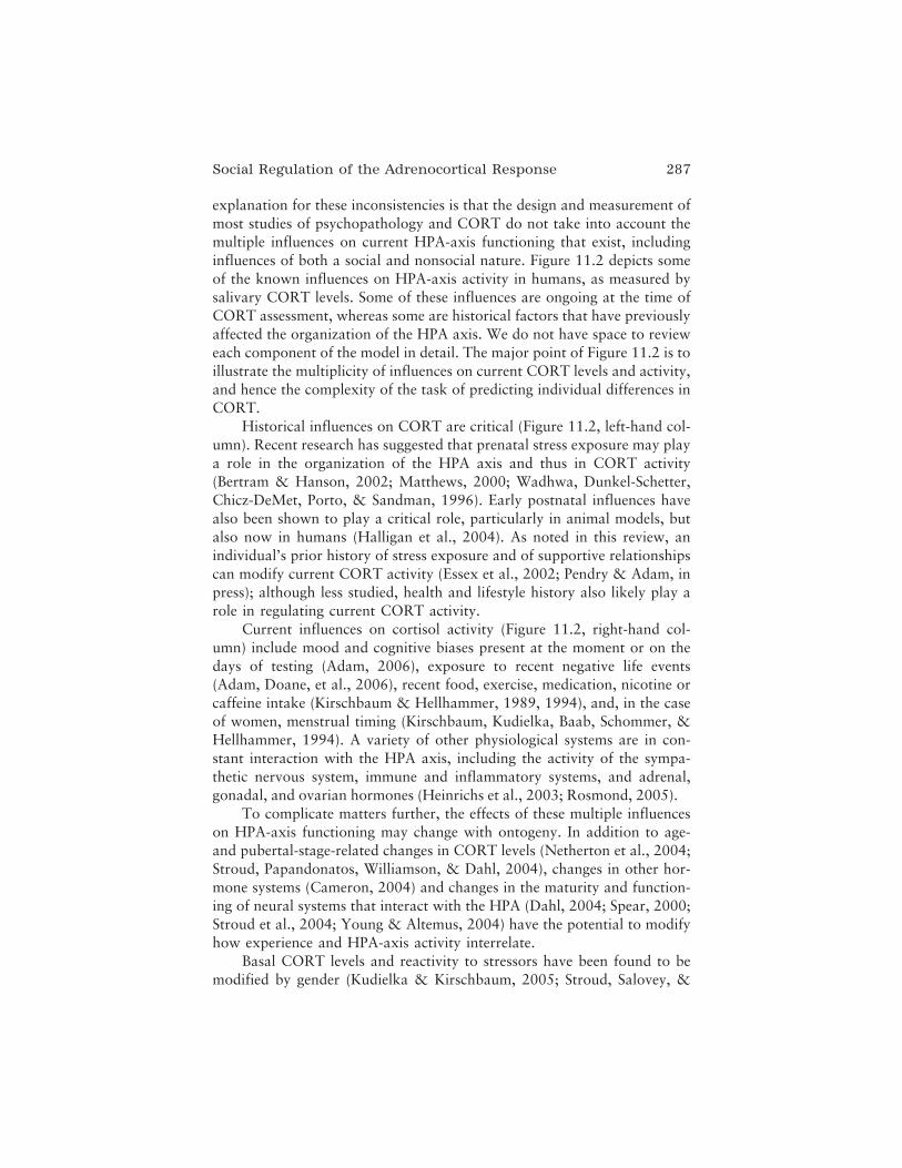
explanation for these inconsistencies is that the design and measurement ofmost studies of psychopathology and CORT do not take into account themultiple influences on current HPA-axis functioning that exist, includinginfluences of both a social and nonsocial nature. Figure 11.2 depicts someof the known influences on HPA-axis activity in humans, as measured bysalivary CORT levels. Some of these influences are ongoing at the time ofCORT assessment, whereas some are historical factors that have previouslyaffected the organization of the HPA axis. We do not have space to revieweach component of the model in detail. The major point of Figure 11.2 is toillustrate the multiplicity of influences on current CORT levels and activity,and hence the complexity of the task of predicting individual differences inCORT.
Historical influences on CORT are critical (Figure 11.2, left-hand col-umn). Recent research has suggested that prenatal stress exposure may playa role in the organization of the HPA axis and thus in CORT activity(Bertram & Hanson, 2002; Matthews, 2000; Wadhwa, Dunkel-Schetter,Chicz-DeMet, Porto, & Sandman, 1996). Early postnatal influences havealso been shown to play a critical role, particularly in animal models, butalso now in humans (Halligan et al., 2004). As noted in this review, anindividual’s prior history of stress exposure and of supportive relationshipscan modify current CORT activity (Essex et al., 2002; Pendry & Adam, inpress); although less studied, health and lifestyle history also likely play arole in regulating current CORT activity.
Current influences on cortisol activity (Figure 11.2, right-hand col-umn) include mood and cognitive biases present at the moment or on thedays of testing (Adam, 2006), exposure to recent negative life events(Adam, Doane, et al., 2006), recent food, exercise, medication, nicotine orcaffeine intake (Kirschbaum & Hellhammer, 1989, 1994), and, in the caseof women, menstrual timing (Kirschbaum, Kudielka, Baab, Schommer, &Hellhammer, 1994). A variety of other physiological systems are in con-stant interaction with the HPA axis, including the activity of the sympa-thetic nervous system, immune and inflammatory systems, and adrenal,gonadal, and ovarian hormones (Heinrichs et al., 2003; Rosmond, 2005).
To complicate matters further, the effects of these multiple influenceson HPA-axis functioning may change with ontogeny. In addition to age-and pubertal-stage-related changes in CORT levels (Netherton et al., 2004;Stroud, Papandonatos, Williamson, & Dahl, 2004), changes in other hor-mone systems (Cameron, 2004) and changes in the maturity and function-ing of neural systems that interact with the HPA (Dahl, 2004; Spear, 2000;Stroud et al., 2004; Young & Altemus, 2004) have the potential to modifyhow experience and HPA-axis activity interrelate.
Basal CORT levels and reactivity to stressors have been found to bemodified by gender (Kudielka & Kirschbaum, 2005; Stroud, Salovey, &
Social Regulation of the Adrenocortical Response 287
288
FIG
UR
E1
1.2
.H
isto
rica
lan
dcu
rren
tin
flue
nces
onH
PA-a
xis
func
tion
ing,
asin
dexe
dby
saliv
ary
cort
isol
leve
ls,
high
light
ing
the
role
ofso
cial
fact
ors.
Epel, 2002; Taylor et al., 2000) and by temperament or personality charac-teristics (Gunnar et al., 2003; Kirschbaum, Barussek, & Strasburger, 1992;Nachmias et al., 1996). Both behavioral genetic and molecular geneticstudies have noted moderate to strong genetic contributions to aspectsof basal CORT and also to CORT reactivity (Bartels, Van den Berg,Sluyter, Boomsma, & de Geus, 2003; Wüst, Federenko, Hellhammer, &Kirschbaum, 2000; Wüst, Federenko, et al., 2004a; Wüst, Van Rossum, etal., 2004b), providing evidence for a partial genetic basis for preexistingHPA-axis differences and differences in the reactivity and modifiability ofthe HPA with experience.
It is also important to note that the various influences identified in Figure11.2 may have differential degrees of impact on the various parameters ofCORT measurement, such as average levels, diurnal slopes, and the size of theCORT response to awakening or reactivity to laboratory stressors. For exam-ple, genetic predispositions may make a larger contribution to the size of theCORT response to awakening than to evening CORT levels (e.g., Wüst et al.,2000); social–environmental contributions may have a larger acute impacton evening CORT levels than they do on morning levels (e.g., Pendry &Adam, in press); and a history of trauma or childhood chronic stress may bemore likely reflected in a current flattening of the diurnal rhythm than in anoverall elevation of levels (Gunnar & Vazquez, 2006).
Rather than being discouraged by this complexity, our goal is to sug-gest that thorough measurement of each (or at least several) of these fac-tors, in relation to multiple parameters of CORT functioning, should leadto better characterization and understanding of CORT activity, whichshould, in turn, reveal stronger and more consistent associations betweenCORT, behavior, and pathology. At the very least, it should make us moreforgiving when only a small proportion of the variation in CORT levels isaccounted for in any one study.
Also apparent in Figure 11.2 are many places in which social variablesenter the model to influence both the developmental course and currentactivity of the HPA axis (see the darkly shaded boxes in the model). Forexample, social environments that influence the mother’s emotional well-being and the hormonal environment of the pregnancy may alter the orga-nization of the fetal HPA axis (Avishai-Eliner, Brunson, Sandman, &Baram, 2002; Wadhwa et al., 1996; Wellberg & Seckl, 2001). In a sophisti-cated set of rodent experiments (Weaver et al., 2004), early mother–pupinteractions were shown to produce organizational effects on the structureand functioning of the offspring hippocampus, through permanent changesin the methylation of a portion of the DNA responsible for glucocorticoidreceptor development. The long-term effects of early maternal depression(Halligan et al., 2004) and institutionalization (Gunnar et al., 2001), dis-
Social Regulation of the Adrenocortical Response 289
cussed previously, suggest the possibility of similar early social influenceson the organization of HPA-axis functioning in humans.
Given the continuing plasticity and growth of the brain, however, it islikely that social experiences may continue to have organizational influ-ences on the development of the HPA axis in early childhood and even intothe adolescent years. Examining the cumulative effects of lifelong historiesof social stress and support on HPA-axis activity, health, and development,using longitudinal data on social, behavioral, and physiological function-ing, is a critical direction for future investigation. This need is beginning tobe addressed by the incorporation of stress biomarkers in large-scale longi-tudinal studies in which social influences on CORT and other stressbiomarkers will be studied from the prenatal period onward.
Another rich direction for future research involves the investigation ofthe impact of macrosocial or macrosystemic factors, such as poverty, lackof opportunity, and discrimination, on stress physiology, with racial andsocioeconomic (SES) differences in stress exposure and stress system activa-tion hypothesized to account partially for group disparities in adult func-tioning and health (Cohen et al., 2006; Flinn & England, 1997; Lupien etal., 2001; Sapolsky, 2004). Indeed, beyond its clear effects on social–emotional functioning, the known impacts of CORT on cognition, mem-ory, immune system functioning, and physical health suggest possible path-ways by which stress may jeopardize one important pathway to prosperity:educational performance.
IMPLICATIONS OF SOCIAL INFLUENCESON HPA-AXIS ACTIVITY FOR EDUCATION
Research on the potential implications of individual differences in HPA-axis activity for educational functioning are only just beginning to beexamined, but these differences represent a ripe area for future investiga-tion. Existing research shows strong associations between experimentalmanipulations in CORT levels and cognition and memory processes inadults (Lupien, Gillin, & Hauger, 1999; Lupien & McEwen, 1997; Wolf,Schommer, Hellhammer, McEwen, & Kirschbaum, 2001). Associationsbetween CORT levels and cognitive and memory functioning are thoughtto follow a reverse U-shaped curve, with too low and/or too high levelsassociated with impaired performance (note the similarity to CORT associ-ations with emotional and physical health—in all cases, moderate andeffectively regulated levels of this hormone are considered advantageous).More recently, some work on CORT and memory in children has sug-gested that negative effects of elevated CORT on memory are also present
290 Adam, Klimes-Dougan, and Gunnar
in preschool-age children (Heffelfinger & Newcomer, 2001; Lupien &McEwen, 1997).
In addition to direct effects on cognitive functioning, individual differ-ences in HPA-axis functioning may have the potential to disrupt achieve-ment through several indirect pathways. CORT levels may influence boththe ability to fall asleep and the quality of sleep (Gillin, Jacobs, Fram, &Snyder, 1972). Decrements in number of hours of sleep have been shown tohave strong effects on educational performance in middle school children(Sadeh, Gruber, & Raviv, 2003). In adults, high levels of CORT duringsleep have been shown to impair declarative memory consolidation pro-cesses that typically occur during slow-wave sleep (Born & Wagner, 2004).
Another indirect pathway by which HPA-axis activity may influenceeducational performance is through its effects on physical and mentalhealth. Increased absenteeism associated with the presence of physicalhealth problems, and absenteeism and social problems associated with thepresence of depression and anxiety could contribute to, and interact with,poor cognitive, attentional, and memory processing to cause additionalimpairments in educational functioning. Problems in classroom functioningand educational performance may then feed back to cause additional psy-chological strain and CORT elevations; failure experiences have beenshown to be important activators of the HPA axis, particularly among indi-viduals with low self-esteem (Pruessner, Hellhammer, & Kirschbaum,1999).
Due to the relative lack of research in this area on children, much ofour discussion on the potential educational implications of increasedCORT has been based on extensions of experimental animal work andresearch with adult humans in laboratory settings. What do we knowabout how CORT levels relate to educational functioning in naturalisticacademic settings? Initial findings by Adam and Snell (2006) show thatCORT levels are typically lower when adolescents are productively en-gaged in academic activities than when they are engaged in nonacademictasks (controlling for the effects of time of day and of being alone onCORT levels). Whether low CORT levels facilitate attention, focus, andachievement, or whether positive engagement in academic activities lowersCORT levels remains unclear from these correlational data; experimentalresearch on CORT, attention, and performance in school-age children andadolescents is required to further explore these associations.
Another important implication for education emerges from Gunnarand Donzella’s (2001) findings that individual differences in the quality ofchild care settings are important in determining the extent to which chil-dren will display increased HPA-axis activation in these environments.Whether CORT differences, in turn, relate to the degree of intellectual and
Social Regulation of the Adrenocortical Response 291
social benefits children obtain from their day care settings remains to beexamined, and whether the quality of elementary school, middle school,and high school classroom environments also influence CORT levels isanother important direction for future research. Child attentional processesand readiness to learn upon entering the first grade are important determi-nants of later success in school (Alexander, Entwisle, & Dauber, 1993;Duncan et al., 2005; McClelland, Morrison, & Holmes, 2000). If elevatedadrenocortical activity due to low-quality child care or learning environ-ments does contribute to impairments in attention and social functioning inthe classroom, this research would provide another important reason whythe quality of these environments needs to be closely regulated, both interms of the intellectual stimulation they offer and the quality of the socialand emotional environment they provide.
In general, research on social influences on the HPA axis leads us to con-clude what should already be obvious: that providing children with support-ive social environments that promote positive emotion and feelings of safetyand security is good for them and their development, and that adverse socialenvironments can cause wounds that cut far beneath the skin, influencingchildren’s mental and physical well-being and their cognitive potential.
ACKNOWLEDGMENTS
Writing of this review was facilitated by awards from the W. T. Grant Foundationand the Spencer Foundation to Emma Adam and by a K05 award (No. MH66208)to Megan Gunnar. We wish to thank Carol Worthman for her helpful comments onan earlier draft of the chapter.
REFERENCES
Adam, E. K. (2006). Transactions among trait and state emotion and adolescentdiurnal and momentary cortisol activity in naturalistic settings. Psycho-neuroendocrinology, 31(5), 664–679.
Adam, E. K., Doane, L. D., Mineka, S., Zinbarg, R., & Craske, M. (2006). Cumu-lative affective strain and cortisol levels in adolescents at risk for mood andanxiety disorders. Manuscript submitted for publication.
Adam, E. K., & Gunnar, M. R. (2001). Relationship functioning and home andwork demands predict individual differences in diurnal cortisol patterns inwomen. Psychoneuroendocrinology, 26, 189–208.
Adam, E. K., Hawkley, L., Cacioppo, 1., & Kudielka, B. (2006). Day-to-daydynamics of experience-cortisol associations in a population-based sample ofolder adults. Proceedings of the National Academy of Sciences, 103, 17058–17063.
292 Adam, Klimes-Dougan, and Gunnar
Adam, E. K., & Snell, E. (2006). Schooling, parental involvement, adolescent emo-tion and cortisol. Manuscript in preparation.
Ahnert, L., Gunnar, M., Lamb, M., & Barthel, M. (2004). Transition to child care:Associations with infant–mother attachment, infant negative emotion andcortisol elevations. Child Development, 75, 639–650.
Alexander, K. L., Entwisle, D. R., & Dauber, S. L. (1993). First grade classroombehavior: Its short- and long-term consequences for school performance. ChildDevelopment, 64, 801–514.
Ashman, S. B., Dawson, G., Panagiotides, H., Yamada, E., & Wilkinson, C. W.(2002). Stress hormone levels of children of depressed mothers. Developmentand Psychopathology, 14, 333–349.
Avishai-Eliner, S., Brunson, K. L., Sandman, C., & Baram, T. (2002). Stressed-outor in (utero)? Trends in Neurosciences, 25, 518–524.
Azar, R., Zoccolillo, M., Paquette, D., Quiros, E., Baltzer, F., & Tremblay, R. E.(2004). Cortisol levels and conduct disorder in adolescent mothers. Journal ofthe American Academy of Child and Adolescent Psychiatry, 43(4), 461–468.
Bartels, M., de Geus, E. J. C., Kirschbaum, C., Sluyter, F., & Boomsma, D. I.(2003). Heritability of daytime cortisol levels in children. Behavior Genetics,22, 421–433.
Bartels, M., Van den Berg, M., Sluyter, F., Boomsma, D. I., & de Geus, E. J. C.(2003). Heritability of cortisol levels: review and simultaneous analysis oftwinstudies. Psychoneuroendocrinology, 28(2), 121–137.
Bertram, C. E., & Hanson, M. A. (2002). Prenatal programming of postnatal endo-crine responses by glucocorticoids. Reproduction, 124, 459–467.
Born, J., & Wagner, W. (2004). Memory consolidation during sleep: Role ofcortisol feedback. Annals of the New York Academy of Sciences, 1032, 198–201.
Boyce, T., Champoux, M., Gunnar, M., & Suomi, S. (1995). Salivary cortisol innursery-reared rhesus monkeys: Reactivity to peer interactions and altered cir-cadian activity. Developmental Psychobiology, 28, 257–268.
Brooks-Gunn, J., Auth, J. J., Petersen, A. C., & Compas, B. E. (2001). Physiologicalprocesses and the development of childhood and adolescent depression. In I.Goodyer (Ed.), The depressed child and adolescent (2nd ed.). New York:Cambridge University Press.
Bruce, J., Davis, E. P., & Gunnar, M. (2002). Individual differences in children’scortisol response to the beginning of a new school year. Psychoneuro-endocrinology, 27(6), 635–650.
Cameron, J. L. (2004). Interrelationships between hormones, behavior and affectduring adolescence: Complex relationships exist between reproductive hor-mones, stress-related hormones, and the activity of neural systems that regu-late behavioral affect. Annals of the New York Academy of Sciences, 1021,134–142.
Carrion, V. G., Weems, C. F., Ray, R. D., Glaser, B., Hessl, D., & Reiss, A. L.(2002). Diurnal salivary cortisol in pediatric posttraumatic stress disorder.Biological Psychiatry, 51, 575–582.
Chrousos, G. P, & Gold, P. W. (1992). The concepts of stress and stress system dis-
Social Regulation of the Adrenocortical Response 293
orders. Overview of physical and behavioral homeostasis. Journal of theAmerican Medical Association, 267, 1244–1252.
Cicchetti, D., & Rogosch, F. A. (2001). Diverse patterns of neuroendocrine activityin maltreated children. Development and Psychopathology, 13, 677–693.
Cohen, S., Schwartz, J. E., Epel, E., Kirschbaum, C., Sidney, S., & Seeman, T.(2006). Socioeconomic status, race and diurnal cortisol decline in the coronaryartery risk development in young adults (CARDIA) study. PsychosomaticMedicine, 68, 41–50.
Dabbs, J. M., Jurkovic, G. J., & Frady, R. L. (1991). Salivary testosterone andcortisol among late adolescent male offenders. Journal of Abnormal ChildPsychology, 19, 469–478.
Dahl, R. E. (2004). Adolescent brain development: A period of vulnerabilities andopportunities. Annals of the New York Academy of Sciences, 1021, 1–22.
Dahl, R. E., & Ryan, N. D. (1996). The psychobiology of adolescent depression. InD. Cicchetti & S. L. Toth (Eds.), Adolescence: Opportunities and challenges—Rochester symposium on developmental psychopathology (Vol. 7, pp. 197–232). Mahwah, NJ: Erlbaum.
Dahl, R., Ryan, N., Puig-Antich, J., Nguyen, N., Al-Shabbout, M., Meyer, V., et al.(1991). 24–hour cortisol measures in adolescents with major depression: Acontrolled study. Biological Psychiatry, 30, 25–36.
Davis, E. P., Donzella, B., Krueger, W. K., & Gunnar, M. R. (1999). The start of anew school year: Individual differences in salivary cortisol response in relationto child temperament. Developmental Psychobiology, 35(3), 188–196.
De Bellis, M. D., Baum, A. S., Birmaher, B., Keshavan, M. S., Eccard, C. H., Boring,A. M., et al. (1999). Developmental traumatology, Part 1: Biological stress sys-tems. Biological Psychiatry, 9, 1259–1270.
de Kloet, E. R. (2003). Hormones, brain and stress. Endocrine Regulations, 37, 51–68.
de Kloet, E. R., Vreugdenhil, E., Oitzl, M., & Joels, A. (1998). Brain corticosteroidreceptor balance in health and disease. Endocrine Reviews, 19, 269–301.
Dettling, A. C., Gunnar, M. R., & Donzella, B. (1999). Cortisol levels of youngchildren in full-day childcare centers: Relations with age and temperament.Psychoneuroendocrinology, 24(5), 505–518.
Dickerson, S. S., & Kemeny, M. E. (2004). Acute stressors and cortisol responses: Atheoretical integration and synthesis of laboratory research. Psychological Bul-letin, 130, 355–391.
Dorn, L. D., Campo, J. C., Thato, S., Dahl, R. E., Lewin, D., Chandra, R., et al.(2003). Psychological comorbidity and stress reactivity in children and adoles-cents with recurrent abdominal pain and anxiety disorders. Journal of theAmerican Academy of Child and Adolescent Psychiatry, 42, 66–75.
Duncan, G. J., Dowsett, C. J., Brooks-Gunn, J., Claessens, A., Duckworth, K.,Engel, M., et al., (2005). School readiness and later achievement. Manuscriptsubmitted for publication.
Elmlinger, M. W., Kuhnel, W., & Ranke, M. B. (2002). Reference ranges for serumconcentrations of flutropin (LH), follitropin (FSH), estradiol (E2), prolactin,progesterone, sex hormone-binding globulin (SHBG), dehydroepiandrosterone
294 Adam, Klimes-Dougan, and Gunnar
sulfate (DHEA), cortisol and ferritin in neonates, children and young adults.Clinical Chemistry and Laboratory Medicine, 401, 1151–1160.
Essex, M. J., Klein, M. H., Cho, E., & Kalin, N. H. (2002). Maternal stress begin-ning in infancy may sensitize children to later stress exposure: Effects oncortisol and behavior. Biological Psychiatry, 52(8), 776–784.
Feder, A., Coplan, J. D., Goetz, R. R., Mathew, S. J., Pine, D. S., Dahl, R. E., et al.(2004). Prepubertal children with anxiety or depressive disorders, BiologicalPsychiatry, 56, 198–204.
Flinn, M. V., & England, B. G. (1995). Childhood stress and family environment.Current Anthropology, 36, 854–866.
Flinn, M. V., & England, B. G. (1997). Social economics of childhood gluco-corticoid response and health. American Journal of Physical Anthropology,102, 33–53.
Gaab, J., Blattler, N., Menzi, T., Pabst, B., Stoyer, S., & Ehlert, U. (2003). Ran-domized controlled evaluation of the effects of cognitive-behavioral stressmanagement on cortisol responses to acute stress in healthy subjects. Psycho-neuroendocrinology, 28, 767–779.
Gillin, J. C., Jacobs, L. S., Fram, D. H., & Snyder, F. (1972). Acute effect of aglucocorticoid on normal human sleep. Nature, 237, 398–399.
Goodyer, I. M., Herbert, J., & Altham, P. M. (1998). Adrenal steroid secretion andmajor depression in 8- to 16-yr olds: III. Influence of cortisol/DHEA ratio atpresentation on subsequent rates of disappointing life events and persistentmajor depression. Psychological Medicine, 28, 265–273.
Goodyer, I. M., Herbert, J., Tamplin, A., & Altham, P. M. (2000). First-episodemajor depression in adolescents: Affective, cognitive and endocrine character-istics of risk status and predictors of onset. British Journal of Psychiatry, 176,142–149.
Goodyer, I. M., Park, R. J., & Herbert, J. (2001). Psychosocial and endocrine fea-tures of chronic first-episode major depression in 8–16 year olds. BiologicalPsychiatry, 50, 351–357.
Granger, D. A., Stansbury, K., & Henker, B. (1994). Preschoolers’ behavioral andneuroendocrine responses to social challenge. Merrill–Palmer Quarterly, 40,190–211.
Granger, D. A., Weisz, J. R., & Kauneckis, D. (1994). Neuroendocrine reactivity,internalizing behavior problems, and control-related cognitions in clinic-referred children and adolescents. Journal of Abnormal Psychology, 103, 267–276.
Granger, D. A., Weisz, J. R., & McCracken, J. T. (1996). Reciprocal influencesamong adrenocortical activation, psychosocial processes, and the behavioraladjustment of clinic-referred children. Child Development, 67(6), 3250–3262.
Gray, J. A. (1994). Three fundamental emotion systems. In P. Elkman & R. J.Davidson (Eds.), The nature of emotion: Fundamental questions (pp. 243–247). New York: Oxford University Press.
Gunnar, M. (1992). Reactivity of the hypothalamic–pituitary–adrenocortical sys-tem to stressors in normal infants and children. Pediatrics, 80(3), 491–497.
Gunnar, M. R. (2000). Early adversity and the development of stress reactivity and
Social Regulation of the Adrenocortical Response 295
regulation. In C. A. Nelson (Ed.), The effects of adversity on neurobehavioraldevelopment: Minnesota symposium on child psychology (Vol. 31, pp. 163–200). Mahwah, NJ: Erlbaum.
Gunnar, M. R., & Donzella, B. (2002). Social regulation of the cortisol levels inearly human development. Psychoneuroendocrinology, 27, 199–222.
Gunnar, M. R., Marvinney, D., Isensee, J., & Fisch, R. O. (Eds.). (1988). Copingwith uncertainty: New models of the relations between hormonal behavioraland cognitive processes. Hillsdale, NJ: Erlbaum.
Gunnar, M. R., Morison, S. J., Chisholm, K., & Schuder, M. (2001). Long-termeffects of institutional rearing on cortisol levels in adopted Romanian children.Development and Psychopathology, 13(3), 611–628.
Gunnar, M. R., Sebanc, A., Tout, K., Donzella, M. A., & van Dulman, M. (2003).Temperament, peer relationships, and cortisol activity in preschoolers. Devel-opmental Psychobiology, 43, 346–368.
Gunnar, M. R., & Vazquez, D. M. (2006). Stress neurobiology and developmentalpsychopathology. In D. Cicchetti & D. Cohen (Eds.), Developmental psycho-pathology: Vol. 2. Developmental neuroscience (pp. 533–577). New York:Wiley.
Halligan, S., Herbert, J., Goodyer, I. M., & Murray, L. (2004). Exposure topostnatal depression predicts elevated cortisol in adolescent offspring. Biologi-cal Psychiatry, 55, 376–381.
Hardie, T. L., Moss, H. B., Vanyukov, M. M., Yao, J. K., & Kirillovac, G. P.(2002). Does adverse family environment or sex matter in the salivary cortisolresponses to anticipatory stress? Psychiatry Research, 111, 121–131.
Hartup, W. W. (1979). Peer relations and the growth of social competence. In M.W. Kent & J. E. Rolf (Eds.), Promoting social competence and coping in chil-dren (Vol. 3, pp. 150–170). Hanover, NH: University Press of New England.
Heffelfinger, A. K., & Newcomer, J. W. (2001). Glucocorticoid effects on memoryfunction over the human life span. Development and Psychopathology, 13,491–513.
Heinrichs, M., Baumgartner, T., Kirschbaum, C., & Ehlert, U. (2003). Social sup-port and oxytocin interact to suppress cortisol and subjective responses topsychosocial stress. Biological Psychiatry, 54, 1389–1398.
Herman, J. P., & Cullinan, W. E. (1997). Neurocircuitry of stress: Central controlof the hypothalamo–pituitary–adrenocortical axis. Trends in Neurosciences,20, 78–84.
Hertsgaard, L., Gunnar, M. R., Erickson, M., & Nachmias, M. (1995). Adrenocor-tical responses to the strange situation in infants with disorganized/disorientedattachment relationships. Child Development, 66(4), 1100–1106.
Hruschka, D. J., Kohrt, B. A., & Worthman, C. M. (2005). Estimating between-and within-individual variation in cortisol levels using multilevel models.Psychoneuroendocrinology, 30, 698–714.
Jansen, L. M. C., Gispen-deWied, C. C., Jasen, M. A., vander Gaag, R. J., Matthys,W., & van Engeland, H. (1999). Pituitary–adrenal reactivity in a child psychi-atric population: Salivary cortisol response to stressors. European Neuro-psychopharmacology, 9, 67–75.
296 Adam, Klimes-Dougan, and Gunnar
Jonetz-Mentzel, L., & Wiedemann, G. (1993). Establishment of reference rangesfor cortisol in neonates, infants, children and adolescents. European Journal ofClinical Chemistry and Clinical Biochemistry, 31, 525–529.
Kalin, N. H., Shelton, S. E., & Barksdale, C. M. (1989). Behavioral and physiologiceffects of CRH administered to infant primates undergoing maternal separa-tion. Neuropsychopharmacology, 2(2), 97–104.
Kaufman, J., Martin, A., King, R. A., & Charney, D. (2001). Are child-, adolescent-,and adult-onset depression one and the same disorder? Biological Psychiatry,49, 980–1001.
Kenny, F. M., Gancayo, G., Heald, F. P., & Hung, W. (1966). Cortisol productionrate in adolescent males at different stages of sexual maturation. Journal ofClinical Endocrinology, 26, 1232–1236.
Kenny, F. M., Preeyasambat, C., & Migeon, C. J. (1966). Cortisol production rate:II. Normal infants, children and adults. Pediatrics, 37, 34–42.
Kertes, D. A., Gunnar, M., Madsen, N. J., & Long, J. (in press). Early deprivationand home basal cortisol levels: A study of internationally-adopted children.Development and Psychopathology.
Kiess, W., Meidert, A., Dressendorfer, R. A., Schriever, K., Kessler, U., Konig, A., etal. (1995). Salivary cortisol levels throughout childhood and adolescence: Re-lation with age, pubertal stage, and weight. Pediatric Research, 37, 502–506.
King, J. A. (1998). Attention-deficit hyperactivity disorder and the stress response.Biological Psychiatry, 44, 72–74.
Kirschbaum, C., Barussek, D., & Strasburger, C. J. (1992). Cortisol responses topsychological stress and correlations with personality traits. Personality andIndividual Differences, 13, 1353–1357.
Kirschbaum, C., & Hellhammer, D. H. (1989). Salivary cortisol in psychobiologicalresearch: An overview. Neuropsychobiology, 22, 150–169.
Kirschbaum, C., & Hellhammer, D. H. (1994). Salivary cortisol is psychoneuro-endocrine research: Recent developments and applications. Psychoneuro-endocrinology, 19(4), 313–333.
Kirschbaum, C., Klauer, T., Filipp, S. H., & Hellhammer, D.H. (1995). Sex-specificeffects of social support on cortisol and subjective responses to acute psycho-logical stress. Psychosomatic Medicine, 57, 23–31.
Kirschbaum, C., Kudielka, B. M., Gaab, J., Schommer, N., & Hellhammer, D. H.(1999). Impact of gender, menstrual cycle phase, and oral contraceptives onthe activity of the hypothalamus–pituitary–adrenal axis. Psychosomatic Medi-cine, 61, 154–162.
Kirschbaum, C., Pirke, K., & Hellhammer, D. (1993). The “Trier Social StressTest”: A tool for investigating psychobiological stress responses in a labora-tory setting. Neuropsychobiology, 28, 76–81.
Kirschbaum, C., Steyer, R., Eid, M., Patalla, U., Schwenkmezger, P., &Hellhammer, D. H. (1990). Cortisol and behavior: 2. Application of a latentstate–trait model to salivary cortisol. Psychoneuroendocrinology, 15(4), 297–307.
Klimes-Dougan, B., Hastings, P. D., Granger, D. A., Usher, B. A., & Zahn-Waxler,C. (2001). Adrenocortical activity in at-risk and normally developing adoles-
Social Regulation of the Adrenocortical Response 297
cents: Individual differences in salivary cortisol basal levels, diurnal variation,and responses to social challenges. Development and Psychopathology, 13,695–719.
Klimes-Dougan, B., Shirtcliff, E., & Zahn-Waxler, C. (2006, August). Diurnal andstress reactivity in the L-HPA axis for suicidal and nonsuicidal adolescents.Paper presented at the annual meeting of the International Society of Psycho-neuroendocrinology, Leiden, The Netherlands.
Kudielka, B. M., & Kirschbaum, C. (2005). Sex differences in HPA axis responsesto stress: A review. Biological Psychology, 69, 113–132.
Larson, M., Gunnar, M., & Hertsgaard, L. (1991). The effects of morning naps, cartrips, and maternal separation on adrenocortical activity of human infants.Child Development, 62(2), 362–372.
Larson, R. (1989). Beeping children and adolescents: A method for studying timeuse and daily experience. Journal of Youth and Adolescence, 18, 511–530.
Legendre, A., & Trudel, M. (1996). Cortisol and behavioral responses of youngchildren coping with a group of unfamiliar peers. Merrill–Palmer Quarterly,42(4), 554–577.
Levine, S. (1957). Infantile experience and resistance to physiological stress. Sci-ence, 126, 405–406.
Levine, S., & Wiener, S. G. (1988). Psychoendocrine aspects of mother-infant rela-tionships in nonhuman primates. Psychoneuroendocrinology, 13(1–2), 143–154.
Luby, J. L., Heffelfinger, A., Mrakotsky, C., Brown, K., Hessler, M., & Spitznagel,E. (2003). Alterations in stress cortisol reactivity in depressed preschoolers rel-ative to psychiatric and no-disorder comparison groups. Archives of GeneralPsychiatry, 60, 1248–1255.
Lupien, S. J., Gillin, C. J., & Hauger, R. L. (1999). Working memory is more sensi-tive than declarative memory to the acute effects of corticosteroids: A dose–response study in humans. Behavioral Neuroscience, 113, 420–430.
Lupien, S. J., King, S., Meaney, M. J., & McEwen, B. S. (2001). Can poverty getunder your skin? Basal cortisol levels and cognitive function in children fromlow and high socioeconomic status. Development and Psychopathology, 13,653–676.
Lupien, S. J., & McEwen, B. S. (1997). The acute affects of corticosteroids on cog-nition: Integration of animal and human model studies. Brain ResearchReview, 24, 1–27.
Makino, S., Gold, P. W., & Schulkin, J. (1994). Effects of corticosterone on CRHmRNA and content in the bed nucleus of the stria terminals: Comparison withthe effects in the central nucleus of the amygdala and the paraventricularnucleus of the hypothalamus. Brain Research, 657, 141–149.
Martel, F. L., Hayward, C., Lyons, D. M., Sanborn, K., Varady, S., & Schatzberg,A. F. (1999). Salivary cortisol levels in socially phobic adolescent girls. Depres-sion and Anxiety, 10, 25–27.
Matthews, S. (2000). Antenatal glucocorticoids and the programming of the devel-oping CNS. Pediatric Research, 47, 291–298.
McBurnett, K., Lahey, B. B., Frick, P. J., Risch, C., Loeber, R., Hart, E. L., et al.
298 Adam, Klimes-Dougan, and Gunnar
(1991). Anxiety, inhibition, and conduct disorder in children: II. Relation tosalivary cortisol. Journal of the American Academy of Child and AdolescentPsychiatry, 30(2), 192–196.
McBurnett, K., Lahey, B. B., Rathouz, P. J., & Loeber, R. (2000). Low salivarycortisol and persistent aggression in boys referred for disruptive behavior.Archives of General Psychiatry, 57, 38–43.
McBurnett, K., Pfiffner, L. J., Capasso, L., Lahey, B. B., & Loeber, R. (1997).Children’s aggression and DSM-III-R symptoms predicted by parent psycho-pathology, parenting practices, cortisol, and SES. NATO ASI Series: Series A:Life Sciences, 292, 345–348.
McClelland, M., Morrison, F. J., & Holmes, D. L. (2000). Children at risk for earlyacademic problems: The role of learning-related social skills. Early ChildhoodResearch Quarterly, 15, 307–309.
McEwen, B. S., Weiss, J. M., & Schwartz, L. S. (1968). Selective retention ofcorticosterone by limbic structures in rat brain. Nature, 220, 911–912.
Miller Brotman, L., Kiely Gouley, K., Pine, D., & Rafferty, E. (2002, July).Neuroendocrine functioning and behavior change among low-income, urbanpreschoolers at familiar risk for conduct problems: Preliminary results from arandomized prevention trial. Paper presented at the Society for Research onAggression, Montreal, Canada.
Mirescu, C., Peters, J. D., & Gould, E. (2004). Early life experience alters responseof adult neurogenesis to stress. Nature Neuroscience, 7(8), 841–846.
Moss, H. B., Vanyukov, M. M., & Martin, C. S. (1995). Salivary cortisol responsesand the risk for substance abuse in prepubertal boys. Biological Psychiatry,38, 547–555.
Moss, H. B., Vanyukov, M. M., Yao, J. K., & Kirillova, G. P. (1999). Salivarycortisol responses in prepubertal boys: The effects of parental substance abuseand association with drug use behavior during adolescence. Biological Psychi-atry, 45, 1293–1299.
Nachmias, M., Gunnar, M. R., Mangelsdorf, S., Parritz, R., & Buss, K. A. (1996).Behavioral inhibition and stress reactivity: Moderating role of attachmentsecurity. Child Development, 67(2), 508–522.
Netherton, C., Goodyer, I., Tamplin A., & Herbert J. (2004). Salivary cortisol anddehydroepiandrosterone in relation to puberty and gender. Psychoneuro-endocrinology, 29, 125–140.
Oosterlaan, J., Geurts, H. M., Knol, D. L., & Sergeant, J. A. (2005). Low basal sali-vary cortisol associated with teacher-reported symptoms of conduct disorder.Psychiatry Research, 134, 1–10.
Pajer, K., Gardner, W., Kirillova, G. P., & Vanyukov, M. M. (2001). Sex differ-ences in cortisol level and neurobehavioral disinhibition in children of sub-stance abusers. Journal of Child and Adolescent Substance Abuse, 10, 65–76.
Pajer, K., Gardner, W., Rubin, R. T., Perel, J., & Neal, S. (2001). Decreasedcortisol levels in adolescent girls with conduct disorder. Archives of GeneralPsychiatry, 58, 297–302.
Pariante, C. M. (2003). Depression, stress and the adrenal axis. Journal ofNeuroendocrinology, 15, 811–812.
Social Regulation of the Adrenocortical Response 299
Peeters, F., Nicholson, N.A., & Berkhof, J. (2004). Levels and variability of dailylife cortisol secretion in major depression. Psychiatry Research, 126, 1–13.
Pendry, P., & Adam, E. K. (in press). Associations between interparental discord,parenting quality, parent emotion and cortisol levels in adolescent andkindergarten-aged children. International Journal of Behavioral Development.
Plihal, W., & Born, J. (1999). Memory consolidation in human sleep depends oninhibition of glucocorticoid release. NeuroReport, 10(13), 2741–2747.
Pruessner, J. C., Gaab, J., Hellhammer, D. H., Lintz, D., Schommer, N., &Kirschbaum, C. (1997). Increasing correlations between personality traits andcortisol stress responses obtained by data aggregation. Psychoneuroendo-crinology, 22, 615–625.
Pruessner, J. C., Hellhammer, D. H., & Kirschbaum, C. (1999). Low self-esteem,induced failure, and the adrenocortical stress response. Personality and Indi-vidual Differences, 27, 477–489.
Pruessner, J. C., Wolf, O. T., Hellhammer, D. H., Buske-Kirschbaum, A., von Auer,K., Jobst, S., et al. (1997). Free cortisol levels after awakening: A reliable bio-logical marker for the assessment of adrenocortical activity. Life Sciences, 61,2539–2549.
Raine, A. (1996). The psychopathology of crime: Criminal behavior as a clinicaldisorder. San Diego, CA: Academic Press.
Repetti, R. L., Taylor, S. E., & Seeman, T. E. (2002). Risky families: Family socialenvironments and the mental and physical health of offspring. PsychologicalBulletin, 128, 330–366.
Ronsaville, D. S., Municchi, G., Laney, C., Cizza, G., Meyer, S. E., Haim, A., et al.(2006). Maternal and environmental factors influence the hypothalamic–pituitary–adrenal axis response to corticotropin-releasing hormone infusion inoffspring of mothers with or without mood disorders. Development and Psy-chopathology, 18(1), 173–194.
Rosen, J. B., & Schulkin, J. (1998). From normal fear to pathological anxiety. Psy-chological Review, 105, 325–350.
Rosmond, R. (2005). Role of stress in the pathogenesis of the metabolic syndrome.Psychoneuroendocrinology, 30, 1–10.
Ryan, N. D. (1998). Psychoneuroendocrinology of children and adolescents. Psy-chiatric Clinics of North America, 21, 435–441.
Sadeh, A., Gruber, R., & Raviv, A. (2003). The effects of sleep restriction andextension on school-age children: What a difference an hour makes. ChildDevelopment, 74, 444–455.
Sanchez, M. M., Ladd, C. O., & Plotsky, P. M. (2001). Early adverse experience asa developmental risk factor for later psychopathology: Evidence from rodentand primate models. Development and Psychopathology, 13, 419–449.
Sapolsky, R. M. (2004). Social status and health in humans and other animals.Annual Review of Anthropology, 33, 393–418.
Sapolsky, R. M., Romero, L. M., & Munck, A. U. (2000). How do glucocorticoidsinfluence stress responses? Integrating permissive, suppressive, stimulatory,and preparative actions. Endocrine Reviews, 21, 55–89.
300 Adam, Klimes-Dougan, and Gunnar
Scerbo, A. S., & Kolko, D. J. (1994). Salivary testosterone and cortisol in disruptivechildren: Relationship to aggressive, hyperactive and internalizing behaviors.Journal of the American Academy of Child and Adolescent Psychiatry, 33,1174–1184.
Schneider, M. L., & Moore, C. F. (2000). Effects of prenatal stress on development:A nonhuman primate model. In C. A. Nelson (Ed.), The effects of early adver-sity on neurobehavioral development: The Minnesota Symposium on ChildPsychology (Vol. 31, pp. 201–244). Mahwah, NJ: Erlbaum.
Selye, H. (1936). A syndrome produced by diverse nocuous agents. Nature, 38, 32.Shirtcliff, E. A., Granger, D., Booth, A., & Johnson, D. (2005). Low salivary
cortisol level and externalizing behavior problems in youth. Development andPsychopathology, 17, 167–184.
Shirtcliff, E. A., Zahn-Waxler, C., & Klimes-Dougan, B. (2005, April). Cortisoldeclivity during social challenge is related to stress and coping strategies in atrisk adolescents. Paper presented at the Society for Research in Child Develop-ment, Atlanta, GA.
Shoal, G., Giancola, P. R., & Kirrilova, G. (2003). Salivary cortisol, personality andaggressive behavior in adolescent boys: A 5-year longitudinal study. Journal ofthe American Academy of Child and Adolescent Psychiatry, 42, 1101–1107.
Smider, N. A., Essex, M. J., Kalin, N. H., Buss, K. A., Klein, M. H., Davidson, R. J.,et al. (2002). Salivary cortisol as a predictor of socioemotional adjustmentduring kindergarten: A prospective study. Child Development, 73, 75–92.
Snoek, H., Van Goozen, S. H. M., Matthys, W., Buitelaar, J. K., & Van Engeland,H. (2004). Stress responsivity in children with externalizing behavior disor-ders. Development and Psychopathology, 16, 389–406.
Snoek, H., Van Goozen, S. H. M., Matthys, W., Sigling, H. O., Koppenschaar, H.P. F., Westenberg, H. G. M., et al. (2002). Serotonergic functioning in childrenwith oppositional defiant disorder: A sumatriptan challenge study. BiologicalPsychiatry, 51, 319–325.
Soloff, P. H., Lynch, K. G., & Moss, H. B. (2000). Serotonin, impulsivity, and alco-hol use disorders in the older adolescent: A psychobiological study. Alcohol-ism, Clinical and Experimental Research, 24(11), 1609–1619.
Spangler, G. (1995). School performance, type A behavior, and adrenocorticalactivity in primary school children. Anxiety, Stress, and Coping, 8, 299–310.
Spangler, G., & Schieche, M. (1998). Emotional and adrenocortical responses ininfants to the Strange Situation: The differential function of emotion expres-sion. International Journal of Behavioral Development, 22, 681–706.
Spear, L. P. (2000). The adolescent brain and age-related behavioral manifestations.Neuroscience and Biobehavioral Reviews, 24, 417–463.
Stroud, L. R., Papandonatos, G. D., Williamson, D. E., & Dahl, R. E. (2004).Sex differences in the effects of pubertal development on responses to acorticotropin-releasing hormone challenge: The Pittsburgh PsychobiologicStudies. Annals of the New York Academy of Sciences, 1021, 348–351.
Stroud, L. R., Salovey, P., & Epel, E. S. (2002). Sex differences in stress responses:Social rejection vs. achievement stress. Biological Psychiatry, 52, 318–327.
Social Regulation of the Adrenocortical Response 301
Suchecki, D., Rosenfeld, P., & Levine, S. (1993). Maternal regulation of thehypothalamic–pituitary–adrenal axis in the rat: The roles of feeding and strok-ing. Developmental Brain Research, 75(2), 185–192.
Sullivan, R. M., & Gratton, A. (1999). Lateralized effects of medial prefrontal cor-tex lesions on neuroendocrine and autonomic stress responses in rats. Journalof Neuroscience, 19, 2834–2840.
Susman, E. J., Dorn, E. D., Inoff-Germain, G., Nottelman, E. D., & Chrousos, G. P.(1997). Cortisol reactivity, distress behavior, and behavioral and psychologi-cal problems in young adolescents: A longitudinal perspective. Journal ofResearch on Adolescence, 7, 81–105.
Susman, E. J., Dorn, L. D., & Chrousos, P. (1991). Negative affect and hormonelevels in young adolescents: Concurrent and predictive perspectives. Journal ofYouth and Adolescence, 20(2), 167–190.
Susman, E. J., Schmeelk, K. H., Worrall, B. K., Granger, D. A., Ponirakis, A., &Chrousos, G. P. (1999). Corticotropin-releasing hormone and cortisol: Longi-tudinal associations with depression and antisocial behavior in pregnant ado-lescents. Journal of the American Academy of Child and Adolescent Psychia-try, 38, 460–467.
Tafet, G. E., & Bernardini, R. (2003). Psychoneuroendocrinological links betweenchronic stress and depression. Progress in Neuro-Psychopharmacology andBiological Psychiatry, 27, 893–903.
Targum, S. D., Clarkson, L. L., Magac-Harris, K., Marshall, L. E., & Skwerer, R.G. (1990). Measurement of cortisol and lymphocyte subpopulations in de-pressed and conduct-disordered adolescents. Journal of Affective Disorders,18, 91–96.
Taylor, S. E., Klein, L. C., Lewis, B. P., Gruenewald, T. L., Gurung, R. A., &Updegraff, J. A. (2000). Biobehavioral responses to stress in females: Tend-and-befriend, not fight-or-flight. Psychological Review, 107, 411–429.
Tennes, K., & Kreye, M. (1985). Children’s adrenocortical responses to classroomactivities and tests in elementary school. Psychosomatic Medicine, 47(5), 451–460.
Tennes, K., Kreye, M., Avitable, N., & Wells, R. (1986). Behavioral correlates ofexcreted catecholamines and cortisol in second-grade children. Journal of theAmerican Academy of Child Psychiatry, 25, 764–770.
Tornhage, C. J. (2002). Reference values for morning salivary cortisol concentra-tions in healthy school-aged children. Journal of Pediatric Endocrinology andMetabolism, 15, 197–204.
Van De Wiel, N. M., Van Goozen, S. H., Matthys, W., Snoek, H., & van Engeland,H. (2004). Cortisol and treatment effect in children with disruptive behaviordisorders: A preliminary study. Journal of the American Academy of Childand Adolescent Psychiatry, 43, 1011–1018.
van Eck, M., Berkhof, H., Nicolson, N., & Sulon, J. (1996). The effects of per-ceived stress, traits, mood states, and stressful daily events on salivary cortisol.Psychosomatic Medicine, 58, 447–458.
van Eck, M., Nicolson, N. A., Berkhof, H., & Sulon, J. (1996). Individual differ-
302 Adam, Klimes-Dougan, and Gunnar
ences in cortisol responses to a laboratory speech task and their relationship toresponses to stressful daily events. Biological Psychology, 43, 69–84.
Van Goozen, S. H., Matthys, W., Cohen-Kettenis, P. T., Buittelaar, J. K., & vanEngeland, H. (2000). Hypothalamic–pituitary–adrenal axis and autonomicnervous system activity in disruptive children and matched controls. Journal ofthe American Academy of Child and Adolescent Psychiatry, 39, 1438–1445.
Van Goozen, S. H., Matthys, W., Cohen-Kettenis, P. T., Gispen-de Wied, C.,Wiegant, V. M., & van Engeland, H. (1998). Salivary cortisol and cardiovas-cular activity during stress in oppositional defiant disordered boys and normalcontrols. Biological Psychiatry, 43, 531–539.
Vanyukov, M. M., Moss, H. B., Plial, J. A., Blackson, T., Mezzich, A. C., & Tarter,R. E. (1993). Antisocial symptoms in preadolescent boys and in their parents:Associations with cortisol. Psychiatry Research, 46(1), 9–17.
Vazquez, D. M. (1998). Stress and the developing limbic–hypothalamic–pituitary–adrenal axis. Psychoneuroendocrinology, 23, 663–700.
Wadhwa, P. D., Dunkel-Schetter, C., Chicz-DeMet, A., Porto, M., & Sandman, C.A. (1996). Prenatal psychosocial factors and the neuroendocrine axis inhuman pregnancy. Psychosomatic Medicine, 58, 432–446.
Walker, E. F., Walder, D. J., & Reynolds, F. (2001). Developmental changes incortisol secretion in normal and at-risk youth. Development and Psychopath-ology, 13, 721–732.
Watamura, S. E., Donzella, B., Alwin, J., & Gunnar, M. (2003). Morning to after-noon increases in cortisol concentrations for infants and toddlers at child care:Age differences and behavioral correlates. Child Development, 74(4), 1006–1020.
Watamura, S., Donzella, B., Kertes, D., & Gunnar, M. (2004). Developmentalchanges in baseline cortisol activity in early childhood: Relations with nappingand effortful control. Developmental Psychobiology, 45, 125–133.
Watamura, S., Sebanc, A., Donzella, B., & Gunnar, M. (2002). Naptime atchildcare: Effects on salivary cortisol levels. Developmental Psychobiology,40, 33–42.
Weaver, I. C., Cervoni, N., Champagne, F. A., D’Alessio, A. C., Sharma, S., Seckl,J. R., et al. (2004). Epigenetic programming by maternal behavior. NatureNeuroscience, 7(8), 847–854.
Wellberg, L. A., & Seckl, J. R. (2001). Prenatal stress, glucocorticoids, and the pro-gramming of the brain. Journal of Neuroendocrinology, 13, 113–128.
Wolf, O. T., Schommer, N. C., Hellhammer, D. H., McEwen, B. S., & Kirschbaum,C. (2001). The relationship between stress induced cortisol levels and mem-ory differs between men and women. Psychoneuroendocrinology, 26, 711–720.
Wüst, S., Federenko, I., Hellhammer, D. H., & Kirschbaum, C. (2000). Genetic fac-tors, perceived chronic stress, and the free cortisol response to awakening.Psychoneuroendocrinology, 25, 707–20.
Wüst, S., Federenko, I., Van Rossum, E., Koper, J. W., Kumsta, R., Entringer, S., etal. (2004). A psychobiological perspective on genetic determinants of
Social Regulation of the Adrenocortical Response 303
hypothalamic–pituitary–adrenal axis activity. Annals of the New York Acad-emy of Sciences, 1032, 52–62.
Wüst, S., Van Rossum, E., Federenko, I., Koper, J. W., Kumsta, R., & Hellhammer,D. H. (2004). Common polymorphism in the glucocorticoid receptor gene areassociated with adrenocortical responses to psychosocial stress. Journal ofClinical Endocrinology and Metabolism, 89(2), 565–573.
Yehuda, R., Halligan, S. L., & Grossman, R. (2001). Childhood trauma and riskfor PTSD: Relationship to intergenerational effects of trauma, parental PTSD,and CORT excretion. Development and Psychopathology, 13, 733–753.
Yehuda, R., Teicher, R. L., Trestman, R., & Siever, L. J. (1996). Cortisol regulationin posttraumatic stress disorder and major depression: A chronobiologicalanalysis. Biological Psychiatry, 40, 79–88.
Young, L. A., & Altemus, M. (2004). Puberty, ovarian steroids, and stress. Annalsof the New York Academy of Sciences, 1021, 124–133.
Zahn-Waxler, C., Klimes-Dougan, B., & Slattery, M. (2000). Internalizing disor-ders of childhood and adolescence: Progress and prospects for advances inunderstanding anxiety and depression. Development and Psychopathology,12, 443–466.
304 Adam, Klimes-Dougan, and Gunnar
C H A P T E R 1 2
Child Maltreatmentand the Development of AlternatePathways in Biology and Behavior
Catherine C. AyoubGabrielle Rappolt-Schlichtmann
Approximately one million cases of child abuse are substantiated throughchild protective services in the United States each year, but experts indicatethat this figure is actually an underestimate of child maltreatment (U.S.Department of Health and Human Services, 2005). The effects of childmaltreatment are not only acute distress, but also lasting changes in psy-chological and physiological functioning throughout childhood and intoadulthood (Briere, 1992a). Child maltreatment is a risk factor for psychiat-ric illness, including depression, suicide, borderline personality disorder,posttraumatic stress disorder (PTSD), dissociative disorders, and somatiza-tion disorders (Walker et al., 1999), violence and criminal behavior, sub-stance abuse, learning and school problems, and poor physical health(Felitti et al., 1998).
Over the past decade researchers have begun to document an associa-tion between the experience of trauma related to maltreatment and neuro-anatomical and neurophysiological differences (Cicchetti & Toth, 2005;DeBellis, 2001; Teicher, 2000). Such differences might be related to the
305
stress associated with the experience of maltreatment; there is now goodevidence that exposure to severe stress early in life leads to atypicalneurodevelopment (e.g., see Adam, Klimes-Dougan, & Gunnar, Chapter11, this volume). As a result of severely stressful experiences early in life, astress-sensitive and atypical neurodevelopmental pathway may emerge thatappears to be adaptive for the child in the context of a high-stress environ-ment, but which is maladaptive in the context of more benign environ-ments the individual may face later in life.
The study of alternative developmental pathways is based in the newfield of developmental traumatology, a term coined by DeBellis (2001) todescribe the interface of development and biology in the context ofinterpersonal violence. Developmental traumatology synthesizes know-ledge from developmental psychology, developmental psychopathology,and trauma research to offer hypotheses about the brain–behavior connec-tions, over time, that influence human functioning. The field focuses onchildren with complex developmental disorders that result from environ-mental influences; child maltreatment is one of the most pervasive andpowerful of such influences (DeBellis, 2001).
In this chapter we describe a framework for understanding thepsychobiological impact of maltreatment on children’s development withinthe field of developmental traumatology. Our framework, called a develop-mental pathways model, emphasizes the highly complex and adaptivenature of maltreated children’s behavior and biology. We extend earlierwriting regarding the psychological and behavioral effects of maltreatment(Fischer et al., 1997) to include a discussion of the biological effects of suchexperiences. We outline hypotheses regarding the link between aspects ofthe atypical developmental pathway associated with maltreatment and spe-cific brain functions.
ALTERNATE DEVELOPMENTAL PATHWAYSAND THE EMERGENCE OF ADAPTIVE SKILLS
Traditional views on psychopathology resulting from early maltreatmentexperiences have posited that such experiences lead to immaturity or devel-opmental delay characterized by fixation or regression (Freud, 1966). Infact, evidence has not supported the notion that maltreated children aredelayed, fixated, or regressed in their development. Instead, evidence sug-gests that maltreated children demonstrate complex skills, requisite to theirparticular, unique experience, which are on par with their nonmaltreatedage-mates, even when psychopathology is evident (Ayoub et al., 2006;Fischer et al., 1997). Our research applies this alternate lens to trauma,
306 Ayoub and Rappolt-Schlichtmann
focusing on how seemingly atypical behavior and biology among mal-treated children can be both adaptive and complex.
Rather than viewing development as unidirectional and domain gen-eral, with one universal and normative developmental trajectory, the devel-opmental pathways approach is one in which development is viewed asmultidimensional and increasingly complex over time. Through this lens ofever-increasing complexity involving integrative and differentiating devel-opmental functions over time, variation among individuals is dependentupon the interface of the timing and nature of individuals’ experiences withtheir developmental processes. In turn, the nature and context of traumaticexperiences have the potential to change physiological and psychologicalfunctioning in a manner that leads these children on an alternative develop-mental trajectory.
Consider, for example, the effects of maltreatment on the developmentof attachment. Research has shown that abusive or neglectful parentalbehavior often results in the formation of insecure attachment on the partof the child (Crittenden, 1985; Egeland & Sroufe, 1981; Schneider-Rosen,Braunwald, Carlson, & Cicchetti, 1985; Schneider-Rosen & Cicchetti,1984). As a result, in the context of attachment situations, maltreated chil-dren’s behavior toward their parents tends to be disorganized and erratic,shifting between approach and avoidance behaviors (Cicchetti, 1991;Crittenden, 1988). This pattern of interaction with parents may later gener-alize and become elaborated in the individuals’ other relationships withadults and peers, such that individuals who have experienced abuse mayappear unpredictable, volatile, and rigid (Darwish, Esquivel, Houtz, &Alfonso, 2001; Mueller & Silverman, 1989). The pattern of approach–avoidance exhibited by abused children, however, is likely adaptive in ahigh-risk context, serving a self-regulatory and self-protective function. Inthe context of an environment in which the primary caregiver might bealternatively nurturing and abusive, such behavior can be considered bothcomplex and organized (Ayoub, Fischer, & O’Connor, 2003). Moreover,these adaptive attachment behaviors likely become even more sophisticatedas the maltreated children hone their defenses over time. In this way, chil-dren who have experienced maltreatment remain on par with nonabusedpeers with respect to the relative complexity of skills but are nevertheless atsignificant risk for psychopathology (Briere, 1992b; Putnam, 1994). Thus,abused children’s behavior is both adaptive and maladaptive, making sur-vival in the home possible but survival in other contexts challenging.
As demonstrated in this example, the developmental pathways frame-work places emphasis on the unique experiences of maltreated children andthe resulting organization of their behavior into adaptive and complex skillsets. The pathways view is based on the theoretical perspective of Fischer
Child Maltreatment 307
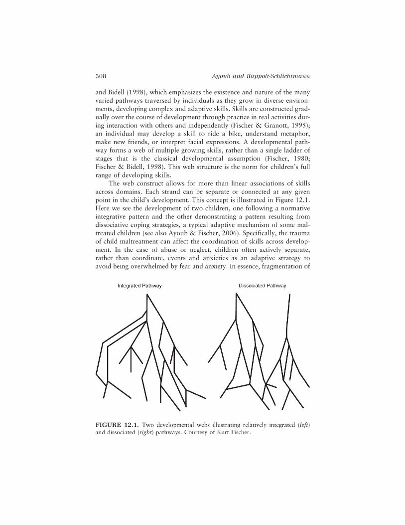
and Bidell (1998), which emphasizes the existence and nature of the manyvaried pathways traversed by individuals as they grow in diverse environ-ments, developing complex and adaptive skills. Skills are constructed grad-ually over the course of development through practice in real activities dur-ing interaction with others and independently (Fischer & Granott, 1995);an individual may develop a skill to ride a bike, understand metaphor,make new friends, or interpret facial expressions. A developmental path-way forms a web of multiple growing skills, rather than a single ladder ofstages that is the classical developmental assumption (Fischer, 1980;Fischer & Bidell, 1998). This web structure is the norm for children’s fullrange of developing skills.
The web construct allows for more than linear associations of skillsacross domains. Each strand can be separate or connected at any givenpoint in the child’s development. This concept is illustrated in Figure 12.1.Here we see the development of two children, one following a normativeintegrative pattern and the other demonstrating a pattern resulting fromdissociative coping strategies, a typical adaptive mechanism of some mal-treated children (see also Ayoub & Fischer, 2006). Specifically, the traumaof child maltreatment can affect the coordination of skills across develop-ment. In the case of abuse or neglect, children often actively separate,rather than coordinate, events and anxieties as an adaptive strategy toavoid being overwhelmed by fear and anxiety. In essence, fragmentation of
308 Ayoub and Rappolt-Schlichtmann
FIGURE 12.1. Two developmental webs illustrating relatively integrated (left)and dissociated (right) pathways. Courtesy of Kurt Fischer.
thoughts and feelings serves an adaptive function, allowing the child tolimit the experience of severe anxiety and helplessness and to maintainfunctioning in order to survive. If maltreatment is recurrent, which it oftenis, these traumatogenic responses become habitual and generalized. Theroutine and generalized use of these different ways of thinking and feelingmark what we call a traumatogenic alternative developmental pathway.
Although the behavioral and psychological literature on child mal-treatment has shifted to allow for the consideration of maltreated children’sbehavior as adaptive and complex, the research literature on the neurobio-logical sequelae of child maltreatment has not. In particular, researchershave characterized the excessive stress of child maltreatment as a toxicagent, interfering with normal brain development and resulting in alteredand impaired brain function (e.g., Bremner et al., 1997; DeBellis et al.,1999; Schiffer, Teicher, & Papanicolaou, 1995). Although it is true thatearly stress can affect developmental processes at both the biological andbehavioral levels, we argue that the result is not an impaired brain butrather a unique brain, actively adapted to survive in a malevolent socialworld. In the remaining sections of this chapter we elaborate the develop-mental pathways framework to include the neurobiological level of analy-sis, beginning with a review of the literature on the neurobiological conse-quences and corresponding behavioral correlates of child maltreatment.
THE NEUROBIOLOGY OF CHILD MALTREATMENT
To date, very few prospective, longitudinal, or even cross-sectional studiesof the neurodevelopment of maltreated children have been conducted.Rather, most studies have been correlational in design, and as such, we cau-tion against taking a causal view of the neurobiological findings reportedhere. As more prospective and cross-sectional studies are completed, thenature of the effects described here will become clearer. In the meantimeunderstanding how the neurobiology of maltreated children, particularlythose with negative behavioral and psychiatric outcomes, is different fromthat of their healthy agemates will provide a preliminary lens for interpret-ing the alternate developmental pathways maltreated children exhibit,informing both future research and our understanding of clinical outcomes.The evidence for brain correlates of maltreatment suggests several hypothe-ses about relations between alternate pathways, illustrated through behav-ioral studies and possible brain functions.
Neuroimaging studies comparing maltreated and nonmaltreated chil-dren have demonstrated variable structural and functional differences (seeTeicher et al., 2003, for a review), providing evidence for one or more
Child Maltreatment 309
alternative neurodevelopmental pathways in maltreated children. Amongthe differences found in maltreated children are a smaller corpus callosum;attenuation of the development of the left neocortex, hippocampus, andamygdala; enhanced electrical activity in the limbic system; and reducedactivity of the cerebellar vermis. For example, Carrion and colleagues(2001) assessed the frontal lobe asymmetry of 24 children with trauma his-tories and PTSD symptoms. They found that the maltreated children hadattenuated frontal lobe asymmetry. In 2003, Teicher and his colleaguesmeasured the regional corpus callosum from magnetic resonance imaging(MRI) scans in 52 children admitted for psychiatric evaluation, of whom28 were identified as abuse or neglect victims. These children were com-pared to 115 healthy children. The researchers found that the corpus callo-sum of the maltreated children was 17% smaller than in the comparisonchildren. The greatest difference was evident in the neglected children andwas associated with a 15–18% reduction in regions 3, 4, 5, and 7 of thecorpus callosum. These two studies are examples of recent advances in thefield; such findings suggest ways in which a combination of new imagingtechniques can support the associations between behavioral observationsand brain activity to better understand the development of alternative path-ways.
To generalize advances from a neurodevelopmental perspective, wehave begun to identify brain regions that have a high density of gluco-corticoid receptors and a long period of postnatal development as thosethat are particularly vulnerable to the stress of early maltreatment. Suchfindings help to explain the structural and functional differences observedacross individuals based on their exposure to traumatic experiences.
The Hippocampus, Memory,and Behavioral Inhibition
The hippocampus plays a central role in the verbal recall aspect of explicitmemory; stress experiences strongly shape such memory (Teicher et al.,2003). The stress of abuse trauma influences a number of functions relatedto the hippocampus, including changes in the creation of semantic memory,specifically memory deficits (e.g., amnesia), and alterations in memory(e.g., intrusive memories) and concentration (e.g., increased dissociation)—which are all common in PTSD (Joseph, 1999; Newport & Nemeroff,2000). It comes as no surprise, then, that the hippocampus seems to have aparticular vulnerability to severe stress experiences (Gould & Tanapat,1999; Sapolsky, Uno, Rebert, & Finch, 1990). Specifically, the hippocam-pus has a long postnatal developmental period and a high density ofglucocorticoid receptors, making it vulnerable to cortisol neurotoxicity and
310 Ayoub and Rappolt-Schlichtmann
thus severe stress (Patel et al., 2000; Sapolsky, McEwen, & Rainbow,1983). Studies investigating the effect of childhood abuse on the structureand function of the hippocampus have yielded mixed results in differentage groups. A reduction in left hippocampal volume has been reported inadults with a childhood history of trauma and current PTSD (Bremner etal., 1997; Stein, 1977). Bilateral reduction in hippocampal volume has beenreported in women with a history of child abuse and borderline personalitydisorder (Driessen et al., 2000). In contrast, studies of maltreated childrenand adolescents consistently report no reduction in hippocampal volume incomparison to healthy controls (Carrion et al., 2001; DeBellis et al., 1999;Teicher et al., 2003).
These discrepancies in the data may reflect a developmental trend inwhich the effects of early abuse on hippocampal structure emerge over thecourse of development (Sapolsky, Krey, & McEwen, 1985; Teicher et al.,2003), or it may be the case that preexisting small hippocampal volume is arisk factor among adult individuals for the development of PTSD and otherpsychiatric illnesses following child abuse (Teicher et al., 2003). It is alsopossible that the psychological and behavioral sequelae of early stress andabuse may play a direct role in the effects on hippocampal volume seen inadults. Specifically, the onset of psychiatric illness and risky coping behav-iors such as alcohol and drug abuse in adolescence may precipitate thereduction in hippocampal volume that has been reported among adult sur-vivors of child maltreatment (Dunn, Ryan, & Dunn, 1994; Ellason, Ross,Sainton, & Mayran, 1996; van der Kolk, & Fisler, 1994). More research isneeded to decipher the nature of the relationship between early severe stressand later hippocampal structure. However, concern remains because dam-age to the hippocampus may affect an individual’s ability to form newmemories and access or organize previously formed memories (Newport &Nemeroff, 2000; Teicher et al., 2003).
The behavioral and psychological sequelae of alterations to hippocam-pal functioning among maltreated children may manifest primarily as dis-ruptions to the encoding and retrieval of episodic memories, and second-arily as the inhibition of ongoing behavior that is inappropriate to theenvironment (Teicher et al., 2003). We have documented these behaviorsamong maltreated children in our laboratory. Specifically, we have exam-ined maltreated toddlers’ performance in a structured task for negative andpositive storytelling (Ayoub et al., 2006). In this study, children were askedto retell and/or act out a series of stories involving interactions that werenice, mean, or a combination of nice and mean. The stories began simplywith a nice interaction between the self doll and a friend doll—or a childdoll and a parent doll—at the developmental level of single representations,defined in terms of dynamic skill theory (Fischer, 1980; Fischer, Shaver, &
Child Maltreatment 311
Carnochan, 1990). Stories were told in order of increasing complexity,forming a developmental sequence based on skill complexity. Children’sperformance on each story was coded on two variables. First, each storywas coded on whether the participant successfully retold it. To successfullypass a story task at a particular level of complexity the participant had toattend to the story and accurately represent the characters as nice or mean,including the various component parts of the story. The representationscould be performed either verbally or nonverbally. Each child received askill level score for correct completions. Second, the children’s stories werecoded for complexity of representation regardless of whether or not thestory was retold correctly. For example, a child might fail to accuratelyretell the story by switching the mean and nice characters, or by making anice story mean, but his or her story might be highly complex nonetheless.In this procedure, children were scored according to the complexity of thestory with which they exhibited the highest skill level, regardless of whetherthey recounted the story correctly. These two forms of coding made it pos-sible to separate the developmental level of performance that matched themodeled stories from the developmental level in general.
Hierarchical regression analyses were conducted in order to investigatewhether having a history of maltreatment affected children’s complexity ofperformance in the nice and mean storytelling tasks. Among both mal-treated and nonmaltreated participants, older children correctly retoldhigher-level stories and told more complex stories regardless of passing.There was an effect of having a maltreatment history on children’s com-plexity of storytelling performance, but only for the structured componentof the assessment. That is, children with a maltreatment history tended tosuccessfully complete fewer story tasks prior to three failures than did theirnonmaltreated peers. This “gap” between the maltreated and nonmal-treated performances was larger among older children. Importantly, therewere no differences in the complexity of stories told between maltreatedand nonmaltreated participants when correctness was not taken intoaccount. These effects are illustrated in Figure 12.2.
Maltreated children’s alternate developmental pathway in this domainis clearly illustrated in this example. In Figure 12.2A we see that older chil-dren, whether maltreated or nonmaltreated, tend to exhibit higher skill lev-els in correctly representing nice/mean interactions during this structuredstorytelling task. However, in comparison, the maltreated and nonmal-treated groups exhibit different developmental trajectories. Specifically,young maltreated children tend to produce more complex stories when cor-rect early on, but the rate at which they become more complex and correctover time is slower in comparison to their nonmaltreated peers. This devel-
312 Ayoub and Rappolt-Schlichtmann
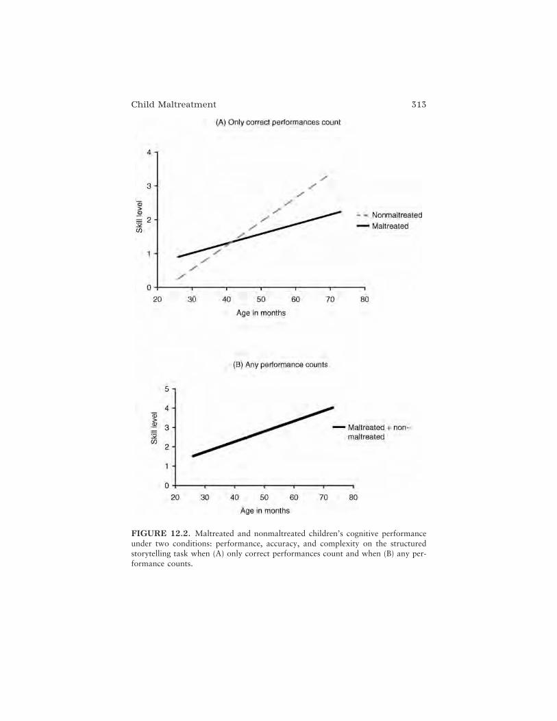
Child Maltreatment 313
FIGURE 12.2. Maltreated and nonmaltreated children’s cognitive performanceunder two conditions: performance, accuracy, and complexity on the structuredstorytelling task when (A) only correct performances count and when (B) any per-formance counts.
opmental trajectory results in a large “gap” in performance by 5 years ofage, with the nonmaltreated children tending to retell more complex niceand mean stories correctly.
From these findings it would seem that the traditional characterizationof maltreated children as “delayed” might be correct at least in part, butthere is more to this story—as evidenced by the performance of childrenwho have been maltreated on incorrect and negative stories. In Figure12.2B we see that without the constraint of having to correctly retell thestory, there are no differences in the maltreated and nonmaltreated chil-dren’s performances throughout early childhood. The maltreated childrentell mean and nice relationship stories just as complex as their peers, butonly under particular conditions. On closer examination of the data wefound that the maltreated children were more distractible, often refusing todo the task as presented, and tended to “switch” the content of nice storiesto mean. They also tended to “fail” at telling the nice stories and to spendmuch more time and energy than their counterparts telling richly detailednegative stories. Overall, they had difficulty inhibiting their negativeimpulses in the stories.
Our behavioral data illustrate that it is not that the maltreated childrenare “delayed”; rather, they exhibit an alternate developmental path com-mensurate with their early experience, demonstrating more skill with, andselective attention to, negative situations and relationships (Fischer et al.,1997). Our work, like that of others, demonstrates that this developmentalpathway seems to render attention and persistence on structured school-like tasks difficult (Porter, 2003), especially for tasks that do not capitalizeon these children’s honed skills around threat-related signals (Pollak &Tolley-Schell, 2003). These findings support those noted in older mal-treated children. For example, Pollak and Sinha (2002) found that school-age, physically abused children detected angry emotions at lower steps in avisual sequence of increasingly angry facial expressions than their non-maltreated counterparts, but showed no differences when responding toother facial displays. Similarly, Dodge and his colleagues (Dodge, Pettit,Bates, & Valente, 1995) found that school-age abused children attributedhostility to others in situations in which most people would not make suchan attribution. All of these behavioral findings of memory-related differ-ences in maltreated children, including both intrusive memories that dis-tract the child from responding to the current surroundings and avoidanceof painful memories that skew the child’s responses to daily interactions,can be linked to changes in hippocampal functioning. Future studies of theinterface will strengthen the brain–behavior argument and provide furthersupport for a pathways model of alternative development.
314 Ayoub and Rappolt-Schlichtmann
The Amygdala, the Cerebellar Vermis, Negative Affect,and Complex Behavior in Alternate Pathways
The amygdala has also been identified as a structure affected by the traumaof childhood abuse, although findings pertaining to the association be-tween maltreatment and abnormal amygdala volume are even less consis-tent than those reported for the hippocampus. The amygdala is a compo-nent of the limbic system and is believed to play a key role in emotions,particularly in fear and vigilance (Pollak, Cicchetti & Klorman, 1998).Although most studies of amygdala volume have revealed no differencesbetween adults with a childhood history of abuse and age-matched controls(Bremner et al., 1997; DeBellis et al., 1999; Stein, 1997), Driessen and col-leagues (2000) reported an 8% reduction in amygdala volume bilaterally inadult female survivors of child abuse who also were diagnosed with border-line personality disorder. Other studies have reported that adults with ahistory of combined physical and sexual abuse and children with concur-rent psychiatric issues showed abnormal and hyperactive amygdala activity(Ito et al., 1993; Teicher, Glod, Surrey, & Swett, 1993).
The functioning of the cerebellar vermis may also be affected by theexperience of child maltreatment. The cerebellar vermis is a narrow struc-ture that links the two hemispheres of the cerebellum; it is one of the lastbrain structures to fully develop in the postnatal period and is vulnerable toglucocorticoid toxicity and thus severe stress (Giedd et al., 1999). It isthought to play an inhibitory role in limbic system activation (Anderson,Teicher, Polcari, & Renshaw, 2002). Abnormalities of the cerebellar vermishave been linked to a wide array of psychiatric disorders, including schizo-phrenia (Andreasen, Paradiso, & O’Leary, 1998), autism (Courchesne,1991), attention-deficit/hyperactivity disorder (Mostofsky, Reiss, Lockhart,& Denckla, 1998), and depression (Lauterbach, 1996). Anderson and col-leagues (2002) found the activity of the cerebellar vermis to be lower inadults with a history of child abuse and neglect as compared to healthycontrols. They used steady-state functional magnetic resonance imaging(fMRI) to assess resting blood flow in the vermis of 24 young adults, athird of whom had a history of repeated child sexual abuse. Child sexualabuse survivors had higher T2 relaxation times when compared to the com-munity controls. The vermal T2-RT also correlated strongly with LimbicSystem Checklist–33 (LSCL-33) ratings of temporal lobe epilepsy-likesymptomatology. These findings, however promising, have not yet beenreplicated. Much more research on the effects of child maltreatment on thedevelopment of the cerebellar vermis is needed before implications can bedrawn definitively.
Child Maltreatment 315
Altered amygdala or cerebellar vermis functioning may have an impacton children’s emotional functioning; specifically, alterations to amygdala orcerebellar vermis functioning among severely maltreated children and ado-lescents may result in frequent impulsive violence, increased fear andnegativity, and may contribute to the development of PTSD and depression(Teicher et al., 2003). Abuse victims frequently show violence, depression,and PTSD, all of which are consistent with disturbances in the amygdalaeand the cerebellar vermis (Newport & Nemeroff, 2000).
Findings from our behavioral and psychiatric studies of girls who havebeen abused might also be related to alterations in amygdalar and cerebel-lar functioning. In our laboratory, we have examined sexually and physi-cally abused adolescent girls’ perceptions of themselves and their relation-ships in an effort to identify behaviors that were associated with givenabuse experiences. In addition, we reviewed each girl’s psychiatric diagno-ses and compared them to those of girls who were depressed but had notexperienced abuse (Ayoub, Hong, Wright, Fischer, & Noam, 2004).To measure self-perception, we used the Self-in-Relationship Interview(Calverley, Fischer, & Ayoub, 1994). This interview supports adolescentsand adults in characterizing themselves in relationships. It also is designedto detect the full range of a person’s cognitive developmental levels and toassess the effects of variation in contextual support. Each girl was inter-viewed using a semistructured format that asked her to describe what shewas like—her perception of self—in various important relationships: thosewith mother, father, best girlfriend, romantic friend, and “the real you nomatter who you are with.” For each relationship, the girls were asked towrite down five self-descriptions on separate pieces of self-sticking paper,thus coming up with a total of 25 different descriptions of self in relation-ships. The girls then assigned each description a valence: either positive,1
negative,2 or mixed valence. Each label was placed on a “self-diagram”consisting of three concentric circles representing the core, middle, andouter self. Self-descriptions placed in the core circle were labeled mostimportant, whereas those placed in the outer circle were labeled leastimportant.
We coded all maltreated adolescents for the type, severity, frequency,duration, and timing of maltreatment and perpetrator’s identity by episodeon the basis of the Maltreatment Classification System developed byBarnett, Manly, and Cicchetti (1993). Psychiatric diagnoses were obtainedfrom the clinician’s discharge report and the Diagnostic Interview Schedulefor Children (DISC-C).3 The psychiatric diagnoses were based on DSM-III-R, which was in use at the time of data collection. Psychiatric backgroundvariables were obtained from each girl’s medical records, including theadmission and discharge reports, treatment team conference summaries,
316 Ayoub and Rappolt-Schlichtmann
social worker’s notes, clinician’s individual and group therapy notes, andpsychoeducational reports. These variables included the number of psychi-atric hospitalizations, number of psychoactive medications taken whilehospitalized, whether they had received outpatient psychotherapy prior tohospitalization, and psychiatric history of the family.
Girls were divided into groups that differentiated their specific andcumulative maltreatment status, including girls who experienced sexualabuse only and girls with co-occurring sexual and physical abuse/neglect.Two additional groups of depressed girls were identified, one group withno maltreatment history and a second group of girls with a history of phys-ical abuse/neglect but no sexual abuse history.
First, we found that the frequency of negative perceptions of self washighest among sexually abused girls. Furthermore, sexually abused girlswere much more likely to rank their negative perceptions of themselves andtheir relationships as most important to their “true” or core self, whereasgirls who had been physically abused or experienced no abuse saw negativeperceptions as much less important. Those sexually abused girls with co-occurring physical abuse were the most negative in their perceptions of self,followed by the group of girls with sexual abuse only, as illustrated in Fig-ure 12.3. In spite of significant differences in the organization of their per-ceptions of themselves in relationships, the sexually abused girls and theremaining girls in the sample evidenced normal levels of cognitive complex-ity across their descriptions (Fischer et al., 1997).
This work led us to describe at least two distinctive characteristics ofalternative developmental pathways of abuse: representations of self andothers that center on negative evaluation rather than positive, and dissocia-tive splitting that develops at complex and sophisticated levels (Calverley etal., 1994; Fischer & Ayoub, 1994). As we hypothesized, these behavioralfindings are consistent with the biological changes noted in studies of theamygdala and the cerebellar vermis mentioned above. The behavioral char-acteristics of the two key components of our proposed traumatogenicdevelopmental pathway found in the abused girls include symptoms ofincreased negativity, dissociation, and PTSD symptoms that may be attrib-uted to the amygdala, because it plays a central role in consolidating theemotional significance of traumatic events, and to the cerebellar vermisbecause it modulates stress in maltreated children and adolescents.
In a second set of findings, the sexually abused girls—both those withsexual abuse as their sole form of maltreatment and those with sexualabuse as one of multiple forms of maltreatment—experienced a similarconstellation of symptoms unique to their sexual abuse categorization. Wehave combined these self-destructive behaviors into an interaction style wecall self-focused aggression. Such self-focused aggressive indicators include
Child Maltreatment 317
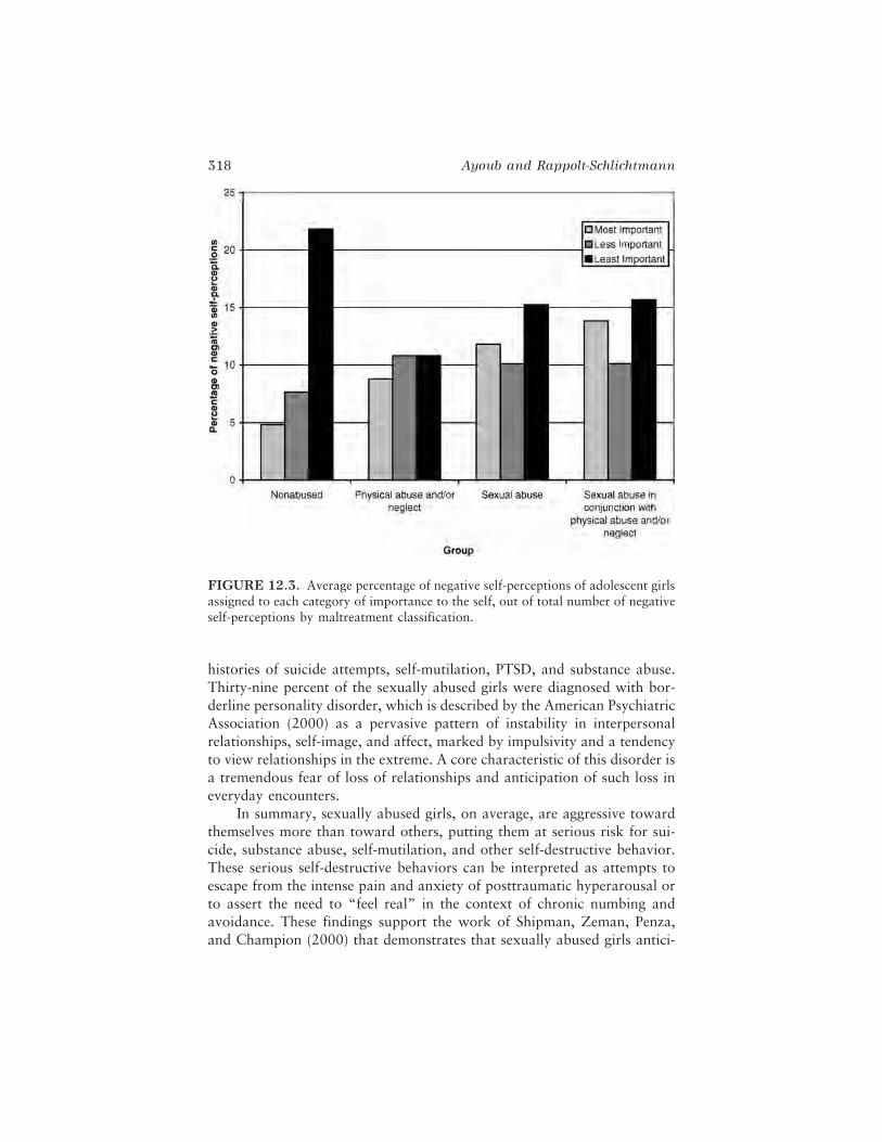
histories of suicide attempts, self-mutilation, PTSD, and substance abuse.Thirty-nine percent of the sexually abused girls were diagnosed with bor-derline personality disorder, which is described by the American PsychiatricAssociation (2000) as a pervasive pattern of instability in interpersonalrelationships, self-image, and affect, marked by impulsivity and a tendencyto view relationships in the extreme. A core characteristic of this disorder isa tremendous fear of loss of relationships and anticipation of such loss ineveryday encounters.
In summary, sexually abused girls, on average, are aggressive towardthemselves more than toward others, putting them at serious risk for sui-cide, substance abuse, self-mutilation, and other self-destructive behavior.These serious self-destructive behaviors can be interpreted as attempts toescape from the intense pain and anxiety of posttraumatic hyperarousal orto assert the need to “feel real” in the context of chronic numbing andavoidance. These findings support the work of Shipman, Zeman, Penza,and Champion (2000) that demonstrates that sexually abused girls antici-
318 Ayoub and Rappolt-Schlichtmann
FIGURE 12.3. Average percentage of negative self-perceptions of adolescent girlsassigned to each category of importance to the self, out of total number of negativeself-perceptions by maltreatment classification.
pate more conflict and less support in relationships. Recent work in traumaand psychopathology supports a connection between the repeated traumaof abuse and a pervasive array of changes in thinking and emotion thatinvolve serious disturbance in perceptions of self (Cole & Putnam, 1992;Herman, 1992; Terr, 1991). Such approach–avoidance mechanisms are anexample of what we have call polarized affective skills for splitting or disso-ciating across domains, in this case, from hypervigilance to numbing. Theother girls, in spite of being depressed like their sexually abused counter-parts, did not see themselves as negative in this way nor did they show dra-matic switching in patterns of activity across contexts.
These behavioral and psychiatric findings are consistent with the pat-terns associated with reported alterations to the amygdala and cerebellarvermis among female sexual abuse survivors as reported in the neurobiolo-gical literature. The emergence of enhanced fight or flight reactions, aggres-sion, hypervigilance, negative worldview, and even complex psychopathol-ogy in the form of PTSD and borderline personality disorder can be viewedfrom the pathways perspective as physiological and psychological self-protective and adaptive mechanisms present in the context of the abusiveenvironment. Such an alternative pathway appears to persist so that oncethe child is in a more benign environment, these adaptations to the abusivecontext become detrimental or less functional than nontraumatic copingstrategies. In some cases, as is evident with the sexually abused girls, long-term patterns of thinking and feeling become the primary patterns of inter-action and are classified as trauma-related characterological disorders suchas borderline personality disorder.
The Corpus Callosum, the Frontal Cortex,Cerebral Lateralization, and Maltreated Children’sNegativity Bias and Affective Splitting and Merging
The corpus callosum, the frontal cortex, and cerebral lateralization alsoappear to be affected by the severe stress of child maltreatment. The corpuscallosum connects the right and left cerebral hemispheres (Berrebi et al.,1988; Juraska & Kopcik, 1988; Lauder, 1983). Most communicationsbetween regions of the cortex in different hemispheres are carried acrossthe corpus callosum. Several studies have found a volume reduction in sec-tions of the corpus callosum in children with substantiated histories ofabuse and concurrent psychiatric disorder (DeBellis et al., 1999; Teicher etal., 1997, 2000). These studies highlight gender differences: boys overalland more markedly boys with a history of neglect, as well as sexuallyabused girls, seem to be particularly susceptible to the effects of abuse onthe corpus callosum. These findings are potentially serious because re-
Child Maltreatment 319
duced size of the corpus callosum is associated with reduced communica-tion between the hemispheres (Yazgan, Wexler, Kinsbourne, Peterson, &Leckman, 1995).
In addition, prefrontal maturation and cerebral lateralization appearto be affected by the early severe stress of child abuse and neglect. The cere-bral cortex and especially the prefrontal cortex are late to develop, withmyelination continuing through adolescence and midadulthood (Fuster,1980), and are sensitive to glucocorticoid toxicity (Diorio, Viau, &Meaney, 1993). Several studies suggest that maltreated children’s corticaldevelopment is attenuated and asymmetrical, especially for those withPTSD (DeBellis, Keshavan, Spencer, & Hall, 2000; Ito, Teicher, Glod, &Ackerman, 1988; Thatcher, 1992). Specifically, the frontal cortex, particu-larly in the left hemisphere, has been shown to lag behind in developmentfor children with a history of abuse, in comparison to nonabused controls(Ito et al., 1988; Teicher et al., 1997). One convenient means of assessinglateralized hemispheric activity is through probe auditory evoked poten-tials. In Schiffer’s study (Schiffer et al., 1995) adult participants with a his-tory of trauma showed a marked suppression of the evoked potentialresponse over the left hemisphere during recall of a neutral memory, whiledemonstrating a suppressed evoked potential response over the right hemi-sphere during recall of a difficult or disturbing memory. Nontraumatizedcomparison participants demonstrated equal probe-evoked amplitudes be-tween hemispheres on both tasks.
These findings raise concerns for maltreated individuals because theprefrontal and frontal cortex regulate key aspects of the planning, coordi-nation, and control of behavior as well as other executive function skills(for reviews, see DeBellis, 2001; Teicher et al., 2003). Alterations in thesefunctions, coupled with possible asymmetrical development in response totrauma, may result in changes in the child’s ability to integrate executivefunctions across context. For example, we have hypothesized that the ten-dency of abused children to split rather than to integrate when problemsolving, especially when the topic or emotion cues memories of past trau-ma, is one way that this dysynchrony between hemispheres may presentbehaviorally. These differences may be observed through the developmentof polarized problem-solving schemas that are applied in specific contextsbut are contradictory when examined in the context of integrated planningand problem-solving skills. This hallmark of the alternative developmentalpathway model we have described may be explained, in part, throughchanges in the development and functioning of the frontal cortex alongwith the tendency of maltreated children to demonstrate asymmetrical useof the right and left hemispheres.
Functionally, alterations to the corpus callosum following child mal-treatment would affect the integration of information across the two hemi-
320 Ayoub and Rappolt-Schlichtmann
spheres. The two cerebral hemispheres are specialized in their information-processing abilities. For example, in most people the right hemisphere has aspecialized role in the expression and perception of emotion, especially neg-ative emotion. In the case of child maltreatment the reduced size of the cor-pus callosum as well as the increased dominance of the right hemispheremay manifest as a negativity bias in emotion expression and perception(DeBellis, 2001).
Such a negativity bias is well documented in research among mal-treated children and adolescents, and it contrasts markedly with the nor-mal, strong positivity bias that pervades behavior among most people(Fischer & Ayoub, 1994; Fischer et al., 1990; Greenwald et al., 2002).Using both the biological and the behavioral frameworks as models, weexamined narratives of young maltreated children. In their earliest narra-tives about interactions with peers and caregivers, maltreated toddlers dem-onstrate a negativity bias, commonly initiating or choosing negative inter-actions, demonstrating a richer understanding of negative interactions, andcharacterizing themselves in predominantly negative terms (Ayoub &Raya, 1993). Specifically, they tend to spontaneously enact negative socialinterchanges even when offered a positive model for interaction and whenscaffolded with social support for producing such a story. For example,2.5-year-old Donald, a child in our lab, when asked to model a nice inter-action of the self doll with the other friend doll, said forcefully, “Guy, youwanna fight? I’ll knock you down. He fight him. They fight. You wannafight? I’m gonna fight you. Fuck my butt, fuck it.” When he is supported bythe examiner who reminds him that the task is to tell a nice story, Donaldtries again to replicate the nice story he has heard. With this social support,Donald is able to say, “Have some Playdough, guy, don’t leave me, pleasedon’t leave me.” To review, Donald at first is unable to repeat a nice story;instead, he describes an elaborate mean interaction. Support from a caringexaminer helps Donald to replicate a positive response, but this nice story isfollowed by an abandonment theme (“don’t leave me, please don’t leaveme”). Even at this young age, children such as Donald, who have experi-enced repeated maltreatment trauma, have come to expect aggression andloss rather than kindness and sustained care.
The maltreated toddlers in our lab describe their interpersonal worldsin terms of three primary schemas: (1) positive relationships are conven-tionalized and relatively bland; (2) positive relationships are reconstructedor converted into negative relationships (as with Donald); and (3) negativerelationships are richly textured, dynamic, and often aggressive or violent(Ayoub & Raya, 1993).
In a pathways model the tendency to choose negative stories, to con-vert neutral and positive situations to negative ones, to worry about theloss of positive caring, and the need to be the powerful one in the interac-
Child Maltreatment 321
tion constitute an adaptive but vigilant approach to recurrent overwhelm-ing negative maltreatment experiences (Crittenden, 1985; Fischer et al.,1997; Westen, 1994). Our work confirms the findings of Shields, Ryan,and Cicchetti (1991), who found that increased negative representations ofpreschoolers affected their relationships with peers, and those of Macfie,Cicchetti, and Toth (2001), who demonstrated that maltreated childrenexhibited higher levels of dissociative or splitting experiences than theirnonabused counterparts. Such cumulative works reinforce both a behavior-al and biological framework for understanding hypervigilance, a character-istic prominent in the alternative pathways in maltreated children. That is,if the world is threatening and dangerous, it is only reasonable to becomehypervigilant to guard against such threats. The experience of trauma leadsthe young maltreated child to anticipate threat and danger. This alteredworldview, when applied to daily living, results in a child with a dramati-cally divergent perspective on the world at large.
These adaptive patterns are markedly different from those of non-maltreated toddlers, who routinely produce a kind of positivity bias inwhich they represent themselves as nice or good and someone else as meanor bad, whether or not the circumstances warrant that evaluation (Ayoub& Fischer, 1994; Fischer, Shaver, & Carnochan, 1990). Findings of differ-ences in critical structures (such as the corpus callosum) that support coor-dination across hemispheres may help to explain the negative perspectivesand hypervigilant presentations of the maltreated children we evaluated. If,as in adults (Schiffer et al., 1995), there is differential use of each hemi-sphere, depending on the threatening nature of the events perceived, as wellas increased negativity expressed by the abused participants, then use of theleft hemisphere in more prominent ways would support a case for increasednegativity in maltreated children. Clearly, this is a beginning hypothesisthat needs further study, but may have promise for understanding the rela-tionship between brain and behavior within the developmental pathwaysmodel.
CONCLUSION: CHILD MALTREATMENTAND THE FORMATION OF ALTERNATEDEVELOPMENTAL PATHWAYS:BRAIN–BEHAVIOR CONNECTIONS
A prevailing view drawn from the current literature on the neurobiology ofchild maltreatment is that the excessive stress of child maltreatment acts asa toxic agent, interfering with normal development and resulting in analtered and impaired brain. Implicit in this view is the assumption that
322 Ayoub and Rappolt-Schlichtmann
there is one “right,” “normal,” “universal” developmental course, whichhas been altered by this extreme social condition. We have argued thatdevelopment does not occur in a domain-general and unidirectional way,without regard for context (Fischer & Bidell, 1998). To accurately describeand begin to understand maltreated children’s development, a frameworkthat emphasizes the enormous variation in human development and therole of the environment in that variation is necessary. Variation is not noiseor error in the data but an essential and important component of develop-ment to be studied. That premise is the starting point for the developmentalpathways approach.
Through the pathways lens, maltreated children’s neurodevelopmentalprofile looks very different. The brain is not impaired by maltreatment, butit adapts to a malevolent environment. When a child is born into a stress-filled world, he or she forms an alternate developmental pathway thatincludes both behavioral and brain changes as adaptations to exposure tohigh levels of stress hormones and other aspects of stress, moving brain andbehavior to organize early in life along a stress-sensitive pathway (Seckl,1998; Seckl, Cleasby, & Nyirenda, 2000; Welberg & Seckl, 2001). Thisstress-sensitive pathway allows maltreated children to protect themselvesby demonstrating an acutely sensitive fight-or-flight response system in theface of environmental challenge. Unfortunately this adaptation comes at ahigh price. The same developmental pathway that allows for survival in amalevolent world leaves the individual at increased risk of serious healthproblems and psychopathology. Furthermore, these stress-sensitive adapta-tions become detrimental and maladaptive when the affected individualsfind themselves in a more benign environment.
Shifting to the pathways approach is not simply a matter of semantics.It potentially can change the way therapists treat children and their fami-lies. In addition, it offers a different theoretical perspective for scientistsstudying the process of change and development in childhood, influencingthe research questions asked, the methods used, and the conclusions drawn.The pathways approach encourages both researchers and therapists tounderstand the multiple domains and transactional nature of developmentand to explore the tremendous variation inherent in developmental pro-cesses and the adaptations of maltreated children. For example, there isnow mounting evidence that the stable and predictable non-normative bio-logical and behavioral patterns exhibited by maltreated children undergo arapid reorganization toward a more normative biobehavioral response pat-tern after transition into sensitive and responsive care (Bruce, Kroupina,Parker, & Gunnar, 2000; Gunnar & Cheatham, 2003). Dismissing varia-tion as error or viewing maltreated children as disordered may result inoverlooking the role of plasticity and change in understanding children’s
Child Maltreatment 323
ever-changing behavioral, psychological, and biological adaptations andoutcomes. A maltreated child may exhibit predictable, relatively stable,non-normative biobehavioral patterns, but at the same time he or sheshows much variation around this state both biologically and behaviorally(Gunnar & Donzella, 2002). These variations within the system providethe potential for new adaptation over time in response to changes in theenvironment.
Although the study of the neurodevelopment of maltreated children isin its infancy, with current findings needing further study and replication,early results suggest that there are likely neurobiological consequences tothe experience of early maltreatment trauma. Studying child maltreatmentfrom the biological point of view holds great promise to inform both theoverall knowledge base and approaches to treatment and intervention.Research to date suggests several hypotheses about relations between biol-ogy and behavior in developmental pathways arising from maltreatment:
1. Effects on memory and behavioral inhibition may relate to changesin the hippocampus, which develops gradually over time.
2. The negative bias in behavior and the development of especiallycomplex negative constructions of self and relationships may corre-spond to alterations in the amygdala and the cerebellar vermis.
3. Maltreated children’s merging of positive and negative affect, com-bined with their strong negativity bias, may relate to changes incomponents of the brain that facilitate coordination, especially thecorpus callosum, the frontal cortex, and hemispheric lateralization.
Of course, these three hypotheses about brain–behavior correlations areonly a rough start in understanding the complex relations between brainand behavior in maltreatment. However, it is important to note that eachone does support the hypothesis of an alternative developmental pathwaythat results from brain–behavior–environment interactions in the contextof childhood maltreatment.
Effective understanding of maltreated children, involving both treat-ment and research, requires moving beyond misunderstanding them asdelayed and dysregulated as a result of their maltreatment. Effective under-standing of both the behavior and biology of children who have been mal-treated requires recognizing that they develop along alternate pathways inadaptation to their maltreating, stressful world. A clear and complex pic-ture of maltreated children becomes possible only when their developmentis understood as combining complex strengths and weaknesses that vary bycontext and are based in the worlds that they experience with their family,community, body, and brain (Fischer et al., 1997).
324 Ayoub and Rappolt-Schlichtmann
ACKNOWLEDGMENTS
We thank Kurt Fischer, Erin O’Connor, Pamela Raya, Elizabeth Nelson, Kyung-wha Hong, Travis Wright, and Gil Noam for their contributions to the work onwhich this chapter is based. The research described in this chapter was made possi-ble in part by the Roche Relief Fund, the Milton Fund, the National Institutes ofHealth, and the Harvard Graduate School of Education.
NOTES
1. “Something I like about myself.” A positive self-description of a girl’s relation-ship with someone does not necessarily indicate that her relationship with thatperson is positive. For example, one of the girls who identified all her self-descriptions as positive had “I’m cranky,” “I’m mad,” “I scream,” “I cry,” “Iwant to die” for her relationship with her father. She indicated that she likes thefact that she is like this with her father and that that is why she identified theseself-descriptions as positive.
2. “Something I do not like about myself.”3. DISC-C is a standardized instrument that elicits information directly from the
child about the presence or absence of symptoms, behaviors, and emotions thatcorrespond to the DSM-III diagnostic criteria for Axis 1 disorders of childhoodand adolescence (Calverley, 1995, p. 70).
REFERENCES
American Psychiatric Association. (2000). Diagnostic and statistical manual ofmental disorders (4th ed., text rev.). Washington, DC: Author.
Anderson, C., Teicher, M., Polcari, A., & Renshaw, P. (2002). Abnormal T2 relax-ation time in the cerebellar vermis of adults sexually abused in childhood:Potential role of the vermis in stress-enhanced risk for drug abuse. Psycho-neuroendocrinology, 27(1–2), 231–244.
Andreasen, N., Paradiso, S., & O’Leary, D. (1998). Cognitive dysmetria as anintegrative theory of schizophrenia: A dysfunction in cortical–subcortical–cerebellar circuitry? Schizophrenia Bulletin, 24(2), 203–218.
Ayoub, C., & Fischer, K. (2006). Intersections among domains of development. InK. McCartney & D. Phillips (Eds.), The Blackwell handbook of early childdevelopment (pp. 62–82). Oxford, UK: Blackwell.
Ayoub, C., Fischer, K., & O’Connor, E. (2003). Analyzing development of workingmodels for disrupted attachments: The case of family violence. Attachmentand Human Development, 5(2), 97–120.
Ayoub, C., Hong, K., Wright, T., Fischer, K., & Noam, G. (2004, April). Develop-mental impact of sexual abuse on adolescent girls: Negative self and self-destructive aggression. Paper presented at the annual conference of the Societyfor Research in Adolescence, Baltimore, MD.
Child Maltreatment 325
Ayoub, C., O’Connor, E., Rappolt-Schlichtmann, G., Fischer, K., Rogosh, F., Toth,S., et al. (2006). Cognitive and emotional differences in young maltreated chil-dren: Application of dynamic skill theory. Development and Psychopathology,18(3), 679–706.
Ayoub, C., & Raya, P. (1993, April). Social interactions in young maltreated tod-dlers: The development of self in relationships. Paper presented at the meetingsof the Society for Research in Child Development, New Orleans.
Barnett, D., Manly, J. T., & Cicchetti, D. (1993). Defining child maltreatment: Theinterface between policy and research. In D. Cicchetti & S. L. Toth (Eds.),Child abuse, child development, and social policy (pp. 7–74). Norwood, NJ:Ablex.
Berrebi, A., Fitch, R., Ralphe, D., Denenberg, J., Friedrich, V., & Denenberg, V.(1988). Corpus callosum: Region-specific effects of sex, early experience andage. Brain Research, 438(1–2), 216–224.
Bremner, J., Randall, P., Vermetten, E., Staib, L., Bronen, R., Mazure, C., et al.(1997). Magnetic resonance imaging-based measurement of hippocampal vol-ume: Posttraumatic stress disorder related to childhood physical and sexualabuse—a preliminary report. Biological Psychiatry, 41(1), 23–32.
Briere, J. (1992a). Child abuse trauma: theory and treatment of the lasting effects.Newbury Park, CA: Sage.
Briere, J. (1992b). Methodological issues in the study of sexual abuse effects. Jour-nal of Consulting and Clinical Psychology, 60, 196–203.
Bruce, J., Kroupina, M., Parker, S., & Gunnar, M. R. (2000, July). The relation-ships between cortisol, growth retardation, and developmental delay in post-institutionalized children. Paper presented at the International Conference onInfant Studies, Brighton, UK.
Calverley, R. (1995). Self in relationship in depressed adolescent girls: Developmen-tal effects of childhood sexual trauma. Unpublished doctoral dissertation,Harvard Graduate School of Education, Cambridge, MA.
Calverley, R., Fischer, K., & Ayoub, C. (1994). Complex splitting of self-representations in sexually abused adolescent girls. Development and Psycho-pathology, 6, 195–213.
Carrion, V., Weems, C., Eliez, S., Patwardhan, A., Brown, W., Ray, R., et al.(2001). Attenuation of frontal asymmetry in pediatric posttraumatic stress dis-order. Biological Psychiatry, 50(12), 943–951.
Cicchetti, D. (1991). Fractures in the crystal: Developmental psychopathology andthe emergence of self. Developmental Review, 11, 271–287.
Cicchetti, D., & Toth, S. (2005). Child maltreatment. Annual Review of ClinicalPsychology, 1, 409–438.
Courchesne, E. (1991). Neuroanatomic imaging in autism.Pediatrics, 87(5), 781–790.
Crittenden, P. (1985). Maltreated infants: Vulnerability and resilience. Journal ofChild Psychology and Psychiatry, 26, 85–96.
Crittenden, P. M. (1988). Relationships at risk. In J. Belsky & T. Nexworski (Eds.),Clinical implications of attachment theory (pp. 136–174). Hillsdale, NJ:Erlbaum.
326 Ayoub and Rappolt-Schlichtmann
Cole, M., & Putnam, F. (1992). Effect of incest on self and social functioning: Adevelopmental psychopathology perspective. Journal of Consulting and Clini-cal Psychology, 60, 174–184.
Darwish, D., Esquivel, G., Houtz, J., & Alfonso, V. (2001). Play and social skills inmaltreated and nonmaltreated preschoolers during peer interactions. ChildAbuse and Neglect, 25, 13–31.
DeBellis, M. (2001). Developmental traumatology: The psychobiological develop-ment of maltreated children and its implications for research, treatment, andpolicy. Development and Psychopathology, 13, 539–564.
DeBellis, M., Keshavan, M., Clark, D., Casey, B., Giedd, J., Boring, M., et al.(1999). Developmental traumatology. Part II: Brain development. BiologicalPsychiatry, 45(10), 1271–1284.
DeBellis, M., Keshavan, M., Spencer, S., & Hall, J. (2000). N-acetylaspartate con-centration in the anterior cingulate of maltreated children and adolescentswith PTSD. American Journal of Psychiatry, 157(7), 1175–1177.
Diorio, D., Viau, V., & Meaney, M. (1993). The role of the medial prefrontal cor-tex (cingulate gyrus) in the regulation of hypothalamic–pituitary–adrenalresponses to stress. Journal of Neuroscience, 13(9), 3839–3847.
Dodge, K. A., Pettit, G. S., Bates, J., & Valente, E. (1995). Social information pro-cessing patterns partially mediate the effect of early physical abuse on laterconduct problems, Journal of Abnormal Psychology, 104, 632–643.
Driessen, M., Herrmann, J., Stahl, K., Zwaan, M., Meier, S., Hill, A., et al. (2000).Magnetic resonance imaging volumes of the hippocampus and the amygdalain women with borderline personality disorder and early traumatization.Archives of General Psychiatry, 57(12), 1115–1122.
Dunn, G., Ryan, J., & Dunn, C. (1994). Trauma symptoms in substance abuserswith and without histories of childhood abuse. Journal of Psychoactive Drugs,26(4), 357–360.
Egeland, B., & Sroufe, L. (1981). Attachment and early maltreatment. Child Devel-opment, 52, 44–52.
Eigsti, I., & Cicchetti, D. (2004). The impact of child maltreatment on expressivesyntax at 60 months. Developmental Science, 7, 88–102.
Ellason, J., Ross, C., Sainton, K., & Mayran, L. (1996). Axis I and II comorbid-ity and childhood trauma history in chemical dependency. Bulletin of theMenninger Clinic, 60(1), 39–51.
Felitti, V., Anda, R., Nordenberg, D., Williamson, D., Spitz, A., Edwards, V., et al.(1998). Relationship of childhood abuse and household dysfunction to manyof the leading causes of death in adults. American Journal of Preventive Medi-cine, 14, 245–258.
Fischer, K. (1980). A theory of cognitive development: The control and construc-tion of hierarchies of skills. Psychological Review, 87, 477–531.
Fischer, K., & Ayoub, C. (1994). Affective splitting and dissociation in normal andmaltreated children: Developmental pathways for self in relationships. In D.Cicchetti & S. L. Toth (Eds.), Rochester Symposium on Development and Psy-chopathology: Vol. 5. Disorders and dysfunctions of the self (pp. 149–222).Rochester, NY: University of Rochester Press.
Child Maltreatment 327
Fischer, K., Ayoub, C., Singh, I., Noam, G., Maraganore, A., & Raya, P. (1997).Psychopathology as adaptive development along distinctive pathways. Devel-opment and Psychopathology, 9(4), 729–748.
Fischer, K., & Bidell, T. R. (1998). Dynamic development of psychological struc-tures in action and thought. In R. M. Lerner (Ed.), Theoretical models ofhuman development (pp. 467–561). New York: Wiley.
Fischer, K., & Granott, N. (1995). Beyond one dimensional change: Parallel, con-current, socially distributed processes in learning and development. HumanDevelopment, 38, 302–314.
Fischer, K., Shaver, P., & Carnochan, P. G. (1990). How emotions develop andhow they organize development. Cognition and Emotion, 4, 81–127.
Freud, A. (1966). The ego and the mechanisms of defense. New York: InternationalUniversities Press. (Original work published 1936)
Fuster, J. (1980). The prefrontal cortex: Anatomy, physiology, and neuropsycholo-gy of the frontal lobe. New York: Raven Press.
Giedd, J., Blumenthal, J., Jeffries, N., Rajapakse, J., Vaituzis, A., Liu, H., et al.(1999). Development of the human corpus callosum during childhood andadolescence: A longitudinal MRI study. Progress in Neuro-Psychopharma-cology and Biological Psychiatry, 23(4), 571–588.
Gould, E., & Tanapat, P. (1999). Stress and hippocampal neurogenesis. BiologicalPsychiatry, 46(11), 1472–1479.
Greenwald, A. G., Banaji, M. R., Rudman, L., Farnham, S., Nosek, B. A., &Mellott, D. (2002). A unified theory of implicit attitudes, stereotypes, self-esteem, and self-concept. Psychological Review, 109, 3–25.
Gunnar, M., & Donzella, B. (2002). Social regulation of the cortisol levels in earlyhuman development. Psychoneuroendocrinology, 27(1–2), 199–220.
Gunnar, M. R., & Cheatham, C. L. (2003). Brain and behavior interface: Stress andthe developing brain. Infant Mental Health Journal, 24, 195–211.
Herman, J. (1992). Trauma and recovery. New York: Basic Books.Ito, Y., Teicher, M., Glod, C., & Ackerman, E. (1998). Preliminary evidence for
aberrant cortical development in abused children: A quantitative EEG study.Journal of Neuropsychiatry Clinical Neuroscience, 10, 298–307.
Ito, Y., Teicher, M., Glod, C., Harper, D., Magnus, E., & Gelbard, H. (1993).Increased prevalence of electrophysiological abnormalities in children withpsychological, physical, and sexual abuse. Journal of Neuropsychiatric Clini-cal Neuroscience, 5, 401–408.
Joseph, R. (1999). The neurobiology of traumatic “dissociative” amnesia: Com-mentary and literature review. Child Abuse and Neglect, 23(8), 715–727.
Juraska, J., & Kopcik, J. (1988). Sex and environmental influences on the size andultrastructure of the rat corpus callosum. Brain Research, 450(1–2), 1–8.
Lauder, J. (1983). Hormonal and humoral influences on brain development.Psychoneuroendocrinology, 8(2), 121–155.
Lauterbach, E. (1996). Bipolar disorders, dystonia, and compulsion after dysfunc-tion of the cerebellum, dentatorubrothalamic tract, and substantia nigra. Bio-logical Psychiatry, 40(8), 726–730.
Macfie, J., Cicchetti, D., & Toth, S. (2001). The development of dissociation in
328 Ayoub and Rappolt-Schlichtmann
maltreated preschool-aged children. Development and Psychopathology, 13,233–234.
Mostofsky, S., Reiss, A., Lockhart, P., & Denckla, M. (1998). Evaluation of cere-bellar size in attention-deficit hyperactivity disorder. Journal of Child Neurol-ogy, 13(9), 434–439.
Mueller, E., & Silverman, N. (1989). Peer relations in maltreated children. In D.Cicchetti & V. Carlson (Eds.), Child maltreatment: Theory and research onthe causes and consequences of child abuse and neglect (pp. 529–578). NewYork: Cambridge University Press.
Newport, D. J., & Nemeroff, C. B. (2000). Neurobiology of posttraumatic stressdisorder. Current Opinion in Neurobiology, 10(2), 211–218.
Patel, P., Lopez, J., Lyons, D., Burke, S., Wallace, M., & Schatzberg, A. (2000).Glucocorticoid and mineralocorticoid receptor mRNA expression in squirrelmonkey brain. Journal of Psychiatric Research, 34(6), 383–392.
Pollak, S., Cicchetti, D., & Klorman, R. (1998). Stress, memory, and emotion:Development considerations from the study of child maltreatment. Develop-ment and Psychopathology, 10, 811–828.
Pollak, S., & Sinha, P. (2002). Effects of early experience on children’s recognitionof facial displays of emotion. Developmental Psychology, 38, 784–791.
Pollak, S., & Tolley-Schell, S. (2003). Selective attention to facial emotion in physi-cally abused children. Journal of Abnormal Psychology, 112(3), 323–338.
Porter, C. (2003). Neurobehavioral sequelae of child sexual abuse. Unpublisheddoctoral dissertation, Brigham Young University, Salt Lake City, UT.
Putnam, F. W. (1994). Dissociation and disturbances of self. In D. Cicchetti & S. L.Toth (Eds.), Rochester Symposium on Development and Psychopathology:Vol. 5. Disorders and dysfunctions of the self (pp. 251–266). Rochester, NY:University of Rochester Press.
Sapolsky, R., Krey, L., & McEwen, B. (1985). Prolonged glucocorticoid exposurereduces hippocampal neuron number: Implications for aging. Journal of Neu-roscience, 5(5), 1222–1227.
Sapolsky, R., McEwen, B., & Rainbow, T. (1983). Quantitative autoradiographyof [3H]corticosterone receptors in rat brain. Brain Research, 271, 331–334.
Sapolsky, R., Uno, H., Rebert, C., & Finch C. (1990). Hippocampal damage associ-ated with prolonged glucocorticoid exposure in primates. Journal of Neurosci-ence, 10, 2897–2902.
Schiffer, F., Teicher, M., & Papanicolaou, A. (1995). Evoked potential evidence forright brain activity during the recall of traumatic memories. Journal of Neuro-psychiatric and Clinical Neuroscience, 7, 169–175.
Schneider-Rosen, K., Braunwald, K., Carlson, V., & Cicchetti, D. (1985). Currentperspectives in attachment theory: Illustration from the study of maltreatedinfants. In I. Bretherington & E. Waters (Eds.), Monographs of the Society forResearch in Child Development: Growing points of attachment theory andresearch (Serial No. 209, pp. 194–210).
Schneider-Rosen, K., & Cicchetti, D. (1984). The relationship between affect andcognition in maltreated infants: Quality of attachment and the development ofvisual self-recognition. Child Development, 55, 648–658.
Child Maltreatment 329
Seckl, J. (1998). Physiologic programming of the fetus. Clinical Perionatal, 25(4),939–962.
Seckl, J., Cleasby, M., & Nyirenda, M. (2000). Glucocorticoids, 11beta-hydroxy-steroid dehydrogenase, and fetal programming. Kidney International, 57(4),1412–1417.
Shields, A., Ryan, R. M., & Cicchetti, D. (1991). Narrative representations of care-givers and emotional dysregulation as predictors of maltreated children’s rejec-tion by peers. Developmental Psychology, 37, 321–337.
Shipman, K., Zeman, J., Penza, S., & Champion, K. (2000). Emotion managementskills in sexually maltreated and nonmaltreated girls: A developmental psycho-pathology perspective. Development and Psychopathology, 12, 47–62.
Stein, M. (1997). Hippocampal volume in women victimized by childhood sexualabuse. Psychological Medicine, 27(4), 951–959.
Teicher, M. (2000). Wounds that time won’t heal: The neurobiology of child abuse.Cerebrum, 2(4), 50–67.
Terr, L. (1991). Childhood traumas: An outline and overview. American Journal ofPsychiatry, 148(1), 10–19.
Teicher, M., Andersen, S., Dumont, N., Ito, Y., Glod, C., Vaituzis, C., et al. (2000)Childhood neglect attenuates development of the corpus callosum. Social Neu-roscience Abstracts, 26, 549.
Teicher, M., Andersen, S., Polcari, P., Anderson, C., Navalta, C., & Kim, D.(2003). The neurobiological consequences of early stress and childhood mal-treatment. Neuroscience and Biobehavioral Reviews, 27, 33–44.
Teicher, M., Glod, C., Surrey, J., & Swett, C. (1993). Early childhood abuse andlimbic system ratings in adult psychiatric outpatients. Journal of Neuropsychi-atric Clinical Neuroscience, 5(3), 301–306.
Teicher, M., Ito, Y., Glod, C., Andersen, S., Dumont, N., & Ackerman, E. (1997).Preliminary evidence for abnormal cortical development in physically and sex-ually abused children using EEG coherence and MRI. Annals of the New YorkAcademy of Sciences, 821, 160–175.
Thatcher, R. (1992). Cyclic cortical reorganization during early childhood. Brainand Cognition, 20, 24–50.
U.S. Department of Health and Human Services. (2005). Child maltreatment 2003.Washington, DC: U.S. Government Printing Office.
van der Kolk, B., & Fisler, R. (1994). Childhood abuse and neglect and loss of self-regulation. Bulletin of the Menninger Clinic, 58(2), 145–168.
Walker, E., Unutzer, J., Rutter, M., Gelfand, A., Saunders, K., VonKorff, M., et al.(1999). Costs of health care use by women HMO members with a history ofchildhood abuse and neglect. Archives of General Psychiatry, 56, 609–613.
Welberg, L., & Seckl, J. (2001). Prenatal stress, glucocorticoids, and the program-ming of the brain. Journal of Neuroendocrinology, 13(2), 113–128.
Westen, D. (1994). The impact of sexual abuse on self structure. In D. Cicchetti &S. L. Toth (Eds.), Disorders and dysfunctions of the self (Vol. 5, pp. 223–250).Rochester, NY: University of Rochester Press.
330 Ayoub and Rappolt-Schlichtmann
C H A P T E R 1 3
Corticolimbic Circuitryand PsychopathologyDEVELOPMENT OF THE CORTICOLIMBIC SYSTEM
Francine M. Benes
Several brain regions play a role in the integration of affective experiencewith higher cognitive function. Papez (1937) was the first to draw attentionto the fact that links between limbic structures and the neocortex areresponsible for the integration of emotion with cognition; he emphasizedthat the hippocampus, with its extensive limbic connections, and thecingulated cortex, with its elaborate reciprocal interactions with other asso-ciative cortical regions, form a central loop of connections. Today, weunderstand that this so-called “loop of Papez” is only one component in amuch more extensive network integrating phylogenetically older and newerportions of the brain. The cortical regions that contribute to this systeminclude not only the hippocampal formation and cingulated cortex, butalso the entorhinal region, the prefrontal area, frontal eye field 8, and theinferior parietal lobe (Benes, 1993a); together, these cortical areas sub-sume, respectively, affective, attentional, motivational, and logical planningcomponents of complex behavioral responses. Although frontal eye field 8and the inferior parietal lobe do not have direct connections with sub-cortical limbic structures, they are nevertheless integral to complex behav-
331
iors involving limbic activity, because phylogeneses has established anobligatory (albeit an indirect) role for these regions.
To understand how emotional responses are integrated with thoughtprocesses, it is useful to examine the anatomy of the various brain regionsthat participate in this integration process. Although a discussion of thesestructures, as they are found in normal adults, can undoubtedly lend someinsight into how the brain may achieve a harmonious collaborationbetween the limbic system and the neocortex, it does not tell us how such acooperative interplay has been achieved. For this reason, it is useful toexamine how such a complex system has been assembled as a result of bothphylogenetic and ontogenetic development of the brain. A developmentalapproach of this type can potentially help us understand not only how thebrain has attained its characteristic functions, but also how these functionscan be disturbed by perturbations of normal ontogeny.
Yakovlev and Lecours (1967) emphasized that maturation of thehuman brain follows a sequentially timed process in which phylogeneticallyolder portions of the brain, such as the spinal cord, mature before phylo-genetically newer portions, such as the neocortex. Although there are simi-larities between the evolution of the brain and its early development, thereare nevertheless also striking differences. For the purposes of the presentdiscussion, the parallelism identified between phylogenetic evolution andontogenetic maturation provides a unique opportunity to explore the rootsof the emotional and cognitive behaviors found in humans. The discussionthat follows begins with a comparison of the hippocampal formation andthe neocortex in premammalian species, such as reptiles, with thoseencountered in mammals, particularly rodents, primates, and humans;however, its main focus is a review of the ontogenetic development of thesestructures and cortical neurotransmitters in the mammalian brain.
ARCHI- AND ALLOCORTICAL STRUCTURES
The most significant advance in the evolution of the corticolimbic systemoccurred during the transition from submammalian to mammalian species.In reptiles, the forebrain is composed of a thin cortical mantle surroundinga region called the anterior dorsal ventricular ridge, which invaginates intothe lateral ventricle (see Figure 13.1). Although simple in organization, thecortical mantle in reptiles has several different subsidiary areas, includingthe medial, mediodorsal, dorsal, and lateral regions (Sarnat & Netsky,1974). Homology of cortical areas found in reptiles with areas found inmammalian brain has been inferred from identified connection patternswith subcortical structures, such as the hypothalamus and the septal region(Bruce & Butler, 1984), as well as with other cortical regions (Ulinski,
332 Benes
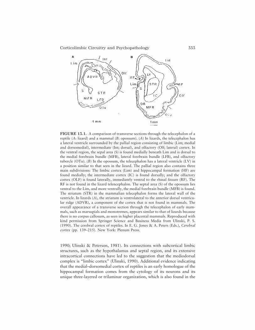
1990; Ulinski & Peterson, 1981). Its connections with subcortical limbicstructures, such as the hypothalamus and septal region, and its extensiveintracortical connections have led to the suggestion that the mediodorsalcomplex is “limbic cortex” (Ulinski, 1990). Additional evidence indicatingthat the medial–dorsomedial cortex of reptiles is an early homologue of thehippocampal formation comes from the cytology of its neurons and itsunique three-layered or trilaminar organization, which is also found in the
Corticolimbic Circuitry and Psychopathology 333
FIGURE 13.1. A comparison of transverse sections through the telecephalon of areptile (A: lizard) and a mammal (B: opossum). (A) In lizards, the telecephalon hasa lateral ventricle surrounded by the pallial region consisting of limbic (Lim; medialand dorsomedial), intermediate (Int; dorsal), and olfactory (Olf; lateral) cortex. Inthe ventral region, the sepal area (S) is found medially beneath Lim and is dorsal tothe medial forebrain bundle (MFB), lateral forebrain bundle (LFB), and olfactorytubercle (OTu). (B) In the opossum, the telecephalon has a lateral ventricle (LV) ina position similar to that seen in the lizard. The pallial region also contains threemain subdivisions: The limbic cortex (Lim) and hippocampal formation (HF) arefound medially; the intermediate cortex (IC) is found dorsally; and the olfactorycortex (OLF) is found laterally, immediately ventral to the rhinal fissure (RF). TheRF is not found in the lizard telencephalon. The septal area (S) of the opossum liesventral to the Lim, and more ventrally, the medial forebrain bundle (MFB) is found.The striatum (STR) in the mammalian telecephalon forms the lateral wall of theventricle. In lizards (A), the striatum is ventrolateral to the anterior dorsal ventricu-lar ridge (ADVR), a component of the cortex that is not found in mammals. Theoverall appearance of a transverse section through the telecephalon of early mam-mals, such as marsupials and monotremes, appears similar to that of lizards becausethere is no corpus callosum, as seen in higher placental mammals. Reproduced withkind permission from Springer Science and Business Media from Ulinski, P. S.(1990). The cerebral cortex of reptiles. In E. G. Jones & A. Peters (Eds.), Cerebralcortex (pp. 139–215). New York: Plenum Press.
area dentate and CA subfields of the mammalian hippocampus or archi-cortex (Ulinski, 1990; see Figure 13.1). Interestingly, neurons in the medialcortex frequently contain zinc (Lopez-Garcia, Martinez-Guijarry, Berbel,& Garcia-Verdugo, 1988; Perez-Clausell, 1988), which is also abundant inthe hippocampal formation of the mammalian brain (Haug, 1965; Rosene& Van Hoesen, 1987). Thus, mediotemporal structures in mammals haveextensive limbic and intracortical connections that are believed to have firstappeared in rudimentary form early in phylogenesis and to have establisheda foundation for the later appearance of the more intricate corticolimbicsystem in mammals. In contrast, the dorsal (general) cortex of reptiles maybe a precursor of other motor, sensory, and tertiary neocortical regions inthe brains of mammalian forms (Bishop, 1959; Herrick, 1933; Orrego,1961).
THE “LIMBIC LOBE” AND THE EXPANSIONOF THE CORTEX
In the course of evolution from reptiles to mammals, there was a transi-tional form, called therapsids, which acquired certain features of mammals(Broom, 1932). Examination of cranial remains of therapsids has revealedevidence consistent with the acquisition of hair, sweat glands, and im-proved masticatory mechanisms (MacLean, 1985). Broca (1878) noted thatin parallel with the first appearance of mammalian features in vertebrates,a limbic lobe, consisting of the septal nuclei, amygdala, hippocampus,parahippocampal (entorhinal) cortex, and cingulate gyrus, appeared alongthe midsagittal plane of virtually all mammalian forms. Interestingly, theelaboration of this so-called limbic lobe coincided with the appearance ofaudition, vocalization, maternal nurturance, and separation calls by theyoung, suggesting that the integration of visceral responses with corticallymediated behaviors might have facilitated the protection of young offspring(MacLean, 1985). Accordingly, the evolutionary trend toward developingmore elaborate behaviors to nurture the young and presumably to perpetu-ate the species probably involved a corresponding increase in both theamount and complexity of cortical ties with limbic structures. Within thespectrum of mammalian forms, there has been a striking increase in the rel-ative proportion of neocortex. Figure 13.2 shows a comparison of thelimbic lobe in rat, cat, and monkey brains (MacLean, 1954).
It is evident from the diagram that there has been a progressiveincrease in the volume of neocortex surrounding the cingulated andparahippocampal gyri. In primates and humans, this expansion has beenaccompanied by the most extensive elaboration of connectivity between
334 Benes
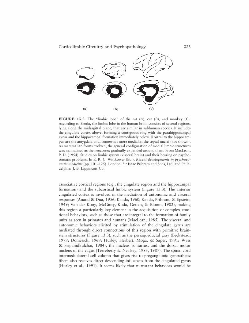
associative cortical regions (e.g., the cingulate region and the hippocampalformation) and the subcortical limbic system (Figure 13.3). The anteriorcingulated cortex is involved in the mediation of autonomic and visceralresponses (Anand & Dua, 1956; Kaada, 1960; Kaada, Pribram, & Epstein,1949; Van der Kooy, McGinty, Koda, Gerfen, & Bloom, 1982), makingthis region a particularly key element in the acquisition of complex emo-tional behaviors, such as those that are integral to the formation of familyunits as seen in primates and humans (MacLean, 1985). The visceral andautonomic behaviors elicited by stimulation of the cingulate gyrus aremediated through direct connections of this region with primitive brain-stem structures (Figure 13.3), such as the periaqueductal gray (Beckstead,1979; Domesick, 1969; Hurley, Herbert, Moga, & Saper, 1991; Wyss& Sripanidkulchai, 1984), the nucleus solitarius, and the dorsal motornucleus of the vagus (Terreberry & Neafsey, 1983, 1987). The spinal cordintermediolateral cell column that gives rise to preganglionic sympatheticfibers also receives direct descending influences from the cingulated gyrus(Hurley et al., 1991). It seems likely that nurturant behaviors would be
Corticolimbic Circuitry and Psychopathology 335
FIGURE 13.2. The “limbic lobe” of the rat (A), cat (B), and monkey (C).According to Broda, the limbic lobe in the human brain consists of several regions,lying along the midsagittal plane, that are similar in subhuman species. It includesthe cingulate cortex above, forming a contiguous ring with the parahippocampalgyrus and the hippocampal formation immediately below. Rostral to the hippocam-pus are the amygdala and, somewhat more medially, the septal nuclei (not shown).As mammalian forms evolved, the general configuration of medial limbic structureswas maintained as the neocortex gradually expanded around them. From MacLean,P. D. (1954). Studies on limbic system (visceral brain) and their bearing on psycho-somatic problems. In E. R. C. Wittkower (Ed.), Recent developments in psychoso-matic medicine (pp. 101–125). London: Sir Isaac Pribram and Sons, Ltd. and Phila-delphia: J. B. Lippincott Co.
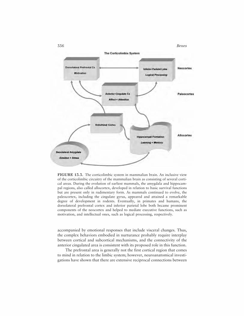
accompanied by emotional responses that include visceral changes. Thus,the complex behaviors embodied in nurturance probably require interplaybetween cortical and subcortical mechanisms, and the connectivity of theanterior cingulated area is consistent with its proposed role in this function.
The prefrontal area is generally not the first cortical region that comesto mind in relation to the limbic system; however, neuroanatomical investi-gations have shown that there are extensive reciprocal connections between
336 Benes
FIGURE 13.3. The corticolimbic system in mammalian brain. An inclusive viewof the corticolimbic circuitry of the mammalian brain as consisting of several corti-cal areas. During the evolution of earliest mammals, the amygdala and hippocam-pal regions, also called allocortex, developed in relation to basic survival functionsbut are present only in rudimentary form. As mammals continued to evolve, thepaleocortex, including the cingulate gyrus, appeared and attained a remarkabledegree of development in rodents. Eventually, in primates and humans, thedorsolateral prefrontal cortex and inferior parietal lobe both became prominentcomponents of the neocortex and helped to mediate executive functions, such asmotivation, and intellectual ones, such as logical processing, respectively.
the dorsolateral prefrontal area and both the cingulated gyrus and theentorhinal cortex (Goldman-Rakic, Selemon, & Schwartz, 1984; Swanson& Kohler, 1986). In this way, the prefrontal area can interface withsubcortical limbic structures via these latter two mesocortical regions andthe hippocampal formation. Accordingly, the volitional and motivationalactivity mediated through the prefrontal region can be driven, in part, bylimbically derived responses processed through the hippocampal formationand its ties with the amygdala, septal nuclei, and lateral hypothalamus (Fig-ure 13.3), or through the anterior cingulated region and its connectionswith subcortical regions at various levels of the neuraxis. Another associa-tive cortical region that is particularly well developed in higher mammals isthe inferior parietal area, which is believed to play a central role in logicalplanning (Luria, 1973). Like the prefrontal cortex, this region also hasextensive connections with the cingulate cortex, but interacts directly withthe hippocampal formation via the presubiculum as well (Seltzer & VanHoesen, 1979).
The above discussion illustrates that the brain, not having a specificmechanism for deleting phylogenetically older circuits, has integrated someolder circuits with newly acquired ones within the corticolimbic system. Asa result, there has been a progressive increase in the integration ofhigher cognitive processes with visceral and instinctual behaviors mediatedthrough subcortical limbic structures. In the human brain, this additive ten-dency that has occurred during phylogeny has probably been adaptive,because it is doubtful that the human species could have survived withoutlimbic-driven emotions and instincts plus cortically driven logical planningand volition.
THREE- AND SIX-LAYERED CORTEX
To understand how the brain has achieved an exquisite harmony betweenthe cortical and subcortical components of the corticolimbic system, it isuseful to examine more closely the cytoarchitectural organization withinthe cortex of higher mammals and to compare it with that found in reptiles.As noted previously, the cortex of reptiles has a simple three-layered orga-nization (Ulinski, 1977). In the mammalian neocortex, however, the intrin-sic organization is much more complex and has been expanded into six lay-ers, which can be distinguished from one another according to the densityand size of neurons, as well as their morphological appearance. For exam-ple, layer I has sparse numbers of relatively small interneurons, whereaslayers II, III, V, and VI contain large numbers of so-called pyramidal neu-rons, which typically project their axons out of the cortex to other distal
Corticolimbic Circuitry and Psychopathology 337
sites. In contrast, layer IV contains abundant numbers of small inter-neurons, called granule cells. With this layer as a reference point, the deeperlayers (V and VI) are called infragranular layers and the superficial layers(I–III) the supragranular layers.
Interestingly, there is a basic dichotomy between infra- and supra-granular layers of the mammalian cortex. Pyramidal neurons in layers Vand VI project axons primarily to subcortical structures, whereas those inlayers II and III send their axons preferentially to other cortical sites (Jones,1984). Layer IV receives a rich supply of afferents from the thalamus(White, 1986), whereas layer I has a large number of incoming associatedinputs from other cortical regions (Marin-Padilla, 1984). Thus, these lattertwo receptive layers reflect the subcortical and corticocortical relationshipof the other infragranular and supragranular projections layers, respec-tively. In a very general sense, then, the phylogenetic development of thecortex has involved an expansion from three to six layers, and it is of morethan passing interest that the three supragranular layers have a more spe-cific role in processing associative activity, with an ever-increasing role inphylogenetically more advanced species such as primates and humans.Interestingly, the thickness of the supragranular layers is proportional tothe amount of associative activity that a particular region mediates. Forexample, the primary visual cortex has a rudimentary amount of thesupragranular layers I–III, whereas the tertiary visual association area 19has a well-developed supragranular zone. Supramodal (tertiary) corticalregions that are purely associative in nature, such as the cingulated cortex,prefrontal areas, and inferior parietal areas, have the most extensive devel-opment of the upper three layers. Overall, the marked expansion of theassociative cortex that has occurred in relation to phylogenesis of the brainhas involved a dramatic increase in supragranular laminar processing.
ONTOGENESIS OF THE HIPPOCAMPAL REGION
The hippocampal region of the mammalian brain includes the area dentate,the hippocampus proper, the subicular complex, and the entorhinal cortex(Stanfield & Cowan, 1988). Generally speaking, the development of thisregion involves a carefully timed sequence of events that occurs in virtuallyall mammalian species, although the period during which the sequentialchanges occur may be quite different among mammalian forms. For exam-ple, in rats the cells of the region superior (CA1-CA2) are produced withinthe last half of gestation (Angevine, 1965; Stanfield & Cowan, 1988),whereas in monkeys they are produced during the first half (Rakic &
338 Benes
Nowakowski, 1981). There is also a general tendency for neurons of thehippocampus to be generated internally by mitotic proliferation of precur-sor elements within a zone closely apposed to the ventricular surface(Angevine, 1965; Rakic & Nowakowski, 1981; Stanfield & Cowan, 1988).When mitoses are complete for a given cell, it begins to migrate upwardand eventually assumes its proper position. This inside-out progressionoccurs for the hippocampus proper, the subicular complex, and theentorhinal cortex. For the area dentate, however, the disposition of thissubregion within the hippocampal formation is such that the cells migratein an outside-in manner. Thus, the direction of migration is not universalbut rather occurs in relation to where the proliferative epithelial zone isfound. A second type of developmental gradient occurs along the axis lyingbetween the entorhinal cortex and the area dentata, the rhinodentate axis(Stanfield & Cowan, 1988). Specifically, neurons in the entorhinal cortexand area dentata develop earlier, whereas cells in the center of this axis(subicular complex and hippocampus proper) appear later (Rakic &Nowakowski, 1981; Schlessinger, Cowan, & Swanson, 1978).
Developmental changes in the area dentata may continue well intopostnatal life. For example, in rats, the numbers of dentate granule cellshave been reported to increase during both the juvenile and adult periods(Bayer, Yackel, & Puri, 1982). Some additional evidence has also suggestedthat the cells arising postnatally may be derived from mitotic neuroblasts(Kaplan & Bell, 1984). These newly generated granule cells of the areadentata may also give rise to axonal projections into subfields CA3 andCA4 of the hippocampus (Stanfield & Trice, 1988). Evidence for postnatalincreases of granule cells has not been found in rhesus monkeys, however(Eckenhoff & Rakic, 1988). As yet, no studies have investigated whethersimilar postnatal changes may occur in the area dentata of the adult humanbrain, although preliminary evidence suggests that they do not (unpub-lished data, 1993).
Interestingly, there are some parallels between the ontogenetic devel-opment of the hippocampal formation and its evolution in vertebrates.Some investigators believe that the development of the human telecephalonat 8 weeks in utero is comparable to that of mature amphibians (Hoffman,1963), whereas it is thought at 10 weeks to be similar to that of reptiles(Crosby, 1917) and early mammalian forms. In the 8-week-old fetus, atrostral levels of the forebrain, the septal nuclei are immediately ventral tothe fornix and medial frontal cortex; at more caudal levels, the fornix iscontiguous with the hippocampus (Rakic & Yakovlev, 1968), which alsooccupies a medial location. Thus, there are similarities in the appearanceand relationships of cortical and limbic structures in the forebrains of
Corticolimbic Circuitry and Psychopathology 339
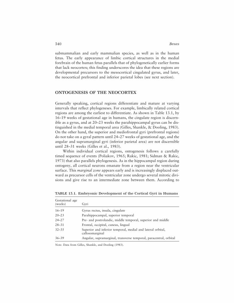
submammalian and early mammalian species, as well as in the humanfetus. The early appearance of limbic cortical structures in the medialforebrain of the human fetus parallels that of phylogenetically earlier formsthat lack neocortex; this finding underscores the idea that these regions aredevelopmental precursors to the mesocortical cingulated gyrus, and later,the neocortical prefrontal and inferior parietal lobes (see next section).
ONTOGENESIS OF THE NEOCORTEX
Generally speaking, cortical regions differentiate and mature at varyingintervals that reflect phylogeneses. For example, limbically related corticalregions are among the earliest to differentiate. As shown in Table 13.1, by16–19 weeks of gestational age in humans, the cingulate region is discern-ible as a gyrus, and at 20–23 weeks the parahippocampal gyrus can be dis-tinguished in the medial temporal area (Gilles, Shankle, & Dooling, 1983).On the other hand, the superior and mediofrontal gyri (prefrontal regions)do not take on a gyral pattern until 24–27 weeks of gestational age, and theangular and supramarginal gyri (inferior parietal area) are not discernibleuntil 28–31 weeks (Gilles et al., 1983).
Within individual cortical regions, ontogenesis follows a carefullytimed sequence of events (Poliakov, 1965; Rakic, 1981; Sidman & Rakic,1973) that also parallels phylogenesis. As in the hippocampal region duringontogeny, all cortical neurons emanate from a region near the ventricularsurface. This marginal zone appears early and is increasingly displaced out-ward as precursor cells of the ventricular zone undergo several mitotic divi-sions and give rise to an intermediate zone between them. According to
340 Benes
TABLE 13.1. Embryonic Development of the Cortical Gyri in Humans
Gestational age(weeks) Gyri
16–19 Gyrus rectus, insula, cingulate
20–23 Parahippocampal, superior temporal
24–27 Pre- and postrolandic, middle temporal, superior and middle
28–31 Frontal, occipital, cuneus, lingual
32–35 Superior and inferior temporal, medial and lateral orbital,callosomarginal
36–39 Angular, supramarginal, transverse temporal, paracentral, orbital
Note. Data from Gilles, Shankle, and Dooling (1983).
Poliakov (1965), there are five stages of human cortical development. Stage1 begins at approximately week 7, when postmitotic cells begin to moveupward to a position between the intermediate and marginal zones andform an area called the cortical plate. In Stage 2, at 10–11 weeks, the corti-cal plate becomes progressively thicker and more compact; afferent fibers,probably originating in the thalamus, begin to appear in the intermediatezone below the cortical plate. During Stage 3, at 11–13 weeks, the corticalplate has two distinct parts: an inner zone with more mature cells that havean elongated shape and a diffuse distribution and an outer zone withdensely packed immature cells. During Stage 4, the ventricular zonebecomes less prominent as cells are completing their mitotic division andmigrating outward, toward the cortical plate. The longest period is Stage 5,which begins at the 16th fetal week and continues into the early postnatalperiod. During this stage, postmitotic neuronal cells continue migratingand reach their final destination within the cortical plate.
As neurons enter the cortical plate, those destined for more superficiallayer arrive later than those that occupy deeper layers. Accordingly, thecortex, like the hippocampus, shows an inside-out progression of develop-ment (Rakic, 1974). Studies in humans have shown that by 7 months ofgestation, layers V and VI have attained a more advanced degree of devel-opment than layers II and III (Marin-Padilla, 1970a). The morphologicaldifferentiation of neurons in the various layers mirrors this migratory pro-cess, such that large pyramidal neurons with a well-developed dendriticarborization and basket cells (inhibitory interneurons) can be distinguishedin deeper layers sooner than in superficial layers (Marin-Padilla, 1970a). By7.5 months in utero, pyramidal neurons in deeper portions of layer III arefirst beginning to show differentiation of an apical dendrite extending intolayer I, whereas in layers V and VI these cells already have elaborate den-dritic arborizations. Interestingly, during this same interval, incoming affer-ent fibers are present in virtually all layers of the cortex. In the monthsimmediately prior to birth, interneurons with a basket cell appearancebegin to appear in the deeper portions of layer III, whereas in layers II andouter portions of III they are first beginning to form. By birth, boththe second and third laminae contain pyramidal neurons; however, basketcells in layer II are largely absent (Marin-Padilla, 1970b). By 2.5 monthspostnatally, pyramidal neurons continue to mature, showing a dramaticincrease in overall size, in the amount of dendritic branching, and in thenumbers of dendritic spines (Michel & Garey, 1984). Interestingly, layersVI and I (the marginal zone) are the first laminae to appear, whereas layersII and III are the last to form and the latest to mature (Marin-Padilla,1970b).
Corticolimbic Circuitry and Psychopathology 341
A COMPARISON OF CORTICAL PHYLOGENYAND ONTOGENY
It can be seen from the discussion above that there is some tendency for theontogenesis of the six-layered cortex in mammalian brain to reflect itsphylogenetic development. As noted previously, the expansion of the neo-cortex that has occurred in relation to mammalian evolution has beenaccompanied by an increase from three to six layers. As shown in Figure13.4, the supragranular layers are predominantly involved in associativeconnections and are most extensive in tertiary supramodel regions thathave expanded in relative size during evolution. The fact that layers II andIII are last to form and mature during mammalian development is in accor-dance with the idea that similarities between normal ontogenesis and phy-logeny can be identified.
Accordingly, the ontogenetic development of layer II may reflect someof the phylogenetic changes that have been traced within this lamina(Sanides, 1969). In certain types of lower mammals, layer II shows anaccentuated appearance, resulting from a higher density of neuronal cellbodies; in higher mammals, a lower density of neurons is typically found inthis layer (Abbie, 1942). Interestingly, 8-month-old human fetuses alsoshow a similar accentuation of the second lamina, but this disappears dur-ing the postnatal period (Sanides, 1969), presumably as increased amountsof neuropil, arising from the elaboration of dendritic branches and synapticconnections, accumulate in layer II of adult primates and humans. Accord-ingly, it is likely that the high density of neurons in layer II in lower mam-mals is the result of a much lower quantity of synaptic connections. Thus,both phylogenetic and ontogenetic information concerning laminar organi-zation point to late developmental changes in layer II as a characteristic fea-ture.
Interestingly, in turtles, fibers projecting from the thalamus to the cor-tex terminate in layer I (Hall & Ebner, 1970); in the mammalian brain, bycontrast, these thalamocortical afferents are preponderantly found in layerIV. Thus, in this respect, layer IV of mammals has some homology withlayer I in reptiles. The fact that the ontogenetic development of the cortexoccurs in an inside-out fashion, with layer IV appearing before layer I andcontaining a phylogenetically older afferent supply from the thalamus, isconsistent with this general idea. It is important to emphasize, however,that neither supragranular nor infragranular layers are exclusively engagedin corticocortical or subcortical processing, respectively. For example, layerI in reptiles receives afferent inputs not only from the thalamus (Hall &Ebner, 1970) but also from other cortical regions, whereas layer I of thecingulated region in mammals also receives afferent fibers from certain
342 Benes
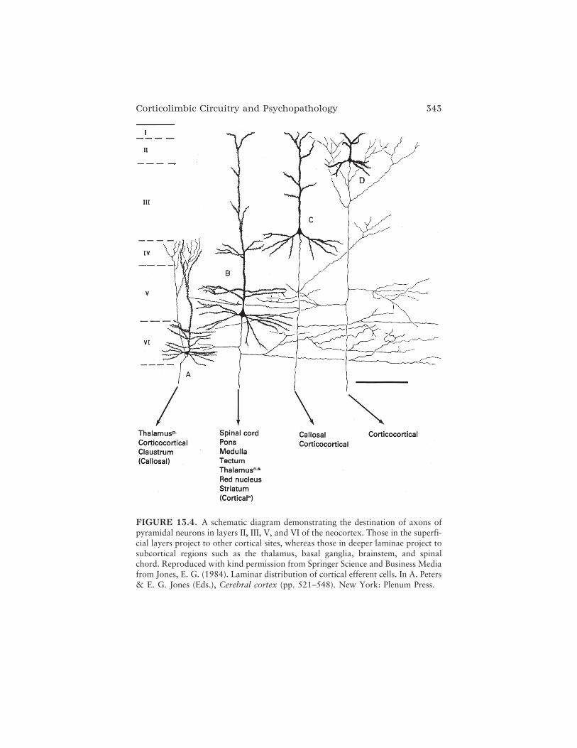
Corticolimbic Circuitry and Psychopathology 343
FIGURE 13.4. A schematic diagram demonstrating the destination of axons ofpyramidal neurons in layers II, III, V, and VI of the neocortex. Those in the superfi-cial layers project to other cortical sites, whereas those in deeper laminae project tosubcortical regions such as the thalamus, basal ganglia, brainstem, and spinalchord. Reproduced with kind permission from Springer Science and Business Mediafrom Jones, E. G. (1984). Laminar distribution of cortical efferent cells. In A. Peters& E. G. Jones (Eds.), Cerebral cortex (pp. 521–548). New York: Plenum Press.
thalamic nuclei (Vogt, Rosene, & Peters, 1981). Thus, it is primarily ofheuristic value to emphasize the similarities between phylogeneses andontogenesis, and in doing so, it is important to avoid the implication thatthe upper cortical layers were simply added on top of the deeper layers dur-ing evolution.
THE DEVELOPMENT OF AFFERENT INPUTSTO THE CORTEX
During normal brain development, the entry of various types of afferentfibers to the cortex also follows a carefully timed sequence. Two principalinputs to the cortex, one arising from the thalamus and one from thecontralateral cortex, show such a sequential progression. During prenatalstages in rats, fibers from the thalamus extend toward the cortical mantleand either stop immediately beneath or begin to enter layer IV (Wise &Jones, 1978). In the rat somatosensory cortex, the ingrowth of thalamo-cortical fibers toward specific neuronal elements, particularly those in layerIV, continues after parturition and is not complete until approximately thethird postnatal day (Wise & Jones, 1978). In contrast, commissural inputsarising from homologous cortex of the opposite hemisphere begin enteringthe somatosensory cortex at 5 days postnatally and attain a mature degreeof connectivity by day 7 (Wise & Jones, 1978). Thus, pyramidal cell axonsgrow toward their sites of termination before their dendritic arbors havedeveloped, suggesting that the elaboration of the dendritic tree and the for-mation of spines on pyramidal neurons may be regulated by extrinsic influ-ences from incoming afferent fibers originating in other cortical areas(Wise, Fleshman, & Jones, 1979).
Interestingly, in primates, the destination toward which commissuralafferents travel is determined by the region from which they originate(Goldman-Rakic, 1981). Thus, commissural afferents arising from cortexand surrounding the principal sulcus of the dorsolateral prefrontal area willgrow toward the principal sulcus of the opposite hemisphere during theprenatal period (Goldman-Rakic, 1981). If, however, the contralateral cor-tex to which the fibers would ordinarily project is surgically removed, thesecommissural afferent fibers deviate from their intended path and coursetoward a more dorsal location, where they will then enter the cortex toform synaptic connections. Although much of normal brain development istightly programmed, cortical afferents appear to have considerable flexibil-ity as to where they can terminate, and perturbations of normal develop-mental sequences can result in the formation of aberrant connections in thecortex of primates (and probably also in humans). Such a process could
344 Benes
contribute to the development of psychopathology later in life (Benes,1993a).
An important developmental change involving projections of theamygdala to the anterior cingulate cortex occurs during the equivalent ofadolescence in the rat brain (Plate 13.1). In a recent study using ante-rograde tracing, a subdivision of the basolateral amygdala showed anincreased penetration of labeled fibers in layers II and V of this corticalregion (Cunningham, Bhattacharyya, & Benes, 2002). The terminations ofthese fibers engage in synaptic connections with dendritic spines of pyrami-dal neurons, but also form nonsynaptic appositions with dendritic shafts ofaspiny neurons. The increase of these connections occurs between birth andthe beginning of the early adult period. It is well established that theamygdala plays a pivotal role in mediating emotional responses, particu-larly under conditions of stress (see next section). It seems likely, therefore,that these connections with cingulate neurons may contribute to the inte-gration of affective experiences, with higher cortical functions mediatedthrough the cingulate region.
MYELINATION OF KEYCORTICOLIMBIC PATHWAYS
A broadly accepted marker for the functional maturation of the central ner-vous system is the acquisition of myelin sheaths around axon shafts.Myelin is the insulating sheath that surrounds axons and enhances conduc-tion velocity; its appearance during development signals the acquisition offunctional capabilities for the pathways along which it occurs. It has longbeen known that various neural pathways myelinate at different stages ofdevelopment (Flechsig, 1920). There is a general tendency, however, formore cephalad structures to myelinate later than those more caudad, andfor subcortical pathways to myelinate before cortical associational paths.For example, the medial longitudinal fasciculus, a pathway that is foundalong the entire extent of the spinal cord and brainstem, begins myelinatingas early as week 20 of the gestational period, whereas the medial lemniscus,a pathway confined to the brainstem, shows similar changes at week 24(Gilles et al., 1983). Within the cerebral hemispheres, the posterior limb ofthe internal capsule begins myelinating at week 32, whereas the anteriorlimb does not show evidence of myelin until week 38. Interestingly, proxi-mal portions of the cingulum bundle do not show evidence of myelin untilgestational weeks 38–39 (Gilles et al., 1983), but probably continuemyelinating well into the postnatal period (see Figure 13.5). Some pathssuch as the fornix and mammillothalamic tract, which are not associative
Corticolimbic Circuitry and Psychopathology 345
in nature, do not begin to myelinate until weeks 44 and 48, respectively.Since the fornix provides an afferent flow from the hippocampal formationto the mammilary body of the posterior hypothalamus, and the mammillo-thalamic tract in turn conveys information to the anterior nucleus of thethalamus, both of these pathways are important components of the loop ofPapez and, in a larger sense, the corticolimbic system (see Figure 13.2).
Other cortically based links within this system, however, do notmyelinate until well into the postnatal period (Benes, 1989; Benes, Turtle,Khan, & Farol, 1994). For example, the perforant pathway (entorhinalcortex to presubiculum) actively myelinates during postnatal life (see Figure13.5). After a plateau during the third and fourth decades, myelin increasesalong the surface of the parahippocampal gyrus during the fifth and sixthdecades (Benes, 1989; Benes et al., 1994). As noted in the section on hippo-campal development, the granule cells of the area dentata, to which theperforant pathway axons send a direct projection, may increase in numberpostnatally in rats. Although similar increases in granule cell numbers havenot, as yet, been demonstrated in the human brain, it seems likely thatchanges in myelination and conduction along the perforant pathway fibersprojecting to granule cells in the area dentata do occur. In this setting,other, subtler changes in granule cells at both the cellular and molecularlevels could occur, even in the absence of increases in their overall number.
The observation of increasing myelination in the perforant pathwayand distal portions of the cingulum bundle, described above, is noteworthyfor two reasons. First, it has long been believed that late postnatal changesof this type may occur in the human brain. Yakovlev and Lecours (1967),in noting sequential myelination of the spinal cord, brainstem, and telen-cephalic structures, suggested that the intracortical associational pathwaysmay continue myelinating as late as the fourth decade. Although this paperis often cited as if it had presented empirical evidence for such changes, nodata were, in fact, included. The first empirical evidence for such adultdevelopmental changes did not appear until the aforementioned study(Benes, 1989; see Plate 13.1) and its more recent replication and extension(Benes et al., 1994). Second, it is interesting to note that the observation oflate myelination of the perforant pathway and distal cingulum bundle pres-ents an unexpected paradox regarding human behavior and development.It has been generally held that late postnatal myelination probably occurslast in the phylogenetically most sophisticated regions of the brain, theassociative neocortex, because this would reflect the overall tendency forbrain development to parallel phylogeny. It is ironic that this latter ideawould have been correct if the associative cortical pathways showing theselate postnatal changes were of the neocortical type. Instead, the ones impli-cated in this late myelination are mesocortical transitional types (i.e., the
346 Benes
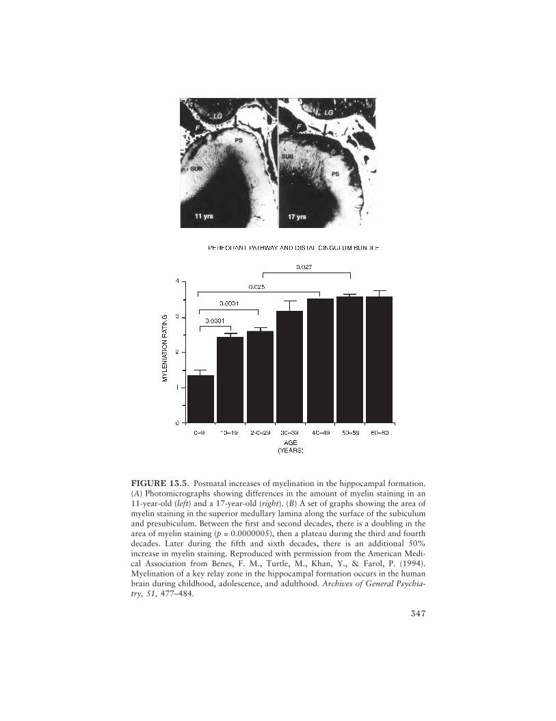
347
FIGURE 13.5. Postnatal increases of myelination in the hippocampal formation.(A) Photomicrographs showing differences in the amount of myelin staining in an11-year-old (left) and a 17-year-old (right). (B) A set of graphs showing the area ofmyelin staining in the superior medullary lamina along the surface of the subiculumand presubiculum. Between the first and second decades, there is a doubling in thearea of myelin staining (p = 0.0000005), then a plateau during the third and fourthdecades. Later during the fifth and sixth decades, there is an additional 50%increase in myelin staining. Reproduced with permission from the American Medi-cal Association from Benes, F. M., Turtle, M., Khan, Y., & Farol, P. (1994).Myelination of a key relay zone in the hippocampal formation occurs in the humanbrain during childhood, adolescence, and adulthood. Archives of General Psychia-try, 51, 477–484.
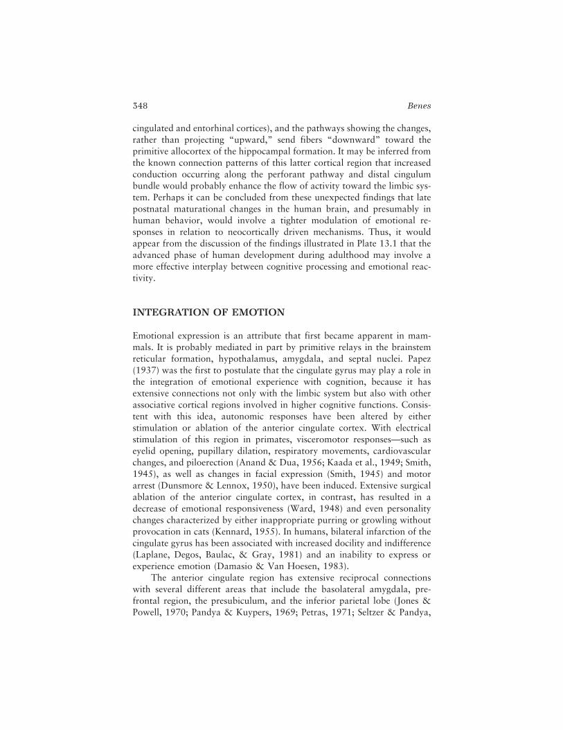
cingulated and entorhinal cortices), and the pathways showing the changes,rather than projecting “upward,” send fibers “downward” toward theprimitive allocortex of the hippocampal formation. It may be inferred fromthe known connection patterns of this latter cortical region that increasedconduction occurring along the perforant pathway and distal cingulumbundle would probably enhance the flow of activity toward the limbic sys-tem. Perhaps it can be concluded from these unexpected findings that latepostnatal maturational changes in the human brain, and presumably inhuman behavior, would involve a tighter modulation of emotional re-sponses in relation to neocortically driven mechanisms. Thus, it wouldappear from the discussion of the findings illustrated in Plate 13.1 that theadvanced phase of human development during adulthood may involve amore effective interplay between cognitive processing and emotional reac-tivity.
INTEGRATION OF EMOTION
Emotional expression is an attribute that first became apparent in mam-mals. It is probably mediated in part by primitive relays in the brainstemreticular formation, hypothalamus, amygdala, and septal nuclei. Papez(1937) was the first to postulate that the cingulate gyrus may play a role inthe integration of emotional experience with cognition, because it hasextensive connections not only with the limbic system but also with otherassociative cortical regions involved in higher cognitive functions. Consis-tent with this idea, autonomic responses have been altered by eitherstimulation or ablation of the anterior cingulate cortex. With electricalstimulation of this region in primates, visceromotor responses—such aseyelid opening, pupillary dilation, respiratory movements, cardiovascularchanges, and piloerection (Anand & Dua, 1956; Kaada et al., 1949; Smith,1945), as well as changes in facial expression (Smith, 1945) and motorarrest (Dunsmore & Lennox, 1950), have been induced. Extensive surgicalablation of the anterior cingulate cortex, in contrast, has resulted in adecrease of emotional responsiveness (Ward, 1948) and even personalitychanges characterized by either inappropriate purring or growling withoutprovocation in cats (Kennard, 1955). In humans, bilateral infarction of thecingulate gyrus has been associated with increased docility and indifference(Laplane, Degos, Baulac, & Gray, 1981) and an inability to express orexperience emotion (Damasio & Van Hoesen, 1983).
The anterior cingulate region has extensive reciprocal connectionswith several different areas that include the basolateral amygdala, pre-frontal region, the presubiculum, and the inferior parietal lobe (Jones &Powell, 1970; Pandya & Kuypers, 1969; Petras, 1971; Seltzer & Pandya,
348 Benes
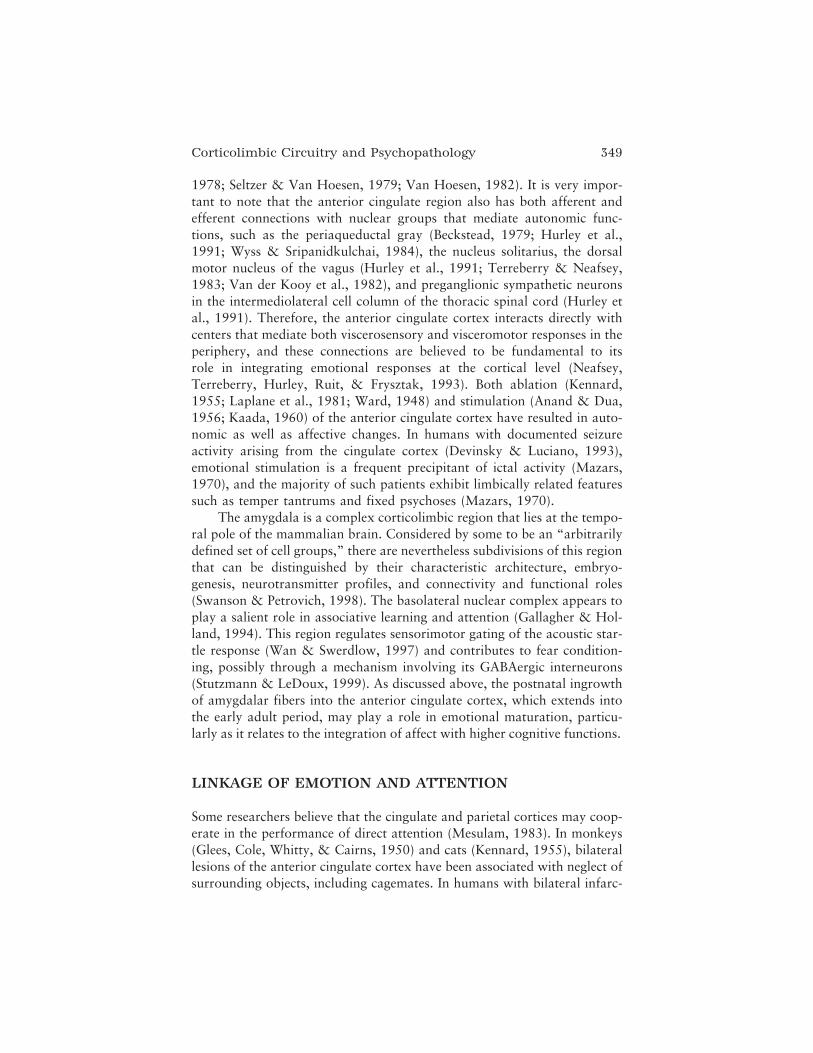
1978; Seltzer & Van Hoesen, 1979; Van Hoesen, 1982). It is very impor-tant to note that the anterior cingulate region also has both afferent andefferent connections with nuclear groups that mediate autonomic func-tions, such as the periaqueductal gray (Beckstead, 1979; Hurley et al.,1991; Wyss & Sripanidkulchai, 1984), the nucleus solitarius, the dorsalmotor nucleus of the vagus (Hurley et al., 1991; Terreberry & Neafsey,1983; Van der Kooy et al., 1982), and preganglionic sympathetic neuronsin the intermediolateral cell column of the thoracic spinal cord (Hurley etal., 1991). Therefore, the anterior cingulate cortex interacts directly withcenters that mediate both viscerosensory and visceromotor responses in theperiphery, and these connections are believed to be fundamental to itsrole in integrating emotional responses at the cortical level (Neafsey,Terreberry, Hurley, Ruit, & Frysztak, 1993). Both ablation (Kennard,1955; Laplane et al., 1981; Ward, 1948) and stimulation (Anand & Dua,1956; Kaada, 1960) of the anterior cingulate cortex have resulted in auto-nomic as well as affective changes. In humans with documented seizureactivity arising from the cingulate cortex (Devinsky & Luciano, 1993),emotional stimulation is a frequent precipitant of ictal activity (Mazars,1970), and the majority of such patients exhibit limbically related featuressuch as temper tantrums and fixed psychoses (Mazars, 1970).
The amygdala is a complex corticolimbic region that lies at the tempo-ral pole of the mammalian brain. Considered by some to be an “arbitrarilydefined set of cell groups,” there are nevertheless subdivisions of this regionthat can be distinguished by their characteristic architecture, embryo-genesis, neurotransmitter profiles, and connectivity and functional roles(Swanson & Petrovich, 1998). The basolateral nuclear complex appears toplay a salient role in associative learning and attention (Gallagher & Hol-land, 1994). This region regulates sensorimotor gating of the acoustic star-tle response (Wan & Swerdlow, 1997) and contributes to fear condition-ing, possibly through a mechanism involving its GABAergic interneurons(Stutzmann & LeDoux, 1999). As discussed above, the postnatal ingrowthof amygdalar fibers into the anterior cingulate cortex, which extends intothe early adult period, may play a role in emotional maturation, particu-larly as it relates to the integration of affect with higher cognitive functions.
LINKAGE OF EMOTION AND ATTENTION
Some researchers believe that the cingulate and parietal cortices may coop-erate in the performance of direct attention (Mesulam, 1983). In monkeys(Glees, Cole, Whitty, & Cairns, 1950) and cats (Kennard, 1955), bilaterallesions of the anterior cingulate cortex have been associated with neglect ofsurrounding objects, including cagemates. In humans with bilateral infarc-
Corticolimbic Circuitry and Psychopathology 349
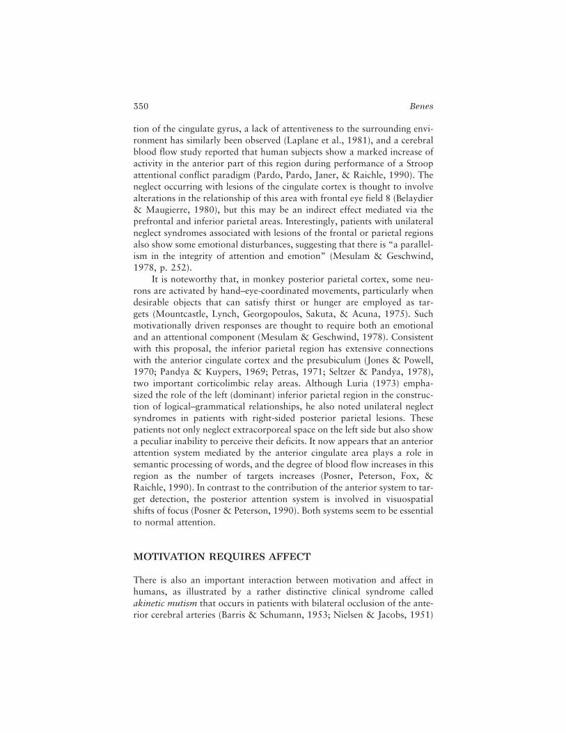
tion of the cingulate gyrus, a lack of attentiveness to the surrounding envi-ronment has similarly been observed (Laplane et al., 1981), and a cerebralblood flow study reported that human subjects show a marked increase ofactivity in the anterior part of this region during performance of a Stroopattentional conflict paradigm (Pardo, Pardo, Janer, & Raichle, 1990). Theneglect occurring with lesions of the cingulate cortex is thought to involvealterations in the relationship of this area with frontal eye field 8 (Belaydier& Maugierre, 1980), but this may be an indirect effect mediated via theprefrontal and inferior parietal areas. Interestingly, patients with unilateralneglect syndromes associated with lesions of the frontal or parietal regionsalso show some emotional disturbances, suggesting that there is “a parallel-ism in the integrity of attention and emotion” (Mesulam & Geschwind,1978, p. 252).
It is noteworthy that, in monkey posterior parietal cortex, some neu-rons are activated by hand–eye-coordinated movements, particularly whendesirable objects that can satisfy thirst or hunger are employed as tar-gets (Mountcastle, Lynch, Georgopoulos, Sakuta, & Acuna, 1975). Suchmotivationally driven responses are thought to require both an emotionaland an attentional component (Mesulam & Geschwind, 1978). Consistentwith this proposal, the inferior parietal region has extensive connectionswith the anterior cingulate cortex and the presubiculum (Jones & Powell,1970; Pandya & Kuypers, 1969; Petras, 1971; Seltzer & Pandya, 1978),two important corticolimbic relay areas. Although Luria (1973) empha-sized the role of the left (dominant) inferior parietal region in the construc-tion of logical–grammatical relationships, he also noted unilateral neglectsyndromes in patients with right-sided posterior parietal lesions. Thesepatients not only neglect extracorporeal space on the left side but also showa peculiar inability to perceive their deficits. It now appears that an anteriorattention system mediated by the anterior cingulate area plays a role insemantic processing of words, and the degree of blood flow increases in thisregion as the number of targets increases (Posner, Peterson, Fox, &Raichle, 1990). In contrast to the contribution of the anterior system to tar-get detection, the posterior attention system is involved in visuospatialshifts of focus (Posner & Peterson, 1990). Both systems seem to be essentialto normal attention.
MOTIVATION REQUIRES AFFECT
There is also an important interaction between motivation and affect inhumans, as illustrated by a rather distinctive clinical syndrome calledakinetic mutism that occurs in patients with bilateral occlusion of the ante-rior cerebral arteries (Barris & Schumann, 1953; Nielsen & Jacobs, 1951)
350 Benes
that supply the anterior cingulate cortices and other midsagittal corticalregions. Acute infarctions of this type are associated with an inability tomove or speak, as well as considerable negativism. This is quite similar tothe catatonic state, wherein muteness, lack of movement, and negativismare also observed. Patients with akinetic mutism who later recover describea sudden loss of the experience of affect and a concomitant absence of thewill to move (Damasio & Van Hoesen, 1983). It seems likely that theextensive reciprocal connectivity between the anterior cingulate region andthe prefrontal cortex that plays a direct role in the mediation and volitionaldrives (Luria, 1973) probably accounts for some of the symptomatologyobserved in akinetic mutism. Similar defects in motivation and emotionalexperience are commonly seen in adults with schizophrenia; these defectsare thought to arise from a disturbance in connectivity between thedorsolateral prefrontal area and the anterior cingulate cortex (Benes,1993b).
MATERNAL NURTURANCE, SEPARATION,AND SOCIAL INTERACTIVENESS
In rodents, ablations of the cingulate gyrus are associated with a loss ofmaternal activities such as nursing, nest building, and retrieval of the young(Slotnick, 1967; Stamm, 1955). It has been suggested that separation callsand play activities—features observed in mammalian offspring—may haveemerged in parallel with the development of the cingulate gyrus during thephylogenetic progression of reptiles into mammals (MacLean, 1985). Insupport of this hypothesis, vocalizations can be elicited by stimulation ofthe anterior cingulate region in monkeys (Ploog, 1979; Smith, 1945),whereas more extensive ablations that also include the medial subcallosaland preseptal cingulate cortices result in a complete loss of spontaneousisolation calls (MacLean, 1985). In a clinical case report, a child with docu-mented cingulate seizures was noted to run toward her mother during ictalepisodes (Geier et al., 1977), an observation suggesting that the cingulatecortex plays a role in human separation behaviors as well (MacLean,1985). It is not known, however, whether the cingulate gyrus can be impli-cated in the separation anxiety seen in children.
CINGULATE CORTEX AS KEYSTONEIN CORTICOLIMBIC FUNCTION
Based on the information presented thus far, it can readily be seen that thecingulate region is a central component in a broadly distributed network of
Corticolimbic Circuitry and Psychopathology 351
cortical and subcortical regions that subsume functions that are altered indisorders of emotion and cognition. In addition to the connections de-scribed in the previous sections, and as shown in Figure 13.3, the cingulatecortex is also connected with the dorsolateral prefrontal area, frontal eyefield 8, and the inferior parietal lobe. The prefrontal and anterior cingulatecortices receive a rich supply of dopamine afferents from the midbrain ven-tral tegmental area (Lindvall & Bjorklund, 1984). Some of the areas indi-cated are phylogenetically newer cortical regions (e.g., the dorsolateralprefrontal and inferior parietal regions) that are most extensively developedin human brain. In contrast, the hippocampal formation is phylogeneticallythe oldest cortical region and is believed to have first appeared, in a rudi-mentary form, in reptiles (Ulinski, 1990). The cingulate gyrus is a transi-tional cortex, or so-called mesocortex, that first appeared in early mam-mals (MacLean, 1990). The anterior portion of this gyrus provides a linkbetween the autonomic centers controlling visceral activity and the neo-cortical regions, represented in Figure 13.3, that are involved in motivation,attention, and logical processing.
By examining the corticolimbic regions and their interconnections, asillustrated in Figure 13.3, it can be appreciated that defective circuitry inone or several of these areas could give rise to abnormalities in the integra-tion of emotional experience, selective attention, and other neocorticallymediated functions such as volition and cognition. Therefore, pathologicalprocesses that have an impact on one or several of these corticolimbicregions could give rise to a broad spectrum of symptoms that encompassboth the emotional and the cognitive spheres.
IMPLICATIONS FOR THE DEVELOPMENTOF PSYCHOPATHOLOGY
It is well known that various forms of psychopathology show characteristicages of onset. For example, autism tends to present between 2 and 3 yearsof age (see also Dawson & Bernier, Chapter 2, and Pelphrey & Carter,Chapter 3, this volume), whereas schizophrenic symptoms begin to appearat approximately 16 years of age, but continue to increase in complexityand intensity for another 5–10 years. It has been suggested that the onset ofmental illness in vulnerable individuals may reflect the occurrence of nor-mal developmental changes in the brain. Increases in the maturity of corti-cal circuitry, including the formation of functional synaptic relationshipsand the myelination of specific pathways, could serve as “triggers” for theonset of psychopathology (Benes, 1989). Whereas some have emphasizedthat a defect of synaptic elimination, which normally occurs in the cortex
352 Benes
between 5 and 15 years of age, may be the “trigger” in schizophrenia(Feinberg, 1982), others have countered this idea with evidence suggestingthat there are major fiber systems growing into the cortex through theperiod of childhood, adolescence, and even early adulthood (Benes, Vin-cent, Molloy, & Khan, 1996; Cunningham et al., 2002). In reality,both phenomena may be occurring at the same time: some synapses—presumably ones that are functionally weak—may be retracting as othersthat have inherently greater strength are undergoing sprouting. Based onwhat is known about the postnatal development of the medial prefrontalcortex, it seems reasonable to assume that there are complex interactionsoccurring among various fiber systems, such as those that employ 5-hydroxytryptamine (5-HT) or dopamine as neurotransmitters (Bolte Tay-lor, Cunningham, & Benes, 1998; Cunningham, Connor, Zhang, & Benes,2005). The convergence and plasticity of these monoaminergic systemswith respect to GABAergic interneurons may provide a neural substrate forthe formation of aberrant connection patterns in individuals who carry thesusceptibility for various types of psychopathology (Benes, Taylor, &Cunningham, 2000).
CONCLUSIONS
It is evident from the above discussion that the anterior cingulate cortexoccupies a unique position in the corticolimbic system of the mammalianbrain. Having no homologue in submammalian species and being amongthe first cortical regions to appear during prenatal development, it is safe toassume that it has an intimate involvement in the mediation of behaviorsthat are involved in emotional responses to environmental stimuli. Al-though it is often considered to be paleocortex, and therefore primitive inthe nature of its functioning, it can also be considered to be neocortical innature, having well-developed supragranular layers. Indeed, it is the upperlayers of the cingulate cortex that allow the anterior portion of this regionto interact extensively with other neocortical regions and create a networkthrough which limbic-driven emotions can be integrated with higher cogni-tive functions. It seems reasonable to assume that the associative connec-tions of the anterior cingulate region are most abundant in the human brainamong mammalian species because the neocortex is of the greatest size inhumans. In terms of cognitive function, this relationship implies that cogni-tive function in humans may involve the greatest degree of emotional con-text. On the surface, this view may seem antithetical to conventional per-spectives of human cognition that emphasize our ability to think logicallyand make judgments based on ethical and moral considerations. Although
Corticolimbic Circuitry and Psychopathology 353
this ability is certainly present, the great paradox and irony inherent in theorganization of the human brain is the fact that these “higher” cognitivefunctions may be driven, not always by the dorsolateral prefrontal cortex,but at least to some degree by the limbic system. What is it that makes us sounique among mammalian species? Is it our massive associational cortices?Or is it the fact that these associations must come to terms with concurrentemotional responses?
This question becomes most critical when we factor in the postnataldevelopment of the human brain during adolescence. The teenage period isa time when active myelination occurs within the limbic lobe (see Figure13.5) as well as in the prefrontal cortex (Paus et al., 1999). Adolescence isalso a period during which major changes in behavior are obviously occur-ring and these, to a large extent, involve emotional responses to the envi-ronment. Indeed, adolescence can be thought of as the period when thegreatest degrees of emotional maturation and maternal separation areoccurring. Because such changes cannot occur in a neural vacuum, it can besafely assumed that the limbic lobe and its connectivity with many otherneocortical areas are changing during this developmental epoch. Accord-ingly, there must be increased interplay between emotional maturation andcognitive functions. Piaget noted that abstract reasoning develops just priorto the early teen period (Piaget, 1952). The ontogenetic changes occurringduring the teen period probably bring the ability to think in the abstractwith the ability to experience emotions at the gut level. When these devel-opmental changes proceed in accordance with a carefully timed set of rulesthat dictates how connections are laid down, then they may “trigger” theability to think in a goal-directed manner. On the other hand, if these samechanges occur in an individual who carries susceptibility genes for a majorpsychiatric disorder, such as schizophrenia, then they may be a trigger forthe appearance of a mental illness phenotype.
Our understanding of the pathophysiology of psychiatric illness willhave to take into account a complex interplay between the various risk fac-tors for these illnesses and normal developmental changes in the brain. Onthe one hand, it is likely that a genetic factor and/or an acquired insult tothe brain can alter normal brain development, perhaps focally or in a gen-eralized way. On the other hand, once the brain of an individual who is atrisk for a mental illness has been affected by adverse environmental factors,then normal ontogenetic changes may trigger their expression during criti-cal periods of postnatal development. The inherited risk factors for autismare probably different from those for schizophrenia, and the types of envi-ronmental factors that influence their expression may also be different. Inapplying a developmental approach to the study of mental illnesses thatpresent during childhood, adolescence, and adulthood, it will be necessary
354 Benes

to define the unique neurobiological consequences of abnormal genesfound in relation to each of these illnesses, and to establish an understand-ing of how the expression of such genes intersects with normal develop-mental brain changes at critical periods of the life cycle. Overall, it seemsreasonable to assume that late maturational changes in the limbic lobe andother regions of the cortex can be related to either normal or abnormalbehavioral manifestations.
REFERENCES
Abbie, A. A. (1942). Cortical lamination in a polyprodont marsupial, peramelesnatusa. Journal of Comparative Neurology, 76, 509–536.
Anand, B. K., & Dua, S. (1956). Circulatory and respiratory changes induced byelectrical stimulation of the limbic system (visceral brain). Journal of Neuro-physiology, 19, 393–400.
Angevine, J. B. (1965). Time of neuron origin in the hippocampal region: Anautoradiographic study in the mouse. Experimental Neurology, 13, 1–70.
Barris, R. W., & Schumann, H. R. (1953). Bilateral anterior cingulate gyrus lesions:Syndrome of the anterior cingulate gyri. Journal of Neurology, 3, 44–52.
Bayer, S. A., Yackel, J. W., & Puri, P. S. (1982). Neurons in the dentate gyrus gran-ular layer substantially increase during juvenile and adult life. Science, 216,890–892.
Beckstead, R. M. (1979). An autoradiographic examination of corticocortical andsubcortical projections of the mediodorsal projections (prefrontal) cortex inthe rat. Journal of Comparative Neurology, 184, 46–62.
Belaydier, C., & Maugierre, F. (1980). The duality of the cingulate gyrus in mon-key. Neuroanatomical study and functional hypothesis. Brain, 130, 525–554.
Benes, F. M. (1989). Myelination of cortical–hippocampal relays during late adoles-cence. Schizophrenia Bulletin, 15, 585–593.
Benes, F. M. (1993a). Neurobiological investigations in cingulate cortex of schizo-phrenic brain. Schizophrenia Bulletin, 19, 537–549.
Benes, F. M. (1993b). Neurobiological investigations in cingulate cortex of schizo-phrenic brain [Review]. Schizophrenia Bulletin, 19, 537–549.
Benes, F. M., Taylor, J. B., & Cunningham, M. C. (2000). Convergence and plastic-ity of monoaminergic systems in the medial prefrontal cortex during thepostnatal period: Implications for the development of psychopathology. Cere-bral Cortex, 10, 1014–1027.
Benes, F. M., Turtle, M., Khan, Y., & Farol, P. (1994). Myelination of a key relay zonein the hippocampal formation occurs in the human brain during childhood, ado-lescence, and adulthood. Archives of General Psychiatry, 51, 477–484.
Benes, F. M., Vincent, S. L., Molloy, R., & Khan, Y. (1996). Increased interactionof dopamine-immunoreactive varicosities with GABA neurons of rat medialprefrontal cortex occurs during the postweanling period. Synapse, 23, 237–245.
Corticolimbic Circuitry and Psychopathology 355
Bishop, V. M. (1959). The relation between nerve implications of the afferentinnervation of cortex. Journal of Nervous and Mental Disease, 1281, 89–114.
Bolte Taylor, J., Cunningham, M. C., & Benes, F. M. (1998). Neonatal raphelesions increase dopamine fibers in prefrontal cortex of adult rats. Neuro-Report, 9, 1811–1815.
Broca, P. (1878). Anatomie comparee des circonvolutions cerebrales: Le grand lobelimbique et la scissure limbique dans la serie des manmiferes. Review ofAnthropology, 1, 385–498.
Broom, R. (1932). The mammal-like reptiles of South Africa and the origin ofmammals. London: Witherby.
Bruce, L. L., & Butler, A. B. (1984). Telencephalic connections in lizards. Journal ofComparative Neurology, 229, 585–601.
Crosby, E. C. (1917). The forebrain of alligator mississippiensis. Journal of Com-parative Neurology, 27, 325–402.
Cunningham, M. G., Bhattacharyya, S., & Benes, F. M. (2002). Amygdalo–corticalsprouting continues into early adulthood: Implications for the development ofnormal and abnormal function during adolescence. Journal of ComparativeNeurology, 453, 116–130.
Cunningham, M. G., Connor, C. M., Zhang, K., & Benes, F. M. (2005). Dimin-ished serotonergic innervation of adult medial prefrontal cortex after 6–OHDA lesions in the newborn rat. Brain Research: Developmental BrainResearch, 157, 124–131.
Damasio, A. R., & Van Hoesen, G. W. (1983). Emotional disturbances associatedwith focal lesions of the limbic frontal lobe. In K. M. Heilman & P.Satz (Eds.), Neuropsychology of human emotion (pp. 85–110). New York:Guilford Press.
Devinsky, O., & Luciano, D. (1993). The contributions of cingulate cortex tohuman behavior. In B. A. Vogt & M. Gabriel (Eds.), Neurobiology ofcingulate cortex and limbic thalamus (pp. 527–556). Boston: Birkhauser.
Domesick, V. B. (1969). Projections from the cingulate cortex in the rat. BrainResearch, 12, 296–320.
Dunsmore, R. H., & Lennox, M. A. (1950). Stimulation and strychninization ofsupracallosal anterior cingulate gyrus. Journal of Neurophysiology, 13, 207–213.
Eckenhoff, M. F., & Rakic, P. (1988). Nature and fate of proliferative cells in thehippocampal dentate gyrus during the life span of the rhesus monkey. Journalof Neuroscience, 8, 2729–2747.
Flechsig, P. (1920). Anatomie des menschlichen Gehirns und Ruckenmarks aufmyelogenetischer. Leipzig: Gundlange.
Gallagher, M., & Holland, P. C. (1994). The amygdala complex: Multiple roles inassociative learning and attention. Proceedings of the National Academy ofSciences, 91, 11771–11776.
Geier, S., Bancaud, J., Talairach, J., Bonis, A., Szikla, G., & Enjelvin, M. (1977).The seizures of frontal lobe epilepsy: A study of clinical manifestations. Neu-rology, 27, 951–958.
356 Benes
Gilles, F. H., Shankle, W., & Dooling, E. C. (1983). Myelinated tracts: Growth pat-terns. In F. H. Gilles, A. Leviton, & E. C. Dooling (Eds.), The developinghuman brain: Growth and epidemiologic neuropathology (pp. 117–183).Boston: Wright-PSG.
Glees, P., Cole, J., Whitty, W. M., & Cairns, H. (1950). The effects of lesions in thecingulate gyrus and adjacent areas in monkeys. Journal of Neurology andNeurosurgery, 13, 178–190.
Goldman-Rakic, P. S. (1981). Development and plasticity of primate frontal associ-ation cortex. In F. O. Schmitt (Ed.), The organization of the cerebral cortex(pp. 69–100). Cambridge, MA: MIT Press.
Goldman-Rakic, P. S., Selemon, L. D., & Schwartz, M. L. (1984). Dual pathwaysconnecting the dorsolateral prefrontal cortex with the hippocampal formationand parahippocampal cortex in the rhesus monkey. Neuroscience, 12, 719–743.
Hall, W. C., & Ebner, F. F. (1970). Thalamotelencephalic projections in the turtle(Pseudemys scripta). Journal of Comparative Neurology, 140, 101–122.
Haug, F. M. (1965). Electron microscopic localization of the zinc in the hippocam-pal mossy fiber synapses by a modified silver sulfide procedure. Histochemis-try, 8, 355–368.
Herrick, C. J. (1933). The function of the olfactory parts of the cerebral cortex.Proceedings of the National Academy of Sciences, 19, 7–14.
Hoffman, H. H. (1963). The olfactory bulb, accessory olfactory bulb and hemi-sphere of some anurans. Journal of Comparative Neurology, 120, 317–368.
Hurley, K. M., Herbert, H., Moga, M. M., & Saper, C. B. (1991). Efferent projec-tions of the infralimbic cortex of the rat. Journal of Comparative Neurology,308, 249–276.
Jones, E. G. (1984). Laminar distribution of cortical efferent cells. In A. Peters & E.G. Jones (Eds.), Cerebral cortex (pp. 521–548). New York: Plenum Press.
Jones, E. G., & Powell, T. P. S. (1970). An anatomical study of converging sensorypathways within the cerebral cortex of the monkey. Brain, 93, 793–820.
Kaada, B. R. (1960). Cingulate, posterior orbital, anterior insular and temporalpole cortex. In F. Field (Ed.), Handbook of physiology (pp. 1345–1372).Washington, DC: American Physiological Society.
Kaada, B. R., Pribram, K. H., & Epstein, J. A. (1949). Respiratory and vascularresponses in monkeys from temporal pole, insula, orbital surface and cingulategyrus. Journal of Neurophysiology, 12, 347–356.
Kaplan, M. S., & Bell, D. H. (1984). Mitotic neuroblasts in the 9-day-old and 1-month-old rodent hippocampus. Journal of Neuroscience, 4, 1429–1441.
Kennard, M. A. (1955). The cingulate gyrus in relation to consciousness. Journal ofNervous and Mental Disease, 121, 34–39.
Laplane, D., Degos, J. D., Baulac, M., & Gray, F. (1981). Bilateral infraction of theanterior cingulate gyri and of the fornices. Journal of Neurological Sciences,51, 289–300.
Lindvall, O., & Bjorklund, A. (1984). General organization of cortical mono-amines. In L. Descarries, T. R. Reader, & H. H. Jasper (Eds.), Monoamineinnervation of cerebral cortex (pp. 9–40). New York: Liss.
Corticolimbic Circuitry and Psychopathology 357
Lopez-Garcia, C., Martinez-Guijarry, F. J., Berbel, P., & Garcia-Verdugo, J. M.(1988). Long-spined polymorphic neurons of the medial cortex of lizards: AGolgi, Timm and electron microscopic study. Journal of Comparative Neurol-ogy, 272, 409–423.
Luria, A. R. (1973). The working brain. New York: Basic Books.MacLean, P. D. (1954). Studies on limbic system (visceral brain) and their bearing
on psychosomatic problems. In E. R. C. Wittkower (Ed.), Recent develop-ments in psychosomatic medicine (pp. 101–125). Philadelphia: Lippincott.
MacLean, P. D. (1985). Brain evolution relating to family, play and the separationcell. Archives of General Psychiatry, 42, 405–417.
MacLean, P. D. (1990). The triune brain in evolution: Role in paleocerebral func-tions. New York: Plenum Press.
Marin-Padilla, M. (1970a). Prenatal and early postnatal ontogenesis of the humanmotor cortex: A Golgi study: I. The sequential development of the cortical lay-ers. Brain Research, 23, 167–183.
Marin-Padilla, M. (1970b). Prenatal and early postnatal ontogenesis of the humanmotor cortex: A Golgi study: II. The basket–pyramidal system. Brain Re-search, 23, 185–191.
Marin-Padilla, M. (1984). Neurons of layer: I. A developmental analysis. In A.Peter & E. G. Jones (Eds.), Cerebral cortex (pp. 447–478). New York: PlenumPress.
Mazars, G. (1970). Criteria for identifying cingulate epilepsies. Epilepsia, 11, 41–47.
Mesulam, M.-M. (1983). The functional anatomy and hemispheric specialization ofdirected attention: The role of the parietal lobe and its commentary. Trends inNeuroscience, 6, 384–387.
Mesulam, M.-M., & Geschwind, N. (1978). On the possible role of neocortex andits limbic connections in the process of attention and schizophrenia: Clinicalcases of inattention in man and experimental anatomy in monkey. Journal ofPsychiatric Research, 14, 249–259.
Michel, A. E., & Garey, L. H. (1984). The development of dendritic spines in thehuman visual cortex. Human Neurobiology, 3, 223–227.
Mountcastle, V. B., Lynch, J. C., Georgopoulos, A., Sakata, H., & Acuna, C.(1975). Posterior parietal association cortex of the monkey: Command func-tions for operations within extrapersonal space. Journal of Neurophysiology,38, 871–908.
Neafsey, E. J., Terreberry, R. R., Hurley, K. M., Ruit, K. G., & Frysztak, R. J.(1993). Anterior cingulate cortex in rodents: Connections, visceral controlfunctions and implications for emotion. In B. A. Vogt & M. Gabriel (Eds.),Neurobiology of cingulate cortex and limbic thalamus (pp. 206–223). Boston:Birkhauser.
Nielsen, J. M., & Jacobs, L. L. (1951). Bilateral lesions of the anterior cingulategyri: Rreport of a case. Bulletin of the Los Angeles Neurological Societies, 18,231–234.
Orrego, F. (1961). The reptilian forebrain: I. The olfactory pathways and corticalareas in the turtle. Archives of Italian Biology, 99, 425–445.
358 Benes
Pandya, D. N., & Kuypers, H. G. J. M. (1969). Cortico–cortical connections in therhesus monkey. Brain Research, 13, 13–36.
Papez, J. W. (1937). A proposed mechanism of emotion. Archives of Neurologyand Pathology, 38, 725–743.
Pardo, J. V., Pardo, P. J., Janer, K. W., & Raichle, M. E. (1990). The anterior cingulatecortex mediated processing selection in the Stroop attentional conflict paradigm.Proceedings of the National Academy of Sciences, 87, 256–259.
Paus, T., Zijdenbos, A., Worsley, K., Collins, D. L., Blumenthal, J., Giedd, J. N., etal. (1999). Structural maturation of neural pathways in children and adoles-cents: In vivo study. Science, 283, 1908–1911.
Perez-Clausell, J. (1988). The organization of zinc-containing fields in the brains ofthe lizard Podaris hispanica: A histochemical study. Journal of ComparativeNeurology, 267, 589–593.
Petras, J. M. (1971). Connections of the parietal lobe. Journal of PsychiatricResearch, 8, 189–201.
Piaget, J. (1952). The origins of intelligence in children. New York: InternationalUniversities Press.
Ploog, D. W. (1979). Phonation, emotion, cognition with references to the brainmechanisms involved. In Brain and Mind Ciba Foundation series (pp. 79–98).Amsterdam: Exerpta Medica.
Poliakov, G. I. (1965). Development of the cerebral neocortex during the first halfof intrauterine life. In S. A. Sarkisov (Ed.), Development of the child’s brain(pp. 22–52). Leningrad: Medicina.
Posner, M. I., & Peterson, S. E. (1990). The attention system of the human brain.Annual Review of Neuroscience, 13, 25–42.
Posner, M. I., Peterson, S. E., Fox, P. T., & Raichle, M. E. (1990). Localization ofcognitive operations in the human brain. Science, 240, 1627–1631.
Rakic, P. (1974). Neurons in rhesus monkey visual cortex: Systematic relationbetween time of origin and eventual disposition. Science, 183, 425–427.
Rakic, P. (1981). Developmental events leading to laminar and area organization ofthe neocortex. In F. O. Schmitt (Ed.), The organization of the cerebral cortex(pp. 7–28). Cambridge, MA: MIT Press.
Rakic, P., & Nowakowski, R. (1981). The time of origin of neurons in the hippo-campal region of the rhesus monkey. Journal of Comparative Neurology, 196,99–128.
Rakic, P., & Yakovlev, P. I. (1968). Development of the corpus callosum and sep-tum cavi in man. Journal of Comparative Neurology, 132, 45–72.
Rosene, D. L., & Van Hoesen, G. W. (1987). The hippocompal formation of theprimate brain. In A. Peters & E. G. Jones (Eds.), Cerebral cortex (pp. 345–456). New York: Plenum Press.
Sanides, F. (1969). Comparative architectonics of the neocortex of mammals andtheir evolutionary interpretation. In J. M. Petras & C. R. Noback (Eds.), Com-parative and evolutionary aspects of the vertebrate central nervous system(pp. 404–423). New York: New York Academy of Sciences.
Sarnat, H. B., & Netsky, M. G. (1974). Evolution of the nervous system. NewYork: Oxford University Press.
Corticolimbic Circuitry and Psychopathology 359
Schlessinger, A. R., Cowan, W. M., & Swanson, L. W. (1978). The time of origin ofneurons in Ammon’s horn and the associated retrohippocampal fields. Anat-omy and Embryology, 154, 153–173.
Seltzer, B., & Pandya, D. N. (1978). Afferent cortical connections and architecton-ics of the superior temporal sulcus and surrounding cortex in the rhesus mon-key. Brain Research, 149, 1–24.
Seltzer, B., & Van Hoesen, G. W. (1979). A direct inferior parietal lobule projectionto the presubiculum in the rhesus monkey. Brain Research, 179, 157–161.
Sidman, R., & Rakic, P. (1973). Neuronal migration with special reference todeveloping human brain. Brain Research, 62, 1–35.
Slotnick, B. M. (1967). Disturbances of maternal behavior in the rat followinglesions of the cingulate cortex. Behavior, 24, 204–236.
Smith, W. D. (1945). The functional significance of the rostral cingulate cortex asrevealed by its responses to electrical excitation. Journal of Neurophysiology,8, 241–255.
Stamm, J. S. (1955). The function of the median cerebral cortex in maternal behav-ior of rats. Journal of Comparative Physiology and Psychology, 48, 347–356.
Stanfield, B. B., & Cowan, W. M. (1988). The development of hippocampal region.In A. Peters & E. G. Jones (Eds.), Cerebral cortex (pp. 9–132). New York: Ple-num Press.
Stanfield, B. B., & Trice, J. E. (1988). Evidence that granule cells generated in thedentate gyrus of adult rats extend axonal projections. Experimental BrainResearch, 72, 399–406.
Stutzmann, G. E., & LeDoux, J. E. (1999). GABAergic antagonists block the inhibi-tory effects of serotonin in the lateral amygdala: A mechanism for modulationof sensory inputs related to fear conditioning. Journal of Neuroscience, 19,RC8.
Swanson, L. W., & Kohler, C. (1986). Anatomical evidence for direct projectionsfrom the entorhinal area to the rat. Journal of Neuroscience, 6, 3010–3023.
Swanson, L. W., & Petrovich, G. D. (1998). What is the amygdala? Trends inNeurosciences, 21, 323–331.
Terreberry, R. R., & Neafsey, E. J. (1983). Rat medial frontal cortex: A viscero-motor region with a direct projection to the nucleus solitarius. Brain Research,278, 245–249.
Terreberry, R. R., & Neafsey, E. J. (1987). The rat medial frontal cortex projectsdirectly to autonomic regions of the brainstem. Brain Research Bulletin, 19,639–649.
Ulinski, P. S. (1977). Intrinsic organization of snake medial cortex: An electronmicroscopic and Golgi study. Journal of Morphology, 152, 247–280.
Ulinski, P. S. (1990). The cerebral cortex of reptiles. In A. Peters & E. G. Jones(Eds.), Cerebral cortex (pp. 139–215). New York: Plenum Press.
Ulinski, P. S., & Peterson, E. (1981). Patterns of olfactory projections in the desertiguana, Dipsosaurus dorsalis. Journal of Morphology, 168, 189–228.
Van der Kooy, D., McGinty, J. F., Koda, L. Y., Gerfen, C. R., & Bloom, F. E.(1982). Visceral cortex: Direct connections from prefrontal cortex to the soli-tary nucleus in the rat. Neuroscience Letters, 33, 123–127.
360 Benes
Van Hoesen, G. W. (1982). The parahippocampal gyrus. Trends in Neurosciences,5, 345–350.
Vogt, B. A., Rosene, D. L., & Peters, A. (1981). Synaptic terminations of thalamicand callosal afferents in cingulate cortex of rat. Journal of Comparative Neu-rology, 201, 265–283.
Wan, F. J., & Swerdlow, N. R. (1997). The basolateral amygdala regulatessensorimotor gating of acoustic startle in the rat. Neuroscience, 76, 715–724.
Ward, A. A. (1948). The cingular gyrus: Area 24. Journal of Neurophysiology, 11,13–23.
White, E. L. (1986). Termination of thalamic afferents in the cerebral cortex. In E.G. Jones & A. Peters (Eds.), Cerebral cortex (pp. 271–289). New York: Ple-num Press.
Wise, S. P., Fleshman, J. W., Jr., & Jones, E. G. (1979). Maturation of pyramidalcell form in relation to developing afferent and efferent connections of rat so-matic sensory cortex. Neuroscience, 4, 1275–1297.
Wise, S. P., & Jones, E. G. (1978). Developmental studies of thalamocortical andcommissural connections in the rat somatic sensory cortex. Journal of Com-parative Neurology, 178, 187–208.
Wyss, J. M., & Sripanidkulchai, K. (1984). The topography of the mesencephalicand pontine projections from the cingulate cortex of the rat. Brain Research,293, 1–15.
Yakovlev, P., & Lecours, A. (1967). The myelinogenetic cycles of regional matura-tion of the brain. In A. Minkowski (Ed.), Regional development of the brainearly in life (pp. 3–70). Oxford, UK: Blackwell.
Corticolimbic Circuitry and Psychopathology 361

Index
Page numbers followed by f indicate figure; n indicate note; and t indicate table.
Abuse. See Maltreatment, childhoodAcalculia
imaging evidence, 216–217lesion evidence for, 215–216overview, 215See also Developmental dyscalculia
Acquired dyscalculia. See AcalculiaAcquired dyslexia, compared to
developmental dyslexia, 118, 146–147
Acquired verbal dyspraxia, 171–174. Seealso Developmental verbal andorofacial dyspraxia (DVOFD)
Acquired verbal dyspraxia (AVD), 170,183–184
Active volitional orienting, 29Adaptive skills, developmental pathways
and, 306–309, 308fAdolescent development
corticolimbic circuitry and, 354social regulation of the HPA axis and,
275–276Affect, motivation and, 350–351Affiliation, oxytocin and, 37Aggression, self-focused, 317–318Alcohol use. See Substance abuseAllocortical structures, 332–334, 333f
Alzheimer’s disease, genetic basis in,132–133
Amygdalaattention-deficit/hyperactivity disorder
(ADHD) and, 246autism and, 60t, 61–63, 64t, 65, 72–75child maltreatment and, 310, 315–319,
318ffusiform gyrus and, 71future directions in researching autism
and, 72–75hypothalamic–pituitary–adrenal (HPA)
axis and, 268integration of emotion and, 348–349overview, 61–62Williams syndrome and, 105–106
Amygdala, basolateralautism and, 36integration of emotion and, 348–349
Anterior cingulate cortexattention-deficit/hyperactivity disorder
(ADHD) and, 244, 249–251emotion and, 348–349, 349–350overview, 353–355
Anticipatory stress reactioncortisol levels and, 270externalizing problems and, 284–286
363
AnxietyHPA-axis activity and, 278–279Williams syndrome and, 97
Aphasia, 171Archicortical structures, 332–334, 333fAsperger syndrome, 57Assessment
attention-deficit/hyperactivity disorder(ADHD) and, 252–253, 256
developmental dyscalculia and, 229hypothalamic–pituitary–adrenal (HPA)
axis and, 269–271Attachment
child maltreatment and, 307oxytocin and, 37social regulation of the HPA axis in
infancy and early childhood and,273–274
Williams syndrome and, 102Attention
corticolimbic circuitry, 331emotion and, 349–350
Attention, active, 41Attention-deficit/hyperactivity disorder
(ADHD)anterior cingulate cortex and, 249–251cerebellar vermis and, 315cerebellum and, 251conduct disorder and, 240–241, 249, 254developmental dyscalculia and, 222developmental processes and, 200–
201, 254–255dopamine neurobiology and, 241–251education and, 256etiological bases of, 252–253language development and, 200–201mesocortical system, 243–246mesolimbic system and, 246–249nigrostriatal system and, 242–243overview, 239–241, 251–255stress physiology and, 285subtypes of, 240
Attention, social, 101–102Attention to emotions of others, 31–32Atypical developmental populations,
model system involving infants ofdiabetic mothers, 5–22, 9f, 10f, 12f,13f, 15f, 16f, 19f
Atypical developmental populations,virtues and limitations of studying,4–5
Auditory processingdevelopmental dyslexia and, 118reading acquisition and, 202
Autismamygdala and, 60t, 61–63, 64t, 65,
72–75attention to emotions of others, 31–
32cerebellar vermis and, 315corticolimbic circuitry and, 352–353early intervention and, 41–42early social impairments in, 29–35education and, 76–78face processing and, 34–35, 59–60,
59ffusiform gyrus and, 60t, 64t, 70–72,
72–75future directions in researching, 72–
75genetic basis in, 42–45, 57–58joint attention and, 30–31motor imitation and, 33neural basis of early social
impairments in, 35–42, 39f, 42foverview, 28–29, 56–61social brain circuitry and, 38–39, 39f,
76–78social orienting and, 29–30social perception and, 58–59superior temporal sulcus and, 60t, 64t,
65–70, 72–75treatment and, 76–78
Autism Diagnostic Interview—Revised(ADI-R), 76
Autism Diagnostic ObservationSchedule—General (ADOS-G), 76
Automatization problems, 137nAutonomic nervous system, 282
B
Basal gangliaattention-deficit/hyperactivity disorder
(ADHD) and, 242developmental verbal and orofacial
dyspraxia (DVOFD) and, 178–179,180–181
Basolateral amygdalaautism and, 36integration of emotion and, 348–349
364 Index
Bayley Scales of Infant Developmentevent-related potentials (ERPs) and,
195model system involving infants of
diabetic mothers, 18Behavioral inhibition, child maltreatment
and, 310–314, 313fBehavioral interventions, autism and, 77Bender–Gestalt test, 196Benton Facial Recognition test, 89Blood oxygenation level–dependent
(BOLD), definition of, 61Borderline personality disorder
child maltreatment and, 305, 318overview, 318
Brain developmentautism and, 40development of afferent inputs and,
344–345developmental dyslexia and, 161–163social experiences and, 266See also Developmental processes
Brain imagingacalculia and, 216–217attention-deficit/hyperactivity disorder
(ADHD) and, 243, 244–245, 252child maltreatment and, 309–310developmental dyslexia and, 122numerical cognition and, 220Williams syndrome and, 95–96, 103–
106See also fMRI; Magnetic resonance
imaging (MRI); Positron emissiontomography (PET) scan
Brain iron deficiency, 8–11, 9f, 10fBrain pathology, 175–176Broader Phenotype Autism Symptom
Scale (BPASS), 44
C
Caregiver behavior, 273–274. See alsoMaternal behavior
Central nervous systemattention-deficit/hyperactivity disorder
(ADHD) and, 240, 254externalizing problems and, 282myelination of key pathways and,
345–348, 347fsocial experiences and, 266
Cerebellar functioningdevelopmental dyslexia and, 118developmental verbal and orofacial
dyspraxia (DVOFD) and, 180–181,182
Cerebellar vermisattention-deficit/hyperactivity disorder
(ADHD) and, 251child maltreatment and, 310, 315–319,
318foverview, 315Williams syndrome and, 104
Cerebellum, 251Cerebral lateralization, 319–322Child maltreatment
amygdala and, 315–319, 318fcerebellar vermis and, 315–319, 318fcerebral lateralization, 319–322, 319–
322corpus callosum and, 319–322frontal cortex and, 319–322hippocampus, memory and behavioral
inhibition and, 310–314, 313fHPA-axis activity and, 277negative affect and, 315–319, 318fneurobiology of, 309–322, 313f, 318f,
322–324overview, 305–306, 322–324
Chinese languagebiological basis of developmental
dyslexia and, 162developmental dyslexia and, 156, 159,
161, 162reading acquisition and, 149–150
Cingulated cortexcorticolimbic circuitry and, 351–352overview, 331–332
Circadian rhythmassessing the (HPA) axis and, 269–
271hypothalamic–pituitary–adrenal (HPA)
axis and, 268Classical conditioning, 41Cognitive development
event-related potentials (ERPs) and, 3–4, 3–4
reading acquisition and, 201–204,203f, 205f
studying populations with atypicaldevelopment, 4–5, 4–5
See also Developmental processes
Index 365
Cognitive functioningcorticolimbic circuitry and, 353–354developmental dyslexia and, 121, 129–
130Williams syndrome and, 88
Cognitive skills, 201–202, 203f, 205fCollaborative Project on Cerebral Palsy,
Mental Retardation, and OtheNeurological and Sensory Disordersof Infant Development, 195
Communicationautism and, 28–29, 45Williams syndrome and, 97
Comorbidityattention-deficit/hyperactivity disorder
(ADHD) and, 240–241developmental dyscalculia and, 222–
223stress physiology and, 285
Comprehension, developmental dyslexiaand, 121
Conduct disorderattention-deficit/hyperactivity disorder
(ADHD) and, 240–241, 249, 254stress physiology and, 286
Continuous recognition memory, 20–22Copying ability, Williams syndrome and,
94Core ability tests, 229Corpus callosum, 310, 319–322Cortical phylogeny, 342–344, 343fCorticolimbic circuity
archi- and allocortical structures, 332–334, 333f
cingulate cortex and, 351–352cytoarchitectural organization of, 337–
338development of afferent inputs and,
344–345expansion of the cortex, 334–337,
335f, 336fintegration of emotion and, 348–349interaction between motivation and
affect, 350–351linkage of emotion and attention and,
349–350myelination of key pathways and,
345–348, 347fontogensis of hippocampus region and,
338–340
ontogensis of the neocortex, 340–341,340t
overview, 331–332, 353–355psychopathology and, 352–353social interactiveness and, 351
Corticotropin-releasing hormone (CRH), 268Cortisol levels
abnormal social environment and,276–277
assessing the (HPA) axis and, 269–271education and, 290–292externalizing problems and, 282–286internalizing psychopathology and,
279–282model of social and nonsocial
contributions to HPA-axisfunctioning, 286–290, 288f
overview, 266–268, 267fsocial experiences and, 266social regulation of the HPA axis,
271–274, 275–276stressors and, 270–271
Counting skillsdevelopment of, 219–220developmental dyscalculia and, 223
Craniofacial characteristics, Williamssyndrome and, 87
Criminal behavior, 305
D
Declivity stress reaction, 270–271Dehydroepiandrosterone (DHEA), 280Depression
cerebellar vermis and, 315child maltreatment and, 305HPA-axis activity and, 277–282
Development, brainautism and, 40development of afferent inputs and,
344–345developmental dyslexia and, 161–163social experiences and, 266See also Developmental processes
Development, cognitiveevent-related potentials (ERPs) and, 3–
4, 3–4reading acquisition and, 201–204,
203f, 205f
366 Index
studying populations with atypicaldevelopment, 4–5, 4–5
See also Developmental processesDevelopmental dyscalculia
adult numerical cognition, 215–217characteristics of, 223–224comorbidity and, 222–223complementary hypothesis of, 214,
220–221core deficit hypothesis of, 213–214,
220–221education and, 228–230hypotheses for, 213–214, 220–221intervention and, 228–230number sense and, 225–227numerical cognition and, 217–220overview, 212–213, 214–215, 221–
225, 227–228subtypes of, 217, 224–225
Developmental dyslexiabiological basis of, 121–126, 161–163compared to acquired dyslexia, 118,
146–147developmental dyscalculia and, 222etiological bases of, 120–126genetic basis in, 127–136, 128f, 132flanguage development and, 200–201languages of the world and, 145–146,
155–157, 157–159, 159–161, 163overview, 117–120phonological awareness development
and, 157–159reading acquisition and, 159–161
Developmental perspectiveautism and, 75future directions in researching autism
and, 75Developmental processes
adaptive skills and, 306–309, 308famygdala and, 62attention-deficit/hyperactivity disorder
(ADHD) and, 254–255autism and, 29–35, 75child maltreatment and, 305–306,
306–309, 308f, 312–314, 313f,322–324
developmental dyslexia and, 129–130,157–159
environmental basis of, 123–124,126
future directions in researching autismand, 75
genetic basis in, 123–125language and, 145–146, 147–150,
148f, 150fmodel system involving infants of
diabetic mothers, 5–22, 9f, 10f, 12f,13f, 15f, 16f, 19f
numerical cognition and, 217–220ontogensis of hippocampus region and,
339ontogensis of the neocortex, 340–341,
340tphonological awareness across
languages, 147–150, 148f, 150fphonological awareness in dyslexia
and, 157–159phonological processing and, 147–
150, 148f, 150f, 157–159, 192–194
social regulation of the HPA axis,271–274, 275–276
studying populations with atypicaldevelopment, 4–5
using ERPs to predict language, 195–199
Williams syndrome and, 88, 94–95,101–103
Developmental Test of Visuo-MotorIntegration, 90–91
Developmental traumatology, 306Developmental verbal and orofacial
dyspraxia (DVOFD)bilateral pathology affecting speech
and language and, 175–176brain abnormalities and, 176–178compared to acquired verbal
dyspraxia, 171–174compared to developmental verbal
dyspraxia, 170–171neural basis of, 174–175neural mechanisms disrupted in, 178–
183overview, 169–170, 183–184phonological processing and, 182–
183Developmental verbal dyspraxia (DVD),
170–171. See also Developmentalverbal and orofacial dyspraxia(DVOFD)
Index 367
Diabetic mothers, model system andbackground of research, 5–8continuous recognition memory, 20–
22event sequence memory, 13–17, 15f,
16fexplicit memory, 17–20, 19firon deficiency at birth, 11, 13recognition memory, 8–11, 9f, 10f,
12f, 13f, 20–22Diagnosis
attention-deficit/hyperactivity disorder(ADHD) and, 251–252
autism and, 36, 76–77comparing AVD and DVOFD, 171–
174comparing DVD and DVOFD, 170–
171developmental dyscalculia, 212–213developmental dyslexia and, 137n
Diagnostic Analysis of NonverbalAccuracy test, 100
Diagnostic Interview Schedule forChildren (DISC-C), 316
Diffusion tensor imaging (DTI)autism and, 74future directions in researching autism
and, 74Disability, reading
developmental dyscalculia and, 224–225
phonological processing and, 202phonological skills and, 192See also Developmental dyslexia
Disruptive behavioral disorders, 282–286
Dissociative disorders, childmaltreatment and, 305
Dopamine systemattention-deficit/hyperactivity disorder
(ADHD) and, 241–251autism and, 36–37, 37–38corticolimbic circuitry and, 353oxytocin and, 37–38
Dorsal stream deficit hypothesis ofWilliams syndrome, 90, 92–93
Dorsolateral prefrontal cortexattention-deficit/hyperactivity disorder
(ADHD) and, 243–244corticolimbic circuitry and, 352
Down syndromejoint attention and, 31Williams syndrome compared to, 88
Drug use. See Substance abuseDutch language, 159Dyscalulia, developmental. See
Developmental dyscalculiaDyscalulia Screener, 229Dyslexia, developmental. See
Developmental dyslexia
E
Early experiences, 202Early Social Communication Scales
(ESCS), 101–102Education
attention-deficit/hyperactivity disorder(ADHD) and, 256
autism and, 76–78developmental dyscalculia and, 228–
230developmental dyslexia and, 135–136HPA-axis activity and, 290–292Williams syndrome and, 106–107
Electrophysiological measures, 20–22Elicited imitation, 13–17, 15f, 16fEmotion, integration of, 348–349Emotional responsiveness, Williams
syndrome and, 105Emotions, attention and, 349–350Emotions, recognizing
autism and, 31–32child maltreatment and, 314Williams syndrome and, 99–101
Empathy, Williams syndrome and, 101,102–103
Entorhinal region, 331–332Environment
developmental dyslexia and, 123–124,126, 131, 132–133, 132f, 133–134
reading acquisition and, 202social regulation of the HPA axis and,
271–276Event-related potentials (ERPs)
attention to emotions of others and,32
atypical developmental populationsand and, 4–5
368 Index
autism and, 39–40, 40–41, 45, 71face processing and, 34–35fusiform gyrus and, 71model system involving infants of
diabetic mothers, 14–17, 15f, 16f,18, 19f, 21, 22–23
overview, 1–3, 194phonological processing and, 193–194reading acquisition and, 202–204,
203f, 205fspeech perceptions and, 40–41, 201using to predict language, 195–199using to study cognitive development,
3–4Event sequence memory, 13–17, 15f, 16fEvolution
expansion of the cortex, 334–337,335f, 336f
ontogensis of hippocampus region and,339–340
Executive dysfunction, 221, 223–224Explicit memory, 17–20, 19fEye-gaze processing
autism and, 67–69, 71–72, 73–74fusiform gyrus and, 71–72future directions in researching autism
and, 73–74superior temporal sulcus and, 67–69Williams syndrome and, 103–104
F
Face processingautism and, 34–35, 39–40, 41, 44–45,
59–60, 59f, 70–72early intervention and, 41fusiform gyrus and, 70–72genetic basis of in autism, 44–45perceptual–cognitive impairment
hypothesis of autism, 36Williams syndrome and, 89, 99–101,
103–104Facial expressions
autism and, 59–60, 59fmotor imitation and, 33social referencing and, 31–32Williams syndrome and, 99–101
Fear conditioning, 62Feedback mechanisms, 245
Ferritin levels, 11, 13Fetal development
background of research, 5–8continuous recognition memory, 20–22event sequence memory, 13–17, 15f,
16fexplicit memory, 17–20, 19firon deficiency at birth, 11, 13recognition memory, 8–11, 9f, 10f,
12f, 13f, 20–22“Flexible unit size” strategies, 154Fluency, comparing AVD and DVOFD,
173fMRI
acalculia and, 216–217attention-deficit/hyperactivity disorder
(ADHD) and, 243, 246autism and, 60–61, 60f, 64t, 69–70,
70–71, 72–75, 77biological basis of developmental
dyslexia and, 162cerebellar vermis and, 315developmental dyscalculia and, 227developmental verbal and orofacial
dyspraxia (DVOFD) and, 169, 175,177, 181–182, 182–183
event-related potentials (ERPs) and, 2–3
fusiform gyrus and, 70–71future directions in researching autism
and, 72–75languages of the world and, 162model system involving infants of
diabetic mothers, 20–22phonological processing and, 182–183superior temporal sulcus and, 69–70Williams syndrome and, 95–96, 103–
106See also Brain imaging
FOXP2 mutation, 169–170, 183–184Fragile X syndrome
autism and, 57developmental dyscalculia and, 227Williams syndrome compared to, 88
Frontal cerebellar loop, 179–180Frontal eye field 8
corticolimbic circuitry and, 352overview, 331–332
Functional magnetic resonance imaging.See fMRI
Index 369
Fusiform face area (FFA), 103Fusiform gyrus
autism and, 60t, 64t, 70–72developmental dyslexia and, 122future directions in researching autism
and, 72–75
G
g-factor model of cognitive functioning,129
Gamma-aminobutyric acid (GABA)corticolimbic circuitry and, 353integration of emotion and, 349overview, 268
Gastrointestinal problems, 87Genetic basis
attention-deficit/hyperactivity disorder(ADHD) and, 240
autism and, 42–45, 57–58developmental dyscalculia and, 227developmental dyslexia and, 118, 123–
125, 127–136, 128f, 132fdevelopmental verbal and orofacial
dyspraxia (DVOFD) and, 174, 179–180
Williams syndrome and, 87, 88–89German language
developmental dyslexia and, 158–159,160
developmental trajectory of readingacquisition and, 152–153, 152t, 160
reading acquisition and, 149Gerstmann’s syndrome
acalculia and, 215, 216developmental dyscalculia and, 226
Glucocorticoids, 266Glucose levels, 6Granule cells, 338Grapheme–phoneme reading skills, 151–
155, 152tGratification delay, 247Greek language, 158
H
Heart defects, 87Hebrew language, 161Hierarchical regression analysis, 312
Hippocampal formationcorticolimbic circuitry and, 352Williams syndrome and, 96
Hippocampusattention-deficit/hyperactivity disorder
(ADHD) and, 246child maltreatment and, 310, 310–314,
313ffetal development and, 6hypothalamic–pituitary–adrenal (HPA)
axis and, 268neuronal metabolism decrease and, 6– 7overview, 331
Hippocampus region, ontogensis of,338–340
Home environment, 126. See alsoEnvironment
Homonym choice judgment, 121Horizontal intraparietal sulcus
acalculia and, 216–217developmental dyscalculia and, 227
Huntington disease, 180Hypercalcemia, 87Hyperglycemia, maternal, 6Hyperinsulinemia, fetal, 6Hypothalamic–pituitary–adrenal (HPA)
axis abnormal social environmentand, 276–277
assessment of, 269–271education and, 290–292externalizing problems, 282–286internalizing psychopathology and,
277–282model of social and nonsocial
contributions to, 286–290, 288foverview, 266–268, 267fsocial experiences and, 265social regulation of, 271–276stress physiology and, 265–266
Hypothalamus, 268, 332–334Hypoxemia, 178–179
I
Ibotenic acid, 62Imitation, motor, 33Imitation, Williams syndrome and, 94Impulsivity
attention-deficit/hyperactivity disorder(ADHD) and, 256
370 Index
education and, 256overview, 241See also Attention-deficit/hyperactivity
disorder (ADHD)Inattentive subtype of ADHD, 240Individuals with Disabilities Education
Act (IDEA), 136Infant development
abnormal social environment and,276–277
autism and, 38–39, 39fface processing and, 34–35joint attention and, 30–31language development and, 199–200model system involving infants of
diabetic mothers, 5–8, 8–11, 9f, 10f,11, 12f, 13, 13–17, 13f, 15f, 16f,17–20, 19f, 20–22, 20–22
motor imitation and, 33numerical cognition and, 217–220ontogensis of the neocortex, 340–341,
340tsocial orienting and, 29–30social referencing and, 31–32social regulation of the HPA axis and,
271–274, 276–277Williams syndrome and, 101–103
Inferior parietal regioncorticolimbic circuitry and, 352integration of emotion and, 348– 349overview, 331–332
Information processing, 40Inhibition, behavioral, 310–314, 313fIntelligence quotients (IQs)
autism and, 56brain imaging and, 95developmental dyscalculia and, 222language skill and, 197Williams syndrome and, 87, 95
Internalizing psychopathology, 277–282International Dyslexia Association, 119,
137nInterpersonal relationships
attention-deficit/hyperactivity disorder(ADHD) and, 239
overview, 264–5Intervention
attention-deficit/hyperactivity disorder(ADHD) and, 256
autism and, 41–42, 76–78developmental dyscalculia and, 228–230
phonological skills and, 206Williams syndrome and, 106–107
Iron deficiencyfetal development and, 6–7, 11, 13model system involving infants of
diabetic mothers, 11, 13recognition memory and, 8–11, 9f, 10f
Isolationautism and, 58Williams syndrome and, 97
Italian language, 148–149
J
Joint attentionautism and, 30–31, 68superior temporal sulcus and, 68
K
Korean language, 159
L
Laboratory Temperament AssessmentBattery, 102
Languageautism and, 40bilateral pathology affecting, 175–176brain responses and, 201–204, 203f,
205fexposure to, 40joint attention and, 31reading development and, 145speech processes and, 168–169using ERPs to predict, 195–199Williams syndrome and, 88, 97, 104–
105Language development
overview, 191–192phonological processing and, 192–
194speech perceptions and, 199–201using ERPs to predict, 195–199
Language disordersdevelopmental verbal and orofacial
dyspraxia (DVOFD) and, 169phonological skills and, 192
Index 371
Language skillphonological processing and, 192–194reading acquisition and, 201
Languages of the worldbiological basis of developmental
dyslexia and, 161–163developmental dyslexia and, 145–146,
155–157, 157–159, 159–161developmental trajectory of reading
acquisition and, 151–155, 152tphonological awareness development
and, 147–150, 148f, 150f, 157–159reading acquisition and, 159–161, 163
Latent variable model, 129Learning disabilities, 87, 88, 99–101. See
also Developmental dyslexiaLimbic lobe, 334–337, 335f, 336fLimbic system
cerebellar vermis and, 315child maltreatment and, 310
Linguistic origins of developmentaldyslexia, 118
Listening, autism and, 40–41Local processing bias hypothesis of
Williams syndrome, 90, 91Logical planning, 331“Loop of Papez,” 331
M
Magnetic resonance imaging (MRI)child maltreatment and, 310developmental verbal and orofacial
dyspraxia (DVOFD) and, 169, 174–175
Williams syndrome and, 104, 105See also Brain imaging
Major depressive disorder, 278Maltreatment, childhood
amygdala and, 315–319, 318fcerebellar vermis and, 315–319, 318fcerebral lateralization, 319–322, 319–
322corpus callosum and, 319–322frontal cortex and, 319–322hippocampus, memory and behavioral
inhibition and, 310–314, 313fHPA-axis activity and, 277negative affect and, 315–319, 318f
neurobiology of, 309–322, 313f, 318f,322–324
overview, 305–306, 322–324Maltreatment Classification System, 316Mapping units of print, 147–150, 148f,
150fMaternal behavior
depression and, 289–290model of social and nonsocial
contributions to HPA-axisfunctioning and, 289–290
oxytocin and, 37social interactiveness and, 351social regulation of the HPA axis in
infancy and early childhood and,271–274
See also ParentingMath disabilities. See Developmental
dyscalculiaMcCarthy Scales of Children’s Abilities,
197–199McCarthy Verbal Index, 197–199Memory
child maltreatment and, 310–314,313f
developmental dyscalculia and, 223–224
explicit, 17–20, 19fface processing and, 34–35reading acquisition and, 201recognition memory, 8–11, 9f, 10f,
12f, 13f, 20–22Williams syndrome and, 90
Mental retardation, Williams syndromeand, 87, 88, 98, 99–101
Merging, affective, 319–322Mesocortical system, 241–242, 243–246,
247Mesolimbic system, 241–242, 246–249,
250, 255Middle childhood development, 275–276Model system involving infants of
diabetic mothersbackground of research, 5–8continuous recognition memory, 20–
22event sequence memory, 13–17, 15f,
16fexplicit memory, 17–20, 19firon deficiency at birth, 11, 13
372 Index
overview, 5–22, 22–23recognition memory, 8–11, 9f, 10f,
12f, 13f, 20–22Monoamine neurotransmitter metabolism
corticolimbic circuitry and, 353iron deficiency and, 6
Motivational processesattention-deficit/hyperactivity disorder
(ADHD) and, 242, 246, 248, 255corticolimbic circuitry, 331mesolimbic system and, 246, 248
Motor developmentattention-deficit/hyperactivity disorder
(ADHD) and, 251event-related potentials (ERPs) latency
and, 195–196See also Developmental processes
Motor execution, 180–181Motor imitation, 33Motor planning, 178–180Multimodal Treatment Study of Children
with ADHD, 254Myelination, iron deficiency and, 6
N
Naming skills, reading acquisition and,202
Negative affect, child maltreatment and,315–319, 318f
Negativity bias, child maltreatment and,319–322, 324
Neglect. See Maltreatment, childhoodNeocortex
child maltreatment and, 310ontogensis of, 340–341, 340t
Neural networksdevelopmental dyslexia and, 129developmental verbal and orofacial
dyspraxia (DVOFD) and, 178–183motor impairment and, 178–180phonological processing and, 193procedural learning and, 181–182
Neural substratesautism and, 28bilateral pathology affecting speech
and language, 175–176Neurocognitive description of
dyscalculia, 228–229
Neurodevelopmental disorders, 174. Seealso Developmental dyslexia
Neuroimagingacalculia and, 216–217attention-deficit/hyperactivity disorder
(ADHD) and, 243, 244–245, 252child maltreatment and, 309–310developmental dyslexia and, 122numerical cognition and, 220Williams syndrome and, 95–96, 103–
106See also fMRI; Magnetic resonance
imaging (MRI); Positron emissiontomography (PET) scan
Neuronal metabolism, 6–7Nigrostriatal system, 242–243Nonsymbolic representations, 221Novelty seeking trait, 246Number sense
development of, 218developmental dyscalculia and, 221,
225–227, 227–228Number sense dyscalculia, 217Numerical cognition
adult numerical cognition, 215–217development of, 217–220developmental dyscalculia and, 223–
224See also Developmental dyscalculia
O
Object recognition, Williams syndromeand, 89
Occupational underachievement, 239Ontogensis
cortical phylogeny and, 342–344,343f
of the hippocampus region, 338–340of the neocortex, 340–341, 340t
Oppositional defiant disorderattention-deficit/hyperactivity disorder
(ADHD) and, 240–241stress physiology and, 285
Optimal arousal theory, 248Orbitofrontal cortex
attention-deficit/hyperactivity disorder(ADHD) and, 253
autism and, 36
Index 373
Organisation for Economic Co-operationand Development definition ofdyslexia, 156
Orofacial dyspraxiacomparing AVD and DVOFD, 173comparing DVD and DVOFD and, 171See also Developmental verbal and
orofacial dyspraxia (DVOFD)Orthographic choice performance, 121Orthographies, developmental dyslexia
across languages and, 155–156,158–159
Oxidative metabolism, 6Oxytocin
autism and, 37–38dopamine reward system, 37–38
P
Parasympathetic nervous system, 248Paraventricular nuclei, 268Parental conflict, 274Parenting
attachment and, 307child maltreatment and, 307social regulation of the HPA axis in
infancy and early childhood and, 274See also Maternal behavior
Parenting training, 256Parkinson disease, 180Pattern construction ability, Williams
syndrome and, 94Peabody Picture Vocabulary Test
(PPVT), 198Peer interactions
in autism, 33child maltreatment and, 307social regulation of the HPA axis, 274,
275–276Perceptual–cognitive impairment
hypothesis of autism, 36Pervasive developmental disorder—not
otherwise specified, 57Phoneme acquisition, 170–171Phonemes, definition of, 147Phonemic awareness
comparing AVD and DVOFD, 173developmental dyslexia and, 121,
137n, 157–159
developmental trajectory of acrosslanguages, 148–150, 148f, 150f,157–159
Phonological awarenessbiological basis of developmental
dyslexia and, 161–162developmental trajectory of across
languages, 147–150, 148f, 150flanguage development and, 199–201overview, 147, 192reading development and, 145–146, 202
Phonological developmentlanguages of the world and, 146, 163overview, 191–192
Phonological memorydevelopmental dyslexia and, 121overview, 192
Phonological processingbrain responses to, 201–204, 203f,
205fdecoding, 121developmental verbal and orofacial
dyspraxia (DVOFD) and, 182–183intervention and, 206language development and, 199–201language skill and, 192–194overview, 192retrieval, 192
Phylogenetic evolution, 332Physical abuse
emotional functioning and, 316–317HPA-axis activity and, 277See also Maltreatment, childhood
Physical health, child maltreatment and,305
Positive slow wave, 11Positron emission tomography (PET)
scanacalculia and, 216–217attention-deficit/hyperactivity disorder
(ADHD) and, 243, 245autism and, 73–74developmental verbal and orofacial
dyspraxia (DVOFD) and, 181future directions in researching autism
and, 73–74Williams syndrome and, 96
Posttraumatic stress disorder (PTSD)child maltreatment and, 305, 318hippocampus and, 310–311
374 Index
HPA-axis activity and, 277neurobiology of, 310
Povertydevelopmental dyslexia and, 126,
131language skill and, 199
Prader–Willi syndromecomparisons of with Williams
syndrome, 98–99, 102–103overview, 98
Pragmatic impairments, Williamssyndrome and, 88
Pre-ejection period (PEP), 248–249Prefrontal cortex
attention-deficit/hyperactivity disorder(ADHD) and, 253–254
child maltreatment and, 320corticolimbic circuitry and, 336–337,
354hypothalamic–pituitary–adrenal (HPA)
axis and, 268Prefrontal region, 348–349Pregnancy, diabetes during
background of research, 5–8continuous recognition memory, 20–
22event sequence memory, 13–17, 15f,
16fexplicit memory, 17–20, 19firon deficiency at birth, 11, 13recognition memory, 8–11, 9f, 10f,
12f, 13f, 20–22Prenatal development. See Fetal
developmentPreschool development
face processing and, 34–35joint attention and, 31numerical cognition and, 217–220phonological awareness development
and, 147–148social referencing and, 32social regulation of the HPA axis and,
271–274stress physiology and, 286
Presubiculum, 348–349Prevention, developmental dyscalculia
and, 229“Primary Mental Abilities” model,
137nProcedural dyscalculia, 225
Procedural learning, 181–182Protective factors, developmental
dyslexia and, 133–134, 135–136Pyramidal neurons, 337–338
R
Reactivity stress reactioncortisol levels and, 270, 280–282externalizing problems and, 284–
286internalizing psychopathology and,
280–282Reading acquisition
brain responses to, 201–204, 203f,205f
developmental dyslexia and, 159–161developmental trajectory of across
languages, 151–155, 152t, 159–161languages of the world and, 163
Reading developmentoverview, 145–147phonological awareness in dyslexia
and, 157–159Reading disability
developmental dyscalculia and, 224–225
phonological processing and, 202phonological skills and, 192See also Developmental dyslexia
Reading-related processes, developmentaldyslexia and, 121
Recognition memory, 8–11, 9f, 10f, 12f,13f, 20–22
Recovery stress reactioncortisol levels and, 270externalizing problems and, 284–286internalizing psychopathology and,
281Reinforcement system
attention-deficit/hyperactivity disorder(ADHD) and, 247–248
oxytocin and, 37Relationships, interpersonal
attention-deficit/hyperactivity disorder(ADHD) and, 239
maltreated children and, 314, 316–317, 318–319, 321–322, 325n
overview, 264–265
Index 375
Reward systemattention-deficit/hyperactivity disorder
(ADHD) and, 245, 247–248autism and, 36–37, 38–39, 39fsocial brain circuitry and, 38–39,
39fRime, 147Risk factors, developmental dyslexia and,
126, 133–134, 135–136
S
Saccade planning, Williams syndromeand, 94–95
Schizophreniacerebellar vermis and, 315corticolimbic circuitry and, 352–353
School problems, child maltreatmentand, 305
Self-focused aggression, 317–318Self-in-Relation Interview, 316Self-mutilation, 318Self–other mapping, 33Self-perception, 317Semantic impairments
comparing AVD and DVOFD, 173Williams syndrome and, 88
Sensory pathway functioning, 119Separation, 351Septal region, 332–334Sexual abuse
cerebellar vermis and, 315emotional functioning and, 316–317HPA-axis activity and, 277See also Maltreatment, childhood
Shared attention, 28Single photon emission computed
tomography (SPECT), 243, 245Smith–Lemli–Opitz syndrome, 57Social affiliation, 37Social attachment, 37Social attention, 101–102Social bonding, 37Social brain circuitry, autism and, 38–39,
39f, 76–78Social cognition
amygdala and, 61–62superior temporal sulcus and, 65–66Williams syndrome and, 97–106
Social competence, 274Social experiences
education and, 290–292HPA-axis activity and, 271–274, 275–
276, 276–277, 286–290, 288f, 290–292
impact of, 264–265internalizing psychopathology and,
277–282maternal behavior and, 351stress physiology and, 265–266
Social impairments, autism and, 29–35,35–42, 39f, 42–45, 42f, 58
Social isolationautism and, 58Williams syndrome and, 97
Social memory, 37Social motivation hypothesis of autism
early intervention and, 41–42overview, 36–37
Social orienting, 29–30, 39–41Social perception
amygdala and, 61–62autism and, 58–59superior temporal sulcus and, 67–69
Social referencing, 31–32Socioeconomic status
developmental dyslexia and, 126, 131language skill and, 199
Somatization disorders, 305Spanish language, 148–149Spatial attention, 217, 221Spatial memory, 224Spatial relations, 92. See also Visual–
spatial cognitionSpearman’s g-factor model of cognitive
functioning, 129, 137nSpecific Language Impairment (SLI), 174
developmental verbal dyspraxia(DVD), 170–171
Specific reading disability/difficulty(SRD). See Developmental dyslexia
Speech impairmentdevelopmental verbal and orofacial
dyspraxia (DVOFD) and, 169, 183–184
reading development and, 145Speech perception
autism and, 40language skill and, 196–198, 199–201
376 Index
Speech processesbilateral pathology affecting, 175–176overview, 168–169phonological processing and, 192–
193Speech sounds
brain responses to, 201–204language development and, 200–201phonological skills and, 192
Spelling patterns, 155Spelling skills, 137nSplitting, affective, 319–322Stress
child maltreatment and, 305–306hippocampus and, 310–311
Stress hyporesponsive period (SHRP),271–272
Stress physiologycortisol reactivity and, 270–271, 280–
282externalizing problems and, 284–286internalizing psychopathology and,
280–282social influences on, 265–266
Stressors, cortisol reactivity to, 270–271Striatum, fetal development and, 6Substance abuse
attention-deficit/hyperactivity disorder(ADHD) and, 239, 246
child maltreatment and, 305, 318mesolimbic system and, 246
Suicide, 305, 318Superior temporal sulcus
autism and, 60t, 64t, 65–70future directions in researching autism
and, 72–75Supragranular layers, 338Supramarginal gyrus, 183Syllables
language development and, 200overview, 147phonological awareness development
and, 149–150, 150fSymbolic capabilities
development of, 219–220developmental dyscalculia and, 221
Symbolic play, 33Sympathetic adrenal medullary (SAM),
265Sympathetic nervous system, 248–249
T
Teacher training, 256Temperament, Williams syndrome and, 102Temporal cortex, 244Temporal processing, 251“The Number Race” software, 229–230Theory of mind tasks
autism and, 59fWilliams syndrome and, 97–98
Thompson’s model of cognitivefunctioning, 130, 137n–138n
Thurstone model of cognitivefunctioning, 129, 137n
Toddler developmentabnormal social environment and,
276–277autism and, 39fchild maltreatment and, 311–312, 321motor imitation and, 33numerical cognition and, 217–220saccade planning and, 94–95social orienting and, 30social referencing and, 31–32social regulation of the HPA axis and,
271–274, 276–277Williams syndrome and, 94–95, 101–103
Tonic activity, 245Traumatogenic alternative developmental
pathway, 309Treatment
attention-deficit/hyperactivity disorder(ADHD) and, 256
autism and, 76–78child maltreatment and, 324developmental dyscalculia and, 228–230disruptive behavior disorders and,
285–286Williams syndrome and, 106–107
Trier Social Stress Test (TSST), 281Tuberous sclerosis, 57Turner syndrome, 227
V
Vasopressinoverview, 268social behavior and, 37
Velocardiofacial syndrome, 227
Index 377
Ventral cerebellar abnormality, 179–180Ventral cortex, 253Ventral striatum, 246Ventral tegmental area, 242, 243–244,
246Verbal dyscalculia, 217Verbal dyspraxia, 169. See also
Developmental verbal and orofacialdyspraxia (DVOFD); Developmentalverbal dyspraxia (DVD)
Verbal memoryacalculia and, 215–216developmental dyscalculia and, 223–
224Verbal symbolic representation, 221Violence, 305Visual short-term memory deficit
hypothesis of Williams syndrome, 90Visual–spatial cognition
numerical cognition and, 218–219Williams syndrome and, 89–97, 95–
96, 106–107
Vocabulary, 121Volitional orienting, 38Voxel-based morphometry (VBM), 176–
177
W
Wechsler cognitive tasks, 45Wechsler Intelligence Scale for
Children—Revised (WISC-R), 196Welsh language, 153Williams syndrome
dorsal stream deficit hypothesis of, 90,92–93
education and, 106–107overview, 87–89, 108social cognition and, 97–106treatment and, 106–107visual–spatial cognition in, 89–97
Working memory, 223–224Written fluency, 173
378 Index