Embed Size (px)
Citation preview
doi:10.1152/ajpheart.00523.2010 300:H1875-H1884, 2011. First published 25 February 2011;Am J Physiol Heart Circ Physiol
DiFrancesco, Maurizio Pesce, Maurizio C. Capogrossi and Andrea BarbutiFrancesca Rusconi, Andrea Biondi, Yuri D'Alessandra, Elisa Vigna, DarioToietta, Paolo Devanna, Mirko Baruscotti, Silvia Truffa, Angela Scavone, Daniele Avitabile, Alessia Crespi, Chiara Brioschi, Valeria Parente, Gabrieleprocessfunctional cardiac properties through a cell fusion
progenitor cells acquire+Human cord blood CD34
You might find this additional info useful...
for this article can be found at:Supplemental materialhttp://ajpheart.physiology.org/content/suppl/2011/03/24/ajpheart.00523.2010.DC1.html
48 articles, 30 of which can be accessed free at:This article cites http://ajpheart.physiology.org/content/300/5/H1875.full.html#ref-list-1
including high resolution figures, can be found at:Updated information and services http://ajpheart.physiology.org/content/300/5/H1875.full.html
can be found at:AJP - Heart and Circulatory Physiologyabout Additional material and information http://www.the-aps.org/publications/ajpheart
This infomation is current as of July 18, 2011.
ISSN: 0363-6135, ESSN: 1522-1539. Visit our website at http://www.the-aps.org/.Physiological Society, 9650 Rockville Pike, Bethesda MD 20814-3991. Copyright © 2011 by the American Physiological Society. intact animal to the cellular, subcellular, and molecular levels. It is published 12 times a year (monthly) by the Americanlymphatics, including experimental and theoretical studies of cardiovascular function at all levels of organization ranging from the
publishes original investigations on the physiology of the heart, blood vessels, andAJP - Heart and Circulatory Physiology
on July 18, 2011ajpheart.physiology.org
Dow
nloaded from
Human cord blood CD34� progenitor cells acquire functional cardiacproperties through a cell fusion process
Daniele Avitabile,2 Alessia Crespi,1 Chiara Brioschi,1 Valeria Parente,2 Gabriele Toietta,3 Paolo Devanna,2
Mirko Baruscotti,1,6 Silvia Truffa,3 Angela Scavone,1 Francesca Rusconi,1 Andrea Biondi,4
Yuri D’Alessandra,2 Elisa Vigna,5 Dario DiFrancesco,1,6 Maurizio Pesce,2 Maurizio C. Capogrossi,3
and Andrea Barbuti1,6
1University of Milan, Department of Biomolecular Sciences and Biotechnology, Laboratory of Molecular Physiologyand Neurobiology, and 2Laboratorio di Biologia Vascolare e Medicina Rigenerativa, Centro Cardiologico Monzino, Istitutodi Ricovero e Cura a Carattere Scientifico (IRCCS), Milan; 3Laboratorio di Patologia Vascolare, Istituto Dermopatico dell’Immacolata, IRCCS, Rome; 4Universita di Milano Bicocca, Monza; 5Istituto per la Ricerca sul Cancro, IRCC, Candiolo;and 6Centro Interuniversitario di Medicina Molecolare e Biofisica Applicata, University of Milano, Milano, Italy
Submitted 3 June 2010; accepted in final form 18 February 2011
Avitabile D, Crespi A, Brioschi C, Parente V, Toietta G,Devanna P, Baruscotti M, Truffa S, Scavone A, Rusconi F, BiondiA, D’Alessandra Y, Vigna E, DiFrancesco D, Pesce M, CapogrossiMC, Barbuti A. Human cord blood CD34� progenitor cells acquirefunctional cardiac properties through a cell fusion process. Am JPhysiol Heart Circ Physiol 300: H1875–H1884, 2011. First publishedFebruary 25, 2011; doi:10.1152/ajpheart.00523.2010.—The efficacyof cardiac repair by stem cell administration relies on a successfulfunctional integration of injected cells into the host myocardium.Safety concerns have been raised about the possibility that stem cellsmay induce foci of arrhythmia in the ischemic myocardium. In aprevious work (36), we showed that human cord blood CD34� cells,when cocultured on neonatal mouse cardiomyocytes, exhibit excita-tion-contraction coupling features similar to those of cardiomyocytes,even though no human genes were upregulated. The aims of thepresent work are to investigate whether human CD34� cells, isolatedafter 1 wk of coculture with neonatal ventricular myocytes, possessmolecular and functional properties of cardiomyocytes and to discrim-inate, using a reporter gene system, whether cardiac differentiationderives from a (trans)differentiation or a cell fusion process. Umbil-ical cord blood CD34� cells were isolated by a magnetic cell sortingmethod, transduced with a lentiviral vector carrying the enhancedgreen fluorescent protein (EGFP) gene, and seeded onto primarycultures of spontaneously beating rat neonatal cardiomyocytes. Cocul-tured EGFP�/CD34�-derived cells were analyzed for their electro-physiological features at different time points. After 1 wk in coculture,EGFP� cells, in contact with cardiomyocytes, were spontaneouslycontracting and had a maximum diastolic potential (MDP) of �53.1mV, while those that remained isolated from the surrounding myo-cytes did not contract and had a depolarized resting potential of �11.4mV. Cells were then resuspended and cultured at low density toidentify EGFP� progenitor cell derivatives. Under these conditions,we observed single EGFP� beating cells that had acquired an hyper-polarization-activated current typical of neonatal cardiomyocytes(EGFP� cells, �2.24 � 0.89 pA/pF; myocytes, �1.99 � 0.63 pA/pF,at �125 mV). To discriminate between cell autonomous differentia-tion and fusion, EGFP�/CD34� cells were cocultured with cardiacmyocytes infected with a red fluorescence protein-lentiviral vector;under these conditions we found that 100% of EGFP� cells were alsored fluorescent protein positive, suggesting cell fusion as the mecha-nism by which cardiac functional features are acquired.
CD34; cord blood; stem cells; cardiac repair; cell fusion
THE ABILITY OF PLURIPOTENT stem cells [embryonic stem cells(ESCs), induced pluripotent stem cells, spermatogonial-de-rived pluripotent cells] to differentiate into functional cardio-myocytes is well established, both in vitro and in vivo, but theclinical research and therapeutic use of these cell types arehampered by their high teratogenic potential and, in the case ofESCs, by ethical issues (10, 21, 32, 46). Mesoderm-derivedadult stem cells, such as cardiac-derived stem cells, mesenchy-mal stem cells, skeletal myoblasts, hematopoietic stem cells(HSCs), and endothelial progenitor cells, represent a moresuitable cell source for cell therapy intervention. These cellshave been reported to differentiate into cardiomyocytes wheninjected in vivo into animal models of myocardial infarction(26, 37) or in vitro by coculturing with neonatal cardiacmyocytes (5, 27, 31); however, controversy still exists aboutthe differentiation potential of these cells (25). The ability ofHSCs to generate functional cardiomyocytes has been chal-lenged by the hypothesis that they may acquire cardiac featuresthrough cell fusion, when coming in contact with cardiacmyocytes (34, 1, 6, 33), and a possible molecular mechanismfor the fusion process has been provided (47).
Recently, the physiological properties of mouse bone mar-row (BM)-derived c-kit� cells and human cord blood CD34�
cells, cocultured with rodent neonatal cardiac myocytes, havebeen investigated (29, 36) by us. However, in those tworeports, the electrophysiological properties of cells reisolatedfrom the coculture and the role of cell fusion in the acquisitionof a molecular repertoire and a typical excitable/contractilephenotype were not investigated. In the present study, we usedgene marking techniques to follow the fate of CD34�-derivedcells when cocultured with newborn myocytes. We providestrong evidence that in vitro acquisition of cardiac features bythese cells is mediated by cell fusion.
MATERIALS AND METHODS
Neonatal cardiomyocytes isolation and culture. The proceduresemployed in this work conform to guidelines for the care and use oflaboratory animals as established by State (D.L. 116/1992) andEuropean directives (86/609/CEE). Animal protocols were reviewedand approved both by the Institutional Review Board of the BiologicalDepartments, University of Milan and by the Italian Ministry of
Address for reprint requests and other correspondence: A. Barbuti, Pacelab,Dept. of Biomolecular Science and Biotechnology Univ. of Milano, Milano,Italy (e-mail: [email protected]).
Am J Physiol Heart Circ Physiol 300: H1875–H1884, 2011.First published February 25, 2011; doi:10.1152/ajpheart.00523.2010.
0363-6135/11 Copyright © 2011 the American Physiological Societyhttp://www.ajpheart.org H1875
on July 18, 2011ajpheart.physiology.org
Dow
nloaded from
Health. Hearts were quickly removed from 2-day-old rats, ventricleswere isolated and minced in small pieces in phosphate-buffered saline(PBS), and five consecutive enzymatic digestions were performed bygentle shaking at 37°C. Collagenase I (136.8 U/ml, Whorthington)and pancreatine (0.6 mg/ml, Sigma) were added to a 1� ADS solution(in mM: 116.4 NaCl, 5.4 KCl, 1 NaH2PO4H2O, 0.8 MgSO4H2O, 5.5glucose, 20 HEPES, pH 7.4). The cell suspension was layered on topof a discontinuous Percoll (Sigma) gradient (40.5%, 58.5% in ADS1�) and centrifuged at 1,750 g for 30 min at room temperature (RT)to separate cardiomyocytes from fibroblasts and blood cells. Myocyteswere then plated at a density of 4 � 105 cells/ml in DMEM/medium-199 (4:1 vol/vol, EuroClone/Sigma) with 10% horse serum, 5% FBS,L-glutamine (2 mM), penicillin, and streptomycin (100 U/ml and 0.1mg/ml, respectively; EuroClone). After 24 h, the plating medium wasswitched to maintenance medium: DMEM/medium-199 (4:1 vol/vol)with 5% horse serum, 5% FBS, 2 mM L-glutamine, 100 U/mlpenicillin-0.1 mg/ml streptomycin, and Ara-C (2.5 �M, Sigma).
CD34� cell isolation from human umbilical cord blood. Humanumbilical cord blood (UCB) was collected, using a clinically approvedmethod, into heparin containing bags immediately after delivery, uponwritten approval by the mothers. Isolation, characterization, culture,and lentiviral infection procedures were carried out as previouslydescribed in detail (38). Briefly, cord blood was diluted at a ratio of1:3 in PBS (Gibco). Cell suspension was carefully layered over 20 mlof lymphoprep (Axis-Shield PocAS) before centrifugation in a swing-ing bucket rotor (400 g, 30 min, RT, without brake). The upper layer,containing the mononuclear cell fraction, was collected. Mononuclearcells were washed twice in wash buffer (WB; PBS supplemented with5% FCS and 2 mM EDTA) by centrifugation (400 g, 30 min, RT) andthen resuspended in 300 �l of WB. CD34� cells were positivelyselected by immunomagnetic separation (MACS) using the miniCD34 progenitor cell isolation kit (Miltenyi Biotec), according to themanufacturer’s instructions. Briefly, Fc receptor-blocking reagent(100 �l/108 cells) was added to the cell suspension before monoclonalmouse anti-human CD34 antibody-coupled magnetic beads (IgG1isotype, 30-min incubation, 4°C, under continuous shaking) wereadded. Cells were washed again as described above, resuspended in500 �l of WB, and applied to an activated column (miniMACS,Miltenyi Biotec) on a magnet. The column was rinsed with WB (3 �500 �l), and CD34� cells were eluted after the column was removedfrom the magnet. CD34� cells were cultured overnight in serum-freeexpansion medium (EM) consisting of StemSpan (SFEM medium,StemCell Technologies) supplemented with IL-3 (20 ng/ml, Biode-sign), IL-6 (20 ng/ml, Endogene), SCF (100 ng/ml, Endogene) Flt-3ligand (100 ng/ml, Biodesign), and next transduced by lentiviralvector. For cocultures, 1.2 � 105 CD34� cells were seeded in each35-mm dish containing a monolayer of neonatal rat ventricular car-diomyocytes. In a series of experiments, CD34� cells were incubatedwith 10 �M 5-azacytidine (5-AZA) for 24 h in the maintenancemedium (see above). 5-AZA was diluted at the desired concentration,directly in the culture medium from a 0.5 mM stock. Medium wasreplaced after 24 h and every 2–3 days. Cells were analyzed at day 6.
Plasmids. The third-generation packaging constructs pMDLg/pRRE and pRSVRev pMD.G-VSV-G, the VSVG envelope encodingplasmid pMD.G, and the self-inactivating transfer vectors werecotransfected with transfer vector to produce high-transduction effi-ciency viral stocks. The plasmids pCCL.SIN.cPPT.hPGK.EGFP.WPRE were used as enhanced green fluorescent protein (EGFP)labeling system and as backbone for cloning. Transfer vector carryingthe red fluorescence protein (RFP) gene was cloned as follows:pLentiBidirez-RFP-DeltaNGFR (a kind gift of A. Bertotti, IRCC,Candiolo, Italy) was digested with PmeI/NheI (NEB), and RFP genewas ligated into the backbone vector previously digested with BamHI/SalI (NEB) and blunt ligated (Quick ligase, NEB). All digestionenzymes and the ligase were from NEB.
Vector production. Vector stocks were produced by calcium phos-phate cotransfection of packaging and transfer vectors into 293T cells.
The calcium phosphate-DNA precipitate was allowed to stay on thecells for 16 h, after which the medium was replaced, collected after 48h posttransfection, and filtered through 0.22-�m-pore nitrocellulosefilter (Nalgene). To obtain high-titer viral stocks, vectors were pre-pared by ultracentrifugation (50,000 g, SW28 rotor, 140 min, RT),resuspended in PBS, and stored at �80°C until use. Vector titer wasdetermined by adding serial dilutions of viral stocks to 105 HeLa cellsin a Costar six-well plate (Corning) and determining the proportionsof green fluorescent protein (GFP) expressing cells by fluorescent-activated cell sorting (FACS) analysis 72 h later. Typical titers ofsupernatants were in the range of 106/107 transducing units (TU)/ml,while titers of concentrated stocks were usually in the range of109/1010 TU/ml.
Cell infection. CD34� viable cells were counted by Trypan blueexclusion method, 24 h post-MACS separation. Depending on theexperiment, a number of cells ranging from 3 � 105 to 5 � 105
CD34� were transduced with Lenti-GFP vector at multiplicity ofinfection of 50 or 100 in the cytokine-containing EM (see above), aspreviously reported (36), in the presence of polybrene (8 �g/ml,Sigma-Aldrich) and incubated at 37°C and 5% CO2 atmosphere for 24h. Next, cells were harvested and extensively washed three times in 10ml of PBS supplemented with 5% FCS. Lenti-RFP vector superna-tants, at a ratio of 1:1 between viral supernatant and rat cardiomyocytemaintaining medium, were used to obtain RFP-labeled neonatal ratcardiomyocytes. Twenty-four hours postinfection, adherent cells werevigorously washed three times with PBS, and fresh medium wasadded to the cell culture dishes. Transduction efficiency rangedbetween 70 and 80%, as evaluated by FACS analysis.
FACS analysis. MACS-sorted cells were cultured for 2 days in EMand then assayed for cell differentiation surface marker expression.Cells were resuspended in 100 �l of PBS containing 0.5% BSA andincubated with 10 �l or 0.5 �g per 105 cells of direct FITC orphycoerythrin-conjugated mouse anti-human CD34, CD133, CD31,CD14, CD146, CD105 (BD-Biosciences Pharmigen), CD144 (eBio-science), and KDR (R&D Systems) monoclonal antibodies. Cellswere incubated in the dark for 10 min at RT, or 30 min in ice whenexpressly specified by the manufacturer. After incubation, cells werewashed with 1 ml of PBS containing 0.5% BSA and then resuspendedin 300 �l of the same buffer. Phycoerythrin- or FITC-conjugatedmouse IgGs or secondary FITC-conjugated antibody (all from BD-Biosciences Pharmigen) were used, at the same concentration asspecific antibodies, as an isotype control during FACS analysis.FACSCalibur fluorescence activated cell sorter (Beckton-Dickinson)was used to acquire 1 � 104 gated events per sample, and Cell Questsoftware (Variety Software House) to analyze data. In a subset ofexperiments, EGFP�/RFP� cocultures and cultures of RFP� onlycardiomyocytes were detached after 6 days, centrifuged, and resus-pended in 250 �l of PBS with 10% FBS and 5 mM EDTA. Cells werethen analyzed by a FACSAria flow cytometer (Beckton-Dickinson).
Immunofluorescence experiments. Cocultures were fixed in para-formaldehyde (4%), permeabilized in Triton X-100 (0.3%), andblocked in BSA (1%). Cells were then labeled with anti-Cx43 anti-bodies (1:50, Chemicon) and rhodamine-conjugated phalloidin (1unit/coverslip, Molecular Probes); secondary antibody was anti-mouse Cy5 (1:1,000). Nuclei were stained by 4,6-diamidino-2-phe-nylindole (Vectashield Mounting medium). Immunolabeling imageswere acquired using a confocal microscope (Leica).
Electrophysiology and data analysis. Action potentials (APs) andmembrane ion currents were recorded with the whole-cell patch-clamp technique. Cells were superfused with Tyrode solution (in mM:140 NaCl; 5.4 KCl; 1.8 CaCl2; 5.5 D-glucose; 5 HEPES-NaOH; pH7.4) at 37°C. Patch-clamp pipettes had a resistance of 3–6 M� whenfilled with an intracellular-like solution containing (in mM) 130potassium-aspartate, 10 NaCl, 5 EGTA-KOH, 2 CaCl2, 2 MgCl2, 2ATP (Na-salt), 5 creatine phosphate, 0.1 GTP (Na-salt), 10 HEPES-KOH, pH 7.2. To record the hyperpolarization-activated current (If),
H1876 CORD BLOOD STEM CELL-CARDIOMYOCYTE CELL FUSION
AJP-Heart Circ Physiol • VOL 300 • MAY 2011 • www.ajpheart.org
on July 18, 2011ajpheart.physiology.org
Dow
nloaded from
1 mM BaCl2 and 2 mM MnCl2 were added to normal Tyrode to blockcontaminating currents.
If was activated by a standard activation protocol (15). Normalizedtail currents measured at �125 mV were used to plot activationcurves, which were fitted to the Boltzmann distribution function: y �1/1 � exp [(V � V1/2)/s], where V is voltage, y is fractional activation,V1/2 the half-activation voltage, and s is the inverse slope factor.Measured values are reported as means � SE. Statistical analysis wasperformed by t-test for independent populations and one-wayANOVA. Pairwise multiple-comparison procedures (Bonferroni’s t-test) were performed to isolate the group or groups differing from theothers. Level of significance was set to P � 0.05.
RESULTS
Electrophysiological features of CD34�/GFP� cells cul-tured onto neonatal rat cardiac myocytes. Purified CD34�
cells from human UCB were obtained using a magnetic cellsorting system (MINI-MACS) (38). Purity and antigen char-acterization of immunoselected cells are shown in Supplemen-tal Fig. S1. (The online version of this article contains supple-mental data.) As expected, sorted cells expressed high levels ofstem cell antigens CD34 and CD133, the endothelial markerCD31 (platelet endothelial cell adhesion molecule-1), andlower levels of stromal derived factor-1 receptor, the CXCR4molecule. They did not express the mature endothelial markerCD144 (VE-cadherin) and the monocyte marker CD14. To
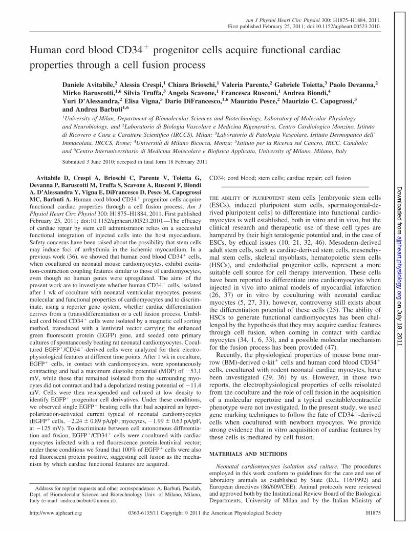
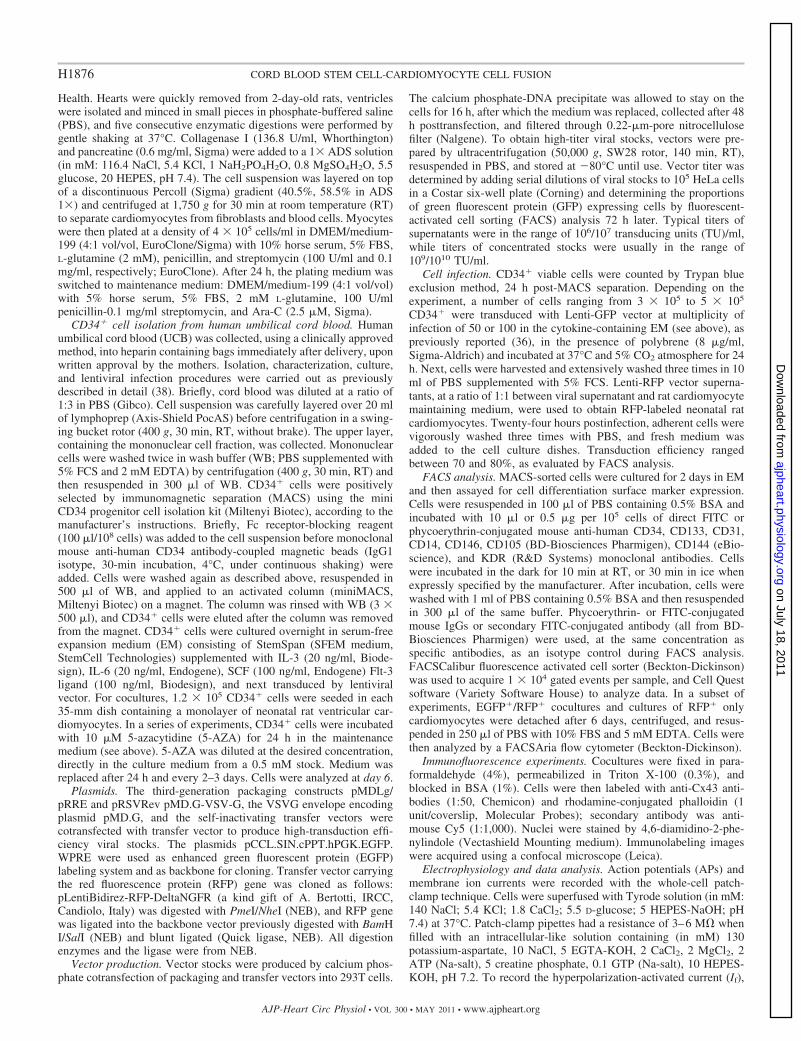
distinguish CD34� cells from the myocytes in cocultures, theywere infected with a lentivirus that provides for stable EGFPexpression. Supplemental Fig. S2A shows a typical FACSanalysis to assess the transduction efficiency of lentiviral in-fection after 72 h, resulting in �70% EGFP�/CD34� cells.The day after lentiviral infection, CD34� cells were seededonto spontaneously beating rat neonatal myocyte primary cul-tures. Although it was previously shown that CD34� UCBcells in coculture with mouse neonatal cardiomyocytes func-tionally integrate within the substrate, no data about the timecourse of this integration were previously described (36). Herewe show that, when in contact with neonatal rat cardiomyo-cytes, the small and rounded EGFP� cells adhered to themyocyte layer and progressively changed their morphology. Atthis stage (day 1), neonatal rat cardiomyocytes contractedspontaneously and showed APs (Fig. 1A), while the EGFP�/CD34� cells did not contract and were electrically inactive anddepolarized (Fig. 1B and Supplemental Video S1). Figure 2Aand Supplemental Video S2 show the membrane potentialrecording and the contractility of an EGFP� cell at day 6 afterseeding onto cardiac myocytes, respectively. At this stage,CD34�-derived EGFP� cells did contract and fired spontane-ous APs, in accordance with our laboratory’s previous findings(36). Recordings from five additional EGFP� cells in cocultureproduced similar results; in these cells, the mean MDP was
Fig. 1. Morphology and action potential (AP) recordings of enhanced green fluorescent protein (EGFP)�/CD34� cells after 1 day in coculture onto neonatal ratcardiac myocytes. A: phase-contrast picture showing a coculture of EGFP�/CD34 with neonatal rat cardiac myocyte layer and AP recording from thecardiomyocyte indicated. B: fluorescence microscopy image of the same field as in A, and membrane potential recording from an EGFP� cell settled on theneonatal rat cardiac myocyte layer (see also Supplemental Video S1). Note the absence of APs and the relatively depolarized resting potential (Vr; �13.5 mV).
H1877CORD BLOOD STEM CELL-CARDIOMYOCYTE CELL FUSION
AJP-Heart Circ Physiol • VOL 300 • MAY 2011 • www.ajpheart.org
on July 18, 2011ajpheart.physiology.org
Dow
nloaded from
�53.1 � 3.2 mV (n � 6). In some dishes, we found fewround-shaped EGFP� cells that were not in contact with thesurrounding myocytes. Voltage-clamp recordings from thesesingle EGFP� cells, directly in the coculture dish, failed toshow either spontaneous activity (Fig. 2B) or contractility (datanot shown). In addition, as reported in Fig. 2B, they maintaineda depolarized resting potential (Vr) (�11.4 � 3.9 mV; n � 7)similar to that of CD34� cells at the beginning of the cocultureprotocol.
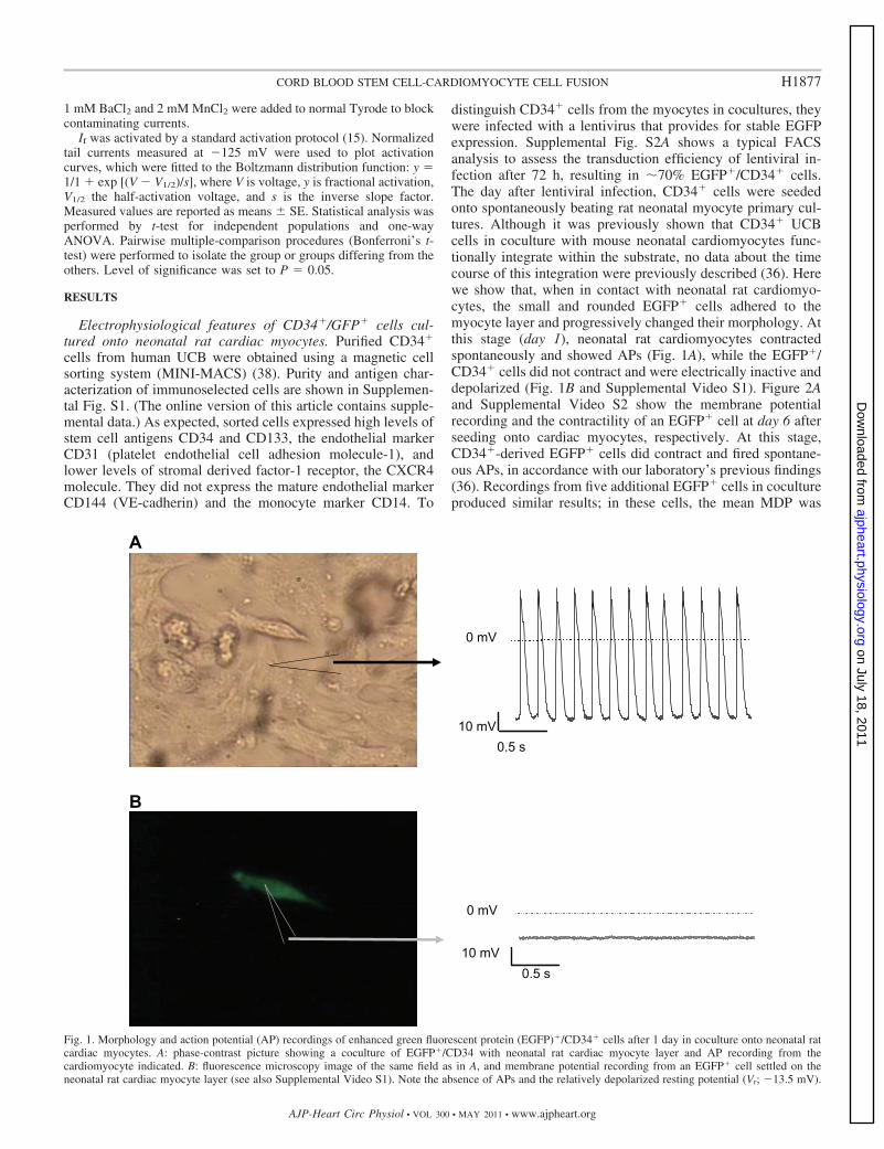
The presence of APs in EGFP� cells indicates gap junction-mediated electrical coupling with the underlying cardiac myo-cytes layer, without necessarily implying the acquisition of acardiac phenotype. To confirm this observation, we performedmulticolor confocal microscopy experiments to detect the ex-pression of connexin-43, the most highly expressed cardiacsubunit of gap junctions, in EGFP� cells stained with rhodamine-conjugated phalloidin, which labels polymerized actin (F-actin)and 4,6-diamidino-2-phenylindole. As shown in Fig. 3 (white
Fig. 2. Morphology and AP recordings of EGFP�/CD34�-derived cells after 6 days in coculture onto neonatal rat cardiac myocytes. A: phase-contrast andoverlapped EGFP fluorescence images showing the integration of an EGFP�/CD34�-derived cell into the rat cardiac myocyte layer and the corresponding APrecording in the same cell. This cell was also synchronously beating with surrounding cardiac myocytes (see Supplemental Video S2). B: EGFP�/CD34�-derived cells that did not contact the cardiomyocyte layer remained essentially round-shaped, lacked electrical activity, and maintained a relativelydepolarized Vr (�13.5 mV).
Fig. 3. Confocal microscopy analysis of EGFP�/CD34�-derived cells and surrounding cardiac myocytes. A: arrows indicate the Cx-43 staining (cyan) in twoEGFP� cells. B: rhodamine-conjugated phalloidin staining in the same cells as in A. Note the sarcomeric structure of the two EGFP� cells. C: merge of allfluorescence signals, showing that EGFP�/CD34� cells acquired a cardiac myocyte phenotype and were connected to surrounding myocytes by gap junctions.Nuclei were labeled with 4,6-diamidino-2-phenylindole (blue).
H1878 CORD BLOOD STEM CELL-CARDIOMYOCYTE CELL FUSION
AJP-Heart Circ Physiol • VOL 300 • MAY 2011 • www.ajpheart.org
on July 18, 2011ajpheart.physiology.org
Dow
nloaded from
arrows), EGFP� cells displayed a cytoskeleton organized insarcomeric structures, connected to each other and to surround-ing cells by gap junctions, thus suggesting that cells arecoupled and that electrical activity can propagate across con-tiguous cells. The percentage of CD34� cells surviving after 6days in coculture was also evaluated. To do this, we havecounted both EGFP� and EGFP� cells in 10 different confocalmicroscopy fields (of 7 different cocultures), and we havefound that 7.7 � 1.6% of the total cells were EGFP�. Com-paring this value with the 13% of CD34� cells in coculture atday 0 (1.2 � 105 CD34� seeded onto 8 � 105 myocytes), wecan estimate that �50% of UCB cells survived.
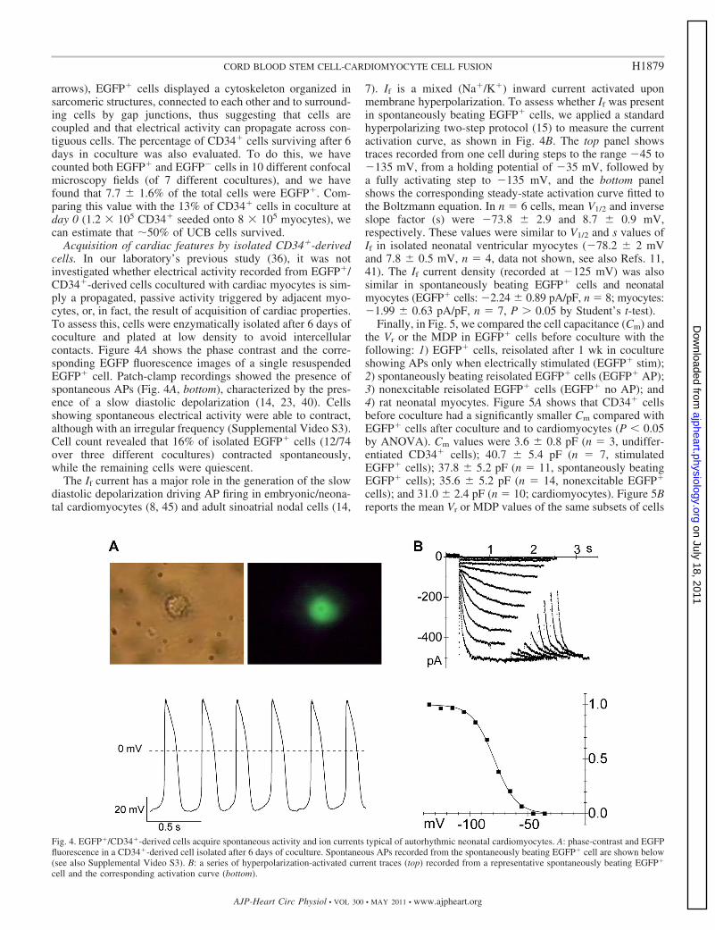
Acquisition of cardiac features by isolated CD34�-derivedcells. In our laboratory’s previous study (36), it was notinvestigated whether electrical activity recorded from EGFP�/CD34�-derived cells cocultured with cardiac myocytes is sim-ply a propagated, passive activity triggered by adjacent myo-cytes, or, in fact, the result of acquisition of cardiac properties.To assess this, cells were enzymatically isolated after 6 days ofcoculture and plated at low density to avoid intercellularcontacts. Figure 4A shows the phase contrast and the corre-sponding EGFP fluorescence images of a single resuspendedEGFP� cell. Patch-clamp recordings showed the presence ofspontaneous APs (Fig. 4A, bottom), characterized by the pres-ence of a slow diastolic depolarization (14, 23, 40). Cellsshowing spontaneous electrical activity were able to contract,although with an irregular frequency (Supplemental Video S3).Cell count revealed that 16% of isolated EGFP� cells (12/74over three different cocultures) contracted spontaneously,while the remaining cells were quiescent.
The If current has a major role in the generation of the slowdiastolic depolarization driving AP firing in embryonic/neona-tal cardiomyocytes (8, 45) and adult sinoatrial nodal cells (14,
7). If is a mixed (Na�/K�) inward current activated uponmembrane hyperpolarization. To assess whether If was presentin spontaneously beating EGFP� cells, we applied a standardhyperpolarizing two-step protocol (15) to measure the currentactivation curve, as shown in Fig. 4B. The top panel showstraces recorded from one cell during steps to the range �45 to�135 mV, from a holding potential of �35 mV, followed bya fully activating step to �135 mV, and the bottom panelshows the corresponding steady-state activation curve fitted tothe Boltzmann equation. In n � 6 cells, mean V1/2 and inverseslope factor (s) were �73.8 � 2.9 and 8.7 � 0.9 mV,respectively. These values were similar to V1/2 and s values ofIf in isolated neonatal ventricular myocytes (�78.2 � 2 mVand 7.8 � 0.5 mV, n � 4, data not shown, see also Refs. 11,41). The If current density (recorded at �125 mV) was alsosimilar in spontaneously beating EGFP� cells and neonatalmyocytes (EGFP� cells: �2.24 � 0.89 pA/pF, n � 8; myocytes:�1.99 � 0.63 pA/pF, n � 7, P 0.05 by Student’s t-test).
Finally, in Fig. 5, we compared the cell capacitance (Cm) andthe Vr or the MDP in EGFP� cells before coculture with thefollowing: 1) EGFP� cells, reisolated after 1 wk in cocultureshowing APs only when electrically stimulated (EGFP� stim);2) spontaneously beating reisolated EGFP� cells (EGFP� AP);3) nonexcitable reisolated EGFP� cells (EGFP� no AP); and4) rat neonatal myocytes. Figure 5A shows that CD34� cellsbefore coculture had a significantly smaller Cm compared withEGFP� cells after coculture and to cardiomyocytes (P 0.05by ANOVA). Cm values were 3.6 � 0.8 pF (n � 3, undiffer-entiated CD34� cells); 40.7 � 5.4 pF (n � 7, stimulatedEGFP� cells); 37.8 � 5.2 pF (n � 11, spontaneously beatingEGFP� cells); 35.6 � 5.2 pF (n � 14, nonexcitable EGFP�
cells); and 31.0 � 2.4 pF (n � 10; cardiomyocytes). Figure 5Breports the mean Vr or MDP values of the same subsets of cells
Fig. 4. EGFP�/CD34�-derived cells acquire spontaneous activity and ion currents typical of autorhythmic neonatal cardiomyocytes. A: phase-contrast and EGFPfluorescence in a CD34�-derived cell isolated after 6 days of coculture. Spontaneous APs recorded from the spontaneously beating EGFP� cell are shown below(see also Supplemental Video S3). B: a series of hyperpolarization-activated current traces (top) recorded from a representative spontaneously beating EGFP�
cell and the corresponding activation curve (bottom).
H1879CORD BLOOD STEM CELL-CARDIOMYOCYTE CELL FUSION
AJP-Heart Circ Physiol • VOL 300 • MAY 2011 • www.ajpheart.org
on July 18, 2011ajpheart.physiology.org
Dow
nloaded from
as in Fig. 5A. CD34� before coculture and quiescent EGFP�
cells had relatively depolarized Vr values (�3.9 � 2 and �23.7 �5.6 mV, respectively). These values were significantly differ-ent (P 0.05) from values recorded in excitable EGFP� cells(�54.4 � 3.4 mV), in spontaneously beating EGFP� cells(�51.8 � 2.3 mV), and in myocytes (�61.1 � 2.0 mV).
Previous studies reported that cell incubation with DNAdemethylating agents, such as 5-AZA, is able to induce acardiogenic program in various cell types (28, 30). We, there-fore, incubated CD34� cells with this drug for 24 h (seeMETHODS for details), followed by culture for 6 days in adrug-free medium. Electrophysiological analysis at day 6 re-vealed that 5-AZA-treated CD34� cells maintained a relativelydepolarized Vr (�15.9 � 2.7 mV, n � 10), and neitherexpressed sodium or pacemaker (f) currents (data not shown),suggesting the lack of cardiac differentiation. Moreover5-AZA-treated CD34� cells had a mean Cm of 24.6 � 7.8 pF(n � 12). These values are not significantly different fromeither those of day-matched untreated CD34� cells (Vr ��15.7 � 7.9 mV, Cm � 35.1 � 1.4 pF, n � 3) or from thoseof CD34� cells not in contact with cardiomyocytes (seeabove). These data indicate that 5-AZA is not sufficient toinduce cardiac differentiation of UCB CD34� cells, in linewith previous reports indicating that such treatment does notinduce cord blood progenitor differentiation, but improvestheir expansion capacity and stemness (4, 43).
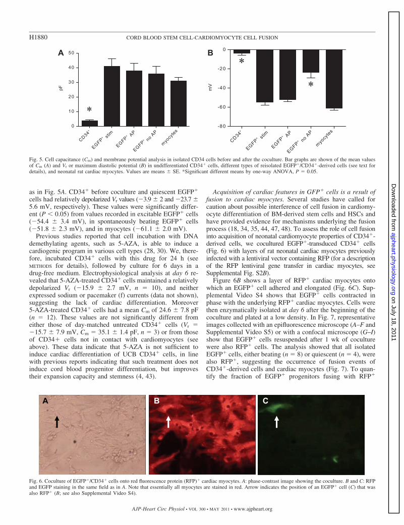
Acquisition of cardiac features in GFP� cells is a result offusion to cardiac myocytes. Several studies have called forcaution about possible interference of cell fusion in cardiomy-ocyte differentiation of BM-derived stem cells and HSCs andhave provided evidence for mechanisms underlying the fusionprocess (18, 34, 35, 44, 47, 48). To assess the role of cell fusioninto acquisition of neonatal cardiomyocyte properties of CD34�-derived cells, we cocultured EGFP�-transduced CD34� cells(Fig. 6) with layers of rat neonatal cardiac myocytes previouslyinfected with a lentiviral vector containing RFP (for a descriptionof the RFP lentiviral gene transfer in cardiac myocytes, seeSupplemental Fig. S2B).
Figure 6B shows a layer of RFP� cardiac myocytes ontowhich an EGFP� cell adhered and elongated (Fig. 6C). Sup-plemental Video S4 shows that EGFP� cells contracted inphase with the underlying RFP� cardiac myocytes. Cells werethen enzymatically isolated at day 6 after the beginning of thecoculture and plated at a low density. In Fig. 7, representativeimages collected with an epifluorescence microscope (A–F andSupplemental Video S5) or with a confocal microscope (G–I)show that EGFP� cells resuspended after 1 wk of coculturewere also RFP� cells. The analysis showed that all isolatedEGFP� cells, either beating (n � 8) or quiescent (n � 4), werealso RFP�, suggesting the occurrence of fusion events ofCD34�-derived cells and cardiac myocytes (Fig. 7). To quan-tify the fraction of EGFP� progenitors fusing with RFP�
Fig. 5. Cell capacitance (Cm) and membrane potential analysis in isolated CD34 cells before and after the coculture. Bar graphs are shown of the mean valuesof Cm (A) and Vr or maximum diastolic potential (B) in undifferentiated CD34� cells, different types of reisolated EGFP�/CD34�-derived cells (see text fordetails), and neonatal rat cardiac myocytes. Values are means � SE. *Significant different means by one-way ANOVA, P � 0.05.
Fig. 6. Coculture of EGFP�/CD34� cells onto red fluorescence protein (RFP)� cardiac myocytes. A: phase-contrast image showing the coculture. B and C: RFPand EGFP staining in the same field as in A. Note that essentially all myocytes are stained in red. Arrow indicates the position of an EGFP� cell (C) that wasalso RFP� (B; see also Supplemental Video S4).
H1880 CORD BLOOD STEM CELL-CARDIOMYOCYTE CELL FUSION
AJP-Heart Circ Physiol • VOL 300 • MAY 2011 • www.ajpheart.org
on July 18, 2011ajpheart.physiology.org
Dow
nloaded from
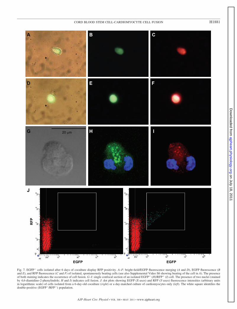
Fig. 7. EGFP� cells isolated after 6 days of coculture display RFP positivity. A–F: bright-field/EGFP fluorescence merging (A and D), EGFP fluorescence (Band E), and RFP fluorescence (C and F) of isolated, spontaneously beating cells (see also Supplemental Video S6 showing beating of the cell in A). The presenceof both staining indicates the occurrence of cell fusion. G–I: single confocal section of an isolated EGFP� (H)/RFP� (I) cell. The presence of two nuclei (stainedby 4,6-diamidino-2-phenylindole; H and I) indicates cell fusion. J: dot plots showing EGFP (X-axes) and RFP (Y-axes) fluorescence intensities (arbitrary unitsin logarithmic scale) of cells isolated from a 6-day-old coculture (right) or a day-matched culture of cardiomyocytes only (left). The white square identifies thedouble-positive (EGFP�/RFP�) population.
H1881CORD BLOOD STEM CELL-CARDIOMYOCYTE CELL FUSION
AJP-Heart Circ Physiol • VOL 300 • MAY 2011 • www.ajpheart.org
on July 18, 2011ajpheart.physiology.org
Dow
nloaded from
cardiomyocytes, we carried out flow cytometry analysis. Infour independent experiments, flow cytometry analysis showedthe absence of EGFP�/RFP� cell population (Fig. 7J). Thisconfirmed that 100% of EGFP� cells were also RFP�, indi-cating the lack of cell-autonomous transdifferentiation ofCD34� cells into cardiac myocytes.
DISCUSSION
In two previous reports (29, 36), it was shown that humancord blood-derived CD34� progenitor cells are able to inte-grate and connect with primary cultures of spontaneouslybeating neonatal cardiomyocytes. The main difference betweenmouse BM-derived c-kit� cells and human cord blood CD34�
cells consisted in a different degree of functional integrationwith the myocyte monolayer (29, 36). In fact, while c-kit� cellsin contact with neonatal cardiac myocytes presented inwardand outward K� currents, but did not show any evidence ofelectromechanical coupling with surrounding cells (absence ofAPs), cord blood CD34� cells exhibited spontaneous calciumtransients and functional gap junctions (36). In the latter study,however, the question as to whether human cord blood-derivedCD34� progenitor cells simply express proteins (connexins)that allow them to electrically connect with the substrate orwhether they may actually acquire a cardiac phenotype and, insuch case, if it was the result of a trans-differentiation or a cellfusion process was not addressed. Furthermore, while severalother reports have described in vitro differentiation (or lack ofdifferentiation) of HSC/progenitor cells into cardiac cells onthe basis of morphological and marker analysis, relatively littleinformation is available about the relationship between func-tional differentiation (i.e., ability to generate APs), or potentialelectrophysiological integration into a contractile and electri-cally active tissue, such as the myocardium, and the fusionprocess. These issues are of relevance in the light of studiesdescribing the modification of electrical impulse propaga-tion by stem cells cocultured with cardiac myocytes (12) andcalling for caution about the generation of potential heartrhythm disturbances following cellular administration (es-pecially using skeletal myoblasts) to the ischemic myocar-dium, both in animal models and in patients (16, 17, 22).
Several sources of stem cells have been used so far in thesearch of a cell type suitable for cardiac regenerative interven-tions; pluripotent stem cells of either embryonic [ESCs (21)] oradult origin [induced pluripotent stem cells and spermatogo-nial-derived cells, (20, 46)] can easily and reproducibly gen-erate electrically active cardiomyocytes and can integrate in ahost myocardium, both in vitro and in vivo. It is less certainwhether other types of stem cells derived from adult tissue, andin particular HSC/progenitor cells, can be used for the samepurpose, despite a wealth of evidence for an improved cardiacfunction following injection of HSCs in the ischemic myocar-dium (reviewed in Ref. 2) and for their myocardial differenti-ation based on morphological, marker (5, 13, 19, 27, 31) andelectrophysiological evidence (42). It is also still controversialwhether in vivo cell fusion events do occur. A major criticismis that an apparent occurrence of fusion may result frommethodological artifacts in experiments (especially histologi-cal analyses), addressed to localize injected stem cells intoischemic tissues (reviewed in Ref. 3). In addition, a mechanisminvolving the interaction of integrin-�4�1 and the VCAM-1
cell adhesion molecule has been recently found to be respon-sible for fusion events of hematopoietic progenitors and car-diomyocytes in culture (47).
Assessment of cell fusion is not easily accomplished. Sys-tems based on the Cre-LoxP system, for example, have beenused mainly in vivo to demonstrate (1, 31, 35) or exclude (9)that stem cells injected in the myocardium undergo cell fusionwith myocytes. This technique, however, is not suitable forquantification of fusion in vitro due to a relatively low effi-ciency of Cre enzyme that does not allow expression ofreporter genes as a result of recombination of LoxP sites in100% of cells (M. Pesce, unpublished observations).
In this study, we provide evidence that acquisition of cardiacfeatures is a consequence of cell fusion. Using a two-colorlabeling system based on transduction with lentiviral vectorscarrying EGFP and RFP, we observed green (hCD34�-de-rived)/red (rat cardiac myocytes-derived) hybrid cells showingspontaneous contractility (Fig. 7). We decided to use thissystem to avoid possible trans-infection, as may occur, forexample, by using adenovirus-based gene transfer (our unpub-lished results and Ref. 31) and possible unspecific cell labelingdue to leakiness of vital fluorescent dyes, such as 1,1’-diocta-decyl-3,3,3’,3’-tetramethylindocarbocyanine perchlorate/ 3,3’-dioctadecyloxacarbocyanine perchlorate labeling [DiI/DiO (5,18)]. In addition, this system has been already used to assess theeffects of cell fusion in studies in which different cell types werecocultured with cardiac myocytes (31, 39) or to show fusion ofBM-derived stem cells and cardiac myocytes in vivo (24).
In agreement with our laboratory’s previous study (36), wehave confirmed that human CD34� hematopoietic progenitorsintegrate within the primary culture of rat neonatal cardiomy-ocytes. Here we show that this coculture system induces achange in morphology of CD34�/EGFP� cells, within the firstday, which is not immediately accompanied by electricalcoupling, as demonstrated by their relatively depolarized Vr.The electrical coupling mediated by the connexin-43 proteinsis then accomplished in the first week, as demonstrated by thehyperpolarization of the Vr of EGFP� cell and by their abilityto propagate APs.
FACS analysis run on transduced cells, isolated after 6 daysof coculture, and showing that 100% of the EGFP� cellsrecovered were also RFP� (Fig. 7) strongly suggests thatacquisition of cardiac features is the result of fusion eventsbetween cardiac myocytes and CD34�-derived cells. Further-more, electrophysiological recordings did not show any differ-ence between EGFP� cells and neonatal cardiac myocytes,supporting the hypothesis that these EGFP� cardiomyocytesoriginate indeed from the fusion between electrically passiveCD34� cells and cardiac myocytes. In accordance with thisobservation, CD34�/GFP� cells that did not contact neonatalcardiomyocytes during the coculture period did not show thefunctional properties of excitable cells (see Figs. 2B and 5B).
Finally, incubation with 5-AZA, a treatment reported toinduce differentiation of mesenchymal progenitor cells towarda cardiogenic lineage (28, 30), failed to induce any electro-physiological change in CD34� cells. These data, in agreementwith the previously reported lack of upregulation of humancardiac genes in CD34� cells during coculture (36), ruleagainst a paracrine effect or a nuclear reprogramming process,induced by soluble factors deriving from the coculture condi-tions.
H1882 CORD BLOOD STEM CELL-CARDIOMYOCYTE CELL FUSION
AJP-Heart Circ Physiol • VOL 300 • MAY 2011 • www.ajpheart.org
on July 18, 2011ajpheart.physiology.org
Dow
nloaded from
In summary, the present study shows that direct culture ofhuman CD34� cells with neonatal cardiac myocytes deter-mines cell fusion events, leading to the acquisition of cardiacphysiological features rather than inducing differentiation. Al-though it remains controversial whether similar fusion eventsbetween hematopoietic-derived progenitors and adult cardiacmyocytes also occur in vivo in the ischemic myocardium, thelack of cell autonomous differentiation in vitro suggest thatCD34� UCB cells do not represent a suitable cell type toderive functional myocardium for cardiac regenerative inter-ventions.
ACKNOWLEDGEMENTS
The authors thank Dr. Daniela Longoni (Clinica Pediatrica, Universita degliStudi di Milano-Bicocca, Monza, Italy), Dr. Domenica Mammoliti (Ospedaledi Melzo, Italy), and the Obstetrics Teams at Clinica Ostetrica Ginecologica atOspedale S. Gerardo, Monza, and Ospedale di Melzo for help with collectionof cord blood units used in this study.
Present address of D. Avitabile and S. Truffa: State University San DiegoHeart Institute, San Diego, CA.
GRANTS
This work was supported by European Union (EU) Normacor CT2006-018676 to D. DiFrancesco; EU Application and process optimization of humanstem cells for myocardium repair LSHB-CT-2004-502988 to M. C. Capogrossiand M. Pesce; EU Thercord LSHB-CT-2005-018817 to M. Pesce; and Fondoper gli Investimenti della Ricerca di Base RBLA035A4X to M. C. Capogrossiand D. DiFrancesco.
DISCLOSURES
No conflicts of interest, financial or otherwise, are declared by the author(s).
REFERENCES
1. Alvarez-Dolado M, Pardal R, Garcia-Verdugo JM, Fike JR, Lee HO,Pfeffer K, Lois C, Morrison SJ, Alvarez-Buylla A. Fusion of bone-marrow-derived cells with Purkinje neurons, cardiomyocytes and hepato-cytes. Nature 425: 968–973, 2003.
2. Anversa P, Kajstura J, Leri A. If I can stop one heart from breaking.Circulation 115: 829–832, 2007.
3. Anversa P, Leri A, Rota M, Hosoda T, Bearzi C, Urbanek K, KajsturaJ, Bolli R. Concise review: stem cells, myocardial regeneration, andmethodological artifacts. Stem Cells 25: 589–601, 2007.
4. Araki H, Baluchamy S, Yoshinaga K, Petro B, Petiwala S, Parajuli R,Milhem M, Lavelle D, DeSimone J, Mahmud N. Cord blood stem cellexpansion is permissive to epigenetic regulation and environmental cues.Exp Hematol 37: 1084–1095, 2009.
5. Badorff C, Brandes RP, Popp R, Rupp S, Urbich C, Aicher A,Fleming I, Busse R, Zeiher AM, Dimmeler S. Transdifferentiation ofblood-derived human adult endothelial progenitor cells into functionallyactive cardiomyocytes. Circulation 107: 1024–1032, 2003.
6. Balsam LB, Wagers AJ, Christensen JL, Kofidis T, Weissman IL,Robbins RC. Haematopoietic stem cells adopt mature haematopoieticfates in ischaemic myocardium. Nature 428: 668–673, 2004.
7. Barbuti A, Baruscotti M, DiFrancesco D. The pacemaker current: frombasics to the clinics. J Cardiovasc Electrophysiol 18: 342–347, 2007.
8. Barbuti A, Crespi A, Capilupo D, Mazzocchi N, Baruscotti M, Di-Francesco D. Molecular composition and functional properties of f-chan-nels in murine embryonic stem cell-derived pacemaker cells. J Mol CellCardiol 46: 343–351, 2009.
9. Bearzi C, Rota M, Hosoda T, Tillmanns J, Nascimbene A, De AA,Yasuzawa-Amano S, Trofimova I, Siggins RW, Lecapitaine N, Cas-capera S, Beltrami AP, D’Alessandro DA, Zias E, Quaini F, UrbanekK, Michler RE, Bolli R, Kajstura J, Leri A, Anversa P. Human cardiacstem cells. Proc Natl Acad Sci U S A 104: 14068–14073, 2007.
10. Behfar A, Perez-Terzic C, Faustino RS, Arrell DK, Hodgson DM,Yamada S, Puceat M, Niederlander N, Alekseev AE, Zingman LV,Terzic A. Cardiopoietic programming of embryonic stem cells for tumor-free heart repair. J Exp Med 204: 405–420, 2007.
11. Cerbai E, Pino R, Sartiani L, Mugelli A. Influence of postnatal-development on I(f) occurrence and properties in neonatal rat ventricularmyocytes. Cardiovasc Res 42: 416–423, 1999.
12. Chang MG, Tung L, Sekar RB, Chang CY, Cysyk J, Dong P, MarbanE, Abraham MR. Proarrhythmic potential of mesenchymal stem celltransplantation revealed in an in vitro coculture model. Circulation 113:1832–1841, 2006.
13. Condorelli G, Borello U, De Angelis L, Latronico M, Sirabella D,Coletta M, Galli R, Balconi G, Follenzi A, Frati G, Cusella De AngelisMG, Gioglio L, Amuchastegui S, Adorini L, Naldini L, Vescovi A,Dejana E, Cossu G. Cardiomyocytes induce endothelial cells to trans-differentiate into cardiac muscle: implications for myocardium regenera-tion. Proc Natl Acad Sci U S A 98: 10733–10738, 2001.
14. DiFrancesco D. Pacemaker mechanisms in cardiac tissue. Annu RevPhysiol 55: 455–472, 1993.
15. DiFrancesco D, Ferroni A, Mazzanti M, Tromba C. Properties of thehyperpolarizing-activated current (if) in cells isolated from the rabbitsino-atrial node. J Physiol 377: 61–88, 1986.
16. Dudley SC Jr. Beware of cells bearing gifts: cell replacement therapy andarrhythmic risk. Circ Res 97: 99–101, 2005.
17. Fernandes S, Amirault JC, Lande G, Nguyen JM, Forest V, BignolaisO, Lamirault G, Heudes D, Orsonneau JL, Heymann MF, Charpen-tier F, Lemarchand P. Autologous myoblast transplantation after myo-cardial infarction increases the inducibility of ventricular arrhythmias.Cardiovasc Res 69: 348–358, 2006.
18. Garbade J, Schubert A, Rastan AJ, Lenz D, Walther T, Gummert JF,Dhein S, Mohr FW. Fusion of bone marrow-derived stem cells withcardiomyocytes in a heterologous in vitro model. Eur J Cardiothorac Surg28: 685–691, 2005.
19. Gruh I, Beilner J, Blomer U, Schmiedl A, Schmidt-Richter I, KruseML, Haverich A, Martin U. No evidence of transdifferentiation ofhuman endothelial progenitor cells into cardiomyocytes after coculturewith neonatal rat cardiomyocytes. Circulation 113: 1326–1334, 2006.
20. Guan K, Wagner S, Unsold B, Maier LS, Kaiser D, Hemmerlein B,Nayernia K, Engel W, Hasenfuss G. Generation of functional cardio-myocytes from adult mouse spermatogonial stem cells. Circ Res 100:1615–1625, 2007.
21. Habib M, Caspi O, Gepstein L. Human embryonic stem cells forcardiomyogenesis. J Mol Cell Cardiol 45: 462–474, 2008.
22. Hagege AA, Marolleau JP, Vilquin JT, Alheritiere A, Peyrard S,Duboc D, Abergel E, Messas E, Mousseaux E, Schwartz K, Desnos M,Menasche P. Skeletal myoblast transplantation in ischemic heart failure:long-term follow-up of the first phase I cohort of patients. Circulation 114:I108–I113, 2006.
23. Hescheler J, Fleischmann BK, Lentini S, Maltsev VA, Rohwedel J,Wobus AM, Addicks K. Embryonic stem cells: a model to studystructural and functional properties in cardiomyogenesis. Cardiovasc Res36: 149–162, 1997.
24. Ishikawa F, Shimazu H, Shultz LD, Fukata M, Nakamura R, Lyons B,Shimoda K, Shimoda S, Kanemaru T, Nakamura K, Ito H, Kaji Y,Perry AC, Harada M. Purified human hematopoietic stem cells contrib-ute to the generation of cardiomyocytes through cell fusion. FASEB J 20:950–952, 2006.
25. Joggerst SJ, Hatzopoulos AK. Stem cell therapy for cardiac repair:benefits and barriers. Expert Rev Mol Med 11: e20, 2009.
26. Kajstura J, Rota M, Whang B, Cascapera S, Hosoda T, Bearzi C,Nurzynska D, Kasahara H, Zias E, Bonafe M, Nadal-Ginard B,Torella D, Nascimbene A, Quaini F, Urbanek K, Leri A, Anversa P.Bone marrow cells differentiate in cardiac cell lineages after infarctionindependently of cell fusion. Circ Res 96: 127–137, 2005.
27. Koyanagi M, Haendeler J, Badorff C, Brandes RP, Hoffmann J,Pandur P, Zeiher AM, Kuhl M, Dimmeler S. Non-canonical Wntsignaling enhances differentiation of human circulating progenitor cells tocardiomyogenic cells. J Biol Chem 280: 16838–16842, 2005.
28. Labovsky V, Hofer EL, Feldman L, Fernandez VV, Garcia RH,Bayes-Genis A, Hernando IA, Levin MJ, Chasseing NA. Cardiomyo-genic differentiation of human bone marrow mesenchymal cells: Role ofcardiac extract from neonatal rat cardiomyocytes. Differentiation 79:93–101, 2010.
29. Lagostena L, Avitabile D, De Falco E, Orlandi A, Grassi F, Iachini-noto MG, Ragone G, Fucile S, Pompilio G, Eusebi F, Pesce M,Capogrossi MC. Electrophysiological properties of mouse bone marrowc-kit� cells co-cultured onto neonatal cardiac myocytes. Cardiovasc Res66: 482–492, 2005.
H1883CORD BLOOD STEM CELL-CARDIOMYOCYTE CELL FUSION
AJP-Heart Circ Physiol • VOL 300 • MAY 2011 • www.ajpheart.org
on July 18, 2011ajpheart.physiology.org
Dow
nloaded from
30. Makino S, Fukuda K, Miyoshi S, Konishi F, Kodama H, Pan J, SanoM, Takahashi T, Hori S, Abe H, Hata J, Umezawa A, Ogawa S.Cardiomyocytes can be generated from marrow stromal cells in vitro. JClin Invest 103: 697–705, 1999.
31. Matsuura K, Wada H, Nagai T, Iijima Y, Minamino T, Sano M,Akazawa H, Molkentin JD, Kasanuki H, Komuro I. Cardiomyocytesfuse with surrounding noncardiomyocytes and reenter the cell cycle. J CellBiol 167: 351–363, 2004.
32. Mummery C, Ward-van Oostwaard D, Doevendans P, Spijker R, vanden BS, Hassink R, van der HM, Opthof T, Pera M, de la Riviere AB,Passier R, Tertoolen L. Differentiation of human embryonic stem cells tocardiomyocytes: role of coculture with visceral endoderm-like cells. Cir-culation 107: 2733–2740, 2003.
33. Murry CE, Soonpaa MH, Reinecke H, Nakajima H, Nakajima HO,Rubart M, Pasumarthi KB, Virag JI, Bartelmez SH, Poppa V, Brad-ford G, Dowell JD, Williams DA, Field LJ. Haematopoietic stem cellsdo not transdifferentiate into cardiac myocytes in myocardial infarcts.Nature 428: 664–668, 2004.
34. Nygren JM, Jovinge S, Breitbach M, Sawen P, Roll W, Hescheler J,Taneera J, Fleischmann BK, Jacobsen SE. Bone marrow-derived he-matopoietic cells generate cardiomyocytes at a low frequency through cellfusion, but not transdifferentiation. Nat Med 10: 494–501, 2004.
35. Oh H, Bradfute SB, Gallardo TD, Nakamura T, Gaussin V, MishinaY, Pocius J, Michael LH, Behringer RR, Garry DJ, Entman ML,Schneider MD. Cardiac progenitor cells from adult myocardium: homing,differentiation, and fusion after infarction. Proc Natl Acad Sci U S A 100:12313–12318, 2003.
36. Orlandi A, Pagani F, Avitabile D, Bonanno G, Scambia G, Vigna E,Grassi F, Eusebi F, Fucile S, Pesce M, Capogrossi MC. Functionalproperties of cells obtained from human cord blood CD34� stem cells andmouse cardiac myocytes in coculture. Am J Physiol Heart Circ Physiol294: H1541–H1549, 2008.
37. Orlic D, Kajstura J, Chimenti S, Jakoniuk I, Anderson SM, Li B,Pickel J, McKay R, Nadal-Ginard B, Bodine DM, Leri A, Anversa P.Bone marrow cells regenerate infarcted myocardium. Nature 410: 701–705, 2001.
38. Pesce M, Orlandi A, Iachininoto MG, Straino S, Torella AR, RizzutiV, Pompilio G, Bonanno G, Scambia G, Capogrossi MC. Myoendo-
thelial differentiation of human umbilical cord blood-derived stem cells inischemic limb tissues. Circ Res 93: e51–e62, 2003.
39. Pfister O, Mouquet F, Jain M, Summer R, Helmes M, Fine A, ColucciWS, Liao R. CD31- but Not CD31� cardiac side population cells exhibitfunctional cardiomyogenic differentiation. Circ Res 97: 52–61, 2005.
40. Qu J, Barbuti A, Protas L, Santoro B, Cohen IS, Robinson RB. HCN2overexpression in newborn and adult ventricular myocytes: distinct effectson gating and excitability. Circ Res 89: E8–E14, 2001.
41. Qu J, Cohen IS, Robinson RB. Sympathetic innervation alters activationof pacemaker current (If) in rat ventricle. J Physiol 526: 561–569, 2000.
42. Rota M, Kajstura J, Hosoda T, Bearzi C, Vitale S, Esposito G,Iaffaldano G, Padin-Iruegas ME, Gonzalez A, Rizzi R, Small N,Muraski J, Alvarez R, Chen X, Urbanek K, Bolli R, Houser SR, LeriA, Sussman MA, Anversa P. Bone marrow cells adopt the cardiomyo-genic fate in vivo. Proc Natl Acad Sci U S A 104: 17783–17788, 2007.
43. Suzuki M, Harashima A, Okochi A, Yamamoto M, Nakamura S,Motoda R, Yamasaki F, Orita K. 5-Azacytidine supports the long-termrepopulating activity of cord blood CD34(�) cells. Am J Hematol 77:313–315, 2004.
44. Terada N, Hamazaki T, Oka M, Hoki M, Mastalerz DM, Nakano Y,Meyer EM, Morel L, Petersen BE, Scott EW. Bone marrow cells adoptthe phenotype of other cells by spontaneous cell fusion. Nature 416:542–545, 2002.
45. Yasui K, Liu W, Opthof T, Kada K, Lee JK, Kamiya K, Kodama I.I(f) current and spontaneous activity in mouse embryonic ventricularmyocytes. Circ Res 88: 536–542, 2001.
46. Zhang J, Wilson GF, Soerens AG, Koonce CH, Yu J, Palecek SP,Thomson JA, Kamp TJ. Functional cardiomyocytes derived from humaninduced pluripotent stem cells. Circ Res 104: e30–e41, 2009.
47. Zhang S, Shpall E, Willerson JT, Yeh ET. Fusion of human hemato-poietic progenitor cells and murine cardiomyocytes is mediated by alpha4 beta 1 integrin/vascular cell adhesion molecule-1 interaction. Circ Res100: 693–702, 2007.
48. Zhang S, Wang D, Estrov Z, Raj S, Willerson JT, Yeh ET. Both cellfusion and transdifferentiation account for the transformation of humanperipheral blood CD34-positive cells into cardiomyocytes in vivo. Circu-lation 110: 3803–3807, 2004.
H1884 CORD BLOOD STEM CELL-CARDIOMYOCYTE CELL FUSION
AJP-Heart Circ Physiol • VOL 300 • MAY 2011 • www.ajpheart.org
on July 18, 2011ajpheart.physiology.org
Dow
nloaded from

![Identification of Novel Human NK Cell Progenitor Subsets · Lin CD34+CD7+CD10+ fraction as a human equivalent of the murine NK cell progenitor (NKP) [8]. Both lymphoid primed multipotent](https://img.pdfslide.net/doc/110x75/611e7eed0504d803c3437279/identiication-of-novel-human-nk-cell-progenitor-subsets-lin-cd34cd7cd10-fraction.jpg)