Embed Size (px)
Citation preview
Human Respiratory Syncytial Virus Nucleoprotein and InclusionBodies Antagonize the Innate Immune Response Mediated by MDA5and MAVS
Aaron W. Lifland,a Jeenah Jung,a Eric Alonas,a Chiara Zurla,a James E. Crowe, Jr.,b and Philip J. Santangeloa
Wallace H. Coulter Department of Biomedical Engineering, Georgia Institute of Technology and Emory University, Atlanta, Georgia, USA,a and Departments of Pediatricsand Pathology, Microbiology and Immunology, Vanderbilt University School of Medicine, Nashville, Tennessee, USAb
Currently, the spatial distribution of human respiratory syncytial virus (hRSV) proteins and RNAs in infected cells is still underinvestigation, with many unanswered questions regarding the interaction of virus-induced structures and the innate immunesystem. Very few studies of hRSV have used subcellular imaging as a means to explore the changes in localization of retinoic-acid-inducible gene-I (RIG-I)-like receptors or the mitochondrial antiviral signaling (MAVS) protein, in response to the infec-tion and formation of viral structures. In this investigation, we found that both RIG-I and melanoma differentiation-associatedgene 5 (MDA5) colocalized with viral genomic RNA and the nucleoprotein (N) as early as 6 h postinfection (hpi). By 12 hpi,MDA5 and MAVS were observed within large viral inclusion bodies (IB). We used a proximity ligation assay (PLA) and deter-mined that the N protein was in close proximity to MDA5 and MAVS in IBs throughout the course of the infection. Similar re-sults were found with the transient coexpression of N and the phosphoprotein (P). Additionally, we demonstrated that the local-ization of MDA5 and MAVS in IBs inhibited the expression of interferon � mRNA 27-fold following Newcastle disease virusinfection. From these data, we concluded that the N likely interacts with MDA5, is in close proximity to MAVS, and localizesthese molecules within IBs in order to attenuate the interferon response. To our knowledge, this is the first report of a specificfunction for hRSV IBs and of the hRSV N protein as a modulator of the innate immune response.
Currently, the spatial biology of human respiratory syncytialvirus (hRSV) is still under investigation, with many questions
still unanswered about the ultrastructure of viral and host proteincomplexes in infected cells. To date, the function and completecomposition of cytoplasmic inclusion bodies, as well as details ofthe host cell interactions with these virus-specific structures, arestill unknown. Host cell interactions are accomplished throughvaried means, but first-line detectors of viral infections primar-ily involve two kinds of receptors: the cytoplasmic pattern rec-ognition receptors, including the retinoic acid-inducible geneI (RIG-I) and melanoma differentiation-associated gene 5(MDA5), both RNA helicases, and the pathogen-associated mo-lecular pattern receptors, known as the Toll-like receptors (TLRs)(1, 18). TLR3 and TLR7 sense double- and single-stranded RNA,respectively, and in most cell types reside within endosomes (18).In contrast, TLR4, expressed on the cell surface, plays a role insensing viral surface glycoproteins (18). RIG-I and MDA5 residewithin the cytosol, and there are reports of them responding toboth single- and double-stranded RNA (18). TLR signaling, me-diated by MyD88, and RIG-I-like receptor signaling, mediated bythe mitochondrial antiviral signaling (MAVS) protein (alsoknown as the interferon promoter-stimulating factor 1 [IPS-1]adaptor protein), results in the production of interferon (18).
In response to hRSV, both TLRs and RIG-I-like receptors havebeen implicated in the general response to the infection. ThehRSV fusion (F) protein has been shown to antagonize TLR4 in amouse model (12). In A549 and mouse embryonic fibroblast(MEF) cells, it has been shown that RIG-I was essential for pro-moting the innate immune response, with MDA5 playing an aux-iliary role (16, 17, 30). Even though there has been some confusionin the literature, recent results depicting the early events (0 to 6 hpostinfection [hpi]) of an hRSV infection have shown that both
RIG-I and MDA5 play a significant role in both NF-�B and IRF3signaling (30).
In addition, a number of viral proteins have been implicated indecreasing the innate response to hRSV. The highly glycosylatedattachment (G) protein has been shown to inhibit TLR3/4 signal-ing in dendritic cells. Nonstructural protein 1 (NS1) expressionhas been correlated with a decrease in TRAF3 levels, and non-structural protein 2 (NS2) has been shown to interact with RIG-Iand its expression to correlate with decreases in TRAF3 andSTAT2 levels (2, 27).
One drawback of many investigations of hRSV induction of theinnate immune response to date has been the lack of spatiotem-poral information at the level of the single cell. Previous work inthis area has provided considerable insight primarily through bio-chemical assays, but these investigations often have neglected tostudy the events of interest within the context of the cell, retainingthe spatial organization of hRSV proteins and RNAs. Many rep-resentations of the signaling events during hRSV infections depicthRSV genomic viral RNA as randomly distributed throughout thecytoplasm even though this pattern has never been shown viamicroscopic investigation. Recent time course imaging of hRSVgenomic RNA using single-molecule sensitive hybridizationprobes clearly showed the RNA and the N protein localized near
Received 25 January 2012 Accepted 14 May 2012
Published ahead of print 23 May 2012
Address correspondence to Philip J. Santangelo, [email protected].
Copyright © 2012, American Society for Microbiology. All Rights Reserved.
doi:10.1128/JVI.00215-12
August 2012 Volume 86 Number 15 Journal of Virology p. 8245–8258 jvi.asm.org 8245
Dow
nloa
ded
from
http
s://j
ourn
als.
asm
.org
/jour
nal/j
vi o
n 01
Dec
embe
r 20
21 b
y 58
.79.
43.6
2.
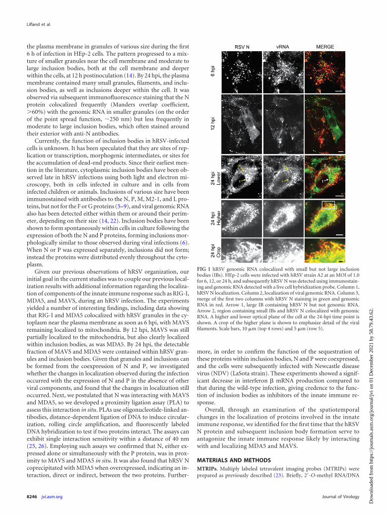
the plasma membrane in granules of various size during the first6 h of infection in HEp-2 cells. The pattern progressed to a mix-ture of smaller granules near the cell membrane and moderate tolarge inclusion bodies, both at the cell membrane and deeperwithin the cells, at 12 h postinoculation (14). By 24 hpi, the plasmamembrane contained many small granules, filaments, and inclu-sion bodies, as well as inclusions deeper within the cell. It wasobserved via subsequent immunofluorescence staining that the Nprotein colocalized frequently (Manders overlap coefficient,�60%) with the genomic RNA in smaller granules (on the orderof the point spread function, �250 nm) but less frequently inmoderate to large inclusion bodies, which often stained aroundtheir exterior with anti-N antibodies.
Currently, the function of inclusion bodies in hRSV-infectedcells is unknown. It has been speculated that they are sites of rep-lication or transcription, morphogenic intermediates, or sites forthe accumulation of dead-end products. Since their earliest men-tion in the literature, cytoplasmic inclusion bodies have been ob-served late in hRSV infections using both light and electron mi-croscopy, both in cells infected in culture and in cells frominfected children or animals. Inclusions of various size have beenimmunostained with antibodies to the N, P, M, M2-1, and L pro-teins, but not for the F or G proteins (5–9), and viral genomic RNAalso has been detected either within them or around their perim-eter, depending on their size (14, 22). Inclusion bodies have beenshown to form spontaneously within cells in culture following theexpression of both the N and P proteins, forming inclusions mor-phologically similar to those observed during viral infections (6).When N or P was expressed separately, inclusions did not form;instead the proteins were distributed evenly throughout the cyto-plasm.
Given our previous observations of hRSV organization, ourinitial goal in the current studies was to couple our previous local-ization results with additional information regarding the localiza-tion of components of the innate immune response such as RIG-I,MDA5, and MAVS, during an hRSV infection. The experimentsyielded a number of interesting findings, including data showingthat RIG-I and MDA5 colocalized with hRSV granules in the cy-toplasm near the plasma membrane as soon as 6 hpi, with MAVSremaining localized to mitochondria. By 12 hpi, MAVS was stillpartially localized to the mitochondria, but also clearly localizedwithin inclusion bodies, as was MDA5. By 24 hpi, the detectablefraction of MAVS and MDA5 were contained within hRSV gran-ules and inclusion bodies. Given that granules and inclusions canbe formed from the coexpression of N and P, we investigatedwhether the changes in localization observed during the infectionoccurred with the expression of N and P in the absence of otherviral components, and found that the changes in localization stilloccurred. Next, we postulated that N was interacting with MAVSand MDA5, so we developed a proximity ligation assay (PLA) toassess this interaction in situ. PLAs use oligonucleotide-linked an-tibodies, distance-dependent ligation of DNA to induce circular-ization, rolling circle amplification, and fluorescently labeledDNA hybridization to test if two proteins interact. The assays canexhibit single interaction sensitivity within a distance of 40 nm(25, 26). Employing such assays we confirmed that N, either ex-pressed alone or simultaneously with the P protein, was in prox-imity to MAVS and MDA5 in situ. It was also found that hRSV Ncoprecipitated with MDA5 when overexpressed, indicating an in-teraction, direct or indirect, between the two proteins. Further-
more, in order to confirm the function of the sequestration ofthese proteins within inclusion bodies, N and P were coexpressed,and the cells were subsequently infected with Newcastle diseasevirus (NDV) (LaSota strain). These experiments showed a signif-icant decrease in interferon � mRNA production compared tothat during the wild-type infection, giving credence to the func-tion of inclusion bodies as inhibitors of the innate immune re-sponse.
Overall, through an examination of the spatiotemporalchanges in the localization of proteins involved in the innateimmune response, we identified for the first time that the hRSVN protein and subsequent inclusion body formation serve toantagonize the innate immune response likely by interactingwith and localizing MDA5 and MAVS.
MATERIALS AND METHODSMTRIPs. Multiply labeled tetravalent imaging probes (MTRIPs) wereprepared as previously described (23). Briefly, 2=-O-methyl RNA/DNA
FIG 1 hRSV genomic RNA colocalized with small but not large inclusionbodies (IBs). HEp-2 cells were infected with hRSV strain A2 at an MOI of 1.0for 6, 12, or 24 h, and subsequently hRSV N was detected using immunostain-ing and genomic RNA detected with a live cell hybridization probe. Column 1,hRSV N localization. Column 2, localization of viral genomic RNA. Column 3,merge of the first two columns with hRSV N staining in green and genomicRNA in red. Arrow 1, large IB containing hRSV N but not genomic RNA.Arrow 2, region containing small IBs and hRSV N colocalized with genomicRNA. A higher and lower optical plane of the cell at the 24-hpi time point isshown. A crop of the higher plane is shown to emphasize detail of the viralfilaments. Scale bars, 10 �m (top 4 rows) and 5 �m (row 5).
Lifland et al.
8246 jvi.asm.org Journal of Virology
Dow
nloa
ded
from
http
s://j
ourn
als.
asm
.org
/jour
nal/j
vi o
n 01
Dec
embe
r 20
21 b
y 58
.79.
43.6
2.
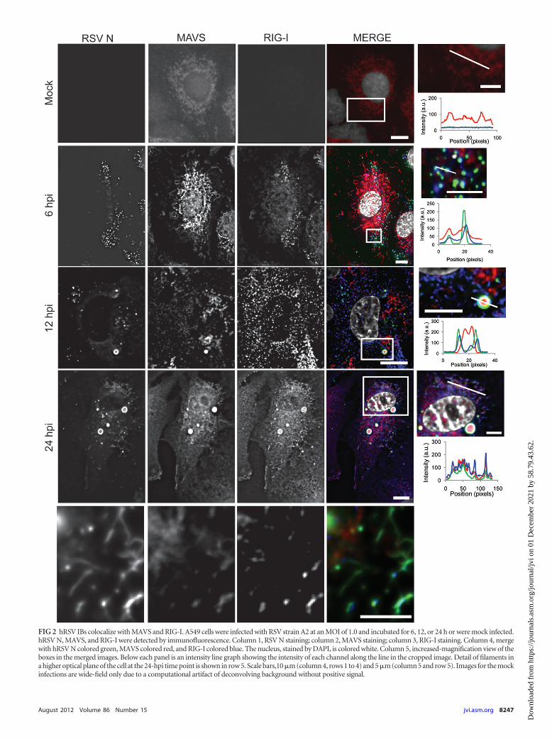
FIG 2 hRSV IBs colocalize with MAVS and RIG-I. A549 cells were infected with RSV strain A2 at an MOI of 1.0 and incubated for 6, 12, or 24 h or were mock infected.hRSV N, MAVS, and RIG-I were detected by immunofluorescence. Column 1, RSV N staining; column 2, MAVS staining; column 3, RIG-I staining. Column 4, mergewith hRSV N colored green, MAVS colored red, and RIG-I colored blue. The nucleus, stained by DAPI, is colored white. Column 5, increased-magnification view of theboxes in the merged images. Below each panel is an intensity line graph showing the intensity of each channel along the line in the cropped image. Detail of filaments ina higher optical plane of the cell at the 24-hpi time point is shown in row 5. Scale bars,10 �m (column 4, rows 1 to 4) and 5 �m (column 5 and row 5). Images for the mockinfections are wide-field only due to a computational artifact of deconvolving background without positive signal.
August 2012 Volume 86 Number 15 jvi.asm.org 8247
Dow
nloa
ded
from
http
s://j
ourn
als.
asm
.org
/jour
nal/j
vi o
n 01
Dec
embe
r 20
21 b
y 58
.79.
43.6
2.
chimeric oligonucleotides were synthesized by Biosearch Technologies,Inc., with the following sequence: 5=-biotin-UXTXTTXAAAAAXGGGGCAAAXAA-3=, where the boldface type denotes 2=-O-methyl RNA, the Xdenotes a dT-C6-NH2, and all other bases are DNA. The binding region isunderlined. MTRIPs were assembled by first conjugating Cy3B-NHS esterfluorophores (GE Healthcare) to the oligonucleotide amine groups usingthe manufacturer’s protocol. Labeled oligonucleotides were then te-tramerized by incubation with Neutravidin (Pierce).
Cell culture and viruses. A549 lung carcinoma cells (American TypeCulture Collection [ATCC] CCL-185), HEp-2 cells (ATCC CCL-23),Vero cells (ATCC CCL-81), and HeLa cells (ATCC CCL-2) were main-tained in high-glucose Dulbecco modified Eagle medium (DMEM)(Lonza) with 10% fetal bovine serum (FBS) (HyClone), 100 U ml�1 pen-icillin, and 100 �g ml�1 streptomycin (Invitrogen). The hRSV used wasthe A2 strain (ATCC VR-1544) prepared in HEp-2 cells at a titer of 1 � 106
PFU ml�1. Virus at a multiplicity of infection (MOI) of 1 was adsorbed tothe cells for 1 h at 37°C. Following adsorption, fresh medium was added tothe inoculum and the cells were incubated for the indicated times. TheNewcastle disease virus (NDV) used was the LaSota strain and was a gift ofClaudio L. Afonso, USDA. NDV was adsorbed onto cells at an MOI of 5for 1 h at 37°C. Following adsorption, fresh medium (DMEM with 5%allantoic fluid) was added to the inoculum and the cells were incubated forthe indicated times.
RNA probe delivery. For RNA probe delivery, cells were washed inDulbecco’s phosphate-buffered saline (DPBS) without Ca2� and withoutMg2� (Lonza), and then incubated with 0.2 U ml�1 activated streptolysinO (Sigma) in Opti-MEM I medium (Invitrogen) containing 30 nMMTRIP for 10 min at 37°C. The delivery medium was replaced withgrowth medium for 15 min to restore membrane integrity before fixation.In these experiments HEp-2 cells were used for expedience in comparisonto previous studies of hRSV RNA and protein localization.
Immunostaining. Cells were fixed with 4% paraformaldehyde (Elec-tron Microscopy Science) in PBS, permeabilized using 0.2% Triton X-100(Sigma), and blocked with 5% bovine serum albumin (EMD). Cells thenwere incubated with primary antibody for 30 min at 37°C, washed threetimes in PBS, and subsequently incubated with the secondary antibody for30 min at 37°C. Multiple labeling was done sequentially. Cells then werewashed three times in PBS, and nuclei were stained using DAPI (4=,6-diamidino-2-phenylindole) (Invitrogen). Cells were mounted on slidesusing Prolong Gold (Invitrogen).
Antibodies. Primary antibodies used were goat anti-RIG-I (SantaCruz), rabbit anti-MDA5 (ProSci Inc.), rabbit anti-MAVS (Abcam),mouse anti-hRSV N (Abcam), and mouse anti-hRSV P (clone 3_5). Sec-ondary antibodies used were donkey anti-mouse Alexa Fluor 488 (Invit-rogen), donkey anti-rabbit Cy3 (Jackson ImmunoResearch), and donkeyanti-goat Cy5 (Jackson ImmunoResearch).
Transfection and mitochondrial staining. Vero and HeLa cells werecultured on glass coverslips in 24-well tissue culture plates for imagingexperiments or directly in 6-well tissue culture plates for reverse transcrip-tion-PCR (RT-PCR). Vero cells were utilized, as they are a commonmodel system for RSV infection, and HeLa cells were used where theinterferon response was measured. Both of these cell lines were used inplace of HEp-2 and A549, as the latter cells are less efficiently transfected.Cells were transfected using Lipofectamine 2000 (Invitrogen) by the man-ufacturer’s protocol. For each well in the six-well plates, 8 �l of Lipo-fectamine 2000 and 4 �g of DNA were used. For each well in the 24-wellplates, 2 �l of Lipofectamine 2000 and 1 �g of DNA were used. For IB andmitochondria colocalization experiments, Vero cells were grown in glass-bottom petri dishes (In Vitro Sci) and transfected as described above using2 �l of Lipofectamine 2000 and 1 �g of DNA. Mitotracker 633 was usedper the manufacturer’s protocols at a dilution of 1:500. Plasmids usedwere pcDNA3.1, containing optimized cDNAs encoding the RSV A2strain nucleoprotein (N) and phosphoprotein (P) genes (synthesized byGeneArt, Regensburg, Germany), GFP-MDA5 in a pEGFP-C2 vector (a
gift of Dong-chul Kang), and a GFP-MAVS in a pEGFP vector (a gift ofStanley M. Lemon).
Imaging and processing. Cells were grown on no. 1.5 coverslips andfixed and immunostained. For hRSV N and viral RNA colocalizationstudies, an LSM 510 confocal microscope (Zeiss) with a Plan-Apochromatprimary objective (63�; NA, 1.4) was used. All images were taken usingmultitrack scanning for each fluorophore to prevent bleed-through. Z-di-mension stacks were taken in 0.5-�m increments; the 543-nm laser wasused for the Cy3B, the 488-nm laser was used for N protein immunostain-ing, and the pinholes were set to an Airy unit of 1 (equal in size to an Airydisk). For all other experiments, images were taken on an Axiovert 200 Mmicroscope (Zeiss) with a Plan-Apochromat primary objective (63�; NA,1.4) and an ORCA-ER AG camera (Hamamatsu). Fluorescent filter setsused were 89000 Sedat Quad - ET for multiple-wavelength imaging(Chroma). All imaging experiments were performed using the Volocityacquisition software (PerkinElmer). Image stacks were recorded at200-nm intervals for fixed cell samples to adequately sample volumes foriterative deconvolution. Images presented are deconvolved from thewide-field data and linearly contrast enhanced unless otherwise noted.Granule counting and object identification were performed in Volocity.Objects were identified by the standard deviation of intensity and filteredto remove objects less than one point spread function in volume.
qRT-PCR. HeLa cells were grown on six-well plates, transfected asdescribed in the previous section, and inoculated with NDV LaSota strain24 h posttransfection. Total RNA was extracted at the indicated timepoints using the RNeasy Minikit (Qiagen). Total RNA was checked sub-sequently for integrity via agarose gel electrophoresis and quantified viaUV-VIS spectroscopy. Total RNA (1 �g) was used for cDNA synthesisusing the RT2 first-strand kit (SA Biosciences) according to the manufac-turer’s instructions. A 1-�l portion of the product then was used forquantitative RT-PCR (qRT-PCR) using the real-time RT2 qPCR primerassay (SYBR green) in the presence of gene-specific primers for �-actin(ACTB) and beta interferon (IFNB1) (SA Biosciences). qRT-PCR wasperformed using the ABI StepOnePlus real-time PCR system (AppliedBiosciences).
Proximity ligation assays. PLA detection to distinguish betweenhRSV N or hRSV P protein and MAVS, RIG-I, or MDA5 was performedusing the Duolink II kit (Olink Bioscience) with anti-rabbit or anti-goatplus and anti-mouse minus probes according to the manufacturer’s pro-tocol. Cells were fixed with 4% paraformaldehyde, permeabilized, andblocked using 0.5% Tween 20 (CalBioChem), 0.1% Triton X-100, 0.1%gelatin (Aurion), 2% donkey serum (VWR), 1% bovine serum albumin innuclease-free phosphate-buffered saline (PBS). Cells then were incubatedwith primary mouse anti-hRSV N or mouse anti-hRSV P antibody (1:500)followed by either rabbit anti-MAVS rabbit anti-MDA5 antibody, or goatanti-RIG-I (1:250). Subsequently, cells were incubated with the oligonu-cleotide-labeled secondary antibodies diluted in 0.05% Tween 20 in nu-clease-free PBS.
Spin capture of RSV filaments on glass. In order to capture singlehRSV filamentous virions on glass, hRSV A2 was propagated in HEp-2cells at an MOI of 0.1. At 4 days postinfection the cell-associated andsupernatant fractions were scraped, freeze-thawed, and spun through5-�m- and 0.45-�m-pore-size centrifugal filters (Millipore) at 5,000 � gand 4°C for 4 min and 1 min, respectively. The fraction between 0.45 �mand 5 �m in diameter was collected and spun down onto a poly-L-lysine(Sigma)-coated cover glass at 3,007 � g and 4°C for 30 min. The immo-bilized virions were fixed using 4% paraformaldehyde and immuno-stained according to the aforementioned protocol. In addition to the hostprotein and viral antibodies previously mentioned, virions were alsostained with palivizumab, which binds to the hRSV F protein (MedIm-mune).
Immunoprecipitation and Western blotting. HeLa cells were trans-fected with green fluorescent protein (GFP)-MAVS and RSV N or GFP-MDA5 and RSV N using Lipofectamine 2000 as described above. Cellswere lysed in RIPA buffer (Thermo) supplemented with complete pro-
Lifland et al.
8248 jvi.asm.org Journal of Virology
Dow
nloa
ded
from
http
s://j
ourn
als.
asm
.org
/jour
nal/j
vi o
n 01
Dec
embe
r 20
21 b
y 58
.79.
43.6
2.
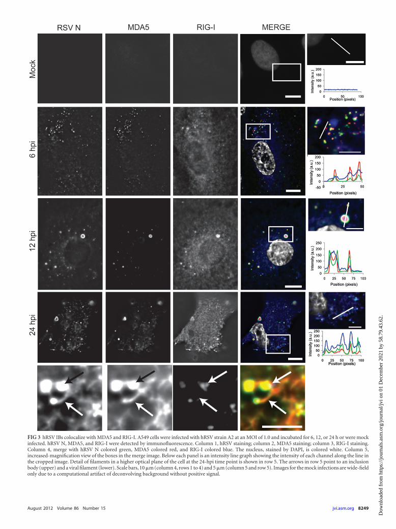
FIG 3 hRSV IBs colocalize with MDA5 and RIG-I. A549 cells were infected with hRSV strain A2 at an MOI of 1.0 and incubated for 6, 12, or 24 h or were mockinfected. hRSV N, MDA5, and RIG-I were detected by immunofluorescence. Column 1, hRSV staining; column 2, MDA5 staining; column 3, RIG-I staining.Column 4, merge with hRSV N colored green, MDA5 colored red, and RIG-I colored blue. The nucleus, stained by DAPI, is colored white. Column 5,increased-magnification view of the boxes in the merge image. Below each panel is an intensity line graph showing the intensity of each channel along the line inthe cropped image. Detail of filaments in a higher optical plane of the cell at the 24-hpi time point is shown in row 5. The arrows in row 5 point to an inclusionbody (upper) and a viral filament (lower). Scale bars, 10 �m (column 4, rows 1 to 4) and 5 �m (column 5 and row 5). Images for the mock infections are wide-fieldonly due to a computational artifact of deconvolving background without positive signal.
August 2012 Volume 86 Number 15 jvi.asm.org 8249
Dow
nloa
ded
from
http
s://j
ourn
als.
asm
.org
/jour
nal/j
vi o
n 01
Dec
embe
r 20
21 b
y 58
.79.
43.6
2.
tease inhibitor (Roche). Lysates were stored at �80°C until use. For im-munoprecipitation, Dynabeads coated with sheep anti-rabbit antibodies(Invitrogen) were incubated overnight at 4°C with rabbit anti-GFP anti-sera (Thermo). The beads were washed, and lysates were added to thebeads. Lysates were tumbled overnight at 4°C. The beads were washed toremove unbound proteins. To release the precipitate, SDS buffer(LI-COR Biosciences) was added to the beads and the solution was incu-bated at 70°C for 10 min. The eluted proteins were loaded into 10%Bis-Tris gels (Invitrogen) and run using MOPS (morpholinepropanesul-fonic acid) buffer (Invitrogen). Western transfer was performed in anXCell II blot module (Invitrogen) onto nitrocellulose paper (Invitrogen)according to the manufacturer’s protocol. Blotting was performed using aSnap i.d. (Millipore). Blots were blocked using Odyssey blocking buffer(LI-COR Biosciences), and primary antibodies, mouse anti-GFP andmouse anti-RSV N, were diluted in Odyssey blocking buffer with 0.1%Tween 20 (CalBioChem) and incubated on the blots for 10 min. Blotswere washed in PBS with 0.1% Tween 20. Secondary goat anti-mouseantibody labeled with DyLight 680B (Thermo), diluted as describedabove, was incubated for 10 min. Blots were washed as described above.Blots were imaged on an Odyssey (LI-COR Biosciences) at 42 �m pixel�1
using the “high” quality setting. Blots were linearly contrast enhanced inImageJ for clarity. Bands corresponding to RSV N and the GFP-MAVSand GFP-MDA5 were differentially enhanced due to the sensitivity differ-ences of the primary antibodies against the respective proteins.
RESULTSLocalization and structure of granules associated with hRSV Nprotein and vRNA during infection. We first analyzed the struc-tures and distribution of viral proteins and genomic RNA at 6, 12,or 24 h postinfection (hpi). HEp-2 cell monolayers were inocu-lated with hRSV strain A2 at an MOI of 1 and incubated in growthmedium for the indicated times. Multiply labeled tetravalent im-
aging probes (MTRIPs) were delivered into the live cells as de-scribed, and they were subsequently fixed and immunostained forhRSV N (Fig. 1). At 6 hpi, both the viral genomic RNA and inclu-sion bodies (IBs) positive for hRSV N protein were punctate andcolocalized (Fig. 1, row 1). Morphologically, the IBs were round,and no filaments were present. The average volume of the IBs atthis time point was 0.9 �m3. At 12 hpi, hRSV N staining was seenboth as a diffuse population that filled the cytoplasm and as inclu-sion bodies, which were larger than those detected at 6 hpi (Fig. 1,row 2). These larger inclusion bodies (�8 �m3, with an averagesize of 37 �m3) did not contain genomic RNA detectable withprobes, unlike those at 6 hpi. The genomic RNA was located pre-dominantly at the plasma membrane and in smaller granuleswithin the cytoplasm. At 24 hpi, N protein could also be detectedin viral filaments on the plasma membrane of the cells in additionto the diffuse hRSV N staining and the staining of IBs (Fig. 1, rows3 to 5). These filaments contained viral genomic RNA, as previ-ously described (14, 22, 29). Notably the larger IBs contained littleto no genomic RNA despite containing high levels of the N pro-tein, which binds to the genomic RNA in the virion.
Viral inclusion bodies colocalize with proteins of the innateimmune response. To test whether the hRSV IBs contained pro-teins of the RIG-I-like receptor (RLR) antiviral response pathway,we infected A549 cells and fixed them at 6, 12, or 24 hpi, or prior toinfection (mock), and stained them for hRSV N, MAVS, andRIG-I. In the mock-infected cells no RSV N or RIG-I staining waspresent and MAVS staining appeared to be mitochondrial (Fig. 2,row 1). At 6 hpi, hRSV N was in a punctate pattern and MAVSstaining continued to appear filamentous throughout the cyto-
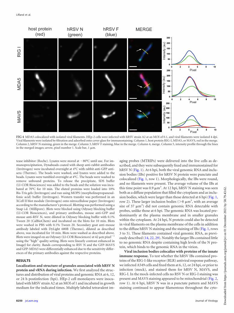
FIG 4 MDA5 colocalized with isolated viral filaments. HEp-2 cells were infected with hRSV strain A2 at an MOI of 0.1, and viral filaments were isolated 4 dpi.Viral filaments were isolated by filtration and adsorbed onto cover glass for immunostaining. Column 1, host protein RIG-I, MDA5, or MAVS, red in the merge.Column 2, hRSV N staining, green in the merge. Column 3, hRSV F staining, blue in the merge. Column 4, merge. Column 5, intensity profile through the linesin the merged images; arrow, pixel number 1. Scale bar, 1 �m.
Lifland et al.
8250 jvi.asm.org Journal of Virology
Dow
nloa
ded
from
http
s://j
ourn
als.
asm
.org
/jour
nal/j
vi o
n 01
Dec
embe
r 20
21 b
y 58
.79.
43.6
2.
plasm, indicative of mitochondria, as would be expected (Fig. 2,row 2) (24). RIG-I staining was similar to hRSV N staining andwas punctate. All three proteins were present inside the hRSV NIBs, with a strong correlation between the hRSV N staining andthe RIG-I staining. At 12 hpi, hRSV N was again in larger IBs thatappeared to be hollow (Fig. 2, row 3). MAVS staining at 12 hpi wasaltered dramatically, with some of the protein localizing to largeIBs that were not observed at earlier time points and appeared tobe surrounded by a “ring” of hRSV N staining. CytoplasmicMAVS staining was still present; however, this staining was nolonger filamentous and did not resemble mitochondria. RIG-Istaining also was increased inside the larger IBs as well as through-out the cytoplasm. At 24 hpi, both MAVS and RIG-I were local-
ized to large IBs and to viral filaments at the plasma membrane(Fig. 2, rows 4 and 5).
In order to see if this change in protein localization was specificto RIG-I/MAVS or was a more general phenomena of the RLRpathway, we next costained for hRSV N, RIG-I, and MDA-5. Inthe mock-infected cells no RSV N, MDA5, or RIG-I staining waspresent (Fig. 3, row 1). At 6 hpi, MDA5 was predominately coin-cident with IBs containing hRSV N, with little cytoplasmic stain-ing apparent outside these regions (Fig. 3, row 2). At 12 hpi,MDA5 colocalized with IBs of all sizes, and there was little to nostaining detected outside these regions (Fig. 3, row 3). At 24 hpi,MDA5, like RIG-I, was present in viral filaments at the plasmamembrane (Fig. 3, rows 4 and 5).
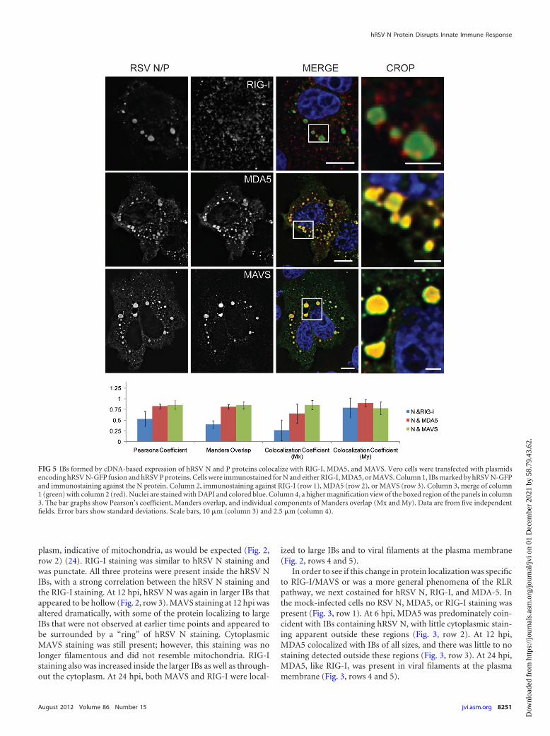
FIG 5 IBs formed by cDNA-based expression of hRSV N and P proteins colocalize with RIG-I, MDA5, and MAVS. Vero cells were transfected with plasmidsencoding hRSV N-GFP fusion and hRSV P proteins. Cells were immunostained for N and either RIG-I, MDA5, or MAVS. Column 1, IBs marked by hRSV N-GFPand immunostaining against the N protein. Column 2, immunostaining against RIG-I (row 1), MDA5 (row 2), or MAVS (row 3). Column 3, merge of column1 (green) with column 2 (red). Nuclei are stained with DAPI and colored blue. Column 4, a higher magnification view of the boxed region of the panels in column3. The bar graphs show Pearson’s coefficient, Manders overlap, and individual components of Manders overlap (Mx and My). Data are from five independentfields. Error bars show standard deviations. Scale bars, 10 �m (column 3) and 2.5 �m (column 4).
hRSV N Protein Disrupts Innate Immune Response
August 2012 Volume 86 Number 15 jvi.asm.org 8251
Dow
nloa
ded
from
http
s://j
ourn
als.
asm
.org
/jour
nal/j
vi o
n 01
Dec
embe
r 20
21 b
y 58
.79.
43.6
2.
Owing to the dense nature of viral and host proteins near theplasma membrane, it was difficult to separate the bright signalfrom IBs from the lower signal inside filaments. To circumventthis problem, viral filaments were isolated from infected cells, de-posited and fixed on glass coverslips, and immunostained forhRSV F and hRSV N, and RIG-I, MDA5, or MAVS (Fig. 4). WhileMDA5 stained strongly in the isolated filaments, neither RIG-Inor MAVS was observed to colocalize with them.
Expression of hRSV N and P proteins was sufficient to local-ize proteins of the innate immune response. In order to testwhether the presence of IBs alone was sufficient to localize RIG-I,MDA5, and MAVS, hRSV N and P were expressed in Vero cellsthat subsequently were fixed and immunostained. As seen in theleft column of Fig. 5, the expression of hRSV N and P resulted inthe formation of both small and large IB-like structures similar tothose seen in the infection model. In these cells, RIG-I staining wasincreased in and around the IBs; however, RIG-I was not localizedexclusively to the IBs. In contrast, both MDA5 and MAVS werelocalized almost exclusively to the IBs, and little staining was de-tected outside these regions. In order to quantify these results,colocalization data were obtained from over 100 cells from fivefields and were summarized in the graph in Fig. 5. A colocalizationanalysis between N and RIG-I yielded a Pearson’s coefficient of 0.5and a Manders overlap of 0.4, suggesting the colocalization wasstrong but not complete. When the Manders overlap was brokeninto its constitutive parts, Mx and My, it was seen that three-quarters of the N staining colocalized with RIG-I (My, �0.75),while RIG-I staining colocalized with N approximately 25% of thetime (Mx, �0.25). In contrast, the Pearson coefficient andManders overlap for N with MDA5 or MAVS were both �0.75,and the Mx and My colocalization coefficients in these two caseswere both �0.5, indicating near-complete colocalization betweenthe expressed inclusion bodies and MDA5 and MAVS.
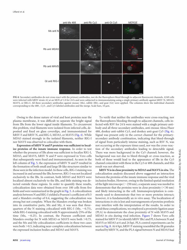
To verify that neither the antibodies were cross-reacting, northe fluorophores bleeding through to adjacent channels, cells in-fected with RSV for 24 h were stained with a single primary anti-body and all three secondary antibodies, anti-mouse Alexa Fluor488, donkey anti-rabbit Cy3, and donkey anti-goat Cy5 (Fig. 6).Signal was present only in the correct channel for the primary-secondary antibody combination, indicating that bleed-throughof signal from particularly intense staining, such as RSV N, wasnot occurring at the exposure times used, nor was the cross-reac-tion of the secondary antibodies leading to detectable signal.There was more background in the Cy5 channel; however, thebackground was not due to bleed-through or cross-reaction, asboth of those would lead to the appearance of IBs in the Cy5channel coincident with those in the Cy3 or 488 channels, and thisresult was not observed.
hRSV N is in close proximity to MDA5 and MAVS in situ. Thecolocalization analysis discussed above suggested an interactionbetween the proteins of the innate immune response and the viralinclusion bodies. However, owing to the limited spatial resolutionof the light microscope (�250 nm), a separate assay was needed todemonstrate that the proteins were in close proximity (30 nm)and likely interacting in the cell. Immunoprecipitation is com-monly used to demonstrate that two or more proteins interact;however, it is fundamentally an in vitro assay. The localization ofinteractions in situ is lost and rearrangements of proteins postlysismay interfere with the interpretation of the results. In order toavoid these complications, we employed a proximity ligation assay(PLA) to demonstrate the close proximity of N with MAVS andMDA5 in situ during viral infection. Figure 7 shows Vero cellsstained for hRSV P (to identify hRSV IBs) and PLA between N andMAVS or MDA5 at 6 and 24 hpi. PLA control experiments can beseen in Fig. 8. At 6 hpi, hRSV P staining resembled the IB granulesmarked by hRSV N, and the PLA signal between N and MDA5 had
FIG 6 Secondary antibodies do not cross-react with the primary antibodies, nor do the fluorophors bleed through to adjacent fluorescent channels. A549 cellswere infected with hRSV strain A2 at an MOI of 1.0 for 24 h and were subjected to immunostaining using a single primary antibody against hRSV N, MDA5,MAVS, or RIG-I. All three secondary antibodies against mouse (Ms), rabbit (Rb), and goat (Gt) were applied. The columns show the individual channelscorresponding to the 488-, Cy3-, and Cy5-labeled antibodies and the merge. Scale bars, 10 �m.
Lifland et al.
8252 jvi.asm.org Journal of Virology
Dow
nloa
ded
from
http
s://j
ourn
als.
asm
.org
/jour
nal/j
vi o
n 01
Dec
embe
r 20
21 b
y 58
.79.
43.6
2.
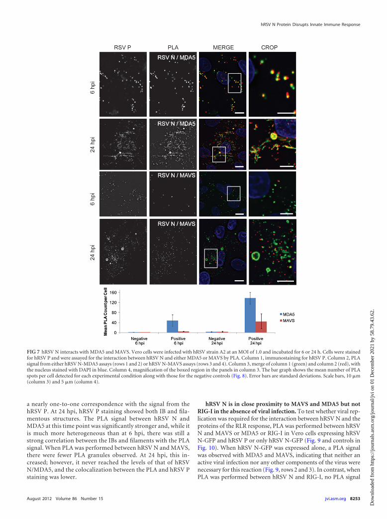
a nearly one-to-one correspondence with the signal from thehRSV P. At 24 hpi, hRSV P staining showed both IB and fila-mentous structures. The PLA signal between hRSV N andMDA5 at this time point was significantly stronger and, while itis much more heterogeneous than at 6 hpi, there was still astrong correlation between the IBs and filaments with the PLAsignal. When PLA was performed between hRSV N and MAVS,there were fewer PLA granules observed. At 24 hpi, this in-creased; however, it never reached the levels of that of hRSVN/MDA5, and the colocalization between the PLA and hRSV Pstaining was lower.
hRSV N is in close proximity to MAVS and MDA5 but notRIG-I in the absence of viral infection. To test whether viral rep-lication was required for the interaction between hRSV N and theproteins of the RLR response, PLA was performed between hRSVN and MAVS or MDA5 or RIG-I in Vero cells expressing hRSVN-GFP and hRSV P or only hRSV N-GFP (Fig. 9 and controls inFig. 10). When hRSV N-GFP was expressed alone, a PLA signalwas observed with MDA5 and MAVS, indicating that neither anactive viral infection nor any other components of the virus werenecessary for this reaction (Fig. 9, rows 2 and 3). In contrast, whenPLA was performed between hRSV N and RIG-I, no PLA signal
FIG 7 hRSV N interacts with MDA5 and MAVS. Vero cells were infected with hRSV strain A2 at an MOI of 1.0 and incubated for 6 or 24 h. Cells were stainedfor hRSV P and were assayed for the interaction between hRSV N and either MDA5 or MAVS by PLA. Column 1, immunostaining for hRSV P. Column 2, PLAsignal from either hRSV N-MDA5 assays (rows 1 and 2) or hRSV N-MAVS assays (rows 3 and 4). Column 3, merge of column 1 (green) and column 2 (red), withthe nucleus stained with DAPI in blue. Column 4, magnification of the boxed region in the panels in column 3. The bar graph shows the mean number of PLAspots per cell detected for each experimental condition along with those for the negative controls (Fig. 8). Error bars are standard deviations. Scale bars, 10 �m(column 3) and 5 �m (column 4).
hRSV N Protein Disrupts Innate Immune Response
August 2012 Volume 86 Number 15 jvi.asm.org 8253
Dow
nloa
ded
from
http
s://j
ourn
als.
asm
.org
/jour
nal/j
vi o
n 01
Dec
embe
r 20
21 b
y 58
.79.
43.6
2.
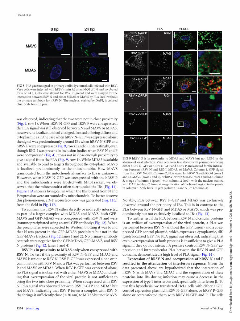
was observed, indicating that the two were not in close proximity(Fig. 9, row 1). When hRSV N-GFP and hRSV P were coexpressed,the PLA signal was still observed between N and MAVS or MDA5;however, its localization had changed. Instead of being diffuse andcytoplasmic as in the case when hRSV N-GFP was expressed alone,the signal was predominantly around IBs when hRSV N-GFP andhRSV P were coexpressed (Fig. 9, rows 5 and 6). Interestingly, eventhough RIG-I was present in inclusion bodies when RSV N and Pwere coexpressed (Fig. 4), it was not in close enough proximity togive a signal from the PLA (Fig. 9, row 4). While MDA5 is solubleand available to bind to targets throughout the cytoplasm, MAVSis localized predominantly to the mitochondria. How MAVStranslocated from the mitochondrial surface to IBs is unknown.However, when hRSV N-GFP was coexpressed with the hRSV Pand the mitochondria were labeled with MitoTracker, we ob-served that the mitochondria often surrounded the IBs (Fig. 11).Figure 11A shows a living cell in which the IBs formed from N andP expression were surrounded by mitochondria. To better observethis phenomenon, a 3-D isosurface view was generated (Fig. 11C)from the field in Fig. 11B.
To confirm that RSV N either directly or indirectly interactedas part of a larger complex with MDA5 and MAVS, both GFP-MAVS and GFP-MDA5 were coexpressed with RSV N and wereimmunoprecipitated using an anti-GFP antibody (Fig. 12). Whenthe precipitates were subjected to Western blotting it was foundthat N was present in the GFP-MDA5 precipitate but not in theGFP-MAVS fraction (Fig. 12, lanes 1 and 2). No primary antibodycontrols were negative for the GFP-MDA5, GFP-MAVS, and RSVN proteins (Fig. 12, lanes 3 and 4).
RSV P is in proximity to MDA5 only when coexpressed withRSV N. To test if the proximity of RSV N-GFP and MDA5 andMAVS is unique to RSV N, RSV P-GFP was expressed alone or incombination with RSV N and a PLA was performed between RSVP and MAVS or MDA5. When RSV P-GFP was expressed alone,no PLA signal was observed with either MAVS or MDA5, indicat-ing that overexpression of the viral protein is not sufficient tobring the two into close proximity. When coexpressed with RSVN, PLA signal was observed between RSV P-GFP and MDA5 butnot MAVS, indicating that RSV P forms a complex with RSV Nthat brings it sufficiently close (30 nm) to MDA5 but not MAVS.
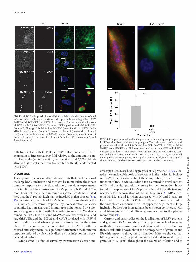
Notably, PLA between RSV P-GFP and MDA5 was exclusivelyobserved around the periphery of IBs. This is in contrast to thePLA between RSV N-GFP and MDA5 or MAVS, which was pre-dominantly but not exclusively localized to IBs (Fig. 13).
To further test if the PLA between RSV N and cellular proteinsis an artifact of overexpression of the viral protein, a PLA wasperformed between RSV N (without the GFP fusion) and a coex-pressed GFP control plasmid, which expresses a cytoplasmic, dif-fusely localized GFP. No PLA signal was observed, indicating thateven overexpression of both proteins is insufficient to give a PLAsignal if they do not interact. A positive control, RSV N-GFP ex-pression and intramolecular PLA between the RSV N and GFPdomains, demonstrated a high level of PLA signal (Fig. 14).
Expression of hRSV N and coexpression of hRSV N and Presulted in the attenuation of interferon response. Given thedata presented above, we hypothesized that the interaction ofhRSV N with MAVS and MDA5 and the sequestration of theseproteins into IBs during infection may cause a decrease in theexpression of type 1 interferons and, specifically, interferon �. Totest this hypothesis, we transfected HeLa cells with either a GFPcontrol expression plasmid, hRSV N-GFP alone, or hRSV P-GFPalone or cotransfected them with hRSV N-GFP and P. The cells
FIG 9 hRSV N is in proximity to MDA5 and MAVS but not RIG-I in theabsence of viral infection. Vero cells were transfected with plasmids encodingeither hRSV N-GFP or hRSV N-GFP and hRSV P and assayed for the interac-tion between hRSV N and RIG-I, MDA5, or MAVS. Column 1, GFP signalfrom the hRSV N-GFP. Column 2, PLA signal for hRSV N with RIG-I (rows 1and 4), MAVS (rows 2 and 5), or hRSV N with MDA5 (rows 3 and 6). Column3, merge of column 1 (green) with column 2 (red), with the nucleus stainedwith DAPI in blue. Column 4, magnification of the boxed region in the panelsin column 3. Scale bars, 10 �m (column 3) and 5 �m (column 4).
FIG 8 PLA gave no signal in primary antibody control cells infected with RSV.Vero cells were infected with hRSV strain A2 at an MOI of 1.0 and incubatedfor 6 or 24 h. Cells were stained for RSV P (green) and were assayed for theinteraction between RSV N and either MDA5 or MAVS by PLA (red) withoutthe primary antibody for hRSV N. The nucleus, stained by DAPI, is coloredblue. Scale bars, 10 �m.
Lifland et al.
8254 jvi.asm.org Journal of Virology
Dow
nloa
ded
from
http
s://j
ourn
als.
asm
.org
/jour
nal/j
vi o
n 01
Dec
embe
r 20
21 b
y 58
.79.
43.6
2.

were subsequently inoculated with the LaSota strain of Newcastledisease virus (NDV), a pathogen known to induce an interferonresponse through the RLR pathway in HeLa cells (31). At 24 h afterNDV inoculation, total RNA was isolated from the cells, and betainterferon (IFNB1) mRNA was quantified relative to �-actin(ACTB) mRNA using reverse transcription real-time PCR
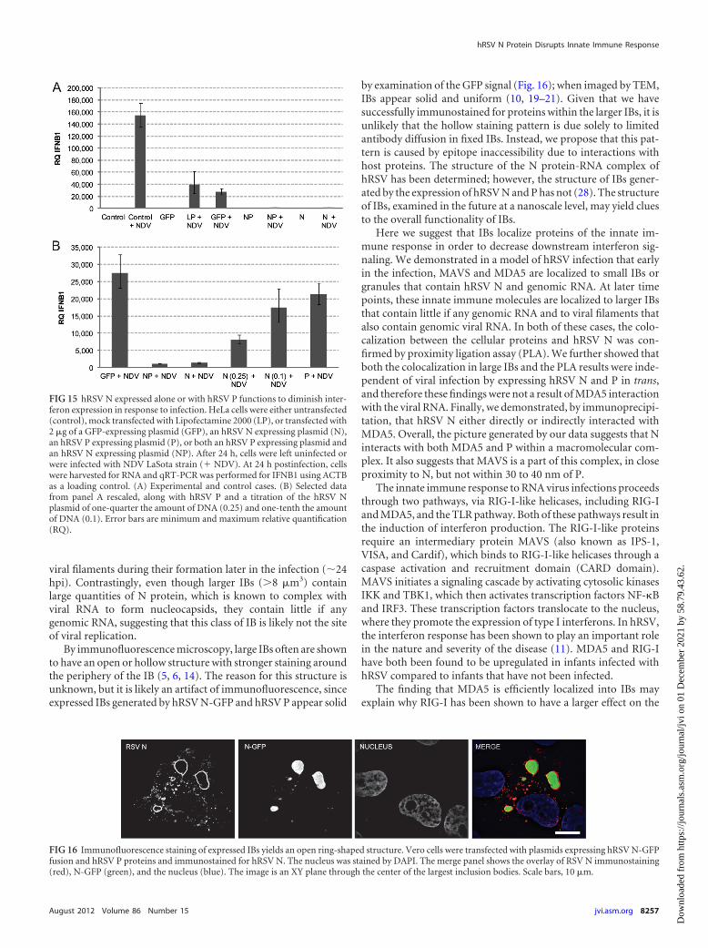
(Fig. 15). Cells transfected with either hRSV N-GFP or hRSVN-GFP and P and infected with NDV expressed IFNB1 at a level 27times lower than in cells transfected with GFP control plasmid andinfected with NDV (Fig. 15B). When smaller amounts of hRSVN-GFP DNA were transfected into the cells, the effect of hRSV Non interferon expression was diminished in a dose-dependentfashion. In addition, the expression of P alone had no effect onIFNB1 expression, as suggested by the PLA data (Fig. 15B). Thesedata indicated that the effect of hRSV N on the innate immuneresponse lowers the production of IFNB1 when hRSV N is presentas a diffuse population throughout the cytoplasm and when it islocalized to cytoplasmic IBs.
In untransfected HeLa cells, NDV infection caused IFNB1 ex-pression to increase 150,000-fold at 24 hpi relative to that in un-infected, untransfected HeLa cells. In cells that had been trans-fected with a GFP control plasmid, NDV infection caused IFNB1expression to increase 5,000-fold when infected with NDV relativeto that in uninfected HeLa cells transfected with GFP. The differ-ence in IFNB1 expression may be due to the transfection processvia Lipofectamine 2000. To test if this was the case, cells weretreated with the transfection agent alone and infected with NDV.As can be seen in Fig. 15A, the transfection agent, without DNA,reduced the susceptibility of the cells to IFN induction when in-fected with NDV. While this reduces the overall effect of the se-questration of RLR components on the induction of IFNB1 byNDV in cells expressing hRSV N, there is still a sizable difference(�27-fold) compared to results for cells expressing GFP alone. In
FIG 10 PLA gave no signal in primary antibody control cells transfected withplasmids encoding RSV N-GFP and RSV P, RSV N-GFP alone, RSV P-GFPand RSV N, or RSV P-GFP alone. Vero cells were transfected with plasmidsencoding hRSV N-GFP, hRSV N-GFP and hRSV P, hRSV P-GFP, or hRSVP-GFP and hRSV N and assayed for the interaction between hRSV N andMAVS (column 1), MDA5 (column 2), or RIG-I (column 3) without theprimary antibody for hRSV N. Row 1, cells transfected with plasmids encodinghRSV N-GFP and hRSV P. Row 2, cells transfected with a plasmid encodinghRSV N-GFP only. Row 3, cells transfected with plasmids encoding hRSVP-GFP and hRSV N. Row 4, cells transfected with a plasmid encoding hRSVP-GFP only. The nucleus, stained by DAPI, is colored blue. Scale bars, 10 �m.Images are single planes of wide-field, undeconvolved Z-stacks.
FIG 11 Mitochondria are found adjacent to hRSV IBs. Vero cells were transfected with plasmids encoding hRSV N-GFP and RSV P. At 24 h posttransfection,mitochondria were labeled with Mitotracker deep red 633 and imaged live. (A) Single cell. (B) Magnification of the boxed region in panel A. (C) Three-dimensional isosurface view of a large IB from panel B surrounded by mitochondria. hRSV N-GFP is colored green, Mitotracker is colored in red, and nucleistained by DAPI are in blue. Scale bar, 10 �m (A) and 5 �m (B).
FIG 12 hRSV N coprecipitates with GFP-MDA5. HeLa cells were transfectedwith plasmids encoding GFP-MDA5 and hRSV N or GFP-MAVS and hRSV N.At 24 h posttransfection, cells were lysed and subjected to IP using an anti-GFPantibody or control antibody. Precipitates were subjected to electrophoresisand Western blotting using an anti-GFP to detect the GFP-MDA5 and GFP-MAVS and an anti-hRSV N antibody to detect the hRSV N. Blots were linearlycontrast enhanced.
hRSV N Protein Disrupts Innate Immune Response
August 2012 Volume 86 Number 15 jvi.asm.org 8255
Dow
nloa
ded
from
http
s://j
ourn
als.
asm
.org
/jour
nal/j
vi o
n 01
Dec
embe
r 20
21 b
y 58
.79.
43.6
2.
cells transfected with GFP alone, NDV infection caused IFNB1expression to increase 27,000-fold relative to the amount in con-trol HeLa cells (no transfection, no infection) and 5,000-fold rel-ative to that in cells that were transfected with GFP and infectedwith NDV.
DISCUSSION
The experiments presented here demonstrate that one function ofthe large hRSV inclusion bodies might be to modulate the innateimmune response to infection. Although previous experimentshave implicated the nonstructural hRSV proteins NS1 and NS2 asmodulators of the innate immune response, we demonstratedhere that the N protein itself may be involved in that process (3, 4,15). We studied the role of hRSV N and IBs in modulating theRLR-induced interferon response by colocalization analysis,proximity ligation assay, and immunoprecipitation and by a bio-assay using an infection with Newcastle disease virus. We deter-mined that RIG-I, MDA5, and MAVS colocalized with small andlarge hRSV IBs and that MDA5 and MAVS localized with hRSV Nboth inside IBs and when expressed diffusely within the cyto-plasm. Furthermore, we demonstrated that hRSV N, both ex-pressed diffusely and in IBs, significantly attenuated the interferonresponse induced by Newcastle disease virus infection in a dose-dependent fashion.
Cytoplasmic IBs, first observed by transmission electron mi-
croscopy (TEM), are likely aggregates of N proteins (19, 20). De-spite the considerable body of knowledge in the molecular biologyof hRSV, little is known about the composition, structure, andfunction of IBs. Previous studies have examined the viral contentof IBs and the viral proteins necessary for their formation. It wasfound that expression of hRSV proteins N and P is sufficient andnecessary for the formation of IB-like structures (6). hRSV pro-teins M, M2-1, and L, when expressed with N and P, also arelocalized to IBs, while hRSV G and F, which are translated inthe endoplasmic reticulum, do not appear to be present in largeinclusion bodies but instead have been shown to interact withviral filaments and small IBs or granules close to the plasmamembrane (9).
Current and past studies on the localization of hRSV proteinsand genomic RNA have shown the important role of imagingmethods in the analysis of viral localization and structure. Even so,there is still little known about the heterogeneity of granules andIBs with respect to time, size, or function. Here we showed thathRSV genomic RNA is predominantly localized to small IBs orgranules (1.0 �m3) throughout the course of infection and to
FIG 13 hRSV P is in proximity to MDA5 and MAVS in the absence of viralinfection. Vero cells were transfected with plasmids encoding either hRSVP-GFP or hRSV P-GFP and hRSV N and assayed for the interaction betweenhRSV P and MDA5 or MAVS. Column 1, GFP signal from the hRSV P-GFP.Column 2, PLA signal for hRSV P with MAVS (rows 1 and 3) or hRSV N withMDA5 (rows 2 and 4). Column 3, merge of column 1 (green) with column 2(red) with the nucleus stained with DAPI in blue. Column 4, magnification ofthe boxed region in the panels in column 3. Scale bars, 10 �m (column 3) and5 �m (column 4). FIG 14 PLA produces a signal in the presence of interacting antigens but not
in diffusely localized, noninteracting antigens. Vero cells were transfected withplasmids encoding either hRSV N and free GFP (N-OPT � GFP) or hRSVN-GFP alone (N-GFP). A PLA was performed against the GFP and hRSV Ndomains in both cases. PLA signal was quantified on a per-cell basis and sum-marized. Nuclei were stained with DAPI. ***, P � 0.001. N.D., not detected.GFP signal is shown in green, PLA signal is shown in red, and DAPI signal isshown in blue. Scale bars, 10 �m. Error bars are standard deviations.
Lifland et al.
8256 jvi.asm.org Journal of Virology
Dow
nloa
ded
from
http
s://j
ourn
als.
asm
.org
/jour
nal/j
vi o
n 01
Dec
embe
r 20
21 b
y 58
.79.
43.6
2.
viral filaments during their formation later in the infection (�24hpi). Contrastingly, even though larger IBs (�8 �m3) containlarge quantities of N protein, which is known to complex withviral RNA to form nucleocapsids, they contain little if anygenomic RNA, suggesting that this class of IB is likely not the siteof viral replication.
By immunofluorescence microscopy, large IBs often are shownto have an open or hollow structure with stronger staining aroundthe periphery of the IB (5, 6, 14). The reason for this structure isunknown, but it is likely an artifact of immunofluorescence, sinceexpressed IBs generated by hRSV N-GFP and hRSV P appear solid
by examination of the GFP signal (Fig. 16); when imaged by TEM,IBs appear solid and uniform (10, 19–21). Given that we havesuccessfully immunostained for proteins within the larger IBs, it isunlikely that the hollow staining pattern is due solely to limitedantibody diffusion in fixed IBs. Instead, we propose that this pat-tern is caused by epitope inaccessibility due to interactions withhost proteins. The structure of the N protein-RNA complex ofhRSV has been determined; however, the structure of IBs gener-ated by the expression of hRSV N and P has not (28). The structureof IBs, examined in the future at a nanoscale level, may yield cluesto the overall functionality of IBs.
Here we suggest that IBs localize proteins of the innate im-mune response in order to decrease downstream interferon sig-naling. We demonstrated in a model of hRSV infection that earlyin the infection, MAVS and MDA5 are localized to small IBs orgranules that contain hRSV N and genomic RNA. At later timepoints, these innate immune molecules are localized to larger IBsthat contain little if any genomic RNA and to viral filaments thatalso contain genomic viral RNA. In both of these cases, the colo-calization between the cellular proteins and hRSV N was con-firmed by proximity ligation assay (PLA). We further showed thatboth the colocalization in large IBs and the PLA results were inde-pendent of viral infection by expressing hRSV N and P in trans,and therefore these findings were not a result of MDA5 interactionwith the viral RNA. Finally, we demonstrated, by immunoprecipi-tation, that hRSV N either directly or indirectly interacted withMDA5. Overall, the picture generated by our data suggests that Ninteracts with both MDA5 and P within a macromolecular com-plex. It also suggests that MAVS is a part of this complex, in closeproximity to N, but not within 30 to 40 nm of P.
The innate immune response to RNA virus infections proceedsthrough two pathways, via RIG-I-like helicases, including RIG-Iand MDA5, and the TLR pathway. Both of these pathways result inthe induction of interferon production. The RIG-I-like proteinsrequire an intermediary protein MAVS (also known as IPS-1,VISA, and Cardif), which binds to RIG-I-like helicases through acaspase activation and recruitment domain (CARD domain).MAVS initiates a signaling cascade by activating cytosolic kinasesIKK and TBK1, which then activates transcription factors NF-�Band IRF3. These transcription factors translocate to the nucleus,where they promote the expression of type I interferons. In hRSV,the interferon response has been shown to play an important rolein the nature and severity of the disease (11). MDA5 and RIG-Ihave both been found to be upregulated in infants infected withhRSV compared to infants that have not been infected.
The finding that MDA5 is efficiently localized into IBs mayexplain why RIG-I has been shown to have a larger effect on the
FIG 15 hRSV N expressed alone or with hRSV P functions to diminish inter-feron expression in response to infection. HeLa cells were either untransfected(control), mock transfected with Lipofectamine 2000 (LP), or transfected with2 �g of a GFP-expressing plasmid (GFP), an hRSV N expressing plasmid (N),an hRSV P expressing plasmid (P), or both an hRSV P expressing plasmid andan hRSV N expressing plasmid (NP). After 24 h, cells were left uninfected orwere infected with NDV LaSota strain (� NDV). At 24 h postinfection, cellswere harvested for RNA and qRT-PCR was performed for IFNB1 using ACTBas a loading control. (A) Experimental and control cases. (B) Selected datafrom panel A rescaled, along with hRSV P and a titration of the hRSV Nplasmid of one-quarter the amount of DNA (0.25) and one-tenth the amountof DNA (0.1). Error bars are minimum and maximum relative quantification(RQ).
FIG 16 Immunofluorescence staining of expressed IBs yields an open ring-shaped structure. Vero cells were transfected with plasmids expressing hRSV N-GFPfusion and hRSV P proteins and immunostained for hRSV N. The nucleus was stained by DAPI. The merge panel shows the overlay of RSV N immunostaining(red), N-GFP (green), and the nucleus (blue). The image is an XY plane through the center of the largest inclusion bodies. Scale bars, 10 �m.
hRSV N Protein Disrupts Innate Immune Response
August 2012 Volume 86 Number 15 jvi.asm.org 8257
Dow
nloa
ded
from
http
s://j
ourn
als.
asm
.org
/jour
nal/j
vi o
n 01
Dec
embe
r 20
21 b
y 58
.79.
43.6
2.
innate immune response to hRSV infection (17). The data alsosuggest a mechanism by which cells can be potentiated towardsubsequent infection or coinfection. We have shown that the ex-pression of IBs by hRSV localizes pattern recognition receptors ofthe RLR pathway and additionally localized their downstream ef-fector MAVS. We demonstrated that N and IBs alone, and not Palone, are enough to diminish the cell’s ability to produce inter-feron in response to a viral infection. To our knowledge, this is thefirst report of hRSV IBs having a functional role in the life cycle ofthe virus. It is important to note that while cytoplasmic, diffusehRSV N expression can decrease the interferon response withoutthe formation of the inclusion bodes, in vivo hRSV N is highlylocalized to IBs (13).
In summary, there are few previous data describing the hostcell protein content inside IBs or their precise function. The studypresented here suggests an interaction of hRSV N protein withimportant host proteins of the innate immune response and aspecific function for inclusion bodies. The findings also clearlypoint to the need for further studies regarding IB formation, struc-ture, and function in terms of time and space during hRSV infec-tions.
ACKNOWLEDGMENTS
This work was supported by NIH grant R01GM094198 (P.J.S.) and theMarch of Dimes (J.E.C.).
In addition, we thank Claudio L. Afonso, ARS, USDA, for the NDVLaSota virus used in this study, Dong-chul Kang, Hallym University, forthe GFP-MDA5 vector, and Stanley M. Lemon, University of North Car-olina at Chapel Hill, for the GFP-MAVS plasmid.
REFERENCES1. Barbalat R, Ewald SE, Mouchess ML, Barton GM. 2011. Nucleic acid
recognition by the innate immune system. Annu. Rev. Immunol. 29:185–214.
2. Bitko V, et al. 2007. Nonstructural proteins of respiratory syncytial virussuppress premature apoptosis by an NF-kappaB-dependent, interferon-independent mechanism and facilitate virus growth. J. Virol. 81:1786 –1795.
3. Bossert B, Conzelmann KK. 2002. Respiratory syncytial virus (RSV)nonstructural (NS) proteins as host range determinants: a chimeric bo-vine RSV with NS genes from human RSV is attenuated in interferon-competent bovine cells. J. Virol. 76:4287– 4293.
4. Bossert B, Marozin S, Conzelmann KK. 2003. Nonstructural proteinsNS1 and NS2 of bovine respiratory syncytial virus block activation ofinterferon regulatory factor 3. J. Virol. 77:8661– 8668.
5. Carromeu C, Simabuco FM, Tamura RE, Farinha Arcieri LE, VenturaAM. 2007. Intracellular localization of human respiratory syncytial virus Lprotein. Arch. Virol. 152:2259 –2263.
6. Garcia J, Garcia-Barreno B, Vivo A, Melero JA. 1993. Cytoplasmicinclusions of respiratory syncytial virus-infected cells: formation of inclu-sion bodies in transfected cells that coexpress the nucleoprotein, the phos-phoprotein, and the 22K protein. Virology 195:243–247.
7. Garcia-Barreno B, Delgado T, Melero JA. 1996. Identification of proteinregions involved in the interaction of human respiratory syncytial virusphosphoprotein and nucleoprotein: significance for nucleocapsid assem-bly and formation of cytoplasmic inclusions. J. Virol. 70:801– 808.
8. Ghildyal R, et al. 2005. Interaction between the respiratory syncytial virusG glycoprotein cytoplasmic domain and the matrix protein. J. Gen. Virol.86:1879 –1884.
9. Ghildyal R, Mills J, Murray M, Vardaxis N, Meanger J. 2002. Respira-
tory syncytial virus matrix protein associates with nucleocapsids in in-fected cells. J. Gen. Virol. 83:753–757.
10. Jeffree CE, et al. 2007. Ultrastructural analysis of the interaction betweenF-actin and respiratory syncytial virus during virus assembly. Virology369:309 –323.
11. Johnson TR, et al. 2005. Role for innate IFNs in determining respiratorysyncytial virus immunopathology. J. Immunol. 174:7234 –7241.
12. Kurt-Jones EA, et al. 2000. Pattern recognition receptors TLR4 and CD14mediate response to respiratory syncytial virus. Nat. Immunol. 1:398 –401.
13. Li D, et al. 2008. Association of respiratory syncytial virus M protein withviral nucleocapsids is mediated by the M2-1 protein. J. Virol. 82:8863–8870.
14. Lindquist ME, Lifland AW, Utley TJ, Santangelo PJ, Crowe JE, Jr. 2010.Respiratory syncytial virus induces host RNA stress granules to facilitateviral replication. J. Virol. 84:12274 –12284.
15. Ling Z, Tran KC, Teng MN. 2009. Human respiratory syncytial virusnonstructural protein NS2 antagonizes the activation of beta interferontranscription by interacting with RIG-I. J. Virol. 83:3734 –3742.
16. Liu P, et al. 2007. Retinoic acid-inducible gene I mediates early antiviralresponse and Toll-like receptor 3 expression in respiratory syncytial virus-infected airway epithelial cells. J. Virol. 81:1401–1411.
17. Loo YM, et al. 2008. Distinct RIG-I and MDA5 signaling by RNA virusesin innate immunity. J. Virol. 82:335–345.
18. Mogensen TH. 2009. Pathogen recognition and inflammatory signalingin innate immune defenses. Clin. Microbiol. Rev. 22:240 –273.
19. Norrby E, Marusyk H, Orvell C. 1970. Morphogenesis of respiratorysyncytial virus in a green monkey kidney cell line (Vero). J. Virol. 6:237–242.
20. Norrby E, Marusyk H, Orvell C. 1970. Ultrastructural studies of themultiplication of RS (respiratory syncytial) virus. Acta Pathol. Microbiol.Scand. B Microbiol. Immunol. 78:268.
21. Roberts SR, Compans RW, Wertz GW. 1995. Respiratory syncytial virusmatures at the apical surfaces of polarized epithelial cells. J. Virol. 69:2667–2673.
22. Santangelo PJ, Bao G. 2007. Dynamics of filamentous viral RNPs prior toegress. Nucleic Acids Res. 35:3602–3611.
23. Santangelo PJ, et al. 2009. Single molecule-sensitive probes for imagingRNA in live cells. Nat. Methods 6:347–349.
24. Seth RB, Sun L, Ea CK, Chen ZJ. 2005. Identification and characteriza-tion of MAVS, a mitochondrial antiviral signaling protein that activatesNF-kappaB and IRF 3. Cell 122:669 – 682.
25. Soderberg O, et al. 2006. Direct observation of individual endogenousprotein complexes in situ by proximity ligation. Nat. Methods 3:995–1000.
26. Soderberg O, et al. 2008. Characterizing proteins and their interactions incells and tissues using the in situ proximity ligation assay. Methods 45:227–232.
27. Swedan S, Andrews J, Majumdar T, Musiyenko A, Barik S. 2011.Multiple functional domains and complexes of the two nonstructural pro-teins of human respiratory syncytial virus contribute to interferon sup-pression and cellular location. J. Virol. 85:10090 –10100.
28. Tawar RG, et al. 2009. Crystal structure of a nucleocapsid-like nucleo-protein-RNA complex of respiratory syncytial virus. Science 326:1279 –1283.
29. Utley TJ, et al. 2008. Respiratory syncytial virus uses a Vps4-independentbudding mechanism controlled by Rab11-FIP2. Proc. Natl. Acad. Sci.U. S. A. 105:10209 –10214.
30. Yoboua F, Martel A, Duval A, Mukawera E, Grandvaux N. 2010.Respiratory syncytial virus-mediated NF-kappa B p65 phosphorylation atserine 536 is dependent on RIG-I, TRAF6, and IKK beta. J. Virol. 84:7267–7277.
31. Yoneyama M, et al. 2004. The RNA helicase RIG-I has an essential func-tion in double-stranded RNA-induced innate antiviral responses. Nat.Immunol. 5:730 –737.
Lifland et al.
8258 jvi.asm.org Journal of Virology
Dow
nloa
ded
from
http
s://j
ourn
als.
asm
.org
/jour
nal/j
vi o
n 01
Dec
embe
r 20
21 b
y 58
.79.
43.6
2.